28 Skeletal Structures and Functions
Bones and Their Articulations
Bones are the primary organs in the skeletal system. Their functions include:
- protection of vital structures, such as the spinal cord, brain, heart, and lungs.
- support of body structures.
- body locomotion through coordination with the muscular system.
- hematopoiesis, or generation of blood cells, within the red marrow spaces of bones.
- storage and release of the inorganic minerals calcium and phosphorous, which are needed for functions such as muscle contraction and neural signal conduction.
It’s common to think of the skeletal system as being made up of only bones, but the skeletal system contains many types of structures. In addition to localizing blood cell formation and storing calcium, bones come together at locations called articulations (or joints) to allow for locomotion and work. In any complex system that moves (such as a bicycle or car), allowing functional, repetitive motion requires a lot of control and support.
Connective Tissues
In addition to bones, the skeletal system contains other important connective tissues including cartilage and ligaments. Tendons, technically a structure of the muscular system, are also an important connective tissue for stabilizing joints and supporting structures.
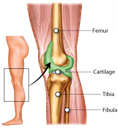
Cartilage is a firm yet pliable substance that performs a variety of functions: protection, shape maintenance and support, lubrication, and shock absorption. Its primary function is to coat the end of the bones where they articulate with one another, providing a smooth, cushioned surface. Furthermore, cartilage can serve as a template for bone formation during development and bone healing (this will be further discussed in the section about bone ossification).
Ligaments and tendons support articulations and control the muscle attachment to the bones. Ligaments connect bones to one another and stabilize articulations. Tendons connect bones to muscles.
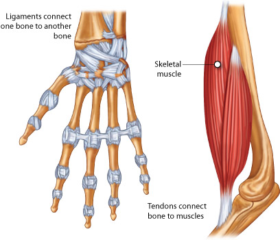
The structures of ligaments and tendons are similar, in that both tissues are made of fibrous proteins aligned in the direction of force. The types of proteins and differences in stretch and recoil distinguish the mechanical behavior of ligaments and tendons. Ligaments are stiffer, but deform more, since they stabilize articulations. Tendons are wrapped with a continuation of the fascia that surrounds muscle cells to help transmit and dissipate force.
Axial Skeleton Anatomy
The adult human skeletal system is composed of 206 bones. The skeleton is divided into two functional groupings—the axial skeleton and the appendicular skeleton.
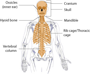
The axial skeleton is composed of the skull, hyoid bone, vertebral column, and the thorax (ribs and sternum). The axial skeleton functions to protect and support organs of the head, neck, and trunk.
The skull consists of 22 facial and cranial bones that interlock to form openings for the eyes and protection for the brain. The cranium, a collection of bones that protect the brain, and the mandible are the two major parts of the skull.
The hyoid bone is not attached to any other bone and is located in the neck, and it supports tongue movement.
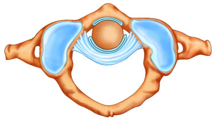
The vertebral column consists of individual vertebrae separated by cartilagenous disks. The vertebral column forms the middle axis of the skeleton.
The thoracic cage protects the internal organs of the thorax and upper abdomen. The thoracic cage consists of the ribs and the sternum. The ribs articulate anteriorly with the sternum and posteriorly with the vertebrae of the thorax.
The table below lists the location and function of the major bones of the axial skeleton:
| Bone(s) | Location | Function | Major grouping of axial skeleton |
|---|---|---|---|
| Cranium | Head | Supports facial structures, encloses and protects the brain, provides muscle attachments for chewing and moving the head | Skull |
| Mandible | Lower jaw | Permits chewing | Skull |
| Vertebrae | Spine | Permit mechanical stability for the body and protect the spinal cord | Vertebral column |
| Ribs | Chest wall | Provide protection for the organs of the upper body | Thoracic cage |
| Sternum | Center of the chest | Provides attachment for many (not all) ribs | Thoracic cage |
Appendicular Skeleton Anatomy
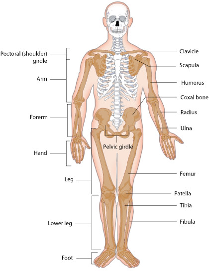
The appendicular skeleton is composed of the upper limbs, lower limbs, pectoral girdle, and pelvic girdle. The appendicular skeleton functions to anchor the limbs to the axial skeleton.
The pectoral girdle consists of a scapula and clavicle on each side of the body. The pectoral (shoulder) girdle permits movement of the upper limbs by connecting the upper limbs to the axial skeleton.
The upper limbs of the appendicular skeleton are composed of the humerus or upper arm bone, the radius and ulna, which complement each other to form the forearm, and the wrist. The hand subdivides into smaller bones of the palm and fingers.
The pelvic girdle of the appendicular skeleton is composed of two coxal bones (fused ilium, ischium and pubis bones), which attach to the vertebral column and the lower limbs.
The lower limbs each consist of the femur, or thigh bone; the tibia, or shinbone and the fibula, or calf bone; and the foot. The patella is the bone located at the point where the femur and tibia articulate with each other. The foot subdivides into smaller bones of the ankle, instep, and toes.
The table below lists the location and function of the major bones of the appendicular skeleton:
| Bone(s) | Location | Function | Major grouping of appendicular skeleton |
|---|---|---|---|
| Scapula | Flat, triangular bone located on the posterior side of each shoulder | Articulates with the clavicle and humerus | Pectoral girdle |
| Clavicle | Located in each shoulder at the base of the neck | Helps to keep the shoulders in place as part of the pectoral girdle | Pectoral girdle |
| Humerus | Extends from the scapula to the elbow | Provides attachments for muscles that move the shoulder and upper arm at the proximal end; articulates with the radius and ulna at the distal end | Upper limbs |
| Radius | Located on the lateral side of the forearm between the elbow and wrist | Provides attachment for muscles that rotate and bend the arm at the elbow and muscles that allow movement of the wrist | Upper limbs |
| Ulna | Located on the medial side of the forearm between the elbow and wrist | Provides attachment for muscles that bend and straighten the arm at the elbow and muscles that allow movement of the wrist | Upper limbs |
| Ilium | Located on the superior portion of the coxal bone | Connects the bones of the lower limbs to the axial skeleton | Pelvic girdle |
| Femur | Extends from the hip to the knee | Provides attachment for muscles of the lower limbs and buttocks; distal end articulates with the tibia and patella | Lower limbs |
| Tibia | Located on the medial side of the leg between the knee and the ankle | Articulates with the femur, on its superior side, to form the knee joint; articulates with the fibula on the lateral side; articulates with the patella on the anterior side; and the tarsals to form the ankle joint | Lower limbs |
| Fibula | Located on the lateral side of the tibia between the knee and ankle | Forms the lateral part of the ankle joint | Lower limbs |
| Patella | Located on the anterior surface of the articulation between the femur and tibia | Supports movement of the knee joint | Lower limbs |
Bone Classifications
Classification by Microscopic Structure
The bones of your body are mineralized structures that make up a major portion of your skeleton. In the broad sense, a bone is composed of bone tissue, cartilage, ligaments, tendons, vasculature, and nervous tissue. Bone tissue is a collection of specialized cells (osteoblasts, osteoclasts, osteocytes), organic extracellular matrix proteins (collagen and proteoglycans) and inorganic salt crystals that work together to provide strength and flexibility.
Although all bones have a similar composition, their large-scale structures and functions differ. One way to classify bone tissue is based on their microscopic structure.
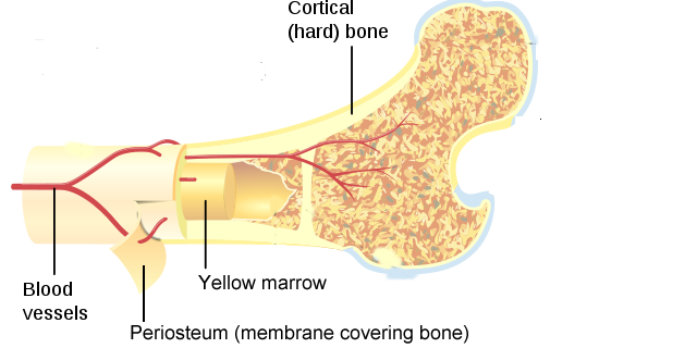
- Bone tissue with a tightly packed microstructure arranged into rings is called compact bone (also called cortical or lamellar bone). Compact bone structure gives bones their stiffness. However, if bone structures were made only of compact bone, they would be very heavy and brittle.
- Bone tissue with a porous microstructure is spongy bone (also called cancellous or trabecular bone). Spongy bone consists of branching bone structures called trabeculae. Spongy bone helps to reduce the weight and brittleness of bone, and is found at the ends of bone where forces are high. Spongy bone allows bones to bend a slight amount without cracking.
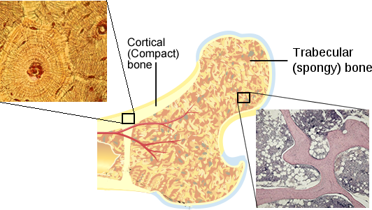
Most bones of the body are composed of both spongy and compact bone tissues, so that they are stiff, resilient and lightweight.
Classification by Shape
A common way to classify individual bones of the skeletal system is based on their shapes. The table below describes the four main shapes of bones.
| Shape | Description | Examples |
|---|---|---|
| long bones | significantly longer in one direction than in either of the other two directions | humerus (upper arm); femur (thigh) |
| short bones | similar length in all directions | most carpal (wrist) and tarsal (ankle) bones |
| flat bones | flat and plate-like | bones of the skull |
| irregular bones | not regular or systematic in shape | vertebrae; hip bones |
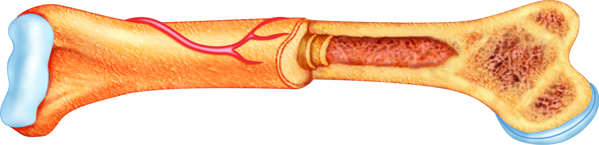
Components of Long Bones
A typical long bone, such as the femur, has three main components:
- the diaphysis or shaft;
- the epiphyses (singular, epiphysis), found at the bone ends;
- and the metaphyses (singular, metaphysis), transitional areas between the diaphysis and epiphyses.
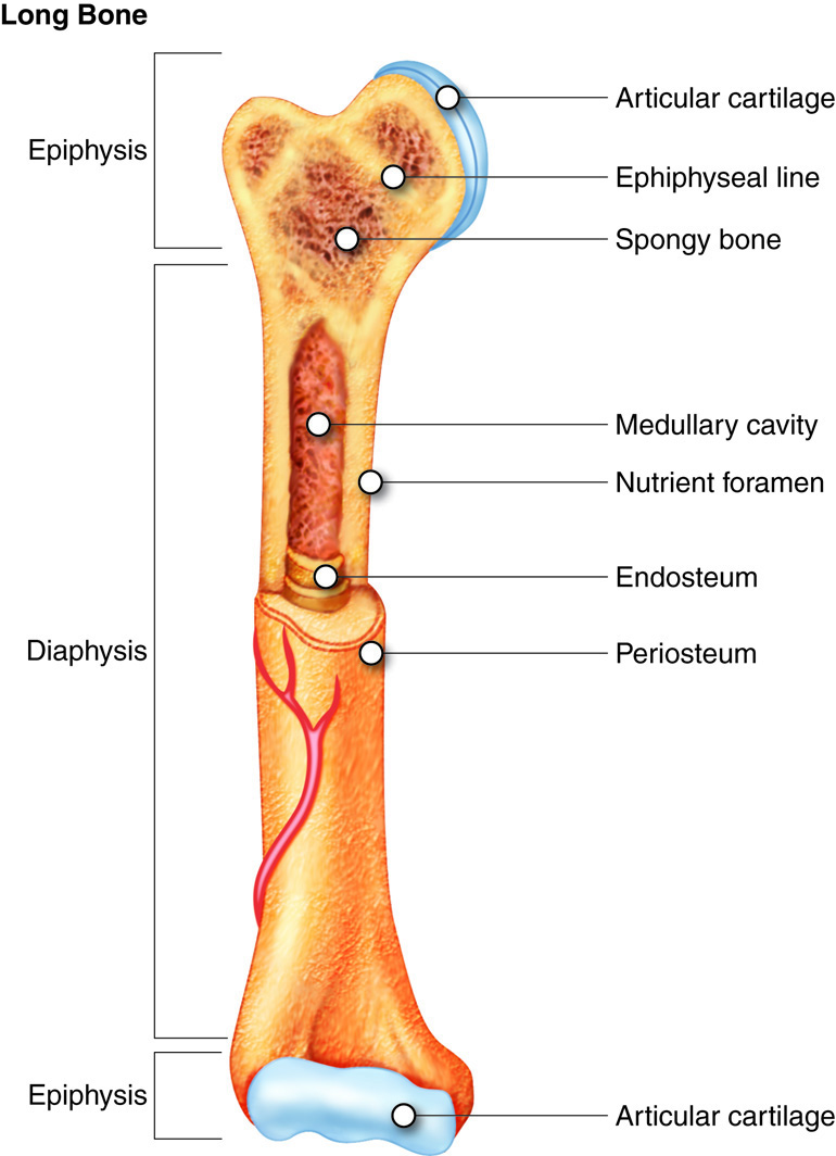
The diaphysis or shaft is the longest part of a long bone. The epiphyses (epiphysis if singular) are the ends of a long bone that articulate (connect) with other bones at articulations or joints. The transitional area between the diaphysis and epiphysis is the metaphysis (meta- means “in between”). The medullary cavity, which houses yellow bone marrow in adults, begins at the boundary between the metaphysis and diaphysis of each bone end and runs throughout the shaft or diaphysis of the bone. This marrow is rich in fat which is why it is termed yellow bone marrow. The epiphyses contain mainly spongy (cancellous) bone and red bone marrow. In children, the junction between the epiphysis and metaphysis contains a layer of hyaline cartilage, termed the epiphyseal plate. You may have heard of this as the growth plate. This layer of cartilage is what allows bone to continue to lengthen. The cartilage cells undergo mitosis and move away from the centerline of the epiphyseal plate. The margins of the plate are then converted to bone tissue. In adult bone, the entire epiphyseal plate has become calcified such that there is no longer active cartilage in this area. At this point it is referred to as the epiphyseal line. After the calcification of the epiphyseal plate occurs (typically around the age of 20), the only area of hyaline cartilage left is on the outer edge of each epiphysis. This cartilage is termed articular cartilage and functions to cushion and reduce friction between bones at articulations.
Articulations
It’s common to think of the skeletal system as being made up of only bones, and performing only the function of supporting the body. However, the skeletal system also contains other structures, and performs a variety of functions for the body.
While the bones of the skeletal system are fascinating, it is our ability to move segments of the skeleton in relation to one another that allows us to move around. Each connection of bones is called an articulation or a joint. Articulations are classified based on material at the joint and the movement allowed at the joint.
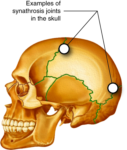
Synarthrosis Articulations
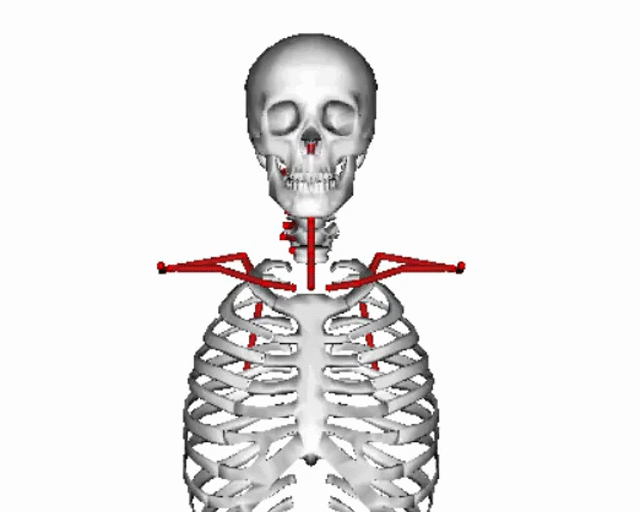
Immovable articulations are synarthrosis articulations (“syn” means together and “arthrosis” means joint); immovable articulations sounds like a contradiction, but all regions where bones come together are called articulations, so there are articulations that don’t move, including in the skull, where bones have fused, and where your teeth meet your jaw. These synarthroses are joined with fibrous connective tissue. Some synarthroses are formed by hyaline cartilage, such as the articulation between the first rib and the sternum (via costal cartilage). This immoveable joint helps stabilize the shoulder girdle and the cartilage can ossify in adults with age. The epiphyseal plate or “growth plate” at the end of long bones is also a synarthrosis until hyaline cartilage ossification is completed around the time of puberty.
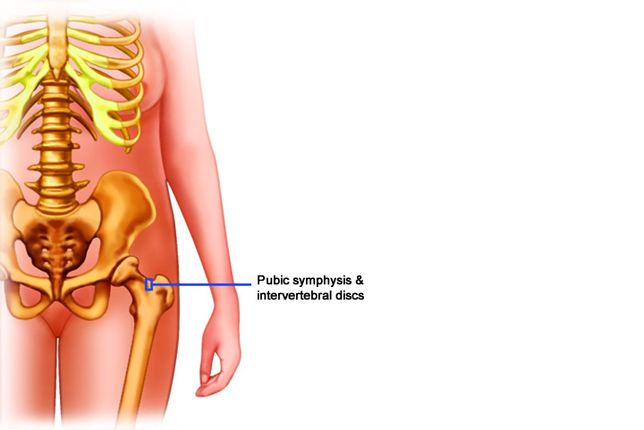
Amphiarthrosis Articulations
There are some articulations which have limited motion called amphiarthrosis articulations. They are held in place with fibrocartilage or fibrous connective tissue. The anterior pelvic girdle joint between pubic bones (pubic symphysis) and the intervertebral joints of the spinal column (discs) are examples of cartilaginous amphiarthroses. The two parallel bones in the arms and legs are considered amphiarthrosis articulations with a sheet-like interosseous membrane. The lateral distal articulation between tibia and fibula at ankle is also an amphiarthrosis with a fibrous ligament.
Diarthrosis Articulations
The articulations that people are most familiar with are diarthrosis articulations, which have wide ranges of motion. Diarthrotic articulations are said to be freely moveable and have a joint cavity. A joint cavity is a structure that consists of a joint capsule which surrounds the joint, and a synovial membrane which is inside the joint and produces a fluid known as synovial fluid. These joints are known as synovial joints and are further classified according to the type of movement allowed at the joint.
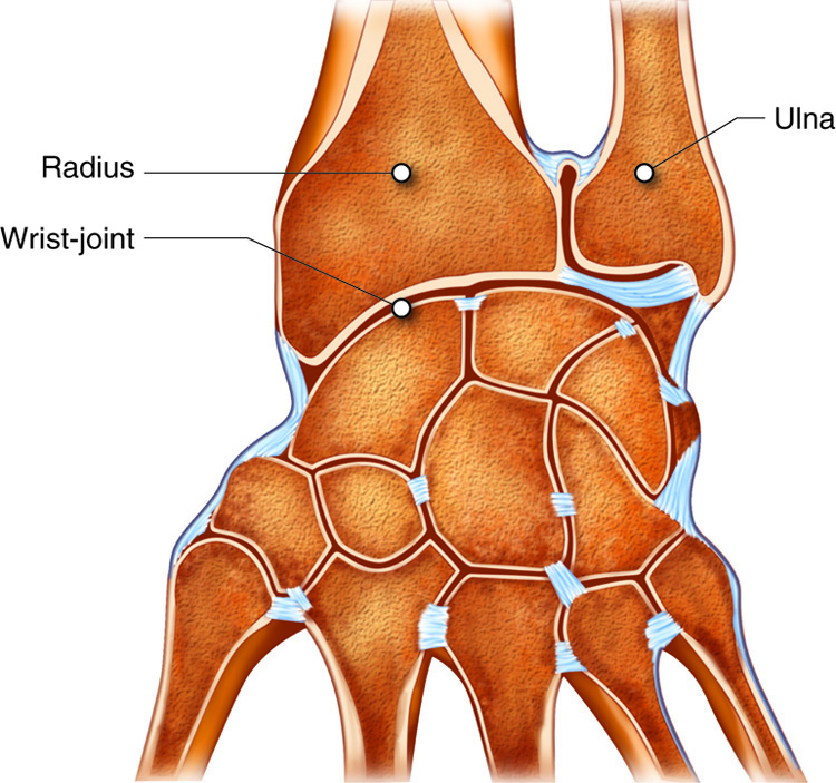
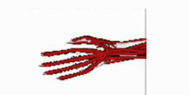
Nonaxial Joints
Nonaxial joints do not have a pivot or axis of movement. An example are gliding joints, also known as plane joints. These joints do not allow much movement other than sliding and twisting. These are often found in certain articulations in the wrist and ankle.
.gif)
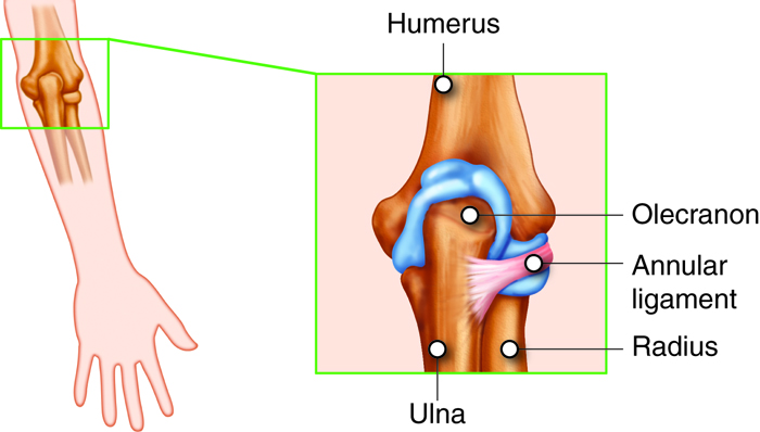
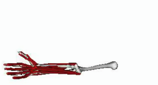
Uniaxial or Monoaxial Joints
Biaxial Joints
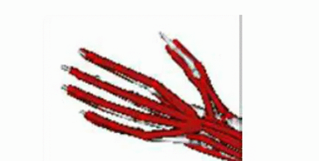
Biaxial joints have two axes of movement and therefore allow more movements than uniaxial joints. One example of a biaxial joint are saddle joints, such as the thumb. These joints were appropriately named because they look like saddles; there are two curved surfaces that meet and allow limited motion in many directions.
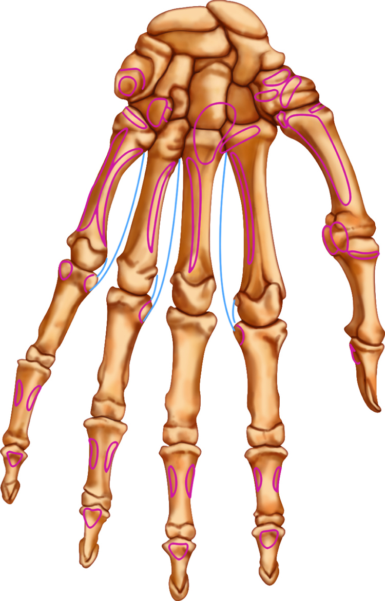
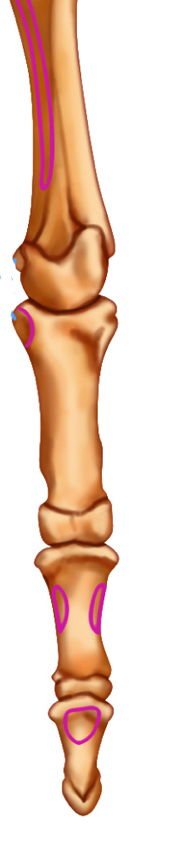
Another example of a biaxial joint is an ellipsoid or condlyar joint. Ellipsoid articulations, also called condyloid articulations, have oddly shaped or elliptical interactions that articulate to allow movement in two planes with no rotation. These are present in the fingers and toes.
Triaxial Joints
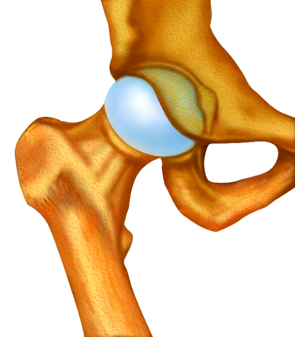
As you can guess, triaxial joints are the most moveable with three axes of movement. The ball and socket joints such as the shoulder and hip, have the widest range of motion. They are aptly named as the head of the bone resembles a “ball” and the articulating bone of the joint (either the scapula or coxal bone) has a deep pit for the “socket”.
.gif)
