33 Muscular Structures and Functions
Muscle Tissue Function
Skeletal muscles (organs) are almost never completely relaxed. Even if a muscle is not producing movement, some of the muscle fibers (cells) in the organ contract to produce muscle tone. The tension produced by muscle tone prevents the muscle from becoming weak, allowing the muscle to stabilize joints and maintain posture. Muscle tone is accomplished by the contraction of a small fraction of muscle cells so muscles won’t fatigue completely; some cells can recover while others are active. This can help with stronger movements, as the motor units being used to maintain tone can produce a quicker, stronger contraction than the motor units that are resting, avoiding the treppe that occurs with muscle fibers that are stimulated from a state of rest.
The absence of the low-level contractions that lead to muscle tone is referred to as hypotonia. Hypotonia can be caused by nervous system dysfunction or imbalances in certain ions in the blood stream. Hypotonic muscles have a flaccid appearance and display functional impairments. Conversely, excessive muscle tone is referred to as hypertonia. This takes two forms: spasticity, which is a type of stiffness related to uncontrolled reflexes, and rigidity, a stiffness not associated with reflexes.
Isometric and Isotonic
There are two main types of skeletal muscle contractions: isotonic and isometric. When a muscle exerts a force, it exerts this force on a load. At the molecular level, cross-bridge formation occurs in both isotonic and isometric contractions, but the whole muscle acts differently depending on the load and the outcome to be achieved. In isotonic (iso- same, tonic – tension) contractions, a load is moved as the length of the muscle changes.
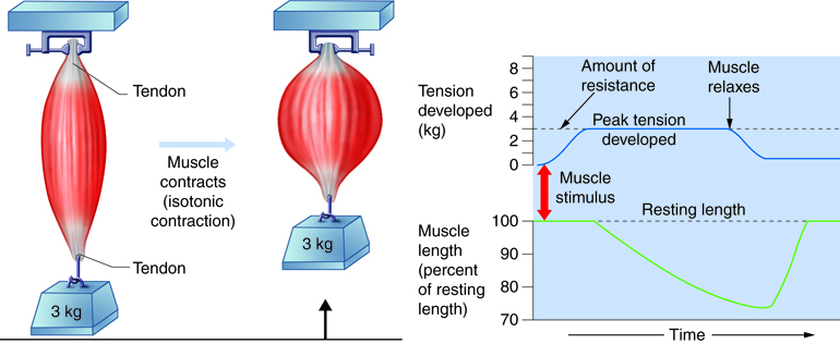
There are two types of isotonic contraction: concentric and eccentric. Concentric contractions involve the muscle shortening to move a load. An example of this is the biceps muscle shortening as a hand weight is brought upward and the angle of the elbow decreases. Eccentric contractions occur as the muscle lengthens. Eccentric contractions do not produce adequate force to move a load but are used for movement, balance, and resisting movement. An example of an eccentric contraction is the movement of a bicep as it performs the lowering portion of a biceps curl. A concentric contraction brings the arm upward, reducing the angle of the elbow, and then an eccentric contraction lowers the arm, resisting gravity and increasing the elbow angle slowly. The same muscle tension is required whether the muscle is shortening or lengthening.
Isometric (iso- same, metric- length) contractions occur as the muscle produces tension without changing length. Isometric contractions do not produce movement or lift loads; these contractions maintain posture and joint stability. For example, pushing against a wall produces no movement because the muscle simply cannot produce enough force and thus does not result in changes in muscle length. Conversely, holding your head in an upright position occurs not because the muscles cannot move the head, but because the goal is to remain stationary and not produce movement. In both of these cases, cross-bridge cycling occurs in the same manner as it would if the load had been moved. Most actions are the result of a combination of isotonic and isometric contractions working together to produce a wide range of outcomes.
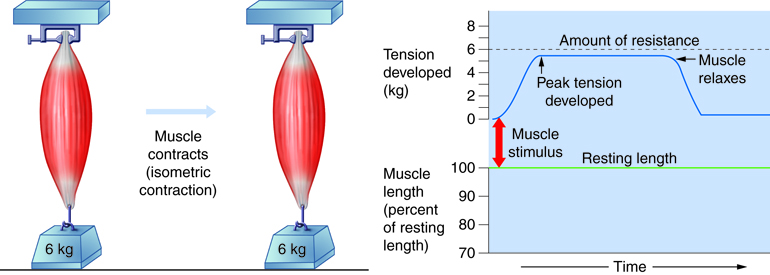
All of the contraction that occurs before a load is lifted is isometric. Even when muscles carry no load, they must still develop a tension equal to the muscles’ own weight before they can shorten. When contractile forces exceed the weight of the load, muscles begin shortening. Shortening stops when active tension falls to the point that it can no longer overcome and move the load. This converts it back to an isometric contraction.
Take the example of lifting a glass of water. It’s relatively easy for someone to lift the glass. However, holding a glass of water at arm’s length for one full minute is difficult. A concentric isotonic contraction is required to lift a glass. The bicep will shorten as the glass is lifted. Cross-bridges are formed and will be released as you set the glass back down. In order to hold a glass of water at arm’s length for one minute you are using an isometric contraction. You are attempting to maintain stability at your shoulder and elbow joints in a position that is not “resting” for an extended time.
Skeletal Muscle Organ Anatomy
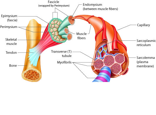
Skeletal muscles, which are the organs of the skeletal muscle system, include three layers of connective tissue that enclose and provide structure to the muscle as a whole. The outer layer of each muscle fiber is wrapped with a layer of connective tissue called the epimysium (“epi-” means “outside” or “over,” and “-mysium” refers to “muscle”). The epimysium allows muscle to contract and move powerfully while maintaining its structural integrity; it also separates muscle from other tissues and organs. It is composed of a thick layer of collagen fibers. The epimysium is surrounded by fascia, which is a type of connective tissue that is found around body organs. Deep fascia surrounds groups of muscles, sometimes joining with tendons to strengthen the bone attachment, whereas superficial fascia lies between muscle and skin. Some fascia contain adipose tissue that insulates and protects muscle.
Inside each skeletal muscle, muscle fibers (a single muscle cell is called a muscle fiber; not to be confused with fibers found in connective tissue) are organized into groups called fascicles, or bundles of muscle fibers (cells). Some of these fascicles can be seen without a microscope when a muscle is cut open. Each fascicle contains many long muscle fibers bound by a middle layer of connective tissue called the perimysium (“peri-” means “around”). Similar to the epimysium, the perimysium contains collagen fibers, but the perimysium also contains elastic fibers.
Inside each fascicle, each individual muscle fiber is encased in a connective tissue layer called the endomysium (“endo-” means “inside”). The endomysium forms a thinner layer than the dense epimysium and perimysium, and contains areolar and reticular tissues which form loose, delicate networks. The endomysium of muscle fibers connect to each other to form a loose complex within a fascicle. Small blood vessels and motor neurons pass through the endomysium to support and activate each muscle fiber.
The plasma membrane, or sarcolemma, of a skeletal muscle fiber is located just under the endomysium. The sarcolemma is the site of action potential conduction, which triggers muscle contraction. Within the sarcolemma is the sarcoplasm, the cytoplasm of the muscle cell.
Example: Muscular Dystrophy
Muscular Dystrophy (MD) is a progressive weakening of skeletal muscles. Duchenne’s Muscular Dystrophy (DMD), the most common type of MD, is caused by a mutation in the dystrophin gene. Dystrophin helps the thin filaments of myofibrils bind to the sarcolemma and maintains equal force transmission through the muscle tissue. Without sufficient dystrophin, muscle contractions cause the sarcolemma to tear, causing an influx of Ca2+, leading to cellular damage and muscle fiber degradation. Over time, muscles are damaged and functional impairments develop.
DMD is an inherited disorder caused by an abnormal gene found on the X chromosome. It is one of many genetic diseases which are referred to as X-linked; X-linked disorders affect males since they only carry one copy of the X chromosome, which they inherited from their mother. Girls inherit one X chromosome from their mother and one from their father, so they are usually not affected. DMD is usually diagnosed in early childhood. It usually first appears as difficulty with balance and motion, and then progresses to an inability to walk. It continues progressing upwards in the body from the lower extremities to the upper body, where it affects the muscles responsible for breathing. It ultimately causes death due to respiratory failure, and those afflicted do not usually live past their twenties.
Muscle Fiber Organization
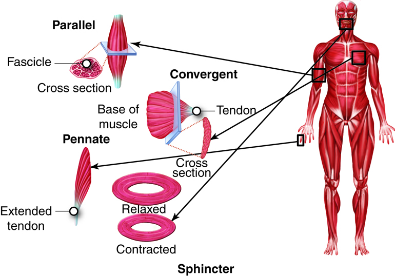
The fascicles of parallel muscles are arranged parallel to the long axis of the muscle. They are equidistant and run in the same direction. The majority of skeletal muscles in the body follow this type of organization. With muscles that seem to be plump, the large mass of tissue located in the middle of the muscle is known as the central body, but its more common name is the belly. When the muscle contracts, the contractile fibers shorten into a large bulge. To see this, extend and then flex your biceps muscle. The large mass of the muscle is called the belly; tendons emerge from both ends of the muscle. When a muscle contracts, it is the pulling on the tendon by the muscle that actually produces the movement.
The fascicles of circular muscles, also called sphincters, are arranged around an opening in rings. When the muscle contracts, the size of the sphincter opening decreases. The orbicularis oris muscle is a circular muscle around the mouth under the lips. Another example is the orbicularis oculi (“ocular” means “eye”), which surrounds each eye. The circular muscles expand and contract in a circular way. The circular muscles contract and relax in a circular way. Think about how you can contract and shape your lips to whistle or pucker.
In convergent muscle, the fascicles extend from a broad, fan-shape area and converge on single attachment site where the muscle interacts with a tendon. .The large muscle on the chest, the pectoralis major, is an example of a convergent muscle. Because of the arrangement of motor units within a convergent muscle, it can be stimulated in different, smaller areas. This can change the direction of the muscle force slightly from when the entire muscle contracts at once.
Pennate muscles are feather-shaped and form different fascicle arrangements at an angle to the tendon. Contracting pennate muscles pull at an angle and can produce high tension, but don’t move tendons very far. There are three subtypes of pennate muscles: unipennate, bipennate, and multipennate muscles. In unipennate muscles, such as the extensor digitorum in the forearm, the fascicles are located on one side of the tendon. A bipennate muscle, such as the rectus femoris in the thigh, has fascicles on both sides of the tendon. If the tendon branches within a pennate muscle, as it does in the deltoid muscle of the shoulder, the muscle is referred to as multipennate.
Some parallel muscles are fusiform and have fascicles that are spindle-shaped to give the muscle a large belly, such as the biceps brachii, whereas triangular muscles can be convergent or multipennate to create triangular shapes. Triangular muscles include the trapezius, which extends from the head down the back and out to the shoulder.
Skeletal Muscle Movement
Skeletal muscles, which are the organs of the skeletal muscle system, generally act by pulling on bones to produce movement. To pull the bone, muscles need to attach to the bone, either directly or indirectly. The epimysium of a muscle can attach directly to bone or cartilage to form a direct attachment. An indirect attachment is formed when the connective tissue layers, the epimysium, perimysium, and endomysium form a complex at the end of the muscle.
For connections, muscles require either a tendon or a broader sheet of connective tissue called an aponeurosis. Muscles connect to muscles via aponeuroses, and muscles attach to bones via tendons or aponeuroses. Thus, when a muscle contracts, the force of movement is transmitted through the attachment, which pulls on the bone to produce skeletal movement.
Tendons also help to stabilize the joints. Tendons are a common form of attachment because the collagen fibers are more resistant to tearing than direct muscle tissue attachment to bone would be, and the compact form of the tendon requires a small amount of space. Tendons can be easily seen as the hand is flexed, causing the thick, cordlike tendons of the forearm to stand out prominently. The calcaneal (Achilles) tendon is visible from the heel to the calf. Some tendons are also surrounded by a connective tissue layer called a tendon sheath, which protects the tendon as it moves. Fluid-filled sacs called bursae also join to tendons to reduce friction as the tendon moves. Bursae are present in connective tissue near bone, where tendons experience friction, but they occasionally arise in other areas due to stress caused by movement.
Examples: Bursae Inflammation as Bursitis
Bursae can become inflamed, a condition known as bursitis. This is usually caused by overuse of a joint or by other mechanical stress, which can result in pain and swelling. Bursitis is common in knees, elbows, and shoulders.
Keep in mind that the range of motion produced by muscles is restricted by the anatomy of the bones and other support structures involved in a particular joint. Consider the movements of the hip and shoulder joints, both of which are freely moveable. The hip is the attachment point for the thigh and leg. The shoulder is the attachment point for the arm and forearm. The shoulder can produce movements that the hip cannot because of the anatomy of the bones involved and the ligaments (connective tissue) associated with the joint. Hold your arm out to the side and move your thumb around the axis of the arm in a 360-degree circle. Can you do that with your hip and leg? No, you can’t. There are some people who appear to be “double-jointed”; they are able to push their joints past the normal limits of movement. The reason they can do this is because the ligaments of their joints are “loose” compared with someone who is not “double-jointed.” This ability appears to have a genetic basis.
For muscles attached to the bones of the skeleton, the location of the connection determines the force, speed, and type of movement. These characteristics depend on each other and can explain the general organization of the muscular and skeletal systems.
Levers and Movement
We can consider the mechanisms by which muscles act on bones using descriptions based on levers. A lever is a rigid structure, in this case a bone, that moves on a fixed point called the fulcrum, in this case an articulation. A lever moves when an applied force or effort is sufficient to overcome any load or resistance that would otherwise oppose or prevent such movement. In the body, each bone is a lever and each joint is a fulcrum, and muscles supply the applied forces. Movement of the skeleton occurs at joints, so there has to be sufficient muscle power to move all the bones at these joints.
Levers can change the direction and effective strength of a force as well as the distance and speed of movement produced by the force. Imagine a seesaw in a playground. You and a friend could sit on opposite ends of it. You would then take turns pushing off the ground with your legs as you each went up in the air. A seesaw is an example of a first-class lever, and there are three classes of levers. The fulcrum (F) lies at the midpoint of the seesaw, between the applied force (AF) and the load (L). What this means is that the seesaw balances you and your friend, as you both provide the applied force with the push of your legs and the load with the weight of your bodies.
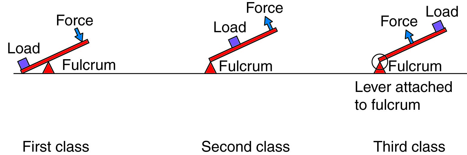
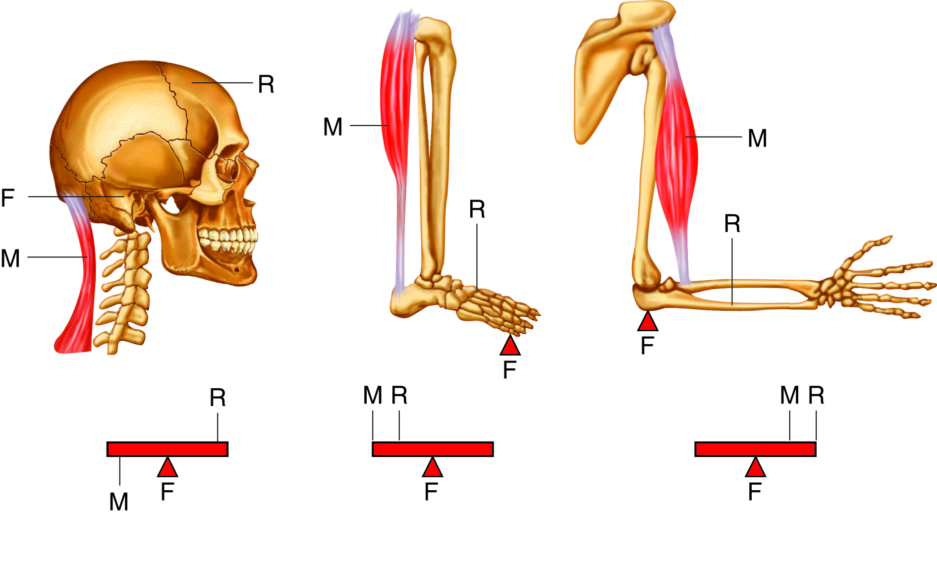
The body has only a few first-class levers. One is involved in head flexion (pulling the head down toward the chest) and head extension (pulling the head back up into normal position). The fulcrum is where the head moves in the sagittal plane on the first cervical vertebrae. The load is the weight of the head, and the applied force comes from the muscles. Extension occurs when the muscles pull the head back up. Moving your head forward and backward mirrors the action of a seesaw.
In a second-class lever, the load is located between the applied force and the fulcrum. When you move a load in a wheelbarrow, you lift upward on the handle and the wheel acts as the fulcrum. The force is further from the fulcrum than the load is, so you can move a larger weight with less effort. In the body, you achieve the same effect by standing on your toes. The fulcrum is on the ball of the foot, the load is your body weight, and the effort comes from the muscles in the back of the leg.
In third-class levers the force is applied between the load and the fulcrum. We don’t often see these types of levers in artificial machines, but they are the most common levers in the body. Speed and distance traveled are increased at the expense of force. For the biceps brachii in the arm, the load will be located in or around the hand. The biceps muscle applies the force, while the fulcrum is the elbow. For instance, when you pick up a full bag of groceries, you can lift it quickly using the third-class lever of the biceps brachii and the forearm. However, the location of fulcrum prevents great enhancements of load carrying.
Muscle Movement and Gross Anatomy
Skeletal muscles are arranged in pairs for balance and to work efficiently.
Some muscles function as agonists, or prime movers. An antagonist is a muscle that opposes the action of the agonist. When an agonist contracts to produce a movement, the corresponding antagonist will be stretched and contract with sufficient tension to control the movement. For instance, when your biceps muscle acts to flex your forearm, the triceps muscle contracts slightly to prevent you from flexing your forearm too quickly or too strongly.
Agonist and antagonist muscles work against one another to control and balance movements. Other muscles, called synergists (from synergy, to work together), improve the efficiency of an agonist muscle. Synergists may provide additional pull or stabilization, and their assistance to a particular movement may change throughout the progression of the movement.
This table provides examples of agonist-antagonist combinations.
| Agonist | Antagonist | Movement |
|---|---|---|
| Biceps brachii—located on the anterior surface of the upper arm | Triceps brachii—located on the posterior surface of the upper arm | The biceps brachii flexes the forearm, while the triceps brachii extends it. |
| Hamstrings—group of three muscles located on the posterior of the thigh | Quadriceps femoris—group of four muscles located on the anterior of the thigh | The hamstrings flex the lower leg. The quadriceps femoris extend the lower leg. |
| Flexor digitorum superficialis and flexor digitorum profundus—located in and around the hand | Extensor digitorum—located in and around the hand | The flexor digitorum superficialis and flexor digitorum profundus flex the fingers and the hand at the wrist while the extensor digitorum extends the fingers and the hand at the wrist. |
Gross Anatomy of Muscles
Nomenclature for skeletal muscles is based on numerous criteria including: the location of the muscle, the shape, the orientation, the number of the muscle in a group, the action it generates and the location of its origin(s) and insertion(s). The origin is the unmoving, stable connection of the muscle to a structure, usually a bone. The insertion is the moveable end of the muscle, and it inserts onto various structures in the body.
Location: Some muscle names indicate the bones or body region with which the muscle is associated. For example, the frontalis muscle is located on top of the frontal bone of the skull. The rectus femoris (femur) and brachioradialis (arm and radius) are also good examples.
Shapes: Muscles with distinctive shapes are often named for the shape. The trapezius in the neck and back is similar to a trapezoid. You have already learned about muscles that have “orbicularis” in the name. The size of the muscle is the source of the names of the muscles of the buttocks: gluteus maximus (largest), gluteus medius (medium) and gluteus minimus (smallest). There are also muscles with names that contain brevis (short), longus (long), lateralis (lateral side or away from midline) and medialis (toward the midline). Some practice with the names of common muscles will help you with the descriptions that indicate muscle size or location.
Orientation: The direction of the muscle fibers and fascicles relative to the midline are also used to describe muscles, such as the rectus (straight) abdominis or the internal and external oblique (at an angle) muscles of the abdomen.
Numbers in a group: Some muscles are named according to the number of muscles in a “group.” One example of this is the quadriceps, a group of four muscles located on the anterior (front) of the thigh. Other muscle names can give you a clue as to how many origins a particular muscle has, such as the biceps brachii or triceps brachii. Bi- indicates that the muscle has two origins and tri- indicates three.
Action: When muscles are named for the movement they produce, you will find action words in their name. Some examples are flexor (decreases the angle at the joint), extensor (increases the angle at the joint), abductor (moves the bone away from the midline) or adductor (moves the bone toward the midline).
Attachments: The location of a muscle’s orientation and insertion can also be used in its name. For this classification, the origin is always named first. For instance, the sternocleidomastoid muscle of the neck has a dual origin on the sternum (sterno) and clavicle (cleido) and it inserts on the mastoid process of the temporal bone.
The following table lists important muscle terminology.
| Name | Definition | Example |
|---|---|---|
| Direction Relative to the Midline of the Body | ||
| rectus | straight | rectus abdominis |
| transverse | at right angle | transverse abdominis |
| oblique | diagonal | external oblique |
| Relative Size | ||
| maximus | largest | gluteus maximus |
| medius | medium | gluteus medius |
| minimus | smallest | gluteus minimus |
| longus | long | longus capitis |
| brevis | short | extensor carpi radialis brevis |
| latissimus | widest | latissimus dorsi |
| longissimus | longest | longissimus thoracis |
| magnus | large | adductor magnus |
| major | larger | rhomboid major |
| minor | smaller | rhomboid minor |
| vastus | huge | vastus medialis |
| Relative Shape | ||
| deltoid | triangular | deltoid |
| trapezius | trapezoidal | trapezius |
| serratus | serrated | serratus anterior |
| rhomboid | diamond-shaped | rhomboid minor |
| orbicularis | circular | orbicularis oris |
| pectinate | comb-like | pectineus |
| piriformis | pear-shaped | piriformis |
| platys | flat | platysma |
| quadratus | four-sided and square | quadratus lumborum |
| gracilis | slender | gracilis |
| Action | ||
| flexor | decreases the angle at a joint | flexor digitorum superficialis |
| extensor | increases the angle at a joint | extensor digitorum |
| abductor | moves the bone away from the midline | abductor pollicis longus |
| adductor | moves the bone toward the midline | adductor magnus |
| levator | elevates a body part | levator scapulae |
| depressor | lowers a body part | depressor labii inferioris |
| supinator | turns the palm anteriorly | supinator |
| pronator | turns the palm posteriorly | pronator teres |
| sphincter | decreases the size of an opening | external anal sphincter |
| tensor | tenses a body part | tensor fasciae latae |
| rotator | rotates a bone around its longitudinal axis | rotator |
| Number of Origins | ||
| biceps | two | biceps brachii |
| triceps | three | triceps brachii |
| quadriceps | four | quadriceps femoris |
Example: Explicit Muscle Anatomy: Axial Muscles of the Face, Neck, and Head
There are more than 600 individually identified skeletal muscles in the human body. An entire course could be spent just learning the names, origins, insertions and actions of skeletal muscles and muscle groups of the body. This course presents an example of integrated muscle units, so that you can appreciate the complex integration of muscle anatomy and physiology.
Muscles can be classified either as axial or appendicular muscles, depending on whether they act on bones of the axial or appendicular skeleton, respectively. If we just look at the axial muscles, we can further divide them into groups on the basis of location, function, or both. Some of the axial muscles may seem to blur the boundaries due to the fact that they cross over to the appendicular skeleton. The muscles of the head and neck would be considered a subgroup of the axial muscles based on location.
This group can also be divided into those that insert into skin or those that insert on bones. The muscles in the face create facial expressions by inserting into the skin rather than onto bone. These muscles include the occipitofrontalis, orbicularis oris, buccinators, orbicularis oculi, and corrugator supercilii among others.
