38 Nervous Structures and Functions
The Brain
The large brain of humans is perhaps the most important evolutionary advance for the species. At the minimum, it is the characteristic most of us consider the distinguishing characteristic of a human. This module outlines the structural and functional relationships of the human brain.
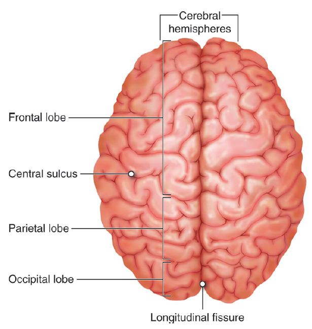
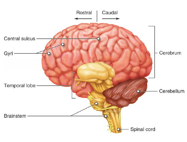
The dominant portion of the human brain is the cerebrum. It is the large upper part of the brain, distinguished by the gyri (folds) and sulci (folds) of the surface. The cerebrum is clearly split into left and right hemispheres; the split is the deep longitudinal fissure. The cerebrum sits atop and around the midbrain, which leads into the brainstem. Situated essentially behind the midbrain and under the cerebrum is the distinctive cerebellum.
The inside of the brain is characterized by regions of gray matter and white matter. The gray matter is mostly cell bodies, dendrites, and synapses and forms a cortex over the cerebrum and cerebellum, and also forms some nuclei deeper in the cerebrum. White matter is myelinated axons forming tracts. (These definitions and components of gray and white matter are similar to the ones for the spinal cord, although their arrangement will be different as you will discover later in this unit.)
The cerebral white matter tracts are classified as
- Projection tracts – from higher to lower, from cerebrum to brainstem and spinal cord
- Commissural – across hemispheres
- Association – within same hemisphere
The gray matter of the cerebral cortex includes:
- Stellate cells – receive sensory input and process information locally
- Pyramidal cells – extend to other parts of the CNS
- Neocortex – 6 layered tissue of recent evolutionary origin
Meninges, Cerebrospinal Fluid and Blood Supply
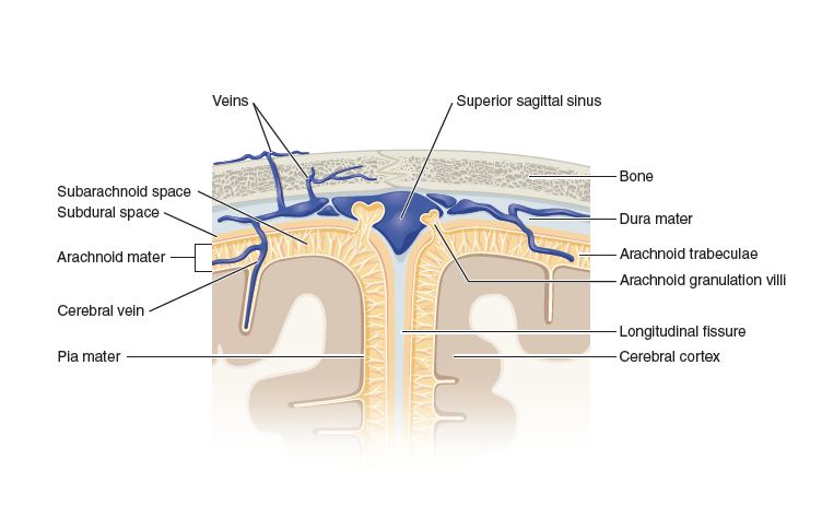
Like the spinal cord, the brain is covered and partially protected by connective tissue meninges. From outermost (bordering the skull bones) to the innermost (adjacent to the nervous tissue) they are the dura mater, arachnoid mater, and pia mater. The dura mater folds into two layers, a periosteal layer fused to the skull bones, and a meningeal layer. In some areas, these layers are separated by a dural sinus, a space used to collect blood. Some areas may also contain a subarachnoid space or a subdural space.
Cerebrospinal fluid (CSF) is a clear, colorless liquid that bathes the external surfaces of the brain. It is constantly produced, flows through the network of ventricles, and is reabsorbed. CSF functions in cushioning and supporting the brain by buoyance, and in chemical stability of the brain, by transporting nutrients and wastes respectively.
The ependymal cells lining the ventricles produce the CSF. Then the CSF flows throughout the brain in the ventricles. Each cerebral hemisphere contains a lateral ventricle. Each lateral ventricle drains through an interventricular foramen into the third ventricle. The third ventricle sits in the midbrain region. The CSF then flows into the cerebral aqueduct to the fourth ventricle. Before being reabsorbed, the CSF enters one of two lateral apertures or a median aperture, and then fills the subarachnoid space. Reabsorption of CSF occurs there by the arachoid villi and enters the venous blood.
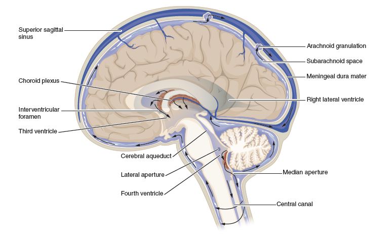
The choriod plexus is the network of blood vessels and ependymal cells on surface of the ventricles. The ependymal cells of this choroid plexus secrete the CSF. Overall the close proximity of ependymal cells and blood vessels create a blood-brain barrier (BBB). The brain requires large amounts of oxygen and glucose but other items in blood may harm it, hence the barrier. At the capillaries there is a BBB of tight endothelial cells and basement membrane; at the choriod plexus the blood-CSF barrier due to tight junctions between ependymal cells
Next, the main structural areas of the brain will be surveyed with some of their major functions. We will go in general order of most primitive brain region to most evolutionary advanced region, or, in other words, from basic functions to more advanced functions.
Hindbrain and Midbrain
The most inferior part of the brain, the medulla oblongata, appears as a thickening of the spinal cord. Many of the cranial nerves originate here (see below). The medulla oblongata contains nuclei that control many basic functions, including the cardiac center, the vasomotor center, the respiratory centers, and many other involuntary functions such as swallowing, coughing, salivating, sweating, and gastrointestinal secretion.
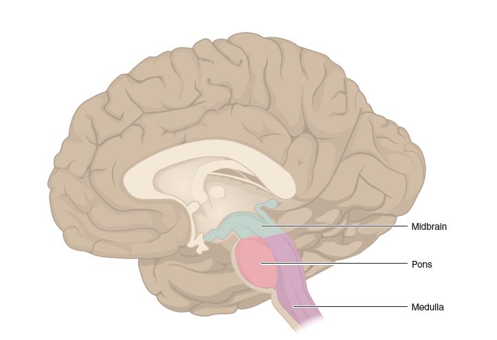
In humans, the pons is the next most superior feature of the brain; the pons looks like a forward-facing bulge in the brainstem above the medulla oblongata. The pons relays signals between cerebrum and cerebellum, including sleep, hearing, taste, and posture to name a few.
The cerebellum is a smaller, highly folded structure in the back of the brain, behind the pons. Like the cerebrum, it is split into hemispheres, with a flattened area down the center called the vermis. The folds are folia and grooves are sulci. The white matter forms a distinctive arbor vitae (“tree of life”). The cerebellum is concerned with muscular coordination, special perception, and tactile perception, and some planning and scheduling tasks.
The midbrain is a small region of gray matter nuclei involved in different motor and sensory functions and connecting white matter pathways. These structures include:
- Cerebral peduncles – anchor cerebrum to brainstem
- Tegmentum – to/from cerebellum for motor control
- Substantia nigra – inhibitory relay (the area destroyed in Parkinson disease)
- Central gray matter – pain awareness
- Tectum – include the inferior and superior colliculi for hearing and vision
- Red nucleus – subconscious motor commands and muscle tone
The reticular formation is a series of gray matter extensions from the midbrain through the cerebellum. They are involved in
- Somatic motor control, including the pattern generators
- Cardiovascular control
- Pain modulation
- Sleep and consciousness, including habituation
Forebrain
The forebrain is the large overarching region of the brain. In the center, above the midbrain are the thalamus and hypothalamus.
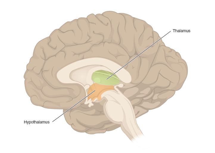
The thalamus is a set of nuclei mainly involved in the relay of sensory signals. Those will be covered in more depth in sensory units. Other thalamic nuclei are involved in memory and emotions.
The hypothalamus is a set of nuclei situated underneath the thalamus. The main function of the hypothalamus is control of the endocrine system and as such will be covered in more detail there. Other nuclei of the hypothalamus are involved in many autonomic functions such as thermoregulation, food and water intake, biological cycles, and emotions.
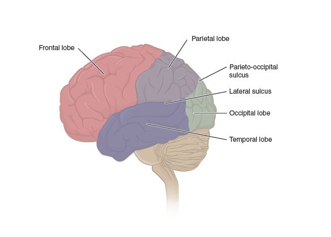
The cerebrum is the major anatomic feature of the human brain. The cerebrum is made of lobes. The frontal lobe is from the frontal bone to central sulcus and is involved in voluntary motor functions, planning and foresight, memory, mood, emotion, social judgment, and aggression.
The parietal lobe is the upper part of brain in each hemisphere from the central sulcus to parietal-occipital sulcus; this lobe is primarily involved in sensory reception and integration.
The temporal lobe of each hemisphere sits under the parietal lobe and the lateral sulcus; this lobe has roles in hearing, smell, learning, memory, visual recognition and emotional behavior.
The lobe furthest to the rear of the head is the occipital lobe, and it contains the visual center.
The insula is a mass of cortex underneath the outer lobes, found beneath the frontal and temporal lobes.
The basal nuclei are clusters of cell bodies found at the bottom (base) of the cerebrum, surrounding the thalamus. They include the caudate nucleus, and the putamen, and globus pallidus, (last two collectively known as the lentiform nucleus and all 3 are sometimes referred to as the corpus striatum) and are involved in motor control.
The limbic system is a loop of cortical structures in temporal lobe surrounding corpus callosum and thalamus. These structures include the hippocampus, amygdala, fornix, and cingulate gyrus. More on the hippocampus and amygdala later in the presentation of higher level functions.
Brain Regions
Higher brain functions are ones generally assigned to regions in the forebrain. Most of these locations were discovered by studying people who had lesions in these regions, and as a result, were defective in one of these functions.
For most functions, there is a localization between the left and right hemispheres, called cerebral lateralization. The left hemisphere generally is stronger in motor, mathematical, and language skills, while the right hemisphere generally emphasizes spatial and tactile skills. The two hemispheres are connected by the corpus callosum.
Cognition is awareness perception, thinking, knowledge, and memory. Its most basic definition is the integration of sensory and motor systems. The cerebral lobes contain most of the regions associated with cognition. An important memory forming center is the hippocampus.
Emotion is deeper feeling, resulting from memory and learned behavior. Many emotions are rooted in the hypothalamus and amygdala.
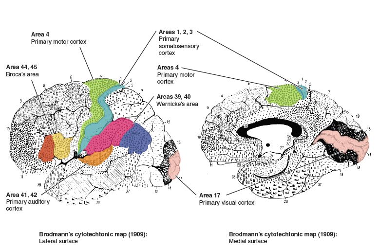
Sensation is the perception of one of the senses. Here, we will simply point out the importance of the post-central gyrus for the interpretation of general senses. General senses are ones from widely distributed receptors, such as touch, pressure, temperature, pain. These pathways end at the post-central gyrus, or sensory cortex. The cortex exhibits somatotophy: point by point correspondence of body locations to brain locations. The point by point correspondence gives more area of the cortex to regions that are well innervated with sensory receptors, such as fingers, and face; meanwhile regions that do not have large sensory innervation have correspondingly smaller areas on the sensory cortex.
The special senses are interpreted in their own specialized cortical regions, and are discussed in another section of this course.
Motor control refers to the initiation and proper coordination of the movement of a muscle. For a skeletal muscle, the intent to contract a skeletal muscle begins in motor association area (frontal lobe). The signal is then sent to the precental gyrus or primary motor area, which is the origin of the upper motor neuron. The precentral gyrus also exhibits somatotophy. Body areas, such as lips and fingers, which have fine motor control, have a large area dedicated on the primary motor cortex while areas that do not have fine control have a correspondingly smaller area.
Language includes abilities such as reading, writing, speaking, and comprehending words. At least two major areas are involved in the recognition and formation of language. Wernicke’s area, within the parietal and temporal lobes, is involved in the recognition of language. Broca’s area is involved in the formation of words.
Cranial Nerves
The cranial nerves provide input to and output from the brain. The numbers, names, and a short functional description are below.
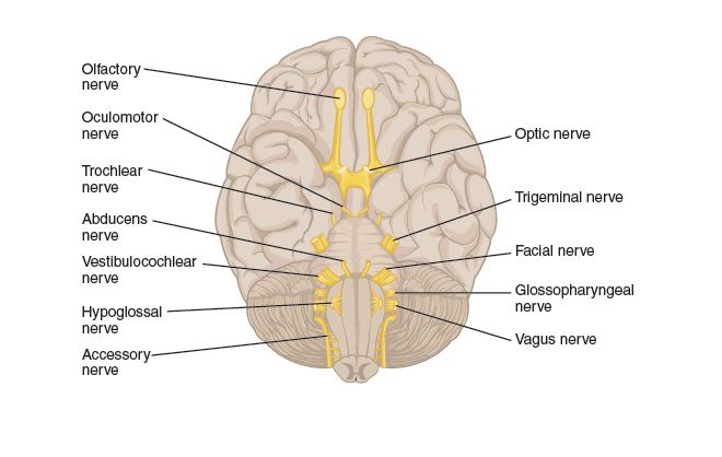
Cranial Nerve Functions
- Olfactory: sensory for smell
- Optic: sensory, process visual information
- Oculomotor: motor, movement of eyes and smooth muscles controlling pupil and lens
- Trochlear: motor, eye movements
- Trigeminal: sensory of upper, and mid face and upper jaw; motor for muscles of chewing
- Abducens: motor, eye movements
- Facial: motor for facial expression, tears and salivary glands; sensory for taste
- Vestibulocochlear: sensory, hearing and equilibrium
- Glossopharyngeal: motor for mouth (swallowing) and for regulation of blood pressure; sensory for tongue and pharynx and outer ear
- Vagus: motor for swallowing, speech, cardivascular and digestive regulation; hunger and fullness; sensory from visceral organs and taste. Main parasympathetic nerve
- Accessory: swallowing, and head, neck, shoulder movement
- Hypoglossal: tongue movements
The Spinal Cord
The nervous system is critical to many of our homeostatic feedback loops. In most of these loops, the structures of the nervous system make up more than one component, and carry out more than one function in these loops. For example, specialized nerve endings often act as sensors (receptors), information is carried along nerves and/or tracts of the spinal cord, integration occurs within the CNS, and spinal cord tracts and nerves carry the responding information back out to the effectors. The spinal cord is a nervous system structure dedicated to relaying information from the periphery to the brain and back, as well as carrying out certain levels of integration, such as those found in many reflexes. The structure of the spinal cord aids it in carrying out these relaying and integrative functions.
The spinal cord is a central nervous system structure that extends inferiorly from the brain stem and into the lower back. Throughout its length, it is enclosed within the spinal column, with the cord passing through the vertebral foramen of the vertebrae. In an adult, the spinal cord itself terminates at a point called the medullary cone, at approximately the level of the first lumbar vertebrae (L1). Below the medullary cone, the vertebral canal contains a bundle of nerve roots called the cauda equina.
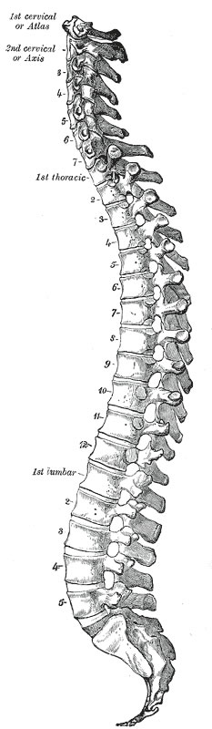
This figure shows other important features of the spinal cord, many of them related to the spinal cord’s function of relaying information. Starting between the base of the skull and the first cervical vertebrae, and continuing into the sacral region of the spinal column, a pair of spinal nerves extend from the spinal cord (although information is transmitted in both directions on sensory and motor neurons within these mixed nerves). All but the first spinal nerve (C1) pass through the intervertebral foramen of the spinal cord, whereas spinal nerve C1 passes between the occipital bone and vertebrae C1. In all there are 31 pairs of spinal nerves that carry information to and from the spinal cord and the periphery of the body. Note that not all of the spinal nerves arise from the cord at the level of the vertebrae between which they pass. This is most obvious when considering those spinal nerves arising in the lower lumbar and sacral regions. The nerve roots for these nerves arise from the spinal cord at, or near, the medullary cone, which you will recall is near the L1 vertebrae. These roots are contained within the cauda equina until passing out of the spinal column. Because the spinal nerve roots don’t always originate at the level of the vertebrae that they pass through, the segments of the spinal cord are named for the spinal nerve to which they give rise. For example, segment S2 of the spinal cord would be located near the T12 vertebrae.
Because the spinal cord terminates near vertebrae L1, and there is a lot of body tissue that needs to be innervated below this level, there are a significant number of nerves arising from the lower aspect of the spinal cord. This leads to an area of increased spinal cord thickness in the lumbosacral regions of the spinal cord (corresponding to a region associated with the inferior thoracic vertebrae) called the lumbar enlargement. There is a corresponding cervical enlargement in the cervical segments that give rise to nerves innervating the upper limbs.
Spinal Cord Structure
Recall that the central nervous system tissues can generally be divided into white matter and gray matter. White matter is the myelin-containing region composed of axons, which make up the tracts of the CNS. These carry information between different regions and structures in the CNS. Gray matter contains the cell bodies and dendrites and therefore is the site of synaptic transmission.
In the cortex of the brain, gray matter makes up the cortical (outer) regions, while the white matter tracts tend to make up the majority of the deep tissues of the brain, although there are exceptions to the latter, such as the deep basal and thalamic nuclei that are composed of gray matter. In contrast to this general arrangement of the brain, the spinal cord is arranged with the white matter surrounding the central gray matter, indicating that the spinal tracts carry information up and down the cord along the outer aspects, while synaptic transmission tends to occur more centrally.
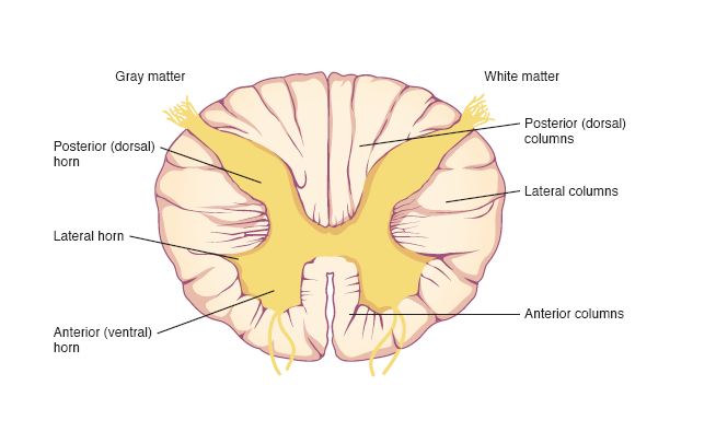
In the image above, you can see how the central gray matter is somewhat butterfly shaped, with each side of the “butterfly” containing a posterior (dorsal) horn and an anterior (ventral) horn. Each of the horns is contiguous with the posterior and anterior spinal nerve roots, respectively. The posterior root of the nerve carries sensory information into the posterior horn, often synapsing there. The anterior horn contains the cell bodies of somatic motor neurons, and it sends its axons out the anterior root of the spinal nerve to the muscle cells it innervates. The lateral horn is not found at all levels of the spinal cord, but is limited to thoracic and lumber segments of the cord. This is because the lateral horns contain the neurons of the sympathetic nervous system, which leave the cord only in these segments. Even though the cell bodies are found in the lateral horns, their axons leave via the anterior nerve roots, just like those that control skeletal muscle. The matched horns on each side of the “butterfly” are connected via the gray commissure, which also surrounds the cerebrospinal fluid filled central canal.
The white matter of the spinal cord is divided into columns. Each segment of the cord contains matched posterior, lateral and anterior columns. The anterior columns and posterior columns are partially separated by the anterior median fissure and posterior median sulcus, respectively. Each pair is also connected by a commissure of white matter that runs adjacent to the gray commissure, termed the anterior and posterior commissures. The columns are further divided into tracts that carry sensory information up the spinal cord (ascending tracts) and motor information down the spinal cord (descending tracts).
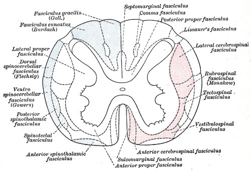
Spinal Cord Tracts
The white matter of the spinal cord is divided into the paired posterior (dorsal), lateral, and anterior (ventral) columns. These columns are sometimes called funiculi (or funiculus when singular) and are made up of axons that are traveling up (ascending) or down (descending) the spinal cord. The ascending tracts generally carry sensory information from the periphery to the brain, while the descending tracts carry motor signals to muscles and glands.
The columns can be further divided into tracts (sometimes called fasciculi), which is a way of functionally grouping the neurons based on similar origin, destination and function. These tracts are often named for the structures that they connect. For example, the spinothalamic tract indicates that the fibers are carrying information from the spinal cord to the thalamus of the brainstem. You may note from its name that it is an ascending tract, so the information that it carries is sensory.
Some of the tracts cross over (decussate) either in the spinal cord or brainstem, and when this occurs, the relationship between the origin and destination is termed contralateral. Much of our motor control is contralateral. For example, your right arm is mainly controlled by the motor area in your left brain. When the origin and destination of a tract are on the same side of the body, it is referred to as an ispsilateral relationship.
Spinal Cord Communication
The spinal cord acts as a conduit for information traveling up and down its length. But because most of this information has to either exit the spinal cord to send signals to peripheral tissues (efferent transmission), or information from peripheral tissues needs to be carried into the spinal cord (afferent transmission), there must be appropriate structures for these types of transmission to occur. It is the 31 pairs of spinal nerves and their related structures that provide the pathways for this interaction.
Sensory and motor fibers enter and exit the cord via rootlets that arise from both the posterior and anterior aspects of the cord. Anterior rootlets carry motor information out of the spinal cord (i.e. they contain efferent fibers) while the posterior rootlets carry sensory information into the spinal cord (i.e. they contain afferent fibers). Several posterior rootlets merge together to form the posterior root, while several anterior rootlets similarly converge to form the anterior root.
Along the posterior root is a ganglion, where cell bodies of many of the sensory neurons are found. These are unipolar neurons, such that their dendrites extend out to the peripheral tissues, and their axons project into the dorsal horn of the spinal cord, where they synapse. These unipolar peripheral neurons are considered first order neurons in the sensory pathway, while the neurons they synapse with in the posterior horn are considered the second order neurons of the sensory pathway. It is the axons of these second order neurons that make up the various ascending white matter tracts.
The anterior root does not contain a ganglion. This is because motor control is typically a two neuron pathway. It starts with an upper motor neuron, whose cell body is in the cerebral cortex or gray matter of the brainstem. This neuron projects its axon via a descending white matter tract to a point in the spinal cord where it synapses in the ventral horn with a lower motor neuron. The cell body of the lower motor neuron is in the gray matter of the spinal cord, and it projects its axon out one of the anterior rootlets and through the anterior root. Ganglia are only found where neuron cell bodies are outside the CNS.
Distal to the posterior root ganglion, the fibers of the anterior and posterior root merge together and pass through the dura to become the spinal nerve. Because the spinal nerves contain both sensory and motor fibers, they are considered a mixed nerve, as opposed to either a sensory or motor nerve.
Just distal to the intervertebral foramen of the spinal column, the spinal nerve branches into rami (singular: ramus). In general, the posterior ramus communicates with structures posterior to the cord, while the anterior ramus communicates with structures anterior to the cord. In spinal nerves T1-L2, the anterior ramus gives rise to a communicating ramus that communicates with the sympathetic ganglia in the region. The sympathetic motor pathway involves two motor neurons, so this ganglion houses the second motor neuron’s cell body. In various regions of the body, the anterior rami from several spinal nerves join together and then branch again, in a complex network of nerves called a plexus.
Spinal Plexus
A plexus is a network of anterior rami from neighboring spinal nerves that come together in a weblike or tangled network adjacent to the spinal cord, and from which new nerves arise. These nerves contain fibers from several spinal nerves. The four main plexuses are the cervical, brachial, lumbar and sacral. Some people consider the coccygeal plexus a fifth plexus, although it is much smaller than the others.
The plexuses are complex networks, with four or more spinal nerve rami contributing to each of the four main plexuses, and with several nerves arising from each. The following table lists the four main plexuses, the spinal nerves that contribute to each, and some of the main nerves that arise from them.
| Plexus | Spinal Nerves that Contribute | Example Nerves Arising From |
|---|---|---|
| Cervical | C1-C5 | Lesser Occipital
Great Auricular Supraclavicular Phrenic |
| Brachial | C5-C8, T1 | Axillary
Radial Musculocutaneous Median Ulnar |
| Lumbar | T1-T4 | Femoral
Saphenous Obturator |
| Sacral | L4-L5, S1-S4 | Superior gluteal
Inferior gluteal Tibial |
So what could be a functional significance of having spinal plexuses? Damage to a single spinal nerve is less likely to result in complete loss of some critical motor functions, such as the phrenic nerve that goes to the diaphragm for breathing, or others innervating the upper and lower limbs. These peripheral body parts would still be able to send sensory and receive motor control through the spinal cord via alternate spinal nerves superior or inferior to the damage. If, however, the spinal cord itself was damaged, the plexus would not be able to compensate.
Nervous Structures and Functions Review
Consider again the learning objectives for this module. Could you demonstrate each of these objectives? If not, consider reviewing content related to these objectives.
- Describe the basic (overall) structure of the human brain.
- Explain the roles of CSF, ventricles, and the blood brain barrier.
- Correlate hindbrain and midbrain regions to their major function(s).
- Correlate forebrain regions to their major functions(s).
- Assign function(s) to each of the cranial nerves.
- Describe the gross anatomy of the spinal cord and spinal nerves and specify their location relative to the anatomy of the vertebral column.
- Contrast the relative position of gray matter and white matter in the spinal cord with the corresponding arrangement of gray and white matter in the brain.
- Compare and contrast the anatomical features of the spinal cord in the cervical, thoracic and lumbar regions.
- Identify how spinal structures relate to each other: tract, root, ganglion, nerve, ramus, plexus.
