3 Body Planes and Directional Terms
Another commonality across body types are the body planes and directional terms. Those in the health professions must speak the same language with regard to locating and identifying specific body parts and organs. Body planes and directional terms are part of this common language. The imaginary vertical and horizontal planes run through the body, essentially cutting it into parts. This section provides an introduction to this new “language” and opportunities to practice using it in context so that you become comfortable locating and describing all organs and parts in the body and in relation to each other. Everything that you learn after body planes and directional terms will be referring to this terminology to help you visualize, identify, and locate anatomical structures.
Body Planes
To better identify the locations of the organs that contribute to vital functions, you need some points of reference for description. To serve that function, we will now define different planes of the body. These imaginary flat surfaces run through the body in different directions. They are used by medical professionals to examine various internal body parts. Directional orientation is another anatomical tool used to describe how parts of the body are related to one another.
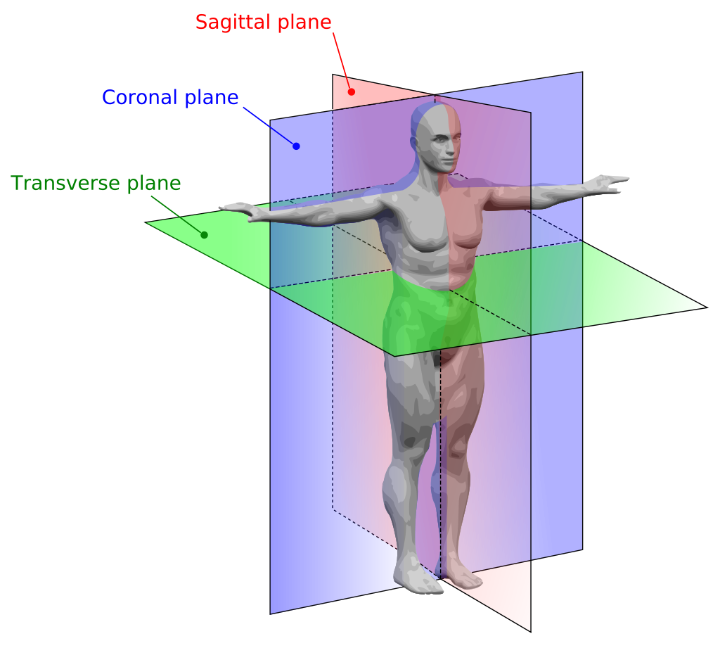
Each organ system spans large regions of the human body. It is helpful, therefore, to establish reference planes and directions that can help us describe specific locations of structures as we discuss them. To make sure everyone is talking about the same thing, anatomists and physiologists often refer to anatomical position and the body planes that penetrate it. Anatomical position describes a person standing upright, with the arms at the sides and the palms facing forward (as demonstrated in the image below). Body planes (a plane is a flat, two-dimensional surface) are imaginary surfaces that run through the body and divide it into different sections. We can talk about a specific location using the planes as reference points within the anatomical position.
There are an infinite number of planes running through the human body in all directions. However, we will focus on the three planes that are traditionally used when discussing human anatomy. First is the transverse plane, (also called the horizontal plane), which divides the body into top and bottom. In anatomical position, transverse planes are parallel to the ground. The second is the coronal plane, which is a vertical plane that divides the body into the front and back sections. If you do a “belly flop” into the water, you sink into the water via the coronal planes. Finally, we will refer to the sagittal plane, which divides the body into left and right sections with a vertical plane that passes from the front to the rear.
Examples: Body Imaging and Cross Sections
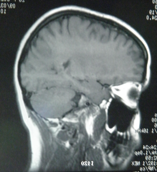
Many imaging modalities used in medicine (CT scans, MRI scans and ultrasounds) image the body in cross sections. The three main planes described above are used to orient and describe where the cross section is and how it passes through the body so that the viewer knows what they are viewing. For example, the image below from a CT (or CAT) scan shows a cross section of the body that runs along the sagittal plane.
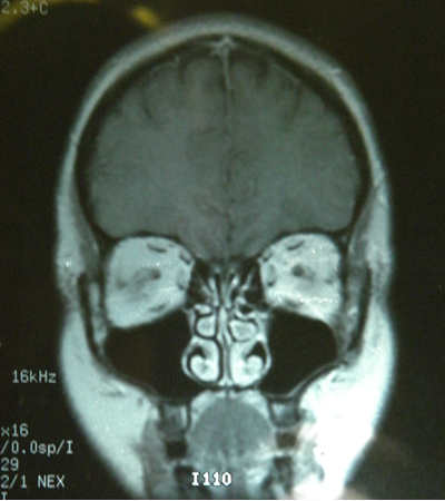
These images can sometimes be reconstructed in a computer to show the same body in a different plane, making some features of the body easier to see. Below is a coronal reconstruction of a CT:
Directions and Orientation
You can use other terms to further pinpoint an anatomical location. These terms are used to describe a location in relation to other structures. Some of them may be terms you have heard in everyday conversation; a lateral pass in football, for example, is a pass toward the sideline.
The following table lists all of the human anatomical directions that we will discuss. You will practice using these planes and directional terms when describing the locations of organs and organ systems in the following sections.
| Directional Term | Meaning |
|---|---|
| superior | above (or toward the head) |
| inferior | below (or toward the feet) |
| distal | farther from the trunk or origin |
| proximal | closer to the trunk or origin |
| superficial | toward or on the surface |
| deep (internal) | away from the surface |
| anterior (ventral) | toward the front (or toward the belly) |
| posterior (dorsal) | toward the rear (or toward the back) |
| medial | toward the midline |
| lateral | toward the side |
Superior, Inferior, Anterior and Posterior
The first set of directions that we will explore are superior, inferior, anterior, and posterior.
In humans, which stand upright on two feet, there are other terms that are synonymous with these four terms. Cephalic means toward the head and is the same as superior for a human in anatomical position. Caudal means toward the tail, or same as inferior for a human in anatomical position. Dorsal means toward the back and ventral means toward the belly; so dorsal and posterior are the same direction, and ventral and anterior are the same direction for a human in anatomical position. This would not be true for a four-legged animal, such as a rat or cat you might dissect in lab.
Medial and Lateral
Next, we will discuss terms that relate structures to the midline. These are medial, lateral and intermediate.
Proximal, Distal, Superficial, Deep
These next terms are used when referring to either appendicular parts of the body (arms and legs) or position in body relative to the external surface. These are Proximal, Distal, Superficial, Deep.
Anatomical Regions
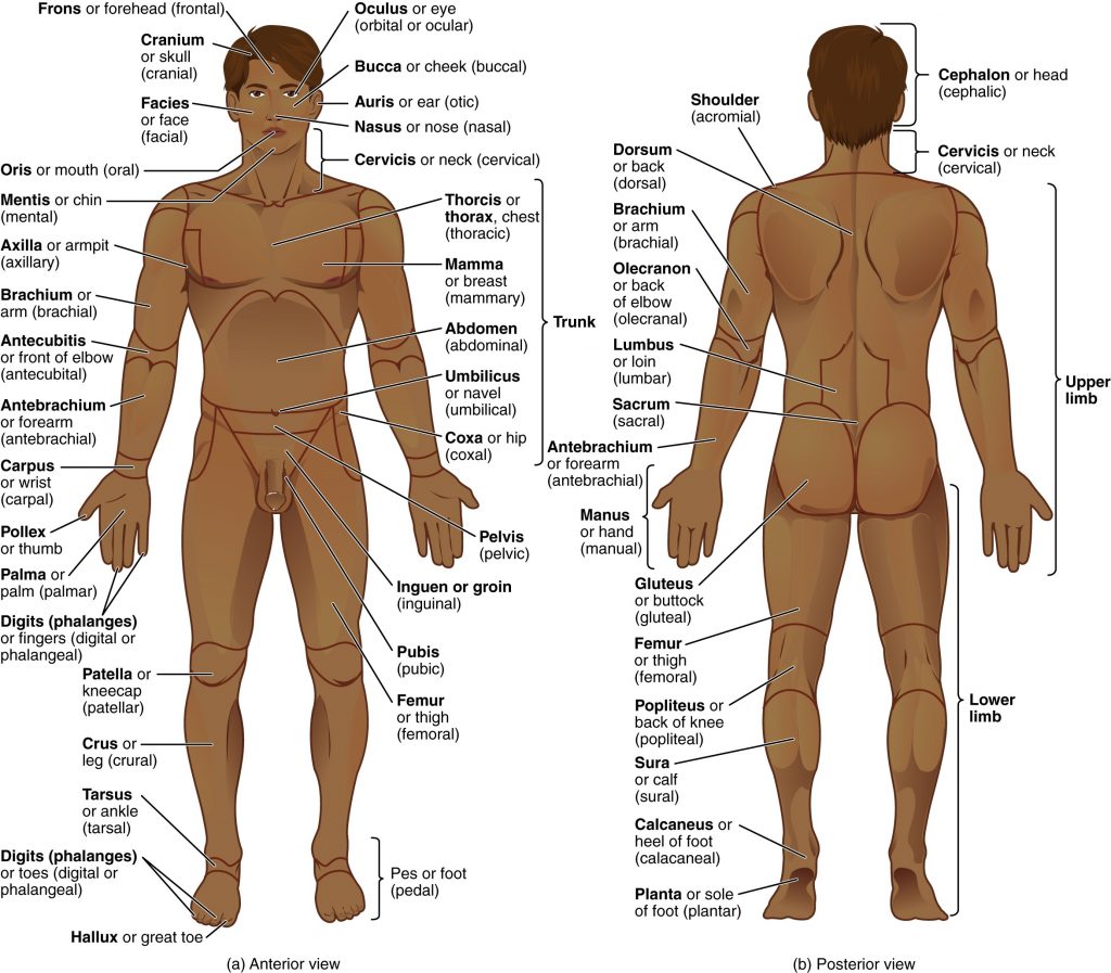
You can use even more terms to describe an anatomical location. These terms are used to describe regions of the body, and often will serve as a source for names of organs in these areas. Some of the terms may be familiar, but some will likely be new to you.
Practice using the terms in parenthesis until they become familiar to you [1].
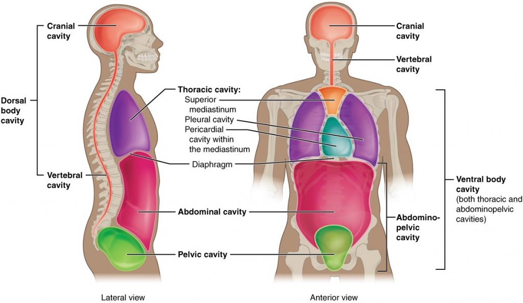
Body Cavities
When you look at the human body, there are several different cavities that contain different organs. You may remember dorsal as meaning towards the back or spinal column, and ventral as being towards the belly. While posterior and anterior have replaced those in common usage, the body cavities still maintain the older terminology.
Dorsal Body Cavity
The dorsal (posterior) body cavity is separated into two areas. The cranial cavity contains the brain, while the spinal cavity (or vertebral cavity) contains the spinal cord. These two spaces are continuous.
Ventral Body Cavity
The ventral (anterior) body cavity is separated anatomically by the diaphragm. Superior to the diaphragm is the thoracic cavity that contains the heart and lungs. Inferior to the diaphragm is the abdominopelvic cavity that is further divided into the abdominal and pelvic cavities. This is an artificial separation, as the two spaces are continuous. The abdominal cavity contains many organs, including the liver, kidneys, spleen, pancreas, and most of the digestive organs. The pelvic cavity is within the pelvic brim, which provides a little more protection for the urinary bladder, rectum, and female reproductive system.
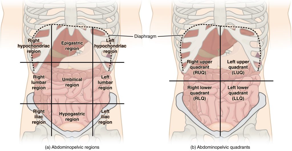
Abdominal Regions and Quadrants
Sometimes it is helpful for doctors to connect pain in one area of the abdomen to the organ located there. One version has nine regions, and the other has four quadrants [2].
- Image name: Regions of the Human Body Section: Anatomical Regions, Image description: Cephalon or head (cephalic) Frons or forehead (frontal) Cranium or skull (cranial) Facies or face (facial) Oculus or eye (orbital or ocular) Bucca or cheek (buccal) Auris or ear (otic) Nasus or nose (nasal) Oris or mouth (oral) Mentis or chin (mental) Cervicis or neck (cervical) Trunk Shoulder (acromial) Dorsum or back (dorsal) Thorcis or thorax, chest (thoracic) Mamma or breast (mammary) Abdomen (abdominal) Umbilicus or naval (umbilical) Lumbus or loin (lumbar) Sacrum (sacral) Hip (coxal) Pelvis (pelvic) Inguen or groin (inguinal) Pubis (pubic) Gluteus or buttock (gluteal) Upper limb Brachium or arm (brachial) Antecubitis or front of elbow (antecubital) Olecranon or back of elbow (olecranal) Antebrachium or forearm (antebrachial) Carpus or wrist (carpal) Manus or hand (manual) Pollex or thumb Palma or palm (palmar) Digits (phalanges) or fingers (digital or phalangeal) Lower limb Crus or leg (crural) Femur or thigh (femoral) Popliteus or back of knee (popliteal) Patella or kneecap (patellar) Sura or calf (sural) Tarsus or ankle (tarsal) Pes or foot (pedal) Calcaneus or heel of foot (calcanea) Planta or sole of foot (plantar) Digits (phalanges) or toes (digital or phalangeal) Hallux or great toe ↵
- Image: Regions and Quadrants of the Peritoneal Cavity Section, Image description: Nine abdominopelvic regions subdivide the abdominal cavity using two horizontal lines (one inferior to the ribs and the other superior to the pelvis), and two vertical lines (extending down from the midpoint of each clavicle). Top region: Right hypochondriac region, Epigastric region, Left hypochondriac region Center region: Right lumbar region, Umbilical region, Left lumbar region Bottom region: Right iliac region, Hypogastric region, Left iliac region Four abdominopelvic quadrants subdivide the abdominal cavity using two lines (one horizontal and one vertical), that intersect at the naval. Right upper quadrant (RUQ), Left upper quadrant (LUQ), Right lower quadrant (RLQ), Left lower quadrant (LLQ) ↵
