24 Integumentary Structures and Functions
Gross Anatomy of the Integumentary System
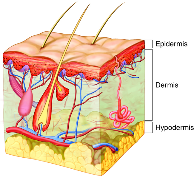
The skin is made up of two mutually dependent layers that are distinguished based on their structure and location. These layers – the epidermis and the dermis – contain a variety of structures, including blood vessels, hair follicles, and sweat glands. Beneath the dermis lies the hypodermis (subcutis). It is composed mainly of fatty tissue.

The most superficial layer, the epidermis, is composed of stratified squamous epithelia that are keratinized at the outermost surface, melanocytes, immune cells (Langerhans that modulate immune response) and sensory receptors (Merkel cells that detect light touch). The function of the epidermis layer is “protection.” The keratinocytes and immune cells help protect the skin.
The dermis lies beneath the epidermis and is composed of two layers of connective tissue: a loose layer (papillary) and a dense irregular layer (reticular). Both layers of the dermis contain connective tissue components (collagen, elastin, fibroblasts), plus blood vessels, sensory receptors and lymphatics. The dermis is a “functional” layer. The dermis is connective tissue that can stretch and retract because of the strong and elastic extracellular matrix. The dermis also contains nerves.
Beneath these two layers lies the hypodermis, composed of loose connective tissue (adipose and areolar). The hypodermis is the “connection” layer. It connects the integument (epidermis and dermis) to organs and muscles in the body. This layer contains adipose tissue and connective tissue as well as blood vessels, nerves and immune cells.

