Chapter 9: Estimating Sex in Human Skeletal Remains
SEXUAL DIMORPHISM
Forensic anthropologists are often called upon to identify skeletal remains and the first step in this process is building the biological profile.6 Estimation of sex is often one of the first things considered when establishing a biological profile because several other parts, such as age and stature estimations, rely on an estimation of sex to make the calculations more accurate.2 Sex estimation can be accomplished because of sexual dimorphism.
According to its general definition, “sexual dimorphism is the growth of visible morphological differences between males and females in a species or population”.3 Humans have a very moderate level of sexual dimorphism is comparison to other species. In some primate species, for example, males are much larger than females. Sexual dimorphism also varies between different human populations. Because of this moderate level of dimorphism there is overlap between smaller males and larger females which could lead to the possibility of misidentification.1
Although the most obvious attribute of sexual dimorphism is stature in humans with males, overall, being taller than females, sexual dimorphism is present in many aspects of the human skeleton. In many skeletal elements, it is present in the form of morphological differences; in others, it exclusively relates to a size variation.3
Population genetics are the most important contributing factor to sexual dimorphism, but environmental factors—especially diet and mechanical loading —also affect its expression. Several studies have demonstrated that secular changes associated with lifestyle modifications have led to a variation in the degree of sexual dimorphism, especially in geographically and temporally isolated populations.3
Traits that differentiate male from female human skeletons are due to the testosterone hormone. As testosterone levels are low before puberty, sexually dimorphic characteristics develop after skeletal maturity. Therefore, a skeletal sex estimation is usually restricted to adults, although various methods for juveniles have also been proposed.3
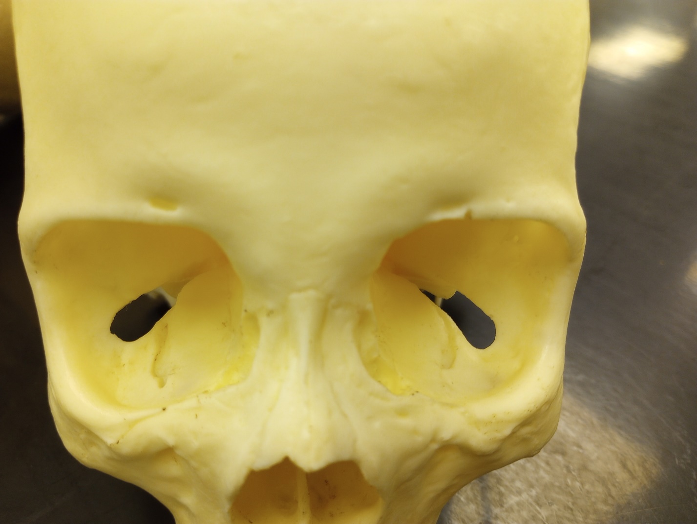
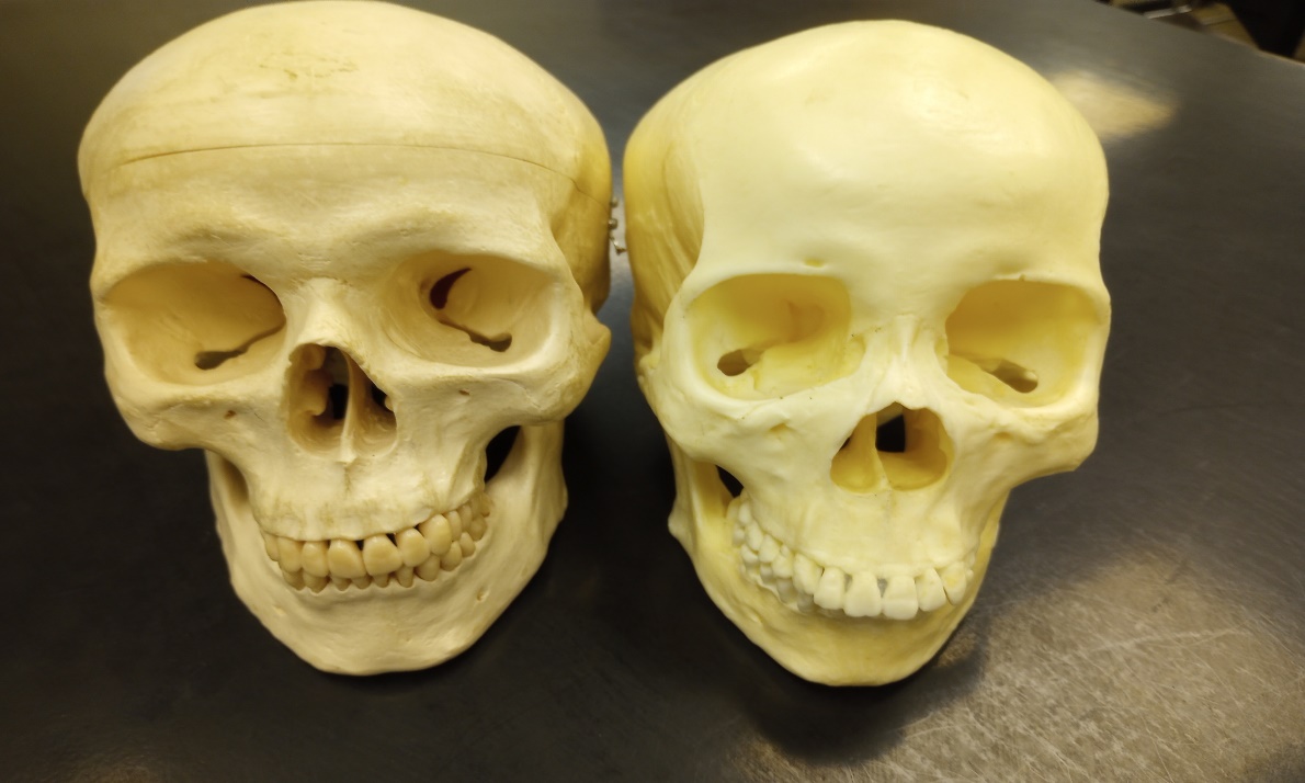
Estimations of sex look at differences in both morphological (form or structure) and metric (measured) traits in individuals. When assessing morphological traits, the skull and the pelvis are the most commonly used areas of the skeleton for estimations. These differences are related to sexual dimorphism usually varying in the amount of robusticity seen between males and females. Robusticity deals with strength and size; it is frequently used as a term to describe a large size or thickness. In general, males will show a greater degree of robusticity than females. For example, the length and width of the mastoid process, a bony projection located behind the opening for the ear, is typically larger in males. The mastoid process is an attachment point for muscles of the neck, and this bony projection tends to be wider and longer in males. In general, cranial features tend to be more robust in males.2
Sex vs. Gender
Biological sex is a different concept than gender. While biological anthropologists can estimate sex from the skeleton, estimating an individual’s gender would require a greater context as gender is culturally defined rather than biologically defined. Take for example an individual who identifies as transgender. This is an individual who has a gender identity that is different from their biological sex. The gender identity of any individual depends on factors related to self-identification, situation, or context, and cultural factors. While in the U.S. we have historically thought of sex and gender as binary concepts (male or female), many cultures throughout the world recognize several possible gender identities. In this sense, gender is seen as a continuous or fluid variable rather than a fixed one.2
Estimating Sex in the Skull
While virtually all bones display some sexual dimorphism, the pelvis is the most reliable for identification. Using the skull alone is less accurate. In cases of adult crania with which there is neither lower jaw, nor any other part of the skeleton, the diagnosis is about 80 percent reliable. This proportion rises to 90 percent where a well-preserved lower jaw is present; and will reach 96 to 98 percent when a whole skeleton is present. Although there will still remain skeletons which, even though complete, show such ambiguous sexual characteristics that it will be impossible to identify them as either male or female with certainty.6The following are cranial traits used in sex assessment:
Overall size – Larger in males; smaller in females.6
Muscle attachments – Stronger in males exhibited by roughening; females are generally more smooth overall.6
Cranium External occipital protuberance (nuchal crest) – More pronounced in males; rounded and smooth in females.6
Forehead – Retreating in males; smooth, round, more vertical and better developed frontal eminences in females.6
Glabella – Protrudes in males; smooth in females.6
Mastoid process – Large in males; small in females.6
Palate – Males are larger and broader; females display less depth.6
Supraorbital margins – Rounded and thick in males; sharp and thin in females.6
Supraorbital ridges – More pronounced in males; flat and smooth in females.6
Zygomatic processes – Heavier in males; more slender in females.6
Mandible Mental eminence – Square and broad in males; v-shaped and narrow in females.6
Gonial angle (angle at the back of the mandibular ramus) – Less obtuse in males (stouter, rougher, and more everted angles); an angle over 125 degrees suggests female sex.6

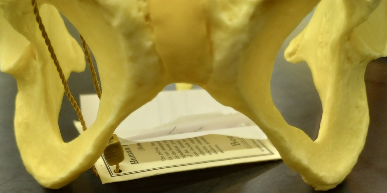
Estimation of the Pelvis
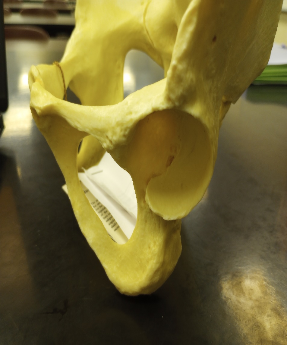
When considering the pelvis, the features associated with the ability to give birth help distinguish females from males. During puberty, estrogen causes a widening of the female pelvis. ² Because humans give birth to large headed babies, the female pelvis needs to be wider to facilitate the passage of the infant’s head during childbirth. In species that do not give birth to large headed babies the male and female pelvises show little difference. ¹ If the adult skeleton is absolute or at least has an unbroken pelvis, the sex can usually be determined with nearly 100% accuracy.4
Many studies have been done to determine pelvic characteristics useful in sex estimations. The first two indicators listed here are generally the most useful, especially for the budding osteologist. Clearly, the more experience an osteologist has in making sex estimations and the greater number and range of pelvis examined, the better the estimations will be.6
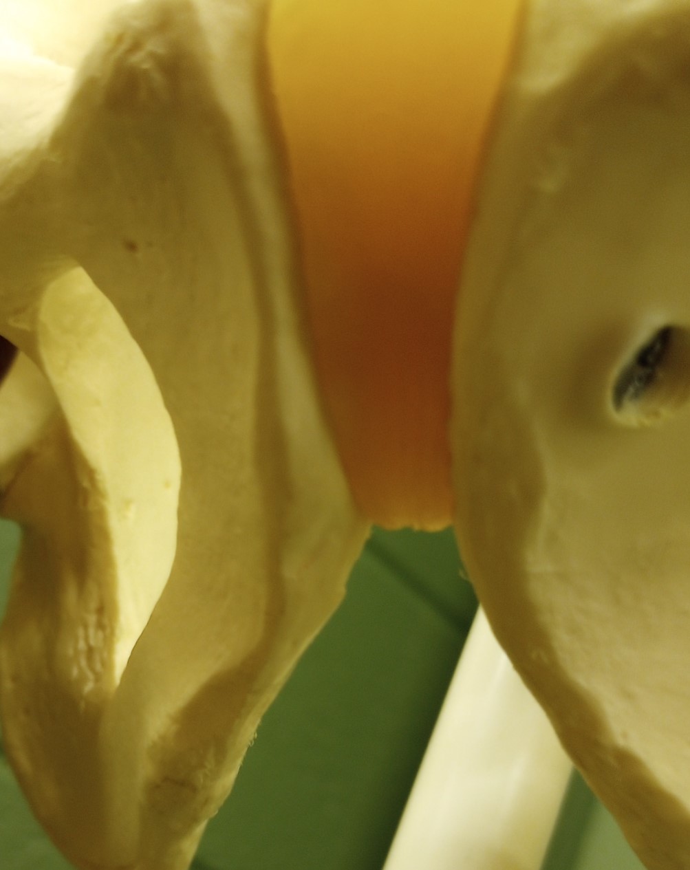
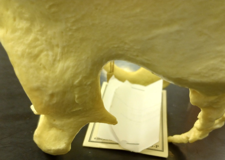
1. Sub-pubic angle. The inferior angle that the right and left pubic bones make when in articulation tend to be wider in females than in males; a wider angle produces a larger pelvic outlet. Angles closer to 90 degrees suggest male sex, while those 120 degrees and over would suggest a female. The female pelvis is shorter and broad to aid in the birthing process, since the male pelvis lacks this necessity, it is slightly taller and more narrow.6
2. Greater Sciatic notch. A narrow sciatic notch is associated with a restricted pelvic outlet and is more commonly found in males; the sciatic notch of females tends to be wider.6
3. Acetabulum. The acetabulum is larger in males, due to the larger size of the femoral head in males. As males are generally larger, the femur is larger to transmit the weight of the body.6
4. Obturator foramen. The obturator foramen tends to be larger in males and rather oval in outline, whereas in females it is smaller and more triangular.6
5. Sacrum. The sacrum of the male tends to be relatively longer, narrow, and curved. On the other hand, the female sacrum is broader, short, and straight.6
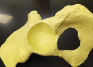
6. Pelvic inlet. The pelvic inlet of the male pelvis, when viewed from above with the ventral aspect facing you, will be heart shaped in appearance. The female pelvic inlet, when held in the same aspect, is described as elliptical in appearance.6
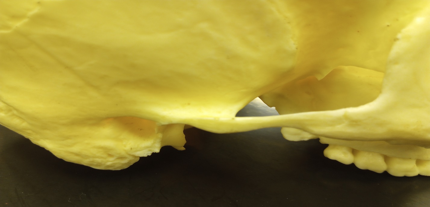
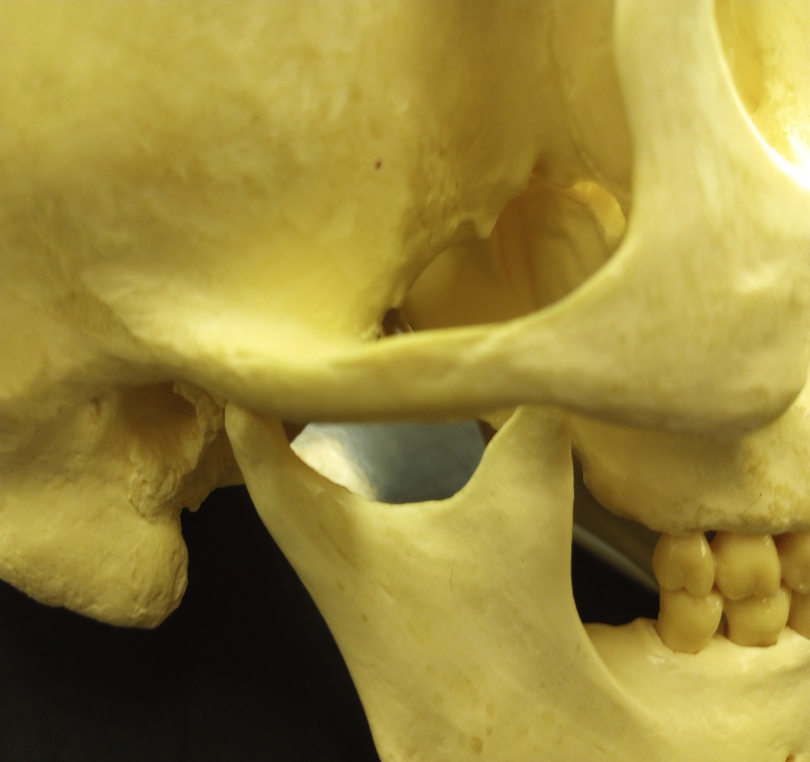
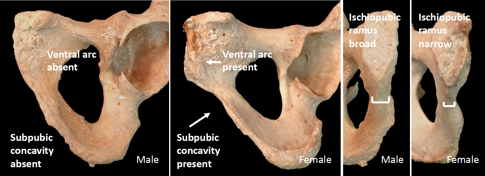
7. The Phenice Method (Phenice 1969) is traditionally the most common reference used to assess morphological characteristics associated with sex. The Phenice Method specifically looks at the presence or absence of (1) a ventral arc, (2) the presence or absence of a sub-pubic concavity, and (3) the width of the medial aspect of the ischiopubic ramus (Figure 9.1). When present, the ventral arc, a ridge of bone located on the ventral surface of the pubic bone, is indicative of female remains. Likewise, the presence of a sub-pubic concavity and a narrow medial aspect of the ischiopubic ramus is associated with a female sex estimation. Assessments of these features, as well as those of the skull (when both the pelvis and skull are present), are combined for an overall estimation of sex.2
Sex Estimation of the Non-Pelvic Postcranial Bones
Sex estimation of the non-pelvic postcranial bones can be very difficult, based on research with accuracy well below 90%. Keep this in mind when employing sex estimation on these bones. However, if observing several of the bones of a single individual you will greatly increase your sex estimation accuracy.6
All human femurs angle inward down from the pelvis to the knee, but because of the wider female pelvis this angle (the Q-angle) is greater in females than males. This is also true of the elbow angle (the carrying angle). This larger carrying angle is needed to allow the arms to pass by the hips when swinging your arms during walking. Otherwise, the arms would bash into the pelvis.1
Metric Analysis
Metric analyses are also used in the estimation of sex. Measurements taken from every region of the body can contribute to estimating sex through statistical approaches that assign a predictive value of sex. These approaches can include multiple measurements from several skeletal elements in what is called multivariate (multiple variables) statistics. Other approaches consider a single measurement, such as the diameter of the head of the femur, of a specific element in a univariate (single variable) analysis.2 Of the metric methods long bone measurements are considered the most accurate.1
Measurements:
Femur – A measurement of the greatest diameter of the femoral head may be useful in determining sex. Keep in mind there may be populational differences.6
Humerus – A vertical (superior/inferior) measurement of the head can be used in sexing.6
Male Female
Femur head diameter >47.5 mm <42.5 mm
Humerus head diameter >47 mm <43 mm
Radius head diameter ≥24 mm ≤21 mm4
Fordisc
These measurements can also be plugged into statistical software which can aid in sex estimation as well. The computer program Fordisc is an anthropological tool used to estimate different components of the biological profile, including ancestry, sex, and stature. When using Fordisc, skeletal measurements are input into the computer software and the program employs multivariate statistical classification methods, including discriminant function analysis, to generate a statistically validated prediction for the geographic origin of unknown remains. Fordisc will also tell the analyst the likelihood of the prediction being correct, as well as how typical the metric data is for the assigned group.2
Subadults
It is important to note that, although forensic anthropologists usually begin assessment of biological profile with sex, there is one major instance in which this is not appropriate. The case of two individuals found near Willits, California, on July 8, 1979, is one example that demonstrates the effect age has on the estimation of sex. The identities of the two individuals found in Willits were unknown; therefore, law enforcement sent them to a lab for identification. A skeletal analysis determined that the remains represented one adolescent male and one adolescent female, both younger than 18 years of age. This information did not match with any known missing children at the time.2
In 2015, the cold case was reanalyzed, and DNA samples were extracted. The results indicated that the remains were actually those of two girls who went missing in 1978. The girls were 15 years old and 14 years old at the time of death. It is clear that the 1979 results were incorrect, but this mistake also provides the opportunity to discuss the limitations of assessing sex from a subadult skeleton.2
Assessing sex from the human skeleton is based on biological and genetic traits associated with females and males. These traits are linked to differences in sexual dimorphism and reproductive characteristics between females and males.2 Prior to the development of secondary sexual characteristics, however, there is limited sexual dimorphism in skeletal features, rendering macroscopic methods unreliable before puberty. While DNA analysis provides a potential solution, it does not always preserve and is too destructive and expensive to use routinely. The inability to reliably determine the sex of nonadults has placed constraints on the identification of children and infants.5
However, some sexual dimorphism has been noted in subadults. This includes features such as a raised auricular surface and wider subpubic angle in females. Some metric analysis using radiographs have also been developed which are accurate more than 80% of the time.1
Another method that anthropologists have begun to use for sex estimation involves sexually dimorphic enamel peptides in both deciduous and permanent teeth at different stages of development. This method identifies sex chromosome-linked proteins from human tooth enamel using a minimally destructive acid etching procedure and a mass spectrometer.5
Tooth enamel is the hardest human tissue and is highly resistant to diagenesis in burial contexts; it is therefore ideal for the biochemical estimation of sex. Tooth enamel consists of only a small number of proteins, and is primarily comprised of the heterogeneous, dimorphic proteins (AMELX coded for on the x chromosome and AMELY coded for on the Y chromosome). Several studies in recent years have successfully demonstrated that proteins can be extracted from human tooth enamel and used as a method for identifying the sex of individuals. The method has worked successfully on samples that are poorly preserved and on juvenile remains.5
Tables
Table 1: Cranial features between males and females
|
Characteristic |
Male |
Female |
|
Occipital protuberance |
More prominent |
Less prominent |
|
Mastoid process |
Larger |
Smaller |
|
Supraorbital ridges |
Prominent |
Absent |
|
Upper orbital margin |
Rounded |
Sharper |
|
Ramus of mandible |
Wide, sharply angled, flared |
Narrow, chin less angled |
|
Chin |
Squared, protuberant |
Rounded or pointed |
|
Zygomatic arch |
Broader |
Narrower4 |
|
Forehead |
Retreating |
Smooth, round, more vertical and better developed frontal eminences6 |
|
Palate
|
Larger |
Smaller4 |
Table 1 Cranial features which differentiate between male and female for sex determination.4
Table 2: Post-Cranial Features Between Males and Females
|
Characteristic |
Male |
Female |
|
Ossification of ribs |
Marginal |
Central |
|
Ribs’ ends’ appearance |
Crab claw-like |
Rough planar end |
|
Pelvis |
Larger and more robust |
Wider and larger pelvic inlet |
|
Pubic body |
Triangular |
Rectangular |
|
Subpubic angle |
Narrow |
Wide |
|
Subpubic concavity |
Absent |
Developed4 |
|
Acetabulum |
Larger |
Smaller6 |
|
Ventral arc |
Absent |
Present2 |
|
Ischiopubic ramus |
Wide |
Narrow2 |
|
Ilium |
Narrow |
Wide |
|
Greater Sciatic notch |
Narrow and deep |
Wide and shallow |
|
Sacrum |
More curved, longer, and narrower |
Flatter, broader, and smaller4 |
|
Pelvic Inlet |
Heart Shaped |
Oval Shaped6 |
|
Muscle attachment in pelvis |
More |
Less |
|
Q-angle in femur bone |
Smaller (11.2 + 3) |
Larger (15.8 + 4.5)4 |
|
Carrying Angle |
Smaller |
Larger1 |
|
Knee |
Larger |
Smaller |
|
Femur head diameter |
>47.5 mm |
<42.5 mm |
|
Humerus head diameter |
>47 mm |
<43 mm |
|
Radius head diameter |
≥24 mm |
≤21 mm4 |
Table 2 Post-cranial features which differentiate between male and female for sex determination.4
References:
1. Angi M. Christensen, Nicholas V. Passalacqua, and Eric J. Bartelink, Forensic Anthropology: Current Methods and Practice, 2nd ed. (London: Academic Press, 2019): 243-257.
2. Ashley Kendell, Alex Perrone, and Colleen Milligan, “Bioarcheology and Forensic Anthropology” In Explorations, ed. Beth Shook, Katie Nelson, Kelsie Aguilera and Lara Braff (Arlington: American Anthropological Association, 2019). https://pressbooks-dev.oer.hawaii.edu/explorationsbioanth/.
3. Maria-Eleni Chovalopoulou, Efstratios Valakos, and Efthymia Nikita, “Skeletal Sex Estimation Methods Based on the Athens Collection,” Forensic Science 2 (2022): 715-724.
4. Purva Wagisha Upadhyay and Amarnath Mishra, “Forensic Anthropology” in Biological Anthropology – Applications and Case Studies, ed. Alessio Vovlas (London: IntechOpen, 2021). https://www.intechopen.com/chapters/73372.
5. Rebecca Gowland, Nicolas A. Stewart, Kayla D. Crowder, Claire Hodson, Heidi Shaw, Kurt J. Gron, and Janet Montgomery, “Sex estimation of teeth at different developmental stages using dimorphic enamel peptide analysis,” American Journal of Biological Anthropology 174 (2021): 859-869. https://onlinelibrary.wiley.com/doi/full/10.1002/ajpa.24231.
6. Roberta Hall, Kenneth Beals, Holm Neumann, Georg Neumann, and Gwyn Madden, Introduction to Human Osteology (Michigan: Grand Valley State University, 2010). https://pressbooks.gvsu.edu/introhumanosteology/.
Figure Attributions:
Figure 9.1 Original to Introduction to Forensic Anthropology
Figure 9.2 Original to Introduction to Forensic Anthropology
Figure 9.3 Original to Introduction to Forensic Anthropology
Figure 9.4 Features associated with the Phenice Method by Colleen Milligan original to Explorations: An Open Invitation to Biological Anthropology is under a CC BY-NC 4.0 License.
Figure 9.5 Original to Introduction to Forensic Anthropology
Figure 9.6 Original to Introduction to Forensic Anthropology
