Chapter 13: Analysis of Skeletal Pathology
BONE PATHOLOGY
While there is a wide range of variation within the human skeletal system, bone development can also occur pathologically. Bone pathology can occur when there is excessive bone growth (osteoblastic activity or bone building) or bone is destroyed unnecessarily (osteoclastic activity or bone breakdown). Osteoblastic (bone building) and osteoclastic (bone destruction or breakdown) activities are normal processes of bone development, growth, and maintenance; however, when bone growth or breakdown exceeds what is necessary, the bony change can be classified as pathological, resulting in a bone pathology.1
Types of Bone Pathology
For the purposes of this chapter, we will focus on both osteoblastic and osteoclastic pathologies of the human skeleton. In addition to considering whether a pathology is osteoclastic or osteoblastic, it is also important to classify a pathology according to its origin. Bone pathologies can be classified in a number of ways,1 including:
- Joint diseases
- Bone forming diseases.
- Infectious1
- Metabolic1
- Neoplastic1
- Disorders of Growth and Development
- Dental diseases
Joint Diseases
Arthritis is a common disorder of synovial joints that involves inflammation of the joint. This often results in significant joint pain, along with swelling, stiffness, and reduced joint mobility. There are more than 100 different forms of arthritis. Arthritis may arise from aging, damage to the articular cartilage, autoimmune diseases, bacterial or viral infections, or unknown (probably genetic) causes.8
Osteoarthritis
The most common type of arthritis is osteoarthritis, which is associated with aging and “wear and tear” of the articular cartilage.9 The prevalence of hand, knee, or hip joint OA is estimated at 27 million among United States (US) adults. Studies indicate that women are at greater risk for developing knee and hip OA compared to their male counterparts. Hormonal factors, reduced volume of cartilage in the knee, and the fact that women are more likely to self-report have been considered as explanatory factors.10 Risk factors that may lead to osteoarthritis later in life include injury to a joint; jobs that involve physical labor; sports with running, twisting, or throwing actions; and being overweight.9 Obesity is a strong modifiable risk factor for the development of knee OA, but less so for hip OA. In a meta-analysis, those who were obese or overweight were nearly three times as likely to report knee OA.10
These factors put stress on the articular cartilage that covers the surfaces of bones at synovial joints, causing the cartilage to gradually become thinner. As the articular cartilage layer wears down, more pressure is placed on the bones. The joint responds by increasing production of the lubricating synovial fluid, but this can lead to swelling of the joint cavity, causing pain and joint stiffness as the articular capsule is stretched. The bone tissue underlying the damaged articular cartilage also responds by thickening, producing irregularities and causing the articulating surface of the bone to become rough or bumpy.8 Eburnation or Extreme erosion may occur when the soft tissue within a joint when cartilage is no longer present. The result is bone on bone contact that creates grooving on the surface of the joint, overall making a smooth shiny surface. Osteophytes or small spicules of bone may be present at the margins of the joint or within the joint itself. A ridge of osteophytic change may also be present around the margin of the joint.9
Schmorl’s Nodes
Schmorl’s Nodes are depression or cavity caused by herniation of the vertebral disc. Located on either the superior or inferior aspect of the body of a vertebra. The depressions are oval or linear in shape, with relatively smooth margins. Generally seen as a sign of advanced age.9
Gout
Gout is a form of arthritis that results from the deposition of uric acid crystals within a body joint. Usually only one or a few joints are affected, such as the big toe, knee, or ankle. The attack may only last a few days, but may return to the same or another joint. Gout occurs when the body makes too much uric acid or the kidneys do not properly excrete it. A diet with excessive fructose has been implicated in raising the chances of a susceptible individual developing gout.8
Rheumatoid Arthritis
Other forms of arthritis are associated with various autoimmune diseases, bacterial infections of the joint, or unknown genetic causes. Autoimmune diseases, including rheumatoid arthritis, produce arthritis because the immune system of the body attacks the body joints. In rheumatoid arthritis, the joint capsule and synovial membrane become inflamed. As the disease progresses, the articular cartilage is severely damaged or destroyed, resulting in joint deformation, loss of movement, and severe disability. The most commonly involved joints are the hands, feet, and cervical spine, with corresponding joints on both sides of the body usually affected, though not always to the same extent.8
Ankylosing Spondylitis
Ankylosing spondylitis (AS) is an autoimmune disease that mainly involves spine joints, sacroiliac joints (SIJs) and their adjacent soft tissues, such as tendons and ligaments. In more advanced cases, this inflammation can lead to fibrosis and calcification, resulting in the loss of flexibility and the fusion of the spine, resembling “bamboo” with an immobile position. The main clinical manifestations include back pain and progressive spinal rigidity as well as inflammation of the hips, shoulders, peripheral joints and fingers/toes. As an autoimmune disease, AS develops through complex interactions between genetic background and environmental factors. Although significant progress has been achieved in the past decades, the etiology of AS remains unclear to some extent. To date, studies have revealed some factors that may be related to the occurrence of AS, including genetic background, immune reaction, microbial infection, and endocrinal abnormity.11
Bone Forming Diseases
Heterotopic Ossification
Heterotopic ossification (HO) is a diverse pathologic process, defined as the formation of extraskeletal bone in muscle and soft tissues. The word “heterotopic” is derived from the Greek roots “hetero” and “topos,” meaning “other place.” HO can be conceptualized as aberrant tissue repair and is increasingly recognized as a common complication of trauma, surgery, and other local or systemic insults. Nongenetic forms of HO are most common, but rare genetic forms of HO also exist. The spectrum of HO is broad. Some HO lesions may be small and clinically irrelevant, while others may exact a high morbidity. In practice, the designation of HO is applied to bone formation at any extra-osseous site, including skeletal muscle, fascia, tendon, ligament, subcutis, skin, vascular wall, or virtually any site of connective tissue.3
The classic presentation of nongenetic HO is in young adults with a clear history of local trauma or surgery. Approximately half of patients are in their second and third decades of life; however, a broad age distribution is present from infancy to late adulthood. Men are slightly more commonly affected with a sex ratio of 3:2. A history of trauma as the initiating event is present in most cases (up to 75%), and unrecognized or “microtrauma” or repetitive mechanical stress is generally thought to be present in the remaining patients.3
Myositis Ossificans
Nongenetic HO can occur nearly anywhere in the body, but the most common areas include locations that are susceptible to trauma, such as the elbow, thigh, pelvis, and shoulder. Nongenetic HO is often designated by the tissue type it involves, such as myositis ossificans when involving skeletal muscle, or fasciitis ossificans when involving fascia.3
Fibrodysplasia Ossificans Progressiva
The rare genetic causes of HO have a different presentation and clinical severity than the far more common nongenetic cases, and include fibrodysplasia ossificans progressiva (FOP). FOP is a rare, slowly progressive disorder caused by ACVR1 mutations and initially presenting in childhood. Multiple congenital skeletal malformations are associated with FOP, including most frequently an abnormal first toe, abnormalities affecting the digits of the hand, and malformations of the cervical spine. FOP patients eventually develop progressive, painful flares and heterotopic lesions limiting mobility and function. Biopsies should not be performed on FOP patients because any surgical intervention leads to additional spread of heterotopic lesions. Most cases arise from a spontaneous mutation, but autosomal dominant transmission has also been described. FOP is characterized by progressive ossification of muscle, tendon, aponeuroses, and ligaments. Ossifications generally develop from cranial to caudal and axial to appendicular. Eventual soft tissue ossification becomes so severe as to lead to difficulty with posture, gait, and respiration. Median age at death is approximately 40 years.3
Enthesopathy
Enthesopathy are calcified muscular or ligamentous attachments. Most often seen at the site of the Achilles tendon attachment site, ischial tuberosities, and iliac crests.9
Diffuse Idiopathic Skeletal Hyperostosis (DISH)
DISH is a bone pathology characterized by a hardening (calcification or buildup of calcium salts) of the ligaments and tendons of the vertebral column. While DISH is observed in other areas of the skeleton, the vertebral column is the most frequently affected region. DISH is more prevalent in males than females and typically is observed in older adults (50-plus years). Recent medical research suggests that DISH results from abnormal osteoblastic activity in the spine, leading to excessive bone growth.1
Infectious Diseases
Infectious diseases are caused by microorganisms and throughout human history have been a major cause of death in both adults and children. However, most infectious diseases affect the soft tissue but not the skeleton. Thus, in fully skeletonized remains evidence of disease can be limited. There are some infectious agents that can affect the bones,5 such as:
Venereal Syphilis
Venereal syphilis is a disease that is transmitted through sexual contact.5 Chronic syphilis is characterized by development in three clinical stages. The first stage is identified only by a primary lesion or chancre and the second phase features a rash that affects the skin and mucous membranes. In its tertiary stage, the disease can cause serious damage to virtually all parts of the human body; for example, it can lead to hepatic cirrhosis in the liver. The skull is the most common location of tertiary syphilitic lesions. These lesions, which are called caries sicca, are considered by many authors as being pathognomonic for venereal syphilis.4 Caries Sicca can cause erosions of the cranial bone and are associated with the spread of infection to the brain and potentially with the development of neural symptoms.7 The tibia is another bone that can be affected by syphilis. Periosteal bone formation causes the cortical bone to thicken resulting in the Tibia becoming heavier.5 These lesions are rarely seen in modern populations with access to antibiotic treatment.7
Osteomyelitis
Osteomyelitis is an infection that starts in the bone marrow and mostly affects the interior surface of bone. In the vast majority of cases the infectious agent that causes osteomyelitis is a form of streptococcus called Streptococcus aureus. Strep can get into the bone either directly due to a open fracture or during surgery or indirectly from a nearby tissue. This nearby tissue is often the periosteum and if the periosteum is affected the external surface will be affected and the marrow cavity may remain unchanged.5
If osteomyelitis occurs in a postcranial bone a cloaca (hole) may be formed to facilitate the drainage of pus. Part of the bone or the entire bone shaft can become necrotic or die which is caused by lytic lesions in the spongy bone. This can result in small or large sequestration or pieces of dead bone that breaks off the rest of the bone. New bone may also be formed around the shaft by the periosteum called an involucrum. This is used by the body to repair the damage caused by this infection.5
Tuberculosis
Tuberculosis is a infectious disease that is transmitted from one human to another through breathing in the bacteria. This means that the infection will start in the lung and spread to the lymph nodes. If the infected human has a strong immune system, the lungs will heal, and the disease will not spread to other parts of the body. If the immune response is not sufficient this disease can spread to the organs and the bones.5
The vertebra, especially the lumber vertebra, are the most commonly affected area in all age groups. In children the cranial vault is also a commonly affected area. Tuberculosis results in very little new bone formation and instead will primarily destroy the spongy bone which will weaken the bone. In the vertebra this results in a weakening of the vertebral bodies which if weakened enough can result in the collapse of the body and kyphosis. The vertebral arches are generally unaffected.5
Metabolic Diseases
Metabolic disorders are caused by either a lack of certain nutrients or too much of a certain nutrient. Included in these diseases are ones that are caused by an inability to absorb ingested nutrients.
Rickets
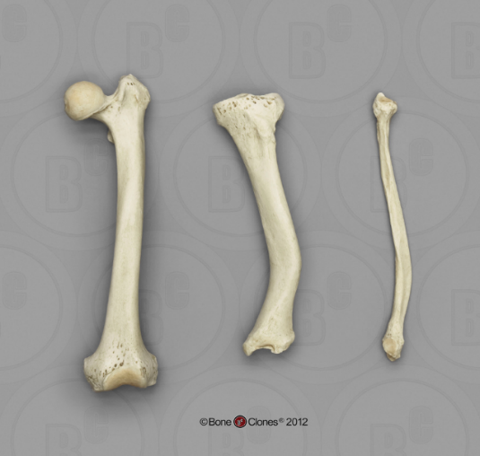
Rickets is a metabolic bone pathology resulting from a Vitamin D deficiency in childhood. Vitamin D is essential to the mineralization of bone tissue and is characterized by a wide variety of cranial and postcranial changes, including the following: asymmetrical deformities of the skull, bowing of the long bones, vertebral compression fractures, and a smaller, thicker pelvis (Figure 13.2).1
Cribra Orbitalia and Porotic Hyperostosis
Cribra Orbitalia appears on the roofs of the orbits as an increase in porosity or expansion of the diploe into the orbital cavity. Cribra Orbitalia is thought to be a general indicator of anemia, although the cause could be a variety of things including malnutrition, parasites, or other physiological illness.9
Porotic Hyperostosis appears ectocranially as increased porosity with an associated thickening of the bone. Can only be confirmed through radiography; as a “hair-on-end” appearance of the diploe. Porotic Hyperostosis is thought to be a general indicator of anemia, although the cause could be a variety of things including malnutrition, parasites, or other physiological illness.9
Osteoporosis
Osteoporosis, literally “porous bone,” is a disease characterized by weak bone. Osteoporosis causes reduced bone mass and disruption of bone architecture.4 This age-related bone disease results in decreased bone density.8 It is a major public health problem, affecting hundreds of millions of people worldwide, predominantly postmenopausal women. The main clinical consequence of the disease is bone fractures. It is estimated that one in three women and one in five men over the age of fifty worldwide will sustain an osteoporotic fracture. Hip and spine fractures are the two most serious fracture types, associated with substantial pain and suffering, disability, and even death.4
Spina Bifida
Spina Bifida is the incomplete closure of the neural arches of the sacral vertebrae. Note that the sacral vertebrae four and five may be open naturally.9 This leaves the spinal cord unprotected and may even be poking through the opening left be the neural arch not forming.5 This condition is both genetic and environmentally controlled.9
Neoplastic
Tumors are either defined as benign or malignant. Benign tumors do not spread to other tissues, while malignant tumors do spread to other tissues. Malignant tumors either arise in the bone itself, in which case they are called a primary tumor, or the spread from other tissues, in which case they are called a secondary tumor. Primary bones tumors are usually found in juveniles, while secondary tumors are usually found in older adults.5
Button Osteoma
A Button osteoma is a benign tumor usually found on the parietal or frontal bone. It generally takes the form of a single small lump of bone. In rarer cases multiple button osteomas have been found.5
Osteosarcoma
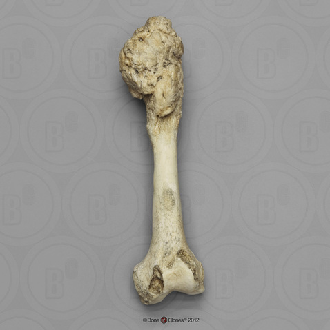
Osteosarcoma is a type of neoplastic bone pathology. Characterized by malignant tumors that begin within bone tissues, osteosarcoma is a primary bone cancer (meaning it begins directly in bone tissue, rather than spreading to bone from other body tissues). Malignant tumors associated with osteosarcoma usually occur during growth and development and are observed most often in adolescents and young adults. Tumors are most frequently observed near the ends of long bones (Figure 13.3).1
Disorders of Growth and Development
Disorders that affect development are called skeletal dysplasia. Dysplasia’s result from a condition that leads to abnormal bone formation and can affect some bones or all the bones in the skeleton. Quite often these are congenital disorders, but not always.
Osteogenesis Imperfecta
Osteogenesis Imperfecta (OI) is a congenital bone pathology characterized by bones with low collagen content, leading to frequent fracturing. However, OI can also occur as a result of a spontaneous mutation. The disease is characterized by multiple fractures throughout the skeleton, particularly in the long bones (Figure 13.1). Depending on the type of OI, the disease is either manifest at birth or during childhood or adolescence. In addition to their susceptibility to easily fractured bones, individuals with OI are typically shorter in stature and may be subject to fracturing of tooth enamel and premature tooth loss.1
Paget’s Disease of Bone
Paget’s disease of bone is a disease of unknown origin that causes bones to grow larger and weaker over time. The disease is marked by both osteoblastic and osteoclastic activity, with excessive osteoclastic resorption followed by osteoblastic proliferation leading to unnecessary amounts of new woven bone. ¹ Radiographic images of this disease often demonstrated thickening of the cortex, coarse trabeculae, and expansion in Paget’s disease.6 The disease typically does not appear until the fourth or fifth decade of life and is more common in males than females.1 Research has shown the pathology is also more common in individuals of British white descent. In contrast, Paget’s disease is less common in individuals of African, Asian, Chinese, Indian, Japanese and Scandinavian descent.6 Paget’s disease of bone can affect any bone, but the most commonly affected elements include the spine, pelvis, skull, and legs. The frequency of osteosarcoma is also higher among individuals with Paget’s disease of bone.1
Achondroplasia
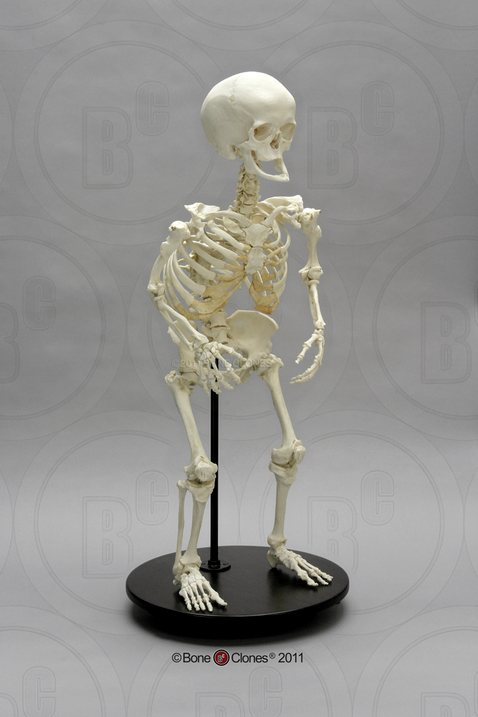
Achondroplasia is a congenital bone pathology resulting from an abnormality in the conversion of cartilage to bone and is the most common form of dwarfism. The skeletal manifestations of achondroplasia are most apparent in the long bones comprising the arms and legs, while the trunk is of relatively normal proportions in individuals with achondroplasia (Figure 13.4). On average, males with achondroplasia are approximately 4'4" tall and females are approximately 4'1" tall.1
Gigantism
Pituitary Gigantism is caused by the release of too much growth hormone which starts in childhood. This results in a person growing will beyond the average height of other member of their sex and ancestral group.5
Dental Diseases
Dental diseases are diseases that affect the teeth.5
Abscess
Abscess are infections the result in cavitations in the bone surrounding the tooth root, resulting in the loss of a tooth and eventually absorption of the bone.5/9
Alveolar Resorption
Alveolar resorption is the product of tooth loss, decrease in bone density associated with age, and also periodontal disease. The alveoli shrink, and as teeth are shed the sockets fill in.9
Caries and Cavities
Caries occur when there is destruction of one of the three dental structures (enamel, dentine, or cementum), caused by bacteria in the mouth. These may be located on the occlusal surface, smooth surface, within the pulp chamber, at the cemento-enamel junction, or on the root. May be seen as a brown spot in the early phase, followed by creation of a cavity within the affected structure.9
Dental Enamel Hypoplasia
Dental enamel hypoplasia are defect in the enamel of the tooth caused by developmental issues during creation of the structure. May cause bands of varying thickness around the circumference of the tooth; also seen in the form of pitting. Thought to be associated with a number of physiological stressors including but not limited to malnutrition, parasites, and weaning.9
References:
1. Ashley Kendell, Alex Perrone, and Colleen Milligan, “Bioarcheology and Forensic Anthropology” In Explorations, ed. Beth Shook, Katie Nelson, Kelsie Aguilera and Lara Braff (Arlington: American Anthropological Association, 2019).
2. A. Svedbom, E. Hernlund, M. Ivergård, J. Compston, C. Cooper, J. Stenmark, E. V. McCloskey, B. Jönsson, J. A. Kanis and the EU review panel of the IOF, “Osteoporosis in the European Union: a compendium of country-specific reports,” Archives of Osteoporosis volume 8 (2013).
3. Carolyn Meyers, Jeffrey Lisiecki, Sarah Miller, Adam Levin, Laura Fayad, Catherine Ding, Takashi Sono, Edward McCarthy, Benjamin Levi, and Aaron W James, “Heterotopic Ossification: A Comprehensive Review,” JBMR Plus 3 (2019).
4. Célia Lopes, Mary Lucas Powell, and Ana Luísa Santos, “Syphilis and cirrhosis: a lethal combination in a XIX century individual identified from the Medical Schools Collection at the University of Coimbra (Portugal),” Memórias do Instituto Oswaldo Cruz 105 (2010). https://www.scielo.br/j/mioc/a/VWRpNf45KyJZcqg3NVWvW6B/?lang=en.
5. David J. Ortner, Identification of Pathological Conditions in Human Skeletal Remains, 2nd ed. (San Diego: Academic Press, 2003), 179-183, 199, 227, 229-231, 235, 278, 285-286, 283, 422, 463, 503-504, 506, and 592.
6. James Elliott, Sarah Stark, Adelina Teoaca, Elizabeth Duffy, and Eleanor Williams, “Fragmented skeletonised remains: Paget’s disease as a method of biological profiling using radiography,” Forensic Imaging 32 (2023). https://www.sciencedirect.com/science/article/pii/S2666225623000039.
7. Laura Lockau, “A Question of Origins: Skeletal Evidence for the History Venereal Syphilis,” Environment & Society Portal, Arcadia (2017). doi.org/10.5282/rcc/7952.
8. Environment & Society Portal, Arcadia (Summer 2017). Layci Harrison, Anatomical Basis of Injury (Houston: University of Houston, 2019). https://uhlibraries.pressbooks.pub/atpanatomy/front-matter/about-this-book/.
9. Roberta Hall, Kenneth Beals, Holm Neumann, Georg Neumann, and Gwyn Madden, Introduction to Human Osteology (Michigan: Grand Valley State University, 2010). https://pressbooks.gvsu.edu/introhumanosteology/.
10. Ronald Plotnikoff, Nandini Karunamuni, Ellina Lytvyak, Christopher Penfold, Donald Schopflocher, Ikuyo Imayama, Steven T. Johnson, and Kim Raine, “Osteoarthritis prevalence and modifiable factors: a population study,” BMC Public Health 15 (2015). https://bmcpublichealth.biomedcentral.com/articles/10.1186/s12889-015-2529-0#rightslink.
11. Wei Zhu, Xuxia He, Kaiyuan Cheng, Linjie Zhang, Di Chen, Xiao Wang, Guixing Qiu, Xu Cao and Xisheng Weng, “Ankylosing spondylitis: etiology, pathogenesis, and treatments,” Bone Research volume 7 (2019). https://www.nature.com/articles/s41413-019-0057-8#rightslink.
Figure References:
Figure 13.1 Osteogenesis Imperfecta Type V by ShakataGaNai is used under a CC BY-SA 4.0 License.
Figure 13.2 Human Femur, Tibia and Fibula, Rickets by ©BoneClones is used by permission and available here is under a CC BY-NC 4.0 License.
Figure 13.3 Human Left Femur, Osteosarcoma by ©BoneClones is used by permission and available here is under a CC BY-NC 4.0 License.
Figure 13.4 Human Female Achondroplasia Dwarf Skeleton, Articulated by ©BoneClones is used by permission and available here is under a CC BY-NC 4.0 License.
