Chapter 11: Estimating Stature in Human Skeletal Remains
ESTIMATING STATURE
The identification of skeletonized human remains is one of the common tasks forensic anthropologists are asked to perform for law enforcement authorities. The aim is to establish the biological profile which consists of estimates of the deceased’s ancestry, sex, age, and stature at time of death, potentially helping in the deceased’s identification.3
Stature, or height, is one of the most prominently recorded components of the biological profile. Our height is recorded from infancy through adulthood. Doctor’s appointments, driver’s license applications, and sports rosters all typically involve a measure of stature for an individual. As such, it is also a component of the biological profile nearly every individual will have on record. Forensic anthropologists use stature estimation methods to provide a range within which an individual’s biological height would fall. Biological height is a person’s true anatomical height. However, the range created through these estimations is often compared to reported stature, which is typically self-reported and based on an approximation of an individual’s true height.2
Methods for estimating stature fall into two types. The more accurate of the two methods are called the full skeleton methods. This type is problematic at times however as it involves measuring all the bones from the top of the skull to the bottom of the ankle. However, the remains themselves maybe incomplete making this method impractical. The full skeleton method can be used for individuals with unusual proportions and without regard to ancestry or sex, which is a strength of the method.1
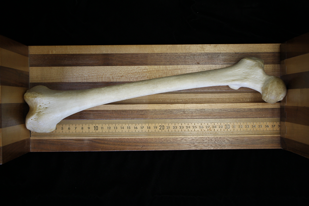
The other type of methods are called regression methods.1 Regression methods examine the relationship between variables such as height and bone length and use the correlation between the variables to create a prediction interval (or range) for estimated stature. This method for calculating stature is the most commonly used method. Figure 11.1 shows the measurement of the bicondylar length of the femur for stature estimations.2
The femur and tibia are the main elements that add the most to your height. Therefore, the most accurate regression formulae result from femoral and tibial lengths. These equations have been created for all of the long bones, however, leg bones are considered more accurate than arm bones. Body proportions differ by both ancestry and sex. Thus, it is essential to determine sex and ancestry to make use of the correct regression formulae for the evaluation of stature.4 Several other factors influence height including nutrition, geographic location, and genetics.5
Long bones are often measured on an osteometric board. The large sliding calipers used for measuring tree diameters are also extremely helpful. Most long bone measurements for stature involves maximum lengths. This includes the measurement of the humerus, radius, ulna, femur, tibia, and fibula. The femur is now and then measured with both condyles in contact with the osteometric board. This is called the bicondylar length or oblique length and is particularly helpful because it orients the femur in anatomical location. After measuring each bone according to directives the measurement is inserted into the suitable formulae.4
These methods are not just used to estimate stature in deceased individuals but can also be used in the living. For example in June 2015, two men were shot and killed in Granite Bay, California, in a double homicide. Investigators were able to locate surveillance camera footage from a gas station where the two victims were spotted in a car with another individual believed to be the perpetrator in the case. The suspect, sitting behind the victims in the car, hung his right arm out of the window as the car drove away. The search for the perpetrator was eventually narrowed down to two suspects. One suspect was 5'8" while the other suspect was 6'4", representing almost a foot difference in height between the two. Forensic anthropologists were given the dimensions of the car (for proportionality of the arm) and were asked to calculate the stature of the suspect in the car from measurements of the suspect’s forearm hanging from the window. Approximate lengths of the bones of the forearm were established from the video footage and used to create a predicted stature range. Stature estimations from skeletal remains typically look at the correlation between the measurements of any individual bone and the overall measurement of body height. In the case above, the length of the right forearm pointed to the taller of the two suspects who was subsequently arrested for the homicide.2
Table 1 (Below) Lists stature estimation formulas.
|
Ethnic group |
Bone |
Male |
Female |
|
European |
Humerus |
2.89 (L) + 78.10 + 4.75 |
3.36 * L + 57.97 + 4.45 |
|
|
Radius |
3.79 (L) + 79.42 + 4.66 |
4.74 * L + 54.93 + 4.24 |
|
|
Ulna |
3.76 (L) + 75.55 + 4.72 |
4.27 * L + 57.76 + 4.30 |
|
|
Femur |
2.32 (L) + 65.53 + 3.94 |
2.47 * L + 54.10 + 3.72 |
|
|
Tibia |
2.42 (L) + 81.93 + 4.00 |
2.90 * L + 61.53 + 3.66 |
|
|
Fibula |
2.60 (L) + 75.50 + 3.86 |
2.93 * L + 59.61 + 3.57 ³ |
|
African |
Humerus |
2.88(L) + 75.48 +/- 4.23 |
3.08(L) + 64.67 +/- 4.25 |
|
|
Radius |
3.32(L) + 85.43 +/- 4. |
3.67(L) + 71.79 +/-4.59 |
|
|
Ulna |
3.20(L) + 82.77 +/-4.74 |
3.31(L) + 75.38 +/- 4.83 |
|
|
Femur |
2.10(L) + 72.22 +/- 3.91 |
2.28(L) + 59.76 +/- 3.41 |
|
|
Tibia |
2.19(L) + 85.36 +/- 3.96 |
2.45(L) + 72.65 +/- 3.70 |
|
|
Fibula |
2.34(L) + 80.07 +/- 4.02 |
2.49(L) + 70.90 +/- 3.80 |
|
Asian |
Humerus |
2.68(L) + 83.19 +/- 4.16 |
Unknown |
|
|
Radius |
3.54(L) + 82.00 +/- 4.60 |
Unknown |
|
|
Ulna |
3.48(L) + 77.45 +/- 4.66 |
Unknown |
|
|
Femur |
2.15(L) + 72.57 +/- 3.80 |
Unknown |
|
|
Tibia |
2.39(L) + 81.45 +/- 3.27 |
Unknown |
|
|
Fibula |
2.40(L) + 80.56 +/- 3.42 |
Unknown |
|
Mexican |
Humerus |
2.92(L) + 73.94 +/- 4.24 |
Unknown |
|
|
Radius |
3.55(L) + 80.71 +/-4.04 |
Unknown |
|
|
Ulna |
3.56(L) + 74.56 +/- 4.05 |
Unknown |
|
|
Femur |
2.44(L) + 58.67 +/- 2.99 |
Unknown |
|
|
Tibia |
2.36(L) + 80.82 +/- 3.73 |
Unknown |
|
|
Fibula |
2.50(L) + 75.44 +/- 3.52 |
Unknown |
Table 1 Stature estimation using Glesser and Bass formulas. 3,4 L= Max Length
Fordisc
These measurements can also be plugged into statistical software which can aid in stature estimation as well. The computer program Fordisc is an anthropological tool used to estimate different components of the biological profile, including ancestry, sex, and stature. When using Fordisc, skeletal measurements are input into the computer software and the program employs multivariate statistical classification methods, including discriminant function analysis, to generate a statistically validated prediction for the geographic origin of unknown remains. Fordisc will also tell the analyst the likelihood of the prediction being correct, as well as how typical the metric data is for the assigned group.2
Fragmented and Nonlimb Bones
Fragmented bones can be an additional obstacle for such investigations. The difficulty of estimating a biological profile varies depending on the condition of the skeletal remains. Due to taphonomic processes such as burial, fire, or animal activity, bones are often found incomplete. Animal activity in particular (which may include trampling, scattering, or scavenging) often leads to the loss of bone structures which help in estimating the biological profile. Trampling and movement have been noted to be the most significant factors leading to disarticulation and alteration of bones by carnivores. Predators, especially carnivores, often disarticulate a human body and remove and consume the body parts in a certain sequence, starting with the neck and thoracic region before moving to the upper and lower extremities. This disarticulation and consumption of the lower leg in the knee joint as well as the upper leg in the hip joint often results in alteration or destruction of the femoral head and neck as well as the distal femoral condyles. Another reason for the more common damage of the femoral extremities is that they feature trabecular bone which is less dense and more nutrient-rich than that of the compact shaft. While the articular parts of the femur are eliminated, the shaft is persistently reduced and covered by teeth marks.3
There are techniques for stature estimation from fragmented bones. Despite problems in correctly determining stature from separate skeletal elements, nearly all studies comparing the accuracy of post-cranial elements have shown the femur to be the best single bone for estimating stature in human adults even in fragmented remains. In most studies, at least the epiphyseal part of the femur must be present to estimate stature. Gidna and Domínguez-Rodrigo developed a method focused on the femoral shaft using the length of the linea aspera, which makes this technique applicable to highly fragmented bones where the shaft is preserved.3
Instead of using the bone fragment itself, measurements for biological profile estimation can be taken on a reconstruction of the full bone, potentially increasing the accuracy or making measurements possible at all. Different methods have been proposed to obtain an estimate of the full bone from a fragment. For example, by using modern 3D digitalization techniques and statistical analysis. ³ Once the maximum length has been estimated it can be plugged into a regression formula. However, these estimates will be less accurate than if the bone had been complete.1
Nonlimb bones can also be used for stature estimation. These estimates will, like fragmented remains, be less accurate. Due to higher inaccuracies nonlimb bones should only be used in the absences of limb bones. Bones that can be used for these purposes include metacarpals, vertebra, the calcaneus bone, metatarsals, and the talus bone.
Sudadults
Formulas for estimating stature in juveniles have been formed. Though for a number of reasons estimating the stature of children can be impractical. Unlike adults children are still growing and reported statures maybe vary from current heights since the individual may have grown since they were last measured. Subadult stature has also been studied less than adult stature. For these reasons height cannot be used to exclude possible matches on the missing persons list.1
Issues with Stature Estimation
Several factors can lead to reported stature not matching up with stature estimations. Diseases can affect bone length which reduces the accuracy of regression formulas. These conditions become more likely with age as will. Osteoporosis can weaken vertebral bodies leading to compression and a decrease in height. Various conditions, including osteoporosis, can lead to increased curvature of the spine which also decreases stature.
The reported stature can be inaccurate in itself. If reported height is based on the recollections of loved ones of the deceased, they could be misremembering their height. Reported stature based on actual measurements can be no less problematic. Forensic scientists cannot be sure how the measurements were taken. If the antemortem stature was taken with shoes on or off can affect the measurement, as can head position. Also due to intervertebral disc compression people actual get slightly shorter just a few hours after the rise from bed. This means that people are taller in the morning.1
Although, there can be issues with stature estimation. These estimates should still be embarked upon because they can help to identify unknown remains.
References:
1. Angi M. Christensen, Nicholas V. Passalacqua, and Eric J. Bartelink, Forensic Anthropology: Current Methods and Practice, 2nd ed. (London: Academic Press, 2019): 351-.
2. Ashley Kendell, Alex Perrone, and Colleen Milligan, “Bioarcheology and Forensic Anthropology” In Explorations, ed. Beth Shook, Katie Nelson, Kelsie Aguilera and Lara Braff (Arlington: American Anthropological Association, 2019). https://pressbooksdev.oer.hawaii.edu/explorationsbioanth/chapter/osteology/.
3. Lars C. Ebert, Dana Rahbani, Marcel Lüthi, Michael J. Thali, Angi M. Christensen, and Barbara Fliss, “Reconstruction of full femora from partial bone fragments for anthropological analyses using statistical shape modeling,” Forensic Science International 332 (2022). https://www.sciencedirect.com/science/article/pii/S0379073822000263.
4. Purva Wagisha Upadhyay and Amarnath Mishra, “Forensic Anthropology” in Biological Anthropology – Applications and Case Studies, ed. Alessio Vovlas (London: IntechOpen, 2021). https://www.intechopen.com/chapters/73372.
5. Roberta Hall, Kenneth Beals, Holm Neumann, Georg Neumann, and Gwyn Madden, Introduction to Human Osteology (Michigan: Grand Valley State University, 2010). https://pressbooks.gvsu.edu/introhumanosteology/
Figure Attributions:
Figure 11.1 Measurement of the bicondylar length of the femur by Alex Perrone original to Explorations: An Open Invitation to Biological Anthropology is under a CC BY-NC 4.0 License.
