7.9 Case Study Conclusion: Under Pressure
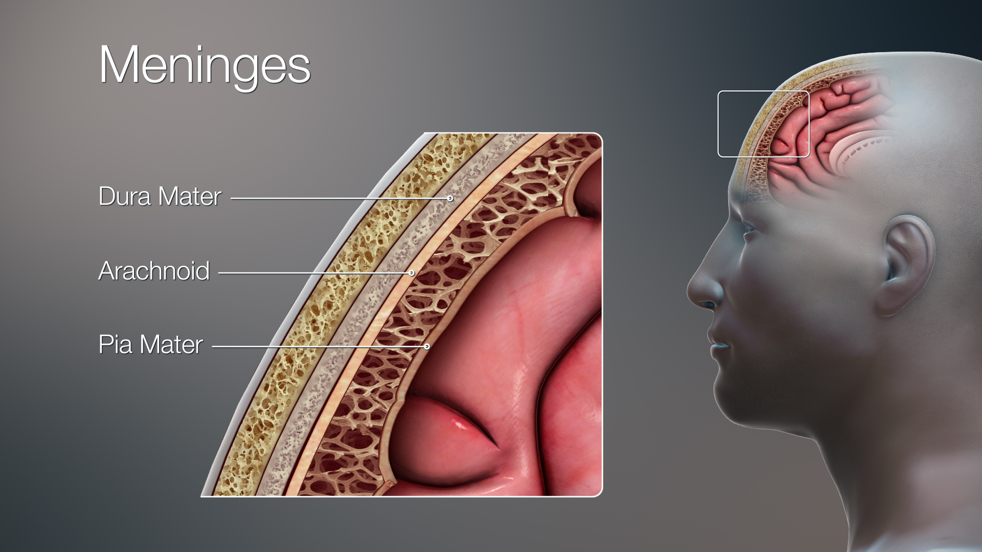
As you learned in this chapter, the human body consists of many complex systems that normally work together efficiently — like a well-oiled machine — to carry out life’s functions. For example, the image above (Figure 7.9.1) illustrates how the brain and spinal cord are protected by layers of membrane called meninges and fluid that flows between the meninges and in spaces called ventricles inside the brain. This fluid is called cerebrospinal fluid, and as you have learned, one of its important functions is to cushion and protect the brain and spinal cord, which make up most of the central nervous system (CNS). Additionally, cerebrospinal fluid circulates nutrients and removes waste products from the CNS. Cerebrospinal fluid is produced continually in the ventricles, circulates throughout the CNS, and is then reabsorbed by the bloodstream. If too much cerebrospinal fluid is produced, its flow is blocked, or not enough is reabsorbed, the system becomes out of balance and it can build up in the ventricles. This causes an enlargement of the ventricles called hydrocephalus that can put pressure on the brain, resulting in the types of neurological problems that former professional football player Jayson, described in the beginning of this chapter, is suffering from.
Recall that Jayson’s symptoms included loss of bladder control, memory loss, and difficulty walking. The cause of his symptoms was not immediately clear, although his doctors suspected that it related to the nervous system, since the nervous system acts as the control centre of the body, controlling and regulating many other organ systems. Jayson’s memory loss directly implicated the brain’s involvement, since that is the site of thoughts and memory. The urinary system is also controlled in part by the nervous system, so the inability to hold urine appropriately can also be a sign of a neurological issue. Jayson’s trouble walking involved the muscular system, which works alongside the skeletal system to enable movement of the limbs. In turn, the contraction of muscles is regulated by the nervous system. You can see why a problem in the nervous system can cause a variety of different symptoms by affecting multiple organ systems in the human body.
To try to find the exact cause of Jayson’s symptoms, his doctors performed a lumbar puncture (or spinal tap), which is the removal of some cerebrospinal fluid through a needle inserted into the lower part of the spinal canal. They then analyzed Jayson’s cerebrospinal fluid for the presence of pathogens (such as bacteria) to determine whether an infection was the cause of his neurological symptoms. When no evidence of infection was found, they used an MRI to observe the structures of his brain. This is when they discovered his enlarged ventricles, which are a hallmark of hydrocephalus.
To treat Jayson’s hydrocephalus, a surgeon implanted a device called a shunt in his brain to remove the excess fluid. An illustration of a brain shunt is shown in Figure 9.7.2 . One side of the shunt consists of a small tube, called a catheter, which was inserted into Jayson’s ventricles. Excess cerebrospinal fluid is then drained through a one-way valve to the other end of the shunt, which was threaded under his skin to his abdominal cavity, where the fluid is released and can be reabsorbed by the bloodstream.
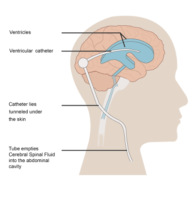
Implantation of a shunt is the most common way to treat hydrocephalus, and for some people, it can allow them to recover almost completely. However, there can be complications associated with a brain shunt. The shunt can have mechanical problems or cause an infection. Also, the rate of draining must be carefully monitored and adjusted to balance the rate of cerebrospinal fluid removal with the rate of its production. If it is drained too fast, it is called overdraining, and if it is drained too slowly, it is called underdraining. In the case of underdraining, the pressure on the brain and associated neurological symptoms will persist. In the case of overdraining, the ventricles can collapse, which can cause serious problems, such as the tearing of blood vessels and hemorrhaging. To avoid these problems, some shunts have an adjustable pressure valve, where the rate of draining can be adjusted by placing a special magnet over the scalp. You can see how the proper balance between cerebrospinal fluid production and removal is so critical – both in the causes of hydrocephalus and in its treatment.
In what other ways does your body regulate balance, or maintain a state of homeostasis? In this chapter you learned about the feedback loops that keep body temperature and blood glucose within normal ranges. Other important examples of homeostasis in the human body are the regulation of the pH in the blood and the balance of water in the body. You will learn more about homeostasis in different body systems in the coming chapters.
Thanks to Jayson’s shunt, his symptoms are starting to improve, but he has not fully recovered. Time may tell whether the removal of the excess cerebrospinal fluid from his ventricles will eventually allow him to recover normal functioning or whether permanent damage to his nervous system has already been done. The flow of cerebrospinal fluid might seem simple, but when it gets out of balance, it can easily wreak havoc on multiple organ systems because of the intricate interconnectedness of the systems within the human “machine.”
To learn more about hydrocephalus and its treatment, watch this video from Boston Children’s Hospital:
Hydrocephalus and its treatment | Boston Children’s Hospital, 2011.
Chapter 7 Summary
This chapter provided an overview of the organization and functioning of the human body. You learned that:
- The human body consists of multiple parts that function together to maintain life. The biology of the human body incorporates the body’s structure — or anatomy — and the body’s functioning, or physiology.
- The organization of the human body is a hierarchy of increasing size and complexity, starting at the level of atoms and molecules and ending at the level of the entire organism.
- Cells are the level of organization above atoms and molecules, and they are the basic units of structure and function of the human body. Each cell carries out basic life functions, as well as other specific roles. Cells of the human body show a lot of variation.
-
- Variations in cell function are generally reflected in variations in cell structure.
- Some cells are unattached to other cells and can move freely. Others are attached to each other and cannot move freely. Some cells can divide readily and form new cells, and others can divide only under exceptional circumstances. Many cells are specialized to produce and secrete particular substances.
- All the different cell types within an individual have the same genes. Cells can vary because different genes are expressed depending on the cell type.
- Many common types of human cells consist of several subtypes of cells, each of which has a special structure and function. For example, subtypes of bone cells include osteocytes, osteoblasts, osteogenic cells, and osteoclasts.
- A tissue is a group of connected cells that have a similar function. There are four basic types of human tissues that make up all the organs of the human body: epithelial, muscle, nervous, and connective tissues.
-
- Connective tissues, such as bone, tendons and blood, are made up of a scattering of living cells that are separated by non-living material, called extracellular matrix.
- Epithelial tissues, such as skin and mucous membranes, protect the body and its internal organs and secrete or absorb substances.
- Muscular tissues are made up of cells that have the unique ability to contract. They include skeletal, smooth, and cardiac muscle tissues.
- Nervous tissues are made up of neurons, which transmit messages, and neuroglia of various types, which play supporting roles.
- An organ is a structure that consists of two or more types of tissues that work together to do the same job. The brain and the heart are two examples.
-
- Many organs are composed of a major tissue that performs the organ’s main function, as well as other tissues that play supporting roles.
- The human body contains five organs that are considered vital for survival: the heart, brain, kidneys, liver, and lungs. If any of these five organs stops functioning, death of the organism is imminent without medical intervention.
- An organ system is a group of organs that work together to carry out a complex overall function. For example, the skeletal system provides structure to the body and protects internal organs.
-
- There are 11 major organ systems in the human organism. They are the integumentary, skeletal, muscular, nervous, endocrine, cardiovascular, lymphatic, respiratory, digestive, urinary, and reproductive systems. Only the reproductive system varies significantly between males and females.
- The human body is divided into a number of body cavities. A body cavity is a fluid-filled space in the body that holds and protects internal organs. The two largest human body cavities are the ventral cavity and dorsal cavity.
-
- The ventral cavity is at the anterior (or front) of the trunk. It is subdivided into the thoracic cavity, abdominal cavity and the pelvic cavity.
- The dorsal cavity is at the posterior (or back) of the body, and includes the head and the back of the trunk. It is subdivided into the cranial cavity and spinal cavity.
- Organ systems of the human body must work together to keep the body alive and functioning normally. This requires communication among organ systems. This is controlled by the autonomic nervous system and endocrine system. The autonomic nervous system controls involuntary body functions, such as heart rate and digestion. The endocrine system secretes hormones into the blood that travel to body cells and influence their activities.
-
- Cellular respiration is a good example of organ system interactions, because it is a basic life process that occurs in all living cells. It is the intracellular process that breaks down glucose with oxygen to produce carbon dioxide and energy. Cellular respiration requires the interaction of the digestive, cardiovascular, and respiratory systems.
- The fight-or-flight response is a good example of how the nervous and endocrine systems control other organ system responses. It is triggered by a message from the brain to the endocrine system and prepares the body for flight or a fight. Many organ systems are stimulated to respond, including the cardiovascular, respiratory, and digestive systems.
- Playing softball or doing other voluntary physical activities may involve the interaction of nervous, muscular, skeletal, respiratory, and cardiovascular systems.
- Homeostasis is the condition in which a system such as the human body is maintained in a more or less steady state. It is the job of cells, tissues, organs, and organ systems throughout the body to maintain homeostasis.
-
- For any given variable (such as body temperature), there is a particular set point that is the physiological optimum value. The spread of values around the set point that is considered insignificant is called the normal range.
- Homeostasis is generally maintained by a negative feedback loop that includes a stimulus, sensor, control centre, and effector. Negative feedback serves to reduce an excessive response and to keep a variable within the normal range. Negative feedback loops control body temperature and the blood glucose level.
- Sometimes homeostatic mechanisms fail, resulting in homeostatic imbalance. Diabetes is an example of a disease caused by homeostatic imbalance. Aging can bring about a reduction in the efficiency of the body’s control system, making the elderly more susceptible to disease.
- Positive feedback loops are not common in biological systems. Positive feedback serves to intensify a response until an end point is reached. Positive feedback loops control blood clotting and childbirth.
The severe and broad impact of hydrocephalus on the body’s systems highlights the importance of the nervous system and its role as the master control system of the body. In the next chapter, you will learn much more about the structures and functioning of this fascinating and important system.
Chapter 7 Review
-
- Compare and contrast tissues and organs.
-
-
- Which type of tissue lines the inner and outer surfaces of the body?
- What is a vital organ? What happens if a vital organ stops working?
- Name three organ systems that transport or remove wastes from the body.
- Name two types of tissue in the digestive system.
-
- Describe one way in which the integumentary and cardiovascular systems work together to regulate homeostasis in the human body.
-
- True or False: Body cavities are filled with air.
- In which organ system is the pituitary gland? Describe how the pituitary gland increases metabolism.
- When the level of thyroid hormone in the body gets too high, it acts on other cells to reduce production of more thyroid hormone. What type of feedback loop does this represent?
- Hypothetical organ A is the control centre in a feedback loop that helps maintain homeostasis. It secretes molecule A1 which reaches organ B, causing organ B to secrete molecule B1. B1 negatively feeds back onto organ A, reducing the production of A1 when the level of B1 gets too high.
- What is the stimulus in this feedback loop?
- If the level of B1 falls significantly below the set point, what do you think happens to the production of A1? Why?
- What is the effector in this feedback loop?
- If organs A and B are part of the endocrine system, what type of molecules do you think A1 and B1 are likely to be?
- What are the two main systems that allow various organ systems to communicate with each other?
- What are two functions of the hypothalamus?
Attributions
Figure 7.9.1
3D Medical Illustration Meninges Details by Scientific Animations on Wikimedia Commons is used under a CC BY-SA 4.0 (https://creativecommons.org/licenses/by-sa/4.0/deed.en) license.
Figure 7.9.2
Hydrocephalus with Shunt from CK-12 Foundation is used under a CC BY-NC 3.0 (https://creativecommons.org/licenses/by-nc/3.0/) license.
 ©CK-12 Foundation Licensed under
 ![]() • Terms of Use • Attribution
• Terms of Use • Attribution
References
Betts, J. G., Young, K.A., Wise, J.A., Johnson, E., Poe, B., Kruse, D.H., Korol, O., Johnson, J.E., Womble, M., DeSaix, P. (2013, April 25). Figure 1.3 Levels of structural organization of the human body [digital image]. In Anatomy and Physiology (Section 1.2). OpenStax. https://openstax.org/books/anatomy-and-physiology/pages/1-2-structural-organization-of-the-human-body
Betts, J. G., Young, K.A., Wise, J.A., Johnson, E., Poe, B., Kruse, D.H., Korol, O., Johnson, J.E., Womble, M., DeSaix, P. (2013, April 25). Figure 1.4 Organ systems of the human body [digital image]. In Anatomy and Physiology (Section 1.2). OpenStax. https://openstax.org/books/anatomy-and-physiology/pages/1-2-structural-organization-of-the-human-body
Betts, J. G., Young, K.A., Wise, J.A., Johnson, E., Poe, B., Kruse, D.H., Korol, O., Johnson, J.E., Womble, M., DeSaix, P. (2013, April 25). Figure 1.15 Dorsal and ventral body cavities [digital image]. In Anatomy and Physiology (Section 1.2). OpenStax. https://openstax.org/books/anatomy-and-physiology/pages/1-6-anatomical-terminology
Boston Children’s Hospital. (2011, ). Hydrocephalus and its treatment | Boston Children’s Hospital. YouTube. https://www.youtube.com/watch?v=bHD8zYImKqA&feature=youtu.be
Brainard, J/ CK-12 Foundation. (2016). Figure 2 An illustration of a brain shunt [digital image]. In CK-12 College Human Biology (Section 9.8) [online Flexbook]. CK12.org. https://www.ck12.org/book/ck-12-college-human-biology/section/9.8/
File:Body cavities lateral view labeled.jpg. (2018, January 4). Wikimedia Commons. https://commons.wikimedia.org/w/index.php?title=File:Body_Cavities_Lateral_view_labeled.jpg&oldid=276851269. (Original image: Figure 1.15 Dorsal and ventral body cavities, from OpenStax, Anatomy and Physiology.)
File:Body cavities lateral view labeled.jpg. (2018, January 4). Wikimedia Commons. https://commons.wikimedia.org/w/index.php?title=File:Body_Cavities_Lateral_view_labeled.jpg&oldid=276851269. (Original image: OpenStax [Version 8.25 from the textbook OpenStax Anatomy and Physiology] adapted for Review questions by Christine Miller].
Clear fluid produced by the brain that forms a thin layer within the meninges and provides protection and cushioning for the brain and spinal cord.
One of two main divisions of the nervous system that includes the brain and spinal cord.
The study of the structure of the body.
Created by CK-12 Foundation/Adapted by Christine Miller
Dowager’s Hump
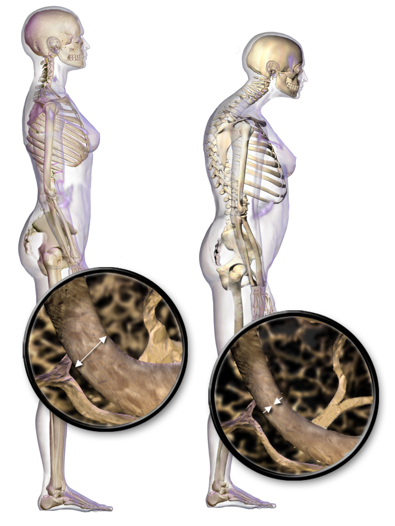
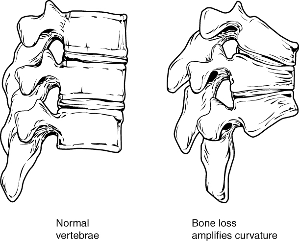
The woman on the right in Figure 11.7.1 has a deformity in her back commonly called dowager’s (widow’s) hump, because it occurs most often in elderly women. Its medical name is kyphosis, and it is defined as excessive curvature of the spinal column in the thoracic region. The curvature generally results from fractures of thoracic vertebrae. As the inset drawings suggest, these fractures may occur due to a significant decrease in bone mass, which is called osteoporosis. Osteoporosis is one of the most prevalent disorders of the skeletal system.
Common Skeletal System Disorders
A number of disorders affect the skeletal system, including bone fractures and bone cancers. However, the two most common disorders of the skeletal system are osteoporosis and osteoarthritis. At least ten million people in the United States have osteoporosis, and more than eight million of them are women. Osteoarthritis is even more common, affecting almost 1.4 million people in Canada, and 1 in 4 women over the age of 50. Because osteoporosis and osteoarthritis are so common, they are the focus of this section. These two disorders are also good examples to illustrate the structure and function of the skeletal system.
Osteoporosis
Osteoporosis is an age-related disorder in which bones lose mass, weaken, and break more easily than normal bones. Bones may weaken so much that a fracture can occur with minor stress — or even spontaneously, without any stress at all. Osteoporosis is the most common cause of broken bones in the elderly, but until a bone fracture occurs, it typically causes no symptoms. The bones that break most often include those in the wrist, hip, shoulder, and spine. When the thoracic vertebrae are affected, there can be a gradual collapse of the vertebrae due to compression fractures, as shown in Figure 11.7.2. This is what causes kyphosis, as pictured above in Figure 11.7.1.
Changes in Bone Mass with Age
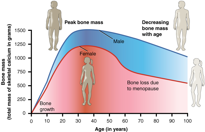
As shown in the Figure 11.7.3, bone mass in both males and females generally peaks when people are in their thirties, with males typically attaining a higher peak mass than females. In both sexes, bone mass usually decreases after that, and this tends to occur more rapidly in females, especially after menopause. The greater decrease in females is generally attributable to low levels of estrogen in the post-menopausal years.
What Causes Osteoporosis?
The underlying mechanism in all cases of osteoporosis is an imbalance between bone formation by osteoblasts and bone resorption by osteoclasts. Normally, bones are constantly being remodeled by these two processes, with up to ten per cent of all bone mass undergoing remodeling at any point in time. As long as these two processes are in balance, no net loss of bone occurs. There are three main ways that an imbalance between bone formation and bone resorption can occur and lead to a net loss of bone. All three ways may occur in the same individual. The three ways are described below:
- An individual never develops normal peak bone mass during the young adult years: If the peak level is lower than normal, then there is less bone mass to begin with, making osteoporosis more likely to develop.
- There is greater than normal bone resorption: Bone resorption normally increases after peak bone mass is reached, but age-related bone resorption may be greater than normal for a variety of reasons. One possible reason is calcium or vitamin D deficiency, which causes the parathyroid gland to release PTH, the hormone that promotes resorption by osteoclasts.
- There is inadequate formation of new bone by osteoblasts during remodeling: Lack of estrogen may decrease the normal deposition of new bone. Inadequate levels of calcium and vitamin D also lead to impaired bone formation by osteoblasts.
An imbalance between bone building and bone destruction leading to bone loss may also occur as a side effect of other disorders. For example, people with alcoholism, anorexia nervosa, or hyperthyroidism have an increased rate of bone loss. Some medications — including anti-seizure medications, chemotherapy drugs, steroid medications, and some antidepressants — also increase the rate of bone loss.
Diagnosing Osteoporosis
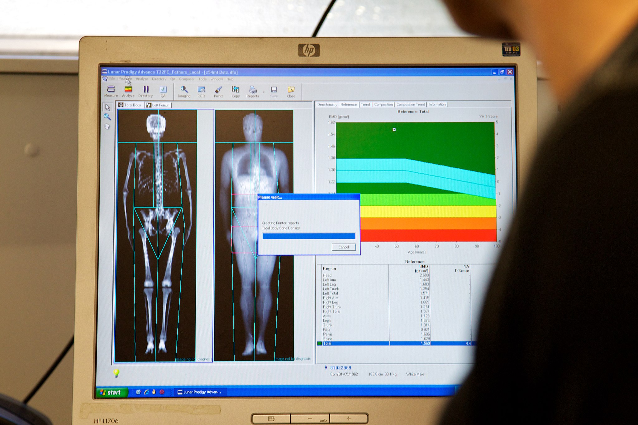
Osteoporosis is diagnosed by measuring a patient’s bone density and comparing it with the normal level of peak bone density in a young adult reference population of the same sex as the patient. If the patient’s bone density is too far below the normal peak level (as measured by a statistic called a T-score), then osteoporosis is diagnosed. Bone density is usually measured by a type of X-ray called dual-energy X-ray absorptiometry (or DEXA), an example of which is shown in Figure 11.7.4. Typically, the density is measured at the hip. Sometimes, other areas are also measured, because there may be variation in bone density in different parts of the skeleton. Osteoporosis Canada recommends that all women 65 years of age and older be screened with DEXA for bone density. Screening may be recommended at younger ages in people with risk factors for osteoporosis (see Risk Factors for Osteoporosis below).
Osteoporotic Fractures
Fractures are the most dangerous aspect of osteoporosis, and osteoporosis is responsible for millions of fractures annually. Debilitating pain among the elderly is often caused by fractures from osteoporosis, and it can lead to further disability and early mortality. Fractures of the long bones (such as the femur) can impair mobility and may require surgery. Hip fracture usually requires immediate surgery, as well. The immobility associated with fractures — especially of the hip — increases the risk of deep vein thrombosis, pulmonary embolism, and pneumonia. Osteoporosis is rarely fatal, but these complications of fractures often are. Older people tend to have more falls than younger people, due to such factors as poor eyesight and balance problems, increasing their risk of fractures even more. The likelihood of falls can be reduced by removing obstacles and loose carpets or rugs in the living environment.
Risk Factors for Osteoporosis
There are a number of factors that increase the risk of osteoporosis. Eleven of them are listed below. The first five factors cannot be controlled, but the remaining factors generally can be controlled by changing behaviors.
- Older age
- Female sex
- European or Asian ancestry
- Family history of osteoporosis
- Short stature and small bones
- Smoking
- Alcohol consumption
- Lack of exercise
- Vitamin D deficiency
- Poor nutrition
- Consumption of soft drinks
Treatment and Prevention of Osteoporosis
Osteoporosis is often treated with medications that may slow or even reverse bone loss. Medications called bisphosphonates, for example, are commonly prescribed. Bisphosphonates slow down the breakdown of bone, allowing bone rebuilding during remodeling to keep pace. This helps maintain bone density and decreases the risk of fractures. The medications may be more effective in patients who have already broken bones than in those who have not, significantly reducing their risk of another fracture. Generally, patients are not recommended to stay on bisphosphonates for more than three or four years. There is no evidence for continued benefit after this time — in fact, there is a potential for adverse side effects.

Preventing osteoporosis includes eliminating any risk factors that can be controlled through changes of behavior. If you smoke, stop. If you drink, reduce your alcohol consumption — or cut it out altogether. Eat a nutritious diet and make sure you are getting adequate amounts of vitamin D. You should also avoid drinking carbonated beverages.
If you’re a couch potato, get involved in regular exercise. Aerobic, weight-bearing, and resistance exercises can all help maintain or increase bone mineral density (for example hiking as in Figure 11.7.5). Exercise puts stress on bones, which stimulates bone building. Good weight-bearing exercises for bone building include weight training, dancing, stair climbing, running, and hiking (see Figure 11.7.5). Biking and swimming are less beneficial, because they don’t stress the bones. Ideally, you should exercise for at least 30 minutes a day most days of the week.
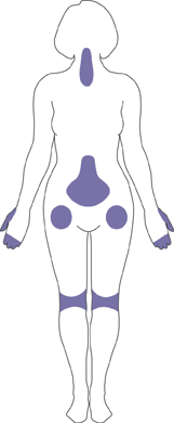
Osteoarthritis (OA) is a joint disease that results from the breakdown of joint cartilage and bone. The most common symptoms are joint pain and stiffness. Other symptoms may include joint swelling and decreased range of motion. Initially, symptoms may occur only after exercise or prolonged activity, but over time, they may become constant, negatively affecting work and normal daily activities. As shown in Figure 11.7.6, the most commonly involved joints are those near the ends of the fingers, at the bases of the thumbs, and in the neck, lower back, hips, and knees. Often, joints on one side of the body are affected more than those on the other side.
What Causes Osteoarthritis?
OA is thought to be caused by mechanical stress on the joints with insufficient self-repair of cartilage. The stress may be exacerbated by low-grade inflammation of the joints, as cells lining the joint attempt to remove breakdown products from cartilage in the synovial space. OA develops over decades as stress and inflammation cause increasing loss of articular cartilage. Eventually, bones may have no cartilage to separate them, so bones rub against one another at joints. This damages the articular surfaces of the bones and contributes to the pain and other symptoms of OA. Because of the pain, movement may be curtailed, leading to loss of muscle, as well.
Diagnosing Osteoarthritis
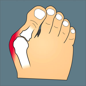
Diagnosis of OA is typically made on the basis of signs and symptoms. Signs include joint deformities, such as bony nodules on the finger joints or bunions on the feet (as illustrated in Figure 11.7.7). Symptoms include joint pain and stiffness. The pain is usually described as a sharp ache or burning sensation, which may be in the muscles and tendons around the affected joints, as well as in the joints themselves. The pain is usually made worse by prolonged activity, and it typically improves with rest. Stiffness is most common when first arising in the morning, and it usually improves quickly as daily activities are undertaken.
X-rays or other tests are sometimes used to either support the diagnosis of OA or to rule out other disorders. Blood tests might be done, for example, to look for factors that indicate rheumatoid arthritis (RA), an autoimmune disease in which the immune system attacks the body’s joints. If these factors are not present in the blood, then RA is unlikely, and a diagnosis of OA is more likely to be correct.
Risk Factors for Osteoarthritis
Age is the chief risk factor for osteoarthritis. By age 65, as many as 80 per cent of all people have evidence of osteoarthritis. However, people are more likely to develop OA — especially at younger ages — if they have had a joint injury. A high school football player might have a bad knee injury that damages the joint, leading to OA in the knee by the time he is in his thirties. If people have joints that are misaligned due to congenital malformations or disease, they are also more likely to develop OA. Excess body weight is another factor that increases the risk of OA, because of the added stress it places on weight-bearing joints.
Researchers have found that people with a family history of OA have a heightened risk of developing the disorder, which suggests that genetic factors are also involved in OA. It is likely that many different genes are needed for normal cartilage and cartilage repair. If such genes are defective and cartilage is abnormal or not normally repaired, OA is more likely to result.
Treatment and Prevention of Osteoarthritis
OA cannot be cured, but the symptoms — especially the pain — can often be treated successfully to maintain good quality of life for people with OA. Treatments include exercise, efforts to decrease stress on joints, pain medications, and surgery.
Exercise
Exercise helps maintain joint mobility and also increases muscle strength. Stronger muscles may help keep the bones in joints correctly aligned, and this can reduce joint stress. Good exercises for OA include swimming, water aerobics (see Figure 11.7.8 below), and biking. These activities are recommended for OA, because they put relatively little stress on the joints.
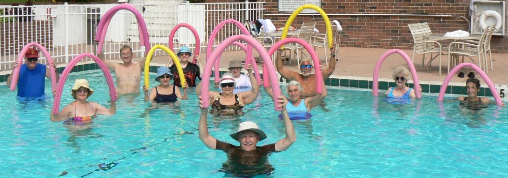
Exercising in water provides buoyancy that places less stress on joints than the same exercises would on the ground or other hard surface.
De-stressing Joints
Efforts to decrease stress on joints include resting and using mobility devices such as canes, which reduce the weight placed on weight-bearing joints and also improve stability. In people who are overweight, losing weight may also reduce joint stress.
Pain Medications
The first type of pain medication likely to be prescribed for OA is acetaminophen (e.g., Tylenol). When taken as prescribed, it has a relatively low risk of serious side effects. If this medication is inadequate to relieve the pain, non-steroidal anti-inflammatory drugs (NSAIDs, such as ibuprofen) may be prescribed. NSAIDs, however, are more likely to cause serious side effects, such as gastrointestinal bleeding, elevated blood pressure, and increased risk of stroke. Opioids usually are reserved for patients who have suffered serious side effects or for whom other medications have failed to relieve pain. Due to the risk of addiction, only short-term use of opioids is generally recommended.
Surgery
Joint-replacement surgery is the most common treatment for serious OA in the knee or hip. In fact, knee and hip replacement surgeries are among the most common of all surgeries. Although they require a long period of healing and physical rehabilitation, the results are usually worth it. The replacement “parts” are usually pain-free and fully functional for at least a couple of decades. Quality, durability, and customization of artificial joints are constantly improving.
Try out this neat Virtual Hip Resurfacing activity by Edheads (you will need to enable Flash).
Feature: Myth vs. Reality
About one out of every 5 adults in Canada suffer from osteoarthritis. The more you know about this disease, the more you can do to avoid it or slow its progression. That means knowing the facts, rather than believing the myths about osteoarthritis.
Myth |
Reality |
| "Cracking my knuckles will cause osteoarthritis." | Cracking your knuckles may lead to inflammation of your tendons, but it will not cause osteoarthritis. |
| "My diet has no effect on my joints." | What and how much you eat does affect your body weight, and every pound you gain translates into an additional four pounds (or more!) of stress on your knees. Being overweight, therefore, increases the chances of developing osteoarthritis — and also the rate at which it progresses. |
| "Exercise causes osteoarthritis or makes it worse, so I should avoid it." | This is one of the biggest myths about osteoarthritis. Low-impact exercise can actually lessen the pain and improve other symptoms of osteoarthritis. If you don’t have osteoarthritis, exercise can reduce your risk of developing it. Low-impact exercise helps keep the muscles around joints strong and flexible, so they can help stabilize and protect the joints. |
| "If my mom or dad has osteoarthritis, I will also develop it." | It is true that you are more likely to develop osteoarthritis if a parent has it, but it isn’t a sure thing. There are several things you can do to decrease your risk, such as getting regular exercise and maintaining a healthy weight. |
| "Bad weather causes osteoarthritis." | Weather conditions do not cause osteoarthritis, although in some people who already have osteoarthritis, bad weather seems to make the symptoms worse. It is primarily low barometric pressure that increases osteoarthritis pain, probably because it leads to greater pressure inside the joints relative to the outside air pressure. Some people think their osteoarthritis pain is worse in cold weather, but systematic studies have not found convincing evidence for this. |
| "Joint pain is unavoidable as you get older, so there is no need to see a doctor for it." | Many people with osteoarthritis think there is nothing that can be done for the pain of osteoarthritis, or that surgery is the only treatment option. In reality, osteoarthritis symptoms often can be improved with a combination of exercise, weight loss, pain management techniques, and pain medications. If osteoarthritis pain interferes with daily life and lasts more than a few days, you should see your doctor. |
| "Osteoarthritis is inevitable in seniors." |
Although many people over 65 develop osteoarthritis, there are many people who never develop it, no matter how old they live to be. You can reduce your risk of developing osteoarthritis in later life by protecting your joints throughout life. |
11.7 Summary
- A number of disorders affect the skeletal system, including bone fractures and bone cancers. The two most common disorders of the skeletal system are osteoporosis and osteoarthritis.
- Osteoporosis is an age-related disorder in which bones lose mass, weaken, and break more easily than normal bones. The underlying mechanism in all cases of osteoporosis is an imbalance between bone formation and bone resorption in bone remodeling. Osteoporosis may also occur as a side effect of other disorders or certain medications.
- Osteoporosis is diagnosed by measuring a patient’s bone density and comparing it with the normal level of peak bone density. Fractures are the most dangerous aspect of osteoporosis. Osteoporosis is rarely fatal, but complications of fractures often are.
- Risk factors for osteoporosis include older age, female sex, European or Asian ancestry, family history of osteoporosis, short stature and small bones, smoking, alcohol consumption, lack of exercise, vitamin D deficiency, poor nutrition, and consumption of soft drinks.
- Osteoporosis is often treated with medications — such as bisphosphonates — that may slow or even reverse bone loss. Preventing osteoporosis includes eliminating any risk factors that can be controlled through changes of behavior, such as undertaking weight-bearing exercise.
- Osteoarthritis (OA) is a joint disease that results from the breakdown of joint cartilage and bone. The most common symptoms are joint pain and stiffness. OA is thought to be caused by mechanical stress on the joints with insufficient self-repair of cartilage, coupled with low-grade inflammation of the joints.
- Diagnosis of OA is typically made on the basis of signs and symptoms, such as joint deformities, pain, and stiffness. X-rays or other tests are sometimes used to either support the diagnosis or rule out other disorders. Age is the chief risk factor for OA. Other risk factors include joint injury, excess body weight, and a family history of OA.
- OA cannot be cured, but the symptoms can often be treated successfully. Treatments may include exercise, efforts to decrease stress on joints, pain medications, and surgery to replace affected hip or knee joints.
11.7 Review Questions
- Create a brochure or poster about osteoporosis to educate others about this disease. Include information about:
- A definition of osteoporosis
- Causes
- Dangers of living with the disease
- Canadian osteoporosis statistics
- Risk factors
- Diagnosis
- Treatment
-
- Why is it important to build sufficient bone mass in your young adult years?
- Explain the difference in cause between rheumatoid arthritis and osteoarthritis.
- Debunk the myth: Osteoarthritis is caused by physical activity, so people who are equally active are equally susceptible to it.
- Explain how we know that estrogen generally promotes production of new bone.
11.7 Explore More
https://www.youtube.com/watch?v=DL0_gcP15Ts
Kevin Stone: The bio-future of joint replacement, TED, 2010.
https://www.youtube.com/watch?v=OyK0oE5rwFY
The benefits of good posture - Murat Dalkilinç, TED-Ed, 2015.
Attributions
Figure 11.7.1
Blausen_0686_Osteoporosis_01 by BruceBlaus on Wikimedia Commons is used under a CC BY 3.0 (https://creativecommons.org/licenses/by/3.0) license.
Figure 11.7.2
Feature_Osteoprosis_of_Spine by OpenStax College on Wikimedia Commons is used under a CC BY 3.0 (https://creativecommons.org/licenses/by/3.0) license.
Figure 11.7.3
Age_and_Bone_Mass by OpenStax College on Wikimedia Commons is used under a CC BY 3.0 (https://creativecommons.org/licenses/by/3.0) license.
Figure 11.7.4
DEXA_scan_screen_ALSPAC by Nick Smith photography on Wikimedia Commons is used under a CC BY-SA 3.0 license.
Figure 11.7.5
Hiking by jake-melara-Yh6K2eTr_FY [photo] by Jake Melara on Unsplash is used under the Unsplash License (https://unsplash.com/license).
Figure 11.7.6
Areas_affected_by_osteoarthritis by National Institute of Arthritis and Musculoskeletal and Skin Diseases (NIAMS)/ NIH on Wikimedia Commons is in the public domain (https://en.wikipedia.org/wiki/Public_domain).
Figure 11.7.7
Hallux_valgus by Malmstajn on Wikimedia Commons is used under a CC BY 3.0 (https://creativecommons.org/licenses/by/3.0) license.
Figure 11.7.8
07-06_WtrAerob1a by Tim Ross on Wikimedia Commons is in the public domain (https://en.wikipedia.org/wiki/Public_domain).
References
Betts, J. G., Young, K.A., Wise, J.A., Johnson, E., Poe, B., Kruse, D.H., Korol, O., Johnson, J.E., Womble, M., DeSaix, P. (2013, June 19). Figure 6.23 Graph showing relationship between age and bone mass digital image]. In Anatomy and Physiology (Section 6.6). OpenStax. https://openstax.org/books/anatomy-and-physiology/pages/6-6-exercise-nutrition-hormones-and-bone-tissue
Betts, J. G., Young, K.A., Wise, J.A., Johnson, E., Poe, B., Kruse, D.H., Korol, O., Johnson, J.E., Womble, M., DeSaix, P. (2013, June 19). Figure 7.22 Osteoporosis [digital image]. In Anatomy and Physiology (Section 7.3). OpenStax. https://openstax.org/books/anatomy-and-physiology/pages/7-3-the-vertebral-column
Blausen.com staff. (2014). Medical gallery of Blausen Medical 2014. WikiJournal of Medicine 1 (2). DOI:10.15347/wjm/2014.010. ISSN 2002-4436.
Mayo Clinic Staff. (n.d.). Kyphosis [online article]. MayoClinic.org. https://www.mayoclinic.org/diseases-conditions/kyphosis/symptoms-causes/syc-20374205
Mayo Clinic Staff. (n.d.). Osteoarthritis [online article]. MayoClinic.org. https://www.mayoclinic.org/diseases-conditions/osteoarthritis/symptoms-causes/syc-20351925
TED. (2010, July 23). Kevin Stone: The bio-future of joint replacement. YouTube. https://youtu.be/DL0_gcP15Ts
TED-Ed. (2015, July 30). The benefits of good posture - Murat Dalkilinç. YouTube. https://www.youtube.com/watch?v=OyK0oE5rwFY&feature=youtu.be
Wheatley, G., Smail, S., Bort, E. (2007). Virtual hip resurfacing [online game]. EdHeads.org. https://edheads.org/page/hip_resurfacing
The smallest particle of an element that still has the properties of that element.
A molecule is an electrically neutral group of two or more atoms held together by chemical bonds.
Created by CK-12 Foundation/Adapted by Christine Miller
Case Study Conclusion: A Pain in the Foot
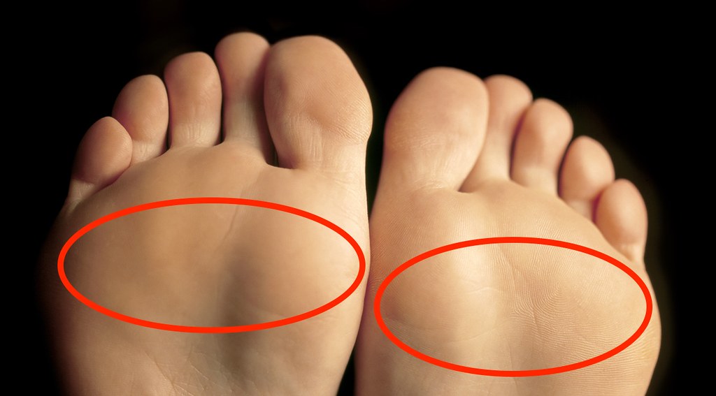
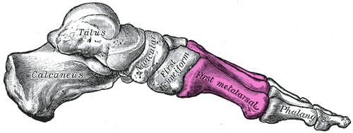
As Sophia discovered in the beginning of the chapter, wearing high heels can result in a condition called metatarsalgia. Metatarsalgia is named for the metatarsal bones, which are the five bones that run through the ball of the foot just behind the toes (highlighted in Figures 11.8.2 and 11.8.3). Wearing high heels causes excessive pressure on the ball of the foot, as described in the beginning of this chapter. Additionally, the toes are forced to pull upward in high heels, which moves the fleshy padding away from the ball of the foot and adds to the overall pressure placed on this region. Over time, this can cause inflammation and direct stress on the bones, resulting in the pain in the ball of the foot known as metatarsalgia. The pain occurs especially in weight-bearing positions, such as standing, walking, or running — which is what Sophia was experiencing. There may also be pain, numbness, or tingling in the toes associated with metatarsalgia.
 |
 |
Wearing high heels can also cause stress fractures in the feet, which are tiny breaks in bone that occur due to repeated mechanical stress. This is caused by the excessive pressure that high heels put on some of the bones of the feet. These fractures are somewhat similar to what occurs in osteoporosis when the bone mass decreases to the point where bones can fracture easily as a person goes about their daily activities. In both cases, a major noticeable injury is not necessary to create the tiny fractures. As you have learned, tiny fractures that accrue over time are the cause of dowager’s hump (or kyphosis), which is often seen in women with osteoporosis.
Don’t think you are immune to stress fractures just because you don’t wear high heels! This injury also commonly occurs in people who participate in sports involving repetitive striking of the foot on the ground, such as running, tennis, basketball, or gymnastics. They may be avoided by taking preventative measures. You should ramp up any increase in activity slowly, cross-train by engaging in a variety of different sports or activities, rest if you experience pain, and wear well-cushioned and supportive running shoes. It is important to know that your cardiovascular and muscular systems adapt to an increase in physical activity much more quickly than the skeletal system.

Sophia learned through her online research that wearing high heels can also lead to foot deformities, such as bunions and hammertoes. As you learned in an earlier chapter, a bunion is a protrusion on the side of the foot, most often at the base of the big toe. It can be caused by wearing shoes with a narrow, pointed toe box — a common shape for high heels (see Figure 11.8.4). The pressure of the shoes on the side of the foot causes an enlargement of bone or inflammation of other tissues in the region, which pushes the big toe toward the other toes.
Hammertoes are an abnormal bend in the middle joint of the second, third, or fourth toe (with the big toe being the first toe), causing the toe to be shaped similarly to a hammer. The narrow, pointed toe box of many high heels, combined with the way the toes are squished into the front of the shoe as a result of the height of the heel, can cause the toes to become deformed this way. Treatments for bunions and hammertoe include wearing shoes with a roomy toe box, padding or taping the toes, and toe exercises and stretches. If the bunion or hammertoe does not respond to these treatments, surgery may be necessary to correct the deformity.
Because the bones of the skeleton are connected and work together with other systems to support the body, wearing high heels can also cause physical problems in areas other than the feet. Wearing high heels shifts a person’s posture and alignment, and can put strain on tendons, muscles, and other joints in the body. Research published in 2014 from a team at Stanford University suggests that wearing high heels, particularly if the person is overweight or the heels are very high, may increase the risk of osteoarthritis (OA) in the knee, due to added stress on the knee joint as the person walks. As you have learned, OA results from the breakdown of cartilage and bone at the joint. Because it can only be treated to minimize symptoms — and not for a cure — OA could be an unfortunate long-term consequence of wearing high heels.
Sophia has decided that wearing high heels regularly is not worth the pain and potential long-term damage to her body. After consulting with her doctor, who confirmed she had metatarsalgia, she was able to successfully treat it with ice, rest, and wearing comfortable, supportive shoes instead of heels.
High heels are not the only kind of shoes that can cause problems. Flip-flops, worn-out sneakers, and shoes that are too tight can all cause foot issues. To prevent future problems from her shoe choices, Sophia is following guidelines recommended by medical experts. The guidelines include:
- Wearing shoes that fit well, have plenty of room in the toes, are supportive, and are comfortable right away. There should be no “break-in” period needed for shoes.
- Avoiding high heels, especially those with heels over two inches high, or those that have narrow, pointed toe boxes or very thin heels. The heels pictured in Figure 11.8.4 are an example of a type of shoe that should be avoided.
- If high heels must be worn, it should only be for a limited period of time.
As you have learned in this chapter, your skeletal system carries out a variety of important functions in your body, including physical support. But even though it is strong, your skeletal system can become damaged and deformed — even through such a seemingly innocuous act as wearing a certain type of shoe. Taking good care of your skeletal system is necessary to help it continue to take good care of the rest of you.
Chapter 11 Summary
In this chapter, you learned about the skeletal system. Specifically, you learned that:
- The skeletal system is the organ system that provides an internal framework for the human body. In adults, the skeletal system contains 206 bones.
- Bones are organs made of supportive connective tissues, mainly the tough protein collagen. Bones also contain blood vessels, nerves, and other tissues. Bones are hard and rigid, due to deposits of calcium and other mineral salts within their living tissues. Besides bones, the skeletal system includes cartilage and ligaments.
- The skeletal system has many different functions, including supporting the body and giving it shape, protecting internal organs, providing attachment surfaces for skeletal muscles, allowing body movements, producing blood cells, storing minerals, helping to maintain mineral homeostasis, and producing endocrine hormones.
- There is relatively little sexual dimorphism in the human skeleton, although the female skeleton tends to be smaller and less robust than the male skeleton. The greatest sex difference is in the pelvis, which is adapted for childbirth in females.
- The skeleton is traditionally divided into two major parts: the axial skeleton and the appendicular skeleton.
- The axial skeleton consists of a total of 80 bones. It includes the skull, vertebral column, and rib cage. It also includes the three tiny ossicles in the middle ear and the hyoid bone in the throat.
-
- The skull provides a bony framework for the head. It consists of 22 different bones: eight in the cranium, which encloses the brain, and 14 in the face, which includes the upper and lower jaw.
- The vertebral column is a flexible, S-shaped column of 33 vertebrae that connects the trunk with the skull and encloses the spinal cord. The vertebrae are divided into five regions: cervical, thoracic, lumbar, sacral, and coccygeal regions. The S shape of the vertebral column allows it to absorb shocks and distribute the weight of the body.
- The rib cage holds and protects the organs of the upper part of the trunk, including the heart and lungs. It includes the 12 thoracic vertebrae, the sternum, and 12 pairs of ribs.
- The appendicular skeleton consists of a total of 126 bones. It includes the bones of the four limbs, shoulder girdle, and pelvic girdle. The girdles attach the appendages to the axial skeleton.
-
- Each upper limb consists of 30 bones. There is one bone (called the humerus) in the upper arm, and two bones (called the ulna and radius) in the lower arm. The wrist contains eight carpal bones, the hand contains five metacarpals, and the fingers consist of 14 phalanges. The thumb is opposable to the palm and fingers of the same hand.
- Each lower limb also consists of 30 bones. There is one bone (called the femur) in the upper leg, and two bones (called the tibia and fibula) in the lower leg. The patella covers the knee joint. The ankle contains seven tarsal bones, and the foot contains five metatarsals. The tarsals and metatarsals form the heel and arch of the foot. The bones in the toes consist of 14 phalanges.
- The shoulder girdle attaches the upper limbs to the trunk of the body. It is connected to the axial skeleton only by muscles, allowing mobility of the upper limbs. Bones of the shoulder girdle include a right and left clavicle, and a right and left scapula.
- The pelvic girdle attaches the legs to the trunk of the body and supports the organs of the abdomen. It is connected to the axial skeleton by ligaments. The pelvic girdle consists of two halves that are fused together in adults. Each half consists of three bones: the ilium, pubis, and ischium.
- Bones are organs that consist mainly of bone (or osseous) tissue. Osseous tissue is a type of supportive connective tissue consisting of a collagen matrix that is mineralized with calcium and phosphorus crystals. The combination of flexible collagen and minerals makes bone hard, without making it brittle.
-
- There are two types of osseous tissues: compact bone tissue and spongy bone tissue. Compact bone tissue is smooth and dense. It forms the outer layer of bones. Spongy bone tissue is porous and light, and it is found inside many bones.
- Besides osseous tissues, bones also contain nerves, blood vessels, bone marrow, and periosteum.
- Bone tissue is composed of four different types of bone cells: osteoblasts, osteocytes, osteoclasts, and osteogenic cells. Osteoblasts form new collagen matrix and mineralize it, osteoclasts break down bone, osteocytes regulate the formation and breakdown of bone, and osteogenic cells divide and differentiate to form new osteoblasts. Bone is a very active tissue, constantly being remodeled by the work of osteoblasts and osteoclasts.
- There are six types of bones in the human body: long bones (such as the limb bones), short bones (such as the wrist bones), sesamoid bones (such as the patella), sutural bones in the skull, and irregular bones (such as the vertebrae).
- Early in the development of a human fetus, the skeleton is made almost entirely of cartilage. The relatively soft cartilage gradually turns into hard bone — a process that is called ossification. It begins at a primary ossification center in the middle of bone, and later also occurs at secondary ossification centers in the ends of bone. The bone can no longer grow in length after the areas of ossification meet and fuse at the time of skeletal maturity.
- Throughout life, bone is constantly being replaced in the process of bone remodeling. In this process, osteoclasts resorb bone and osteoblasts make new bone to replace it. Bone remodeling shapes the skeleton, repairs tiny flaws in bones, and helps maintain mineral homeostasis in the blood.
- Bone repair is the natural process in which a bone repairs itself following a bone fracture. This process may take several weeks. In the process, the periosteum produces cells that develop into osteoblasts, and the osteoblasts form new bone matrix to heal the fracture. Bone repair may be affected by diet, age, pre-existing bone disease, or other factors.
- Joints are locations at which bones of the skeleton connect with one another.
- Joints can be classified structurally or functionally, and there is significant overlap between the two types of classifications.
- The structural classification of joints depends on the type of tissue that binds the bones to each other at the joint. There are three types of joints in the structural classification: fibrous, cartilaginous, and synovial joints.
- The functional classification of joints is based on the type and degree of movement that they allow. There are three types of joints in the functional classification: immovable, partly movable, and movable joints.
- A number of disorders affect the skeletal system, including bone fractures and bone cancers. The two most common disorders of the skeletal system are osteoporosis and osteoarthritis.
- Osteoporosis is an age-related disorder in which bones lose mass, weaken, and break more easily than normal bones. The underlying mechanism in all cases of osteoporosis is an imbalance between bone formation and bone resorption in bone remodeling. Osteoporosis may also occur as a side effect of other disorders or certain medications.
-
- Osteoporosis is diagnosed by measuring a patient’s bone density and comparing it with the normal level of peak bone density. Fractures are the most dangerous aspect of osteoporosis. Osteoporosis is rarely fatal, but complications of fractures often are.
- Risk factors for osteoporosis include older age, female sex, European or Asian ancestry, family history of osteoporosis, short stature and small bones, smoking, alcohol consumption, lack of exercise, vitamin D deficiency, poor nutrition, and consumption of soft drinks.
- Osteoporosis is often treated with medications (such as bisphosphonates) that may slow or even reverse bone loss. Preventing osteoporosis includes eliminating any risk factors that can be controlled through changes of behavior, such as undertaking weight-bearing exercise.
- Osteoarthritis (OA) is a joint disease that results from the breakdown of joint cartilage and bone. The most common symptoms are joint pain and stiffness. OA is thought to be caused by mechanical stress on the joints with insufficient self-repair of cartilage, coupled with low-grade inflammation of the joints.
-
- Diagnosis of OA is typically made on the basis of signs and symptoms, such as joint deformities, pain, and stiffness. X-rays or other tests are sometimes used to either support the diagnosis or rule out other disorders. Age is the chief risk factor for OA. Other risk factors include joint injury, excess body weight, and a family history of OA.
- OA cannot be cured, but the symptoms can often be treated successfully. Treatments may include exercise, efforts to decrease stress on joints, pain medications, and surgery to replace affected hip or knee joints.
As you have learned in this chapter, one of the important functions of the skeletal system is to allow movement of the body. But it doesn’t do it alone. Movement is caused by the contraction of muscles, which pull on the bones, causing them to move. Read the next chapter to learn about this and other important functions of the muscular system.
Chapter 11 Review
-
-
- Why does the rib cage need to be flexible? Why can it be flexible?
- In general, what do “girdles” in the skeletal system do?
- Would swimming be more effective as an exercise for preventing osteoporosis or as a treatment for osteoarthritis? Explain your answer.
- Explain why some of the vertebrae become misshapen in the condition called dowager’s hump (or kyphosis).
- Explain why osteoarthritis often involves inflammation in the joints.
- Osteoporosis can involve excess bone resorption, as well as insufficient production of new bone tissue. What are the two main bone cell types that carry out these processes, respectively?
- Describe two roles that calcium in bones play in the body.
-
Attributions
Figure 11.8.1
Running Shoes by bruno-nascimento-PHIgYUGQPvU [photo] by Bruno Nascimento on Unsplash is used under the Unsplash License (https://unsplash.com/license).
Figure 11.8.2
Metatarsalgia/ Best Shoes for Metatarsalgia by Esther Max on Flickr is used under a CC BY 2.0 (https://creativecommons.org/licenses/by/2.0/) license.
Figure 11.8.3
Gray290_-_Mratatarsus (1) by Henry Vandyke Carter (1831-1897) (Revised by Warren H. Lewis, coloured by Was a bee) on Wikimedia Commons is in the public domain (https://en.wikipedia.org/wiki/Public_domain. (Bartleby.com: Gray’s Anatomy, Plate 290)
Figure 11.8.4
Heels by gavin-allanwood-ndpX28miBtE-unsplash by Photo by Gavin Allanwood on Unsplash is used under the Unsplash License (https://unsplash.com/license).
References
Mayo Clinic Staff. (n.d.). Hammertoe and mallet toe [online article]. MayoClinic.org. https://www.mayoclinic.org/diseases-conditions/hammertoe-and-mallet-toe/symptoms-causes/syc-20350839
VanDyke Carter, H. (1858). Illustration plate 290. In H. Gray, Anatomy of the Human Body. Lea & Febiger. Bartleby.com, 2000. www.bartleby.com/107/.
The smallest unit of life, consisting of at least a membrane, cytoplasm, and genetic material.
a bone cell, formed when an osteoblast becomes embedded in the matrix it has secreted.
Created by CK-12 Foundation/Adapted by Christine Miller
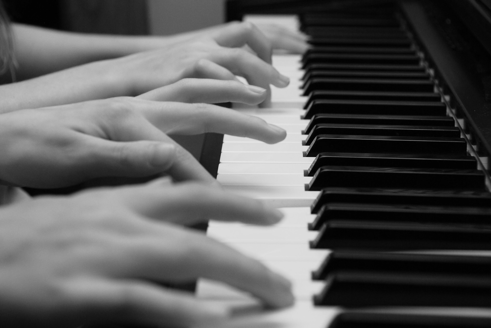
One Piano, Four Hands
Did you ever see two people play the same piano? How do they coordinate all the movements of their own fingers — let alone synchronize them with those of their partner? The peripheral nervous system plays an important part in this challenge.
What Is the Peripheral Nervous System?
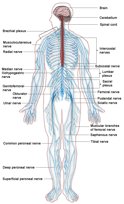
The peripheral nervous system (PNS) consists of all the nervous tissue that lies outside of the central nervous system (CNS). The main function of the PNS is to connect the CNS to the rest of the organism. It serves as a communication relay, going back and forth between the CNS and muscles, organs, and glands throughout the body.
Tissues of the Peripheral Nervous System
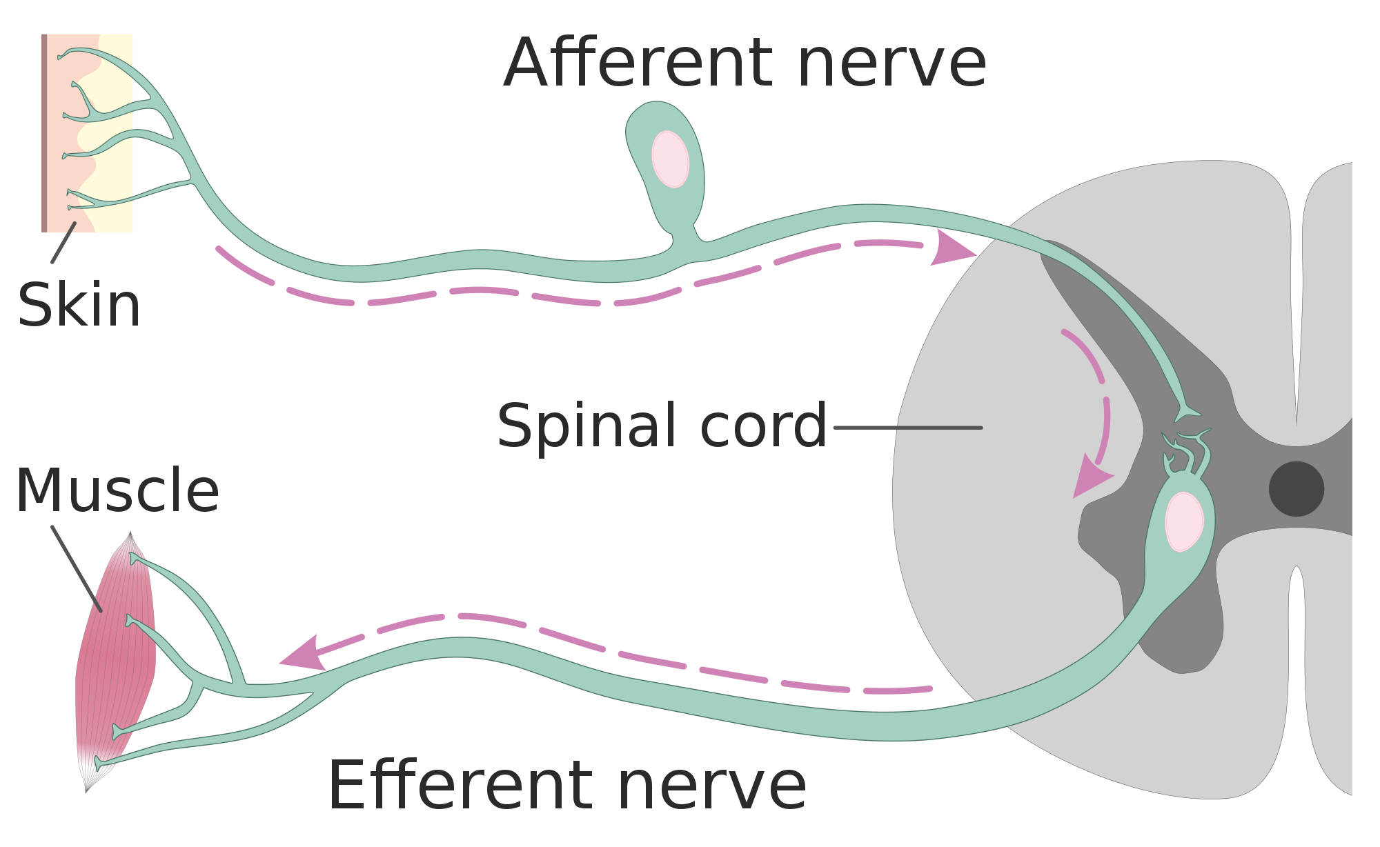
The PNS is mostly made up of cable-like bundles of axons called nerves, as well as clusters of neuronal cell bodies called ganglia (singular, ganglion). Nerves are generally classified as sensory, motor, or mixed nerves based on the direction in which they carry nerve impulses.
- Sensory nerves transmit information from sensory receptors in the body to the CNS. Sensory nerves are also called afferent nerves. You can see an example in the figure below.
- Motor nerves transmit information from the CNS to muscles, organs, and glands. Motor nerves are also called efferent nerves. You can see one in the figure below.
- Mixed nerves contain both sensory and motor neurons, so they can transmit information in both directions. They have both afferent and efferent functions.
Divisions of the Peripheral Nervous System
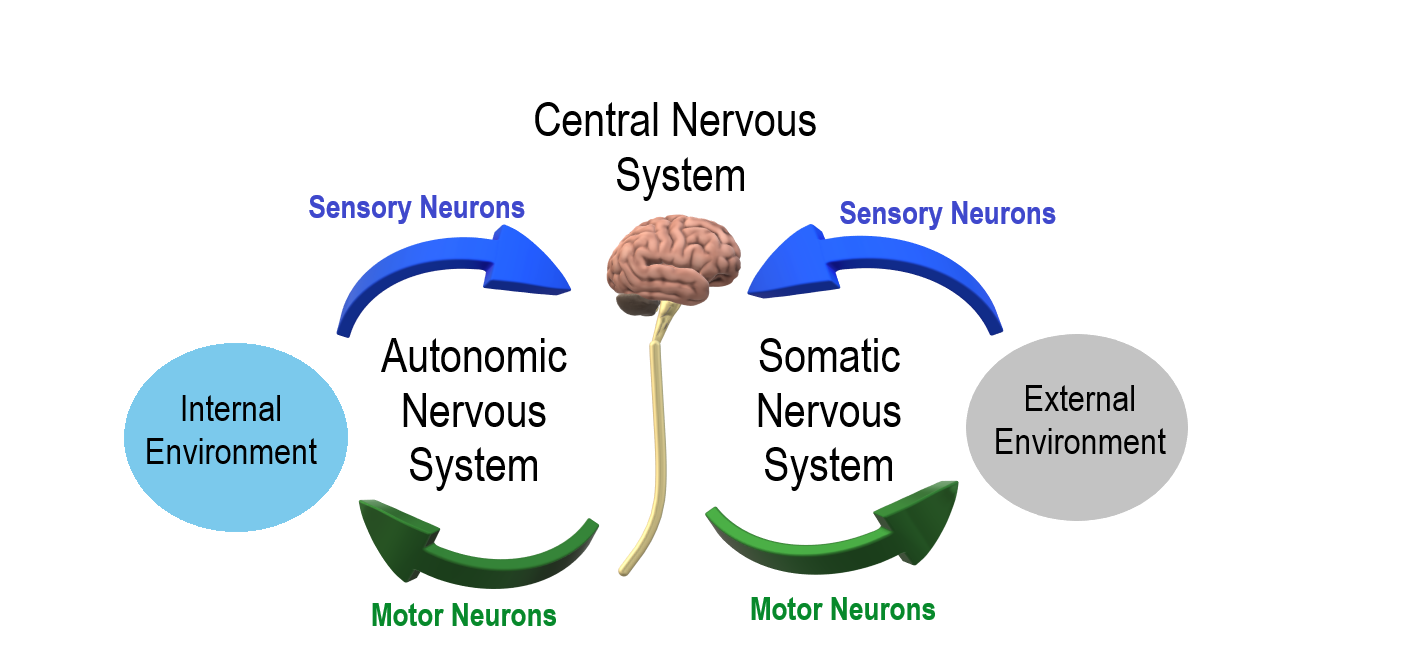
The PNS is divided into two major systems, called the autonomic nervous system and the somatic nervous system. In the diagram below, the autonomic system is shown on the left, and the somatic system on the right. Both systems of the PNS interact with the CNS and include sensory and motor neurons, but they use different circuits of nerves and ganglia.
Somatic Nervous System
The somatic nervous system primarily senses the external environment and controls voluntary activities about which decisions and commands come from the cerebral cortex of the brain. When you feel too warm, for example, you decide to turn on the air conditioner. As you walk across the room to the thermostat, you are using your somatic nervous system. In general, the somatic nervous system is responsible for all of your conscious perceptions of the outside world, as well as all of the voluntary motor activities you perform in response. Whether it’s playing a piano, driving a car, or playing basketball, you can thank your somatic nervous system for making it possible.
Somatic sensory and motor information is transmitted through 12 pairs of cranial nerves and 31 pairs of spinal nerves. Cranial nerves are in the head and neck and connect directly to the brain. Sensory components of cranial nerves transmit information about smells, tastes, light, sounds, and body position. Motor components of cranial nerves control skeletal muscles of the face, tongue, eyeballs, throat, head, and shoulders. Motor components of cranial nerves also control the salivary glands and swallowing. Four of the 12 cranial nerves participate in both sensory and motor functions as mixed nerves, having both sensory and motor neurons.
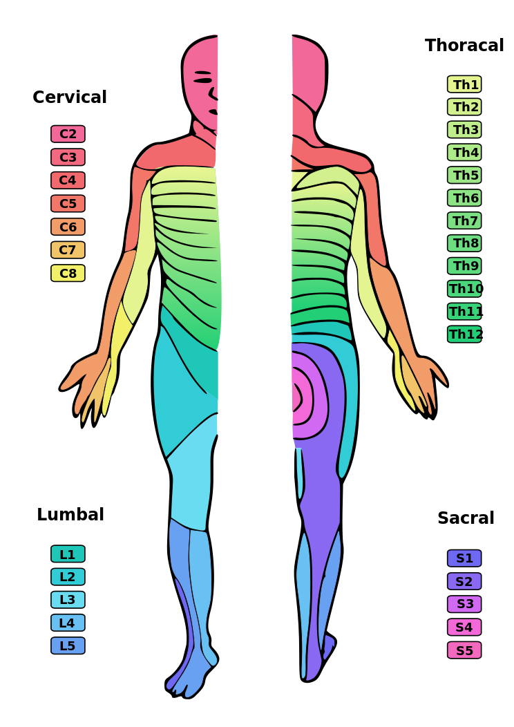
Spinal nerves emanate from the spinal column between vertebrae. All of the spinal nerves are mixed nerves, containing both sensory and motor neurons. The areas of skin innervated by the 31 pairs of spinal nerves are shown in the figure below. These include sensory nerves in the skin that sense pressure, temperature, vibrations, and pain. Other sensory nerves are in the muscles, and they sense stretching and tension. Spinal nerves also include motor nerves that stimulate skeletal muscles to contract, allowing for voluntary body movements.
Autonomic Nervous System
The autonomic nervous system primarily senses the internal environment and controls involuntary activities. It is responsible for monitoring conditions in the internal environment and bringing about appropriate changes in them. In general, the autonomic nervous system is responsible for all the activities that go on inside your body without your conscious awareness or voluntary participation.
Structurally, the autonomic nervous system consists of sensory and motor nerves that run between the CNS (especially the hypothalamus in the brain), internal organs (such as the heart, lungs, and digestive organs), and glands (such as the pancreas and sweat glands). Sensory neurons in the autonomic system detect internal body conditions and send messages to the brain. Motor nerves in the autonomic system affect appropriate responses by controlling contractions of smooth or cardiac muscle, or glandular tissue. For example, when sensory nerves of the autonomic system detect a rise in body temperature, motor nerves signal smooth muscles in blood vessels near the body surface to undergo vasodilation, and the sweat glands in the skin to secrete more sweat to cool the body.
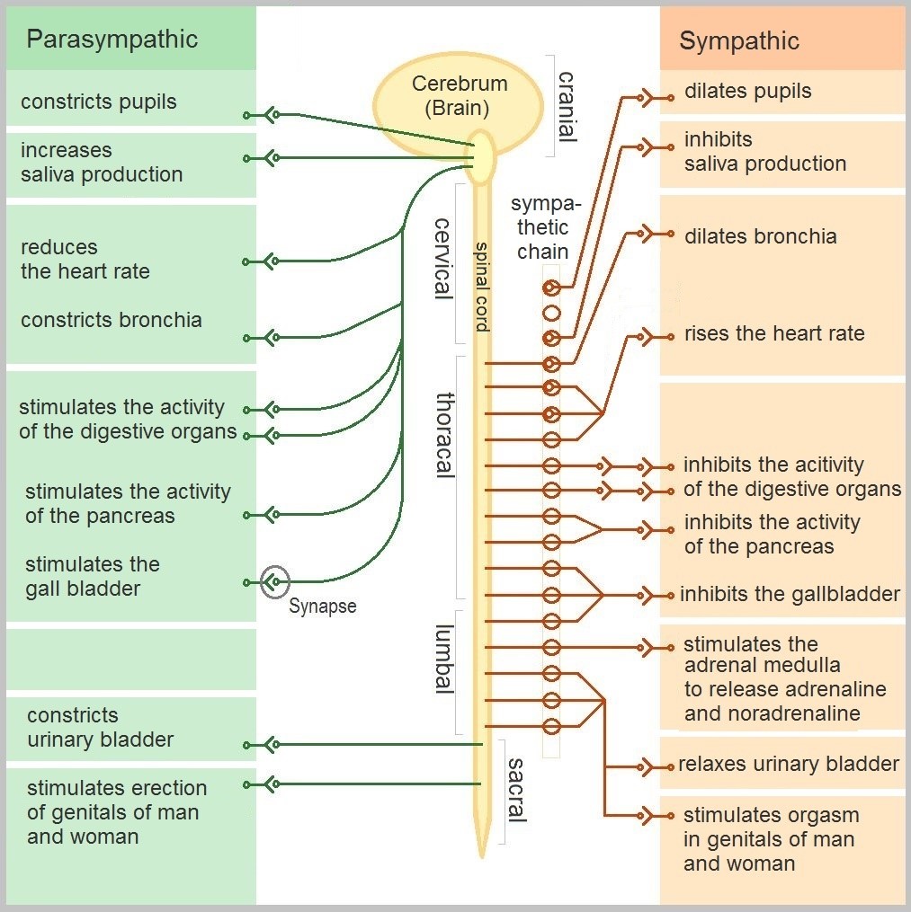
The autonomic nervous system, in turn, has three subdivisions: the sympathetic division, parasympathetic division, and enteric division. The first two subdivisions of the autonomic system are summarized in the figure below. Both affect the same organs and glands, but they generally do so in opposite ways.
- The sympathetic division controls the fight-or-flight response. Changes occur in organs and glands throughout the body that prepare the body to fight or flee in response to a perceived danger. For example, the heart rate speeds up, air passages in the lungs become wider, more blood flows to the skeletal muscles, and the digestive system temporarily shuts down.
- The parasympathetic division returns the body to normal after the fight-or-flight response has occurred. For example, it slows down the heart rate, narrows air passages in the lungs, reduces blood flow to the skeletal muscles, and stimulates the digestive system to start working again. The parasympathetic division also maintains internal homeostasis of the body at other times.
- The enteric division is made up of nerve fibres that supply the organs of the digestive system. This division allows for the local control of many digestive functions.
Disorders of the Peripheral Nervous System
Unlike the CNS — which is protected by bones, meninges, and cerebrospinal fluid — the PNS has no such protections. The PNS also has no blood-brain barrier to protect it from toxins and pathogens in the blood. Therefore, the PNS is more subject to injury and disease than is the CNS. Causes of nerve injury include diabetes, infectious diseases (such as shingles), and poisoning by toxins (such as heavy metals). PNS disorders often have symptoms like loss of feeling, tingling, burning sensations, or muscle weakness. If a traumatic injury results in a nerve being transected (cut all the way through), it may regenerate, but this is a very slow process and may take many months.
Two other diseases of the PNS are Guillain-Barre syndrome and Charcot-Marie-Tooth disease.
- Guillain-Barre syndrome is a rare disease in which the immune system attacks nerves of the PNS, leading to muscle weakness and even paralysis. The exact cause of Guillain-Barre syndrome is unknown, but it often occurs after a viral or bacterial infection. There is no known cure for the syndrome, but most people eventually make a full recovery. Recovery can be slow, however, lasting anywhere from several weeks to several years.
- Charcot-Marie-Tooth disease is a hereditary disorder of the nerves, and one of the most common inherited neurological disorders. It affects predominantly the nerves in the feet and legs, and often in the hands and arms, as well. The disease is characterized by loss of muscle tissue and sense of touch. It is presently incurable.
Feature: My Human Body
The autonomic nervous system is considered to be involuntary because it doesn't require conscious input. However, it is possible to exert some voluntary control over it. People who practice yoga or other so-called mind-body techniques, for example, can reduce their heart rate and certain other autonomic functions. Slowing down these otherwise involuntary responses is a good way to relieve stress and reduce the wear-and-tear that stress can place on the body. Such techniques may also be useful for controlling post-traumatic stress disorder and chronic pain. Three types of integrative practices for these purposes are breathing exercises, body-based tension modulation exercises, and mindfulness techniques.
Breathing exercises can help control the rapid, shallow breathing that often occurs when you are anxious or under stress. These exercises can be learned quickly, and they provide immediate feelings of relief. Specific breathing exercises include paced breath, diaphragmatic breathing, and Breathe2Relax or Chill Zone on MindShift™ CBT, which are downloadable breathing practice mobile applications, or "Apps". Try syncing your breathing with Eric Klassen's "Triangle breathing, 1 minute" video:
https://www.youtube.com/watch?v=u9Q8D6n-3qw
Triangle breathing, 1 minute, Erin Klassen, 2015.
Body-based tension modulation exercises include yoga postures (also known as “asanas”) and tension manipulation exercises. The latter include the Trauma/Tension Release Exercise (TRE) and the Trauma Resiliency Model (TRM). Watch this video for a brief — but informative — introduction to the TRE program:
https://www.youtube.com/watch?v=67R974D8swM&feature=youtu.be
TRE® : Tension and Trauma Releasing Exercises, an Introduction with Jessica Schaffer, Jessica Schaffer Nervous System RESET, 2015.
Mindfulness techniques have been shown to reduce symptoms of depression, as well as those of anxiety and stress. They have also been shown to be useful for pain management and performance enhancement. Specific mindfulness programs include Mindfulness Based Stress Reduction (MBSR) and Mindfulness Mind-Fitness Training (MMFT). You can learn more about MBSR by watching the video below.
https://www.youtube.com/watch?v=0TA7P-iCCcY&feature=youtu.be
Mindfulness-Based Stress Reduction (UMass Medical School, Center for Mindfulness), Palouse Mindfulness, 2017.
8.6 Summary
- The peripheral nervous system (PNS) consists of all the nervous tissue that lies outside the central nervous system (CNS). Its main function is to connect the CNS to the rest of the organism.
- The PNS is made up of nerves and ganglia. Nerves are bundles of axons, and ganglia are groups of cell bodies. Nerves are classified as sensory, motor, or a mix of the two.
- The PNS is divided into the somatic and autonomic nervous systems. The somatic system controls voluntary activities, whereas the autonomic system controls involuntary activities.
- The autonomic nervous system is further divided into sympathetic, parasympathetic, and enteric divisions. The sympathetic division controls fight-or-flight responses during emergencies, the parasympathetic system controls routine body functions the rest of the time, and the enteric division provides local control over the digestive system.
- The PNS is not as well protected physically or chemically as the CNS, so it is more prone to injury and disease. PNS problems include injury from diabetes, shingles, and heavy metal poisoning. Two disorders of the PNS are Guillain-Barre syndrome and Charcot-Marie-Tooth disease.
8.6 Review Questions
- Describe the general structure of the peripheral nervous system. State its primary function.
- What are ganglia?
- Identify three types of nerves based on the direction in which they carry nerve impulses.
- Outline all of the divisions of the peripheral nervous system.
- Compare and contrast the somatic and autonomic nervous systems.
- When and how does the sympathetic division of the autonomic nervous system affect the body?
- What is the function of the parasympathetic division of the autonomic nervous system? Specifically, how does it affect the body?
- Name and describe two peripheral nervous system disorders.
- Give one example of how the CNS interacts with the PNS to control a function in the body.
- For each of the following types of information, identify whether the neuron carrying it is sensory or motor, and whether it is most likely in the somatic or autonomic nervous system:
- Visual information
- Blood pressure information
- Information that causes muscle contraction in digestive organs after eating
- Information that causes muscle contraction in skeletal muscles based on the person’s decision to make a movement
-
8.6 Explore More
https://www.youtube.com/watch?v=ySIDMU2cy0Y&feature=emb_logo
Phantom Limbs Explained, Plethrons, 2015.
https://www.youtube.com/watch?time_continue=1&v=73yo5nJne6c&feature=emb_logo
Why Do Hot Peppers Cause Pain? Reactions, 2015.
Attributions
Figure 8.6.1
Kid’s piant duet by PJMixer on Flickr is used under a CC BY-NC-ND 2.0 (https://creativecommons.org/licenses/by-nc-nd/2.0/) license.
Figure 8.6.2
Nervous_system_diagram by ¤~Persian Poet Gal on Wikimedia Commons is released into the public domain (https://en.wikipedia.org/wiki/Public_domain).
Figure 8.6.3
Afferent_and_efferent_neurons_en.svg by Helixitta on Wikimedia Commons is used under a CC BY-SA 4.0 (https://creativecommons.org/licenses/by-sa/4.0) license.
Figure 8.6.4
Autonomic and Somatic Nervous System by Christinelmiller on Wikimedia Commons is used under a CC BY-SA 4.0 (https://creativecommons.org/licenses/by-sa/4.0) license.
Figure 8.6.5
Dermatoms.svg by Ralf Stephan (mailto:ralf@ark.in-berlin.de) on Wikimedia Commons is released into the public domain (https://en.wikipedia.org/wiki/Public_domain).
Figure 8.6.6
The_Autonomic_Nervous_System by Geo-Science-International on Wikimedia Commons is used and adapted by Christine Miller under a CC0 1.0 Universal
Public Domain Dedication license (https://creativecommons.org/publicdomain/zero/1.0/).
References
Erin Klassen. (2015, December 15). Triangle breathing, 1 minute. YouTube. https://www.youtube.com/watch?v=u9Q8D6n-3qw&feature=youtu.be
Jessica Schaffer Nervous System RESET. (2015, January 15). TRE® : Tension and trauma releasing exercises, an Introduction with Jessica Schaffer. YouTube. https://www.youtube.com/watch?v=67R974D8swM&feature=youtu.be
Mayo Clinic Staff. (n.d.). Charcot-Marie-Tooth disease [online article]. MayoClinic.org. https://www.mayoclinic.org/diseases-conditions/charcot-marie-tooth-disease/symptoms-causes/syc-20350517
Mayo Clinic Staff. (n.d.). Diabetes [online article]. MayoClinic.org. https://www.mayoclinic.org/diseases-conditions/diabetes/symptoms-causes/syc-20371444
Mayo Clinic Staff. (n.d.). Guillain-Barre syndrome [online article]. MayoClinic.org. https://www.mayoclinic.org/diseases-conditions/guillain-barre-syndrome/symptoms-causes/syc-20362793
Mayo Clinic Staff. (n.d.). Shingles [online article]. MayoClinic.org. https://www.mayoclinic.org/diseases-conditions/shingles/symptoms-causes/syc-20353054
Mayo Clinic Staff. (n.d.). Stroke [online article]. MayoClinic.org. https://www.mayoclinic.org/diseases-conditions/stroke/symptoms-causes/syc-20350113
Palouse Mindfulness. (2017, March 25). Mindfulness-based stress reduction (UMass Medical School, Center for Mindfulness), YouTube. https://www.youtube.com/watch?v=0TA7P-iCCcY&feature=youtu.be
Plethrons, (2015, March 23). Phantom limbs explained. YouTube. https://www.youtube.com/watch?v=ySIDMU2cy0Y&feature=youtu.be
Reactions. (2015, December 1). Why do hot peppers cause pain? YouTube. https://www.youtube.com/watch?v=73yo5nJne6c&feature=youtu.be
Created by CK-12 Foundation/Adapted by Christine Miller
Goose Bumps
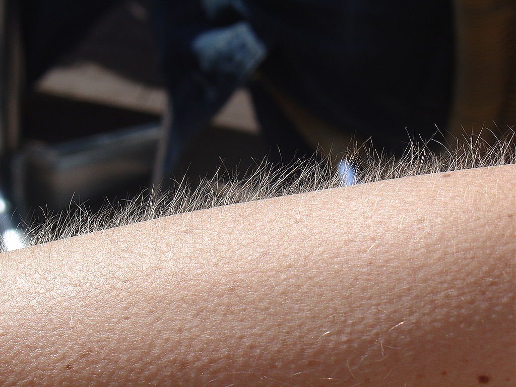
No doubt you’ve experienced the tiny, hair-raising skin bumps called goose bumps, like those you see in Figure 10.4.1. They happen when you feel chilly. Do you know what causes goose bumps, or why they pop up when you are cold? The answers to these questions involve the layer of skin known as the dermis.
What is the Dermis?
The dermis is the inner of the two major layers that make up the skin, the outer layer being the epidermis. The dermis consists mainly of connective tissues. It also contains most skin structures, such as glands and blood vessels. The dermis is anchored to the tissues below it by flexible collagen bundles that permit most areas of the skin to move freely over subcutaneous (“below the skin”) tissues. Functions of the dermis include cushioning subcutaneous tissues, regulating body temperature, sensing the environment, and excreting wastes.
Anatomy of the Dermis
The basic anatomy of the dermis is a matrix, or sort of scaffolding, composed of connective tissues. These tissues include collagen fibres — which provide toughness — and elastin fibres, which provide elasticity. Surrounding these fibres, the matrix also includes a gel-like substance made of proteins. The tissues of the matrix give the dermis both strength and flexibility.
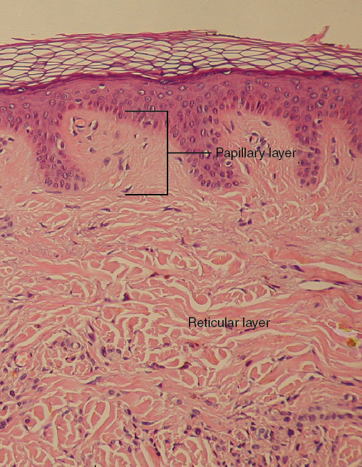
The dermis is divided into two layers: the papillary layer and the reticular layer. Both layers are shown in Figure 10.4.2 below and described in the text that follows.
Papillary Layer
The papillary layer is the upper layer of the dermis, just below the basement membrane that connects the dermis to the epidermis above it. The papillary layer is the thinner of the two dermal layers. It is composed mainly of loosely arranged collagen fibres. The papillary layer is named for its fingerlike projections — or papillae — that extend upward into the epidermis. The papillae contain capillaries and sensory touch receptors.
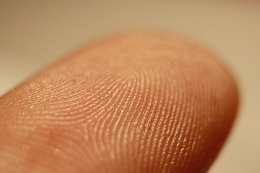
The papillae give the dermis a bumpy surface that interlocks with the epidermis above it, strengthening the connection between the two layers of skin. On the palms and soles, the papillae create epidermal ridges. Epidermal ridges on the fingers are commonly called fingerprints (see Figure 10.4.3). Fingerprints are genetically determined, so no two people (other than identical twins) have exactly the same fingerprint pattern. Therefore, fingerprints can be used as a means of identification, for example, at crime scenes. Fingerprints were much more commonly used forensically before DNA analysis was introduced for this purpose.
Reticular Layer
The reticular layer is the lower layer of the dermis, located below the papillary layer. It is the thicker of the two dermal layers. It is composed of densely woven collagen and elastin fibres. These protein fibres give the dermis its properties of strength and elasticity. This layer of the dermis cushions subcutaneous tissues of the body from stress and strain. The reticular layer of the dermis also contains most of the structures in the dermis, such as glands and hair follicles.
Structures in the Dermis
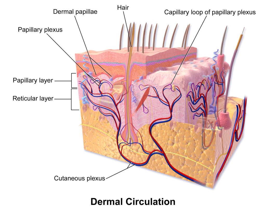
Both papillary and reticular layers of the dermis contain numerous sensory receptors, which make the skin the body’s primary sensory organ for the sense of touch. Both dermal layers also contain blood vessels. They provide nutrients to remove wastes from dermal cells, as well as cells in the lowest layer of the epidermis, the stratum basale. The circulatory components of the dermis are shown in Figure 10.4.4 below.
Glands
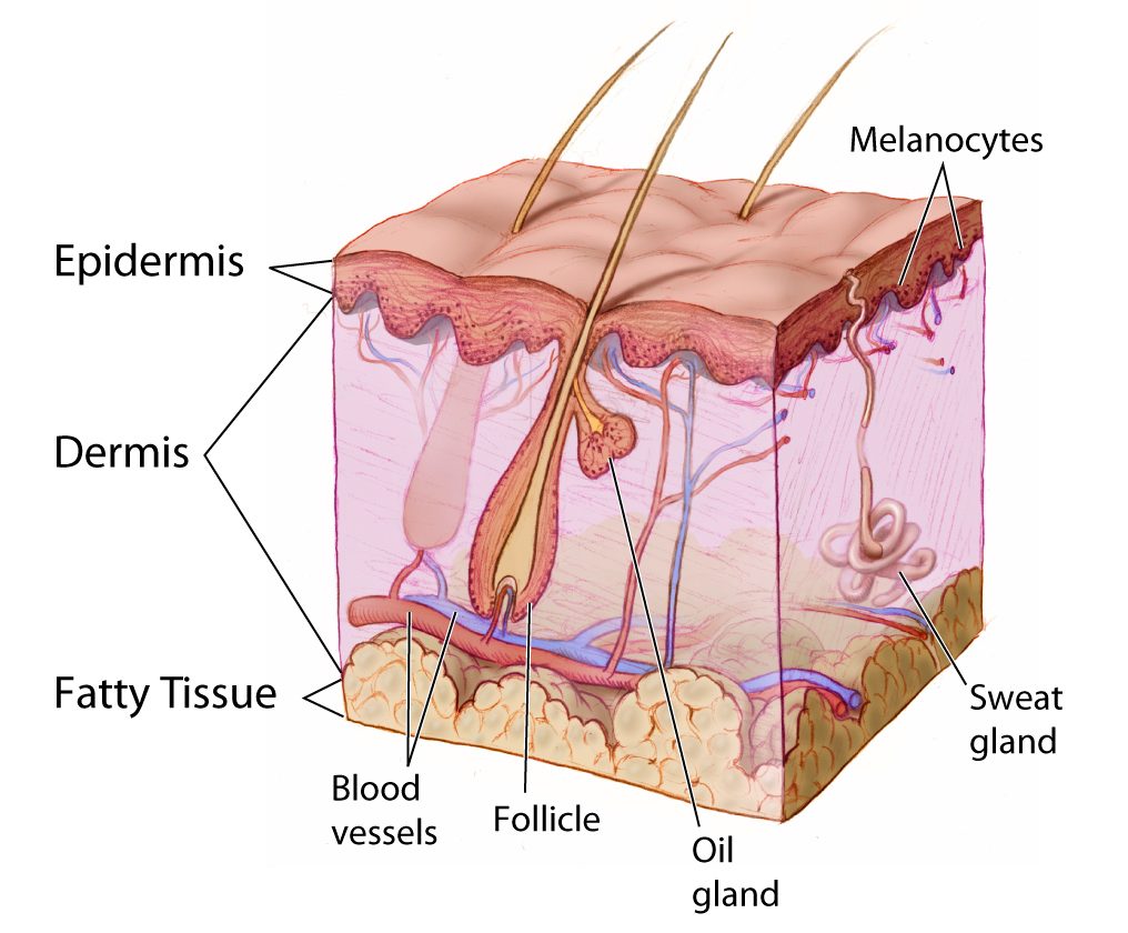
Glands in the reticular layer of the dermis include sweat glands and sebaceous (oil) glands. Both are exocrine glands, which are glands that release their secretions through ducts to nearby body surfaces. The diagram in Figure 10.4.5 shows these glands, as well as several other structures in the dermis.
Sweat Glands
Sweat glands produce the fluid called sweat, which contains mainly water and salts. The glands have ducts that carry the sweat to hair follicles, or to the surface of the skin. There are two different types of sweat glands: eccrine glands and apocrine glands.
- Eccrine sweat glands occur in skin all over the body. Their ducts empty through tiny openings called pores onto the skin surface. These sweat glands are involved in temperature regulation.
- Apocrine sweat glands are larger than eccrine glands, and occur only in the skin of the armpits and groin. The ducts of apocrine glands empty into hair follicles, and then the sweat travels along hairs to reach the surface. Apocrine glands are inactive until puberty, at which point they start producing an oily sweat that is consumed by bacteria living on the skin. The digestion of apocrine sweat by bacteria causes body odor.
Sebaceous Glands
Sebaceous glands are exocrine glands that produce a thick, fatty substance called sebum. Sebum is secreted into hair follicles and makes its way to the skin surface along hairs. It waterproofs the hair and skin, and helps prevent them from drying out. Sebum also has antibacterial properties, so it inhibits the growth of microorganisms on the skin. Sebaceous glands are found in every part of the skin — except for the palms of the hands and soles of the feet, where hair does not grow.
Hair Follicles
Hair follicles are the structures where hairs originate (see the diagram above). Hairs grow out of follicles, pass through the epidermis, and exit at the surface of the skin. Associated with each hair follicle is a sebaceous gland, which secretes sebum that coats and waterproofs the hair. Each follicle also has a bed of capillaries, a nerve ending, and a tiny muscle called an arrector pili.
Functions of the Dermis
The main functions of the dermis are regulating body temperature, enabling the sense of touch, and eliminating wastes from the body.
Temperature Regulation
Several structures in the reticular layer of the dermis are involved in regulating body temperature. For example, when body temperature rises, the hypothalamus of the brain sends nerve signals to sweat glands, causing them to release sweat. An adult can sweat up to four litres an hour. As the sweat evaporates from the surface of the body, it uses energy in the form of body heat, thus cooling the body. The hypothalamus also causes dilation of blood vessels in the dermis when body temperature rises. This allows more blood to flow through the skin, bringing body heat to the surface, where it can radiate into the environment.
When the body is too cool, sweat glands stop producing sweat, and blood vessels in the skin constrict, thus conserving body heat. The arrector pili muscles also contract, moving hair follicles and lifting hair shafts. This results in more air being trapped under the hairs to insulate the surface of the skin. These contractions of arrector pili muscles are the cause of goose bumps.
Sensing the Environment
Sensory receptors in the dermis are mainly responsible for the body’s tactile senses. The receptors detect such tactile stimuli as warm or cold temperature, shape, texture, pressure, vibration, and pain. They send nerve impulses to the brain, which interprets and responds to the sensory information. Sensory receptors in the dermis can be classified on the basis of the type of touch stimulus they sense. Mechanoreceptors sense mechanical forces such as pressure, roughness, vibration, and stretching. Thermoreceptors sense variations in temperature that are above or below body temperature. Nociceptors sense painful stimuli. Figure 10.4.6 shows several specific kinds of tactile receptors in the dermis. Each kind of receptor senses one or more types of touch stimuli.
- Free nerve endings sense pain and temperature variations.
- Merkel cells sense light touch, shapes, and textures.
- Meissner’s corpuscles sense light touch.
- Pacinian corpuscles sense pressure and vibration.
- Ruffini corpuscles sense stretching and sustained pressure.
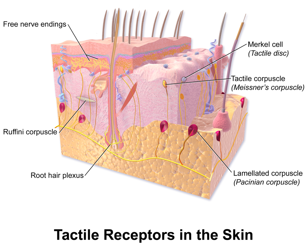
Excreting Wastes
The sweat released by eccrine sweat glands is one way the body excretes waste products. Sweat contains excess water, salts (electrolytes), and other waste products that the body must get rid of to maintain homeostasis. The most common electrolytes in sweat are sodium and chloride. Potassium, calcium, and magnesium electrolytes may be excreted in sweat, as well. When these electrolytes reach high levels in the blood, more are excreted in sweat. This helps to bring their blood levels back into balance. Besides electrolytes, sweat contains small amounts of waste products from metabolism, including ammonia and urea. Sweat may also contain alcohol in someone who has been drinking alcoholic beverages.
Feature: My Human Body
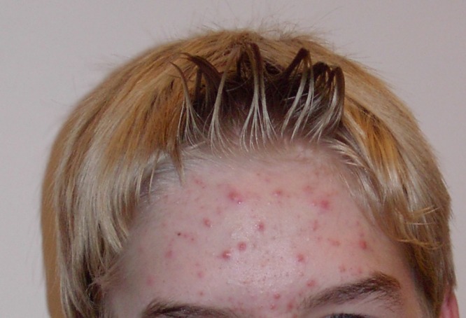
Acne is the most common skin disorder in the Canada. At least 20% of Canadians have acne at any given time and it affects approximately 90% of adolescents (as in Figure 10.4.7). Although acne occurs most commonly in teens and young adults, but it can occur at any age. Even newborn babies can get acne.
The main sign of acne is the appearance of pimples (pustules) on the skin, like those in the photo above. Other signs of acne may include whiteheads, blackheads, nodules, and other lesions. Besides the face, acne can appear on the back, chest, neck, shoulders, upper arms, and buttocks. Acne can permanently scar the skin, especially if it isn’t treated appropriately. Besides its physical effects on the skin, acne can also lead to low self-esteem and depression.
Acne is caused by clogged, sebum-filled pores that provide a perfect environment for the growth of bacteria. The bacteria cause infection, and the immune system responds with inflammation. Inflammation, in turn, causes swelling and redness, and may be associated with the formation of pus. If the inflammation goes deep into the skin, it may form an acne nodule.
Mild acne often responds well to treatment with over-the-counter (OTC) products containing benzoyl peroxide or salicylic acid. Treatment with these products may take a month or two to clear up the acne. Once the skin clears, treatment generally needs to continue for some time to prevent future breakouts.
If acne fails to respond to OTC products, nodules develop, or acne is affecting self-esteem, a visit to a dermatologist is in order. A dermatologist can determine which treatment is best for a given patient. A dermatologist can also prescribe prescription medications (which are likely to be more effective than OTC products) and provide other medical treatments, such as laser light therapies or chemical peels.
What can you do to maintain healthy skin and prevent or reduce acne? Dermatologists recommend the following tips:
- Wash affected or acne-prone skin (such as the face) twice a day, and after sweating.
- Use your fingertips to apply a gentle, non-abrasive cleanser. Avoid scrubbing, which can make acne worse.
- Use only alcohol-free products and avoid any products that irritate the skin, such as harsh astringents or exfoliants.
- Rinse with lukewarm water, and avoid using very hot or cold water.
- Shampoo your hair regularly.
- Do not pick, pop, or squeeze acne. If you do, it will take longer to heal and is more likely to scar.
- Keep your hands off your face. Avoid touching your skin throughout the day.
- Stay out of the sun and tanning beds. Some acne medications make your skin very sensitive to UV light.
10.4 Summary
- The dermis is the inner and thicker of the two major layers that make up the skin. It consists mainly of a matrix of connective tissues that provide strength and stretch. It also contains almost all skin structures, including sensory receptors and blood vessels.
- The dermis has two layers. The upper papillary layer has papillae extending upward into the epidermis and loose connective tissues. The lower reticular layer has denser connective tissues and structures, such as glands and hair follicles. Glands in the dermis include eccrine and apocrine sweat glands and sebaceous glands. Hair follicles are structures where hairs originate.
- Functions of the dermis include cushioning subcutaneous tissues, regulating body temperature, sensing the environment, and excreting wastes. The dense connective tissues of the dermis provide cushioning. The dermis regulates body temperature mainly by sweating and by vasodilation or vasoconstriction. The many tactile sensory receptors in the dermis make it the main organ for the sense of touch. Wastes excreted in sweat include excess water, electrolytes, and certain metabolic wastes.
10.4 Review Questions
- What is the dermis?
- Describe the basic anatomy of the dermis.
- Compare and contrast the papillary and reticular layers of the dermis.
- What causes epidermal ridges, and why can they be used to identify individuals?
- Name the two types of sweat glands in the dermis, and explain how they differ.
- What is the function of sebaceous glands?
- Describe the structures associated with hair follicles.
- Explain how the dermis helps regulate body temperature.
- Identify three specific kinds of tactile receptors in the dermis, along with the type of stimuli they sense.
- How does the dermis excrete wastes? What waste products does it excrete?
- What are subcutaneous tissues? Which layer of the dermis provides cushioning for subcutaneous tissues? Why does this layer provide most of the cushioning, instead of the other layer?
- For each of the functions listed below, describe which structure within the dermis carries it out.
- Brings nutrients to and removes wastes from dermal and lower epidermal cells
- Causes hairs to move
- Detects painful stimuli on the skin
10.4 Explore More
https://www.youtube.com/watch?v=FX-FwK0IIrE
How do you get rid of acne? SciShow, 2016.
https://www.youtube.com/watch?v=VcHQWMAClhQ&feature=emb_logo
When You Can't Scratch Away An Itch, Seeker, 2013.
Attributions
Figure 10.4.1
Goose_bumps by EverJean on Wikimedia Commons is used under a CC BY 2.0 (https://creativecommons.org/licenses/by/2.0) license.
Figure 10.4.2
Layers_of_the_Dermis by OpenStax College on Wikimedia Commons is used under a CC BY 3.0 (https://creativecommons.org/licenses/by/3.0) license.
Figure 10.4.3
Fingerprint_detail_on_male_finger_in_Třebíč,_Třebíč_District by Frettie on Wikimedia Commons is used under a CC BY 3.0 (https://creativecommons.org/licenses/by/3.0) license.
Figure 10.4.4
Blausen_0802_Skin_Dermal Circulation by BruceBlaus on Wikimedia commons is used under a CC BY 3.0 (https://creativecommons.org/licenses/by/3.0) license.
Figure 10.4.5
Anatomy_The_Skin_-_NCI_Visuals_Online by Don Bliss (artist) / National Cancer Institute (National Institutes of Health, with the ID 4604) is in the public domain (https://en.wikipedia.org/wiki/public_domain).
Figure 10.4.6
Blausen_0809_Skin_TactileReceptors by BruceBlaus on Wikimedia commons is used under a CC BY 3.0 (https://creativecommons.org/licenses/by/3.0) license.
Figure 10.4.7
Akne-jugend by Ellywa on Wikimedia Commons is released into the public domain (https://en.wikipedia.org/wiki/public_domain). (No machine-readable author provided. Ellywa assumed, based on copyright claims).
References
Betts, J. G., Young, K.A., Wise, J.A., Johnson, E., Poe, B., Kruse, D.H., Korol, O., Johnson, J.E., Womble, M., DeSaix, P. (2013, June 19). Figure 5.7 Layers of the dermis [digital image]. In Anatomy and Physiology (Section 5.1 Layers of the skin). OpenStax. https://openstax.org/books/anatomy-and-physiology/pages/5-1-layers-of-the-skin
Blausen.com staff. (2014). Medical gallery of Blausen Medical 2014. WikiJournal of Medicine 1 (2). DOI:10.15347/wjm/2014.010. ISSN 2002-4436.
SciShow. (2016, October 26). How do you get rid of acne? YouTube. https://www.youtube.com/watch?v=FX-FwK0IIrE
Seeker. (2013, October 26). When you can't scratch away an itch. YouTube. https://www.youtube.com/watch?v=VcHQWMAClhQ&feature=emb_logo
Created by CK-12 Foundation/Adapted by Christine Miller
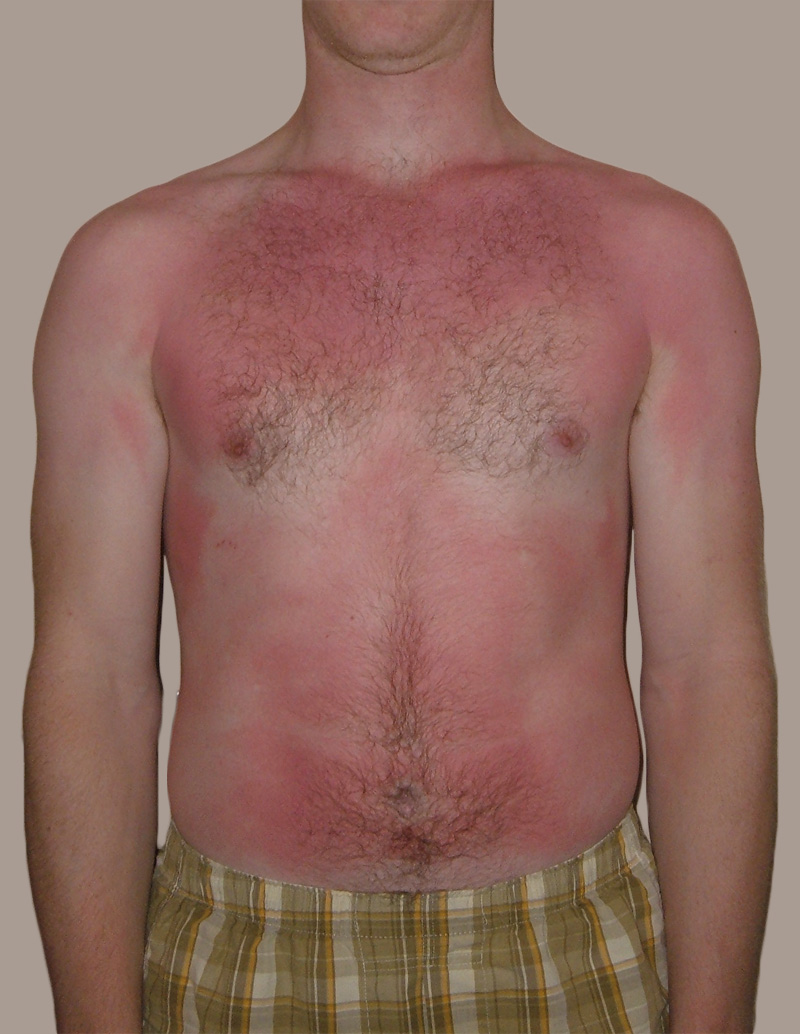
Feel the Burn
The person in Figure 10.3.1 is no doubt feeling the burn — sunburn, that is. Sunburn occurs when the outer layer of the skin is damaged by UV light from the sun or tanning lamps. Some people deliberately allow UV light to burn their skin, because after the redness subsides, they are left with a tan. A tan may look healthy, but it is actually a sign of skin damage. People who experience one or more serious sunburns are significantly more likely to develop skin cancer. Natural pigment molecules in the skin help protect it from UV light damage. These pigment molecules are found in the layer of the skin called the epidermis.
What is the Epidermis?
The epidermis is the outer of the two main layers of the skin. The inner layer is the dermis. It averages about 0.10 mm thick, and is much thinner than the dermis. The epidermis is thinnest on the eyelids (0.05 mm) and thickest on the palms of the hands and soles of the feet (1.50 mm). The epidermis covers almost the entire body surface. It is continuous with — but structurally distinct from — the mucous membranes that line the mouth, anus, urethra, and vagina.
Structure of the Epidermis
There are no blood vessels and very few nerve cells in the epidermis. Without blood to bring epidermal cells oxygen and nutrients, the cells must absorb oxygen directly from the air and obtain nutrients via diffusion of fluids from the dermis below. However, as thin as it is, the epidermis still has a complex structure. It has a variety of cell types and multiple layers.
Cells of the Epidermis
There are several different types of cells in the epidermis. All of the cells are necessary for the important functions of the epidermis.
- The epidermis consists mainly of stacks of keratin-producing epithelial cells called keratinocytes. These cells make up at least 90 per cent of the epidermis. Near the top of the epidermis, these cells are also called squamous cells.
- Another eight per cent of epidermal cells are melanocytes. These cells produce the pigment melanin that protects the dermis from UV light.
- About one per cent of epidermal cells are Langerhans cells. These are immune system cells that detect and fight pathogens entering the skin.
- Less than one per cent of epidermal cells are Merkel cells, which respond to light touch and connect to nerve endings in the dermis.
Layers of the Epidermis
The epidermis in most parts of the body consists of four distinct layers. A fifth layer occurs in the palms of the hands and soles of the feet, where the epidermis is thicker than in the rest of the body. The layers of the epidermis are shown in Figure 10.3.2, and described in the following text.
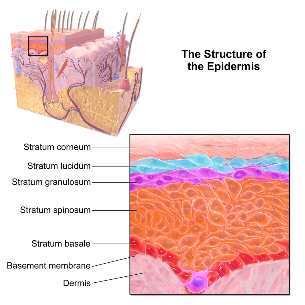
Stratum Basale
The stratum basale is the innermost (or deepest) layer of the epidermis. It is separated from the dermis by a membrane called the basement membrane. The stratum basale contains stem cells — called basal cells — which divide to form all the keratinocytes of the epidermis. When keratinocytes first form, they are cube-shaped and contain almost no keratin. As more keratinocytes are produced, previously formed cells are pushed up through the stratum basale. Melanocytes and Merkel cells are also found in the stratum basale. The Merkel cells are especially numerous in touch-sensitive areas, such as the fingertips and lips.
Stratum Spinosum
Just above the stratum basale is the stratum spinosum. This is the thickest of the four epidermal layers. The keratinocytes in this layer have begun to accumulate keratin, and they have become tougher and flatter. Spiny cellular projections form between the keratinocytes and hold them together. In addition to keratinocytes, the stratum spinosum contains the immunologically active Langerhans cells.
Stratum Granulosum
The next layer above the stratum spinosum is the stratum granulosum. In this layer, keratinocytes have become nearly filled with keratin, giving their cytoplasm a granular appearance. Lipids are released by keratinocytes in this layer to form a lipid barrier in the epidermis. Cells in this layer have also started to die, because they are becoming too far removed from blood vessels in the dermis to receive nutrients. Each dying cell digests its own nucleus and organelles, leaving behind only a tough, keratin-filled shell.
Stratum Lucidum
Only on the palms of the hands and soles of the feet, the next layer above the stratum granulosum is the stratum lucidum. This is a layer consisting of stacks of translucent, dead keratinocytes that provide extra protection to the underlying layers.
Stratum Corneum
The uppermost layer of the epidermis everywhere on the body is the stratum corneum. This layer is made of flat, hard, tightly packed dead keratinocytes that form a waterproof keratin barrier to protect the underlying layers of the epidermis. Dead cells from this layer are constantly shed from the surface of the body. The shed cells are continually replaced by cells moving up from lower layers of the epidermis. It takes a period of about 48 days for newly formed keratinocytes in the stratum basale to make their way to the top of the stratum corneum to replace shed cells.
Functions of the Epidermis
The epidermis has several crucial functions in the body. These functions include protection, water retention, and vitamin D synthesis.
Protective Functions
The epidermis provides protection to underlying tissues from physical damage, pathogens, and UV light.
Protection from Physical Damage
Most of the physical protection of the epidermis is provided by its tough outer layer, the stratum corneum. Because of this layer, minor scrapes and scratches generally do not cause significant damage to the skin or underlying tissues. Sharp objects and rough surfaces have difficulty penetrating or removing the tough, dead, keratin-filled cells of the stratum corneum. If cells in this layer are pierced or scraped off, they are quickly replaced by new cells moving up to the surface from lower skin layers.
Protection from Pathogens
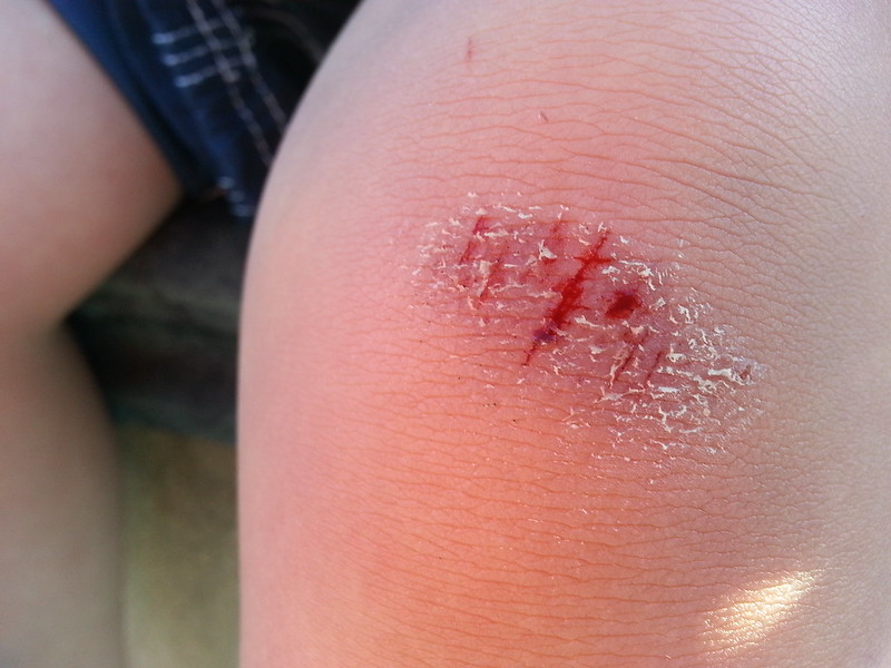
When pathogens such as viruses and bacteria try to enter the body, it is virtually impossible for them to enter through intact epidermal layers. Generally, pathogens can enter the skin only if the epidermis has been breached, for example by a cut, puncture, or scrape (like the one pictured in Figure 10.3.3). That’s why it is important to clean and cover even a minor wound in the epidermis. This helps ensure that pathogens do not use the wound to enter the body. Protection from pathogens is also provided by conditions at or near the skin surface. These include relatively high acidity (pH of about 5.0), low amounts of water, the presence of antimicrobial substances produced by epidermal cells, and competition with non-pathogenic microorganisms that normally live on the epidermis.
Protection from UV Light
UV light that penetrates the epidermis can damage epidermal cells. In particular, it can cause mutations in DNA that lead to the development of skin cancer, in which epidermal cells grow out of control. UV light can also destroy vitamin B9 (in forms such as folate or folic acid), which is needed for good health and successful reproduction. In a person with light skin, just an hour of exposure to intense sunlight can reduce the body’s vitamin B9 level by 50 per cent.
Melanocytes in the stratum basale of the epidermis contain small organelles called melanosomes, which produce, store, and transport the dark brown pigment melanin. As melanosomes become full of melanin, they move into thin extensions of the melanocytes. From there, the melanosomes are transferred to keratinocytes in the epidermis, where they absorb UV light that strikes the skin. This prevents the light from penetrating deeper into the skin, where it can cause damage. The more melanin there is in the skin, the more UV light can be absorbed.
Water Retention
Skin's ability to hold water and not lose it to the surrounding environment is due mainly to the stratum corneum. Lipids arranged in an organized way among the cells of the stratum corneum form a barrier to water loss from the epidermis. This is critical for maintaining healthy skin and preserving proper water balance in the body.
Although the skin is impermeable to water, it is not impermeable to all substances. Instead, the skin is selectively permeable, allowing certain fat-soluble substances to pass through the epidermis. The selective permeability of the epidermis is both a benefit and a risk.
- Selective permeability allows certain medications to enter the bloodstream through the capillaries in the dermis. This is the basis of medications that are delivered using topical ointments, or patches (see Figure 10.3.4) that are applied to the skin. These include steroid hormones, such as estrogen (for hormone replacement therapy), scopolamine (for motion sickness), nitroglycerin (for heart problems), and nicotine (for people trying to quit smoking).
- Selective permeability of the epidermis also allows certain harmful substances to enter the body through the skin. Examples include the heavy metal lead, as well as many pesticides.
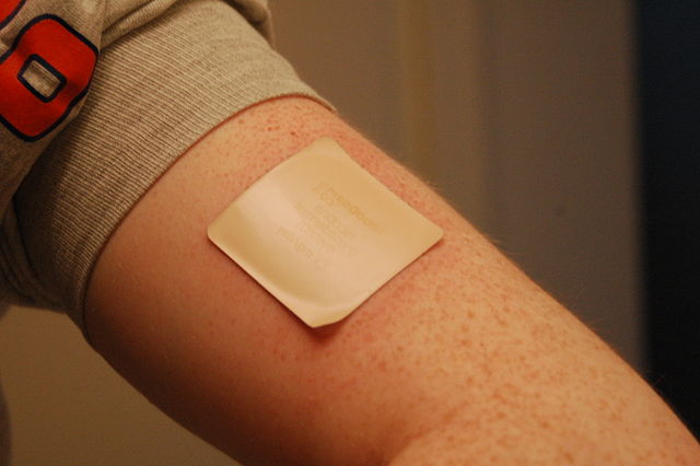
Vitamin D Synthesis
Vitamin D is a nutrient that is needed in the human body for the absorption of calcium from food. Molecules of a lipid compound named 7-dehydrocholesterol are precursors of vitamin D. These molecules are present in the stratum basale and stratum spinosum layers of the epidermis. When UV light strikes the molecules, it changes them to vitamin D3. In the kidneys, vitamin D3 is converted to calcitriol, which is the form of vitamin D that is active in the body.
What Gives Skin Its Colour?
Melanin in the epidermis is the main substance that determines the colour of human skin. It explains most of the variation in skin colour in people around the world. Two other substances also contribute to skin colour, however, especially in light-skinned people: carotene and hemoglobin.
- The pigment carotene is present in the epidermis and gives skin a yellowish tint, especially in skin with low levels of melanin.
- Hemoglobin is a red pigment found in red blood cells. It is visible through skin as a pinkish tint, mainly in skin with low levels of melanin. The pink colour is most visible when capillaries in the underlying dermis dilate, allowing greater blood flow near the surface.
Hear what Bill Nye has to say about the subject of skin colour in the video here.
Bacteria on Skin
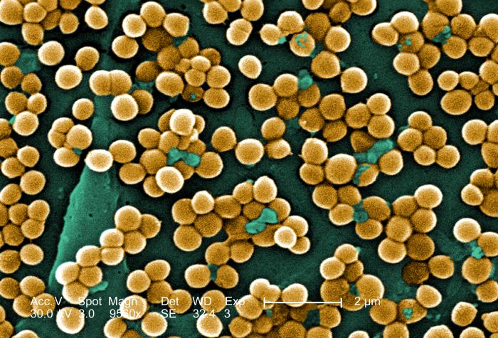
The surface of the human skin normally provides a home to countless numbers of bacteria. Just one square inch of skin normally has an average of about 50 million bacteria. These generally harmless bacteria represent roughly one thousand bacterial species (including the one in Figure 10.3.5) from 19 different bacterial phyla. Typical variations in the moistness and oiliness of the skin produce a variety of rich and diverse habitats for these microorganisms. For example, the skin in the armpits is warm and moist and often hairy, whereas the skin on the forearms is smooth and dry. These two areas of the human body are as diverse to microorganisms as rainforests and deserts are to larger organisms. The density of bacterial populations on the skin depends largely on the region of the skin and its ecological characteristics. For example, oily surfaces, such as the face, may contain over 500 million bacteria per square inch. Despite the huge number of individual microorganisms living on the skin, their total volume is only about the size of a pea.
In general, the normal microorganisms living on the skin keep one another in check, and thereby play an important role in keeping the skin healthy. If the balance of microorganisms is disturbed, however, there may be an overgrowth of certain species, and this may result in an infection. For example, when a patient is prescribed antibiotics, it may kill off normal bacteria and allow an overgrowth of single-celled yeast. Even if skin is disinfected, no amount of cleaning can remove all of the microorganisms it contains. Disinfected areas are also quickly recolonized by bacteria residing in deeper areas (such as hair follicles) and in adjacent areas of the skin.
Feature: Myth vs. Reality
Because of the negative health effects of excessive UV light exposure, it is important to know the facts about protecting the skin from UV light.
Myth |
Reality |
| "Sunblock and sunscreen are just different names for the same type of product. They both work the same way and are equally effective." | Sunscreens and sunblocks are different types of products that protect the skin from UV light in different ways. They are not equally effective. Sunblocks are opaque, so they do not let light pass through. They prevent most of the rays of UV light from penetrating to the skin surface. Sunblocks are generally stronger and more effective than sunscreens. Sunblocks also do not need to be reapplied as often as sunscreens. Sunscreens, in contrast, are transparent once they are applied the skin. Although they can prevent most UV light from penetrating the skin when first applied, the active ingredients in sunscreens tend to break down when exposed to UV light. Sunscreens, therefore, must be reapplied often to remain effective. |
| "The skin needs to be protected from UV light only on sunny days. When the sky is cloudy, UV light cannot penetrate to the ground and harm the skin." | Even on cloudy days, a significant amount of UV radiation penetrates the atmosphere to strike Earth’s surface. Therefore, using sunscreens or sunblocks to protect exposed skin is important even when there are clouds in the sky. |
| "People who have dark skin, such as African Americans, do not need to worry about skin damage from UV light." | No matter what colour skin you have, your skin can be damaged by too much exposure to UV light. Therefore, even dark-skinned people should use sunscreens or sunblocks to protect exposed skin from UV light. |
| "Sunscreens with an SPF (sun protection factor) of 15 are adequate to fully protect the skin from UV light." | Most dermatologists recommend using sunscreens with an SPF of at least 35 for adequate protection from UV light. They also recommend applying sunscreens at least 20 minutes before sun exposure and reapplying sunscreens often, especially if you are sweating or spending time in the water. |
| "Using tanning beds is safer than tanning outside in natural sunlight." | The light in tanning beds is UV light, and it can do the same damage to the skin as the natural UV light in sunlight. This is evidenced by the fact that people who regularly use tanning beds have significantly higher rates of skin cancer than people who do not. It is also the reason that the use of tanning beds is prohibited in many places in people who are under the age of 18, just as youth are prohibited from using harmful substances, such as tobacco and alcohol. |
10.3 Summary
- The epidermis is the outer of the two main layers of the skin. It is very thin, but has a complex structure.
- Cell types in the epidermis include keratinocytes that produce keratin and make up 90 per cent of epidermal cells, melanocytes that produce melanin, Langerhans cells that fight pathogens in the skin, and Merkel cells that respond to light touch.
- The epidermis in most parts of the body consists of four distinct layers. A fifth layer occurs only in the epidermis of the palms of the hands and soles of the feet.
- The innermost layer of the epidermis is the stratum basale, which contains stem cells that divide to form new keratinocytes. The next layer is the stratum spinosum, which is the thickest layer and contains Langerhans cells and spiny keratinocytes. This is followed by the stratum granulosum, in which keratinocytes are filling with keratin and starting to die. The stratum lucidum is next, but only on the palms and soles. It consists of translucent dead keratinocytes. The outermost layer is the stratum corneum, which consists of flat, dead, tightly packed keratinocytes that form a tough, waterproof barrier for the rest of the epidermis.
- Functions of the epidermis include protecting underlying tissues from physical damage and pathogens. Melanin in the epidermis absorbs and protects underlying tissues from UV light. The epidermis also prevents loss of water from the body and synthesizes vitamin D.
- Melanin is the main pigment that determines the colour of human skin. The pigments carotene and hemoglobin, however, also contribute to skin colour, especially in skin with low levels of melanin.
- The surface of healthy skin normally is covered by vast numbers of bacteria representing about one thousand species from 19 phyla. Different areas of the body provide diverse habitats for skin microorganisms. Usually, microorganisms on the skin keep each other in check unless their balance is disturbed.
10.3 Review Questions
- What is the epidermis?
- Identify the types of cells in the epidermis.
- Describe the layers of the epidermis.
-
- State one function of each of the four epidermal layers found all over the body.
- Explain three ways the epidermis protects the body.
- What makes the skin waterproof?
- Why is the selective permeability of the epidermis both a benefit and a risk?
- How is vitamin D synthesized in the epidermis?
- Identify three pigments that impart colour to skin.
- Describe bacteria that normally reside on the skin, and explain why they do not usually cause infections.
- Explain why the keratinocytes at the surface of the epidermis are dead, while keratinocytes located deeper in the epidermis are still alive.
- Which layer of the epidermis contains keratinocytes that have begun to die?
-
- Explain why our skin is not permanently damaged if we rub off some of the surface layer by using a rough washcloth.
10.3 Explore More
https://www.youtube.com/watch?v=27lMmdmy-b8
Jonathan Eisen: Meet your microbes, TED, 2015.
https://www.youtube.com/watch?v=9AcQXnOscQ8
Why Do We Blush?, SciShow, 2014.
https://www.youtube.com/watch?v=_r4c2NT4naQ
The science of skin colour - Angela Koine Flynn, TED-Ed, 2016.
Attributions
Figure 10.3.1
Sunburn by QuinnHK at English Wikipedia on Wikimedia Commons is released into the public domain (https://en.wikipedia.org/wiki/Public_domain).
Figure 10.3.2
Blausen_0353_Epidermis by BruceBlaus on Wikimedia Commons is used under a CC BY 3.0 (https://creativecommons.org/licenses/by/3.0) license.
Figure 10.3.3
Isaac's scraped knee close-up by Alpha on Flickr is used under a CC BY-NC-SA 2.0 (https://creativecommons.org/licenses/by-nc-sa/2.0/) license.
Figure 10.3.4
Nicoderm by RegBarc on Wikimedia Commons is used under a CC BY-SA 3.0 (http://creativecommons.org/licenses/by-sa/3.0/) license. (No machine-readable author provided for original.)
Figure 10.3.5
Staphylococcus aureus bacteria, MRSA by Microbe World on Flickr is used under a CC BY-NC-SA 2.0 (https://creativecommons.org/licenses/by-nc-sa/2.0/) license.
References
Blausen.com staff. (2014). Medical gallery of Blausen Medical 2014. WikiJournal of Medicine 1 (2). DOI:10.15347/wjm/2014.010. ISSN 2002-4436.
Jeff Bone 'n' Pookie. (2020, July 19). Bill Nye the science guy explains we have different skin color. Youtube. https://www.youtube.com/watch?v=zOkj5jgC4sM&feature=youtu.be
SciShow. (2014, July 15). Why do we blush? YouTube. https://www.youtube.com/watch?v=9AcQXnOscQ8
TED. (2015, July 17). Jonathan Eisen: Meet your microbes. YouTube. https://www.youtube.com/watch?v=27lMmdmy-b8
TED-Ed. (2016, February 16). The science of skin color - Angela Koine Flynn. YouTube. https://youtu.be/_r4c2NT4naQ
Created by CK-12 Foundation/Adapted by Christine Miller

Nail Art
Painting nails with coloured polish for aesthetic reasons is nothing new. In fact, there is evidence of this practice dating back to at least 3000 BCE. Today, painting and otherwise decorating the nails is big business, with annual revenues in the billions of dollars in North America alone! With all the attention (and money) given to nails as decorative objects, it’s easy to forget that they also have important biological functions.
What Are Nails?
Nails are accessory organs of the skin. They are made of sheets of dead keratinocytes and are found on the far (or distal) ends of the fingers and toes. The keratin in nails makes them hard, but flexible. Nails serve a number of purposes, including protecting the digits, enhancing sensations, and acting as tools.
Nail Anatomy
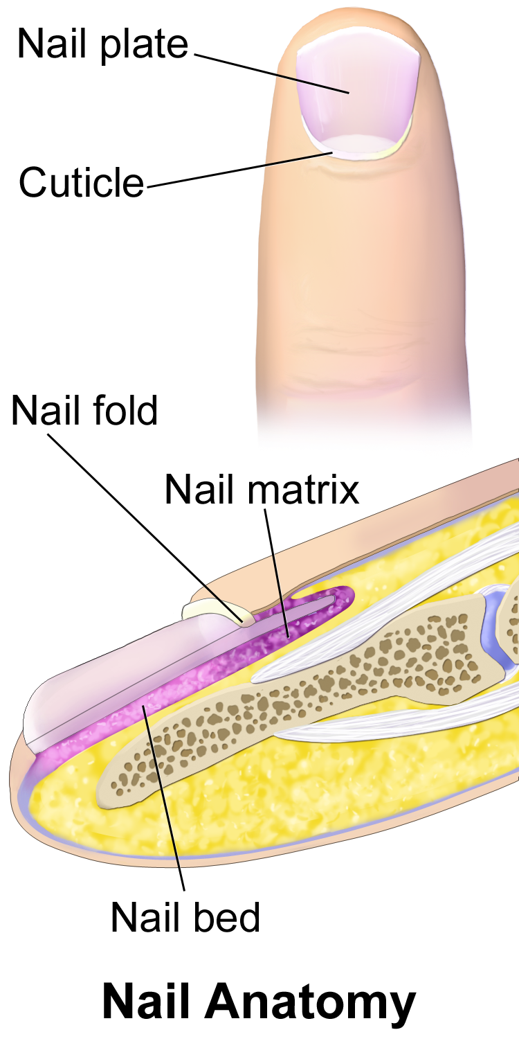
A nail has three main parts: the root, plate, and free margin. Other structures around or under the nail include the nail bed, cuticle, and nail fold. These structures are shown in Figure 10.6.2.
- The nail root is the portion of the nail found under the surface of the skin at the near (or proximal) end of the nail. It is where the nail begins.
- The nail plate (or body) is the portion of the nail that is external to the skin. It is the visible part of the nail.
- The free margin is the portion of the nail that protrudes beyond the distal end of the finger or toe. This is the part that is cut or filed to keep the nail trimmed.
- The nail bed is the area of skin under the nail plate. It is pink in colour, due to the presence of capillaries in the dermis.
- The cuticle is a layer of dead epithelial cells that overlaps and covers the edge of the nail plate. It helps to seal the edges of the nail to prevent infection of the underlying tissues.
- The nail fold is a groove in the skin in which the side edges of the nail plate are embedded.
Nail Growth
Nails grow from a deep layer of living epidermal tissue, known as the nail matrix, at the proximal end of the nail (see the bottom of the diagram in Figure 10.6.2). The nail matrix surrounds the nail root. It contains stem cells that divide to form keratinocytes, which are cells that produce keratin and make up the nail.
Formation of the Nail Root and Nail Plate
The keratinocytes produced by the nail matrix accumulate to form tough, hard, translucent sheets of dead cells filled with keratin. The sheets make up the nail root, which slowly grows out of the skin and becomes the nail plate when it reaches the skin surface. As the nail grows longer, the cells of the nail root and nail plate are pushed toward the distal end of the finger or toe by new cells being formed in the nail matrix. The upper epidermal cells of the nail bed also move along with the nail plate as it grows toward the tip of the digit.
The proximal end of the nail plate near the root has a whitish crescent shape called the lunula. This is where a small amount of the nail matrix is visible through the nail plate. The lunula is most pronounced in the nails of the thumbs, and may not be visible in the nails of the little fingers.
Rate of Nail Growth
Nails grow at an average rate of 3 mm a month. Fingernails, however, grow up to four times as fast as toenails. If a fingernail is lost, it takes between three and six months to regrow completely, whereas a toenail takes between 12 and 18 months to regrow. The actual rate of growth of an individual’s nails depends on many factors, including age, sex, season, diet, exercise level, and genes. If protected from breaking, nails can sometimes grow to be very long. The Chinese doctor in the photo below (Figure 10.6.3) has very long nails on two fingers of his left hand. This picture was taken in 1920 in China, where having long nails was a sign of aristocracy since it implied that one was wealthy enough to not have to do physical labour.

Functions of Nails
Both fingernails and toenails protect the soft tissues of the fingers and toes from injury. Fingernails also serve to enhance sensation and precise movements of the fingertips through the counter-pressure exerted on the pulp of the fingers by the nails. In addition, fingernails can function as several different types of tools. For example, they enable a fine precision grip like tweezers, and can also be used for cutting and scraping.
Nails and Health
Healthcare providers, particularly EMTs, often examine the fingernail beds as a quick and easy indicator of oxygen saturation of the blood, or the amount of blood reaching the extremities. If the nail beds are bluish or purple, it is generally a sign of low oxygen saturation. To see if blood flow to the extremities is adequate, a blanch test may be done. In this test, a fingernail is briefly depressed to turn the nail bed white by forcing the blood out of its capillaries. When the pressure is released, the pink colour of the nail bed should return within a second or two if there is normal blood flow. If the return to a pink colour is delayed, then it can be an indicator of low blood volume, due to dehydration or shock.
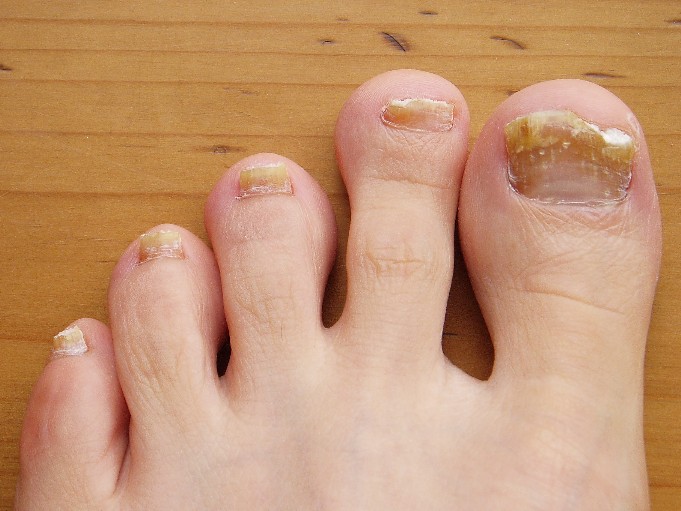
How the visible portion of the nails appears can be used as an indicator of recent health status. In fact, nails have been used as diagnostic tools for hundreds — if not thousands — of years. Nail abnormalities, such as deep grooves, brittleness, discolouration, or unusually thin or thick nails, may indicate various illnesses, nutrient deficiencies, drug reactions, or other health problems.
Nails — especially toenails — are common sites of fungal infections (shown in Figure 10.6.4), causing nails to become thickened and yellowish in colour. Toenails are more often infected than fingernails because they are often confined in shoes, which creates a dark, warm, moist environment where fungi can thrive. Toes also tend to have less blood flow than fingers, making it harder for the immune system to detect and stop infections in toenails.
Although nails are harder and tougher than skin, they are also more permeable. Harmful substances may be absorbed through the nails and cause health problems. Some of the substances that can pass through the nails include the herbicide Paraquat, fungicidal agents such as miconazole (e.g., Monistat), and sodium hypochlorite, which is an ingredient in common household bleach. Care should be taken to protect the nails from such substances when handling or immersing the hands in them by wearing latex or rubber gloves.
Feature: Reliable Sources
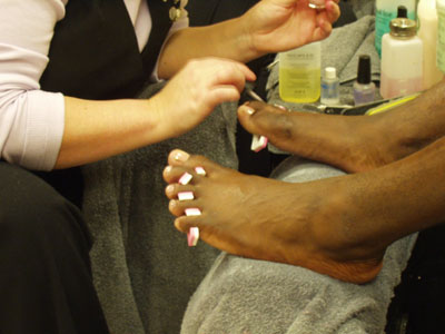
Do you get regular manicures or pedicures from a nail technician? If so, there is a chance that you are putting your health at risk. Nail tools that are not properly disinfected between clients may transmit infections from one person to another. Cutting the cuticles with scissors may create breaks in the skin that let infective agents enter the body. Products such as acrylics, adhesives, and UV gels that are applied to the nails may be harmful, especially if they penetrate the nails and enter the skin.
Use the Internet to find several reliable sources that address the health risks of professional manicures or pedicures. Try to find answers to the following questions:
- What training and certification are required for professional nail technicians?
- What licenses and inspections are required for nail salons?
- What hygienic practices should be followed in nail salons to reduce the risk of infections being transmitted to clients?
- Which professional nail products are potentially harmful to the human body and which are safer?
- How likely is it to have an adverse health consequence when you get a professional manicure or pedicure?
- What steps can you take to ensure that a professional manicure or pedicure is safe?
10.6 Summary
- Nails are accessory organs of the skin, consisting of sheets of dead, keratin-filled keratinocytes. The keratin in nails makes them hard, but flexible.
- A nail has three main parts: the nail root (which is under the epidermis), the nail plate (which is the visible part of the nail), and the free margin (which is the distal edge of the nail). Other structures under or around a nail include the nail bed, cuticle, and nail fold.
- A nail grows from a deep layer of living epidermal tissues — called the nail matrix — at the proximal end of the nail. Stem cells in the nail matrix keep dividing to allow nail growth, forming first the nail root and then the nail plate as the nail continues to grow longer and emerges from the epidermis.
- Fingernails grow faster than toenails. Actual rates of growth depend on many factors, such as age, sex, and season.
- Functions of nails include protecting the digits, enhancing sensations and precise movements of the fingertips, and acting as tools.
- The colour of the nail bed can be used to quickly assess oxygen and blood flow in a patient. How the nail plate grows out can reflect recent health problems, such as illness or nutrient deficiency.
- Nails — and especially toenails — are prone to fungus infections. Nails are more permeable than skin and can absorb several harmful substances, such as herbicides.
10.6 Review Questions
- What are nails?
-
- Explain why most of the nail plate looks pink.
- Describe a lunula.
- Explain how a nail grows.
- Identify three functions of nails.
- Give several examples of how nails are related to health.
- What is the cuticle of the nail composed of? What is the function of the cuticle? Why is it a bad idea to cut the cuticle during a manicure?
- Is the nail plate composed of living or dead cells?
10.6 Explore More
https://www.youtube.com/watch?v=G35kPhbUZdg
Longest Fingernails - Guinness World Records 60th Anniversary,
Guinness World Records, 2014.
https://www.youtube.com/watch?v=aTSVHwzkYI4&feature=emb_logo
5 Things Your Nails Can Say About Your Health, SciShow, 2015.
https://www.youtube.com/watch?v=7w2gCBL1MCg
Claws vs. Nails - Matthew Borths, TED-Ed, 2019.
Attributions
Figure 10.6.1
Nails by allison-christine-vPrqHSLdF28 [photo] by allison christine on Unsplash is used under the Unsplash License (https://unsplash.com/license).
Figure 10.6.2
Blausen_0406_FingerNailAnatomy by BruceBlaus on Wikimedia Commons is used under a CC BY 3.0 (https://creativecommons.org/licenses/by/3.0) license.
Figure 10.6.3
Chinese_doctor_with_long_finger_nails_(an_aristocrat),_ca.1920_(CHS-249) by Pierce, C.C. (Charles C.), 1861-1946 from the USC Digital Library on Wikimedia Commons is in the public domain (https://en.wikipedia.org/wiki/public_domain).
Figure 10.6.4
Toenail fungus Nagelpilz-3 by Pepsyrock on Wikimedia Commons is released into the public domain (https://en.wikipedia.org/wiki/public_domain).
Figure 10.6.5
OLYMPUS DIGITAL CAMERA by Stoive at the English language Wikipedia, on Wikimedia Commons is used under a CC BY-SA 3.0 (http://creativecommons.org/licenses/by-sa/3.0/) license.
References
Blausen.com staff. (2014). Medical gallery of Blausen Medical 2014. WikiJournal of Medicine 1 (2). DOI:10.15347/wjm/2014.010. ISSN 2002-4436.
Guiness World Records. (2014, December 8). Longest fingernails - Guinness World Records 60th Anniversary. YouTube. https://www.youtube.com/watch?v=G35kPhbUZdg
SciShow. (2015, September 14). 5 things your nails can say about your health. YouTube. https://www.youtube.com/watch?v=aTSVHwzkYI4
TED-Ed. (2019, October 29). Claws vs. nails - Matthew Borths. YouTube. https://www.youtube.com/watch?v=7w2gCBL1MCg
Created by CK-12 Foundation/Adapted by Christine Miller
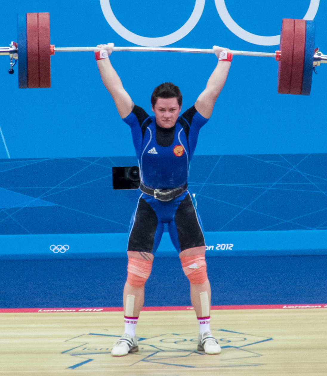
Marvelous Muscles
Does the word muscle make you think of the well-developed muscles of a weightlifter, like the woman in Figure 12.2.1? Her name is Natalia Zabolotnaya, and she’s a Russian Olympian. The muscles that are used to lift weights are easy to feel and see, but they aren’t the only muscles in the human body. Many muscles are deep within the body, where they form the walls of internal organs and other structures. You can flex your biceps at will, but you can’t control internal muscles like these. It’s a good thing that these internal muscles work without any conscious effort on your part, because movement of these muscles is essential for survival. Muscles are the organs of the muscular system.
What Is the Muscular System?
The muscular system consists of all the muscles of the body. The largest percentage of muscles in the muscular system consists of skeletal muscles, which are attached to bones and enable voluntary body movements (shown in Figure 12.2.2). There are almost 650 skeletal muscles in the human body, many of them shown in Figure 12.2.2. Besides skeletal muscles, the muscular system also includes cardiac muscle, which makes up the walls of the heart, and smooth muscles, which control movement in other internal organs and structures.
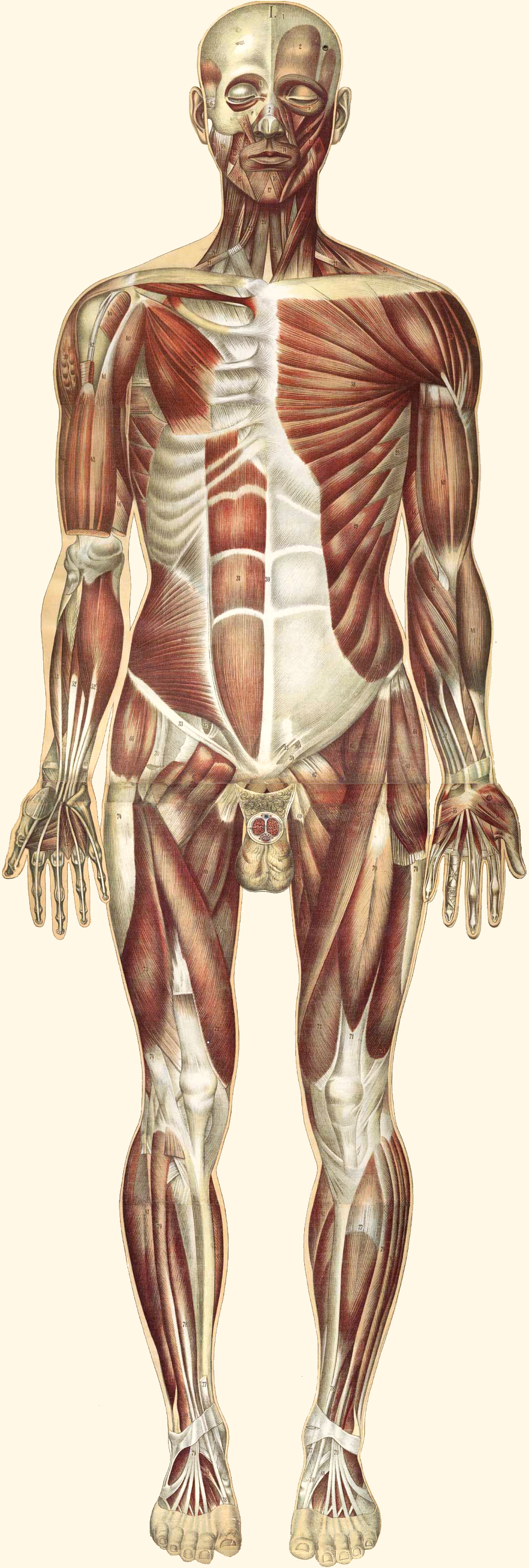
Muscle Structure and Function
Muscles are organs composed mainly of muscle cells, which are also called muscle fibres (mainly in skeletal and cardiac muscle) or myocytes (mainly in smooth muscle). Muscle cells are long, thin cells that are specialized for the function of contracting. They contain protein filaments that slide over one another using energy in ATP. The sliding filaments increase the tension in — or shorten the length of — muscle cells, causing a contraction. Muscle contractions are responsible for virtually all the movements of the body, both inside and out.
Skeletal muscles are attached to bones of the skeleton. When these muscles contract, they move the body. They allow us to use our limbs in a variety of ways, from walking to turning cartwheels. Skeletal muscles also maintain posture and help us to keep balance.
Smooth muscles in the walls of blood vessels contract to cause vasoconstriction, which may help conserve body heat. Relaxation of these muscles causes vasodilation, which may help the body lose heat. In the organs of the digestive system, smooth muscles squeeze food through the gastrointestinal tract by contracting in sequence to form a wave of muscle contractions called peristalsis. Think of squirting toothpaste through a tube by applying pressure in sequence from the bottom of the tube to the top, and you have a good idea of how food is moved by muscles through the digestive system. Peristalsis of smooth muscles also moves urine through the urinary tract.
Cardiac muscle tissue is found only in the walls of the heart. When cardiac muscle contracts, it makes the heart beat. The pumping action of the beating heart keeps blood flowing through the cardiovascular system.
Muscle Hypertrophy and Atrophy
Muscles can grow larger, or hypertrophy. This generally occurs through increased use, although hormonal or other influences can also play a role. The increase in testosterone that occurs in males during puberty, for example, causes a significant increase in muscle size. Physical exercise that involves weight bearing or resistance training can increase the size of skeletal muscles in virtually everyone. Exercises (such as running) that increase the heart rate may also increase the size and strength of cardiac muscle. The size of muscle, in turn, is the main determinant of muscle strength, which may be measured by the amount of force a muscle can exert.
Muscles can also grow smaller, or atrophy, which can occur through lack of physical activity or from starvation. People who are immobilized for any length of time — for example, because of a broken bone or surgery — lose muscle mass relatively quickly. People in concentration or famine camps may be so malnourished that they lose much of their muscle mass, becoming almost literally just “skin and bones.” Astronauts on the International Space Station may also lose significant muscle mass because of weightlessness in space (see Figure 12.2.3).
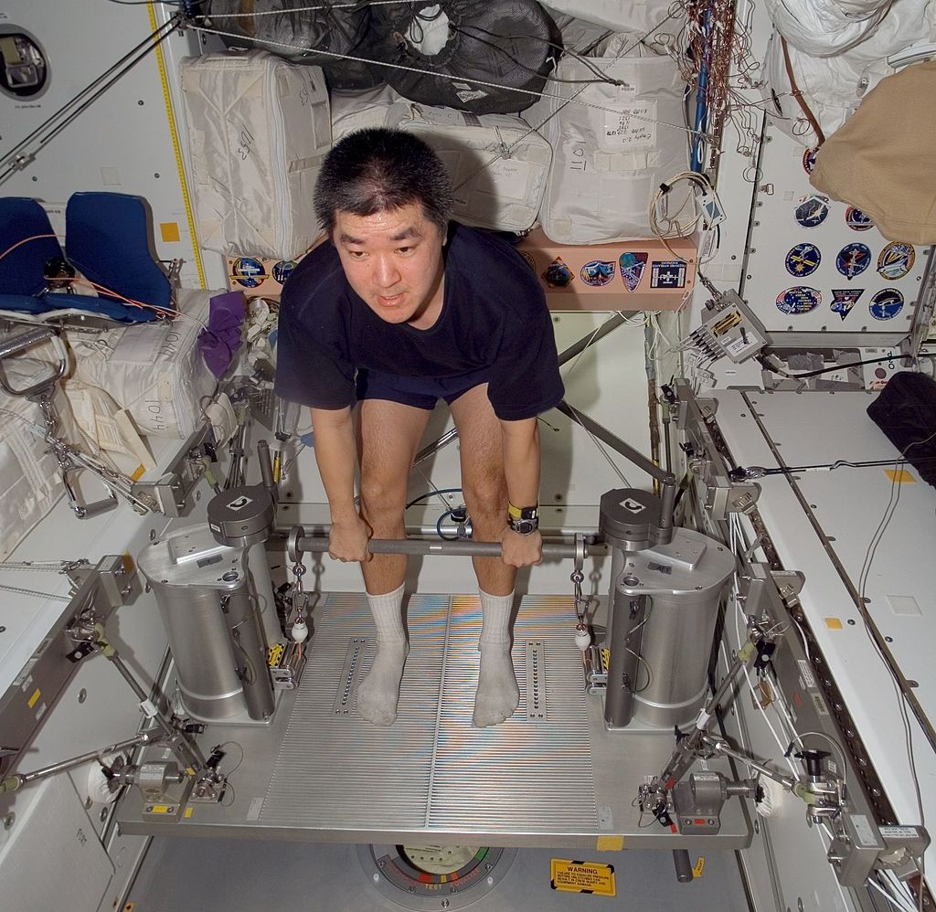
Many diseases, including cancer and AIDS, are often associated with muscle atrophy. Atrophy of muscles also happens with age. As people grow older, there is a gradual decrease in the ability to maintain skeletal muscle mass, known as sarcopenia. The exact cause of sarcopenia is not known, but one possible cause is a decrease in sensitivity to growth factors that are needed to maintain muscle mass. Because muscle size determines strength, muscle atrophy causes a corresponding decline in muscle strength.
In both hypertrophy and atrophy, the number of muscle fibres does not change. What changes is the size of the muscle fibres. When muscles hypertrophy, the individual fibres become wider. When muscles atrophy, the fibres become narrower.
Interactions with Other Body Systems
Muscles cannot contract on their own. Skeletal muscles need stimulation from motor neurons in order to contract. The point where a motor neuron attaches to a muscle is called a neuromuscular junction. Let’s say you decide to raise your hand in class. Your brain sends electrical messages through motor neurons to your arm and shoulder. The motor neurons, in turn, stimulate muscle fibres in your arm and shoulder to contract, causing your arm to rise.
Involuntary contractions of smooth and cardiac muscles are also controlled by electrical impulses, but in the case of these muscles, the impulses come from the autonomic nervous system (smooth muscle) or specialized cells in the heart (cardiac muscle). Hormones and some other factors also influence involuntary contractions of cardiac and smooth muscles. For example, the fight-or-flight hormone adrenaline increases the rate at which cardiac muscle contracts, thereby speeding up the heartbeat.
Muscles cannot move the body on their own. They need the skeletal system to act upon. The two systems together are often referred to as the musculoskeletal system. Skeletal muscles are attached to the skeleton by tough connective tissues called tendons. Many skeletal muscles are attached to the ends of bones that meet at a joint. The muscles span the joint and connect the bones. When the muscles contract, they pull on the bones, causing them to move. The skeletal system provides a system of levers that allow body movement. The muscular system provides the force that moves the levers.
12.2 Summary
- The muscular system consists of all the muscles of the body. There are three types of muscle: skeletal muscle (which is attached to bones and enables voluntary body movements), cardiac muscle (which makes up the walls of the heart and makes it beat), and smooth muscle (which is found in the walls of internal organs and other internal structures and controls their movements).
- Muscles are organs composed mainly of muscle cells, which may also be called muscle fibres or myocytes. Muscle cells are specialized for the function of contracting, which occurs when protein filaments inside the cells slide over one another using energy in ATP.
- Muscles can grow larger, or hypertrophy. This generally occurs through increased use (physical exercise), although hormonal or other influences can also play a role. Muscles can also grow smaller, or atrophy. This may occur through lack of use, starvation, certain diseases, or aging. In both hypertrophy and atrophy, the size — but not the number — of muscle fibres changes. The size of muscles is the main determinant of muscle strength.
- Skeletal muscles need the stimulus of motor neurons to contract, and to move the body, they need the skeletal system to act upon. Involuntary contractions of cardiac and smooth muscles are controlled by special cells in the heart, nerves of the autonomic nervous system, hormones, or other factors.
12.2 Review Questions
- What is the muscular system?
- Describe muscle cells and their function.
- Identify three types of muscle tissue and where each type is found.
- Define muscle hypertrophy and muscle atrophy.
- What are some possible causes of muscle hypertrophy?
- Give three reasons that muscle atrophy may occur.
- How do muscles change when they increase or decrease in size?
- How do changes in muscle size affect strength?
- Explain why astronauts can easily lose muscle mass in space.
- Describe how the terms muscle cells, muscle fibres, and myocytes relate to each other.
-
- Name two systems in the body that work together with the muscular system to carry out movements.
- Describe one way in which the muscular system is involved in regulating body temperature.
12.2 Explore More
https://www.youtube.com/watch?v=VVL-8zr2hk4
How your muscular system works - Emma Bryce, TED-Ed, 2017.
https://www.youtube.com/watch?v=Ujr0UAbyPS4&feature=emb_logo
3D Medical Animation - Peristalsis in Large Intestine/Bowel || ABP ©, AnimatedBiomedical, 2013.
https://www.youtube.com/watch?v=LkXwfTsqQgQ&feature=emb_logo
Muscle matters: Dr Brendan Egan at TEDxUCD, TEDx Talks, 2014.
Attributions
Figure 12.2.1
Natalia_Zabolotnaya_2012b by Simon Q on Wikimedia Commons is used under a CC BY 2.0 (https://creativecommons.org/licenses/by/2.0/deed.en) license.
Figure 12.2.2
Bougle_whole2_retouched by Bouglé, Julien from the National LIbrary of Medicine (NLM) on Wikimedia Commons is in the public domain (https://en.wikipedia.org/wiki/Public_domain).
Figure 12.2.3
Daniel_Tani_iss016e027910 by NASA/ International Space Station Imagery on Wikimedia Commons is in the public domain (https://en.wikipedia.org/wiki/Public_domain).
References
AnimatedBiomedical. (2013, January 30). 3D Medical animation - Peristalsis in large intestine/bowel || ABP ©. YouTube. https://www.youtube.com/watch?v=Ujr0UAbyPS4&feature=youtu.be
Bouglé, J. (1899). Le corps humain en grandeur naturelle : planches coloriées et superposées, avec texte explicatif. J. B. Baillière et fils. In Historical Anatomies on the Web. http://www.nlm.nih.gov/exhibition/historicalanatomies/bougle_home.html
TED-Ed. (2017, October 26). How your muscular system works - Emma Bryce. YouTube. https://www.youtube.com/watch?v=VVL-8zr2hk4&feature=youtu.be
TEDx Talks. (2014, June 27). Muscle matters: Dr Brendan Egan at TEDxUCD. YouTube. https://www.youtube.com/watch?v=LkXwfTsqQgQ&feature=youtu.be
Wikipedia contributors. (2020, June 15). Natalya Zabolotnaya. In Wikipedia. https://en.wikipedia.org/w/index.php?title=Natalya_Zabolotnaya&oldid=962630409
Created by CK-12 Foundation/Adapted by Christine Miller

Work Those Eye Muscles!
Imagine the man in Figure 12.3.1 turns his eyes in your direction. This is a very small movement, considering the conspicuously large and strong external eye muscles that control eyeball movements. These muscles have been called the strongest muscles in the human body relative to the work they do. However, the external eye muscles actually do a surprising amount of work. Eye movements occur almost constantly during waking hours, especially when we are scanning faces or reading. Eye muscles are also exercised nightly during the phase of sleep called rapid eye movement sleep. External eye muscles can move the eyes because they are made mainly of muscle tissue.
What is Muscle Tissue?
Muscle tissue is a soft tissue that makes up most of the tissues in the muscles of the human muscular system. Other tissues in muscles are connective tissues, such as tendons that attach skeletal muscles to bones and sheaths of connective tissues that cover or line muscle tissues. Only muscle tissue per se, has cells with the ability to contract.
There are three major types of muscle tissues in the human body: skeletal, smooth, and cardiac muscle tissues. Figure 12.3.2 shows how the three types of muscle tissues appear under magnification. When you read about each type below, you will learn why the three types appear as they do.
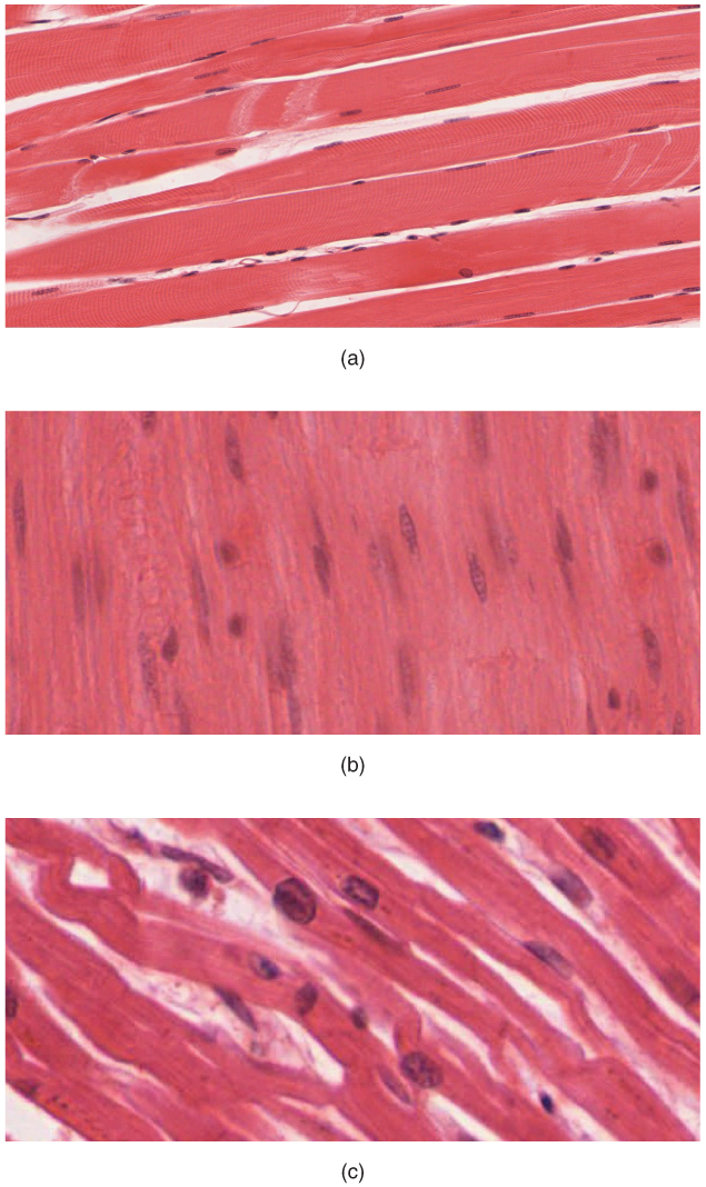
Skeletal Muscle Tissue
Skeletal muscle is muscle tissue that is attached to bones by tendons, which are bundles of collagen fibres. Whether you are moving your eyes or running a marathon, you are using skeletal muscles. Contractions of skeletal muscles are voluntary, or under conscious control of the central nervous system via the somatic nervous system. Skeletal muscle tissue is the most common type of muscle tissue in the human body. By weight, an average adult male is about 42% skeletal muscles, and the average adult female is about 36% skeletal muscles. Some of the major skeletal muscles in the human body are labeled in Figure 12.3.3 below.
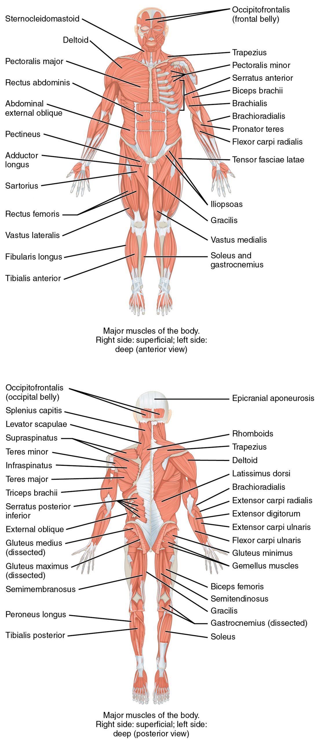
Skeletal Muscle Pairs
To move bones in opposite directions, skeletal muscles often consist of muscle pairs that work in opposition to one another, also called antagonistic muscle pairs. For example, when the biceps muscle (on the front of the upper arm) contracts, it can cause the elbow joint to flex or bend the arm, as shown in Figure 12.3.4. When the triceps muscle (on the back of the upper arm) contracts, it can cause the elbow to extend or straighten the arm. The biceps and triceps muscles, also shown in Figure 12.3.4, are an example of a muscle pair where the muscles work in opposition to each other.
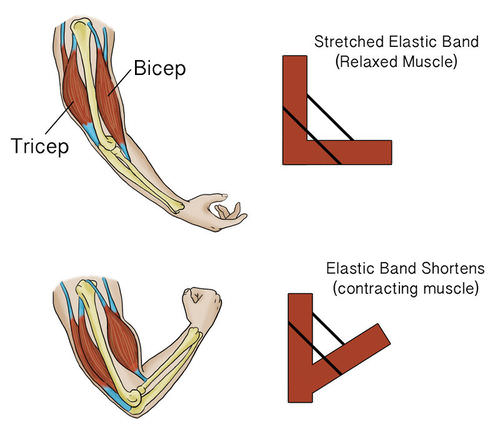
Skeletal Muscle Structure
Each skeletal muscle consists of hundreds — or even thousands — of skeletal muscle fibres, which are long, string-like cells. As shown in Figure 12.3.5 below, skeletal muscle fibres are individually wrapped in connective tissue called endomysium. The skeletal muscle fibres are bundled together in units called muscle fascicles, which are surrounded by sheaths of connective tissue called perimysium. Each fascicle contains between ten and 100 (or even more!) skeletal muscle fibres. Fascicles, in turn, are bundled together to form individual skeletal muscles, which are wrapped in connective tissue called epimysium. The connective tissues in skeletal muscles have a variety of functions. They support and protect muscle fibres, allowing them to withstand the forces of contraction by distributing the forces applied to the muscle. They also provide pathways for nerves and blood vessels to reach the muscles. In addition, the epimysium anchors the muscles to tendons.
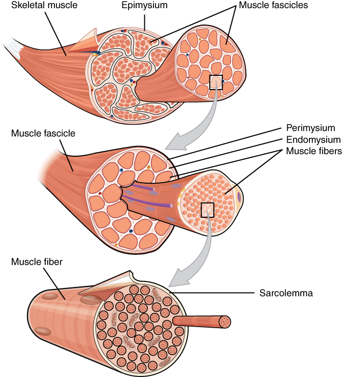
The same bundles-within-bundles structure is replicated within each muscle fibre. As shown in Figure 12.3.6, a muscle fibre consists of a bundle of myofibrils, which are themselves bundles of protein filaments. These protein filaments consist of thin filaments of the protein actin, which are anchored to structures called Z discs, and thick filaments of the protein myosin. The filaments are arranged together within a myofibril in repeating units called sarcomeres, which run from one Z disc to the next. The sarcomere is the basic functional unit of skeletal and cardiac muscles. It contracts as actin and myosin filaments slide over one another. Skeletal muscle tissue is said to be striated, because it appears striped. It has this appearance because of the regular, alternating A (dark) and I (light) bands of filaments arranged in sarcomeres inside the muscle fibres. Other components of a skeletal muscle fibre include multiple nuclei and mitochondria.
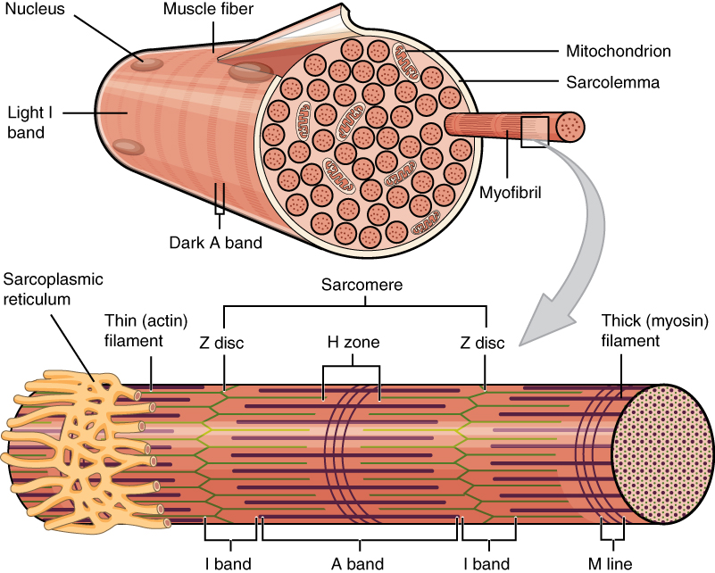
Slow- and Fast-Twitch Skeletal Muscle Fibres
Skeletal muscle fibres can be divided into two types, called slow-twitch (or type I) muscle fibres and fast-twitch (or type II) muscle fibres.
- Slow-twitch muscle fibres are dense with capillaries and rich in mitochondria and myoglobin, which is a protein that stores oxygen until needed for muscle activity. Relative to fast-twitch fibres, slow-twitch fibres can carry more oxygen and sustain aerobic (oxygen-using) activity. Slow-twitch fibres can contract for long periods of time, but not with very much force. They are relied upon primarily in endurance events, such as distance running or cycling.
- Fast-twitch muscle fibres contain fewer capillaries and mitochondria and less myoglobin. This type of muscle fibre can contract rapidly and powerfully, but it fatigues very quickly. Fast-twitch fibres can sustain only short, anaerobic (non-oxygen-using) bursts of activity. Relative to slow-twitch fibres, fast-twitch fibres contribute more to muscle strength and have a greater potential for increasing in mass. They are relied upon primarily in short, strenuous events, such as sprinting or weightlifting.
Proportions of fibre types vary considerably from muscle to muscle and from person to person. Individuals may be genetically predisposed to have a larger percentage of one type of muscle fibre than the other. Generally, an individual who has more slow-twitch fibres is better suited for activities requiring endurance, whereas an individual who has more fast-twitch fibres is better suited for activities requiring short bursts of power.
Smooth Muscle
Smooth muscle is muscle tissue in the walls of internal organs and other internal structures such as blood vessels. When smooth muscles contract, they help the organs and vessels carry out their functions. When smooth muscles in the stomach wall contract, for example, they squeeze the food inside the stomach, helping to mix and churn the food and break it into smaller pieces. This is an important part of digestion. Contractions of smooth muscles are involuntary, so they are not under conscious control. Instead, they are controlled by the autonomic nervous system, hormones, neurotransmitters, and other physiological factors.
Structure of Smooth Muscle
The cells that make up smooth muscle are generally called myocytes. Unlike the muscle fibres of striated muscle tissue, the myocytes of smooth muscle tissue do not have their filaments arranged in sarcomeres. Therefore, smooth tissue is not striated. However, the myocytes of smooth muscle do contain myofibrils, which in turn contain bundles of myosin and actin filaments. The filaments cause contractions when they slide over each other, as shown in Figure 12.3.7.

Functions of Smooth Muscle
Unlike striated muscle, smooth muscle can sustain very long-term contractions. Smooth muscle can also stretch and still maintain its contractile function, which striated muscle cannot. The elasticity of smooth muscle is enhanced by an extracellular matrix secreted by myocytes. The matrix consists of elastin, collagen, and other stretchy fibres. The ability to stretch and still contract is an important attribute of smooth muscle in organs such as the stomach and uterus (see Figures 12.3.8 and 12.3.9), both of which must stretch considerably as they perform their normal functions.
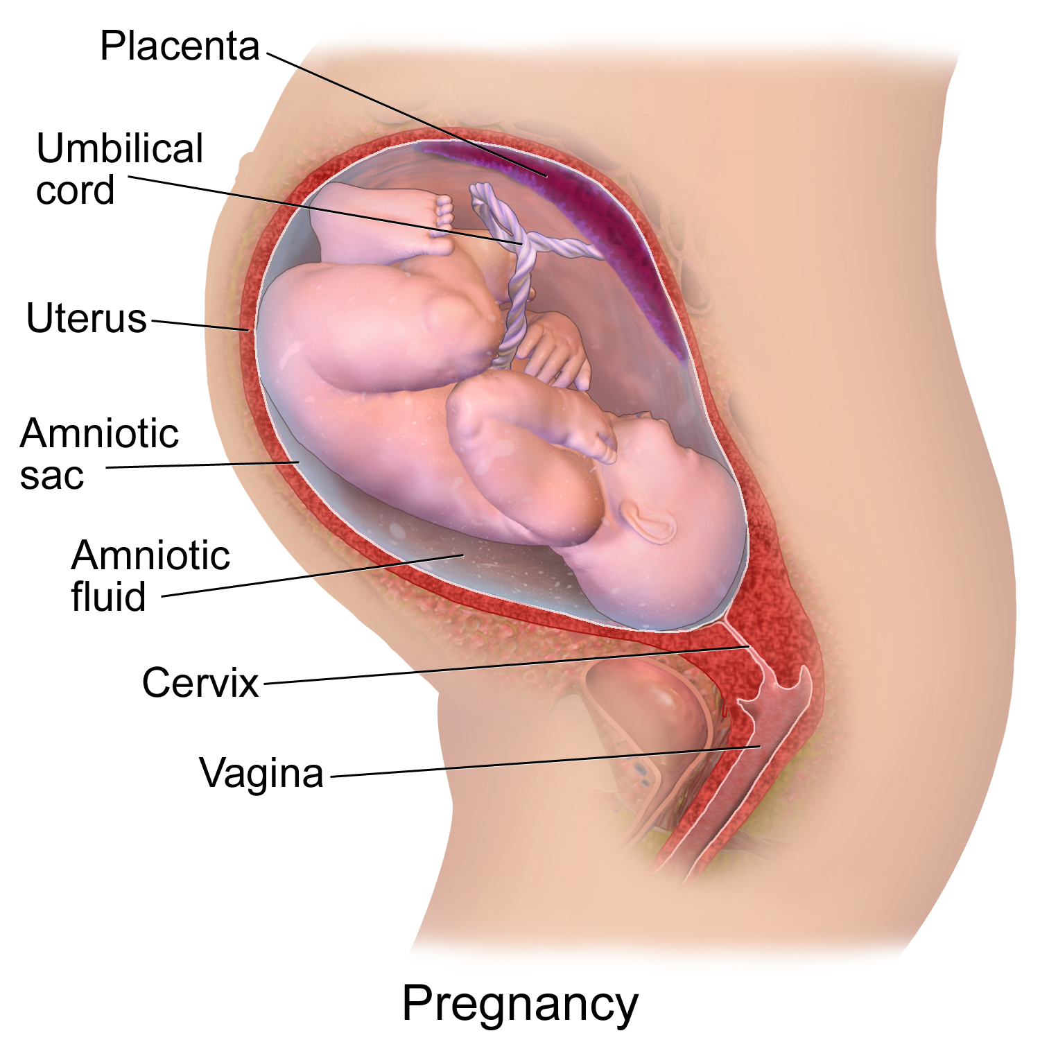 |
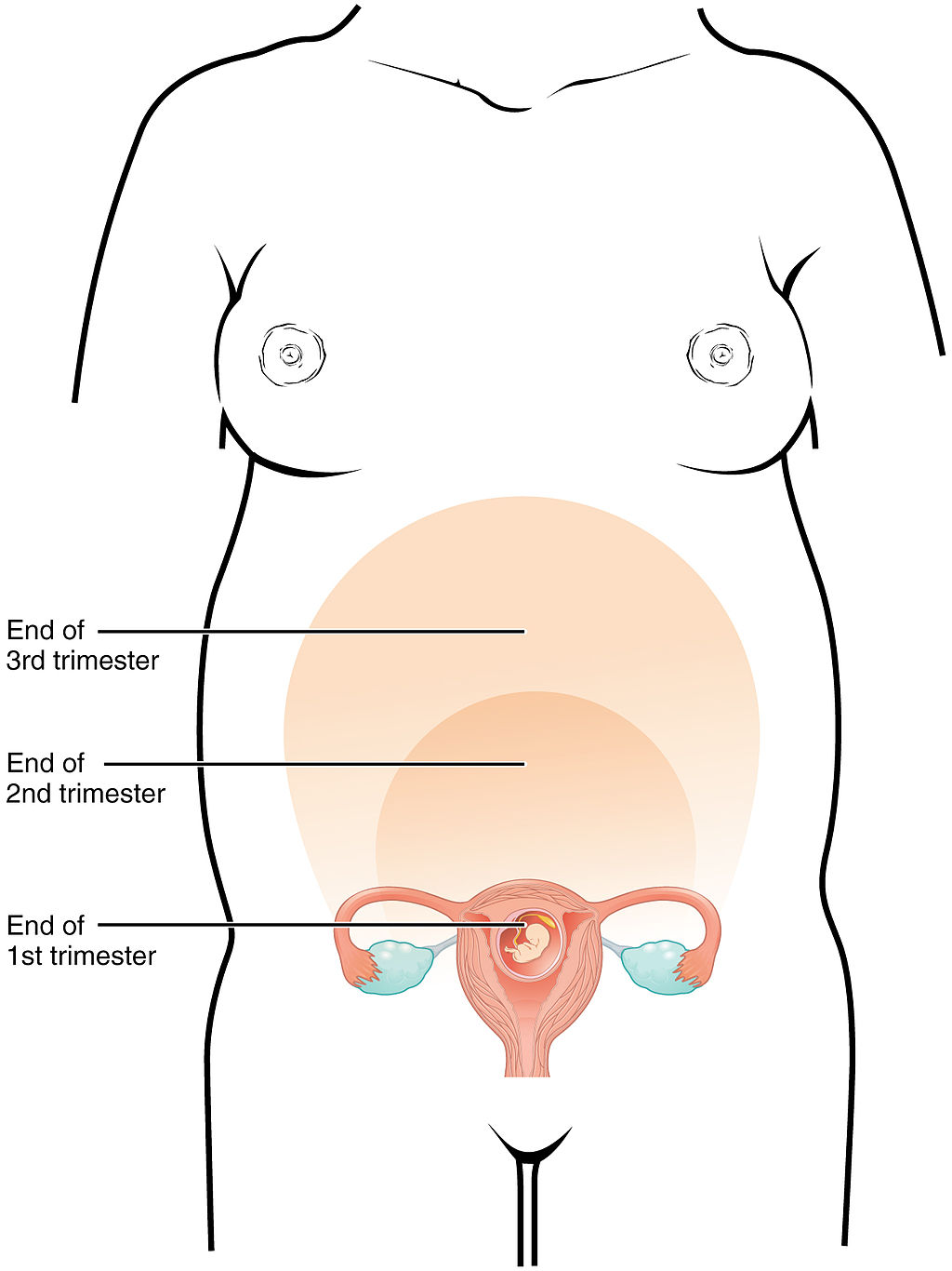 |
The following list indicates where many smooth muscles are found, along with some of their specific functions.
- Walls of organs of the gastrointestinal tract (such as the esophagus, stomach, and intestines), moving food through the tract by peristalsis
- Walls of air passages of the respiratory tract (such as the bronchi), controlling the diameter of the passages and the volume of air that can pass through them
- Walls of organs of the male and female reproductive tracts; in the uterus, for example, pushing a baby out of the uterus and into the birth canal
- Walls of structures of the urinary system, including the urinary bladder, allowing the bladder to expand so it can hold more urine, and then contract as urine is released
- Walls of blood vessels, controlling the diameter of the vessels and thereby affecting blood flow and blood pressure
- Walls of lymphatic vessels, squeezing the fluid called lymph through the vessels
- Iris of the eyes, controlling the size of the pupils and thereby the amount of light entering the eyes
- Arrector pili in the skin, raising hairs in hair follicles in the dermis
Cardiac Muscle
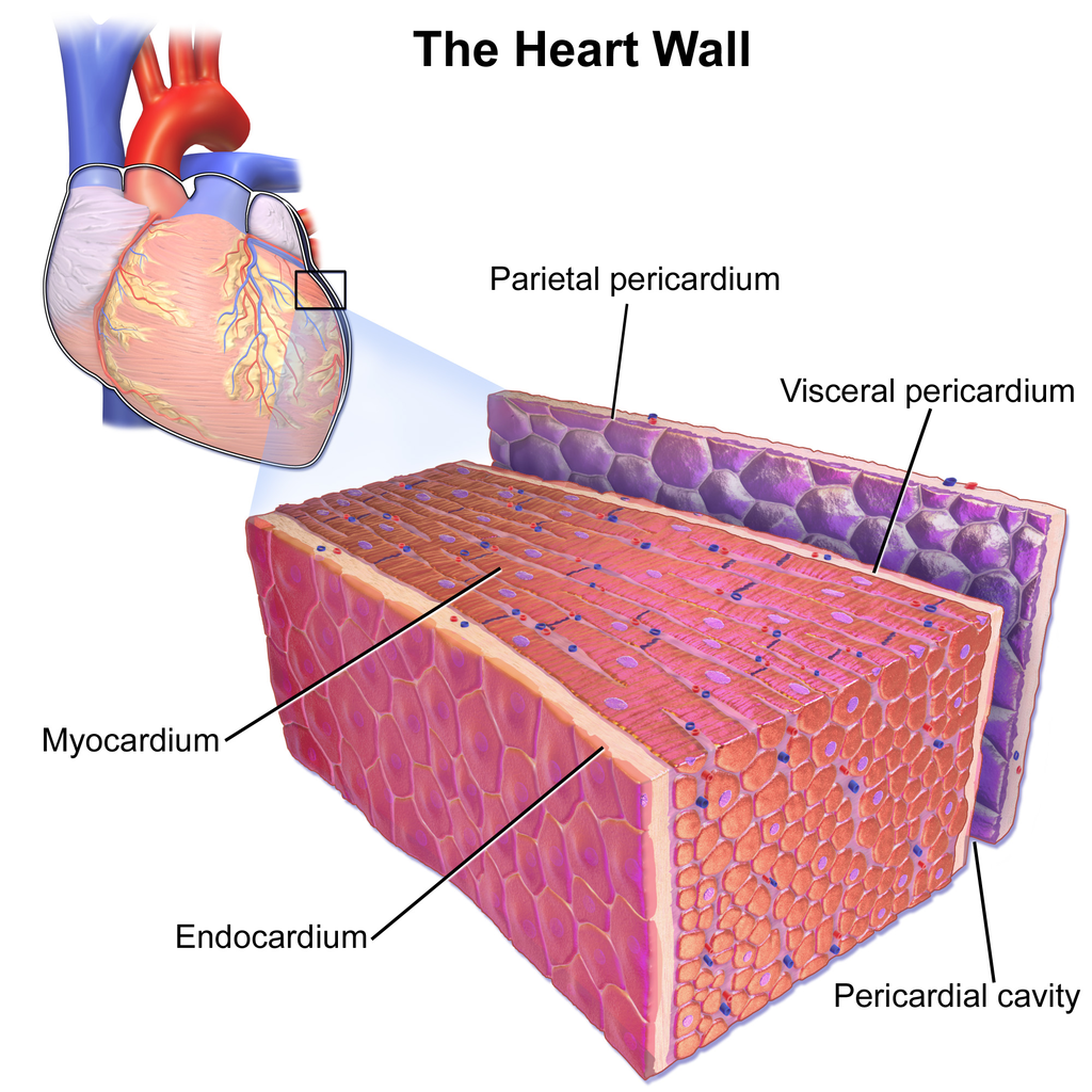
Cardiac muscle is found only in the wall of the heart. It is also called myocardium. As shown in Figure 12.3.10, myocardium is enclosed within connective tissues, including the endocardium on the inside of the heart and pericardium on the outside of the heart. When cardiac muscle contracts, the heart beats and pumps blood. Contractions of cardiac muscle are involuntary, like those of smooth muscles. They are controlled by electrical impulses from specialized cardiac muscle cells in an area of the heart muscle called the sinoatrial node.
Like skeletal muscle, cardiac muscle is striated because its filaments are arranged in sarcomeres inside the muscle fibres. However, in cardiac muscle, the myofibrils are branched at irregular angles rather than arranged in parallel rows (as they are in skeletal muscle). This explains why cardiac and skeletal muscle tissues look different from one another.
The cells of cardiac muscle tissue are arranged in interconnected networks. This arrangement allows rapid transmission of electrical impulses, which stimulate virtually simultaneous contractions of the cells. This enables the cells to coordinate contractions of the heart muscle.
The heart is the muscle that performs the greatest amount of physical work in the course of a lifetime. Although the power output of the heart is much less than the maximum power output of some other muscles in the human body, the heart does its work continuously over an entire lifetime without rest. Cardiac muscle contains a great many mitochondria, which produce ATP for energy and help the heart resist fatigue.
Feature: Human Biology in the News
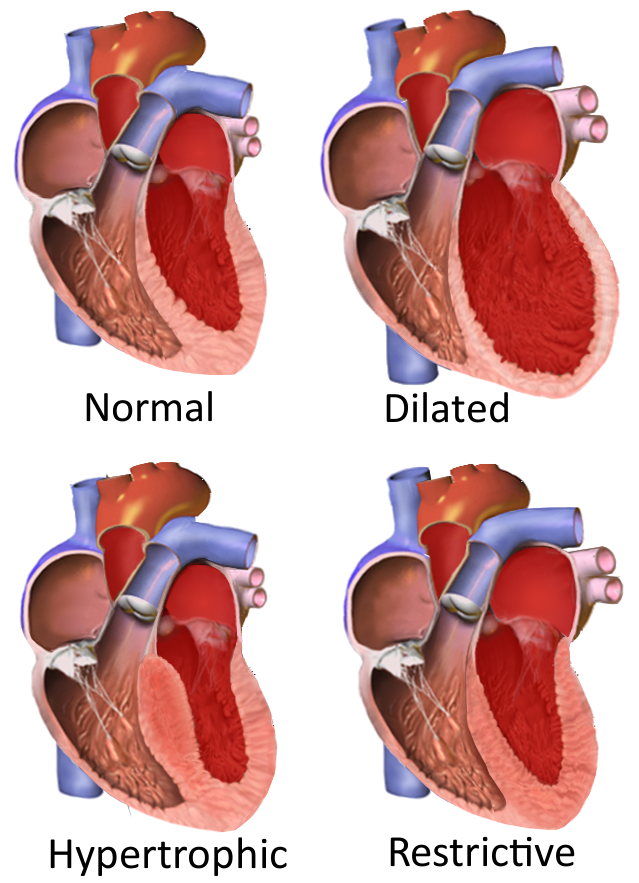
Cardiomyopathy is a disease in which the muscles of the heart are no longer able to effectively pump blood to the body — extreme forms of this disease can lead to heart failure. There are four main types of cardiomyopathy (also illustrated in Figure 12.3.11):
- Dilated (congestive) cardiomyopathy: the left ventricle (the chamber itself) of the heart becomes enlarged and can't pump blood our to the body. This is normally related to coronary artery disease and/or heart attack
- Hypertrophic cardiomyopathy: abnormal thickening of the muscular walls of the left ventricle make the chamber less able to work properly. This condition is more common in patients with a family history of the disease.
- Restrictive cardiomyopathy: the myocardium becomes abnormally rigid and inelastic and is unable to expand in between heartbeats to refill with blood. Restrictive cardiomyopathy typically affects older people.
- Arrhythmogenic right ventricular cardiomyopathy: the right ventricular muscle is replaced by adipose or scar tissue, reducing elasticity and interfering with normal heartbeat and rhythm. This disease is often caused by genetic mutations.
Cardiomyopathy is typically diagnosed with a physical exam supplemented by medical and family history, an angiogram, blood tests, chest x-rays and electrocardiograms. In some cases your doctor would also requisition a CT scan and/or genetic testing.
When treating cardiomyopathy, the goal is to reduce symptoms that affect everyday life. Certain medications can help regularize and slow heart rate, decrease chances of blood clots and cause vasodilation in the coronary arteries. If medication is not sufficient to manage symptoms, a pacemaker or even a heart transplant may be the best option. Lifestyle can also help manage the symptoms of cardiomyopathy — people living with this disease are encouraged to avoid drug and alcohol use, control high blood pressure, eat a healthy diet, get ample rest and exercise, as well as reduce stress levels.
12.3 Summary
- Muscle tissue is a soft tissue that makes up most of the tissues in the muscles of the human muscular system. It is the only type of tissue that has cells with the ability to contract.
- Skeletal muscle tissue is attached to bones by tendons. It allows voluntary body movements.
- Skeletal muscle is the most common type of muscle tissue in the human body. To move bones in opposite directions, skeletal muscles often consist of pairs of muscles that work in opposition to one another to move bones in different directions at joints.
- Skeletal muscle fibres are bundled together in units called muscle fascicles, which are bundled together to form individual skeletal muscles. Skeletal muscles also have connective tissue supporting and protecting the muscle tissue.
- Each skeletal muscle fibre consists of a bundle of myofibrils, which are bundles of protein filaments. The filaments are arranged in repeating units called sarcomeres, which are the basic functional units of skeletal muscles. Skeletal muscle tissue is striated because of the pattern of sarcomeres in its fibres.
- Skeletal muscle fibres can be divided into two types, called slow-twitch and fast-twitch fibres. Slow-twitch fibres are used mainly in aerobic endurance activities, such as long-distance running. Fast-twitch fibres are used mainly for non-aerobic, strenuous activities, such as sprinting. Proportions of the two types of fibres vary from muscle to muscle and person to person.
- Smooth muscle tissue is found in the walls of internal organs and vessels. When smooth muscles contract, they help the organs and vessels carry out their functions. Contractions of smooth muscles are involuntary and controlled by the autonomic nervous system, hormones, and other substances.
- Cells of smooth muscle tissue are not striated because they lack sarcomeres, but the cells contract in the same basic way as striated muscle cells. Unlike striated muscle, smooth muscle can sustain very long-term contractions and maintain its contractile function, even when stretched.
- Cardiac muscle tissue is found only in the wall of the heart. When cardiac muscle contracts, the heart beats and pumps blood. Contractions of cardiac muscle are involuntary, like those of smooth muscles. They are controlled by electrical impulses from specialized cardiac cells.
- Like skeletal muscle, cardiac muscle is striated because its filaments are arranged in sarcomeres inside the muscle fibres. However, the myofibrils are branched instead of arranged in parallel rows, making cardiac and skeletal muscle tissues look different from one another.
- The heart is the muscle that performs the greatest amount of physical work in the course of a lifetime. Its cells contain a great many mitochondria to produce ATP for energy and help the heart resist fatigue.
12.3 Review Questions
- What is muscle tissue?
-
-
- Where is skeletal muscle found, and what is its general function?
- Why do many skeletal muscles work in pairs?
- Describe the structure of a skeletal muscle.
- Relate muscle fibre structure to the functional units of muscles.
- Why is skeletal muscle tissue striated?
- Where is smooth muscle found? What controls the contraction of smooth muscle?
- Where is cardiac muscle found? What controls its contractions?
- The heart muscle is smaller and less powerful than some other muscles in the body. Why is the heart the muscle that performs the greatest amount of physical work in the course of a lifetime? How does the heart resist fatigue?
- Give one example of connective tissue that is found in muscles. Describe one of its functions.
12.3 Explore More
https://www.youtube.com/watch?v=3_PYnWVoUzM
What happens during a heart attack? - Krishna Sudhir, TED-Ed, 2017.
https://www.youtube.com/watch?v=bwOE1MEginA&feature=emb_logo
Three types of muscle | Circulatory system physiology | NCLEX-RN | KhanAcademyMedicine, 2012.
Attributions
Figure 12.3.1
Look by ali-yahya-155huuQwGvA [photo] by Ali Yahya on Unsplash is used under the Unsplash License (https://unsplash.com/license).
Figure 12.3.2
Skeletal_Smooth_Cardiac by OpenStax College on Wikimedia Commons is used under a CC BY 3.0 (https://creativecommons.org/licenses/by/3.0) license.
Figure 12.3.3
Anterior_and_Posterior_Views_of_Muscles by OpenStax on Wikimedia Commons is used under a CC BY 4.0 (https://creativecommons.org/licenses/by/4.0) license.
Figure 12.3.4
Antagonistic Muscle Pair by Laura Guerin at CK-12 Foundation on Wikimedia Commons is used under a CC BY-NC 3.0 (https://creativecommons.org/licenses/by-nc/3.0/) license.
Figure 12.3.5
Muscle_Fibes_(large) by OpenStax on Wikimedia Commons is used under a CC BY 4.0 (https://creativecommons.org/licenses/by/4.0) license.
Figure 12.3.6
Muscle_Fibers_(small) by OpenStax on Wikimedia Commons is used under a CC BY 4.0 (https://creativecommons.org/licenses/by/4.0) license.
Figure 12.3.7
Smooth_Muscle_Contraction by OpenStax on Wikimedia Commons is used under a CC BY 4.0 (https://creativecommons.org/licenses/by/4.0) license.
Figure 12.3.8
Blausen_0747_Pregnancy by BruceBlaus on Wikimedia Commons is used under a CC BY 3.0 (https://creativecommons.org/licenses/by/3.0) license.
Figure 12.3.9
Size_of_Uterus_Throughout_Pregnancy-02 by OpenStax College on Wikimedia Commons is used under a CC BY 3.0 (https://creativecommons.org/licenses/by/3.0) license.
Figure 12.3.10
1024px-Blausen_0470_HeartWall by BruceBlaus on Wikimedia Commons is used under a CC BY 3.0 (https://creativecommons.org/licenses/by/3.0) license.
Figure 12.3.11
Tipet_e_kardiomiopative by Npatchett at English Wikipedia on Wikimedia Commons is used under a CC BY-SA 3.0 (https://creativecommons.org/licenses/by-sa/3.0) license. (Work derived from Blausen 0165 Cardiomyopathy Dilated by BruceBlaus)
References
Betts, J. G., Young, K.A., Wise, J.A., Johnson, E., Poe, B., Kruse, D.H., Korol, O., Johnson, J.E., Womble, M., DeSaix, P. (2013, June 19). Figure 4.18 Muscle tissue [digital image]. In Anatomy and Physiology (Section 4.4). OpenStax. https://openstax.org/books/anatomy-and-physiology/pages/4-4-muscle-tissue-and-motion
Betts, J. G., Young, K.A., Wise, J.A., Johnson, E., Poe, B., Kruse, D.H., Korol, O., Johnson, J.E., Womble, M., DeSaix, P. (2013, June 19). Figure 28.18 Size of uterus throughout pregnancy [digital image]. In Anatomy and Physiology (Section 28.4). OpenStax. https://openstax.org/books/anatomy-and-physiology/pages/28-4-maternal-changes-during-pregnancy-labor-and-birth
Betts, J. G., Young, K.A., Wise, J.A., Johnson, E., Poe, B., Kruse, D.H., Korol, O., Johnson, J.E., Womble, M., DeSaix, P. (2016, May 18). Figure 10.3 The three connective tissue layers [digital image]. In Anatomy and Physiology (Section 10.2). OpenStax. https://openstax.org/books/anatomy-and-physiology/pages/10-2-skeletal-muscle
Betts, J. G., Young, K.A., Wise, J.A., Johnson, E., Poe, B., Kruse, D.H., Korol, O., Johnson, J.E., Womble, M., DeSaix, P. (2016, May 18). Figure 10.4 Muscle fiber [digital image]. In Anatomy and Physiology (Section 10.2). OpenStax. https://openstax.org/books/anatomy-and-physiology/pages/10-2-skeletal-muscle
Betts, J. G., Young, K.A., Wise, J.A., Johnson, E., Poe, B., Kruse, D.H., Korol, O., Johnson, J.E., Womble, M., DeSaix, P. (2016, May 18). Figure 10.24 Muscle contraction [digital image]. In Anatomy and Physiology (Section 10.8). OpenStax. https://openstax.org/books/anatomy-and-physiology/pages/10-8-smooth-muscle
Betts, J. G., Young, K.A., Wise, J.A., Johnson, E., Poe, B., Kruse, D.H., Korol, O., Johnson, J.E., Womble, M., DeSaix, P. (2016, May 18). Figure 11.5 Overview of the muscular system [digital image]. In Anatomy and Physiology (Section 11.2). OpenStax. https://openstax.org/books/anatomy-and-physiology/pages/11-2-naming-skeletal-muscles
Blausen.com staff. (2014). Medical gallery of Blausen Medical 2014. WikiJournal of Medicine 1 (2). DOI:10.15347/wjm/2014.010. ISSN 2002-4436.
Brainard, J/ CK-12 Foundation. (2012). Figure 5 Triceps and biceps muscles in the upper arm are opposing muscles. [digital image]. In CK-12 Biology (Section 21.3) [online Flexbook]. CK12.org. https://www.ck12.org/book/ck-12-biology/section/21.3/ (Last modified August 11, 2017.)
khanacademymedicine. (2012, October 19). Three types of muscle | Circulatory system physiology | NCLEX-RN | Khan Academy. YouTube.
TED-Ed. (2017, February 14). What happens during a heart attack? - Krishna Sudhir. YouTube. https://www.youtube.com/watch?v=3_PYnWVoUzM&feature=youtu.be
Involuntary, striated muscle found only in the walls of the heart; also called myocardium.
Created by CK-12 Foundation/Adapted by Christine Miller

Bathing in Sunshine
Summer sun may feel good on your body, but its invisible UV rays wreak havoc on your skin. Exposing the skin to UV light causes photo-aging: premature wrinkling, brown discolourations, and other unattractive signs of sun exposure. Even worse, UV light increases your risk of skin cancer.
What Is Skin Cancer?
Skin cancer is a disease in which skin cells grow out of control. It is caused mainly by excessive exposure to UV light, which damages DNA. Therefore, skin cancer most often develops on areas of the skin that are frequently exposed to UV light. However, it can also occur on areas that are rarely exposed to UV light. Skin cancer affects people of all skin colours, including those with dark skin. It also affects more people altogether than all other cancers combined. One in five Canadians develops skin cancer in his or her lifetime.
Types of Skin Cancer
Skin cancer begins in the outer layer of skin, the epidermis. There are three common types of skin cancer: basal cell carcinoma, squamous cell carcinoma, and melanoma.
Basal Cell Carcinoma
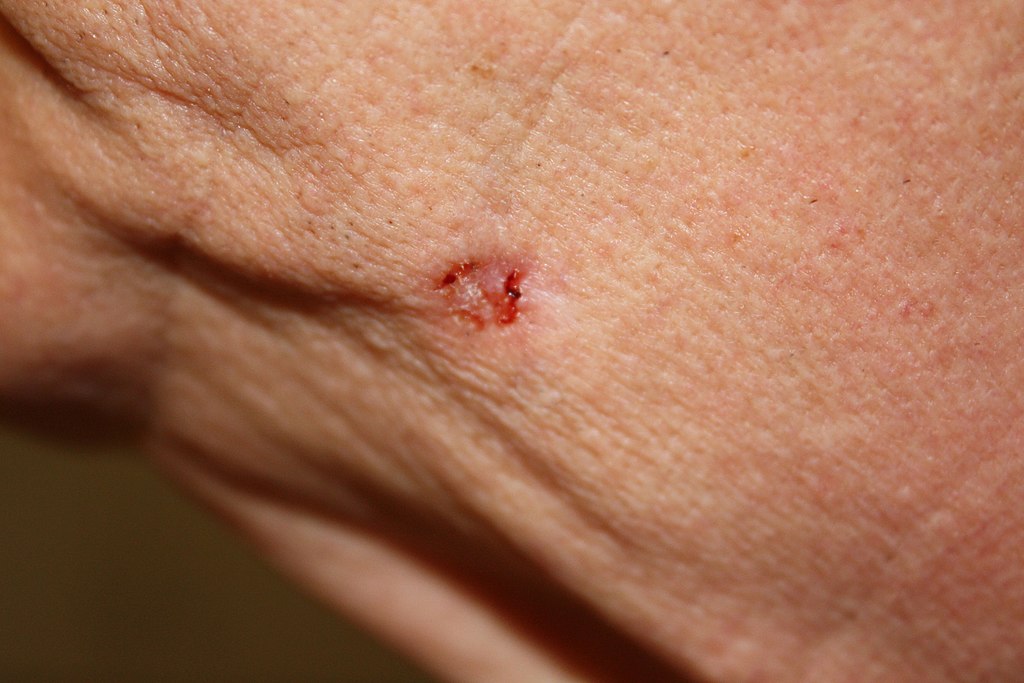
Basal cell carcinoma occurs in basal cells of the epidermis. Basal cells are stem cells in the stratum basale layer that divide to form all the keratinocytes of the epidermis. Basal cell carcinoma is the most common form of skin cancer and 1 in 8 Canadians will develop basal cell carcinoma during their lifetime. A basal cell carcinoma may appear as a pearly or waxy sore, like the one shown in Figure 10.7.2. Basal cell carcinomas rarely spread (or undergo metastasis), so they can generally be cured with a biopsy, in which the lesion is cut out of the skin and analyzed in a medical lab.
Squamous Cell Carcinoma
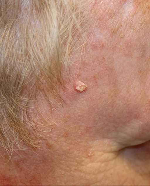
Squamous cell carcinoma occurs in squamous cells of the epidermis. Squamous cells are flattened, keratin-filled cells in upper layers of the epidermis. Squamous cell carcinoma is the second most common form of skin cancer. More than two million cases occur in the United States each year. A squamous cell carcinoma may appear as a firm, red nodule, or as a flat lesion with a scaly or crusty surface, like the one pictured in Figure 10.7.3. Squamous cell carcinomas are generally localized and unlikely to metastasize, so they are usually curable surgically.
Melanoma
Melanoma occurs in melanocytes of the epidermis. Melanocytes are the melanin-producing cells in the stratum basale of the epidermis. Melanoma is the rarest type of skin cancer, accounting for less than one per cent of all skin cancer cases. Melanoma, however, is the most deadly type of skin cancer. It causes the vast majority of skin cancer deaths, because melanoma is malignant. If not treated, it will metastasize and spread to other parts of the body. If melanoma is detected early and while it is still localized in the skin, most patients survive for at least five years. If melanoma is discovered only after it has already metastasized to distant organs, there is only a 17% of patients surviving for five years. You can see an example of a melanoma in Figure 10.7.4.
Melanoma can develop anywhere on the body. It may develop in otherwise normal skin, or an existing mole may become cancerous. Signs of melanoma may include a:
- Mole that changes in size, feel, or colour.
- Mole that bleeds.
- Large brown spot on the skin sprinkled with darker specks.
- Small lesion with an irregular border and parts that appear red, white, blue, or blue-black.
- Dark lesion on the palms, soles, fingertips, toes, or mucous membranes.
Skin Cancer Risk Factors
Exposure to UV radiation causes about 90 per cent of all skin cancer cases. The connection between skin cancer and UV light is so strong that the World Health Organization has classified UV radiation (whether from tanning beds or the sun) as a Group 1 carcinogen (cancer-causing agent). Group 1 carcinogens are those carcinogens that are known with virtual certainty to cause cancer. In addition to UV light, Group 1 carcinogens include tobacco and plutonium. In terms of numbers of cancers caused, UV radiation is far worse than tobacco. More people develop skin cancer because of UV light exposure than develop lung cancer because of smoking. The increase in cancer risk due to UV light is especially great if you have ever had blistering sunburns as a child or teen.
Besides UV light exposure, other risk factors for skin cancer include:
- Having light coloured skin.
- Having a lot of moles.
- Being diagnosed with precancerous skin lesions.
- Having a family history of skin cancer.
- Having a personal history of skin cancer.
- Having a weakened immune system.
- Being exposed to other forms of radiation or to certain toxic substances such as arsenic.
Feature: My Human Body
As with most types of cancer, skin cancer is easiest to treat and most likely to be cured the earlier it is detected. The skin is one of the few organs that you can monitor for cancer yourself, as long as you know what to look for. A brown spot on the skin is likely to be a harmless mole, but it could be a sign of skin cancer. As shown in Figure 10.7.5 below, unlike moles, skin cancers may be asymmetrical, have irregular borders, may be very dark in colour, and may have a relatively great diameter. These characteristics can be remembered with the acronym ABCD.

With the help of mirrors, you should check all of your skin regularly. Look for new skin growths or changes in any existing moles, freckles, bumps, or birthmarks. Report anything suspicious or different to your doctor.
If you have risk factors for skin cancer, it’s a good idea to have an annual skin check by a dermatologist. This helps ensure that cancerous or precancerous lesions will be detected before they grow too large and become difficult to cure, or in the case of melanoma, before they metastasize.
10.7 Summary
- Skin cancer is a disease in which skin cells grow out of control. It is caused mainly by excessive exposure to UV light, which damages DNA. Skin cancer affects more Canadians than all other cancers combined. There are three common types of skin cancer: basal cell carcinoma, squamous cell carcinoma, and melanoma. Carcinomas are more common and unlikely to metastasize. Melanoma is rare and likely to metastasize. It causes most skin cancer deaths.
- Besides exposure to UV light, risk factors for skin cancer include having light coloured skin, having lots of moles, and a family history of skin cancer, among several others.
10.7 Review Questions
- What is skin cancer?
- How common is skin cancer?
-
- Compare and contrast the three common types of skin cancer.
- Identify factors that increase the risk of skin cancer.
- How does exposure to UV light cause skin cancer?
- In which layer of the skin does skin cancer normally start?
- Which two skin cancers described in this section start in the same sub-layer? Include the name of the sub-layer and the cells affected in each of these cancers.
- Which type of skin cancer is most likely to spread to other organs? Explain your answer.
- Which form of skin cancer is the most deadly?
- What are some ways people can reduce their risk of getting skin cancer? Explain your answer.
10.7 Explore More
https://www.youtube.com/watch?v=60e-t4zglBk&feature=emb_logo
The skin 'beauty' and the sun 'beast': Charareh Pourzand at TEDxBathUniveristy, TEDx Talks, 2014.
https://www.youtube.com/watch?v=ID-O-Ion3EQ&feature=emb_logo
Cancer of the Vulva, Robert Miller, 2014.
https://www.youtube.com/watch?v=BmFEoCFDi-w
How do cancer cells behave differently from healthy ones? - George Zaidan, TED-Ed, 2012.
Attributions
Figure 10.7.1
Stolen_Moment_in_the_Sun by Angie Garrett on Wikimedia Commons is used under a CC BY 2.0 (https://creativecommons.org/licenses/by/2.0) license.
Figure 10.7.2
Basal_cell_carcinoma,_ulcerated by Kelly Nelson (Photographer) from National Cancer Institute (part of the National Institutes of Health) with the ID 9237 on Wikimedia Commons was released into the public domain (https://en.wikipedia.org/wiki/Public_domain).
Figure 10.7.3
Squamous_cell_carcinoma_(1) by Kelly Nelson (Photographer) from National Cancer Institute (part of the National Institutes of Health) with the ID 9248 on Wikimedia Commons was released into the public domain (https://en.wikipedia.org/wiki/Public_domain).
Figure 10.7.4
Melanoma by Unknown author (Photographer) from National Cancer Institute (part of the National Institutes of Health) with the AV-8500-3850/ ID 9186 on Wikimedia Commons was released into the public domain (https://en.wikipedia.org/wiki/Public_domain).
Figure 10.7.5
ABCDs of skin cancer by CK-12 Foundation is used under a CC BY-NC 3.0 (https://creativecommons.org/licenses/by-nc/3.0/) license. (Original images courtesy of NCI: ID numbers 2362; 2363; 2364; and 2184)
References
Brainard, J/ CK-12 Foundation. (2016). Figure 5 ABCDs of skin cancer[digital image]. In CK-12 College Human Biology (Section 12.7) [online Flexbook]. CK12.org. https://www.ck12.org/book/ck-12-college-human-biology/section/12.7/
Public Health Agency of Canada. (2019, December 9). Non melanoma skin cancer. Canada.ca. https://www.canada.ca/en/public-health/services/chronic-diseases/cancer/non-melanoma-skin-cancer.html
Robert Miller. (2014, July 22). Cancer of the vulva. YouTube. https://www.youtube.com/watch?v=ID-O-Ion3EQ
TED-Ed. (2012, December 5). How do cancer cells behave differently from healthy ones? - George Zaidan. YouTube. https://www.youtube.com/watch?v=BmFEoCFDi-w
TEDx Talks. (2014, March 28). The skin 'beauty' and the sun 'beast': Charareh Pourzand at TEDxBathUniveristy. YouTube. https://www.youtube.com/watch?v=60e-t4zglBk
Created by CK-12 Foundation/Adapted by Christine Miller
Stroller Moms
These moms (Figure 12.5.1) are setting a great example for their children by engaging in physical exercise. Adopting a habit of regular physical exercise is one of the most important ways to maintain fitness and good health. From higher self-esteem to a healthier heart, physical exercise can have a positive effect on virtually all aspects of health, including physical, mental, and emotional health.
What Is Physical Exercise?
Physical exercise is any bodily activity that enhances or maintains physical fitness and overall health and wellness. We generally think of physical exercise as activities that are undertaken for the main purpose of improving physical fitness and health. However, physical activities that are undertaken for other purposes may also count as physical exercise. Scrubbing a floor, raking a lawn, or playing active games with young children or a pet are all activities that can have fitness and health benefits, even though they generally are not done mainly for this purpose.
How much physical exercise should people get? In the Canada, both the Canadian Food Guide and the Canadian Society for Exercise Physiology recommend that every child and adult who is able should participate in moderate exercise for a minimum of 60 minutes a day. This might include walking, swimming, and/or household or yard work.
Types of Physical Exercise
Physical exercise can be classified into three types, depending on the effects it has on the body: aerobic exercise, anaerobic exercise, and flexibility exercise. Many specific examples of physical exercise (including playing soccer and rock climbing) can be classified as more than one type.
Aerobic Exercise

Aerobic exerciseis any physical activity in which muscles are used at well below their maximum contraction strength, but for long periods of time. Aerobic exercise uses a relatively high percentage of slow-twitch muscle fibres that consume a large amount of oxygen. The main goal of aerobic exercise is to increase cardiovascular endurance, although it can have many other benefits, including muscle toning. Examples of aerobic exercise include cycling, swimming, brisk walking, jumping rope, rowing, hiking, tennis, and kayaking as shown in Figure 12.5.2 .
Anaerobic Exercise
Anaerobic exercise is any physical activity in which muscles are used at close to their maximum contraction strength, but for relatively short periods of time. Anaerobic exercise uses a relatively high percentage of fast-twitch muscle fibres that consume a small amount of oxygen. Goals of anaerobic exercise include building and strengthening muscles, as well as improving bone strength, balance, and coordination. Examples of anaerobic exercise include push-ups, lunges, sprinting, interval training, resistance training, and weight training (such as biceps curls with a dumbbell, as pictured in Figure 12.5.3).

Flexibility Exercise

Flexibility exercise is any physical activity that stretches and lengthens muscles. Goals of flexibility exercise include increasing joint flexibility, keeping muscles limber, and improving the range of motion, all of which can reduce the risk of injury. Examples of flexibility exercise include stretching, yoga (as in Figure 12.5.4), and tai chi.
Health Benefits of Physical Exercise
Many studies have shown that physical exercise is positively correlated with a diversity of health benefits. Some of these benefits include maintaining physical fitness, losing weight and maintaining a healthy weight, regulating digestive health, building and maintaining healthy bone density, increasing muscle strength, improving joint mobility, strengthening the immune system, boosting cognitive ability, and promoting psychological well-being. Some studies have also found a significant positive correlation between exercise and both quality of life and life expectancy. People who participate in moderate to high levels of physical activity have been shown to have lower mortality rates than people of the same ages who are not physically active and daily exercise has been shown to increase life expectancy up to an average of five years.
The underlying physiological mechanisms explaining why exercise has these positive health benefits are not completely understood. However, developing research suggests that many of the benefits of exercise may come about because of the role of skeletal muscles as endocrine organs. Contracting muscles release hormones called myokines, which promote tissue repair and the growth of new tissue. Myokines also have anti-inflammatory effects, which, in turn, reduce the risk of developing inflammatory diseases. Exercise also reduces levels of cortisol, the adrenal cortex stress hormone that may cause many health problems — both physical and mental — at sustained high levels.
Cardiovascular Benefits of Physical Exercise
The beneficial effects of exercise on the cardiovascular system are well documented. Physical inactivity has been identified as a risk factor for the development of coronary artery disease. There is also a direct correlation between physical inactivity and cardiovascular disease mortality. Physical exercise, in contrast, has been demonstrated to reduce several risk factors for cardiovascular disease, including hypertension (high blood pressure), “bad” cholesterol (low-density lipoproteins), high total cholesterol, and excess body weight. Physical exercise has also been shown to increase “good” cholesterol (high-density lipoproteins), insulin sensitivity, the mechanical efficiency of the heart, and exercise tolerance, which is the ability to perform physical activity without undue stress and fatigue.
Cognitive Benefits of Physical Exercise
Physical exercise has been shown to help protect people from developing neurodegenerative disorders, such as dementia. A 30-year study of almost 2,400 men found that those who exercised regularly had a 59 per cent reduction in dementia when compared with those who did not exercise. Similarly, a review of cognitive enrichment therapies for the elderly found that physical activity — in particular, aerobic exercise — can enhance the cognitive function of older adults. Anecdotal evidence suggests that frequent exercise may even help reverse alcohol-induced brain damage. There are several possible reasons why exercise is so beneficial for the brain. Physical exercise:
- Increases blood flow and oxygen availability to the brain.
- Increases growth factors that promote new brain cells and new neuronal pathways in the brain.
- Increases levels of neurotransmitters (such as serotonin), which increase memory retention, information processing, and cognition.
Mental Health Benefits of Physical Exercise
Numerous studies suggest that regular aerobic exercise works as well as pharmaceutical antidepressants in treating mild-to-moderate depression. A possible reason for this effect is that exercise increases the biosynthesis of at least three neurochemicals that may act as euphoriants. The euphoric effect of exercise is well known. Distance runners may refer to it as “runner’s high,” and people who participate in crew (as in Figure 12.5.5) may refer to it as “rower’s high.” Because of these effects, health care providers often promote the use of aerobic exercise as a treatment for depression.

Additional mental health benefits of physical exercise include reducing stress, improving body image, and promoting positive self-esteem. Conversely, there is evidence to suggest that being sedentary is associated with increased risk of anxiety.
Sleep Benefits of Physical Exercise
A recent review of published scientific research suggests that exercise generally improves sleep for most people, and helps sleep disorders, such as insomnia. In fact, exercise is the most recommended alternative to sleeping pills for people with insomnia. For sleep benefits, the optimum time to exercise may be four to eight hours before bedtime, although exercise at any time of day seems to be beneficial. The only possible exception is heavy exercise undertaken shortly before bedtime, which may actually interfere with sleep.
Other Benefits of Physical Exercise
Some studies suggest that physical activity may benefit the immune system. For example, moderate exercise has been found to be associated with a decreased incidence of upper respiratory tract infections. Evidence from many studies has found a correlation between physical exercise and reduced death rates from cancer, specifically breast cancer and colon cancer. Physical exercise has also been shown to reduce the risk of type 2 diabetes and obesity.
Variation in Responses to Physical Exercise
Not everyone benefits equally from physical exercise. When participating in aerobic exercise, most people will have a moderate increase in their endurance, but some people will as much as double their endurance. Some people, on the other hand, will show little or no increase in endurance from aerobic exercise. Genetic differences in slow-twitch and fast-twitch skeletal muscle fibres may play a role in these different results. People with more slow-twitch fibres may be able to develop greater endurance, because these muscle fibres have more capillaries, mitochondria, and myoglobin than fast-twitch fibres. As a result, slow-twitch fibres can carry more oxygen and sustain aerobic activity for a longer period of time than fast-twitch fibres. Studies show that endurance athletes (like the marathoner pictured in Figure 12.5.6) generally do tend to have a higher proportion of slow-twitch fibres than other people.
There is also great variation in individual responses to muscle building as a result of anaerobic exercise. Some people have a much greater capacity to increase muscle size and strength, whereas other people never develop large muscles, no matter how much they exercise them. People who have more fast-twitch than slow-twitch muscle fibres may be able to develop bigger, stronger muscles, because fast-twitch muscle fibres contribute more to muscle strength and have greater potential to increase in mass. Evidence suggests that athletes who excel at power activities (such as throwing and jumping) tend to have a higher proportion of fast-twitch fibres than do endurance athletes.
Can You “Overdose” on Physical Exercise?
Is it possible to exercise too much? Can too much exercise be harmful? Evidence suggests that some adverse effects may occur if exercise is extremely intense and the body is not given proper rest between exercise sessions. Athletes who train for multiple marathons have been shown to develop scarring of the heart and heart rhythm abnormalities. Doing too much exercise without prior conditioning also increases the risk of injuries to muscles and joints. Damage to muscles due to overexertion is often seen in new military recruits (see Figure 12.5.7). Too much exercise in females may cause amenorrhea, which is a cessation of menstrual periods. When this occurs, it generally indicates that a woman is pushing her body too hard.
Many people develop delayed onset muscle soreness (DOMS), which is pain or discomfort in muscles that is felt one to three days after exercising, and generally subsides two or three days later. DOMS was once thought to be caused by the buildup of lactic acid in the muscles. Lactic acid is a product of anaerobic respiration in muscle tissues. However, lactic acid disperses fairly rapidly, so it is unlikely to explain pain experienced several days after exercise. The current theory is that DOMS is caused by tiny tears in muscle fibres, which occur when muscles are used at too high a level of intensity.
Feature: My Human Body
Most people know that exercise is important for good health, and it’s easy to find endless advice about exercise programs and fitness plans. What is not so easy to find is the motivation to start exercising — and to stick with it. This is the main reason why so many people fail to get regular exercise. Practical concerns like a busy schedule and bad weather can certainly make exercising more of a challenge, but the biggest barriers to adopting a regular exercise routine are mental. If you want to exercise but find yourself making excuses or getting discouraged and giving up, here are some tips that may help you get started and stay moving:
- Avoid an all-or-nothing point of view. Don’t think you need to spend hours sweating at the gym or training for a marathon to get healthy. Even a little bit of exercise is better than nothing at all. Start out with ten or 15 minutes of moderate activity each day. Taking a walk around your neighborhood is a great way to begin! From there, gradually increase the amount of time until you are exercising to at least 30 minutes a day, five days a week. Make it a routine.
- Be kind to yourself, and reinforce positive behaviors with rewards. Don’t be down on yourself because you are overweight or out of shape. Don’t beat yourself up because of a supposed lack of willpower. Instead, look at any past failures as opportunities to learn and do better. When you do achieve even small exercise goals, treat yourself to something special. Did you just complete your first workout? Reward yourself with a relaxing bath or other treat.
- Don’t make excuses for not exercising. Common complaints include being too busy or tired or not athletic enough. Such excuses are not valid reasons to avoid exercising, and they will sabotage any plans to improve your fitness. If you can’t find a 30-minute period to work out, try to find ten minutes, three times a day. If you’re feeling tired, know that exercise can actually reduce fatigue and boost your energy level. If you feel clumsy and uncoordinated, remind yourself that you don’t need to be athletic to take a walk or engage in vigorous house or yard work.
- Find an activity that you truly enjoy doing. Don’t think you have to lift weights or run on a treadmill to exercise your muscles. If you find such activities boring or unpleasant, you won’t stick with them. Any activity that increases your heart rate and uses large muscles can provide a workout, especially if you’re not in the habit of exercising, so find something you like to do. Do you like to dance? Put on some music and dance up a sweat! Do you enjoy gardening? Get out in the yard and dig up some dirt! Still not interested? Try an activity-based video game, such as Wii or Kinect. You may find it so much fun that it doesn’t seem like exercise until you realize you’ve worked up a sweat.
- Make yourself accountable. Tell friends and family members that you’re going to start exercising. You’ll be letting them — as well as yourself — down if you don’t follow through. Some people find that keeping an exercise log to track their progress is a good way to be accountable and stick to an exercise program. Perhaps the best way to keep at it is to find an exercise partner. If you’ve got someone waiting to exercise with you, you will be less likely to make excuses for not exercising.
- Add more physical activity to your daily life. You don’t need to follow a structured exercise program to increase your activity level. Do your house or yard work briskly for a workout. Park your car further than necessary from work or the mall, and walk the extra distance. If you live close enough, leave the car at home and walk to and from your destination. Rather than taking elevators or escalators, walk up and down stairs. When you take breaks at work, take a walk instead of sitting. Every time a commercial comes on while you’re watching TV, take a quick exercise break — run in place or do some curls with hand weights.
12.5 Summary
- Physical exercise is any bodily activity that enhances or maintains physical fitness and overall health. Activities such as household chores may count as physical exercise, even if they are not done for their health benefits. Current recommendations for adults are 30 minutes a day of moderate exercise.
- Aerobic exercise is any physical activity that uses muscles at less than their maximum contraction strength, but for long periods of time. This type of exercise uses a relatively high percentage of slow-twitch muscle fibres that consume large amounts of oxygen. Aerobic exercises increase cardiovascular endurance and include cycling and brisk walking.
- Anaerobic exercise is any physical activity that uses muscles at close to their maximum contraction strength, but for short periods of time. This type of exercise uses a relatively high percentage of fast-twitch muscle fibres that consume small amounts of oxygen. Anaerobic exercises increase muscle and bone mass and strength, and they include push-ups and sprinting.
- Flexibility exercise is any physical activity that stretches and lengthens muscles, thereby improving range of motion and reducing risk of injury. Examples include stretching and yoga.
- Many studies have shown that physical exercise is positively correlated with a diversity of physical, mental, and emotional health benefits. Physical exercise also increases quality of life and life expectancy.
- Many of the benefits of exercise may come about because contracting muscles release hormones called myokines, which promote tissue repair and growth and have anti-inflammatory effects.
- Physical exercise can reduce risk factors for cardiovascular disease, including hypertension and excess body weight. Physical exercise can also increase factors associated with cardiovascular health, such as mechanical efficiency of the heart.
- Physical exercise has been shown to offer protection from dementia and other cognitive problems, perhaps because it increases blood flow or neurotransmitters in the brain, among other potential effects.
- Numerous studies suggest that regular aerobic exercise works as well as pharmaceutical antidepressants in treating mild-to-moderate depression, possibly because it increases synthesis of natural euphoriants in the brain.
- Research shows that physical exercise generally improves sleep for most people and helps sleep disorders, such as insomnia. Other health benefits of physical exercise include better immune system function and reduced risk of type 2 diabetes and obesity.
- There is great variation in individual responses to exercise, partly due to genetic differences in proportions of slow-twitch and fast-twitch skeletal muscle fibres. People with more slow-twitch fibres may be able to develop greater endurance from aerobic exercise, whereas people with more fast-twitch fibres may be able to develop greater muscle size and strength from anaerobic exercise.
- Some adverse effects may occur if exercise is extremely intense and the body is not given proper rest between exercise sessions. Many people who overwork their muscles develop delayed onset muscle soreness (DOMS), which may be caused by tiny tears in muscle fibres.
12.5 Review Questions
- How do we define physical exercise?
- What are current recommendations for physical exercise for adults?
-
- Define flexibility exercise, and state its benefits. What are two examples of flexibility exercises?
- In general, how does physical exercise affect health, quality of life, and longevity?
- What mechanism may underlie many of the general health benefits of physical exercise?
- Relate physical exercise to cardiovascular disease risk.
- What may explain the positive benefits of physical exercise on cognition?
- How does physical exercise compare with antidepressant drugs in the treatment of depression?
- Identify several other health benefits of physical exercise.
- Explain how genetics may influence the way individuals respond to physical exercise.
- Can too much physical exercise be harmful?
12.5 Explore More
https://www.youtube.com/watch?v=hmFQqjMF_f0
How playing sports benefits your body ... and your brain - Leah Lagos and Jaspal Ricky Singh, TED-Ed, 2016.
https://www.youtube.com/watch?v=rLsimrBoYXc&t=12s
The surprising reason our muscles get tired - Christian Moro, TED-Ed, 2019.
https://youtu.be/2tM1LFFxeKg
What makes muscles grow? - Jeffrey Siegel, TED-Ed, 2015.
https://www.youtube.com/watch?v=QeIrdqU0o9s
Why some people find exercise harder than others | Emily Balcetis, TED, 2014.
Attributions
Figure 12.5.1
stroller fit by Serge Melki from Indianapolis, USA on Wikimedia Commons is used under a CC BY 2.0 (https://creativecommons.org/licenses/by/2.0) license.
Figure 12.5.2
Children kayaking young sport by Hagerty Ryan, USFWS on Pixnio is used under a public domain (CC0) Certification (https://creativecommons.org/licenses/publicdomain/).
Figure 12.5.3
Bicep curls [photo] by Senior Airman Jarrod Grammel from U.S. Moody Air Force Base is released into the public domain (https://en.wikipedia.org/wiki/Public_domain).
Figure 12.5.4
Flexibility exercise by carl-barcelo-nqUHQkuVj3c [photo] by Carl Barcelo on Unsplash is used under the Unsplash License (https://unsplash.com/license).
Figure 12.5.5
Canadian women’s double scull silver Rio 2016 by Gerhard Pratt on Flickr is used under a CC BY-NC 2.0 (https://creativecommons.org/licenses/by-nc/2.0/) license.
Figure 12.5.6
Toronto Marathon 2012 by Marc Roberts on Flickr is used under a CC BY-NC-SA 2.0 (https://creativecommons.org/licenses/by-nc-sa/2.0/) license.
Figure 12.5.7
Muscle damage in military recruits by Lance Cpl. Bridget M. Keane from the United States Marine Corps Recruit Depot is in the public domain (https://en.wikipedia.org/wiki/Public_domain).
References
Elwood, P., Galante, J., Pickering, J., Palmer, S., Bayer, A., Ben-Shlomo, Y., Longley, M., & Gallacher, J. (2013). Healthy lifestyles reduce the incidence of chronic diseases and dementia: evidence from the Caerphilly cohort study. PloS one, 8(12), e81877. https://doi.org/10.1371/journal.pone.0081877
Mayo Clinic Staff. (n.d.). Amenorrhea [online article]. MayoClinic.org. https://www.mayoclinic.org/diseases-conditions/amenorrhea/symptoms-causes/syc-20369299#
Mayo Clinic Staff. (n.d.). Coronary artery disease [online article]. MayoClinic.org. https://www.mayoclinic.org/diseases-conditions/coronary-artery-disease/symptoms-causes/syc-20350613
TED-Ed. (2016, June 28). How playing sports benefits your body ... and your brain - Leah Lagos and Jaspal Ricky Singh. YouTube. https://www.youtube.com/watch?v=hmFQqjMF_f0&feature=youtu.be
TED-Ed. (2019, April 18). The surprising reason our muscles get tired - Christian Moro. YouTube. https://www.youtube.com/watch?v=rLsimrBoYXc&feature=youtu.be
TED-Ed. (2015, November 3). What makes muscles grow? - Jeffrey Siegel. YouTube https://www.youtube.com/watch?v=2tM1LFFxeKg&feature=youtu.be
TED. (2014, November 14). Why some people find exercise harder than others | Emily Balcetis, YouTube. https://www.youtube.com/watch?v=QeIrdqU0o9s&feature=youtu.be
Wikipedia contributors. (2020, August 1). Delayed onset muscle soreness. In Wikipedia. https://en.wikipedia.org/w/index.php?title=Delayed_onset_muscle_soreness&oldid=970682631
Image shows a diagram of locations in the body where the effects of anemia are experienced. Some of these include: Central nervous system: fatigue, dizziness and possibly fainting. Low blood pressure. In the heart: heart palpitations, rapid heart rate, chest palpitations, in extreme cases chest pain, angina and heart attack. Enlargement of the spleen. Changed stool (poo) colour. Muscular weakness. Shortness of breath. Pale, cold and/or yellowing skin. Yellowing eyes.
Created by CK-12 Foundation/Adapted by Christine Miller

Art in a Cup
Who knew that a cup of coffee could also be a work of art? A talented barista can make coffee look as good as it tastes. If you are a coffee drinker, you probably know that coffee can also affect your mental state. It can make you more alert, and it may improve your concentration. That’s because the caffeine in coffee is a psychoactive drug. In fact, caffeine is the most widely consumed psychoactive substance in the world. In North America, for example, 90 per cent of adults consume caffeine daily.
What Are Psychoactive Drugs?
Psychoactive drugs are substances that change the function of the brain and result in alterations of mood, thinking, perception, and/or behavior. Psychoactive drugs may be used for many purposes, including therapeutic, ritual, or recreational purposes. Besides caffeine, other examples of psychoactive drugs include cocaine, LSD, alcohol, tobacco, codeine, and morphine. Psychoactive drugs may be legal prescription medications (codeine and morphine), legal nonprescription drugs (alcohol and tobacco), or illegal drugs (cocaine and LSD).
Cannabis (or marijuana) is also a psychoactive drug that while illegal in many countries is legal for use in Canada by individuals over the age of 19 years. Legal prescription medications (such as opioids) are also used illegally by increasingly large numbers of people. Some legal drugs, such as alcohol and nicotine, are readily available almost everywhere, as illustrated by the images below.
Figure 8.8.2 These psychoactive drugs are legal and accessible almost anywhere.
Classes of Psychoactive Drugs
Psychoactive drugs are divided into different classes based on their pharmacological effects. Several classes are listed below, along with examples of commonly used drugs in each class.
- Stimulants are drugs that stimulate the brain and increase alertness and wakefulness. Examples of stimulants include caffeine, nicotine, cocaine, and amphetamines (such as Adderall).
- Depressants are drugs that calm the brain, reduce anxious feelings, and induce sleepiness. Examples of depressants include ethanol (in alcoholic beverages) and opioids, such as codeine and heroin.
- Anxiolytics are drugs that have a tranquilizing effect and inhibit anxiety. Examples of anxiolytic drugs include benzodiazepines (such as diazepam/Valium), barbiturates (such as phenobarbital), opioids, and antidepressant drugs (such as sertraline/Zoloft).
- Euphoriants are drugs that bring about a state of euphoria, or intense feelings of well-being and happiness. Examples of euphoriants include the so-called "club drug" MDMA (ecstasy), amphetamines, ethanol, and opioids (such as morphine).
- Hallucinogens are drugs that can cause hallucinations and other perceptual anomalies. They also cause subjective changes in thoughts, emotions, and consciousness. Examples of hallucinogens include LSD, mescaline, nitrous oxide, and psilocybin.
- Empathogens are drugs that produce feelings of empathy, or sympathy with other people. Examples of empathogens include amphetamines and MDMA.
Many psychoactive drugs have multiple effects, so they may be placed in more than one class. One example is MDMA, pictured below, which may act both as a euphoriant and as an empathogen. In some people, MDMA may also have stimulant or hallucinogenic effects. As of 2016, MDMA had no accepted medical uses, but it was undergoing testing for use in the treatment of post-traumatic stress disorder and certain other types of anxiety disorders.
Mechanisms of Action
Psychoactive drugs generally produce their effects by affecting brain chemistry, which in turn may cause changes in a person’s mood, thinking, perception, and behavior. Each drug tends to have a specific action on one or more neurotransmitters or neurotransmitter receptors in the brain. Generally, they act as either agonists or antagonists.
- Agonists are drugs that increase the activity of particular neurotransmitters. They might act by promoting the synthesis of the neurotransmitters, reducing their reuptake from synapses, or mimicking their action by binding to receptors for the neurotransmitters.
- Antagonists are drugs that decrease the activity of particular neurotransmitters. They might act by interfering with the synthesis of the neurotransmitters or by blocking their receptors so the neurotransmitters cannot bind to them.
Consider the example of the neurotransmitter GABA. This is one of the most common neurotransmitters in the brain, and it normally has an inhibitory effect on cells. GABA agonists — which increase its activity — include ethanol, barbiturates, and benzodiazepines, among other psychoactive drugs. All of these drugs work by promoting the activity of GABA receptors in the brain.
Uses of Psychoactive Drugs
You may have been prescribed psychoactive drugs by your doctor. For example, your doctor may have prescribed you an opioid drug, such as codeine for pain (most likely in the form of Tylenol with added codeine). Chances are you also use nonprescription psychoactive drugs (like caffeine) for mental alertness. These are just two of the many possible uses of psychoactive drugs.
Medical Uses
General anesthesia is one use of psychoactive drugs in medicine. With general anesthesia, pain is blocked and unconsciousness is induced. General anesthetics are most often used during surgical procedures and may be administered in gaseous form, as in Figure 8.8.4. General anesthetics include the drugs halothane and ketamine. Other psychoactive drugs are used to manage pain without affecting consciousness. They may be prescribed either for acute pain in cases of trauma (such as broken bones) or for chronic pain caused by arthritis, cancer, or fibromyalgia. Most often, the drugs used for pain control are opioids, such as morphine and codeine.
Many psychiatric disorders are also managed with psychoactive drugs. Antidepressants like sertraline, for example, are used to treat depression, anxiety, and eating disorders. Anxiety disorders may also be treated with anxiolytics, such as buspirone and diazepam. Stimulants (such as amphetamines) are used to treat attention deficit disorder. Antipsychotics (such as clozapine and risperidone) — as well as mood stabilizers, such as lithium — are used to treat schizophrenia and bipolar disorder.
Ritual Uses
Certain psychoactive drugs, particularly hallucinogens, have been used for ritual purposes since prehistoric times. For example, Native Americans have used the mescaline-containing peyote cactus (pictured in Figure 8.8.5) for religious ceremonies for as long as 5,700 years. In prehistoric Europe, the mushroom Amanita muscaria, which contains a hallucinogenic drug called muscimol, was used for similar purposes. Various other psychoactive drugs — including jimsonweed, psilocybin mushrooms, and cannabis — have also been used for millennia, by various peoples, for ritual purposes.
Recreational Uses
The recreational use of psychoactive drugs generally has the purpose of altering one’s consciousness and creating a feeling of euphoria commonly called a “high.” Some of the drugs used most commonly for recreational purposes are cannabis, ethanol (alcohol), opioids, and stimulants (such as nicotine). Hallucinogens are also used recreationally, primarily for the alterations they cause in thinking and perception.
Some investigators have suggested that the urge to alter one’s state of consciousness is a universal human drive, similar to the drive to satiate thirst, hunger, or sexual desire. They think that this instinct is even present in children, who may attain an altered state by repetitive motions, such as spinning or swinging. Some nonhuman animals also exhibit a drive to experience altered states. They may consume fermented berries or fruit and become intoxicated. The way cats respond to catnip (see Figure 8.8.6) is another example.

Addiction, Dependence, and Rehabilitation
Psychoactive substances often bring about subjective changes that the user may find pleasant (euphoria) or advantageous (increased alertness). These changes are rewarding and positively reinforcing, so they have the potential for misuse, addiction, and dependence. Addiction refers to the compulsive use of a drug, despite negative consequences that such use may entail. Sustained use of an addictive drug may produce dependence on the drug. Dependence may be physical and/or psychological. It occurs when cessation of drug use produces withdrawal symptoms. Physical dependence produces physical withdrawal symptoms, which may include tremors, pain, seizures, or insomnia. Psychological dependence produces psychological withdrawal symptoms, such as anxiety, depression, paranoia, or hallucinations.
Rehabilitation for drug dependence and addiction typically involves psychotherapy, which may include both individual and group therapy. Organizations such as Alcoholics Anonymous (AA) and Narcotics Anonymous (NA) may also be helpful for people trying to recover from addiction. These groups are self-described as international mutual aid fellowships, and their primary purpose is to help addicts achieve and maintain sobriety. In some cases, rehabilitation is aided by the temporary use of psychoactive substances that reduce cravings and withdrawal symptoms without creating addiction themselves. The drug methadone, for example, is commonly used to treat heroin addiction.
Feature: Human Biology in the News
In North America, a lot of media attention is currently given to a rising tide of opioid addiction and overdose deaths. Opioids are drugs derived from the opium poppy or synthetic versions of such drugs. They include the illegal drug heroin, as well as prescription painkillers such as codeine, morphine, hydrocodone, oxycodone, and fentanyl. In 2016, fentanyl received wide media attention when it was announced that an accidental fentanyl overdose was responsible for the death of music icon Prince. Fentanyl is an extremely strong and dangerous drug, said to be 50 to 100 times stronger than morphine, making risk of overdose death from fentanyl very high.
The dramatic increase in opioid addiction and overdose deaths has been called an opioid epidemic. It is considered to be the worst drug crisis in Canadian history. Consider the following facts:
- In 2016, there were almost 2,500 opioid-related deaths in Canada — almost 7 per day.
- The number of prescriptions written for opioids quadrupled between 1999 and 2010. If you have been prescribed codeine, fentanyl, morphine, oxycodone, hydromorphone or medical heroin, then you have been prescribed an opiate.
- There are many long-term health effects of using opioids, which include:
- Increased tolerance to the drug.
- Liver damage.
- Substance use disorder or addiction.
Doctors, public health professionals, and politicians have all called for new policies, funding, programs, and laws to address the opioid epidemic. Changes that have already been made include a shift from criminalizing to medicalizing the problem, more treatment programs, and more widespread distribution and use of the opioid-overdose antidote naloxone (Narcan). Opioids can slow or stop a person's breathing, which is what usually causes overdose deaths. Naloxone helps the person wake up and keeps them breathing until emergency medical treatment can be provided.
What, if anything, will work to stop the opioid epidemic in Canada and the United States? Keep watching the news to find out.
8.8 Summary
- Psychoactive drugs are substances that change the function of the brain and result in alterations of mood, thinking, perception, and behavior. They include prescription medications (such as opioid painkillers), legal substances (such as nicotine and alcohol), and illegal drugs (such as LSD and heroin).
- Psychoactive drugs are divided into different classes according to their pharmacological effects. They include stimulants, depressants, anxiolytics, euphoriants, hallucinogens, and empathogens. Many psychoactive drugs have multiple effects, so they may be placed in more than one class.
- Psychoactive drugs generally produce their effects by affecting brain chemistry. Generally, they act either as agonists — which enhance the activity of particular neurotransmitters — or as antagonists, which decrease the activity of particular neurotransmitters.
- Psychoactive drugs are used for various purposes, including medical, ritual, and recreational purposes.
- Misuse of psychoactive drugs may lead to addiction, which is the compulsive use of a drug despite the negative consequences such use may entail. Sustained use of an addictive drug may produce physical or psychological dependence on the drug. Rehabilitation typically involves psychotherapy, and sometimes the temporary use of other psychoactive drugs.
8.8 Review Questions
- What are psychoactive drugs?
- Identify six classes of psychoactive drugs, along with an example of a drug in each class.
- Compare and contrast psychoactive drugs that are agonists and psychoactive drugs that are antagonists.
- Describe two medical uses of psychoactive drugs.
- Give an example of a ritual use of a psychoactive drug.
- Generally speaking, why do people use psychoactive drugs recreationally?
- Define addiction.
- Identify possible withdrawal symptoms associated with physical dependence on a psychoactive drug.
- Why might a person with a heroin addiction be prescribed the psychoactive drug methadone?
- The prescription drug Prozac inhibits the reuptake of the neurotransmitter serotonin, causing more serotonin to be present in the synapse. Prozac can elevate mood, which is why it is sometimes used to treat depression. Answer the following questions about Prozac:
- Is Prozac an agonist or an antagonist for serotonin? Explain your answer.
- Is Prozac a psychoactive drug? Explain your answer.
- Name three classes of psychoactive drugs that include opioids.
- True or False: All psychoactive drugs are either illegal or available by prescription only.
- True or False: Anxiolytics might be prescribed by a physician.
- Name two drugs that activate receptors for the neurotransmitter GABA. Why do you think these drugs generally have a depressant effect?
8.8 Explore More
https://www.youtube.com/watch?v=foLf5Bi9qXs
How does caffeine keep us awake? - Hanan Qasim, TED-Ed, 2017.
https://www.youtube.com/watch?v=8qK0hxuXOC8
How do drugs affect the brain? - Sara Garofalo, TED-Ed, 2017.
https://www.youtube.com/watch?v=Nlcr1jd_Tok
Is marijuana bad for your brain? - Anees Bahji, TED-Ed, 2019.
Attributions
Figure 8.8.1
Cappucino Art by drew-coffman-tZKwLRO904E [photo] by Drew Coffman on Unsplash is used under the Unsplash License (https://unsplash.com/license).
Figure 8.8.2
- 3804, Saint-Laurent, Montreal - Cannabis Culture shop by Exile on Ontario St (Montreal, Canada) on Wikimedia Commons is used under a CC BY SA 2.0 (https://creativecommons.org/licenses/by-sa/2.0/deed.en) license.
- Drive Through Cigarette Store by Cosmo Spacely on Flickr is used under CC BY-NC-SA 2.0 (https://creativecommons.org/licenses/by-nc-sa/2.0/) license.
- Franklin-Nicollet Liquors by Max Sparber on Flickr is used under a CC BY 2.0 (https://creativecommons.org/licenses/by/2.0/deed.en) license.
Figure 8.8.3
Ecstasy_monogram by Drug Enforcement Administration on Wikimedia Commons is in the public domain (https://en.wikipedia.org/wiki/Public_domain).
Figure 8.8.4
US Navy 030513-N-1577S-001 Lt. Cmdr. Joe Casey, Ship's Anesthetist, trains on anesthetic procedures with Hospital Corpsman 3rd Class Eric Wichman aboard USS Nimitz (CVN 68) by U.S. Navy photo by Photographer’s Mate Airman Timothy F. Sosais on Wikimedia Commons is in the public domain (https://en.wikipedia.org/wiki/Public_domain).
Figure 8.8.5
Peyote Lophophora_williamsii_pm by Peter A. Mansfeld on Wikimedia Commons is used under a CC BY 3.0 (https://creativecommons.org/licenses/by/3.0/deed.en) license.
Figure 8.8.6
Cat under effects of catnip/Self Indulgence by Katieb50 on Flickr is used under a CC BY 2.0 (https://creativecommons.org/licenses/by/2.0/deed.en) license.
Alcoholics Anonymous World Services, Inc. (n.d.). Regional correspondent U.S. and Canada [website]. https://www.aa.org/pages/en_US/regional-correspondent-us-and-canada
Belzak, L., & Halverson, J. (2018). The opioid crisis in Canada: a national perspective. La crise des opioïdes au Canada : une perspective nationale. Health promotion and chronic disease prevention in Canada : research, policy and practice, 38(6), 224–233. https://doi.org/10.24095/hpcdp.38.6.02
British Columbia Regional Service Committee of Narcotics Anonymous. (n.d.). Welcome to the B.C. region of N.A. [website]. https://www.bcrna.ca/
Centers for Disease Control and Prevention (CDC). (2011 November 4). Vital signs: overdoses of prescription opioid pain relievers—United States, 1999–2008. Morbidity and Mortality Weekly Report (MMWR),60(43):1487-1492. https://www.cdc.gov/mmwr/preview/mmwrhtml/mm6043a4.htm
TED-Ed. (2017, June 29). How do drugs affect the brain? - Sara Garofalo. YouTube. https://www.youtube.com/watch?v=8qK0hxuXOC8&feature=youtu.be
TED-Ed. (2017, July 17). How does caffeine keep us awake? - Hanan Qasim. YouTube. https://www.youtube.com/watch?v=foLf5Bi9qXs&feature=youtu.be
TED-Ed. (2019, December 2). Is marijuana bad for your brain? - Anees Bahji. YouTube. https://www.youtube.com/watch?v=Nlcr1jd_Tok&feature=youtu.be
The central nervous system organ inside the skull that is the control center of the nervous system.
Created by CK-12 Foundation/Adapted by Christine Miller
Case Study Conclusion: Needing to Relax
As you learned in the beginning of this chapter, botulinum toxin — one form of which is sold under the brand name Botox — does much more than smooth out wrinkles. It can be used to treat a number of disorders involving excessive muscle contraction, including cervical dystonia. You also learned that cervical dystonia, which Edward suffers from, causes abnormal, involuntary muscle contractions of the neck. This results in jerky movements of the head and neck, and/or a sustained abnormal tilt to the head. It is often painful and can significantly interfere with a person’s life.

How could a toxin actually help treat a muscular disorder? The botulinum toxin is produced by the soil bacterium, Clostridium botulinum, and it is the cause of the potentially deadly disease called botulism. Botulism is often a foodborne illness, commonly caused by foods that are improperly canned. Other forms of botulism are caused by wound infections, or occur when infants consume spores of the bacteria from soil or honey.
Botulism can be life-threatening, because it paralyzes muscles throughout the body, including those involved in breathing. When a very small amount of botulinum toxin is injected carefully into specific muscles by a trained medical professional, however, it can be useful in inhibiting unwanted muscle contractions.
For cosmetic purposes, botulinum toxin injected into the facial muscles relaxes them to reduce the appearance of wrinkles. When used to treat cervical dystonia, it is injected into the muscles of the neck to inhibit excessive muscle contractions. For many patients, this helps relieve the abnormal positioning, movements, and pain associated with the disorder. The effect is temporary, so the injections must be repeated every three to four months to keep the symptoms under control.
How does botulinum toxin inhibit muscle contraction? First, recall how skeletal muscle contraction works. A motor neuron instructs skeletal muscle fibres to contract at a synapse between them called the neuromuscular junction. A nerve impulse called an action potential travels down to the axon terminal of the motor neuron, where it causes the release of the neurotransmitter acetylcholine (ACh) from synaptic vesicles. The ACh travels across the synaptic cleft and binds to ACh receptors on the muscle fibre, signaling the muscle fibre to contract. According to the sliding filament theory, the contraction of the muscle fibre occurs due to the sliding of myosin and actin filaments across each other. This causes the Z discs of the sacromeres to move closer together, shortening the sacromeres and causing the muscle fibre to contract.
If you wanted to inhibit muscle contraction, at what points could you theoretically interfere with this process? Inhibiting the action potential in the motor neuron, the release of ACh, the activity of ACh receptors, or the sliding filament process in the muscle fibre would all theoretically impair this process and inhibit muscle contraction. For example, in the disease myasthenia gravis, the function of the ACh receptors is impaired, causing a lack of sufficient muscle contraction. As you have learned, this results in muscle weakness that can eventually become life-threatening. Botulinum toxin works by inhibiting the release of ACh from the motor neurons, thereby removing the signal instructing the muscles to contract.
Fortunately, Edward’s excessive muscle contractions and associated pain improved significantly thanks to botulinum toxin injections. Although cervical dystonia cannot currently be cured, botulinum toxin injections have improved the quality of life for many patients with this and other disorders involving excessive involuntary muscle contractions.
As you have learned in this chapter, our muscular system allows us to do things like make voluntary movements, digest our food, and pump blood through our bodies. Whether they are in your arm, heart, stomach, or blood vessels, muscle tissue works by contracting. But as you have seen here, too much contraction can be a very bad thing. Fortunately, scientists and physicians have found a way to put a potentially deadly toxin — and wrinkle-reducing treatment — to excellent use as a medical treatment for some muscular system disorders.
Chapter 12 Summary
In this chapter, you learned about the muscular system. Specifically, you learned that:
- The muscular system consists of all the muscles of the body. There are three types of muscle: skeletal muscle (which is attached to bones by tendons and enables voluntary body movements), cardiac muscle (which makes up the walls of the heart and makes it beat) and smooth muscle (which is found in the walls of internal organs and other internal structures and controls their movements).
- Muscles are organs composed mainly of muscle cells, which may also be called muscle fibres or myocytes. Muscle cells are specialized for the function of contracting, which occurs when protein filaments inside the cells slide over one another using energy from ATP. Muscle tissue is the only type of tissue that has cells with the ability to contract.
- Muscles can grow larger, or hypertrophy. This generally occurs through increased use, although hormonal or other influences can also play a role. Muscles can also grow smaller, or atrophy. This may occur through lack of use, starvation, certain diseases, or aging. In both hypertrophy and atrophy, the size — but not the number — of muscle fibres changes. The size of muscles is the main determinant of muscle strength.
- Skeletal muscles need the stimulus of motor neurons to contract, and to move the body, they need the skeletal system to act upon.
- Skeletal muscle is the most common type of muscle tissue in the human body. To move bones in opposite directions, skeletal muscles often consist of pairs of muscles that work in opposition to one another to move bones in different directions at joints.
- Skeletal muscle fibres are bundled together in units called muscle fascicles, which are bundled together to form individual skeletal muscles. Skeletal muscles also have connective tissue supporting and protecting the muscle tissue.
-
- Each skeletal muscle fibre consists of a bundle of myofibrils, which are bundles of protein filaments. The filaments are arranged in repeating units called sarcomeres, which are the basic functional units of skeletal muscles. Skeletal muscle tissue is striated, because of the pattern of sarcomeres in its fibres.
- Skeletal muscle fibres can be divided into two types, called slow-twitch and fast-twitch fibres. Slow-twitch fibres are used mainly in aerobic endurance activities (such as long-distance running). Fast-twitch fibres are used mainly for non-aerobic, strenuous activities (such as sprinting). Proportions of the two types of fibres vary from muscle to muscle and person to person.
- Smooth muscle tissue is found in the walls of internal organs and vessels. When smooth muscles contract, they help the organs and vessels carry out their functions. The pattern of smooth muscle contraction to move substances through body tubes is called peristalsis. Contractions of smooth muscles are involuntary and controlled by the autonomic nervous system, hormones, and other substances.
-
- Cells of smooth muscle tissue are not striated because they lack sarcomeres, but the cells contract in the same basic way as striated muscle cells. Unlike striated muscle, smooth muscle can sustain very long-term contractions and maintain its contractile function, even when stretched.
- Cardiac muscle tissue is found only in the wall of the heart. When cardiac muscle contracts, the heart beats and pumps blood. Contractions of cardiac muscle are involuntary, like those of smooth muscles. They are controlled by electrical impulses from specialized cardiac cells.
-
- Like skeletal muscle, cardiac muscle is striated because its filaments are arranged in sarcomeres. The exact arrangement, however, differs, making cardiac and skeletal muscle tissues look different from one another.
- The heart is the muscle that performs the greatest amount of physical work in the course of a lifetime. Its cells contain a great many mitochondria to produce ATP for energy and to help the heart resist fatigue.
- A muscle contraction is an increase in the tension or a decrease in the length of a muscle. A muscle contraction is isometric if muscle tension changes, but muscle length remains the same. It is isotonic if muscle length changes, but muscle tension remains the same.
-
- A skeletal muscle contraction begins with electrochemical stimulation of a muscle fibre by a motor neuron. This occurs at a chemical synapse called a neuromuscular junction. The neurotransmitter acetylcholine diffuses across the synaptic cleft and binds to receptors on the muscle fibre. This initiates a muscle contraction.
- Once stimulated, the protein filaments within the skeletal muscle fibre slide past each other to produce a contraction. The sliding filament theory is the most widely accepted explanation for how this occurs. According to this theory, thick myosin filaments repeatedly attach to and pull on thin actin filaments, thus shortening sarcomeres.
- Crossbridge cycling is a cycle of molecular events that underlies the sliding filament theory. Using energy in ATP, myosin heads repeatedly bind with and pull on actin filaments. This moves the actin filaments toward the center of a sarcomere, shortening the sarcomere and causing a muscle contraction.
- The ATP needed for a muscle contraction comes first from ATP already available in the cell, and more is generated from creatine phosphate. These sources are quickly used up. Glucose and glycogen can be broken down to form ATP and pyruvate. Pyruvate can then be used to produce ATP in aerobic respiration if oxygen is available, or it can be used in anaerobic respiration if oxygen is not available.
- Physical exercise is defined as any bodily activity that enhances or maintains physical fitness and overall health. Activities such as household chores may even count as physical exercise! Current recommendations for adults are 30 minutes of moderate exercise a day.
- Aerobic exercise is any physical activity that uses muscles at less than their maximum contraction strength, but for long periods of time. This type of exercise uses a relatively high percentage of slow-twitch muscle fibres that consume large amounts of oxygen. Aerobic exercises increase cardiovascular endurance, and include cycling and brisk walking.
- Anaerobic exercise is any physical activity that uses muscles at close to their maximum contraction strength, but for short periods of time. This type of exercise uses a relatively high percentage of fast-twitch muscle fibres that consume small amounts of oxygen. Anaerobic exercises increase muscle and bone mass and strength, and they include push-ups and sprinting.
- Flexibility exercise is any physical activity that stretches and lengthens muscles, thereby improving range of motion and reducing risk of injury. Examples include stretching and yoga.
- Many studies have shown that physical exercise is positively correlated with a diversity of physical, mental, and emotional health benefits. Physical exercise also increases quality of life and life expectancy.
-
- Many of the benefits of exercise may come about because contracting muscles release hormones called myokines, which promote tissue repair and growth and have anti-inflammatory effects.
- Physical exercise can reduce risk factors for cardiovascular disease, including hypertension and excess body weight. Physical exercise can also increase factors associated with cardiovascular health, such as mechanical efficiency of the heart.
- Physical exercise has been shown to offer protection from dementia and other cognitive problems, perhaps because it increases blood flow or neurotransmitters in the brain, among other potential effects.
- Numerous studies suggest that regular aerobic exercise works as well as pharmaceutical antidepressants in treating mild-to-moderate depression, possibly because it increases synthesis of natural euphoriants in the brain.
- Research shows that physical exercise generally improves sleep for most people, and helps sleep disorders, such as insomnia. Other health benefits of physical exercise include better immune system function and reduced risk of type 2 diabetes and obesity.
- There is great variation in individual responses to exercise, partly due to genetic differences in proportions of slow-twitch and fast-twitch muscle fibres. People with more slow-twitch fibres may be able to develop greater endurance from aerobic exercise, whereas people with more fast-twitch fibres may be able to develop greater muscle size and strength from anaerobic exercise.
- Some adverse effects may occur if exercise is extremely intense and the body is not given proper rest between exercise sessions. Many people who overwork their muscles develop delayed onset muscle soreness (DOMS), which may be caused by tiny tears in muscle fibres.
- Musculoskeletal disorders are injuries that occur in muscles or associated tissues (such as tendons) because of biomechanical stresses. The disorders may be caused by sudden exertion, over-exertion, repetitive motions, and similar stresses.
-
- A muscle strain is an injury in which muscle fibres tear as a result of overstretching. First aid for a muscle strain includes the five steps represented by the acronym PRICE (protection, rest, ice, compression, and elevation). Medications for inflammation and pain (such as NSAIDs) may also be used.
- Tendinitis is inflammation of a tendon that occurs when it is over-extended or worked too hard without rest. Tendinitis may also be treated with PRICE and NSAIDs.
- Carpal tunnel syndrome is a biomechanical problem that occurs in the wrist when the median nerve becomes compressed between carpal bones. It may occur with repetitive use, a tumor, or trauma to the wrist. It may cause pain, numbness, and eventually — if untreated — muscle wasting in the thumb and first two fingers of the hand.
- Neuromuscular disorders are systemic disorders that occur because of problems with the nervous control of muscle contractions, or with the muscle cells themselves.
-
- Muscular dystrophy is a genetic disorder caused by defective proteins in muscle cells. It is characterized by progressive skeletal muscle weakness and death of muscle tissues.
- Myasthenia gravis is a genetic neuromuscular disorder characterized by fluctuating muscle weakness and fatigue. More muscles are affected, and muscles become increasingly weakened as the disorder progresses. Myasthenia gravis most often occurs because immune system antibodies block acetylcholine receptors on muscle cells, and because of the actual loss of acetylcholine receptors.
- Parkinson’s disease is a degenerative disorder of the central nervous system that mainly affects the muscular system and movement. It occurs because of the death of neurons in the midbrain. Characteristic signs of the disorder are muscle tremor, muscle rigidity, slowness of movement, and postural instability. Dementia and depression also often characterize advanced stages of the disease.
As you saw in this chapter, muscles need oxygen to provide enough ATP for most of their activities. In fact, all of the body’s systems require oxygen, and also need to remove waste products, such as carbon dioxide. In the next chapter, you will learn about how the respiratory system obtains and distributes oxygen throughout the body, as well as how it removes wastes, such as carbon dioxide.
Chapter 12 Review
-
- What are tendons? Name a muscular system disorder involving tendons
- Describe the relationship between muscles, muscle fibres, and fascicles.
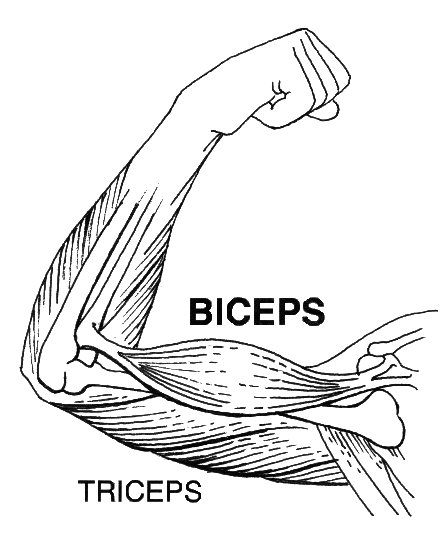
- The biceps and triceps muscles are shown above. Answer the following questions about these arm muscles.
- When the biceps contract and become shorter (as in the picture above), what kind of motion does this produce in the arm?
- Is the situation described in part (a) more likely to be an isometric or isotonic contraction? Explain your answer.
- If the triceps were to then contract, which way would the arm move?
- What are Z discs? What happens to them during muscle contraction?
- What is the function of mitochondria in muscle cells? Which type of muscle fibre has more mitochondria — slow-twitch or fast-twitch?
- What is the difference between primary and secondary Parkinson’s disease?
- Why can carpal tunnel syndrome cause muscle weakness in the hands?
Attributions
Figure 12.7.1
Botox, he whispered by Michael Reuter on Flickr is used under a CC BY 2.0 (https://creativecommons.org/licenses/by/2.0/) license.
Figure 12.7.2
botulism by jason wilson on Flickr is used under a CC BY 2.0 (https://creativecommons.org/licenses/by/2.0/) license.
Reference
Pearson Scott Foresman. (2020, April 14). File:Biceps (PSF).jpg [digital image]. Wikimedia Commons. https://commons.wikimedia.org/w/index.php?title=File:Biceps_(PSF).jpg&oldid=411251538. [Public Domain (https://en.wikipedia.org/wiki/Public_domain)]
Created by CK-12 Foundation/Adapted by Christine Miller

Seeing Your Breath
Why can you “see your breath” on a cold day? The air you exhale through your nose and mouth is warm like the inside of your body. Exhaled air also contains a lot of water vapor, because it passes over moist surfaces from the lungs to the nose or mouth. The water vapor in your breath cools suddenly when it reaches the much colder outside air. This causes the water vapor to condense into a fog of tiny droplets of liquid water. You release water vapor and other gases from your body through the process of respiration.
What is Respiration?
Respiration is the life-sustaining process in which gases are exchanged between the body and the outside atmosphere. Specifically, oxygen moves from the outside air into the body; and water vapor, carbon dioxide, and other waste gases move from inside the body to the outside air. Respiration is carried out mainly by the respiratory system. It is important to note that respiration by the respiratory system is not the same process as cellular respiration —which occurs inside cells — although the two processes are closely connected. Cellular respiration is the metabolic process in which cells obtain energy, usually by “burning” glucose in the presence of oxygen. When cellular respiration is aerobic, it uses oxygen and releases carbon dioxide as a waste product. Respiration by the respiratory system supplies the oxygen needed by cells for aerobic cellular respiration, and removes the carbon dioxide produced by cells during cellular respiration.
Respiration by the respiratory system actually involves two subsidiary processes. One process is ventilation, or breathing. Ventilation is the physical process of conducting air to and from the lungs. The other process is gas exchange. This is the biochemical process in which oxygen diffuses out of the air and into the blood, while carbon dioxide and other waste gases diffuse out of the blood and into the air. All of the organs of the respiratory system are involved in breathing, but only the lungs are involved in gas exchange.
Respiratory Organs
The organs of the respiratory system form a continuous system of passages, called the respiratory tract, through which air flows into and out of the body. The respiratory tract has two major divisions: the upper respiratory tract and the lower respiratory tract. The organs in each division are shown in Figure 13.2.2. In addition to these organs, certain muscles of the thorax (body cavity that fills the chest) are also involved in respiration by enabling breathing. Most important is a large muscle called the diaphragm, which lies below the lungs and separates the thorax from the abdomen. Smaller muscles between the ribs also play a role in breathing.
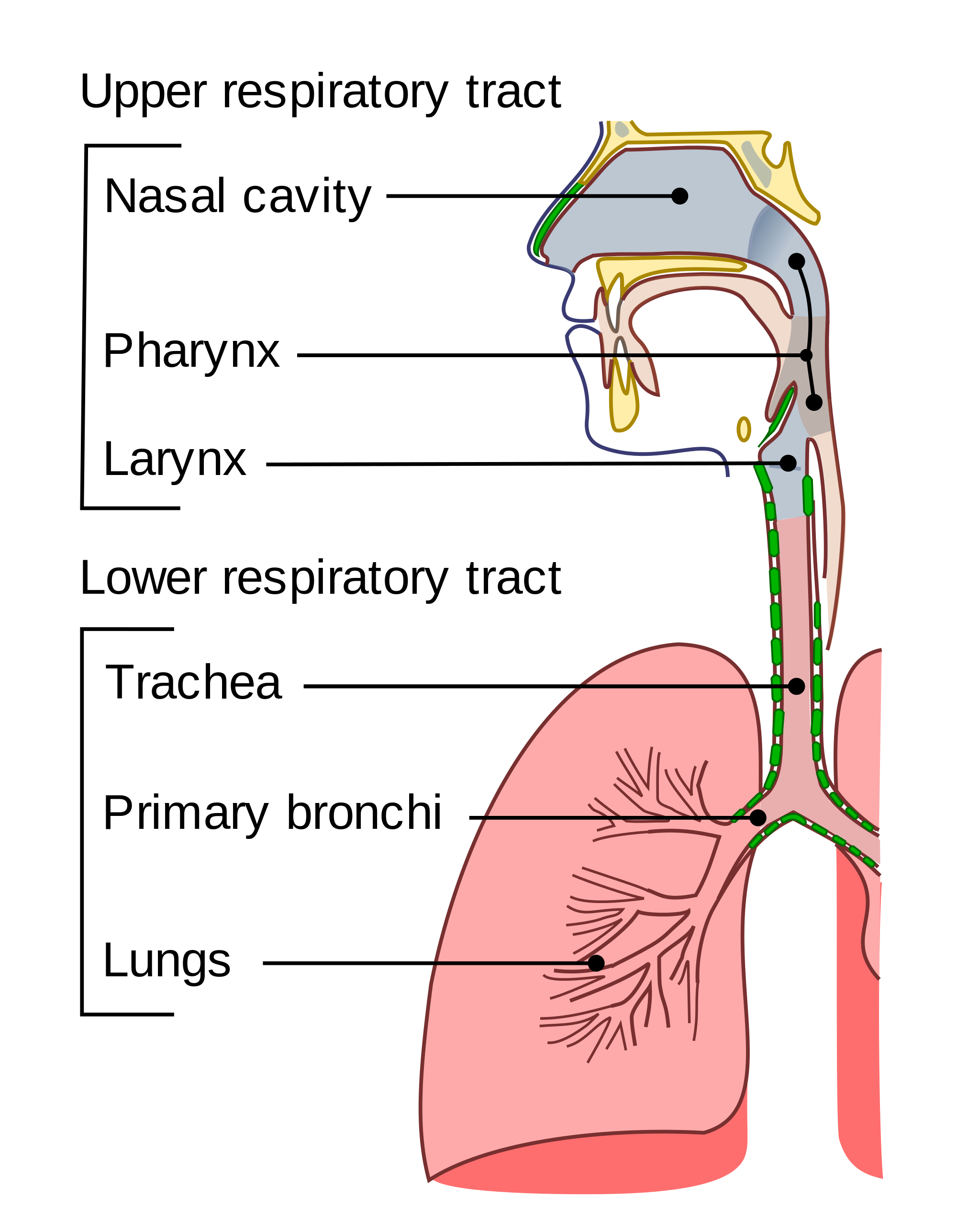
Upper Respiratory Tract
All of the organs and other structures of the upper respiratory tract are involved in conduction, or the movement of air into and out of the body. Upper respiratory tract organs provide a route for air to move between the outside atmosphere and the lungs. They also clean, humidify, and warm the incoming air. No gas exchange occurs in these organs.
Nasal Cavity
The nasal cavity is a large, air-filled space in the skull above and behind the nose in the middle of the face. It is a continuation of the two nostrils. As inhaled air flows through the nasal cavity, it is warmed and humidified by blood vessels very close to the surface of this epithelial tissue . Hairs in the nose and mucous produced by mucous membranes help trap larger foreign particles in the air before they go deeper into the respiratory tract. In addition to its respiratory functions, the nasal cavity also contains chemoreceptors needed for sense of smell, and contribution to the sense of taste.
Pharynx
The pharynx is a tube-like structure that connects the nasal cavity and the back of the mouth to other structures lower in the throat, including the larynx. The pharynx has dual functions — both air and food (or other swallowed substances) pass through it, so it is part of both the respiratory and the digestive systems. Air passes from the nasal cavity through the pharynx to the larynx (as well as in the opposite direction). Food passes from the mouth through the pharynx to the esophagus.
Larynx
The larynx connects the pharynx and trachea, and helps to conduct air through the respiratory tract. The larynx is also called the voice box, because it contains the vocal cords, which vibrate when air flows over them, thereby producing sound. You can see the vocal cords in the larynx in Figures 13.2.3 and 13.2.4. Certain muscles in the larynx move the vocal cords apart to allow breathing. Other muscles in the larynx move the vocal cords together to allow the production of vocal sounds. The latter muscles also control the pitch of sounds and help control their volume.
 |
Figure 13.2.4 The larynx as viewed from the top. |
A very important function of the larynx is protecting the trachea from aspirated food. When swallowing occurs, the backward motion of the tongue forces a flap called the epiglottis to close over the entrance to the larynx. (You can see the epiglottis in both Figure 13.2.3 and 13.2.4.) This prevents swallowed material from entering the larynx and moving deeper into the respiratory tract. If swallowed material does start to enter the larynx, it irritates the larynx and stimulates a strong cough reflex. This generally expels the material out of the larynx, and into the throat.
https://www.youtube.com/watch?v=BsyB88mq5rQ
Larynx Model - Respiratory System, Dr. Lotz, 2018.
Lower Respiratory Tract
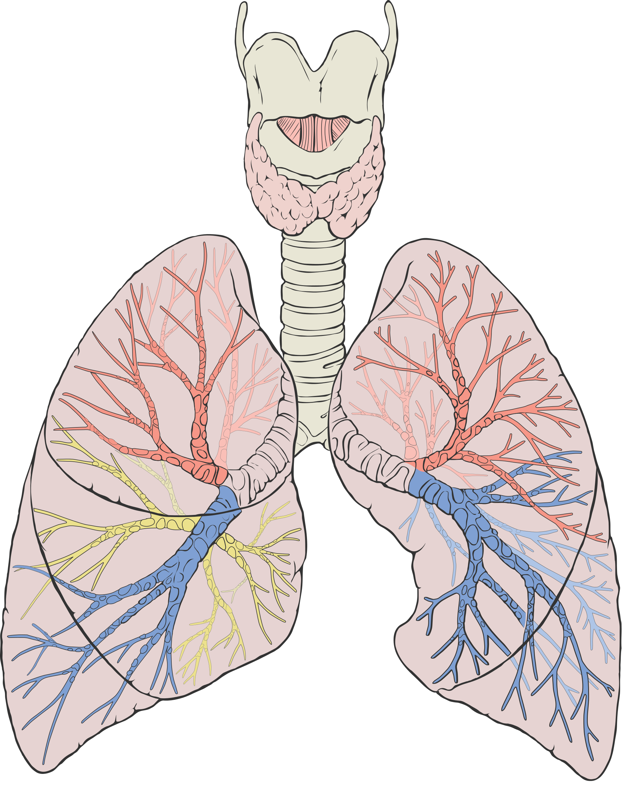
The trachea and other passages of the lower respiratory tract conduct air between the upper respiratory tract and the lungs. These passages form an inverted tree-like shape (Figure 13.2.5), with repeated branching as they move deeper into the lungs. All told, there are an astonishing 2,414 kilometres (1,500 miles) of airways conducting air through the human respiratory tract! It is only in the lungs, however, that gas exchange occurs between the air and the bloodstream.
Trachea
The trachea, or windpipe, is the widest passageway in the respiratory tract. It is about 2.5 cm wide and 10-15 cm long (approximately 1 inch wide and 4–6 inches long). It is formed by rings of cartilage, which make it relatively strong and resilient. The trachea connects the larynx to the lungs for the passage of air through the respiratory tract. The trachea branches at the bottom to form two bronchial tubes.
Bronchi and Bronchioles
There are two main bronchial tubes, or bronchi (singular, bronchus), called the right and left bronchi. The bronchi carry air between the trachea and lungs. Each bronchus branches into smaller, secondary bronchi; and secondary bronchi branch into still smaller tertiary bronchi. The smallest bronchi branch into very small tubules called bronchioles. The tiniest bronchioles end in alveolar ducts, which terminate in clusters of minuscule air sacs, called alveoli (singular, alveolus), in the lungs.
Lungs
The lungs are the largest organs of the respiratory tract. They are suspended within the pleural cavity of the thorax. The lungs are surrounded by two thin membranes called pleura, which secrete fluid that allows the lungs to move freely within the pleural cavity. This is necessary so the lungs can expand and contract during breathing. In Figure 13.2.6, you can see that each of the two lungs is divided into sections. These are called lobes, and they are separated from each other by connective tissues. The right lung is larger and contains three lobes. The left lung is smaller and contains only two lobes. The smaller left lung allows room for the heart, which is just left of the center of the chest.
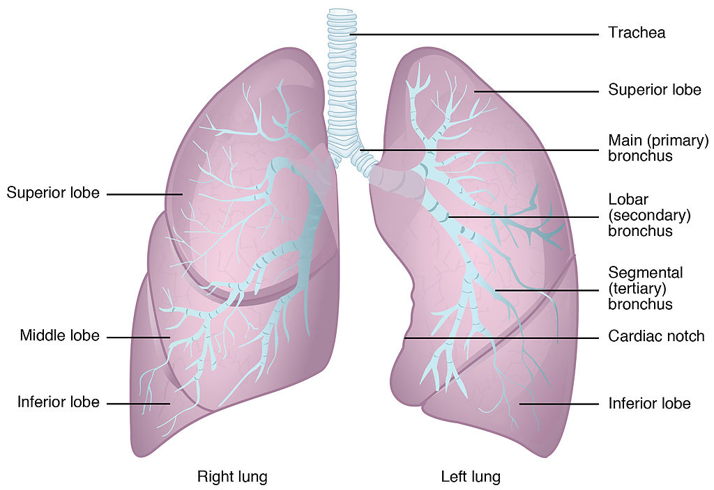
As mentioned previously, the bronchi terminate in bronchioles which feed air into alveoli, tiny sacs of simple squamous epithelial tissue which make up the bulk of the lung. The cross-section of lung tissue in the diagram below (Figure 13.2.7) shows the alveoli, in which gas exchange takes place with the capillary network that surrounds them.
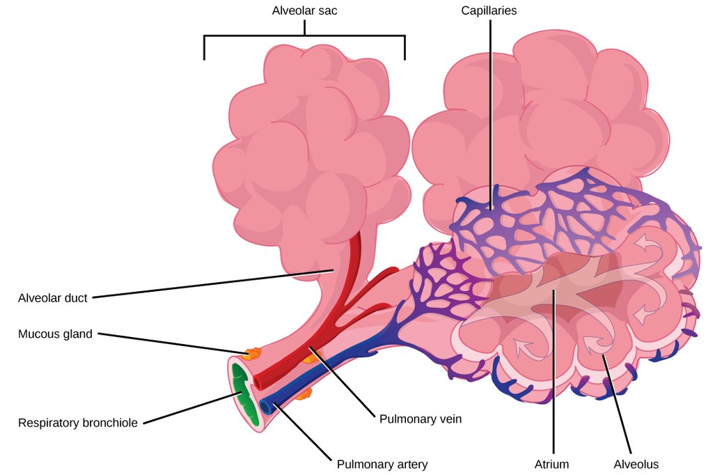 |
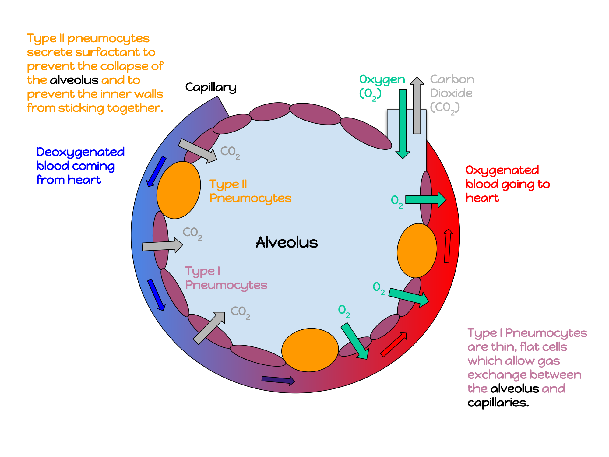 |
Lung tissue consists mainly of alveoli (see Figures 13.2.7 and 13.2.8). These tiny air sacs are the functional units of the lungs where gas exchange takes place. The two lungs may contain as many as 700 million alveoli, providing a huge total surface area for gas exchange to take place. In fact, alveoli in the two lungs provide as much surface area as half a tennis court! Each time you breathe in, the alveoli fill with air, making the lungs expand. Oxygen in the air inside the alveoli is absorbed by the blood via diffusion in the mesh-like network of tiny capillaries that surrounds each alveolus. The blood in these capillaries also releases carbon dioxide (also by diffusion) into the air inside the alveoli. Each time you breathe out, air leaves the alveoli and rushes into the outside atmosphere, carrying waste gases with it.
The lungs receive blood from two major sources. They receive deoxygenated blood from the right side of the heart. This blood absorbs oxygen in the lungs and carries it back to the left side heart to be pumped to cells throughout the body. The lungs also receive oxygenated blood from the heart that provides oxygen to the cells of the lungs for cellular respiration.
Protecting the Respiratory System
You may be able to survive for weeks without food and for days without water, but you can survive without oxygen for only a matter of minutes — except under exceptional circumstances — so protecting the respiratory system is vital. Ensuring that a patient has an open airway is the first step in treating many medical emergencies. Fortunately, the respiratory system is well protected by the ribcage of the skeletal system. The extensive surface area of the respiratory system, however, is directly exposed to the outside world and all its potential dangers in inhaled air. It should come as no surprise that the respiratory system has a variety of ways to protect itself from harmful substances, such as dust and pathogens in the air.
The main way the respiratory system protects itself is called the mucociliary escalator. From the nose through the bronchi, the respiratory tract is covered in epithelium that contains mucus-secreting goblet cells. The mucus traps particles and pathogens in the incoming air. The epithelium of the respiratory tract is also covered with tiny cell projections called cilia (singular, cilium), as shown in the animation. The cilia constantly move in a sweeping motion upward toward the throat, moving the mucus and trapped particles and pathogens away from the lungs and toward the outside of the body. The upward sweeping motion of cilia lining the respiratory tract helps keep it free from dust, pathogens, and other harmful substances.
Watch "Mucociliary clearance" by I-Hsun Wu to learn more:
https://www.youtube.com/watch?v=HMB6flEaZwI
Mucociliary clearance, I-Hsun Wu, 2015.
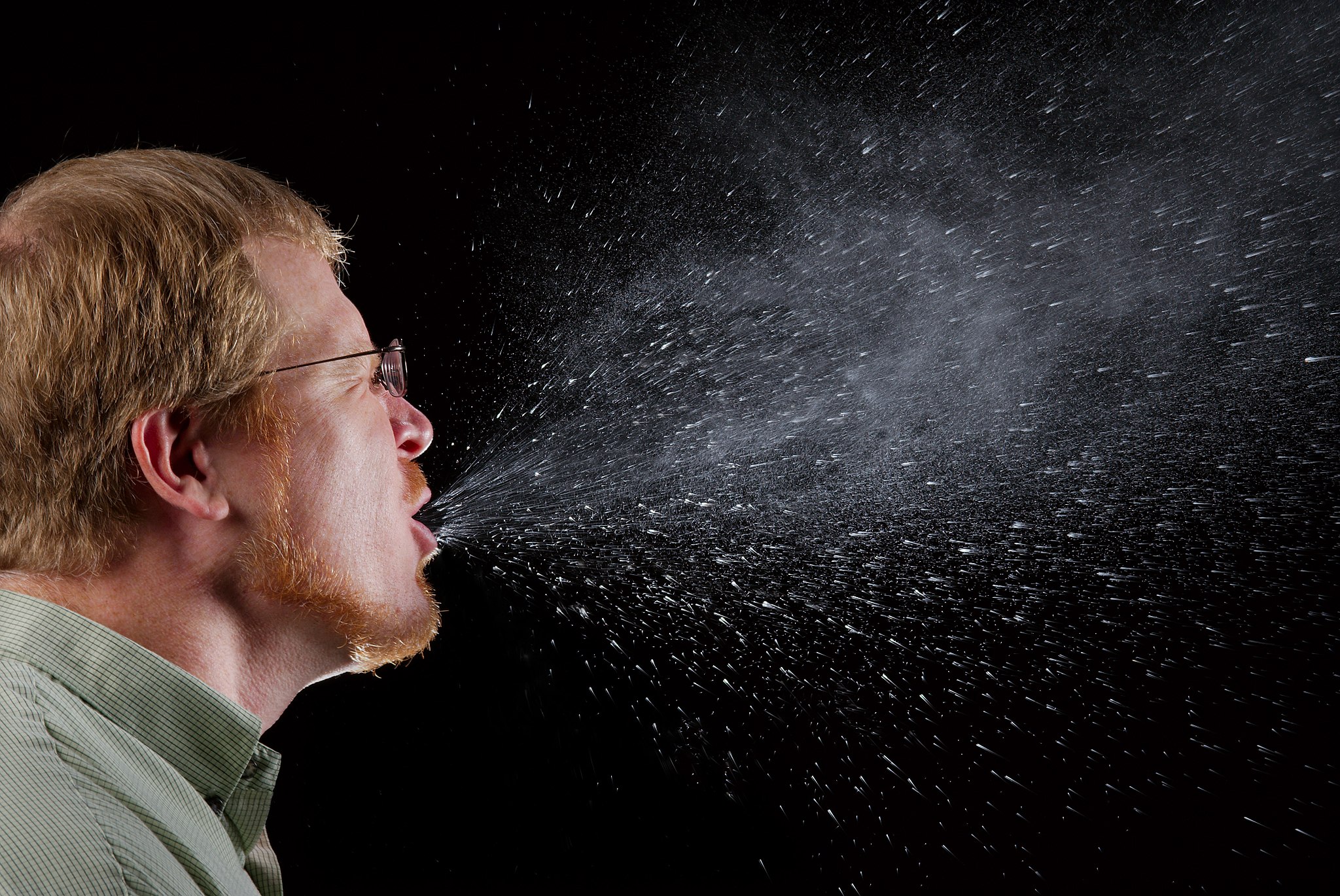
Sneezing is a similar involuntary response that occurs when nerves lining the nasal passage are irritated. It results in forceful expulsion of air from the mouth, which sprays millions of tiny droplets of mucus and other debris out of the mouth and into the air, as shown in Figure 13.2.9. This explains why it is so important to sneeze into a tissue (rather than the air) if we are to prevent the transmission of respiratory pathogens.
How the Respiratory System Works with Other Organ Systems
The amount of oxygen and carbon dioxide in the blood must be maintained within a limited range for survival of the organism. Cells cannot survive for long without oxygen, and if there is too much carbon dioxide in the blood, the blood becomes dangerously acidic (pH is too low). Conversely, if there is too little carbon dioxide in the blood, the blood becomes too basic (pH is too high). The respiratory system works hand-in-hand with the nervous and cardiovascular systems to maintain homeostasis in blood gases and pH.
It is the level of carbon dioxide — rather than the level of oxygen — that is most closely monitored to maintain blood gas and pH homeostasis. The level of carbon dioxide in the blood is detected by cells in the brain, which speed up or slow down the rate of breathing through the autonomic nervous system as needed to bring the carbon dioxide level within the normal range. Faster breathing lowers the carbon dioxide level (and raises the oxygen level and pH), while slower breathing has the opposite effects. In this way, the levels of carbon dioxide, oxygen, and pH are maintained within normal limits.
The respiratory system also works closely with the cardiovascular system to maintain homeostasis. The respiratory system exchanges gases with the outside air, but it needs the cardiovascular system to carry them to and from body cells. Oxygen is absorbed by the blood in the lungs and then transported through a vast network of blood vessels to cells throughout the body, where it is needed for aerobic cellular respiration. The same system absorbs carbon dioxide from cells and carries it to the respiratory system for removal from the body.
Feature: My Human Body
Choking due to a foreign object becoming lodged in the airway results in nearly 5 thousand deaths in Canada each year. In addition, choking accounts for almost 40% of unintentional injuries in infants under the age of one. For the sake of your own human body, as well as those of loved ones, you should be aware of choking risks, signs, and treatments.
Choking is the mechanical obstruction of the flow of air from the atmosphere into the lungs. It prevents breathing, and may be partial or complete. Partial choking allows some — though inadequate — air flow into the lungs. Prolonged or complete choking results in asphyxia, or suffocation, which is potentially fatal.
Obstruction of the airway typically occurs in the pharynx or trachea. Young children are more prone to choking than are older people, in part because they often put small objects in their mouth and do not understand the risk of choking that they pose. Young children may choke on small toys or parts of toys, or on household objects, in addition to food. Foods that are round (hotdogs, carrots, grapes) or can adapt their shape to that of the pharynx (bananas, marshmallows), are especially dangerous, and may cause choking in adults, as well as children.
How can you tell if a loved one is choking? The person cannot speak or cry out, or has great difficulty doing so. Breathing, if possible, is laboured, producing gasping or wheezing. The person may desperately clutch at his or her throat or mouth. If breathing is not soon restored, the person’s face will start to turn blue from lack of oxygen. This will be followed by unconsciousness, brain damage, and possibly death if oxygen deprivation continues beyond a few minutes.
If an infant is choking, turning the baby upside down and slapping him on the back may dislodge the obstructing object. To help an older person who is choking, first encourage the person to cough. Give them a few hard back slaps to help force the lodged object out of the airway. If these steps fail, perform the Heimlich maneuver on the person. See the series of videos below, from ProCPR, which demonstrate several ways to help someone who is choking based on age and consciousness.
https://www.youtube.com/watch?v=XOTbjDGZ7wg&t=46s
Conscious Adult Choking, ProCPR, 2016.
https://www.youtube.com/watch?v=5kmsKNvKAvU
Unconscious Adult Choking, ProCPR, 2011.
https://www.youtube.com/watch?v=ZjmbD7aIaf0
Conscious Child Choking, ProCPR, 2009.
https://www.youtube.com/watch?v=Sba0T2XGIn4
Unconscious Child Choking, ProCPR, 2009.
https://www.youtube.com/watch?v=axqIju9CLKA
Conscious Infant Choking, ProCPR, 2011.
https://www.youtube.com/watch?v=_K7Dwy6b2wQ
Unconscious Infant Choking, ProCPR, 2011.
13.2 Summary
- Respiration is the process in which oxygen moves from the outside air into the body, and in which carbon dioxide and other waste gases move from inside the body into the outside air. It involves two subsidiary processes: ventilation and gas exchange. Respiration is carried out mainly by the respiratory system.
- The organs of the respiratory system form a continuous system of passages, called the respiratory tract. It has two major divisions: the upper respiratory tract and the lower respiratory tract.
- The upper respiratory tract includes the nasal cavity, pharynx, and larynx. All of these organs are involved in conduction, or the movement of air into and out of the body. Incoming air is also cleaned, humidified, and warmed as it passes through the upper respiratory tract. The larynx is called the voice box, because it contains the vocal cords, which are needed to produce vocal sounds.
- The lower respiratory tract includes the trachea, bronchi and bronchioles, and the lungs. The trachea, bronchi, and bronchioles are involved in conduction. Gas exchange takes place only in the lungs, which are the largest organs of the respiratory tract. Lung tissue consists mainly of tiny air sacs called alveoli, which is where gas exchange takes place between air in the alveoli and the blood in capillaries surrounding them.
- The respiratory system protects itself from potentially harmful substances in the air by the mucociliary escalator. This includes mucus-producing cells, which trap particles and pathogens in incoming air. It also includes tiny hair-like cilia that continually move to sweep the mucus and trapped debris away from the lungs and toward the outside of the body.
- The level of carbon dioxide in the blood is monitored by cells in the brain. If the level becomes too high, it triggers a faster rate of breathing, which lowers the level to the normal range. The opposite occurs if the level becomes too low. The respiratory system exchanges gases with the outside air, but it needs the cardiovascular system to carry the gases to and from cells throughout the body.
13.2 Review Questions
-
- What is respiration, as carried out by the respiratory system? Name the two subsidiary processes it involves.
- Describe the respiratory tract.
- Identify the organs of the upper respiratory tract. What are their functions?
- List the organs of the lower respiratory tract. Which organs are involved only in conduction?
- Where does gas exchange take place?
- How does the respiratory system protect itself from potentially harmful substances in the air?
- Explain how the rate of breathing is controlled.
- Why does the respiratory system need the cardiovascular system to help it perform its main function of gas exchange?
- Describe two ways in which the body prevents food from entering the lungs.
- What is the relationship between respiration and cellular respiration?
13.2 Explore More
https://www.youtube.com/watch?v=8NUxvJS-_0k
How do lungs work? - Emma Bryce, TED-Ed, 2014.
https://www.youtube.com/watch?time_continue=1&v=6iFPs6JlSzY&feature=emb_logo
Why Do Men Have Deeper Voices? BrainStuff - HowStuffWorks, 2015.
https://www.youtube.com/watch?v=rjibeBSnpJ0
Why does your voice change as you get older? - Shaylin A. Schundler, TED-Ed, 2018.
Attributions
Figure 13.2.1
Exhale by pavel-lozovikov-HYovA7yPPvI [photo] by Pavel Lozovikov on Unsplash is used under the Unsplash License (https://unsplash.com/license).
Figure 13.2.2
Illu_conducting_passages.svg by Lord Akryl, Jmarchn from SEER Training Modules/ National Cancer Institute on Wikimedia Commons is in the public domain (https://en.wikipedia.org/wiki/Public_domain).
Figure 13.2.3
Larynx by www.medicalgraphics.de is used under a CC BY-ND 4.0 (https://creativecommons.org/licenses/by-nd/4.0/) license.
Figure 13.2.4
Larynx top view by Alan Hoofring (Illustrator) for National Cancer Institute is in the public domain (https://en.wikipedia.org/wiki/Public_domain).
Figure 13.2.5
2000px-Lungs_diagram_detailed.svg by Patrick J. Lynch, medical illustrator on Wikimedia Commons is used under a CC BY 2.5 (https://creativecommons.org/licenses/by/2.5) license. (Derivative work of Fruchtwasserembolie.png.)
Figure 13.2.6
Gross_Anatomy_of_the_Lungs by OpenStax College on Wikimedia Commons is used under a CC BY 3.0 (https://creativecommons.org/licenses/by/3.0) license.
Figure 13.2.7
Alveoli Structure by CNX OpenStax on Wikimedia Commons is used under a CC BY 4.0 (https://creativecommons.org/licenses/by/4.0) license.
Figure 13.2.8
annotated_diagram_of_an_alveolus.svg by Katherinebutler1331 on Wikimedia Commons is used under a CC BY-SA 4.0 (https://creativecommons.org/licenses/by-sa/4.0) license.
Figure 13.2.9
Sneeze by James Gathany at CDC Public Health Imagery Library (PHIL) #11162 on Wikimedia Commons is in the public domain (https://en.wikipedia.org/wiki/Public_domain).
References
Betts, J. G., Young, K.A., Wise, J.A., Johnson, E., Poe, B., Kruse, D.H., Korol, O., Johnson, J.E., Womble, M., DeSaix, P. (2013, June 19). Figure 22.2 Major respiratory structures [digital image]. In Anatomy and Physiology (Section 22.1). OpenStax. https://openstax.org/books/anatomy-and-physiology/pages/22-1-organs-and-structures-of-the-respiratory-system [CC BY 4.0 (https://creativecommons.org/licenses/by/4.0)].
Betts, J. G., Young, K.A., Wise, J.A., Johnson, E., Poe, B., Kruse, D.H., Korol, O., Johnson, J.E., Womble, M., DeSaix, P. (2013, June 19). Figure 22.13 Gross anatomy of the lungs [digital image]. In Anatomy and Physiology (Section 22.2). OpenStax. https://openstax.org/books/anatomy-and-physiology/pages/22-2-the-lungs
BrainStuff - HowStuffWorks. (2015, December 1). Why do men have deeper voices? YouTube. https://www.youtube.com/watch?v=6iFPs6JlSzY&feature=youtu.be
Dr. Lotz. (2018, January 25). Larynx model - Respiratory system. YouTube. https://www.youtube.com/watch?v=BsyB88mq5rQ&feature=youtu.be
I-Hsun Wu. (2015, March 31). Mucociliary clearance. YouTube. https://www.youtube.com/watch?v=HMB6flEaZwI&feature=youtu.be
OpenStax. (2016, May 27). Figure 9 Terminal bronchioles are connected by respiratory bronchioles to alveolar ducts and alveolar sacs [digital image]. In OpenStax, Biology (Section 39.1). OpenStax CNX. https://cnx.org/contents/GFy_h8cu@10.53:35-R0biq@11/Systems-of-Gas-Exchange
ProCPR. (2009, November 24). Conscious child choking. YouTube. https://www.youtube.com/watch?v=ZjmbD7aIaf0&feature=youtu.be
ProCPR. (2009, November 24).Unconscious child choking. YouTube. https://www.youtube.com/watch?v=Sba0T2XGIn4&feature=youtu.be
ProCPR. (2011, February 1). Conscious infant choking. YouTube. https://www.youtube.com/watch?v=axqIju9CLKA&feature=youtu.be
ProCPR. (2011, February 1). Unconscious adult choking. YouTube. https://www.youtube.com/watch?v=5kmsKNvKAvU&feature=youtu.be
ProCPR. (2011, February 1). Unconscious infant choking. YouTube. https://www.youtube.com/watch?v=_K7Dwy6b2wQ&feature=youtu.be
ProCPR. (2016, April 8). Conscious adult choking. YouTube. https://www.youtube.com/watch?v=XOTbjDGZ7wg&feature=youtu.be
TED-Ed. (2014, November 24). How do lungs work? - Emma Bryce. YouTube. https://www.youtube.com/watch?v=8NUxvJS-_0k&feature=youtu.be
TED-Ed. (2018, August 2). Why does your voice change as you get older? - Shaylin A. Schundler. YouTube. https://www.youtube.com/watch?v=rjibeBSnpJ0&feature=youtu.be
Created by CK-12 Foundation/Adapted by Christine Miller

Doing the ‘Fly
The swimmer in the Figure 13.3.1 photo is doing the butterfly stroke, a swimming style that requires the swimmer to carefully control his breathing so it is coordinated with his swimming movements. Breathing is the process of moving air into and out of the lungs, which are the organs in which gas exchange takes place between the atmosphere and the body. Breathing is also called ventilation, and it is one of two parts of the life-sustaining process of respiration. The other part is gas exchange. Before you can understand how breathing is controlled, you need to know how breathing occurs.
How Breathing Occurs
Breathing is a two-step process that includes drawing air into the lungs, or inhaling, and letting air out of the lungs, or exhaling. Both processes are illustrated in Figure 13.3.2.
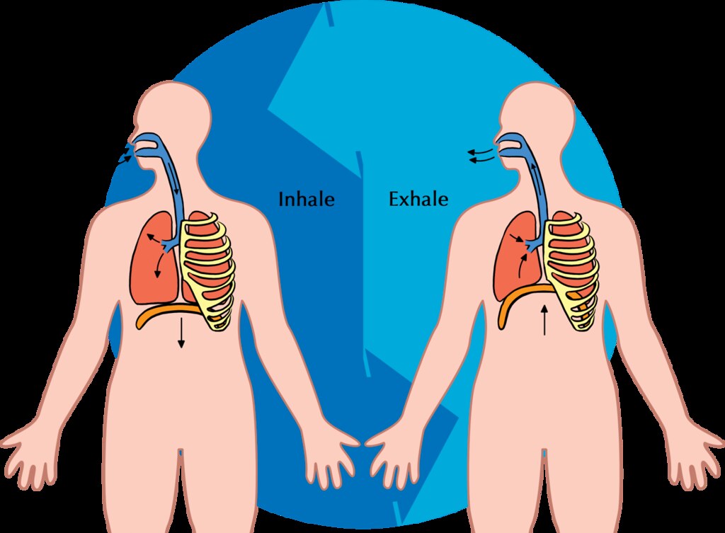
Inhaling
Inhaling is an active process that results mainly from contraction of a muscle called the diaphragm, shown in Figure 13.3.2. The diaphragm is a large, dome-shaped muscle below the lungs that separates the thoracic (chest) and abdominal cavities. When the diaphragm contracts it moves down causing the thoracic cavity to expand, and the contents of the abdomen to be pushed downward. Other muscles — such as intercostal muscles between the ribs — also contribute to the process of inhalation, especially when inhalation is forced, as when taking a deep breath. These muscles help increase thoracic volume by expanding the ribs outward. The increase in thoracic volume creates a decrease in thoracic air pressure. With the chest expanded, there is lower air pressure inside the lungs than outside the body, so outside air flows into the lungs via the respiratory tract according the the pressure gradient (high pressure flows to lower pressure).
Exhaling
Exhaling involves the opposite series of events. The diaphragm relaxes, so it moves upward and decreases the volume of the thorax. Air pressure inside the lungs increases, so it is higher than the air pressure outside the lungs. Exhalation, unlike inhalation, is typically a passive process that occurs mainly due to the elasticity of the lungs. With the change in air pressure, the lungs contract to their pre-inflated size, forcing out the air they contain in the process. Air flows out of the lungs, similar to the way air rushes out of a balloon when it is released. If exhalation is forced, internal intercostal and abdominal muscles may help move the air out of the lungs.
Control of Breathing
Breathing is one of the few vital bodily functions that can be controlled consciously, as well as unconsciously. Think about using your breath to blow up a balloon. You take a long, deep breath, and then you exhale the air as forcibly as you can into the balloon. Both the inhalation and exhalation are consciously controlled.
Conscious Control of Breathing
You can control your breathing by holding your breath, slowing your breathing, or hyperventilating, which is breathing more quickly and shallowly than necessary. You can also exhale or inhale more forcefully or deeply than usual. Conscious control of breathing is common in many activities besides blowing up balloons, including swimming, speech training, singing, playing many different musical instruments (Figure 13.3.3), and doing yoga, to name just a few.

There are limits on the conscious control of breathing. For example, it is not possible for a healthy person to voluntarily stop breathing indefinitely. Before long, there is an irrepressible urge to breathe. If you were able to stop breathing for a long enough time, you would lose consciousness. The same thing would happen if you were to hyperventilate for too long. Once you lose consciousness so you can no longer exert conscious control over your breathing, involuntary control of breathing takes over.
Unconscious Control of Breathing
Unconscious breathing is controlled by respiratory centers in the medulla and pons of the brainstem (see Figure 13.3.4). The respiratory centers automatically and continuously regulate the rate of breathing based on the body’s needs. These are determined mainly by blood acidity, or pH. When you exercise, for example, carbon dioxide levels increase in the blood, because of increased cellular respiration by muscle cells. The carbon dioxide reacts with water in the blood to produce carbonic acid, making the blood more acidic, so pH falls. The drop in pH is detected by chemoreceptors in the medulla. Blood levels of oxygen and carbon dioxide, in addition to pH, are also detected by chemoreceptors in major arteries, which send the “data” to the respiratory centers. The latter respond by sending nerve impulses to the diaphragm, “telling” it to contract more quickly so the rate of breathing speeds up. With faster breathing, more carbon dioxide is released into the air from the blood, and blood pH returns to the normal range.
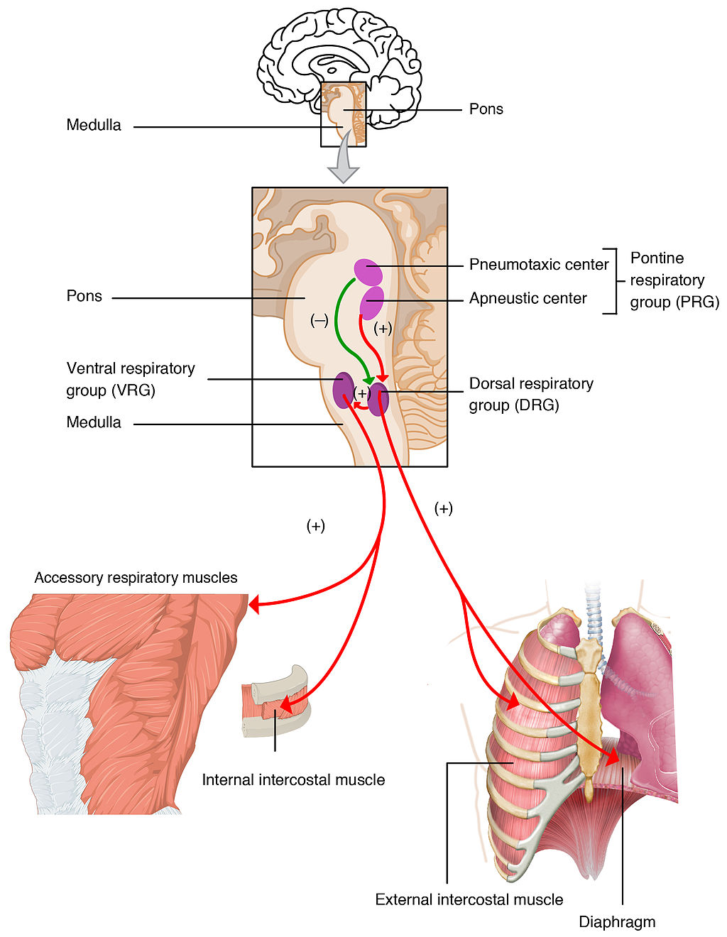
The opposite events occur when the level of carbon dioxide in the blood becomes too low and blood pH rises. This may occur with involuntary hyperventilation, which can happen in panic attacks, episodes of severe pain, asthma attacks, and many other situations. When you hyperventilate, you blow off a lot of carbon dioxide, leading to a drop in blood levels of carbon dioxide. The blood becomes more basic (alkaline), causing its pH to rise.
Nasal vs. Mouth Breathing
Nasal breathing is breathing through the nose rather than the mouth, and it is generally considered to be superior to mouth breathing. The hair-lined nasal passages do a better job of filtering particles out of the air before it moves deeper into the respiratory tract. The nasal passages are also better at warming and moistening the air, so nasal breathing is especially advantageous in the winter when the air is cold and dry. In addition, the smaller diameter of the nasal passages creates greater pressure in the lungs during exhalation. This slows the emptying of the lungs, giving them more time to extract oxygen from the air.
Feature: Myth vs. Reality
Drowning is defined as respiratory impairment from being in or under a liquid. It is further classified according to its outcome into: death, ongoing health problems, or no ongoing health problems (full recovery). Four hundred Canadians die annually from drowning, and drowning is one of the leading causes of death in children under the age of five. There are some potentially dangerous myths about drowning, and knowing what they are might save your life or the life of a loved one, especially a child.
| Myth | Reality |
|---|---|
| "People drown when they aspirate water into their lungs." | Generally, in the early stages of drowning, very little water enters the lungs. A small amount of water entering the trachea causes a muscular spasm in the larynx that seals the airway and prevents the passage of water into the lungs. This spasm is likely to last until unconsciousness occurs. |
| "You can tell when someone is drowning because they will shout for help and wave their arms to attract attention." | The muscular spasm that seals the airway prevents the passage of air, as well as water, so a person who is drowning is unable to shout or call for help. In addition, instinctive reactions that occur in the final minute or so before a drowning person sinks under the water may look similar to calm, safe behavior. The head is likely to be low in the water, tilted back, with the mouth open. The person may have uncontrolled movements of the arms and legs, but they are unlikely to be visible above the water. |
| "It is too late to save a person who is unconscious in the water." | An unconscious person rescued with an airway still sealed from the muscular spasm of the larynx stands a good chance of full recovery if they start receiving CPR within minutes. Without water in the lungs, CPR is much more effective. Even if cardiac arrest has occurred so the heart is no longer beating, there is still a chance of recovery. The longer the brain goes without oxygen, however, the more likely brain cells are to die. Brain death is likely after about six minutes without oxygen, except in exceptional circumstances, such as young people drowning in very cold water. There are examples of children surviving, apparently without lasting ill effects, for as long as an hour in cold water. Rescuers retrieving a child from cold water should attempt resuscitation even after a protracted period of immersion. |
| "If someone is drowning, you should start administering CPR immediately, even before you try to get the person out of the water." | Removing a drowning person from the water is the first priority, because CPR is ineffective in the water. The goal should be to bring the person to stable ground as quickly as possible and then to start CPR. |
| "You are unlikely to drown unless you are in water over your head." | Depending on circumstances, people have drowned in as little as 30 mm (about 1 ½ in.) of water. Inebriated people or those under the influence of drugs, for example, have been known to have drowned in puddles. Hundreds of children have drowned in the water in toilets, bathtubs, basins, showers, pails, and buckets (see Figure 13.3.5). |
13.3 Summary
- Breathing, or ventilation, is the two-step process of drawing air into the lungs (inhaling) and letting air out of the lungs (exhaling). Inhalation is an active process that results mainly from contraction of a muscle called the diaphragm. Exhalation is typically a passive process that occurs mainly due to the elasticity of the lungs when the diaphragm relaxes.
- Breathing is one of the few vital bodily functions that can be controlled consciously, as well as unconsciously. Conscious control of breathing is common in many activities, including swimming and singing. There are limits on the conscious control of breathing, however. If you try to hold your breath, for example, you will soon have an irrepressible urge to breathe.
- Unconscious breathing is controlled by respiratory centers in the medulla and pons of the brainstem. They respond to variations in blood pH by either increasing or decreasing the rate of breathing as needed to return the pH level to the normal range.
- Nasal breathing is generally considered to be superior to mouth breathing because it does a better job of filtering, warming, and moistening incoming air. It also results in slower emptying of the lungs, which allows more oxygen to be extracted from the air.
- Drowning is a major cause of death in Canada, in particular in children under the age of five. It is important to supervise small children when they are playing in, around, or with water.
13.3 Review Questions
- Define breathing.
-
- Give examples of activities in which breathing is consciously controlled.
- Explain how unconscious breathing is controlled.
- Young children sometimes threaten to hold their breath until they get something they want. Why is this an idle threat?
- Why is nasal breathing generally considered superior to mouth breathing?
- Give one example of a situation that would cause blood pH to rise excessively. Explain why this occurs.
13.3 Explore More
https://www.youtube.com/watch?v=Kl4cU9sG_08
How breathing works - Nirvair Kaur, TED-Ed, 2012.
https://www.youtube.com/watch?v=yDtKBXOEsoM
How do ventilators work? - Alex Gendler, TED-Ed, 2020.
https://www.youtube.com/watch?v=XFnGhrC_3Gs&feature=emb_logo
How I held my breath for 17 minutes | David Blaine, TED, 2010.
https://www.youtube.com/watch?v=Vca6DyFqt4c&feature=emb_logo
The Ultimate Relaxation Technique: How To Practice Diaphragmatic Breathing For Beginners, Kai Simon, 2015.
Attributions
Figure 13.3.1
US_Marines_butterfly_stroke by Cpl. Jasper Schwartz from U.S. Marine Corps on Wikimedia Commons is in the public domain (https://en.wikipedia.org/wiki/Public_domain).
Figure 13.3.2
Inhale Exhale/Breathing cycle by Siyavula Education on Flickr is used under a CC BY 2.0 (https://creativecommons.org/licenses/by/2.0/) license.
Figure 13.3.3
Trumpet/ Frenchmen Street [photo] by Morgan Petroski on Unsplash is used under the Unsplash License (https://unsplash.com/license).
Figure 13.3.4
Respiratory_Centers_of_the_Brain by OpenStax College on Wikimedia Commons is used under a CC BY 3.0 (https://creativecommons.org/licenses/by/3.0) license.
Figure 13.3.5
Lily & Ava in the Kiddie Pool by mob mob on Flickr is used under a CC BY-NC 2.0 (https://creativecommons.org/licenses/by-nc/2.0/) license.
References
Betts, J. G., Young, K.A., Wise, J.A., Johnson, E., Poe, B., Kruse, D.H., Korol, O., Johnson, J.E., Womble, M., DeSaix, P. (2013, June 19). Figure 22.20 Respiratory centers of the brain [digital image]. In Anatomy and Physiology (Section 22.3). OpenStax. https://openstax.org/books/anatomy-and-physiology/pages/22-3-the-process-of-breathing
Kai Simon. (2015, January 11). The ultimate relaxation technique: How to practice diaphragmatic breathing for beginners. YouTube. https://www.youtube.com/watch?v=Vca6DyFqt4c&feature=youtu.be
TED. (2010, January 19). How I held my breath for 17 minutes | David Blaine. YouTube. https://www.youtube.com/watch?v=XFnGhrC_3Gs&feature=youtu.be
TED-Ed. (2012, October 4). How breathing works - Nirvair Kaur. YouTube. https://www.youtube.com/watch?v=Kl4cU9sG_08&feature=youtu.be
TED-Ed. (2020, May 21). How do ventilators work? - Alex Gendler. YouTube. https://www.youtube.com/watch?v=yDtKBXOEsoM&feature=youtu.be
Created by CK-12 Foundation/Adapted by Christine Miller
Oxygen Bar
Belly up to the bar and get your favorite... oxygen? That’s right — in some cities, you can get a shot of pure oxygen, with or without your choice of added flavors. Bar patrons inhale oxygen through a plastic tube inserted into their nostrils, paying up to a dollar per minute to inhale the pure gas. Proponents of the practice claim that breathing in extra oxygen will remove toxins from the body, strengthen the immune system, enhance concentration and alertness, increase energy, and even cure cancer! These claims, however, have not been substantiated by controlled scientific studies. Normally, blood leaving the lungs is almost completely saturated with oxygen, even without the use of extra oxygen, so it’s unlikely that a higher concentration of oxygen in air inside the lungs would lead to significantly greater oxygenation of the blood. Oxygen enters the blood in the lungs as part of the process of gas exchange.
What is Gas Exchange?
Gas exchange is the biological process through which gases are transferred across cell membranes to either enter or leave the blood. Oxygen is constantly needed by cells for aerobic cellular respiration, and the same process continually produces carbon dioxide as a waste product. Gas exchange takes place between the blood and cells throughout the body, with oxygen leaving the blood and entering the cells, and carbon dioxide leaving the cells and entering the blood. Gas exchange also takes place between the blood and the air in the lungs, with oxygen entering the blood from the inhaled air inside the lungs, and carbon dioxide leaving the blood and entering the air to be exhaled from the lungs.
Gas Exchange in the Lungs
Alveoli are the basic functional units of the lungs where gas exchange takes place between the air and the blood. Alveoli (singular, alveolus) are tiny air sacs that consist of connective and epithelial tissues. The connective tissue includes elastic fibres that allow alveoli to stretch and expand as they fill with air during inhalation. During exhalation, the fibres allow the alveoli to spring back and expel the air. Special cells in the walls of the alveoli secrete a film of fatty substances called surfactant. This substance prevents the alveolar walls from collapsing and sticking together when air is expelled. Other cells in alveoli include macrophages, which are mobile scavengers that engulf and destroy foreign particles that manage to reach the lungs in inhaled air.
As shown in Figure 13.4.2, alveoli are arranged in groups like clusters of grapes. Each alveolus is covered with epithelium that is just one cell thick. It is surrounded by a bed of pulmonary capillaries, each of which has a wall of epithelium just one cell thick. As a result, gases must cross through only two cells to pass between an alveolus and its surrounding capillaries.
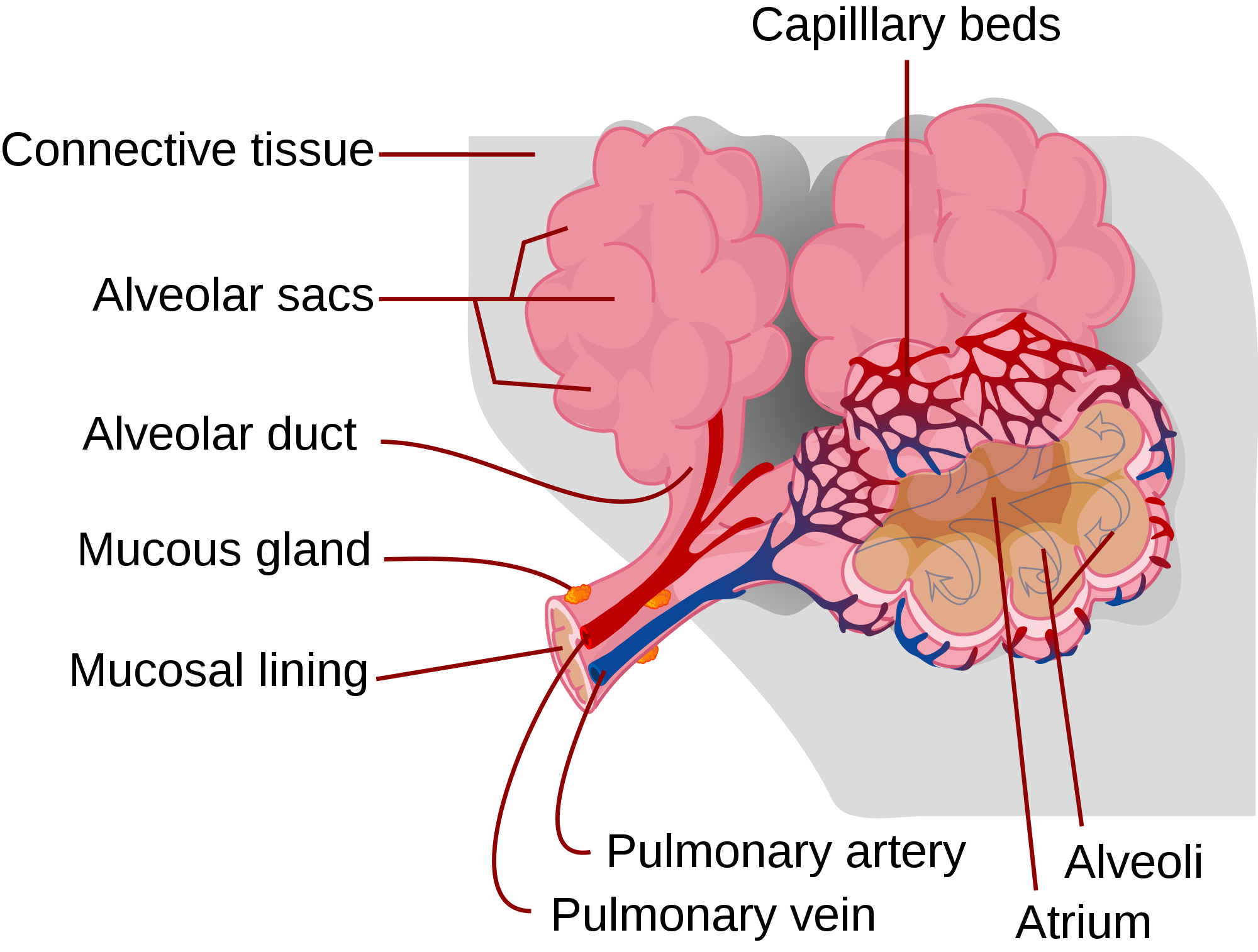
The pulmonary artery (also shown in Figure 13.4.2) carries deoxygenated blood from the heart to the lungs. Then, the blood travels through the pulmonary capillary beds, where it picks up oxygen and releases carbon dioxide. The oxygenated blood then leaves the lungs and travels back to the heart through pulmonary veins. There are four pulmonary veins (two for each lung), and all four carry oxygenated blood to the heart. From the heart, the oxygenated blood is then pumped to cells throughout the body.
Mechanism of Gas Exchange
Gas exchange occurs by diffusion across cell membranes. Gas molecules naturally move down a concentration gradient from an area of higher concentration to an area of lower concentration. This is a passive process that requires no energy. To diffuse across cell membranes, gases must first be dissolved in a liquid. Oxygen and carbon dioxide are transported around the body dissolved in blood. Both gases bind to the protein hemoglobin in red blood cells, although oxygen does so more effectively than carbon dioxide. Some carbon dioxide also dissolves in blood plasma.
As shown in Figure 13.4.3, oxygen in inhaled air diffuses into a pulmonary capillary from the alveolus. Carbon dioxide in the blood diffuses in the opposite direction. The carbon dioxide can then be exhaled from the body.
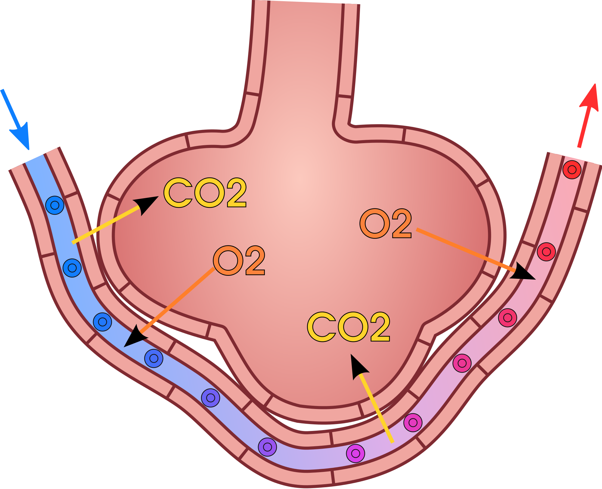
Gas exchange by diffusion depends on having a large surface area through which gases can pass. Although each alveolus is tiny, there are hundreds of millions of them in the lungs of a healthy adult, so the total surface area for gas exchange is huge. It is estimated that this surface area may be as great as 100 m2 (or approximately 1,076 ft²). Often we think of lungs as balloons, but this type of structure would have very limited surface area and there wouldn't be enough space for blood to interface with the air in the alveoli. The structure alveoli take in the lungs is more like a giant mass of soap bubbles — millions of tiny little chambers making up one large mass — this is what increases surface area giving blood lots of space to come into close enough contact to exchange gases by diffusion.
Gas exchange by diffusion also depends on maintaining a steep concentration gradient for oxygen and carbon dioxide. Continuous blood flow in the capillaries and constant breathing maintain this gradient.
- Each time you inhale, there is a greater concentration of oxygen in the air in the alveoli than there is in the blood in the pulmonary capillaries. As a result, oxygen diffuses from the air inside the alveoli into the blood in the capillaries. Carbon dioxide, in contrast, is more concentrated in the blood in the pulmonary capillaries than it is in the air inside the alveoli. As a result, carbon dioxide diffuses in the opposite direction.
- The cells of the body have a much lower concentration of oxygen than does the oxygenated blood that reaches them in peripheral capillaries, which are the capillaries that supply tissues throughout the body. As a result, oxygen diffuses from the peripheral capillaries into body cells. The opposite is true of carbon dioxide. It has a much higher concentration in body cells than it does in the blood of the peripheral capillaries. Thus, carbon dioxide diffuses from body cells into the peripheral capillaries.
13.4 Summary
- Gas exchange is the biological process through which gases are transferred across cell membranes to either enter or leave the blood. Gas exchange takes place continuously between the blood and cells throughout the body, and also between the blood and the air inside the lungs.
- Gas exchange in the lungs takes place in alveoli, which are tiny air sacs surrounded by networks of capillaries. The pulmonary artery carries deoxygenated blood from the heart to the lungs, where it travels through pulmonary capillaries, picking up oxygen and releasing carbon dioxide. The oxygenated blood then leaves the lungs through pulmonary veins.
- Gas exchange occurs by diffusion across cell membranes. Gas molecules naturally move down a concentration gradient from an area of higher concentration to an area of lower concentration. This is a passive process that requires no energy.
- Gas exchange by diffusion depends on the large surface area provided by the hundreds of millions of alveoli in the lungs. It also depends on a steep concentration gradient for oxygen and carbon dioxide. This gradient is maintained by continuous blood flow and constant breathing.
13.4 Review Questions
- What is gas exchange?
- Summarize the flow of blood into and out of the lungs for gas exchange.
-
- Describe the mechanism by which gas exchange takes place.
- Identify the two main factors upon which gas exchange by diffusion depends.
- If the concentration of oxygen were higher inside of a cell than outside of it, which way would the oxygen flow? Explain your answer.
- Why is it important that the walls of the alveoli are only one cell thick?
13.4 Explore More
https://www.youtube.com/watch?v=nRpwdwm06Ic&feature=emb_logo
Oxygen movement from alveoli to capillaries | NCLEX-RN | Khan Academy, khanacademymedicine, 2013.
https://www.youtube.com/watch?v=KmgIqVwytwA&feature=emb_logo
About Carbon Monoxide and Carbon Monoxide Poisoning, EMDPrepare, 2009.
https://www.youtube.com/watch?v=GVU_zANtroE
Oxygen’s surprisingly complex journey through your body - Enda Butler, TED-Ed, 2017.
Attributions
Figure 13.4.1
Oxygen Bar by Farrukh on Flickr is used under a CC BY-NC 2.0 (https://creativecommons.org/licenses/by-nc/2.0/) license.
Figure 13.4.2
Alveolus_diagram.svg by Mariana Ruiz Villarreal [LadyofHats] on Wikimedia Commons is released into the public domain (https://en.wikipedia.org/wiki/Public_domain).
Figure 13.4.3
Gas_exchange_in_the_aveolus.svg by domdomegg on Wikimedia Commons is used under a CC BY 4.0 (https://creativecommons.org/licenses/by/4.0) license.
References
EMDPrepare. (2009, December 21). About carbon monoxide and carbon monoxide poisoning. YouTube. https://www.youtube.com/watch?v=KmgIqVwytwA&feature=youtu.be
khanacademymedicine. (2013, February 25). Oxygen movement from alveoli to capillaries | NCLEX-RN | Khan Academy. YouTube. https://www.youtube.com/watch?v=nRpwdwm06Ic&feature=youtu.be
TED-Ed. (2017, April 13). Oxygen’s surprisingly complex journey through your body - Enda Butler. YouTube. https://www.youtube.com/watch?v=GVU_zANtroE&feature=youtu.be
Created by CK-12 Foundation/Adapted by Christine Miller
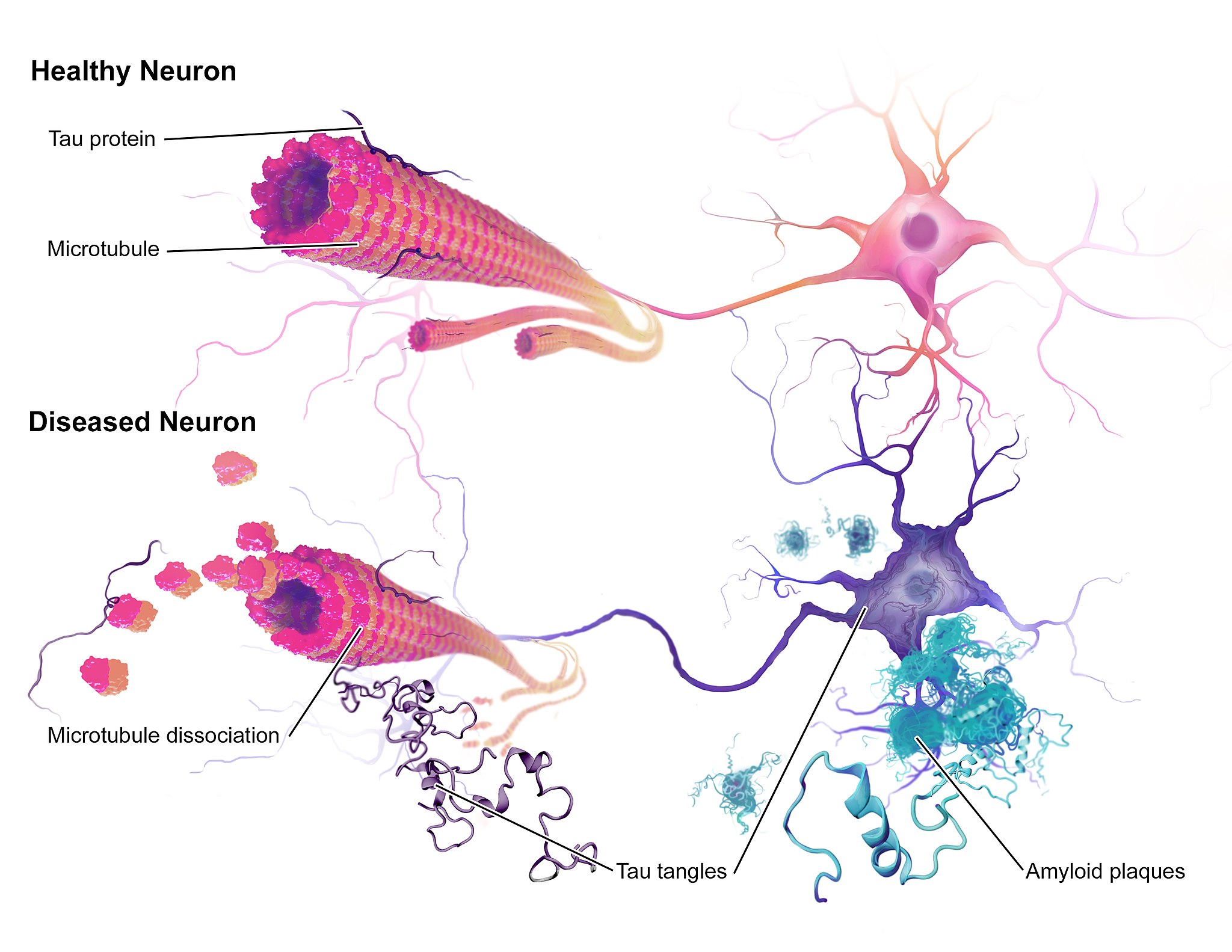
Case Study Conclusion: Fading Memory
The illustration above (Figure 8.9.1) shows some of the molecular and cellular changes that occur in Alzheimer’s disease (AD). Rosa was diagnosed with AD at the beginning of this chapter after experiencing memory problems and other changes in her cognitive functioning, mood, and personality. These abnormal changes in the brain include the development of amyloid plaques between brain cells and neurofibrillary tangles inside of neurons. These hallmark characteristics of AD are associated with the loss of synapses between neurons, and ultimately the death of neurons.
After reading this chapter, you should have a good appreciation for the importance of keeping neurons alive and communicating with each other at synapses. The nervous system coordinates all of the body’s voluntary and involuntary activities. It interprets information from the outside world through sensory systems, and makes appropriate responses through the motor system, through communication between the PNS and CNS. The brain directs the rest of the nervous system and controls everything from basic vital functions (such as heart rate and breathing) to high-level functions (such as problem solving and abstract thought). The nervous system can perform these important functions by generating action potentials in neurons in response to stimulation and sending messages between cells at synapses, typically using chemical neurotransmitter molecules. When neurons are not functioning properly, lose their synapses, or die, they cannot carry out the signaling essential for the proper functioning of the nervous system.
AD is a progressive neurodegenerative disease, meaning that the damage to the brain becomes more extensive as time goes on. The picture in Figure 8.9.2 illustrates how the damage progresses from before AD is diagnosed (preclinical AD), to mild and moderate AD, to severe AD.
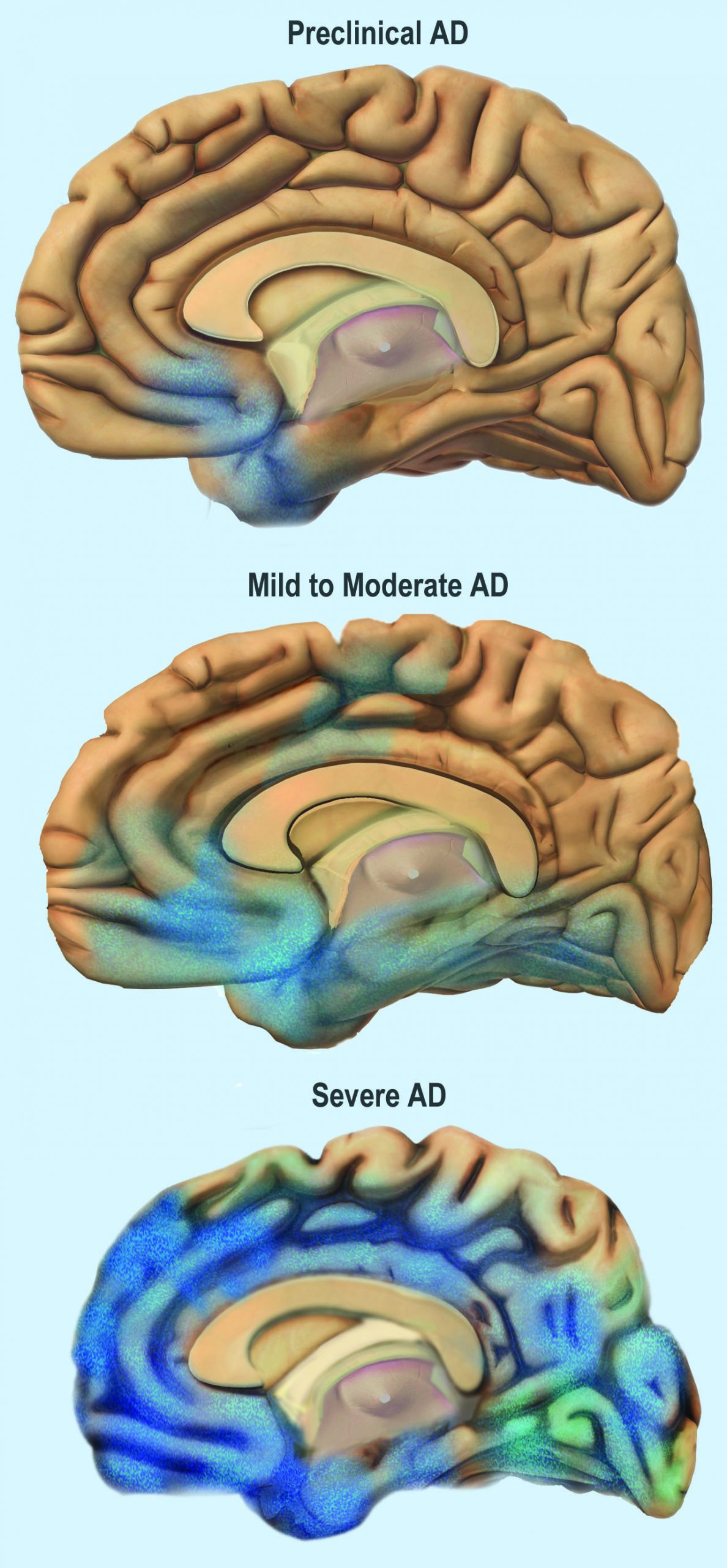
You can see that the damage starts in a relatively small location toward the bottom of the brain. One of the earliest brain areas to be affected by AD is the hippocampus. As you have learned, the hippocampus is important for learning and memory, which explains why many of Rosa’s symptoms of mild AD involve deficits in memory, such as trouble remembering where she placed objects, recent conversations, and appointments.
As AD progresses, more of the brain is affected, including areas involved in emotional regulation, social behavior, planning, language, spatial navigation, and higher-level thought. Rosa is beginning to show signs of problems in these areas, including irritability, lashing out at family members, getting lost in her neighborhood, problems finding the right words, putting objects in unusual locations, and difficulty in managing her finances. You can see that as AD progresses, damage spreads further across the cerebrum, which you now know controls conscious functions like reasoning, language, and interpretation of sensory stimuli. You can also see how the frontal lobe — which controls executive functions such as planning, self-control, and abstract thought — becomes increasingly damaged.
Increasing damage to the brain causes corresponding deficits in functioning. In moderate AD, patients have increased memory, language, and cognitive deficits, compared to mild AD. They may not recognize their own family members, and may wander and get lost, engage in inappropriate behaviors, become easily agitated, and have trouble carrying out daily activities such as dressing. In severe AD, much of the brain is affected. Patients usually cannot recognize family members or communicate, and they are often fully dependent on others for their care. They begin to lose the ability to control their basic functions, such as bladder control, bowel control, and proper swallowing. Eventually, AD causes death, usually as a result of this loss of basic functions.
For now, Rosa only has mild AD and is still able to function relatively well with care from her family. The medication her doctor gave her has helped improve some of her symptoms. It is a cholinesterase inhibitor, which blocks an enzyme that normally degrades the neurotransmitter acetylcholine. With more of the neurotransmitter available, more of it can bind to neurotransmitter receptors on postsynaptic cells. Therefore, this drug acts as an agonist for acetylcholine, which enhances communication between neurons in Rosa’s brain. This increase in neuronal communication can help restore some of the functions lost in early Alzheimer’s disease and may slow the progression of symptoms.
But medication such as this is only a short-term measure, and does not halt the progression of the underlying disease. Ideally, the damaged or dead neurons would be replaced by new, functioning neurons. Why does this not happen automatically in the body? As you have learned, neurogenesis is very limited in adult humans, so once neurons in the brain die, they are not normally replaced to any significant extent. Scientists, however, are studying the ways in which neurogenesis might be increased in cases of disease or injury to the brain. They are also investigating the possibility of using stem cell transplants to replace damaged or dead neurons with new neurons. But this research is in very early stages and is not currently a treatment for AD.
One promising area of research is in the development of methods to allow earlier detection and treatment of AD, given that the changes in the brain may actually start ten to 20 years before diagnosis of AD. A radiolabeled chemical called Pittsburgh Compound B (PiB) binds to amyloid plaques in the brain, and in the future, it may be used in conjunction with brain imaging techniques to detect early signs of AD. Scientists are also looking for biomarkers in bodily fluids (such as blood and cerebrospinal fluid) that might indicate the presence of AD before symptoms appear. Finally, researchers are also investigating possible early and subtle symptoms (such as changes in how people move or a loss of smell) to see whether they can be used to identify people who will go on to develop AD. This research is in the early stages, but the hope is that patients can be identified earlier, allowing for earlier and more effective treatment, as well as more planning time for families.
Scientists are also still trying to fully understand the causes of AD, which affects more than five million Americans. Some genetic mutations have been identified as contributors, but environmental factors also appear to be important. With more research into the causes and mechanisms of AD, hopefully a cure can be found, and people like Rosa can live a longer and better life.
Chapter 8 Summary
In this chapter, you learned about the human nervous system. Specifically, you learned that:
- The nervous system is the organ system that coordinates all of the body’s voluntary and involuntary actions by transmitting signals to and from different parts of the body. It has two major divisions: the central nervous system (CNS) and the peripheral nervous system (PNS).
- The CNS includes the brain and spinal cord.
- The PNS consists mainly of nerves that connect the CNS with the rest of the body. It has two major divisions: the somatic nervous system and the autonomic nervous system. These divisions control different types of functions, and often interact with the CNS to carry out these functions. The somatic system controls activities that are under voluntary control. The autonomic system controls activities that are involuntary.
- The autonomic nervous system is further divided into the sympathetic division (which controls the fight-or-flight response), the parasympathetic division (which controls most routine involuntary responses), and the enteric division (which provides local control for digestive processes).
- Signals sent by the nervous system are electrical signals called nerve impulses. They are transmitted by special, electrically excitable cells called neurons, which are one of two major types of cells in the nervous system.
- Neuroglia are the other major type of nervous system cells. There are many types of glial cells, and they have many specific functions. In general, neuroglia function to support, protect, and nourish neurons.
- The main parts of a neuron include the cell body, dendrites, and axon. The cell body contains the nucleus. Dendrites receive nerve impulses from other cells, and the axon transmits nerve impulses to other cells at axon terminals. A synapse is a complex membrane junction at the end of an axon terminal that transmits signals to another cell.
- Axons are often wrapped in an electrically-insulating myelin sheath, which is produced by oligodendrocytes or schwann cells, both of which are types of neuroglia. Electrical impulses called action potentials occur at gaps in the myelin sheath, called nodes of Ranvier, which speeds the conduction of nerve impulses down the axon.
- Neurogenesis, or the formation of new neurons by cell division, may occur in a mature human brain — but only to a limited extent.
- The nervous tissue in the brain and spinal cord consists of gray matter — which contains mainly unmyelinated cell bodies and dendrites of neurons — and white matter, which contains mainly myelinated axons of neurons. Nerves of the peripheral nervous system consist of long bundles of myelinated axons that extend throughout the body.
- There are hundreds of types of neurons in the human nervous system, but many can be classified on the basis of the direction in which they carry nerve impulses. Sensory neurons carry nerve impulses away from the body and toward the central nervous system, motor neurons carry them away from the central nervous system and toward the body, and interneurons often carry them between sensory and motor neurons.
- A nerve impulse is an electrical phenomenon that occurs because of a difference in electrical charge across the plasma membrane of a neuron.
- The sodium-potassium pump maintains an electrical gradient across the plasma membrane of a neuron when it is not actively transmitting a nerve impulse. This gradient is called the resting potential of the neuron.
- An action potential is a sudden reversal of the electrical gradient across the plasma membrane of a resting neuron. It begins when the neuron receives a chemical signal from another cell or some other type of stimulus. The action potential travels rapidly down the neuron’s axon as an electric current.
- A nerve impulse is transmitted to another cell at either an electrical or a chemical synapse. At a chemical synapse, neurotransmitter chemicals are released from the presynaptic cell into the synaptic cleft between cells. The chemicals travel across the cleft to the postsynaptic cell and bind to receptors embedded in its membrane.
- There are many different types of neurotransmitters. Their effects on the postsynaptic cell generally depend on the type of receptor they bind to. The effects may be excitatory, inhibitory, or modulatory in more complex ways. Both physical and mental disorders may occur if there are problems with neurotransmitters or their receptors.
- The CNS includes the brain and spinal cord. It is physically protected by bones, meninges, and cerebrospinal fluid. It is chemically protected by the blood-brain barrier.
- The brain is the control center of the nervous system and of the entire organism. The brain uses a relatively large proportion of the body’s energy, primarily in the form of glucose.
-
- The brain is divided into three major parts, each with different functions: the forebrain, the midbrain and the hindbrain.
- The forebrain includes the cerebrum, the thalamus, the hypothalamus, the hippocampus and the amygdala. The cerebrum is further divided into left and right hemispheres. Each hemisphere has four lobes: frontal, parietal, temporal, and occipital. Each lobe is associated with specific senses or other functions. The cerebrum has a thin outer layer called the cerebral cortex. Its many folds give it a large surface area. This is where most information processing takes place.
- The thalamus, hypothalamus, hippocampus and amygdala are all part of the limbic system which helps regulate memories, coordination and attention
- The brain is divided into three major parts, each with different functions: the forebrain, the midbrain and the hindbrain.
- The spinal cord is a tubular bundle of nervous tissues that extends from the head down the middle of the back to the pelvis. It functions mainly to connect the brain with the PNS. It also controls certain rapid responses called reflexes without input from the brain.
- A spinal cord injury may lead to paralysis (loss of sensation and movement) of the body below the level of the injury, because nerve impulses can no longer travel up and down the spinal cord beyond that point.
- The PNS consists of all the nervous tissue that lies outside of the CNS. Its main function is to connect the CNS to the rest of the organism.
- The tissues that make up the PNS are nerves and ganglia. Nerves are bundles of axons and ganglia are groups of cell bodies. Nerves are classified as sensory, motor, or a mix of the two.
- The PNS is not as well protected physically or chemically as the CNS, so it is more prone to injury and disease. PNS problems include injury from diabetes, shingles, and heavy metal poisoning. Two disorders of the PNS are Guillain-Barre syndrome and Charcot-Marie-Tooth disease.
- The human body has two major types of senses: special senses and general senses. Special senses have specialized sense organs and include vision (eyes), hearing (ears), balance (ears), taste (tongue), and smell (nasal passages). General senses are all associated with touch and lack special sense organs. Touch receptors are found throughout the body but particularly in the skin.
- All senses depend on sensory receptor cells to detect sensory stimuli and transform them into nerve impulses. Types of sensory receptors include mechanoreceptors (mechanical forces), thermoreceptors (temperature), nociceptors (pain), photoreceptors (light), and chemoreceptors (chemicals).
- Touch includes the ability to sense pressure, vibration, temperature, pain, and other tactile stimuli. The skin includes several different types of touch receptor cells.
- Vision is the ability to sense light and see. The eye is the special sensory organ that collects and focuses light, forms images, and changes them to nerve impulses. Optic nerves send information from the eyes to the brain, which processes the visual information and “tells” us what we are seeing.
- Common vision problems include myopia (nearsightedness), hyperopia (farsightedness), and presbyopia (age-related decline in close vision).
- Hearing is the ability to sense sound waves, and the ear is the organ that senses sound. It changes sound waves to vibrations that trigger nerve impulses, which travel to the brain through the auditory nerve. The brain processes the information and “tells” us what we are hearing.
- The ear is also the organ responsible for the sense of balance, which is the ability to sense and maintain an appropriate body position. The ears send impulses on head position to the brain, which sends messages to skeletal muscle via the peripheral nervous system. The muscles respond by contracting to maintain balance.
- Taste and smell are both abilities to sense chemicals. Taste receptors in taste buds on the tongue sense chemicals in food, and olfactory receptors in the nasal passages sense chemicals in the air. The sense of smell contributes significantly to the sense of taste.
- Psychoactive drugs are substances that change the function of the brain and result in alterations of mood, thinking, perception, and behavior. They include prescription medications (such as opioid painkillers), legal substances (such as nicotine and alcohol), and illegal drugs (such as LSD and heroin).
- Psychoactive drugs are divided into different classes according to their pharmacological effects. They include stimulants, depressants, anxiolytics, euphoriants, hallucinogens, and empathogens. Many psychoactive drugs have multiple effects, so they may be placed in more than one class.
- Psychoactive drugs generally produce their effects by affecting brain chemistry. Generally, they act either as agonists, which enhance the activity of particular neurotransmitters, or as antagonists, which decrease the activity of particular neurotransmitters.
- Psychoactive drugs are used for medical, ritual, and recreational purposes.
- Misuse of psychoactive drugs may lead to addiction, which is the compulsive use of a drug, despite its negative consequences. Sustained use of an addictive drug may produce physical or psychological dependence on the drug. Rehabilitation typically involves psychotherapy, and sometimes the temporary use of other psychoactive drugs.
In addition to the nervous system, there is another system of the body that is important for coordinating and regulating many different functions – the endocrine system. You will learn about the endocrine system in the next chapter.
Chapter 8 Review
- Imagine that you decide to make a movement. To carry out this decision, a neuron in the cerebral cortex of your brain (neuron A) fires a nerve impulse that is sent to a neuron in your spinal cord (neuron B). Neuron B then sends the signal to a muscle cell, causing it to contract, resulting in movement. Answer the following questions about this pathway.
- Which part of the brain is neuron A located in — the cerebellum, cerebrum, or brain stem? Explain how you know.
- The cell body of neuron A is located in a lobe of the brain that is involved in abstract thought, problem solving, and planning. Which lobe is this?
- Part of neuron A travels all the way down to the spinal cord to meet neuron B. Which part of neuron A travels to the spinal cord?
- Neuron A forms a chemical synapse with neuron B in the spinal cord. How is the signal from neuron A transmitted to neuron B?
- Is neuron A in the central nervous system (CNS) or peripheral nervous system (PNS)?
- The axon of neuron B travels in a nerve to a skeletal muscle cell. Is the nerve part of the CNS or PNS? Is this an afferent nerve or an efferent nerve?
- What part of the PNS is involved in this pathway — the autonomic nervous system or the somatic nervous system? Explain your answer.
- What are the differences between a neurotransmitter receptor and a sensory receptor?
-
- If a person has a stroke and then has trouble using language correctly, which hemisphere of their brain was most likely damaged? Explain your answer.
- Electrical gradients are responsible for the resting potential and action potential in neurons. Answer the following questions about the electrical characteristics of neurons.
- Define an electrical gradient, in the context of a cell.
- What is responsible for maintaining the electrical gradient that results in the resting potential?
- Compare and contrast the resting potential and the action potential.
- Where along a myelinated axon does the action potential occur? Why does it happen here?
- What does it mean that the action potential is “all-or-none?”
- Compare and contrast Schwann cells and oligodendrocytes.
- For the senses of smell and hearing, name their respective sensory receptor cells, what type of receptor cells they are, and what stimuli they detect.
- Nicotine is a psychoactive drug that binds to and activates a receptor for the neurotransmitter acetylcholine. Is nicotine an agonist or an antagonist for acetylcholine? Explain your answer.
Attributions
Figure 8.9.1
Alzheimers_Disease by BruceBlaus on Wikimedia Commons is used under a CC BY-SA 4.0 (https://creativecommons.org/licenses/by-sa/4.0/deed.en) license.
Figure 8.9.2
Alzheimer’s Disease stagess by NIH Image Gallery on Flickr is in the public domain (https://en.wikipedia.org/wiki/Public_domain).
Created by: CK-12/Adapted by Christine Miller
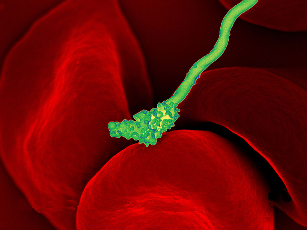
Bacteria Attack!
The colourful image in Figure 4.3.1 shows a bacterial cell (in green) attacking human red blood cells. The bacterium causes a disease called relapsing fever. The bacterial and human cells look very different in size and shape. Although all living cells have certain things in common — such as a plasma membrane and cytoplasm — different types of cells, even within the same organism, may have their own unique structures and functions. Cells with different functions generally have different shapes that suit them for their particular job. Cells vary not only in shape, but also in size, as this example shows. In most organisms, however, even the largest cells are no bigger than the period at the end of this sentence. Why are cells so small?
Explaining Cell Size
Most organisms, even very large ones, have microscopic cells. Why don't cells get bigger instead of remaining tiny and multiplying? Why aren't you one giant cell rolling around school? What limits cell size?
Once you know how a cell functions, the answers to these questions are clear. To carry out life processes, a cell must be able to quickly pass substances in and out of the cell. For example, it must be able to pass nutrients and oxygen into the cell and waste products out of the cell. Anything that enters or leaves a cell must cross its outer surface. The size of a cell is limited by its need to pass substances across that outer surface.
Look at the three cubes in Figure 4.3.2. A larger cube has less surface area relative to its volume than a smaller cube. This relationship also applies to cells — a larger cell has less surface area relative to its volume than a smaller cell. A cell with a larger volume also needs more nutrients and oxygen, and produces more waste. Because all of these substances must pass through the surface of the cell, a cell with a large volume will not have enough surface area to allow it to meet its needs. The larger the cell is, the smaller its ratio of surface area to volume, and the more difficult it will be for the cell to get rid of its waste and take in necessary substances. This is what limits the size of the cell.
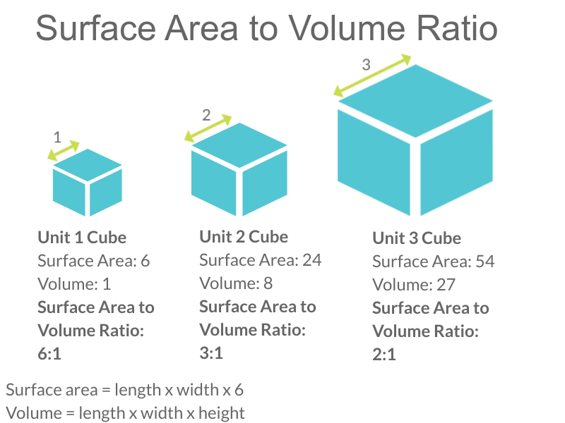
Cell Form and Function
Cells with different functions often have varying shapes. The cells pictured below (Figure 4.3.3) are just a few examples of the many different shapes that human cells may have. Each type of cell has characteristics that help it do its job. The job of the nerve cell, for example, is to carry messages to other cells. The nerve cell has many long extensions that reach out in all directions, allowing it to pass messages to many other cells at once. Do you see the tail of each tiny sperm cell? Its tail helps a sperm cell "swim" through fluids in the female reproductive tract in order to reach an egg cell. The white blood cell has the job of destroying bacteria and other pathogens. It is a large cell that can engulf foreign invaders.
Figure 4.3.3 Human cells may have many different shapes that help them to do their jobs.
Cells With and Without a Nucleus
The nucleus is a basic cell structure present in many — but not all — living cells. The nucleus of a cell is a structure in the cytoplasm that is surrounded by a membrane (the nuclear membrane) and contains DNA. Based on whether or not they have a nucleus, there are two basic types of cells: prokaryotic cells and eukaryotic cells.
Prokaryotic Cells
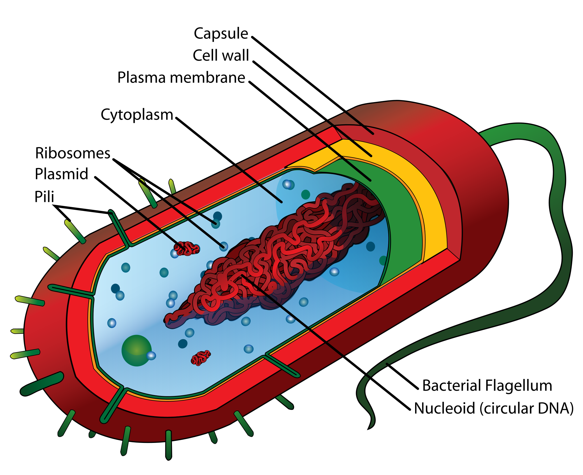
Prokaryotic cells are cells without a nucleus. The DNA in prokaryotic cells is in the cytoplasm, rather than enclosed within a nuclear membrane. In addition, these cells are typically smaller than eukaryotic cells and contain fewer organelles. Prokaryotic cells are found in single-celled organisms, such as the bacterium represented by the model in Figure 4.3.3. Organisms with prokaryotic cells are called prokaryotes. They were the first type of organisms to evolve, and they are still the most common organisms today.
Eukaryotic Cells
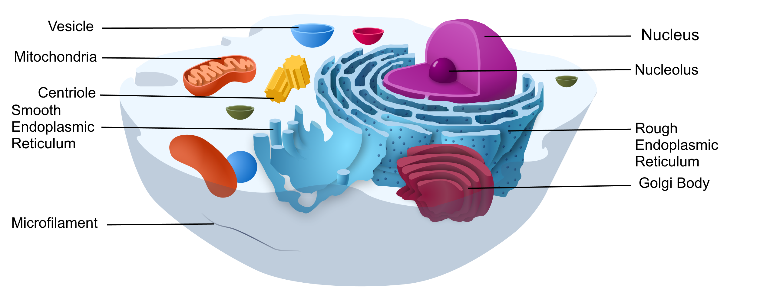
Eukaryotic cells are cells that contain a nucleus. A typical eukaryotic cell is represented by the model in Figure 4.3.4. Eukaryotic cells are usually larger than prokaryotic cells. They are found in some single-celled and all multicellular organisms. Organisms with eukaryotic cells are called eukaryotes, and they range from fungi to humans.
Besides a nucleus, eukaryotic cells also contain other organelles. An organelle is a structure within the cytoplasm that performs a specific job in the cell. Organelles called mitochondria, for example, provide energy to the cell, and organelles called vesicles store substances in the cell. Organelles allow eukaryotic cells to carry out more functions than prokaryotic cells can.
Interestingly, scientists think that mitochondria were once free-living prokaryotes that infected (or were engulfed by) larger cells. The two organisms developed a symbiotic relationship that was beneficial to both of them, resulting in the smaller prokaryote becoming an organelle within the larger cell. This is called endosymbiotic theory, and it is supported by a lot of evidence, including the fact that mitochondria have their own DNA separate from the DNA in the nucleus of the eukaryotic cell. Endosymbiotic theory will be described in more detail in later sections, and it's also discussed in the video below.
https://www.youtube.com/watch?v=FGnS-Xk0ZqU
Endosymbiotic Theory, Amoeba Sisters, 2017.
4.3 Summary
- Cells must be very small so they have a large enough surface area-to-volume ratio to maintain normal cell processes.
- Cells with different functions often have different shapes.
- Prokaryotic cells do not have a nucleus. Eukaryotic cells do have a nucleus, along with other organelles.
4.3 Review Questions
- Explain why most cells are very small.
- Discuss variations in the form and function of cells.
-
-
- Do human cells have organelles? Explain your answer.
- Which are usually larger – prokaryotic or eukaryotic cells? What do you think this means for their relative ability to take in needed substances and release wastes? Discuss your answer.
- DNA in eukaryotes is enclosed within the _______ ________.
- Name three different types of cells in humans.
- Which organelle provides energy in eukaryotic cells?
- What is a function of a vesicle in a cell?
4.3 Explore More
https://www.youtube.com/watch?time_continue=1&v=9i7kAt97XYU&feature=emb_logo
How we think complex cells evolved - Adam Jacobson, TED-Ed, 2015.
https://www.youtube.com/watch?v=Pxujitlv8wc
Prokaryotic vs. Eukaryotic Cells (updated), Amoeba Sisters, 2018.
Attributions
Figure 4.3.1
Borrelia_hermsii_Bacteria_(13758011613) by NAID on Wikimedia Commons is released into the public domain (https://en.wikipedia.org/wiki/Public_domain).
Figure 4.3.2
Cell Size by Christine Miller is released into the Public Domain (https://creativecommons.org/publicdomain/mark/1.0/).
Figure 4.3.3
- Chondrocyte. BioTek-Wikipedia-Image by BioTek Instruments, Inc. on Wikimedia Commons is used under a CC BY-SA 3.0 (https://creativecommons.org/licenses/by-sa/3.0/deed.en) license.
- Neutrophil with anthrax copy by Volker Brinkmann from PLOS Pathogens on Wikimedia Commons is used under a CC BY 2.5 (https://creativecommons.org/licenses/by/2.5/deed.en) license.
- PLoSBiol4.e126.Fig6fNeuron by Lee, et al. from PLOS Biology on Wikimedia Commons is used under a CC BY 2.5 (https://creativecommons.org/licenses/by/2.5/deed.en) license.
- Sperm (265 33) human by Doc. RNDr. Josef Reischig, CSc. on Wikimedia Commons is used under a CC BY-SA 3.0 (https://creativecommons.org/licenses/by-sa/3.0) license.
Figure 4.3.4
Model of a prokaryotic cell: bacterium by Mariana Ruiz Villarreal [LadyofHats] on Wikimedia Commons is released into the public domain (https://en.wikipedia.org/wiki/Public_domain).
Figure 4.3.5
Animal Cell adapted by Christine Miller is used under a CC0 1.0 (https://creativecommons.org/publicdomain/zero/1.0/deed.en) public domain dedication license. (Original image, Animal Cell Unannotated, is by Kelvin Song on Wikimedia Commons.)
References
Amoeba Sisters. (2017, May 3). Endosymbiotic theory. YouTube. https://www.youtube.com/watch?v=FGnS-Xk0ZqU&feature=youtu.be
Amoeba Sisters. (2018, July 30). Prokaryotic vs. eukaryotic cells (updated). YouTube. https://www.youtube.com/watch?v=Pxujitlv8wc&feature=youtu.be
Brinkmann, V. (November 2005). Neutrophil engulfing Bacillus anthracis. PLoS Pathogens 1 (3): Cover page [digital image]. DOI:10.1371. https://journals.plos.org/plospathogens/issue?id=10.1371/issue.ppat.v01.i03
Lee, W.C.A., Huang, H., Feng, G., Sanes, J.R., Brown, E.N., et al. (2005, December 27) Figure 6f, slightly altered (plus scalebar, minus letter "f".) [digital image]. Dynamic Remodeling of Dendritic Arbors in GABAergic Interneurons of Adult Visual Cortex. PLoS Biology, 4(2), e29. doi:10.1371/journal.pbio.0040029. https://journals.plos.org/plosbiology/article?id=10.1371/journal.pbio.0040029
TED-Ed. (2015, February 17). How we think complex cells evolved - Adam Jacobson. https://www.youtube.com/watch?v=9i7kAt97XYU&feature=youtu.be
Created by: CK-12/Adapted by Christine Miller
Figure 4.2.1 Human cells viewed with a very powerful tool called a scanning electron microscope.
Amazing Cells
What are these incredible objects? Would it surprise you to learn that they are all human cells? Cells are actually too small to see with the unaided eye. It is visible here in such detail because it is being viewed with a very powerful tool called a scanning electron microscope. Cells may be small in size, but they are extremely important to life. Like all other living things, you are made of cells. Cells are the basis of life, and without cells, life as we know it would not exist. You will learn more about these amazing building blocks of life in this section.
What Are Cells?
If you look at living matter with a microscope — even a simple light microscope — you will see that it consists of cells. Cells are the basic units of the structure and function of living things. They are the smallest units that can carry out the processes of life. All organisms are made up of one or more cells, and all cells have many of the same structures and carry out the same basic life processes. Knowing the structure of cells and the processes they carry out is necessary to an understanding of life itself.
Discovery of Cells
The first time the word cell was used to refer to these tiny units of life was in 1665 by a British scientist named Robert Hooke. Hooke was one of the earliest scientists to study living things under a microscope. The microscopes of his day were not very strong, but Hooke was still able to make an important discovery. When he looked at a thin slice of cork under his microscope, he was surprised to see what looked like a honeycomb. Hooke made the drawing in the figure to the right to show what he saw. As you can see, the cork was made up of many tiny units. Hooke called these units cells because they resembled cells in a monastery.
Soon after Robert Hooke discovered cells in cork, Anton van Leeuwenhoek in Holland made other important discoveries using a microscope. Leeuwenhoek made his own microscope lenses, and he was so good at it that his microscope was more powerful than other microscopes of his day. In fact, Leeuwenhoek’s microscope was almost as strong as modern light microscopes. Using his microscope, Leeuwenhoek was the first person to observe human cells and bacteria.
Cell Theory
By the early 1800s, scientists had observed cells of many different organisms. These observations led two German scientists named Theodor Schwann and Matthias Jakob Schleiden to propose cells as the basic building blocks of all living things. Around 1850, a German doctor named Rudolf Virchow was studying cells under a microscope, when he happened to see them dividing and forming new cells. He realized that living cells produce new cells through division. Based on this realization, Virchow proposed that living cells arise only from other living cells.
The ideas of all three scientists — Schwann, Schleiden, and Virchow — led to cell theory, which is one of the fundamental theories unifying all of biology.
Cell theory states that:
- All organisms are made of one or more cells.
- All the life functions of organisms occur within cells.
- All cells come from existing cells.
Seeing Inside Cells
Starting with Robert Hooke in the 1600s, the microscope opened up an amazing new world — a world of life at the level of the cell. As microscopes continued to improve, more discoveries were made about the cells of living things, but by the late 1800s, light microscopes had reached their limit. Objects much smaller than cells (including the structures inside cells) were too small to be seen with even the strongest light microscope.
Then, in the 1950s, a new type of microscope was invented. Called the electron microscope, it used a beam of electrons instead of light to observe extremely small objects. With an electron microscope, scientists could finally see the tiny structures inside cells. They could even see individual molecules and atoms. The electron microscope had a huge impact on biology. It allowed scientists to study organisms at the level of their molecules, and it led to the emergence of the molecular biology field. With the electron microscope, many more cell discoveries were made.
Structures Shared By All Cells
Although cells are diverse, all cells have certain parts in common. These parts include a plasma membrane, cytoplasm, ribosomes, and DNA.
- The plasma membrane (a type of cell membrane) is a thin coat of lipids that surrounds a cell. It forms the physical boundary between the cell and its environment. You can think of it as the “skin” of the cell.
- Cytoplasm refers to all of the cellular material inside of the plasma membrane. Cytoplasm is made up of a watery substance called cytosol, and it contains other cell structures, such as ribosomes.
- Ribosomes are the structures in the cytoplasm in which proteins are made.
- DNA is a nucleic acid found in cells. It contains the genetic instructions that cells need to make proteins.
These four parts are common to all cells, from organisms as different as bacteria and human beings. How did all known organisms come to have such similar cells? The similarities show that all life on Earth has a common evolutionary history.
4.2 Summary
- Cells are the basic units of structure and function in living things. They are the smallest units that can carry out the processes of life.
- In the 1600s, Hooke was the first to observe cells from an organism (cork). Soon after, microscopist van Leeuwenhoek observed many other living cells.
- In the early 1800s, Schwann and Schleiden theorized that cells are the basic building blocks of all living things. Around 1850, Virchow observed cells dividing. To previous learnings, he added that living cells arise only from other living cells. These ideas led to cell theory, which states that all organisms are made of cells, that all life functions occur in cells, and that all cells come from other cells.
- It wasn't until the 1950s that scientists could see what was inside the cell. The invention of the electron microscope allowed them to see organelles and other structures smaller than cells.
- There is variation in cells, but all cells have a plasma membrane, cytoplasm, ribosomes, and DNA. These similarities show that all life on Earth has a common ancestor in the distant past.
4.2 Review Questions
- Describe cells.
- Explain how cells were discovered.
- Outline the development of cell theory.
-
- Identify the structures shared by all cells.
- Proteins are made on _____________ .
-
- Robert Hooke sketched what looked like honeycombs — or repeated circular or square units — when he observed plant cells under a microscope.
- What is each unit?
- Of the shared parts of all cells, what makes up the outer surface of each unit?
- Of the shared parts of all cells, what makes up the inside of each unit?
4.2 Explore More
https://www.youtube.com/watch?v=8IlzKri08kk
Introduction to Cells: The Grand Cell Tour, by The Amoeba Sisters, 2016.
Attributions
Figure 4.2.1
- A white blood cell (WBC) known as a neutrophil by National Institute of Allergy and Infectious Diseases (NIAID) on the CDC/ Public Health Image Library (PHIL) Photo ID# 18129. is in the public domain (https://en.wikipedia.org/wiki/Public_domain).
- Healthy Human T Cell by NIAID on Flickr. is used under a CC BY 2.0 (https://creativecommons.org/licenses/by/2.0/) license.
- Human natural killer cell by NIAID on Flickr. is used under a CC BY 2.0 (https://creativecommons.org/licenses/by/2.0/) license.
- Human blood with red blood cells, T cells (orange) and platelets (green) by ZEISS Microscopy on Flickr. is used under a CC BY-NC-ND 2.0 (https://creativecommons.org/licenses/by-nc-nd/2.0/) license.
- Developing nerve cells by ZEISS Microscopy on Flickr. is used under a CC BY-NC-ND 2.0 (https://creativecommons.org/licenses/by-nc-nd/2.0/) license.
Figure 4.2.2
Hooke-microscope-cork by Robert Hooke (1635-1702) [uploaded by Alejandro Porto] on Wikimedia Commons is released into the public domain (https://en.wikipedia.org/wiki/Public_domain).
Figure 4.2.3
Electron Microscope image of a cell by Dartmouth Electron Microscope Facility, Dartmouth College on Wikimedia Commons is released into the public domain (https://en.wikipedia.org/wiki/Public_domain).
Figure 4.2.4
Basic-Components-of-a-cell by Christine Miller is used under a CC0 1.0 (https://creativecommons.org/publicdomain/zero/1.0/) license.
References
Amoeba Sisters. (2016, November 1). Introduction to cells: The grand cell tour. YouTube. https://www.youtube.com/watch?v=8IlzKri08kk&feature=youtu.be
National Institute of Allergy and Infectious Diseases (NIAID). (2011). A white blood cell (WBC) known as a neutrophil, as it was in the process of ingesting a number of spheroid shaped, methicillin-resistant, Staphylococcus aureus (MRSA) bacteria [digital image]. CDC/ Public Health Image Library (PHIL) Photo ID# 18129. https://phil.cdc.gov/Details.aspx?pid=18129.
Wikipedia contributors. (2020, June 24). Antonie van Leeuwenhoek. In Wikipedia. https://en.wikipedia.org/w/index.php?title=Antonie_van_Leeuwenhoek&oldid=964339564
Wikipedia contributors. (2020, May 25). Matthias Jakob Schleiden. In Wikipedia. https://en.wikipedia.org/w/index.php?title=Matthias_Jakob_Schleiden&oldid=958819219
Wikipedia contributors. (2020, June 4). Rudolf Virchow. In Wikipedia,. https://en.wikipedia.org/w/index.php?title=Rudolf_Virchow&oldid=960708716
Wikipedia contributors. (2020, May 16). Theodor Schwann. In Wikipedia. https://en.wikipedia.org/w/index.php?title=Theodor_Schwann&oldid=956919239
Created by: CK-12/Adapted by Christine Miller

A Bag Full of Jell-O
The simple cut-away model of an animal cell (Figure 4.4.1) shows that a cell resembles a plastic bag full of Jell-O. Its basic structure is a plasma membrane filled with cytoplasm. Like Jell-O containing mixed fruit (Figure 4.4.2), the cytoplasm of the cell also contains various structures, including a nucleus and other organelles. Your body is composed of trillions of cells, but all of them perform the same basic life functions. They all obtain and use energy, respond to the environment, and reproduce. How do your cells carry out these basic functions and keep themselves — and you — alive? To answer these questions, you need to know more about the structures that make up cells, starting with the plasma membrane.
What is the Plasma Membrane?
The plasma membrane is a structure that forms a barrier between the cytoplasm inside the cell and the environment outside the cell. Without the plasma membrane, there would be no cell. Although it is very thin and flexible, the plasma membrane protects and supports the cell by controlling everything that enters and leaves it. It allows only certain substances to pass through, while keeping others in or out. To understand how the plasma membrane controls what passes into or out of the cell, you need to know its basic structure.
Phospholipid Bilayer
The plasma membrane is composed mainly of phospholipids, which consist of fatty acids and alcohol. The phospholipids in the plasma membrane are arranged in two layers, called a phospholipid bilayer. As shown in the simplified diagram in Figure 4.4.3, each individual phospholipid molecule has a phosphate group head (in red) and two fatty acid tails (in yellow). The head “loves” water (hydrophilic) and the tails “hate” water (hydrophobic). The water-hating tails are on the interior of the membrane, whereas the water-loving heads point outward, toward either the cytoplasm (intracellular) or the fluid that surrounds the cell (extracellular).
Hydrophobic molecules can easily pass through the plasma membrane if they are small enough, because they are water-hating like the interior of the membrane. Hydrophilic molecules, on the other hand, cannot pass through the plasma membrane — at least not without help — because they are water-loving like the exterior of the membrane.
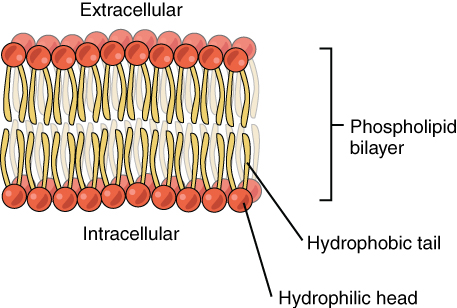
Other Molecules in the Plasma Membrane
The plasma membrane also contains other molecules, primarily other lipids and proteins. The yellow molecules in the diagram here, for example, are the lipid cholesterol. Molecules of the steroid lipid cholesterol help the plasma membrane keep its shape. Proteins in the plasma membrane (shown blue in Figure 4.4.4) include: transport proteins that assist other substances in crossing the cell membrane, receptors that allow the cell to respond to chemical signals in its environment, and cell-identity markers that indicate what type of cell it is and whether it belongs in the body.
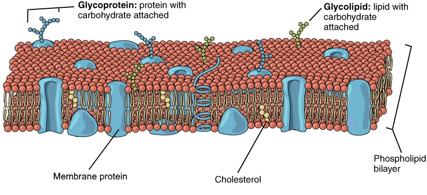
Additional Functions of the Plasma Membrane
The plasma membrane may have extensions, such as whip-like flagella (singular flagellum) or brush-like cilia (singular cilium), shown below (Figure 4.4.5), that give it other functions. In single-celled organisms, these membrane extensions may help the organisms move. In multicellular organisms, the extensions have different functions. For example, the cilia on human lung cells sweep foreign particles and mucus toward the mouth and nose, while the flagellum on a human sperm cell allows it to swim.
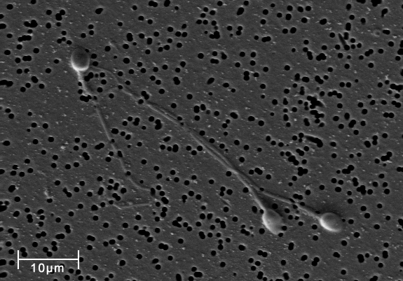
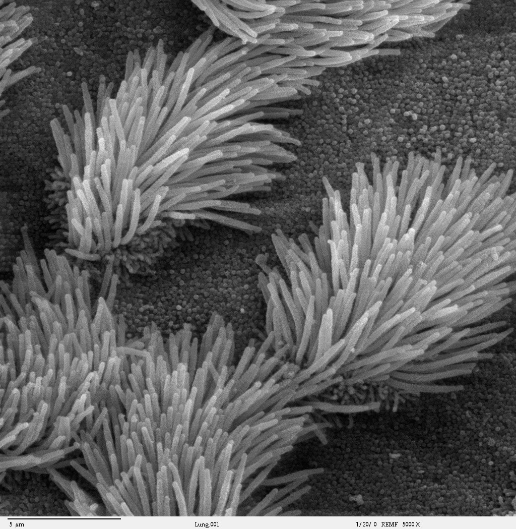
Feature: My Human Body
If you smoke or use e-cigarettes (vaping) and need another reason to quit, here's a good one. We usually think of lung cancer as the major disease caused by smoking. But smoking and vaping can have devastating effects on the body's ability to protect itself from repeated, serious respiratory infections, such as bronchitis and pneumonia.
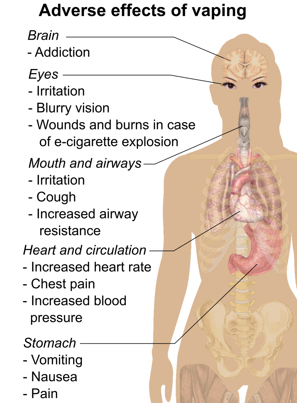
Cilia are microscopic, hair-like projects on cells that line the respiratory, reproductive, and digestive systems. Cilia in the respiratory system line most of your airways, where they have the job of trapping and removing dust, germs, and other foreign particles before they can make you sick. Cilia secrete mucus that traps particles, and they move in a continuous wave-like motion that sweeps the mucus and particles upward toward the throat, where they can be expelled from the body. When you are sick and cough up phlegm, that's what you are doing.
Smoking prevents cilia from performing these important functions. Chemicals in tobacco smoke paralyze the cilia so they can't sweep mucus out of the airways. Those chemicals also inhibit the cilia from producing mucus. Fortunately, these effects start to wear off soon after the most recent exposure to tobacco smoke. If you stop smoking, your cilia will return to normal. Even if prolonged smoking has destroyed cilia, they will regrow and resume functioning in a matter of months after you stop smoking.
4.4 Summary
- The plasma membrane is a structure that forms a barrier between the cytoplasm inside the cell and the environment outside the cell. It allows only certain substances to pass in or out of the cell.
- The plasma membrane is composed mainly of a bilayer of phospholipid molecules. It also contains other molecules, such as the steroid cholesterol, which helps the membrane keep its shape, and transport proteins, which help substances pass through the membrane.
- The plasma membranes of some cells have extensions that have other functions, like flagella to help sperm move, or cilia to help keep our airways clear.
4.4 Review Questions
- What are the general functions of the plasma membrane?
- Describe the phospholipid bilayer of the plasma membrane.
- Identify other molecules in the plasma membrane. State their functions.
- Why do some cells have plasma membrane extensions, like flagella and cilia?
- Explain why hydrophilic molecules cannot easily pass through the cell membrane. What type of molecule in the cell membrane might help hydrophilic molecules pass through it?
- Which part of a phospholipid molecule in the plasma membrane is made of fatty acid chains? Is this part hydrophobic or hydrophilic?
- The two layers of phospholipids in the plasma membrane are called a phospholipid ____________.
-
- Steroid hormones can pass directly through cell membranes. Why do you think this is the case?
- Some antibiotics work by making holes in the plasma membrane of bacterial cells. How do you think this kills the cells?
- What is the name of the long, whip-like extensions of the plasma membrane that helps some single-celled organisms move?
4.4 Explore More
https://www.youtube.com/watch?v=yAXnYcUjn5k&feature=emb_logo
Insights into cell membranes via dish detergent - Ethan Perlstein, TED-Ed, 2013.
https://www.youtube.com/watch?v=qBCVVszQQNs
Inside the cell membrane, by The Amoeba Sisters, 2018.
Attributions
Figure 4.4.1
Animal Cell Unannotated, by Kelvin Song on Wikimedia Commons is used under a CC0 1.0 (https://creativecommons.org/publicdomain/zero/1.0/deed.en) public domain dedication license.
Figure 4.4.2
Jello mold at the mexican bakery photo by Aimée Knight on Flickr is used under a CC BY 2.0 (https://creativecommons.org/licenses/by/2.0/) license.
Figure 4.4.3
Phospholipid_Bilayer by OpenStax on Wikimedia Commons is used under a CC BY 4.0 (https://creativecommons.org/licenses/by/4.0) license.
Figure 4.4.4
Lipid bilayer by OpenStax on Wikimedia Commons is used under a CC BY 4.0 (https://creativecommons.org/licenses/by/4.0) license.
Figure 4.4.5
Spermatozoa-human-3140x by No specific author on Wikimedia Commons is released into the public domain (https://en.wikipedia.org/wiki/Public_domain).
Figure 4.4.6
Cilia/ Bronchiolar epithelium 3 - SEM by Charles Daghlian on Wikimedia Commons is released into the public domain (https://en.wikipedia.org/wiki/Public_domain).
Figure 4.4.7
Adverse effects of vaping (raster) by Mikael Häggström on Wikimedia Commons is released into the public domain (https://en.wikipedia.org/wiki/Public_domain).
References
Amoeba Sisters. (2018, February 27). Inside the cell membrane. YouTube. https://www.youtube.com/watch?v=qBCVVszQQNs&feature=youtu.be
Betts, J.G., Young, K.A., Wise, J.A., Johnson, E., Poe, B., Kruse, D.H., Korol, O., Johnson, J.E.. Womble, M., DeSaix. P. (2013, April 25). Figure 3.3 Phospolipid Bilayer [digital image]. In Anatomy and Physiology. OpenStax. https://openstax.org/books/anatomy-and-physiology/pages/3-1-the-cell-membrane
Betts, J.G., Young, K.A., Wise, J.A., Johnson, E., Poe, B., Kruse, D.H., Korol, O., Johnson, J.E.. Womble, M., DeSaix. P. (2013, April 25). Figure 3.4 Cell Membrane [digital image]. In Anatomy and Physiology. OpenStax. https://openstax.org/books/anatomy-and-physiology/pages/3-1-the-cell-membrane
Ghosh, A., Coakley, R. C., Mascenik, T., Rowell, T. R., Davis, E. S., Rogers, K., Webster, M. J., Dang, H., Herring, L. E., Sassano, M. F., Livraghi-Butrico, A., Van Buren, S. K., Graves, L. M., Herman, M. A., Randell, S. H., Alexis, N. E., & Tarran, R. (n.d.). Chronic E-Cigarette Exposure Alters the Human Bronchial Epithelial Proteome. American Journal of Respiratory and Critical /Care Medicine, 198(1), 67–76. https://doi-org.ezproxy.tru.ca/10.1164/rccm.201710-2033OC
TED-Ed. (2013, February 26). Insights into cell membranes via dish detergent - Ethan Perlstein. YouTube. https://www.youtube.com/watch?v=yAXnYcUjn5k&feature=youtu.be
Created by: CK-12/Adapted by Christine Miller
Ribosome Review
The 25-metre long sculpture shown in Figure 4.6.1 is a recognition of the beauty of one of the metabolic functions that takes place in the cells in your body. This artwork brings to life an important structure in living cells: the ribosome, the cell structure where proteins are synthesized. The slender silver strand is the messenger RNA(mRNA) bringing the code for a protein out into the cytoplasm. The purple and green structures are ribosomal subunits (which together form a single ribosome), which can "read" the code on the mRNA and direct the bonding of the correct sequence of amino acids to create a protein. All living cells — whether they are prokaryotic or eukaryotic — contain ribosomes, but only eukaryotic cells also contain a nucleus and several other types of organelles.
What Are Organelles?
An organelle is a structure within the cytoplasm of a eukaryotic cell that is enclosed within a membrane and performs a specific job. Organelles are involved in many vital cell functions. Organelles in animal cells include the nucleus, mitochondria, endoplasmic reticulum, Golgi apparatus, vesicles, and vacuoles. Ribosomes are not enclosed within a membrane, but they are still commonly referred to as organelles in eukaryotic cells.
The Nucleus
The nucleus is the largest organelle in a eukaryotic cell, and it's considered the cell’s control center. It contains most of the cell’s DNA(which makes up chromosomes), and it is encoded with the genetic instructions for making proteins. The function of the nucleus is to regulate gene expression, including controlling which proteins the cell makes. In addition to DNA, the nucleus contains a thick liquid called nucleoplasm, which is similar in composition to the cytosol found in the cytoplasm outside the nucleus. Most eukaryotic cells contain just a single nucleus, but some types of cells (such as red blood cells) contain no nucleus and a few other types of cells (such as muscle cells) contain multiple nuclei.
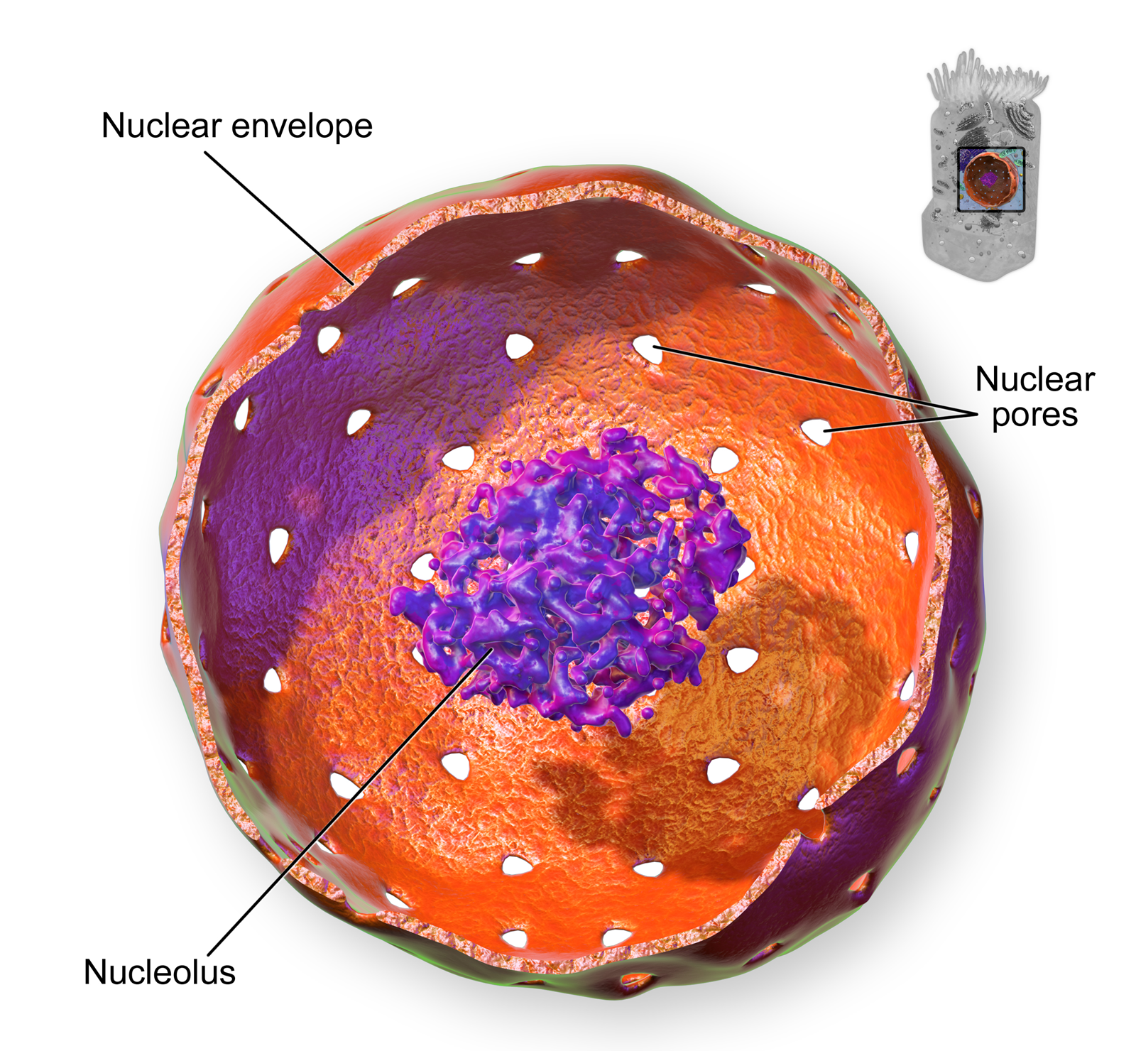
As you can see in the model pictured in Figure 4.6.2, the membrane enclosing the nucleus is called the nuclear envelope. This is actually a double membrane that encloses the entire organelle and isolates its contents from the cellular cytoplasm. Tiny holes called nuclear pores allow large molecules to pass through the nuclear envelope, with the help of special proteins. Large proteins and RNA molecules must be able to pass through the nuclear envelope so proteins can be synthesized in the cytoplasm and the genetic material can be maintained inside the nucleus. The nucleolus shown in the model below is mainly involved in the assembly of ribosomes. After being produced in the nucleolus, ribosomes are exported to the cytoplasm, where they are involved in the synthesis of proteins.
Mitochondria
The mitochondrion (plural, mitochondria) is an organelle that makes energy available to the cell. This is why mitochondria are sometimes referred to as the "power plants of the cell." They use energy from organic compounds (such as glucose) to make molecules of ATP (adenosine triphosphate), an energy-carrying molecule that is used almost universally inside cells for energy.
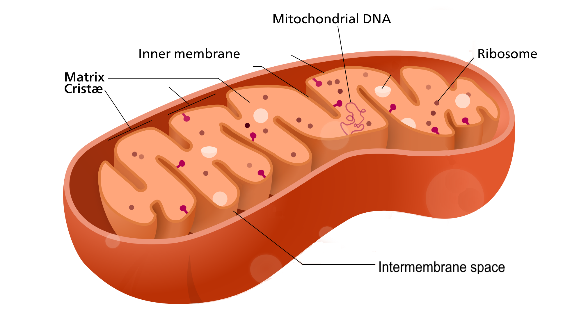
Mitochondria (as in the Figure 4.6.3 diagram) have a complex structure including an inner and out membrane. In addition, mitochondria have their own DNA, ribosomes, and a version of cytoplasm, called matrix. Does this sound similar to the requirements to be considered a cell? That's because they are!
Scientists think that mitochondria were once free-living organisms because they contain their own DNA. They theorize that ancient prokaryotes infected (or were engulfed by) larger prokaryotic cells, and the two organisms evolved a symbiotic relationship that benefited both of them. The larger cells provided the smaller prokaryotes with a place to live. In return, the larger cells got extra energy from the smaller prokaryotes. Eventually, the smaller prokaryotes became permanent guests of the larger cells, as organelles inside them. This theory is called endosymbiotic theory, and it is widely accepted by biologists today. (See the video in section 4.3 to learn all about endosymbiotic theory.)
Endoplasmic Reticulum
The endoplasmic reticulum (ER) is an organelle that helps make and transport proteins and lipids. There are two types of endoplasmic reticulum: rough endoplasmic reticulum (rER) and smooth endoplasmic reticulum (sER). Both types are shown in Figure 4.6.4.
- rER looks rough because it is studded with ribosomes. It provides a framework for the ribosomes, which make proteins. Bits of its membrane pinch off to form tiny sacs called vesicles, which carry proteins away from the ER.
- sER looks smooth because it does not have ribosomes. sER makes lipids, stores substances, and plays other roles.
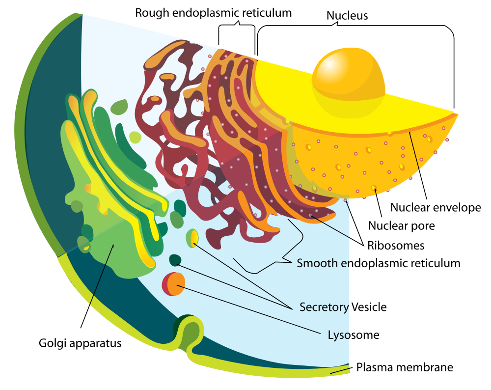
The Figure 4.6.4 drawing includes the nucleus, rER, sER, and Golgi apparatus. From the drawing, you can see how all these organelles work together to make and transport proteins.
Golgi Apparatus
The Golgi apparatus (shown in the Figure 4.6.4 diagram) is a large organelle that processes proteins and prepares them for use both inside and outside the cell. You can see the Golgi apparatus in the figure above. The Golgi apparatus is something like a post office. It receives items (proteins from the ER), then packages and labels them before sending them on to their destinations (to different parts of the cell or to the cell membrane for transport out of the cell). The Golgi apparatus is also involved in the transport of lipids around the cell.
Vesicles and Vacuoles
Both vesicles and vacuoles are sac-like organelles made of phospholipid bilayer that store and transport materials in the cell. Vesicles are much smaller than vacuoles and have a variety of functions. The vesicles that pinch off from the membranes of the ER and Golgi apparatus store and transport protein and lipid molecules. You can see an example of this type of transport vesicle in the Figure 4.6.4. Some vesicles are used as chambers for biochemical reactions.
There are some vesicles which are specialized to carry out specific functions. Lysosomes, which use enzymes to break down foreign matter and dead cells, have a double membrane to make sure their contents don't leak into the rest of the cell. Peroxisomes are another type of specialized vesicle with the main function of breaking down fatty acids and some toxins.
Centrioles
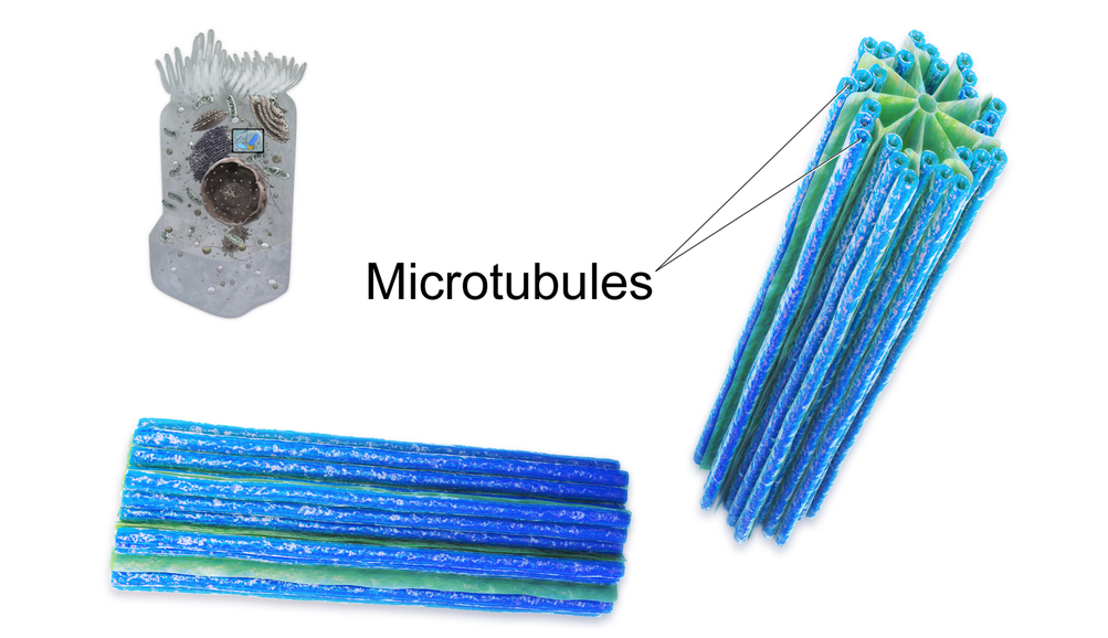
Centrioles are organelles involved in cell division. The function of centrioles is to help organize the chromosomes before cell division occurs so that each daughter cell has the correct number of chromosomes after the cell divides. Centrioles are found only in animal cells, and are located near the nucleus. Each centriole is made mainly of a protein named tubulin. The centriole is cylindrical in shape and consists of many microtubules, as shown in the model pictured in Figure 4.6.5.
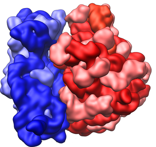
Ribosomes
Ribosomes are small structures where proteins are made. Although they are not enclosed within a membrane, they are frequently considered organelles. Each ribosome is formed of two subunits, like the ones pictured at the beginning of this section (Figure 4.6.1) and in Figure 4.6.6. Both subunits consist of proteins and RNA. mRNA from the nucleus carries the genetic code, copied from DNA, which remains in the nucleus. At the ribosome, the genetic code in mRNA is used to assemble and join together amino acids to make proteins. Ribosomes can be found alone or in groups within the cytoplasm, as well as on the rER.
4.6 Summary
- An organelle is a structure within the cytoplasm of a eukaryotic cell that is enclosed within a membrane and performs a specific job. Although ribosomes are not enclosed within a membrane, they are still commonly referred to as organelles in eukaryotic cells.
- The nucleus is the largest organelle in a eukaryotic cell, and it is considered to be the cell's control center. It controls gene expression, including controlling which proteins the cell makes.
- The mitochondrion (plural, mitochondria) is an organelle that makes energy available to the cells. It is like the power plant of the cell. According to the widely accepted endosymbiotic theory, mitochondria evolved from prokaryotic cells that were once free-living organisms that infected or were engulfed by larger prokaryotic cells.
- The endoplasmic reticulum (ER) is an organelle that helps make and transport proteins and lipids. Rough endoplasmic reticulum (rER) is studded with ribosomes. Smooth endoplasmic reticulum (sER) has no ribosomes.
- The Golgi apparatus is a large organelle that processes proteins and prepares them for use both inside and outside the cell. It is also involved in the transport of lipids around the cell.
- Both vesicles and vacuoles are sac-like organelles that may be used to store and transport materials in the cell or as chambers for biochemical reactions. Lysosomes and peroxisomes are special types of vesicles that break down foreign matter, dead cells, or poisons.
- Centrioles are organelles located near the nucleus that help organize the chromosomes before cell division so each daughter cell receives the correct number of chromosomes.
- Ribosomes are small structures where proteins are made. They are found in both prokaryotic and eukaryotic cells. They may be found alone or in groups within the cytoplasm or on the rER.
4.6 Review Questions
- What is an organelle?
- Describe the structure and function of the nucleus.
- Explain how the nucleus, ribosomes, rough endoplasmic reticulum, and Golgi apparatus work together to make and transport proteins.
- Why are mitochondria referred to as the "power plants of the cell"?
- What roles are played by vesicles and vacuoles?
- Why do all cells need ribosomes — even prokaryotic cells that lack a nucleus and other cell organelles?
- Explain endosymbiotic theory as it relates to mitochondria. What is one piece of evidence that supports this theory?
-
4.6 Explore More
https://www.youtube.com/watch?v=URUJD5NEXC8&t=121s
Biology: Cell Structure I Nucleus Medical Media, Nucleus Medical Media, 2015.
https://www.youtube.com/watch?v=Id2rZS59xSE&feature=youtu.be
David Bolinsky: Visualizing the wonder of a living cell, TED, 2007.
Attributes
Figure 4.6.1
Ribosomes at Work by Pedrik on Flickr is used under a CC BY-NC-SA 2.0 (https://creativecommons.org/licenses/by-nc-sa/2.0/) license.
Figure 4.6.2
Nucleus by BruceBlaus on Wikimedia Commons is used under a CC BY 3.0 (https://creativecommons.org/licenses/by/3.0) license.
Figure 4.6.3
Mitochondrion_structure.svg by Kelvinsong; modified by Sowlos on Wikimedia Commons is used and adapted by Christine Miller under a CC BY-SA 3.0 (https://creativecommons.org/licenses/by-sa/3.0) license.
Figure 4.6.4
Endomembrane_system_diagram_en.svg by Mariana Ruiz [LadyofHats] on Wikimedia Commons is released into the public domain (https://en.wikipedia.org/wiki/Public_domain).
Figure 4.6.5
Centrioles by BruceBlaus on Wikimedia Commons is used under a CC BY 3.0 (https://creativecommons.org/licenses/by/3.0) license.
Figure 4.6.6
Ribosome_shape by Vossman on Wikimedia Commons is used and adapted by Christine Miller under a CC BY-SA 3.0 (https://creativecommons.org/licenses/by-sa/3.0) license.
References
Blausen.com staff. (2014). Nucleus - Medical gallery of Blausen Medical 2014. WikiJournal of Medicine 1 (2). DOI:10.15347/wjm/2014.010. ISSN 2002-4436. https://en.wikiversity.org/wiki/WikiJournal_of_Medicine/Medical_gallery_of_Blausen_Medical_2014
Blausen.com staff (2014). Centrioles - Medical gallery of Blausen Medical 2014. WikiJournal of Medicine 1 (2). DOI:10.15347/wjm/2014.010. ISSN 2002-4436.https://en.wikiversity.org/wiki/WikiJournal_of_Medicine/Medical_gallery_of_Blausen_Medical_2014
Nucleus Medical Media. (2015, March 18). Biology: Cell structure I Nucleus Medical Media. YouTube. https://www.youtube.com/watch?v=URUJD5NEXC8&feature=youtu.be
TED. (2007, July 24). David Bolinsky: Visualizing the wonder of a living cell. YouTube. https://www.youtube.com/watch?v=Id2rZS59xSE&feature=youtu.be
The body system which acts as a chemical messenger system comprising feedback loops of the hormones released by internal glands of an organism directly into the circulatory system, regulating distant target organs. In humans, the major endocrine glands are the thyroid gland and the adrenal glands.
Refers to the body system consisting of the heart, blood vessels and the blood. Blood contains oxygen and other nutrients which your body needs to survive. The body takes these essential nutrients from the blood.
Created by: CK-12/Adapted by Christine Miller

Like Pushing a Humvee Uphill
You can tell by their faces that these airmen (Figure 4.8.1) are expending a lot of energy trying to push this Humvee up a slope. The men are participating in a competition that tests their brute strength against that of other teams. The Humvee weighs about 13 thousand pounds (about 5,897 kilograms), so it takes every ounce of energy they can muster to move it uphill against the force of gravity. Transport of some substances across a plasma membrane is a little like pushing a Humvee uphill — it can't be done without adding energy.
What Is Active Transport?
Some substances can pass into or out of a cell across the plasma membrane without any energy required because they are moving from an area of higher concentration to an area of lower concentration. This type of transport is called passive transport. Other substances require energy to cross a plasma membrane, often because they are moving from an area of lower concentration to an area of higher concentration, against the concentration gradient. This type of transport is called active transport. The energy for active transport comes from the energy-carrying molecule called ATP (adenosine triphosphate). Active transport may also require proteins called pumps, which are embedded in the plasma membrane. Two types of active transport are membrane pumps (such as the sodium-potassium pump) and vesicle transport.
The Sodium-Potassium Pump
The sodium-potassium pump is a mechanism of active transport that moves sodium ions out of the cell and potassium ions into the cells — in all the trillions of cells in the body! Both ions are moved from areas of lower to higher concentration, so energy is needed for this "uphill" process. The energy is provided by ATP. The sodium-potassium pump also requires carrier proteins. Carrier proteins bind with specific ions or molecules, and in doing so, they change shape. As carrier proteins change shape, they carry the ions or molecules across the membrane. Figure 4.8.2 shows in greater detail how the sodium-potassium pump works, as well as the specific roles played by carrier proteins in this process.
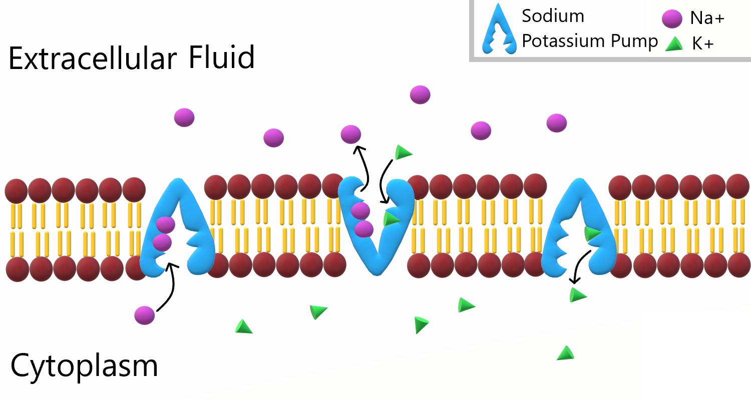
To appreciate the importance of the sodium-potassium pump, you need to know more about the roles of sodium and potassium in the body. Both are essential dietary minerals. You need to get them from the foods you eat. Both sodium and potassium are also electrolytes, which means they dissociate into ions (charged particles) in solution, allowing them to conduct electricity. Normal body functions require a very narrow range of concentrations of sodium and potassium ions in body fluids, both inside and outside of cells.
- Sodium is the principal ion in the fluid outside of cells. Normal sodium concentrations are about ten times higher outside of cells than inside of cells. To move sodium out of the cell is moving it against the concentration gradient
- Potassium is the principal ion in the fluid inside of cells. Normal potassium concentrations are about 30 times higher inside of cells than outside of cells. To move potassium into the cell is moving it against the concentration gradient.
These differences in concentration create an electrical and chemical gradient across the cell membrane, called the membrane potential. Tightly controlling the membrane potential is critical for vital body functions, including the transmission of nerve impulses and contraction of muscles. A large percentage of the body's energy goes to maintaining this potential across the membranes of its trillions of cells with the sodium-potassium pump.
Vesicle Transport
Some molecules, such as proteins, are too large to pass through the plasma membrane, regardless of their concentration inside and outside the cell. Very large molecules cross the plasma membrane with a different sort of help, called vesicle transport. Vesicle transport requires energy input from the cell, so it is also a form of active transport. There are two types of vesicle transport: endocytosis and exocytosis. Both types are shown in Figure 4.8.3.
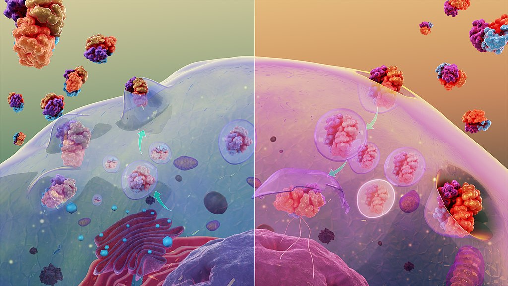
Endocytosis
Endocytosis is a type of vesicle transport that moves a substance into the cell. The plasma membrane completely engulfs the substance, a vesicle pinches off from the membrane, and the vesicle carries the substance into the cell. When an entire cell or other solid particle is engulfed, the process is called phagocytosis. When fluid is engulfed, the process is called pinocytosis.
Exocytosis
Exocytosis is a type of vesicle transport that moves a substance out of the cell (exo-, like "exit"). A vesicle containing the substance moves through the cytoplasm to the cell membrane. Because the vesicle membrane is a phospholipid bilayer like the plasma membrane, the vesicle membrane fuses with the cell membrane, and the substance is released outside the cell.
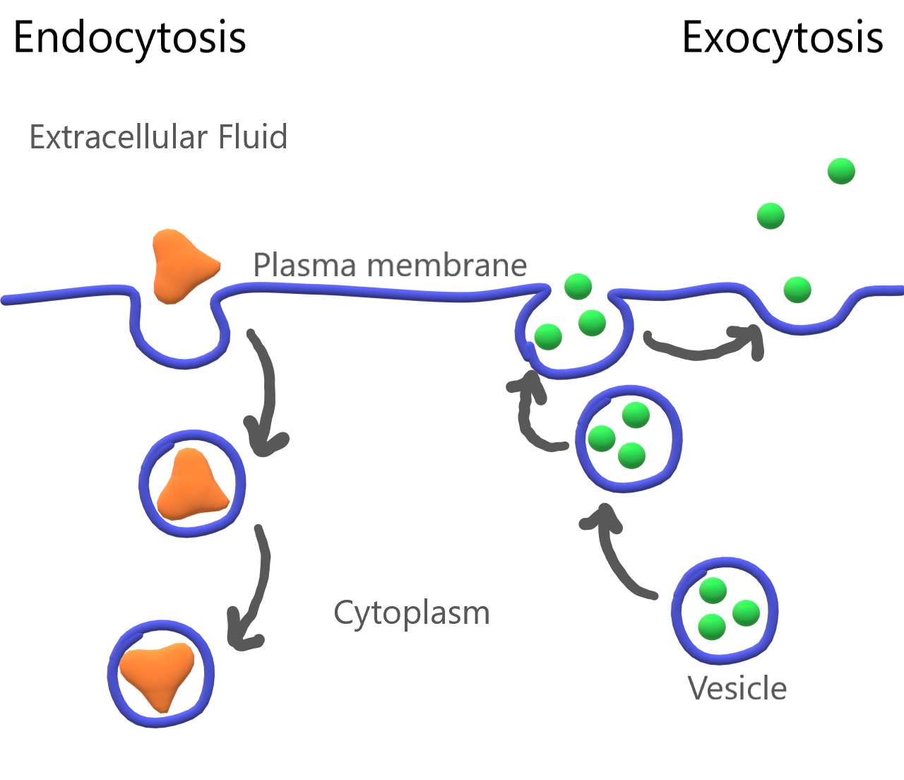
Feature: My Human Body
Maintaining the proper balance of sodium and potassium in body fluids by active transport is necessary for life itself, so it's no surprise that getting the right balance of sodium and potassium in the diet is important for good health. Imbalances may increase the risk of high blood pressure, heart disease, diabetes, and other disorders.
If you are like the majority of North Americans, sodium and potassium are out of balance in your diet. You are likely to consume too much sodium and too little potassium. Follow these guidelines to help ensure that these minerals are balanced in the foods you eat:
- Total sodium intake should be less than 2,300 mg/day. Most salt in the diet is found in processed foods, or added with a salt shaker. Stop adding salt and start checking food labels for sodium content. Foods considered low in sodium have less than 140 mg/serving (or 5 per cent daily value).
- Total potassium intake should be 4,700 mg/day. It's easy to add potassium to the diet by choosing the right foods — and there are plenty of choices! Most fruits and vegetables are high in potassium. Potatoes, bananas, oranges, apricots, plums, leafy greens, tomatoes, lima beans, and avocado are especially good sources. Other foods with substantial amounts of potassium are fish, meat, poultry, and whole grains. The collage below shows some of these potassium-rich foods.
Figure 4.8.5 Potassium power!
4.8 Summary
- Active transport requires energy to move substances across a plasma membrane, often because the substances are moving from an area of lower concentration to an area of higher concentration, or because of their large size. Two types of active transport are membrane pumps (such as the sodium-potassium pump) and vesicle transport.
- The sodium-potassium pump is a mechanism of active transport that moves sodium ions out of the cell and potassium ions into the cell against a concentration gradient, in order to maintain the proper concentrations of ions, both inside and outside the cell, and to thereby control membrane potential.
- Vesicle transport is a type of active transport that uses vesicles to move large molecules into or out of cells.
4.8 Review Questions
- Define active transport.
-
- What is the sodium-potassium pump? Why is it so important?
- The drawing below shows the fluid inside and outside of a cell. The dots represent molecules of a substance needed by the cell. Explain which type of transport — active or passive — is needed to move the molecules into the cell.
Figure 4.8.6 Use this image to answer question #4 - What are the similarities and differences between phagocytosis and pinocytosis?
- What is the functional significance of the shape change of the carrier protein in the sodium-potassium pump after the sodium ions bind?
- A potentially deadly poison derived from plants called ouabain blocks the sodium-potassium pump and prevents it from working. What do you think this does to the sodium and potassium balance in cells? Explain your answer.
4.8 Explore More
https://www.youtube.com/watch?v=Z_mXDvZQ6dU&feature=emb_logo
Neutrophil Phagocytosis - White Blood Cell Eats Staphylococcus Aureus Bacteria,
ImmiflexImmuneSystem, 2013.
https://www.youtube.com/watch?v=Ptmlvtei8hw
Cell Transport, The Amoeba Sisters, 2016.
Attributions
Figure 4.8.1
Humvee challenge by Airman 1st Class Collin Schmidt on Wikimedia Commons is released into the public domain (https://en.wikipedia.org/wiki/Public_domain).
Figure 4.8.2
Sodium Potassium Pump by Christine Miller is used under a CC BY 4.0 (https://creativecommons.org/licenses/by/4.0/) license.
Figure 4.8.3
Cytosis by Manu5 on Wikimedia Commons is used under a CC BY-SA 4.0 (https://creativecommons.org/licenses/by-sa/4.0) license.
Figure 4.8.4
Endocytosis and Exocytosis by Christine Miller is used under a CC BY 4.0 (https://creativecommons.org/licenses/by/4.0/) license.
Figure 4.8.5
- Canteloupes. Image Number K7355-11 by Scott Bauer/ USDA on Wikimedia Commons is in the public domain (https://en.wikipedia.org/wiki/Public_domain).
- Spinach by chiara conti on Unsplash is used under the Unsplash license (https://unsplash.com/license).
- Eleven long purple eggplants by JVRKPRASAD on Wikimedia commons is used under a CC BY-SA 4.0 (https://creativecommons.org/licenses/by-sa/4.0/deed.en) license.
- Bananas by Marco Antonio Victorino on Pexels is used under the Pexels license (https://www.pexels.com/license/).
- Potato picking by Nic D on Unsplash is used under the Unsplash license (https://unsplash.com/license).
- Maldives by Sebastian Pena Lambarri on Unsplash is used under the Unsplash license (https://unsplash.com/license).
Figure 4.8.6
Active Transport by Christine Miller is released into the public domain (https://en.wikipedia.org/wiki/Public_domain).
References
Amoeba Sisters. (2016, June 24). Cell transport [digital image]. YouTube. https://www.youtube.com/watch?v=Ptmlvtei8hw&feature=youtu.be
ImmiflexImmuneSystem. (2013). Neutrophil phagocytosis - White blood cell eats staphylococcus aureus bacteria. YouTube. https://www.youtube.com/watch?v=Z_mXDvZQ6dU
Mayo Clinic Staff. (n.d.). Diabetes [online]. MayoClinic.org. https://www.mayoclinic.org/diseases-conditions/diabetes/symptoms-causes/syc-20371444
Mayo Clinic Staff. (n.d.). High blood pressure (hypertension) [online]. MayoClinic.org. https://www.mayoclinic.org/diseases-conditions/high-blood-pressure/symptoms-causes/syc-20373410
Mayo Clinic Staff. (n.d.). Heart disease [online]. MayoClinic.org. https://www.mayoclinic.org/diseases-conditions/heart-disease/symptoms-causes/syc-20353118
Wikipedia contributors. (2020, June 19). Ouabain. In Wikipedia. https://en.wikipedia.org/w/index.php?title=Ouabain&oldid=963440756
Created by: CK-12/Adapted by Christine Miller
Mush!
These beautiful sled dogs are a metabolic marvel. While running up to 160 kilometres (about 99 miles) a day, they will each consume and burn about 12 thousand calories — about 240 calories per pound per day, which is the equivalent of about 24 Big Macs! A human endurance athlete, in contrast, typically burns only about 100 calories per pound (0.45 kg) each day. Scientists are intrigued by the amazing metabolism of sled dogs, although they still haven't determined how they use up so much energy. But one thing is certain: all living things need energy for everything they do, whether it's running a race or blinking an eye. In fact, every cell of your body constantly needs energy just to carry out basic life processes. You probably know that you get energy from the food you eat, but where does food come from? How does it come to contain energy? And how do your cells get the energy from food?
What Is Energy?
In the scientific world, energy is defined as the ability to do work. You can often see energy at work in living things — a bird flies through the air, a firefly glows in the dark, a dog wags its tail. These are obvious ways that living things use energy, but living things constantly use energy in less obvious ways, as well.
Why Living Things Need Energy
Inside every cell of all living things, energy is needed to carry out life processes. Energy is required to break down and build up molecules, and to transport many molecules across plasma membranes. All of life’s work needs energy. A lot of energy is also simply lost to the environment as heat. The story of life is a story of energy flow — its capture, its change of form, its use for work, and its loss as heat. Energy (unlike matter) cannot be recycled, so organisms require a constant input of energy. Life runs on chemical energy. Where do living organisms get this chemical energy?
How Organisms Get Energy
The chemical energy that organisms need comes from food. Food consists of organic molecules that store energy in their chemical bonds. In terms of obtaining food for energy, there are two types of organisms: autotrophs and heterotrophs.
Autotrophs
Autotrophs are organisms that capture energy from nonliving sources and transfer that energy into the living part of the ecosystem. They are also able to make their own food. Most autotrophs use the energy in sunlight to make food in the process of photosynthesis. Only certain organisms — such as plants, algae, and some bacteria — can make food through photosynthesis. Some photosynthetic organisms are shown in Figure 4.9.2.
 |
 |
 |
| Figure 4.9.2 Photosynthetic autotrophs, which make food using the energy in sunlight, include plants (left), algae (middle), and certain bacteria (right). |
Autotrophs are also called producers. They produce food not only for themselves, but for all other living things (known as consumers), as well. This is why autotrophs form the basis of food chains, such as the food chain shown In Figure 4.9.3.
A food chain shows how energy and matter flow from producers to consumers. Matter is recycled, but energy must keep flowing into the system. Where does this energy come from?
Watch the video "The simple story of photosynthesis and food - Amanda Ooten" from TED-Ed to learn more about photosynthesis:
https://www.youtube.com/watch?time_continue=39&v=eo5XndJaz-Y
The simple story of photosynthesis and food - Amanda Ooten, TED-Ed, 2013.
Heterotrophs
Heterotrophs are living things that cannot make their own food. Instead, they get their food by consuming other organisms, which is why they are also called consumers. They may consume autotrophs or other heterotrophs. Heterotrophs include all animals and fungi, as well as many single-celled organisms. In Figure 4.9.3, all of the organisms are consumers except for the grasses and phytoplankton. What do you think would happen to consumers if all producers were to vanish from Earth?
Energy Molecules: Glucose and ATP
Organisms mainly use two types of molecules for chemical energy: glucose and ATP. Both molecules are used as fuels throughout the living world. Both molecules are also key players in the process of photosynthesis.
Glucose
Glucose is a simple carbohydrate with the chemical formula C6H12O6. It stores chemical energy in a concentrated, stable form. In your body, glucose is the form of energy that is carried in your blood and taken up by each of your trillions of cells. Glucose is the end product of photosynthesis, and it is the nearly universal food for life. In Figure 4.9.4, you can see how photosynthesis stores energy from the sun in the glucose molecule and then how cellular respiration breaks the bonds in glucose to retrieve the energy.
ATP
If you remember from section 3.7 Nucleic Acids, ATP (adenosine triphosphate) is the energy-carrying molecule that cells use to power most cellular processes (nerve impulse conduction, protein synthesis and active transport are good examples of cell processes that rely on ATP as their energy source). ATP is made during the first half of photosynthesis and then used for energy during the second half of photosynthesis, when glucose is made. ATP releases energy when it gives up one of its three phosphate groups (Pi) and changes to ADP (adenosine diphosphate, which has two phosphate groups), as shown in Figure 4.9.5. Thus, the breakdown of ATP into ADP + Pi is a catabolic reaction that releases energy (exothermic). ATP is made from the combination of ADP and Pi, an anabolic reaction that takes in energy (endothermic).
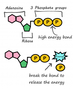
Why Organisms Need Both Glucose and ATP
Why do living things need glucose if ATP is the molecule that cells use for energy? Why don’t autotrophs just make ATP and be done with it? The answer is in the “packaging.” A molecule of glucose contains more chemical energy in a smaller “package” than a molecule of ATP. Glucose is also more stable than ATP. Therefore, glucose is better for storing and transporting energy. Glucose, however, is too powerful for cells to use. ATP, on the other hand, contains just the right amount of energy to power life processes within cells. For these reasons, both glucose and ATP are needed by living things.
How Energy Flows Through Living Things
The flow of energy through living organisms begins with photosynthesis. This process stores energy from sunlight in the chemical bonds of glucose. By breaking the chemical bonds in glucose, cells release the stored energy and make the ATP they need. The process in which glucose is broken down and ATP is made is called cellular respiration.
Photosynthesis and cellular respiration are like two sides of the same coin. This is apparent in Figure 4.9.6. The products of one process are the reactants of the other. Together, the two processes store and release energy in living organisms. The two processes also work together to recycle oxygen in the Earth’s atmosphere.
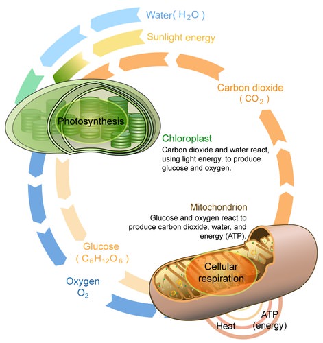
4.9 Summary
- Energy is the ability to do work. It is needed by all living things and every living cell to carry out life processes, such as breaking down and building up molecules, and transporting many molecules across cell membranes.
- The form of energy that living things need for these processes is chemical energy, and it comes from food. Food consists of organic molecules that store energy in their chemical bonds.
- Autotrophs make their own food. Plants, for example, make food by photosynthesis. Autotrophs are also called producers.
- Heterotrophss obtain food by eating other organisms. Heterotrophs are also known as consumers.
- Organisms mainly use the molecules glucose and ATP for energy. Glucose is a compact, stable form of energy that is carried in the blood and taken up by cells. ATP contains less energy and is used to power cell processes.
- The flow of energy through living things begins with photosynthesis, which creates glucose. In a process called cellular respiration, organisms' cells break down glucose and make the ATP they need.
4.9 Review Questions
- Define energy.
- Why do living things need energy?
-
- Compare and contrast the two basic ways that organisms get energy.
- Describe the roles and relationships of the energy molecules glucose and ATP.
- Summarize how energy flows through living things.
- Why does the transformation of ATP to ADP release energy?
4.9 Explore More
https://www.youtube.com/watch?v=eDalQv7d2cs
Learn Biology: Autotrophs vs. Heterotrophs, Mahalodotcom, 2011.
https://www.youtube.com/watch?v=0glkXIj1DgE&feature=emb_logo
Energy Transfer in Trophic Levels, Teacher's Pet, 2015.
Attributions
Figure 4.9.1
Three Airmen participate in dog-sled expedition by U.S. Air Force photo by Tech. Sgt. Dan Rea is released into the public domain (https://en.wikipedia.org/wiki/Public_domain).
Figure 4.9.2
- Plant [photo] by Ren Ran on Unsplash is used under the Unsplash License (https://unsplash.com/license).
- Green Algae by Tristan Schmurr on Flickr is used under a CC BY 2.0 (https://creativecommons.org/licenses/by/2.0/) license.
- Cyanobacteria by Argon National Laboratory on Flickr is used under a CC BY-NC-SA 2.0 (https://creativecommons.org/licenses/by-nc-sa/2.0/) license.
Figure 4.9.3
Biomass_Pyramid by Swiggity.Swag.YOLO.Bro on Wikipedia is used and adapted by Christine Miller under a CC BY-SA 4.0 (https://creativecommons.org/licenses/by-sa/4.0/deed.en) license.
Figure 4.9.4
Photosynthesis and respiration by Christine Miller is used under a CC BY 4.0 (https://creativecommons.org/licenses/by/4.0/) license.
Figure 4.9.5
Photo synthesis and cellular respiration by Lady of Hats/ CK-12 Foundation is used under a CC BY-NC 3.0 (https://creativecommons.org/licenses/by-nc/3.0/) license.
References
LadyofHats/CK-12 Foundation. (2016, August 15). Figure 5: Photosynthesis and cellular respiration [digital image]. In Brainard, J., and Henderson, R., CK-12's College Human Biology FlexBook® (Section 4.9). CK-12 Foundation. https://www.ck12.org/book/ck-12-college-human-biology/section/4.9/
Mahalodotcom. (2011, January 14). Learn biology: Autotrophs vs. heterotrophs. YouTube. https://www.youtube.com/watch?v=eDalQv7d2cs
Teacher's Pet. (2015, March 23). Energy transfer in trophic levels. YouTube. https://www.youtube.com/watch?v=0glkXIj1DgE&feature=emb_logo
TED-Ed. (2013, March 5). The simple story of photosynthesis and food - Amanda Ooten. YouTube. https://www.youtube.com/watch?v=eo5XndJaz-Y&feature=youtu.be
A body system including a series of hollow organs joined in a long, twisting tube from the mouth to the anus. The hollow organs that make up the GI tract are the mouth, esophagus, stomach, small intestine, large intestine, and anus. The liver, pancreas, and gallbladder are the solid organs of the digestive system.
Created by: CK-12/Adapted by Christine Miller

Fast and Furious
These sprinters' muscles will need a lot of energy to complete this short race, because they will be running at top speed. The action won't last long, but it will be very intense. The energy each sprinter needs can't be provided quickly enough by aerobic cellular respiration. Instead, their muscle cells must use a different process to power their activity.
Making ATP Without Oxygen
Living things' cells power their activities with the energy-carrying molecule ATP (adenosine triphosphate). The cells of most living things make ATP from glucose in the process of cellular respiration. This process occurs in three stages: glycolysis, the Krebs cycle, and electron transport. The latter two stages require oxygen, making cellular respiration an aerobic process. When oxygen is not available in cells, the ETS quickly shuts down. Luckily, there are also ways of making ATP from glucose which are anaerobic, which means that they do not require oxygen. These processes are referred to collectively as anaerobic respiration.
Fermentation
Ome important way of making ATP without oxygen is fermentation. Fermentation starts with glycolysis, which does not require oxygen, but it does not involve the latter two stages of aerobic cellular respiration (the Krebs cycle and electron transport). There are two types of fermentation: alcoholic fermentation and lactic acid fermentation. We make use of both types of fermentation using other organisms, but only lactic acid fermentation actually takes place inside the human body.
Alcoholic Fermentation
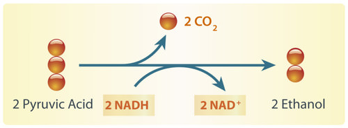
Alcoholic fermentation is carried out by single-celled fungi (called yeasts), as well as some bacteria. We use alcoholic fermentation in these organisms to make biofuels, bread, and wine. The biofuel ethanol (a type of alcohol), for example, is produced by alcoholic fermentation of the glucose in corn or other plants. The process by which this happens is summarized in the diagram below. The two pyruvic acid molecules shown in the diagram come from the splitting of glucose in the first stage of the process (glycolysis). ATP is also made during glycolysis. Two molecules of ATP are produced from each molecule of glucose.
Yeasts in bread dough also use alcoholic fermentation for energy. They produce carbon dioxide gas as a waste product. The carbon dioxide released causes bubbles in the dough and explains why the dough rises. Do you see the small holes in the bread pictured to the right? The holes were formed by bubbles of carbon dioxide gas.
As you have probably guessed, yeast is also used in producing alcoholic beverages. When making beer, brewers will add yeast to a mix of barley and hops. In the absence of oxygen, yeast will carry out alcoholic fermentation in order to convert the glucose in the barley into energy, producing the alcohol content as well as the carbonation present in beer.
Lactic Acid Fermentation
Lactic acid fermentation is carried out by certain bacteria, including the bacteria in yogurt. It is also carried out by your muscle cells when you work them hard and fast. This is how the muscles of the sprinters pictured above get energy for their short-lived — but intense — activity. When this happens, your muscles are using ATP faster than your cardiovascular system can deliver oxygen! The process by which this happens is summarized in the diagram below. Again, the two pyruvic acid molecules shown in the diagram come from the splitting of glucose in the first stage of the process (glycolysis). It is also during this stage that two ATP molecules are produced. The rest of the processes produce lactic acid. Note that, unlike in alcoholic fermentation, there is no carbon dioxide waste product in lactic acid fermentation.
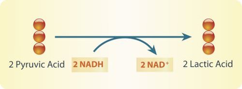
Lactic acid fermentation produces lactic acid and NAD+. The NAD+ cycles back to allow glycolysis to continue so more ATP is made. Each circle represents a carbon atom.
Did you ever run a race, lift heavy weights, or participate in some other intense activity and notice that your muscles start to feel a burning sensation? This may occur when your muscle cells use lactic acid fermentation to provide ATP for energy. The buildup of lactic acid in the muscles causes a burning feeling. This painful sensation is useful if it gets you to stop overworking your muscles and allow them a recovery period, during which cells can eliminate the lactic acid.
Pros and Cons of Anaerobic Respiration
With oxygen, organisms can use aerobic cellular respiration to produce up to 38 molecules of ATP from just one molecule of glucose. Without oxygen, organisms must use anaerobic respiration to produce ATP, and this process produces only two molecules of ATP per molecule of glucose. Although anaerobic respiration produces less ATP, it has the advantage of doing so very quickly. For example, it allows your muscles to get the energy they need for short bursts of intense activity. Aerobic cellular respiration, in contrast, produces ATP more slowly.
Fermentation in Food Production
Anaerobic respiration is also used in the food industry. You read about yeast's role in making bread and beer, but did you know that there are many microbes that are used to create the food we eat, including cheese, sour cream, yogurt, soy sauce, olives, pepperoni, and many more. Watch the video below to learn more about fermentation in the food industry.
https://www.youtube.com/watch?v=eksagPy5tmQ&t=3s
The beneficial bacteria that make delicious food - Erez Garty, TED-Ed, 2016.
4.11 Cultural Connection
Fishing has always been part of the culture and nutrition of Indigenous peoples living on the west coast of Canada. Fish provides delicious important nutrients such as protein, Omega-3 fatty acids, calcium, iron, and Vitamins A, B, C and D. Traditionally, no part of the fish was wasted, including head, eyes, internal organs, and eggs.
Eulachon, also known as candle fish or oolichan, (pictured below) have been prized for their oil for thousands of years. The pathways of these fish dictated "grease trails" and are found from Bristol Bay, Alaska, all the way south to the Klamath River, California. Within BC, the areas of Nass, Knights Inlet, and Bella Coola had large trading centres for this important natural resource.
Photos by Brodie Guy - www.brodieguy.com CC BY-NC-ND 4.0
Euchalon were and are eaten fresh, smoked or dried, and as grease. The grease remains a highly valued food to Indigenous coastal communities. The flavour of the grease varies greatly depending not only on where the fish is from and how it is made, but also how long it is left to ferment. To ferment the eulachon, fish are left in a wood-lined locker dug into the soil for 10 days. Fermentation uses the action of fungi and bacteria to break down the fish making oil extraction much faster and easier.
To learn more, visit the First Nations Health Authority Traditional Foods Fact Sheet and a feature in the Yukon News, "Eulachon, oolicahn, hooligan: A fish by any other name is just as oily."
4.11 Summary
- The cells of most living things produce ATP from glucose by aerobic cellular respiration, which uses oxygen. Some organisms instead produce ATP from glucose by anaerobic respiration, which does not require oxygen.
- An important way of making ATP without oxygen is fermentation. There are two types of fermentation: alcoholic fermentation and lactic acid fermentation. Both start with glycolysis, the first (anaerobic) stage of cellular respiration, in which two molecules of ATP are produced from one molecule of glucose.
- Alcoholic fermentation is carried out by single-celled organisms, including yeasts and some bacteria. We use alcoholic fermentation in these organisms to make biofuels, bread, and wine.
- Lactic acid fermentation is undertaken by certain bacteria, including the bacteria in yogurt, and also by our muscle cells when they are worked hard and fast.
- Anaerobic respiration produces far less ATP than does aerobic cellular respiration, but it has the advantage of being much faster. For example, it allows muscles to get the energy they need for short bursts of intense activity.
4.11 Review Questions
- Explain the primary difference between aerobic cellular respiration and anaerobic respiration.
- What is fermentation?
- Compare and contrast alcoholic and lactic acid fermentation.
- Identify the major pros and the major cons of anaerobic respiration relative to aerobic cellular respiration.
- What process is shared between aerobic cellular respiration and anaerobic respiration? Describe the process briefly. Why can this process happen in anaerobic respiration, as well as aerobic respiration?
-
- What is the reactant (or starting material)common to aerobic respiration and both types of fermentation?
4.11 Explore More
https://www.youtube.com/watch?v=cDC29iBxb3w&t=3s
Anaerobic Respiration, Bozeman Science, 2013.
https://www.youtube.com/watch?v=YbdkbCU20_M
Fermentation, The Amoeba Sisters, 2018.
https://www.youtube.com/watch?time_continue=17&v=TVtqwWGguFk&feature=emb_logo
Science of Beer: Tapping the Power of Brewer's Yeast, KQED Science, 2014.
Attributions
Figure 4.11.1
Sprinters by Jonathan Chng on Unsplash is used under the Unsplash License (https://unsplash.com/license).
Figure 4.11.2
Alcoholic fermentation by Hana Zavadska/ CK-12 Foundation is used under a CC BY-NC 3.0 (https://creativecommons.org/licenses/by-nc/3.0/) license.
Figure 4.11.3
Bread [photo] by Orlova Maria on Unsplash is used under the Unsplash License (https://unsplash.com/license).
Figure 4.11.4
Lactic Acid Fermenation by Hana Zavadska/ CK-12 Foundation is used under a CC BY-NC 3.0 (https://creativecommons.org/licenses/by-nc/3.0/) license.
References
Bozeman Science. (2013, May 2). Anaerobic respiration. YouTube. https://www.youtube.com/watch?v=cDC29iBxb3w&feature=youtu.be
Hana Zavadska/CK-12 Foundation. (2016, August 15). Figure 2: Alcoholic fermentation [digital image]. In Brainard, J., and Henderson, R., CK-12’s College Human Biology FlexBook® (Section 4.11). CK-12 Foundation. https://www.ck12.org/book/ck-12-college-human-biology/section/4.11/
Hana Zavadska/CK-12 Foundation. (2016, August 15). Figure 4: Lactic acid fermentation [digital image]. In Brainard, J., and Henderson, R., CK-12’s College Human Biology FlexBook® (Section 4.11). CK-12 Foundation. https://www.ck12.org/book/ck-12-college-human-biology/section/4.11/
First Nations Health Authority. (2019, September 6). First Nations traditional foods facts Sheet [pdf]. https://www.fnha.ca/Documents/Traditional_Food_Fact_Sheets.pdf
Genest, M. (2017, May 24). Eulachon, oolichan, hooligan: A fish by any other name is just as oily [online article]. YukonNews.com. https://www.yukon-news.com/business/eulachon-oolichan-hooligan-a-fish-by-any-other-name-is-just-as-oily/
KQED Science. (2014, February 11). Science of beer: Tapping the power of brewer's yeast. YouTube. https://www.youtube.com/watch?v=TVtqwWGguFk&feature=youtu.be
TED-Ed. (2016). The beneficial bacteria that make delicious food - Erez Garty. YouTube. https://www.youtube.com/watch?v=eksagPy5tmQ&feature=youtu.be
The Amoeba Sisters. (2018, April 30). Fermentation. YouTube. https://www.youtube.com/watch?v=YbdkbCU20_M&feature=youtu.be
Wikipedia contributors. (2020, June 21). Ethanol fuel. In Wikipedia. https://en.wikipedia.org/w/index.php?title=Ethanol_fuel&oldid=963675942
Created by: CK-12/Adapted by Christine Miller
Divide and Split
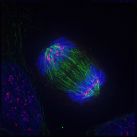
Can you guess what the colourful image in Figure 4.13.1 represents? It shows a eukaryotic cell during the process of cell division. In particular, the image shows the cell in a part of cell division called anaphase, where the DNA is being pulled to opposite ends of the cell. Normally, DNA is located in the nucleus of most human cells. The nucleus divides before the cell itself splits in two, and before the nucleus divides, the cell’s DNA is replicated (or copied). There must be two copies of the DNA so that each daughter cell will have a complete copy of the genetic material from the parent cell. How is the replicated DNA sorted and separated so that each daughter cell gets a complete set of the genetic material? To answer that question, you first need to know more about DNA and the forms it takes.
The Forms of DNA
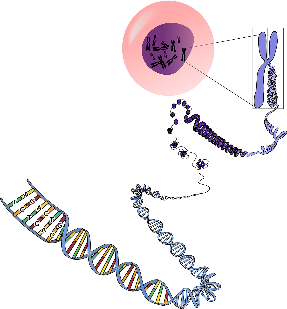
Except when a eukaryotic cell divides, its nuclear DNA exists as a grainy material called chromatin. Only once a cell is about to divide and its DNA has replicated does DNA condense and coil into the familiar X-shaped form of a chromosome, like the one shown below.
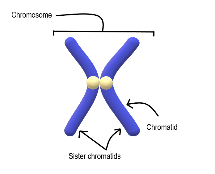
Most cells in the human body have two pairs of 23 different chromosomes, for a total of 46 chromosomes. Cells that have two pairs of chromosomes are called diploid. Because DNA has already replicated when it coils into a chromosome, each chromosome actually consists of two identical structures called sister chromatids. Sister chromatids are joined together at a region called a centromere.
Mitosis
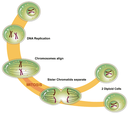
The process in which the nucleus of a eukaryotic cell divides is called mitosis. During mitosis, the two sister chromatids that make up each chromosome separate from each other and move to opposite poles of the cell. This is shown in the figure below.
Mitosis actually occurs in four phases. The phases are called prophase, metaphase, anaphase, and telophase.
Prophase
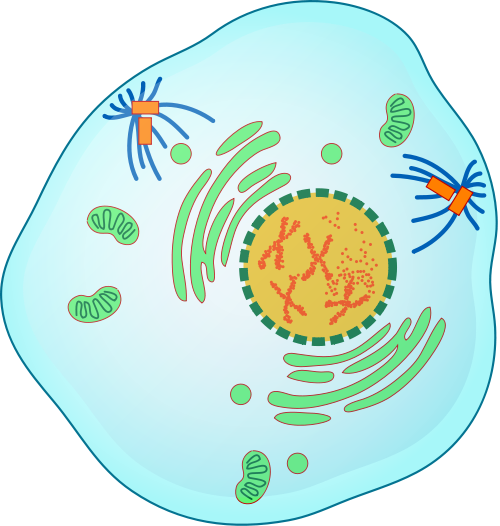
The first and longest phase of mitosis is prophase. During prophase, chromatin condenses into chromosomes, and the nuclear envelope (the membrane surrounding the nucleus) breaks down. In animal cells, the centrioles near the nucleus begin to separate and move to opposite poles of the cell. Centrioles are small organelles found only in eukaryotic cells. They help ensure that the new cells that form after cell division each contain a complete set of chromosomes. As the centrioles move apart, a spindle starts to form between them. The spindle consists of fibres made of microtubules.
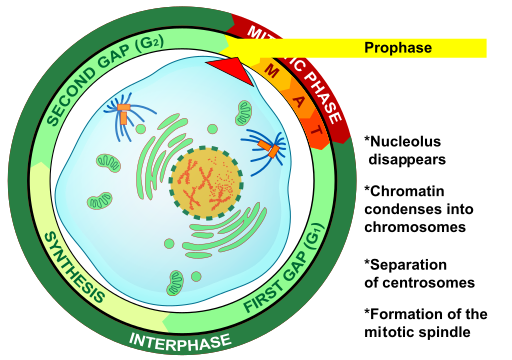
Metaphase
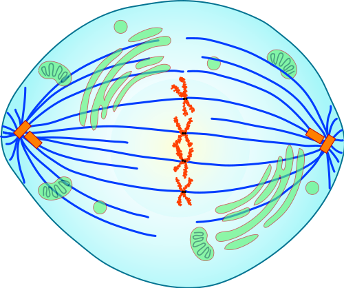
During metaphase, spindle fibres attach to the centromere of each pair of sister chromatids. As you can see in Figure 4.13.7, the sister chromatids line up at the equator (or center) of the cell. The spindle fibres ensure that sister chromatids will separate and go to different daughter cells when the cell divides.
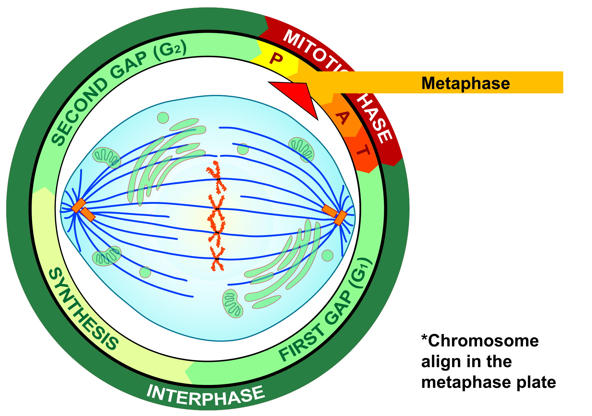
Anaphase
During anaphase, sister chromatids separate and the centromeres divide. The sister chromatids are pulled apart by the shortening of the spindle fibres. This is a little like reeling in a fish by shortening the fishing line. One sister chromatid moves to one pole of the cell, and the other sister chromatid moves to the opposite pole. At the end of anaphase, each pole of the cell has a complete set of chromosomes.
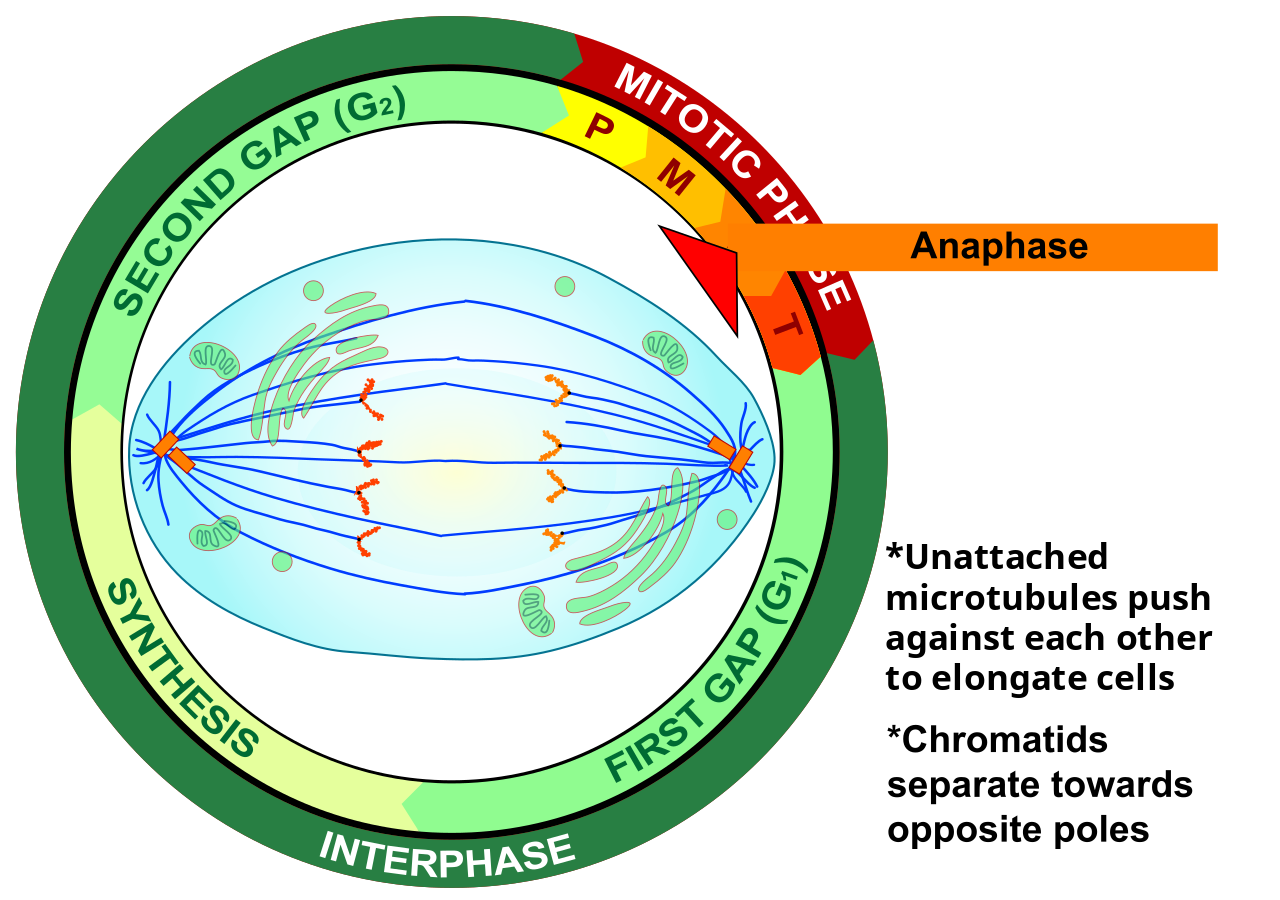
Telophase
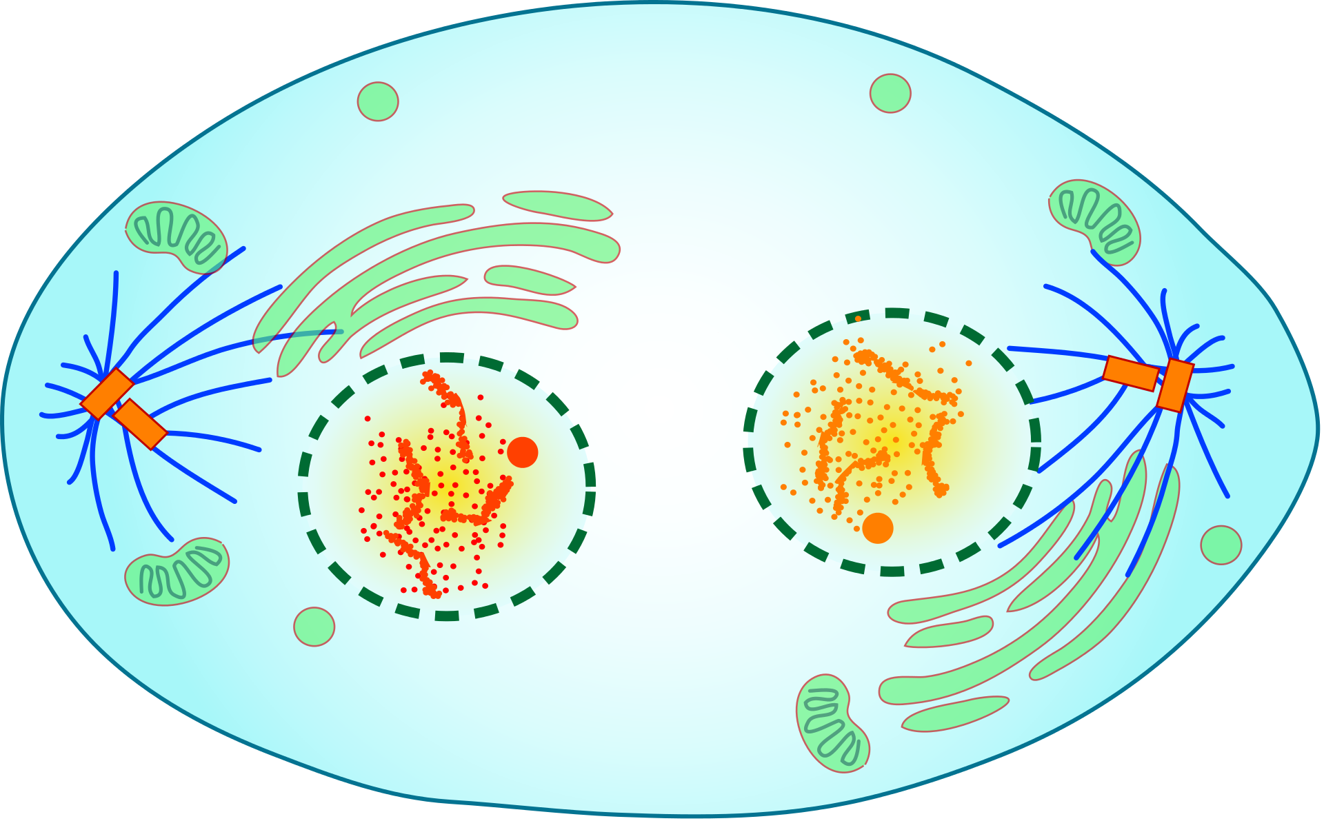
During telophase, the chromosomes begin to uncoil and form chromatin. This prepares the genetic material for directing the metabolic activities of the new cells. The spindle also breaks down, and new nuclear envelopes form.
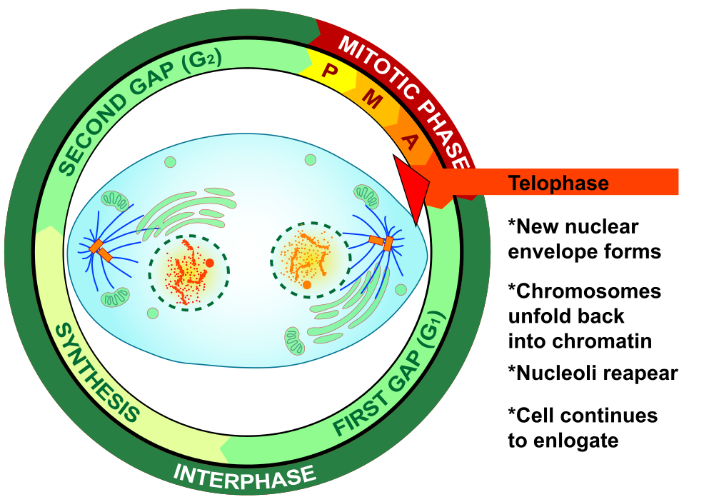
Cytokinesis

Cytokinesis is the final stage of cell division. During cytokinesis, the cytoplasm splits in two and the cell divides, as shown below. In animal cells, the plasma membrane of the parent cell pinches inward along the cell’s equator until two daughter cells form. Thus, the goal of mitosis and cytokinesis is now complete, because one parent cell has given rise to two daughter cells. The daughter cells have the same chromosomes as the parent cell.
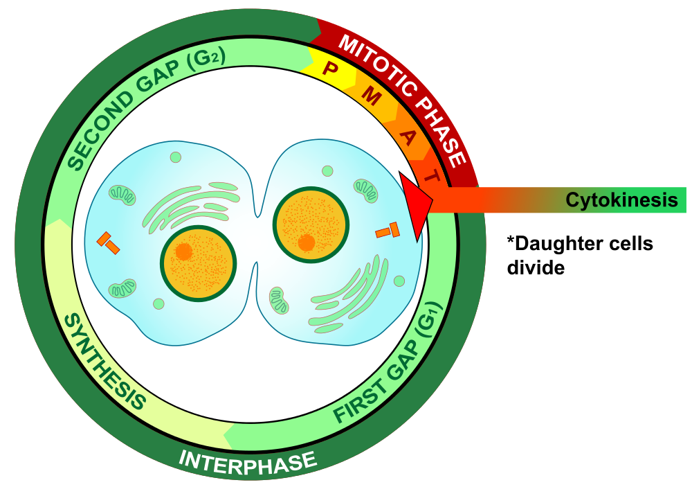
4.13 Summary
- Until a eukaryotic cell divides, its nuclear DNA exists as a grainy material called chromatin. After DNA replicates and the cell is about to divide, the DNA condenses and coils into the X-shaped form of a chromosome. Each chromosome actually consists of two sister chromatids, which are joined together at a centromere.
- Mitosis is the process during which the nucleus of a eukaryotic cell divides. During this process, sister chromatids separate from each other and move to opposite poles of the cell. This happens in four phases: prophase, metaphase, anaphase, and telophase.
- Cytokinesis is the final stage of cell division, during which the cytoplasm splits in two and two daughter cells form.
4.13 Review Questions
- Describe the different forms that DNA takes before and during cell division in a eukaryotic cell.
-
- Identify the four phases of mitosis in an animal cell, and summarize what happens during each phase.
- Order the diagrams of the stages of mitosis:
- Explain what happens during cytokinesis in an animal cell.
- What do you think would happen if the sister chromatids of one of the chromosomes did not separate during mitosis?
- True or False:
4.13 Explore More
https://www.youtube.com/watch?time_continue=3&v=C6hn3sA0ip0&feature=emb_logo
Mitosis, NDSU Virtual Cell Animations project (ndsuvirtualcell), 2012.
https://www.youtube.com/watch?time_continue=19&v=EA0qxhR2oOk&feature=emb_logo
Nondisjunction (Trisomy 21) - An Animated Tutorial, Kristen Koprowski, 2012.
Attributions
Figure 4.13.1
Anaphase_IF by Roy van Heesbeen on Wikimedia Commons is released into the public domain (https://en.wikipedia.org/wiki/Public_domain).
Figure 4.13.2
Chromosomes by OpenClipArt-Vectors on Pixabay is used under the Pixabay License (https://pixabay.com/service/license/).
Figure 4.13.3
Chromosome/ Chromatid/ Sister Chromatid by Christine Miller is released into the public domain (https://en.wikipedia.org/wiki/Public_domain).
Figure 4.13.4
Simple Mitosis by Mariana Ruiz Villarreal [LadyofHats] via CK-12 Foundation is used under a CC BY-NC 3.0 (https://creativecommons.org/licenses/by-nc/3.0/) license.
 ©CK-12 Foundation Licensed under
 ![]() • Terms of Use • Attribution
• Terms of Use • Attribution
Figure 4.13.5
Mitotic Prophase [tiny] by Mariana Ruiz Villarreal [LadyofHats] on Wikimedia Commons is released into the public domain (https://en.wikipedia.org/wiki/Public_domain).
Figure 4.13.6
Prophase Eukaryotic Mitosis by Mariana Ruiz Villarreal [LadyofHats] on Wikimedia Commons is released into the public domain (https://en.wikipedia.org/wiki/Public_domain).
Figure 4.13.7
Mitotic_Metaphase by Mariana Ruiz Villarreal [LadyofHats] on Wikimedia Commons is released into the public domain (https://en.wikipedia.org/wiki/Public_domain).
Figure 4.13.8
Metaphase Eukaryotic Mitosis by Mariana Ruiz Villarreal [LadyofHats] on Wikimedia Commons is released into the public domain (https://en.wikipedia.org/wiki/Public_domain).
Figure 4.13.9
Anaphase [adapted] by Mariana Ruiz Villarreal [LadyofHats] on Wikimedia Commons is released into the public domain (https://en.wikipedia.org/wiki/Public_domain).
Figure 4.13.10
Anaphase_eukaryotic_mitosis.svg by Mariana Ruiz Villarreal [LadyofHats] on Wikimedia Commons is released into the public domain (https://en.wikipedia.org/wiki/Public_domain).
Figure 4.13.11
Mitotic Telophase by Mariana Ruiz Villarreal [LadyofHats] on Wikimedia Commons is released into the public domain (https://en.wikipedia.org/wiki/Public_domain).
Figure 4.13.12
Telophase Eukaryotic Mitosis by Mariana Ruiz Villarreal [LadyofHats] on Wikimedia Commons is released into the public domain (https://en.wikipedia.org/wiki/Public_domain).
Figure 4.13.13
Mitotic Cytokinesis by Mariana Ruiz Villarreal [LadyofHats] on Wikimedia Commons is released into the public domain (https://en.wikipedia.org/wiki/Public_domain).
Figure 4.13.14
Cytokinesis Eukaryotic Mitosis by Mariana Ruiz Villarreal [LadyofHats] on Wikimedia Commons is released into the public domain (https://en.wikipedia.org/wiki/Public_domain).
References
Koprowski, K., Cabey, R. [Kristen Koprowski]. (2012). Nondisjunction (Trisomy 21) - An Animated Tutorial. YouTube. https://www.youtube.com/watch?v=EA0qxhR2oOk&feature=youtu.be
NDSU Virtual Cell Animations project [ndsuvirtualcell]. (2012). Mitosis. YouTube. https://www.youtube.com/watch?v=C6hn3sA0ip0&t=21s
A fluid-filled space inside the body that holds and protects internal organs.
Created by: CK-12/Adapted by Christine Miller
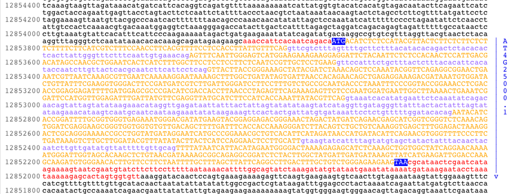
Can You Code?
If someone asks you whether you can code, you probably assume they are referring to computer code. The image in Figure 5.6.1 represents an important code that you use all the time — but not with a computer! It's the genetic code, and it is used by your cells to store information, as well as to make RNA and proteins.
What Is the Genetic Code?
The genetic code consists of the sequence of nitrogen bases in a polynucleotide chain of DNA or RNA. The bases are adenine (A), cytosine (C), guanine (G), and thymine (T) (or uracil, U, in RNA). The four bases make up the “letters” of the genetic code. The letters are combined in groups of three to form code “words,” called codons. Each codon stands for (encodes) one amino acid, unless it codes for a start or stop signal. There are 20 common amino acids in proteins. With four bases forming three-base codons, there are 64 possible codons. This is more than enough to code for the 20 amino acids. The genetic code is shown in Figure 5.6.2.
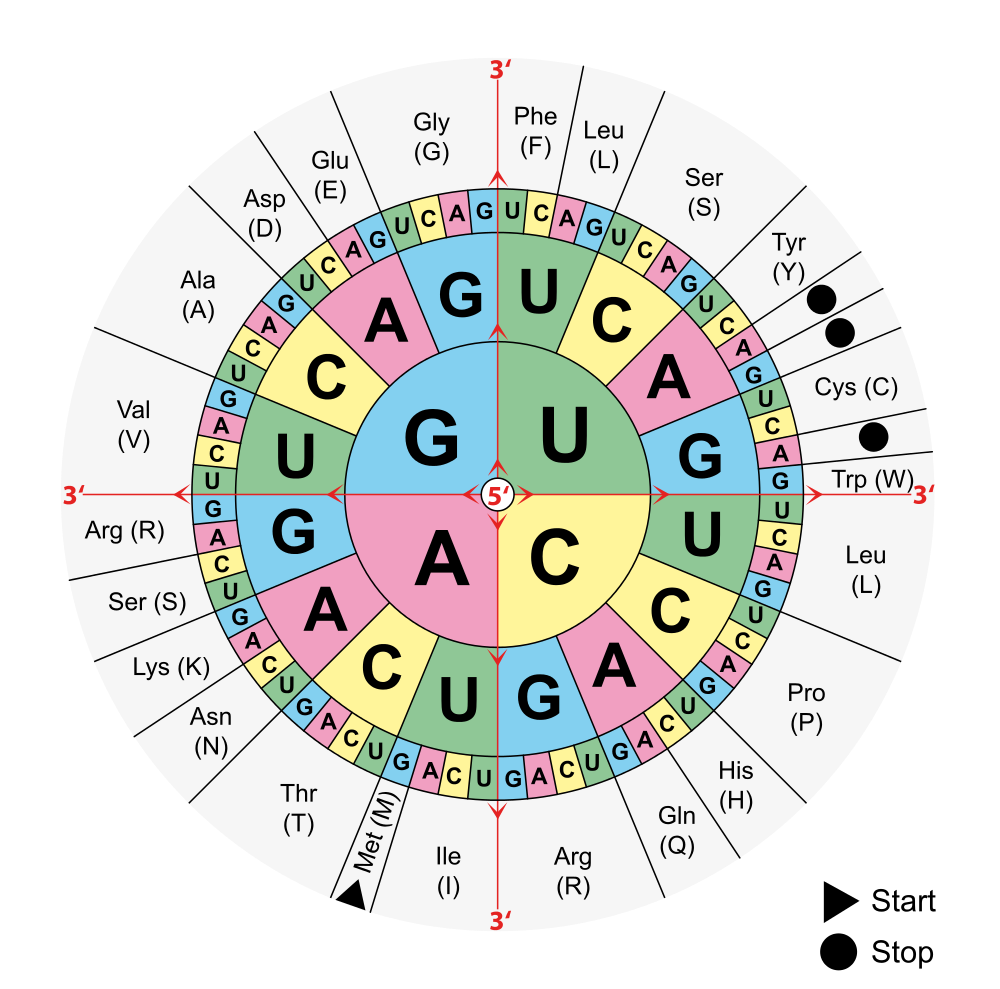
To find the amino acid for a particular codon, find the first base in the codon in the centre of the circle in Figure 5.6.2, then the second base in the middle row out from the center, and finally the third base in the outer ring For example, CUG codes for leucine, AAG codes for lysine, and GGG codes for glycine. Try it out: Can you figure out what the codon AGC codes for?
Reading the Genetic Code
If you find the codon AUG in Figure 5.6.2, you will see that it codes for the amino acid methionine. This codon is also the start codon that establishes the reading frame of the code. The start codon is a necessary tool in translation, since a single chromosome contains many genes. In order to transcribe and translate a gene for a specific protein, we need to know where in the DNA code to start "reading" the instructions. AUG signals the start of a reading frame. After the AUG start codon, the next three bases are read as the second codon. The next three bases after that are read as the third codon, and so on. The sequence of bases is read, codon by codon, until a stop codon is reached. UAG, UGA, and UAA are all stop codons. They do not code for any amino acids.
The importance of the reading frame is illustrated in the hypothetical situation below:
The section of mRNA in Figure 5.6.3 is designed to create a chain of five specific amino acids.
- In Option 1, a string of amino acids is created, but the chain does not have a stop codon, and so keeps on adding amino acids. This means the desired protein was not made.
- In Option 2, a stop codon was encountered and the amino acid chain was stopped after 3 amino acids. This means the desired protein was not made
- In Option 3, using the AUG start codon, a chain of five specific amino acids was successfully formed. Success! This was only possible because the start codon, AUG, specified the reading frame.
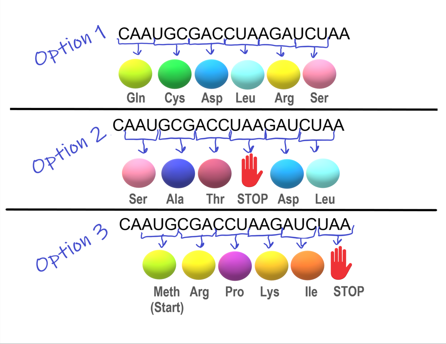
Characteristics of the Genetic Code
The genetic code has a number of important characteristics:
- The genetic code is universal. All known living things have the same genetic code, which shows that all organisms share a common evolutionary history.
- The genetic code is unambiguous. This means that each codon codes for just one amino acid (or start or stop). This is necessary so there is no question about which amino acid is correct.
- The genetic code is redundant. This means that each amino acid is encoded by more than one codon. For example, in Figure 5.6.2, four codons code for the amino acid threonine. Redundancy in the code helps prevent errors in protein synthesis. If a base in a codon changes by accident, there is a good chance that it will still code for the same amino acid.
Cracking the Code
The double helix structure of DNA was discovered in 1953. It took just eight more years to crack the genetic code. The scientist primarily responsible for deciphering the code was American biochemist Marshall Nirenberg, who worked at the National Institutes of Health in the United States. When Nirenberg began the research in 1959, the manner in which proteins are synthesized in cells was not well understood, and messenger RNA had not yet been discovered. At that time, scientists didn't even know whether DNA or RNA was the molecule used as a template for protein synthesis. Nirenberg, along with a collaborator named Heinrich Matthaei, devised an ingenious experiment to determine which molecule — DNA or RNA — has this important role. They also began deciphering the genetic code.
Nirenberg and Matthaei added the contents of bacterial cells to each of 20 test tubes. The cell contents provided the necessary "machinery" for the synthesis of a polypeptide molecule. The researchers also added all 20 amino acids to the test tubes, with a different amino acid "tagged" by a radioactive element in each test tube. That way, if a polypeptide formed in a test tube, they would be able to tell which amino acid it contained. Then, they added synthetic RNA containing just one nitrogen base to all 20 test tubes. They used the base uracil in their first experiment. They discovered that an RNA chain consisting only of uracil bases produces a polypeptide chain of the amino acid phenylalanine. This experiment showed that RNA (rather than DNA) is the template for protein synthesis, but it also showed that a sequence of uracil bases codes for the amino acid phenylalanine. The year was 1961, and it was a momentous occasion. When Nirenberg presented the discovery at a scientific conference later that year, he received a standing ovation. As Nirenberg puts it, "...for the next five years I became like a scientific rock star."
After Nirenberg and Matthaei cracked the first word of the genetic code, they used similar experiments to show that each codon consists of three bases. Before long, they had discovered the codons for all 20 amino acids. In 1968, in recognition of this important achievement, Nirenberg was named a co-winner of the Nobel Prize in Physiology or Medicine.
5.6 Summary
- The genetic code consists of the sequence of nitrogen bases in a polynucleotide chain of DNA or RNA. The four bases make up the "letters" of the code. The letters are combined in groups of three to form code "words" known as codons, each of which encodes for one amino acid or a start or stop signal.
- AUG is the start codon, and it establishes the reading frame of the code. After the start codon, the next three bases are read as the second codon, the three bases after that as the third codon, and so on until a stop codon is reached.
- The genetic code is universal, unambiguous, and redundant.
- The genetic code was cracked in the 1960s, mainly by a series of ingenious experiments carried out by Marshall Nirenberg, who won a Nobel Prize for this achievement.
5.6 Review Questions
- Describe the genetic code and explain how it is "read"
- Identify three important characteristics of the genetic code.
- Summarize how the genetic code was deciphered.

- Use the decoder above to answer the following questions:
- Is the code from DNA or RNA? How do you know?
- Which amino acid does the codon CAA code for?
- What does UGA code for?
- Look at the codons that code for the amino acid glycine. How many of them are there and how are they similar and different?
-
5.6 Explore More
https://www.youtube.com/watch?v=OSKwuOccAak&feature=emb_logo
Comparing DNA Sequences, Bozeman Science, 2012.
https://www.youtube.com/watch?v=LsEYgwuP6ko
How to Read a Codon Chart, Amoeba Sisters, 2019.
Attributions
Figure 5.6.1
AMY1gene by unknown author from National Science Foundation on Wikimedia Commons is released into the public domain (https://en.wikipedia.org/wiki/Public_domain).
Figure 5.6.2
Aminoacids table (Adapted) by Mouagip on Wikimedia Commons is released into the public domain. (Original: Codons sun ("codesonne" in German) by Onie~commonswiki])
Figure 5.6.3
Reading Frame (3 Options) by Christine Miller is used under a CC BY-NC-SA 4.0 (https://creativecommons.org/licenses/by-nc-sa/4.0/) license.
References
Amoeba Sisters. (2019, September 17). How to read a codon chart. YouTube. https://www.youtube.com/watch?v=LsEYgwuP6ko&feature=youtu.be
Bozeman Science. (2012, September 15). Comparing DNA sequences. YouTube. https://www.youtube.com/watch?v=OSKwuOccAak&feature=youtu.be
Wikipedia contributors. (2020, July 2). Marshall Warren Nirenberg. In Wikipedia. https://en.wikipedia.org/w/index.php?title=Marshall_Warren_Nirenberg&oldid=965562106
Created by: CK-12/Adapted by Christine Miller

Mutant Cosplay
You probably recognize these costumed comic fans in Figure 5.8.1 as two of the four Teenage Mutant Ninja Turtles. Can a mutation really turn a reptile into an anthropomorphic superhero? Of course not — but mutations can often result in other drastic (but more realistic) changes in living things.
What Are Mutations?
Mutations are random changes in the sequence of bases in DNA or RNA. The word mutation may make you think of the Ninja Turtles, but that's a misrepresentation of how most mutations work. First of all, everyone has mutations. In fact, most people have dozens (or even hundreds!) of mutations in their DNA. Secondly, from an evolutionary perspective, mutations are essential. They are needed for evolution to occur because they are the ultimate source of all new genetic variation in any species.
Causes of Mutations
Mutations have many possible causes. Some mutations seem to happen spontaneously, without any outside influence. They occur when errors are made during DNA replication or during the transcription phase of protein synthesis. Other mutations are caused by environmental factors. Anything in the environment that can cause a mutation is known as a mutagen. Examples of mutagens are shown in the figure below.
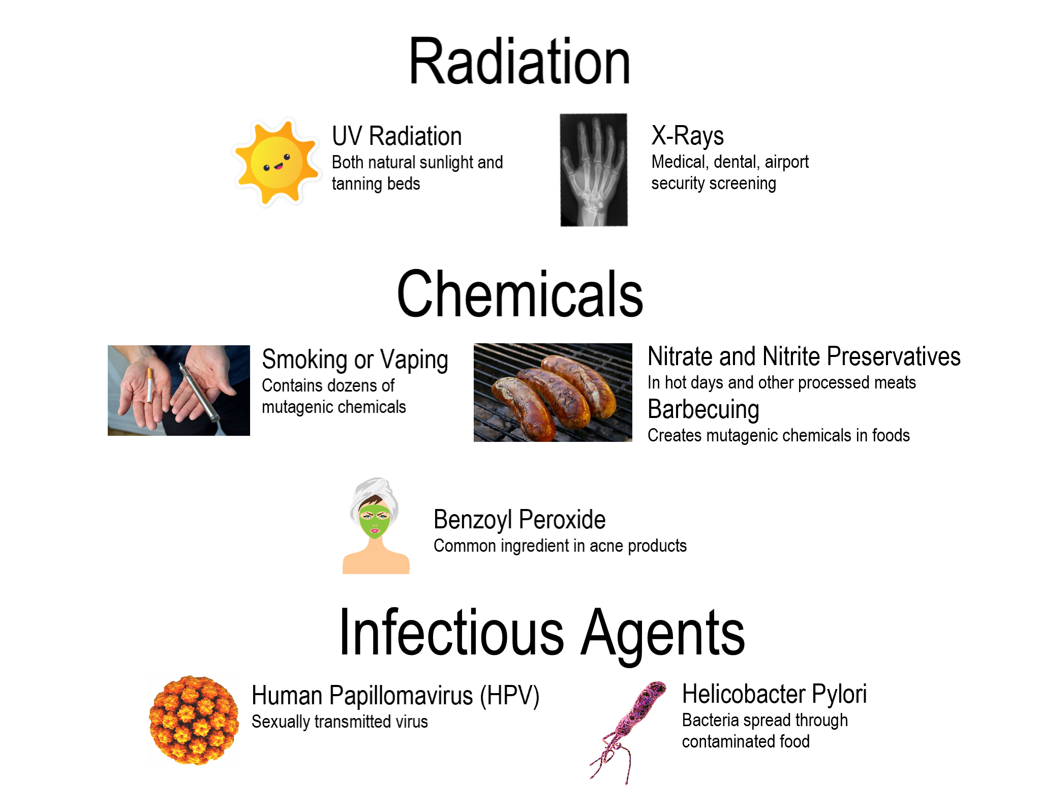
Types of Mutations
Mutations come in a variety of types. Two major categories of mutations are germline mutations and somatic mutations.
- Germline mutations occur in gametes (the sex cells), such as eggs and sperm. These mutations are especially significant because they can be transmitted to offspring, causing every cell in the offspring to carry those mutations.
- Somatic mutations occur in other cells of the body. These mutations may have little effect on the organism, because they are confined to just one cell and its daughter cells. Somatic mutations cannot be passed on to offspring.
Mutations also differ in the way that the genetic material is changed. Mutations may change an entire chromosome, or they may alter just one or a few nucleotides.
Chromosomal Alterations
Chromosomal alterations are mutations that change chromosome structure. They occur when a section of a chromosome breaks off and rejoins incorrectly, or otherwise does not rejoin at all. Possible ways in which these mutations can occur are illustrated in the figure below. Chromosomal alterations are very serious. They often result in the death of the organism in which they occur. If the organism survives, it may be affected in multiple ways. An example of a human disease caused by a chromosomal duplication is Charcot-Marie-Tooth disease type 1 (CMT1). It is characterized by muscle weakness, as well as loss of muscle tissue and sensation. The most common cause of CMT1 is a duplication of part of chromosome 17.
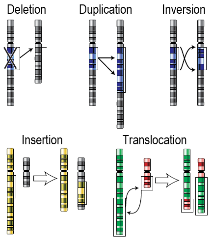
Point Mutations
A point mutation is a change in a single nucleotide in DNA. This type of mutation is usually less serious than a chromosomal alteration. An example of a point mutation is a mutation that changes the codon UUU to the codon UCU. Point mutations can be silent, missense, or nonsense mutations, as described in Table 5.8.1. The effects of point mutations depend on how they change the genetic code.
| Type | Description | Example | Effect |
|---|---|---|---|
| Silent | mutated codon codes for the same amino acid | CAA (glutamine) → CAG (glutamine) | none |
| Missense | mutated codon codes for a different amino acid | CAA (glutamine) → CCA (proline) | variable |
| Nonsense | mutated codon is a premature stop codon | CAA (glutamine) → UAA (stop) usually | serious |
Frameshift Mutations
A frameshift mutation is a deletion or insertion of one or more nucleotides, changing the reading frame of the base sequence. Deletions remove nucleotides, and insertions add nucleotides. Consider the following sequence of bases in RNA:
AUG-AAU-ACG-GCU = start-asparagine-threonine-alanine
Now, assume that an insertion occurs in this sequence. Let’s say an A nucleotide is inserted after the start codon AUG. The sequence of bases becomes:
AUG-AAA-UAC-GGC-U = start-lysine-tyrosine-glycine
Even though the rest of the sequence is unchanged, this insertion changes the reading frame and, therefore, all of the codons that follow it. As this example shows, a frameshift mutation can dramatically change how the codons in mRNA are read. This can have a drastic effect on the protein product.
Effects of Mutations
The majority of mutations have neither negative nor positive effects on the organism in which they occur. These mutations are called neutral mutations. Examples include silent point mutations, which are neutral because they do not change the amino acids in the proteins they encode.
Many other mutations have no effects on the organism because they are repaired before protein synthesis occurs. Cells have multiple repair mechanisms to fix mutations in DNA.
Beneficial Mutations
Some mutations — known as beneficial mutations — have a positive effect on the organism in which they occur. They generally code for new versions of proteins that help organisms adapt to their environment. If they increase an organism’s chances of surviving or reproducing, the mutations are likely to become more common over time. There are several well-known examples of beneficial mutations. Here are two such examples:
- Mutations have occurred in bacteria that allow the bacteria to survive in the presence of antibiotic drugs, leading to the evolution of antibiotic-resistant strains of bacteria.
- A unique mutation is found in people in Limone, a small town in Italy. The mutation protects them from developing atherosclerosis, which is the dangerous buildup of fatty materials in blood vessels despite a high-fat diet. The individual in which this mutation first appeared has even been identified and many of his descendants carry this gene.
Harmful Mutations
Imagine making a random change in a complicated machine, such as a car engine. There is a chance that the random change would result in a car that does not run well — or perhaps does not run at all. By the same token, a random change in a gene's DNA may result in the production of a protein that does not function normally... or may not function at all. Such mutations are likely to be harmful. Harmful mutations may cause genetic disorders or cancer.
- A genetic disorder is a disease, syndrome, or other abnormal condition caused by a mutation in one or more genes, or by a chromosomal alteration. An example of a genetic disorder is cystic fibrosis. A mutation in a single gene causes the body to produce thick, sticky mucus that clogs the lungs and blocks ducts in digestive organs.
- Cancer is a disease in which cells grow out of control and form abnormal masses of cells (called tumors). It is generally caused by mutations in genes that regulate the cell cycle. Because of the mutations, cells with damaged DNA are allowed to divide without restriction.
Feature: My Human Body
Inherited mutations are thought to play a role in roughly five to ten per cent of all cancers. Specific mutations that cause many of the known hereditary cancers have been identified. Most of the mutations occur in genes that control the growth of cells or the repair of damaged DNA.
Genetic testing can be done to determine whether individuals have inherited specific cancer-causing mutations. Some of the most common inherited cancers for which genetic testing is available include hereditary breast and ovarian cancer, caused by mutations in genes called BRCA1 and BRCA2. Besides breast and ovarian cancers, mutations in these genes may also cause pancreatic and prostate cancers. Genetic testing is generally done on a small sample of body fluid or tissue, such as blood, saliva, or skin cells. The sample is analyzed by a lab that specializes in genetic testing, and it usually takes at least a few weeks to get the test results.
Should you get genetic testing to find out whether you have inherited a cancer-causing mutation? Such testing is not done routinely just to screen patients for risk of cancer. Instead, the tests are generally done only when the following three criteria are met:
- The test can determine definitively whether a specific gene mutation is present. This is the case with the BRCA1 and BRCA2 gene mutations, for example.
- The test results would be useful to help guide future medical care. For example, if you found out you had a mutation in the BRCA1 or BRCA2 gene, you might get more frequent breast and ovarian cancer screenings than are generally recommended.
- You have a personal or family history that suggests you are at risk of an inherited cancer.
Criterion number 3 is based, in turn, on such factors as:
- Diagnosis of cancer at an unusually young age.
- Several different cancers occurring independently in the same individual.
- Several close genetic relatives having the same type of cancer (such as a maternal grandmother, mother, and sister all having breast cancer).
- Cancer occurring in both organs in a set of paired organs (such as both kidneys or both breasts).
If you meet the criteria for genetic testing and are advised to undergo it, genetic counseling is highly recommended. A genetic counselor can help you understand what the results mean and how to make use of them to reduce your risk of developing cancer. For example, a positive test result that shows the presence of a mutation may not necessarily mean that you will develop cancer. It may depend on whether the gene is located on an autosome or sex chromosome, and whether the mutation is dominant or recessive. Lifestyle factors may also play a role in cancer risk even for hereditary cancers. Early detection can often be life saving if cancer does develop. Genetic counseling can also help you assess the chances that any children you may have will inherit the mutation.
5.8 Summary
- Mutations are random changes in the sequence of bases in DNA or RNA. Most people have multiple mutations in their DNA without ill effects. Mutations are the ultimate source of all new genetic variation in any species.
- Mutations may happen spontaneously during DNA replication or transcription. Other mutations are caused by environmental factors called mutagens. Mutagens include radiation, certain chemicals, and some infectious agents.
- Germline mutations occur in gametes and may be passed onto offspring. Every cell in the offspring will then have the mutation. Somatic mutations occur in cells other than gametes and are confined to just one cell and its daughter cells. These mutations cannot be passed on to offspring.
- Chromosomal alterations are mutations that change chromosome structure and usually affect the organism in multiple ways. Charcot-Marie-Tooth disease type 1 is an example of a chromosomal alteration in humans.
- Point mutations are changes in a single nucleotide. The effects of point mutations depend on how they change the genetic code and may range from no effects to very serious effects.
- Frameshift mutations change the reading frame of the genetic code and are likely to have a drastic effect on the encoded protein.
- Many mutations are neutral and have no effect on the organism in which they occur. Some mutations are beneficial and improve fitness. An example is a mutation that confers antibiotic resistance in bacteria. Other mutations are harmful and decrease fitness, such as the mutations that cause genetic disorders or cancers.
5.8 Review Question
- Define mutation.
- Identify causes of mutation.
- Compare and contrast germline and somatic mutations.
- Describe chromosomal alterations, point mutations, and frameshift mutations. Identify the potential effects of each type of mutation.
- Why do many mutations have neutral effects?
- Give one example of a beneficial mutation and one example of a harmful mutation.
-
- Why do you think that exposure to mutagens (such as cigarette smoke) can cause cancer?
- Explain why the insertion or deletion of a single nucleotide can cause a frameshift mutation.
- Compare and contrast missense and nonsense mutations.
- Explain why mutations are important to evolution.
5.8 Explore More
https://www.youtube.com/watch?time_continue=51&v=PQjL4ZDuq2o&feature=emb_logo
How Radiation Changes Your DNA, Seeker, 2016.
https://www.youtube.com/watch?v=z9HIYjRRaDE&t=93s
Where do genes come from? - Carl Zimmer, TED-Ed, 2014.
https://www.youtube.com/watch?v=a63t8r70QN0&feature=youtu.be
What you should know about vaping and e-cigarettes | Suchitra Krishnan-Sarin,
TED, 2019.
Attributions
Figure 5.8.1
Ninja Turtles by Pat Loika on Flickr is used under a CC BY 2.0 (https://creativecommons.org/licenses/by/2.0/) license.
Figure 5.8.2
Separate images are all in public domain or CC licensed:
- Beauty treatment face mask by no-longer-here on Pixabay is used under the Pixabay License (https://pixabay.com/service/license/).
- HPV by AJC1 on Flickr is used under a CC BY-NC 2.0 (https://creativecommons.org/licenses/by-nc/2.0/) license.
- H Pylori by AJC1 on Flickr is used under a CC BY-NC 2.0 (https://creativecommons.org/licenses/by-nc/2.0/) license.
- Vape and Cigarette by Vaping360 on Flickr is used under a CC BY 2.0 (https://creativecommons.org/licenses/by/2.0/) license.
- Hand X-Ray by Hellerhoff on Wikimedia Commons - CC BY-SA 3.0 (https://creativecommons.org/licenses/by-sa/3.0/deed.en) license.
- Hot dogs by unknown on PxFuel is used under the Pxfuel Terms (https://www.pxfuel.com/terms-of-use).
- Sunshine face is clipart.
Figure 5.8.3
Scheme of possible chromosome mutations/ Chromosomenmutationen by unknown on Wikimedia Commons is adapted from NIH's Talking Glossary of Genetics. [Changes as described by de:user:Dietzel65]. Further use and adapation (text translated to English) by Christine Miller as image is in the public domain (https://en.wikipedia.org/wiki/Public_domain).
References
Seeker. (2016, April 23). How radiation changes your DNA. YouTube. https://www.youtube.com/watch?v=PQjL4ZDuq2o&feature=youtu.be
TED. (2019, June 5). What you should know about vaping and e-cigarettes | Suchitra Krishnan-Sarin. YouTube. https://www.youtube.com/watch?v=a63t8r70QN0&feature=youtu.be
TED-Ed. (2014, September 22). Where do genes come from? - Carl Zimmer. YouTube. https://www.youtube.com/watch?v=z9HIYjRRaDE&feature=youtu.be
Wikipedia contributors. (2020, July 6). Breast cancer. In Wikipedia. https://en.wikipedia.org/w/index.php?title=Breast_cancer&oldid=966366739
Wikipedia contributors. (2020, July 9). Charcot–Marie–Tooth disease. In Wikipedia. https://en.wikipedia.org/w/index.php?title=Charcot%E2%80%93Marie%E2%80%93Tooth_disease&oldid=966912915
Wikipedia contributors. (2020, July 7). Cystic fibrosis. In Wikipedia. https://en.wikipedia.org/w/index.php?title=Cystic_fibrosis&oldid=966566921
Wikipedia contributors. (2020, June 4). Limone sul Garda. In Wikipedia. https://en.wikipedia.org/w/index.php?title=Limone_sul_Garda&oldid=960771991
Wikipedia contributors. (2020, June 23). Ovarian cancer. In Wikipedia. https://en.wikipedia.org/w/index.php?title=Ovarian_cancer&oldid=964157192
Wikipedia contributors. (2020, May 7). BRCA mutation. In Wikipedia. https://en.wikipedia.org/w/index.php?title=BRCA_mutation&oldid=955463902
Wikipedia contributors. (2020, July 10). Teenage Mutant Ninja Turtles. In Wikipedia. https://en.wikipedia.org/w/index.php?title=Teenage_Mutant_Ninja_Turtles&oldid=967030468
A large cavity found in the torso of mammals between the thoracic cavity, which it is separated from by the thoracic diaphragm, and the pelvic cavity. Organs of the abdominal cavity include the stomach, liver, gallbladder, spleen, pancreas, small intestine, kidneys, large intestine, and adrenal glands.
Created by CK-12 Foundation/Adapted by Christine Miller
Case Study Conclusion: Cough That Won't Quit
Inhaling the moist air from a humidifier or steamy shower can feel particularly good if you have a respiratory system infection, such as bronchitis. The moist air helps to loosen and thin mucus in the respiratory system, allowing you to breathe easier.
In the beginning of this chapter, you learned about Erica, who developed acute bronchitis after getting a cold. She had a worsening cough, a sore throat due to coughing, and chest congestion. She was also coughing up thick mucus.
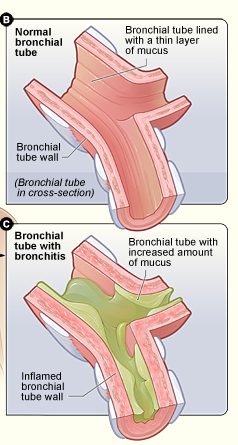
Acute bronchitis usually occurs after a cold or flu, usually due to the same viruses that cause cold or flu. Because bronchitis is not usually caused by bacteria (although it can be), in most cases, antibiotics are not an effective treatment.
Bronchitis affects the bronchial tubes, which, as you have learned, are air passages in the lower respiratory tract. The main bronchi branch off of the trachea and then branch into smaller bronchi, and then bronchioles. In bronchitis, the walls of the bronchi become inflamed, which makes them narrower. There is also excessive production of mucus in the bronchi, which further narrows the pathway where air can flow through. Figure 13.7.2, shows how bronchitis affects the bronchial tubes.
The treatment for most cases of bronchitis involves thinning and loosening the mucus so that it can be effectively coughed out of the airways. This can be done by drinking plenty of fluids, using humidifiers or steam, and — in some cases — using over-the-counter medications (such as expectorants). Dr. Choo recommended some of these treatments to Erica, and also warned against using cough suppressants. Cough suppressants work on the nervous system to suppress the cough reflex. When a patient has a “productive” cough (which means they are coughing up mucus), doctors generally advise them not to take cough suppressants, so that they can cough the mucus out of their bodies.
When Dr. Choo was examining Erica, she used a pulse oximeter to measure the oxygen level in her blood. Why did she do this? As you have learned, the bronchial tubes branch into bronchioles, which ultimately branch into the alveoli of the lungs. The alveoli are where gas exchange occurs between the air and the blood to take in oxygen and remove carbon dioxide and other wastes. By checking Erica’s blood oxygen level, Dr. Choo was making sure that her clogged airways were not impacting her level of much-needed oxygen.
Erica has acute bronchitis, but you may recall that chronic bronchitis was discussed earlier in this chapter (Section 13.5) as a term that describes the symptoms of chronic obstructive pulmonary disease (COPD). COPD is often due to tobacco smoking, and it causes damage to the walls of the alveoli. Acute bronchitis, on the other hand, typically occurs after a cold or flu, and involves inflammation and mucus build-up in the bronchial tubes. As implied by the difference in their names, chronic bronchitis is an ongoing, long-term condition, while acute bronchitis is likely to resolve relatively quickly with proper rest and treatment.
Erica uses e-cigarettes (vaping), so she is more likely to develop chronic respiratory conditions, such as COPD. As you have learned, smoking damages the respiratory system, along with many other systems of the body. Smoking and vaping increases the risk of respiratory infections, including bronchitis and flu, due to its damaging effects on the respiratory and immune systems. Dr. Choo strongly encouraged Erica to quit vaping, not only so that her acute bronchitis resolves, but so that she can avoid future infections and other negative health outcomes associated with vaping and smoking, including COPD and lung cancer.
As you have learned in this chapter, the respiratory system is critical to carry out the gas exchange necessary for life’s functions, and to protect the body from pathogens and other potentially harmful substances in the air. But this ability to interface with the outside air has a cost. The respiratory system is prone to infections, as well as damage and other negative effects from allergens, mold, air pollution, cigarette smoke and vaping. While exposure to most of these things cannot be avoided, not smoking is an important step you can take to protect this organ system — as well as many other systems of your body.
Chapter 13 Summary
In this chapter, you learned about the respiratory system. Specifically, you learned that:
- Respiration is the process in which oxygen moves from the outside air into the body, and carbon dioxide and other waste gases move from inside the body to the outside air. It involves two subsidiary processes: ventilation and gas exchange.
- The organs of the respiratory system form a continuous system of passages, called the respiratory tract. It has two major divisions: the upper respiratory tract and the lower respiratory tract.
-
- The upper respiratory tract includes the nasal cavity, pharynx, and larynx. All of these organs are involved in conduction, or the movement of air into and out of the body. Incoming air is also cleaned, humidified, and warmed as it passes through the upper respiratory tract. The larynx is also called the voice box, because it contains the vocal cords, which are needed to produce vocal sounds.
- The lower respiratory tract includes the trachea, bronchi and bronchioles, and the lungs. The trachea, bronchi, and bronchioles are involved in conduction. Gas exchange takes place only in the lungs, which are the largest organs of the respiratory tract. Lung tissue consists mainly of tiny air sacs called alveoli, which is where gas exchange takes place between air in the alveoli and the blood in capillaries surrounding them.
- The respiratory system protects itself from potentially harmful substances in the air by the mucociliary escalator. This includes mucus-producing cells, which trap particles and pathogens in incoming air. It also includes tiny hair-like cilia that continually move to sweep the mucus and trapped debris away from the lungs and toward the outside of the body.
- The level of carbon dioxide in the blood is monitored by cells in the brain. If the level becomes too high, it triggers a faster rate of breathing, which lowers the level to the normal range. The opposite occurs if the level becomes too low. The respiratory system exchanges gases with the outside air, but it needs the cardiovascular system to carry the gases to and from cells throughout the body.
- Breathing, or ventilation, is the two-step process of drawing air into the lungs (inhalation) and letting air out of the lungs (exhalation). Inhaling is an active process that results mainly from contraction of a muscle called the diaphragm. Exhaling is typically a passive process that occurs mainly due to the elasticity of the lungs when the diaphragm relaxes.
-
- Breathing is one of the few vital bodily functions that can be controlled consciously, as well as unconsciously. Conscious control of breathing is common in many activities, including swimming and singing. However, there are limits on the conscious control of breathing. If you try to hold your breath, for example, you will soon have an irrepressible urge to breathe.
- Unconscious breathing is controlled by respiratory centers in the medulla and pons of the brainstem. They respond to variations in blood pH by either increasing or decreasing the rate of breathing as needed to return the pH level to the normal range.
- Nasal breathing is generally considered to be superior to mouth breathing, because it does a better job of filtering, warming, and moistening incoming air. It also results in slower emptying of the lungs, which allows more oxygen to be extracted from the air.
- Gas exchange is the biological process through which gases are transferred across cell membranes to either enter or leave the blood. Gas exchange takes place continuously between the blood and cells throughout the body, and also between the blood and the air inside the lungs.
-
- Gas exchange in the lungs takes place in alveoli. The pulmonary artery carries deoxygenated blood from the heart to the lungs, where it travels through pulmonary capillaries, picking up oxygen and releasing carbon dioxide. The oxygenated blood then leaves the lungs through pulmonary veins.
- Gas exchange occurs by diffusion across cell membranes. Gas molecules naturally move down a concentration gradient from an area of higher concentration to an area of lower concentration. This is a passive process that requires no energy.
- Gas exchange by diffusion depends on the large surface area provided by the hundreds of millions of alveoli in the lungs. It also depends on a steep concentration gradient for oxygen and carbon dioxide. This gradient is maintained by continuous blood flow and constant breathing.
- Asthma is a chronic inflammatory disease of the airways in the lungs, in which the airways periodically become inflamed. This causes swelling and narrowing of the airways, often with excessive mucus production, leading to difficulty breathing and other symptoms. Asthma is thought to be caused by a combination of genetic and environmental factors. Asthma attacks are triggered by allergens, air pollution, or other factors.
- Pneumonia is a common inflammatory disease of the respiratory tract in which inflammation affects primarily the alveoli, which become filled with fluid that inhibits gas exchange. Most cases of pneumonia are caused by viral or bacterial infections. Vaccines are available to prevent pneumonia. Treatment often includes prescription antibiotics.
- Chronic obstructive pulmonary disease (COPD) is a lung disease characterized by chronic poor airflow, which causes shortness of breath and a productive cough. It is caused most often by tobacco smoking, which leads to breakdown of connective tissues in the lungs. Alveoli are reduced in number and elasticity, making it impossible to fully exhale air from the lungs. There is no cure for COPD, but stopping smoking may reduce the rate at which COPD worsens.
- Lung cancer is a malignant tumor characterized by uncontrolled cell growth in tissues of the lung. It results from accumulated DNA damage, most often caused by tobacco smoking. Lung cancer is typically diagnosed late, so most cases cannot be cured. It may be treated with surgery, chemotherapy, and/or radiation therapy.
- Smoking is the single greatest cause of preventable death worldwide. It has adverse effects on just about every body system and organ. Tobacco smoke affects not only smokers, but also non-smokers who are exposed to secondhand smoke. The nicotine in tobacco is highly addictive, making it very difficult to quit smoking.
-
- A major health risk of smoking is lung cancer. Smoking also increases the risk of many other types of cancer. Tobacco smoke contains dozens of chemicals that are known carcinogens.
- Smoking is the primary cause of COPD. Chemicals — such as carbon monoxide and cyanide in tobacco smoke — reduce the elasticity of alveoli so the lungs can no longer fully exhale air.
- Smoking and/or vaping damages the cardiovascular system and increases the risk of high blood pressure, blood clots, heart attack, and stroke. Smoking also has a negative impact on blood lipid levels.
- A wide diversity of additional adverse health effects — such as erectile dysfunction, female infertility, and slow wound healing — are attributable to smoking.
As you have learned, the respiratory system brings in oxygen to the body and removes waste gases to the atmosphere — but these molecules wouldn’t get to where they need to go without the cardiovascular system to transport them via the bloodstream. Read the next chapter to learn about how the cardiovascular system carries out these critical functions.
Chapter 13 Review
-
- Describe the relationship between the bronchi, secondary bronchi, tertiary bronchi, and bronchioles.
- Deoxygenated and oxygenated blood both travel to the lungs. Describe what happens to that blood when it gets to the lungs.
- Explain the difference between ventilation and gas exchange.
- Which way do oxygen and carbon dioxide flow during gas exchange in the lungs, and why? Which way do oxygen and carbon dioxide flow during gas exchange between the blood and the body’s cells, and why?
- Why does the body require oxygen, and why does it emit carbon dioxide as a waste product?
- What do coughing and sneezing have in common?
- COPD can cause too much carbon dioxide in the blood. Answer the following questions about this:
- How does COPD cause there to be too much carbon dioxide in the blood?
- What does this do to the blood pH?
- How does the body respond to this change in blood pH?
- What are three different types of things that can enter the respiratory system and cause illness or injury? Describe the negative health effects of each in your answer.
- Where are the respiratory centers of the brain located? What is the main function of the respiratory centers of the brain?
- Smoking increases the risk of getting influenza, commonly known as the flu. Explain why this could lead to a greater risk of pneumonia.
- If a person has a gene that caused them to get asthma, could changes to their environment (such as more frequent cleaning) help their asthma? Why or why not?
- Explain why nasal breathing generally stops particles from entering the body at an earlier stage than mouth breathing does.
Attributions
Figure 13.7.1
Tags: Essential Oils Aroma Diffuser Diffuse Led by asundermeier on Pixabay is used under the Pixabay License (https://unsplash.com/license).
Figure 13.7.2
Bronchitis by National Heart Lung and Blood Institute on Wikimedia Commons is in the public domain (https://en.wikipedia.org/wiki/Public_domain).
A major human body cavity that includes the head and the posterior (back) of the trunk and holds the brain and spinal cord.
A cavity that fills most of the upper part of the skull and contains the brain.
Created by: CK-12/Adapted by Christine Miller

Like Father, Like Son
This father-son duo share some similarities. The shape of their faces and their facial features look very similar. If you saw them together, you might well guess that they are father and son. People have long known that the characteristics of living things are similar between parents and their offspring. However, it wasn’t until the experiments of Gregor Mendel that scientists understood how those traits are inherited.
The Father of Genetics
Mendel did experiments with pea plants to show how traits such as seed shape and flower colour are inherited. Based on his research, he developed his two well known laws of inheritance: the law of segregation and the law of independent assortment. When Mendel died in 1884, his work was still virtually unknown. In 1900, three other researchers working independently came to the same conclusions that Mendel had drawn almost half a century earlier. Only then was Mendel's work rediscovered.
Mendel knew nothing about genes, because they were discovered after his death. He did think, however, that some type of "factors" controlled traits, and that those "factors" were passed from parents to offspring. We now call these "factors" genes. Mendel's laws of inheritance, now expressed in terms of genes, form the basis of genetics, the science of heredity. For this reason, Mendel is often called the father of genetics.
The Language of Genetics
Today, we know that traits of organisms are controlled by genes on chromosomes. To talk about inheritance in terms of genes and chromosomes, you need to know the language of genetics. The terms below serve as a good starting point. They are illustrated in the figure that follows.
- A gene is the part of a chromosome that contains the genetic code for a given protein. For example, in pea plants, a given gene might code for flower colour.
- The position of a given gene on a chromosome is called its locus (plural, loci). A gene might be located near the center, or at one end or the other of a chromosome.
- A given gene may have different normal versions, which are called alleles. For example, in pea plants, there is a purple-flower allele (B) and a white-flower allele (b) for the flower-colour gene. Different alleles account for much of the variation in the traits of organisms, including people.
- In sexually reproducing organisms, each individual has two copies of each type of chromosome. Paired chromosomes of the same type are called homologous chromosomes. They are about the same size and shape, and they have all the same genes at the same loci.
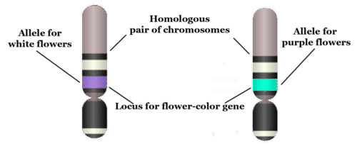
Genotype
When sexual reproduction occurs, sex cells (called gametes) unite during fertilization to form a single cell called a zygote. The zygote inherits two of each type of chromosome, with one chromosome of each type coming from the father, and the other coming from the mother. Because homologous chromosomes have the same genes at the same loci, each individual also inherits two copies of each gene. The two copies may be the same allele or different alleles. The alleles an individual inherits for a given gene make up the individual’s genotype. As shown in Table 5.11.1, an organism with two of the same allele (for example, BB or bb) is called a homozygote. An organism with two different alleles (in this example, Bb) is called a heterozygote.
Table 5.11.1
Allele Combinations Associated With the Terms Homozygous and Heterozygous

Phenotype
The expression of an organism’s genotype is referred to as its phenotype, and it refers to the organism’s traits, such as purple or white flowers in pea plants. As you can see from Table 5.11.1, different genotypes may produce the same phenotype. In this example, both BB and Bb genotypes produce plants with the same phenotype, purple flowers. Why does this happen? In a Bb heterozygote, only the B allele is expressed, so the b allele doesn’t influence the phenotype. In general, when only one of two alleles is expressed in the phenotype, the expressed allele is called dominant, and the allele that isn’t expressed is called recessive.
The terms dominant and recessive may also be used to refer to phenotypic traits. For example, purple flower colour in pea plants is a dominant trait. It shows up in the phenotype whenever a plant inherits even one dominant allele for the trait. Similarly, white flower colour is a recessive trait. Like other recessive traits, it shows up in the phenotype only when a plant inherits two recessive alleles for the trait.
5.11 Summary
- Mendel's laws of inheritance, now expressed in terms of genes, form the basis of genetics, which is the science of heredity. This is why Mendel is often called the father of genetics.
- A gene is the part of a chromosome that codes for a given protein. The position of a gene on a chromosome is its locus. A given gene may have different versions, called alleles. Paired chromosomes of the same type are called homologous chromosomes. They have the same size and shape, and they have the same genes at the same loci.
- The alleles an individual inherits for a given gene make up the individual's genotype. An organism with two of the same allele is called a homozygote, and an individual with two different alleles is called a heterozygote.
- The expression of an organism's genotype is referred to as its phenotype. A dominant allele is always expressed in the phenotype, even when just one dominant allele has been inherited. A recessive allele is expressed in the phenotype only when two recessive alleles have been inherited.
5.11 Review Questions
- Define genetics.
- Why is Gregor Mendel called the father of genetics if genes were not discovered until after his death?
-
- Imagine that there are two alleles, R and r, for a given gene. R is dominant to r. Answer the following questions about this gene:
- What are the possible homozygous and heterozygous genotypes?
- Which genotype or genotypes express the dominant R phenotype? Explain your answer.
- Are R and r on different loci? Why or why not?
- Can R and r be on the same exact chromosome? Why or why not? If not, where are they located?
5.11 Explore More
https://www.youtube.com/watch?v=pv3Kj0UjiLE
Alleles and Genes, Amoeba Sisters, 2018.
https://www.youtube.com/watch?v=OaovnS7BAoc
Genotypes and Phenotypes, Bozeman Science, 2011.
Attributions
Figure 5.11.1
Father holding his baby boy with matching haircut [photo] by Kelly Sikkema on Unsplash is used under the Unsplash License (https://unsplash.com/license).
Figure 5.11.2
Chromosome, Gene, Locus, and Allele by CK-12 Foundation is used under a CC BY-NC 3.0 (https://creativecommons.org/licenses/by-nc/3.0/) license.
 ©CK-12 Foundation Licensed under
 ![]() • Terms of Use • Attribution
• Terms of Use • Attribution
Table 5.11.1
Allele Combinations Associated With the Terms Homozygous and Heterozygous by Christine Miller is released into the public domain (https://en.wikipedia.org/wiki/Public_domain).
References
Amoeba Sisters. (2018, February 1). Alleles and genes. YouTube. https://www.youtube.com/watch?v=pv3Kj0UjiLE&feature=youtu.be
Bozeman Science. (2011, August 4). Genotypes and phenotypes. YouTube. https://www.youtube.com/watch?v=OaovnS7BAoc&feature=youtu.be
Brainard, J/ CK-12 Foundation. (2016). Figure 2 Chromosome, gene, locus, and allele [digital image]. In CK-12 College Human Biology (Section 5.10) [online Flexbook]. CK12.org. https://www.ck12.org/book/ck-12-human-biology/section/5.9/
division of the peripheral nervous system that controls involuntary activities
A hormone is a signaling molecule produced by glands in multicellular organisms that target distant organs to regulate physiology and behavior.
A set of metabolic reactions and processes that take place in the cells of organisms to convert biochemical energy from nutrients into adenosine triphosphate (ATP).
Glucose (also called dextrose) is a simple sugar with the molecular formula C6H12O6. Glucose is the most abundant monosaccharide, a subcategory of carbohydrates. Glucose is mainly made by plants and most algae during photosynthesis from water and carbon dioxide, using energy from sunlight.
An involuntary human body response mediated by the nervous and endocrine systems that prepares the body to fight or flee from perceived danger.
The ability of an organism to maintain constant internal conditions despite external changes.
Created by CK-12 Foundation/Adapted by Christine Miller
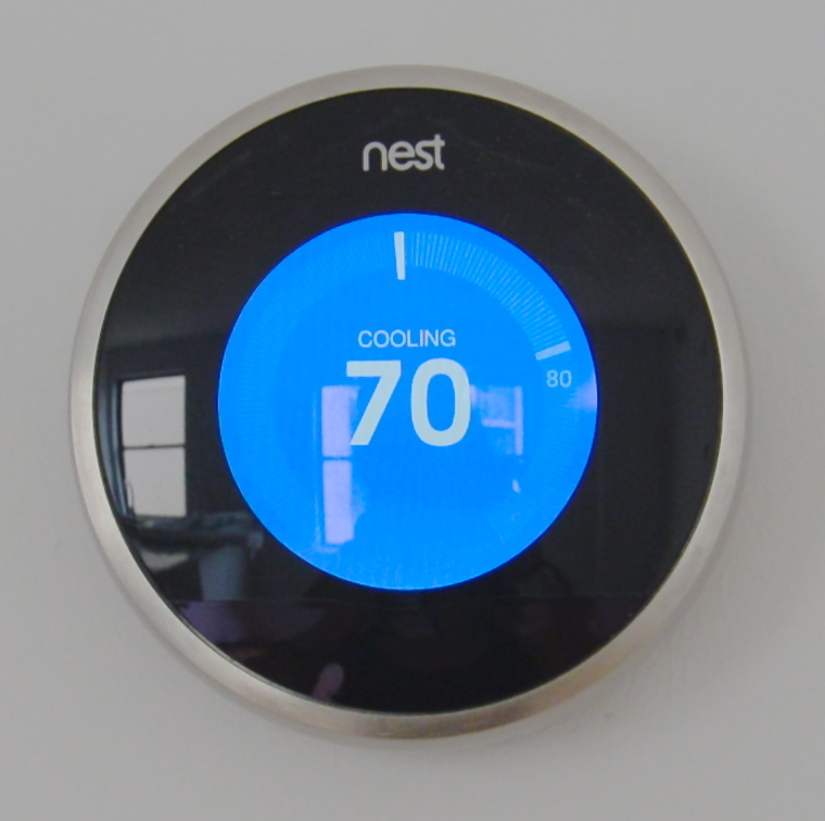
Steady as She Goes
This device (Figure 7.8.1) looks simple, but it controls a complex system that keeps a home at a steady temperature — it's a thermostat. The device shows the current temperature in the room, and also allows the occupant to set the thermostat to the desired temperature. A thermostat is a commonly cited model of how living systems — including the human body— maintain a steady state called homeostasis.
What Is Homeostasis?
Homeostasis is the condition in which a system (such as the human body) is maintained in a more or less steady state. It is the job of cells, tissues, organs, and organ systems throughout the body to maintain many different variables within narrow ranges compatible with life. Keeping a stable internal environment requires continually monitoring the internal environment and constantly making adjustments to keep things in balance.
Set Point and Normal Range
For any given variable, such as body temperature or blood glucose level, there is a particular set point that is the physiological optimum value. The set point for human body temperature, for example, is about 37 degrees C (98.6 degrees F). As the body works to maintain homeostasis for temperature or any other internal variable, the value typically fluctuates around the set point. Such fluctuations are normal, as long as they do not become too extreme. The spread of values within which such fluctuations are considered insignificant is called the normal range. In the case of body temperature, for example, the normal range for an adult is about 36.5 to 37.5 degrees C (97.7 to 99.5 degrees F).
A good analogy for set point, normal range, and maintenance of homeostasis is driving. When you are driving a vehicle on the road, you are supposed to drive in the centre of your lane — this is analogous to the set point. Sometimes, you are not driving in the exact centre of the lane, but you are still within your lines, so you are in the equivalent of the normal range. However, if you were to get too close to the centre line or the shoulder of the road, you would take action to correct your position. You'd move left if you were too close to the shoulder, or right if too close to the centre line — which is analogous to our next concept, negative feedback to maintain homeostasis.
Maintaining Homeostasis
Homeostasis is normally maintained in the human body by an extremely complex balancing act. Regardless of the variable being kept within its normal range, maintaining homeostasis requires at least four interacting components: stimulus, sensor, control centre, and effector.
- The stimulus is provided by the variable being regulated. Generally, the stimulus indicates that the value of the variable has moved away from the set point or has left the normal range.
- The sensor monitors the values of the variable and sends data on it to the control centre.
- The control centre matches the data with normal values. If the value is not at the set point or is outside the normal range, the control centre sends a signal to the effector.
- The effector is an organ, gland, muscle, or other structure that acts on the signal from the control centre to move the variable back toward the set point.
Each of these components is illustrated in Figure 7.8.2. The diagram on the left is a general model showing how the components interact to maintain homeostasis. The diagram on the right shows the example of body temperature. From the diagrams, you can see that maintaining homeostasis involves feedback, which is data that feeds back to control a response. Feedback may be negative (as in the example below) or positive. All the feedback mechanisms that maintain homeostasis use negative feedback. Biological examples of positive feedback are much less common.
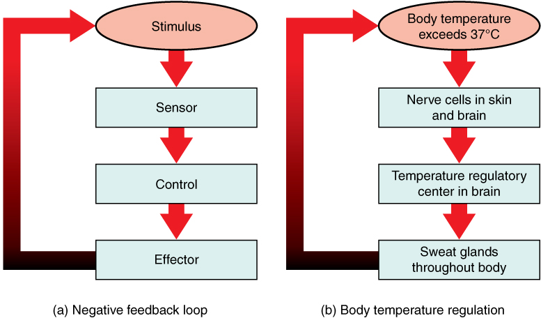
Negative Feedback
In a negative feedback loop, feedback serves to reduce an excessive response and keep a variable within the normal range. Two processes controlled by negative feedback are body temperature regulation and control of blood glucose.
Body Temperature
Body temperature regulation involves negative feedback, whether it lowers the temperature or raises it, as shown in Figure 7.8.3 and explained in the text that follows.
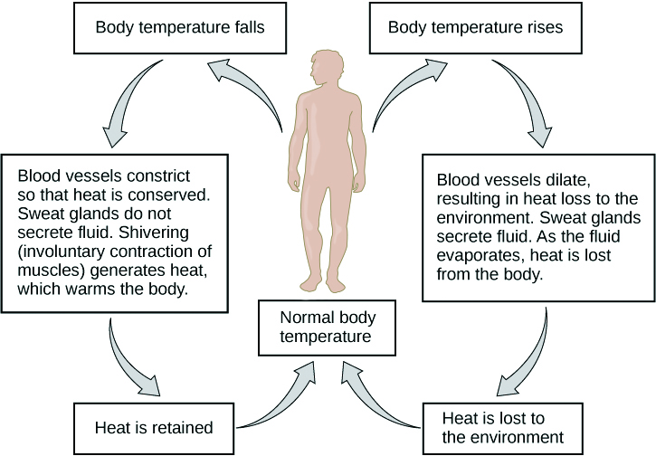
Cooling Down
The human body’s temperature regulatory centre is the hypothalamus in the brain. When the hypothalamus receives data from sensors in the skin and brain that body temperature is higher than the set point, it sets into motion the following responses:
- Blood vessels in the skin dilate (vasodilation) to allow more blood from the warm body core to flow close to the surface of the body, so heat can be radiated into the environment.
- As blood flow to the skin increases, sweat glands in the skin are activated to increase their output of sweat (diaphoresis). When the sweat evaporates from the skin surface into the surrounding air, it takes heat with it.
- Breathing becomes deeper, and the person may breathe through the mouth instead of the nasal passages. This increases heat loss from the lungs.
Heating Up
When the brain’s temperature regulatory centre receives data that body temperature is lower than the set point, it sets into motion the following responses:
- Blood vessels in the skin contract (vasoconstriction) to prevent blood from flowing close to the surface of the body, which reduces heat loss from the surface.
- As temperature falls lower, random signals to skeletal muscles are triggered, causing them to contract. This causes shivering, which generates a small amount of heat.
- The thyroid gland may be stimulated by the brain (via the pituitary gland) to secrete more thyroid hormone. This hormone increases metabolic activity and heat production in cells throughout the body.
- The adrenal glands may also be stimulated to secrete the hormone adrenaline. This hormone causes the breakdown of glycogen (the carbohydrate used for energy storage in animals) to glucose, which can be used as an energy source. This catabolic chemical process is exothermic, or heat producing.
Blood Glucose
In controlling the blood glucose level, certain endocrine cells in the pancreas (called alpha and beta cells) detect the level of glucose in the blood. They then respond appropriately to keep the level of blood glucose within the normal range.
- If the blood glucose level rises above the normal range, pancreatic beta cells release the hormone insulin into the bloodstream. Insulin signals cells to take up the excess glucose from the blood until the level of blood glucose decreases to the normal range.
- If the blood glucose level falls below the normal range, pancreatic alpha cells release the hormone glucagon into the bloodstream. Glucagon signals cells to break down stored glycogen to glucose and release the glucose into the blood until the level of blood glucose increases to the normal range.
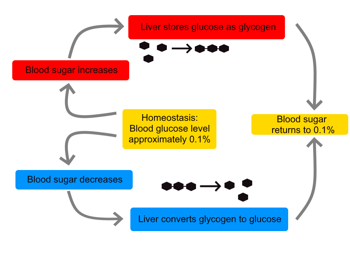
https://www.youtube.com/watch?v=Iz0Q9nTZCw4
Homeostasis and Negative/Positive Feedback, Amoeba Sisters, 2017.
Positive Feedback
In a positive feedback loop, feedback serves to intensify a response until an end point is reached. Examples of processes controlled by positive feedback in the human body include blood clotting and childbirth.
Blood Clotting
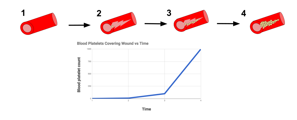
When a wound causes bleeding, the body responds with a positive feedback loop to clot the blood and stop blood loss. Substances released by the injured blood vessel wall begin the process of blood clotting. Platelets in the blood start to cling to the injured site and release chemicals that attract additional platelets. As the platelets continue to amass, more of the chemicals are released and more platelets are attracted to the site of the clot. The positive feedback accelerates the process of clotting until the clot is large enough to stop the bleeding.
Childbirth
Figure 7.8.6 shows the positive feedback loop that controls childbirth. The process normally begins when the head of the infant pushes against the cervix. This stimulates nerve impulses, which travel from the cervix to the hypothalamus in the brain. In response, the hypothalamus sends the hormone oxytocin to the pituitary gland, which secretes it into the bloodstream so it can be carried to the uterus. Oxytocin stimulates uterine contractions, which push the baby harder against the cervix. In response, the cervix starts to dilate in preparation for the passage of the baby. This cycle of positive feedback continues, with increasing levels of oxytocin, stronger uterine contractions, and wider dilation of the cervix until the baby is pushed through the birth canal and out of the body. At that point, the cervix is no longer stimulated to send nerve impulses to the brain, and the entire process stops.
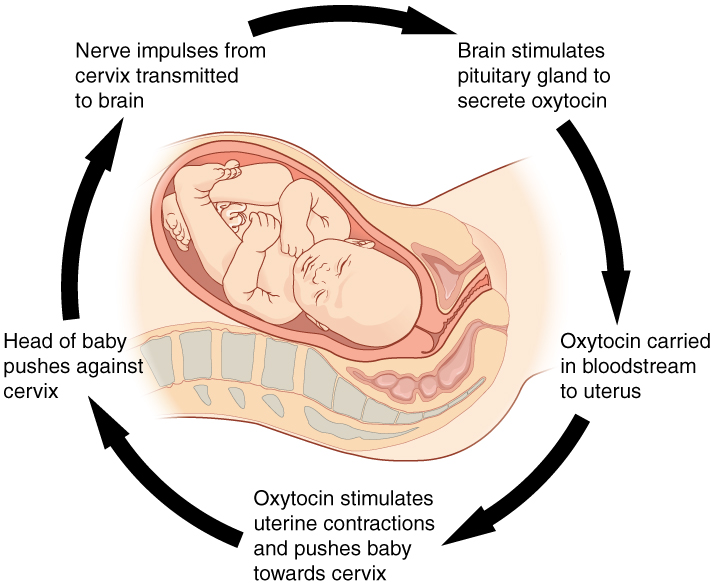
Normal childbirth is driven by a positive feedback loop. Positive feedback causes an increasing deviation from the normal state to a fixed end point, rather than a return to a normal set point as in homeostasis.
When Homeostasis Fails
Homeostatic mechanisms work continuously to maintain stable conditions in the human body. Sometimes, however, the mechanisms fail. When they do, homeostatic imbalance may result, in which cells may not get everything they need or toxic wastes may accumulate in the body. If homeostasis is not restored, the imbalance may lead to disease — or even death. Diabetes is an example of a disease caused by homeostatic imbalance. In the case of diabetes, blood glucose levels are no longer regulated and may be dangerously high. Medical intervention can help restore homeostasis and possibly prevent permanent damage to the organism.
Normal aging may bring about a reduction in the efficiency of the body’s control systems, which makes the body more susceptible to disease. Older people, for example, may have a harder time regulating their body temperature. This is one reason they are more likely than younger people to develop serious heat-induced illnesses, such as heat stroke.
Feature: My Human Body
Diabetes is diagnosed in people who have abnormally high levels of blood glucose after fasting for at least 12 hours. A fasting level of blood glucose below 100 is normal. A level between 100 and 125 places you in the pre-diabetes category, and a level higher than 125 results in a diagnosis of diabetes.
Of the two types of diabetes, type 2 diabetes is the most common, accounting for about 90 per cent of all cases of diabetes in the United States. Type 2 diabetes typically starts after the age of 40. However, because of the dramatic increase in recent decades in obesity in younger people, the age at which type 2 diabetes is diagnosed has fallen. Even children are now being diagnosed with type 2 diabetes. Today, about 3 million Canadians (8.1% of total population) are living with diabetes.
You may at some point have your blood glucose level tested during a routine medical exam. If your blood glucose level indicates that you have diabetes, it may come as a shock to you because you may not have any symptoms of the disease. You are not alone, because as many as one in four diabetics do not know they have the disease. Once the diagnosis of diabetes sinks in, you may be devastated by the news. Diabetes can lead to heart attacks, strokes, blindness, kidney failure, nerve damage, and loss of toes or feet. The risk of death in adults with diabetes is 50 per cent greater than it is in adults without diabetes, and diabetes is the seventh leading cause of death of adults. In addition, controlling diabetes usually requires frequent blood glucose testing, watching what and when you eat, and taking medications or even insulin injections. All of this may seem overwhelming.
The good news is that changing your lifestyle may stop the progression of type 2 diabetes or even reverse it. By adopting healthier habits, you may be able to keep your blood glucose level within the normal range without medications or insulin. Here’s how:
- Lose weight. Any weight loss is beneficial. Losing as little as seven per cent of your weight may be all that is needed to stop diabetes in its tracks. It is especially important to eliminate excess weight around your waist.
- Exercise regularly. You should try to exercise for at least 30 minutes, five days a week. This will not only lower your blood sugar and help your insulin work better, but it will also lower your blood pressure and improve your heart health. Another bonus of exercise is that it will help you lose weight by increasing your basal metabolic rate.
- Adopt a healthy diet. Decrease your consumption of refined carbohydrates, such as sweets and sugary drinks. Increase your intake of fibre-rich foods, such as fruits, vegetables, and whole grains. About one-quarter of each meal should consist of high-protein foods, such as fish, chicken, dairy products, legumes, or nuts.
- Control stress. Stress can increase your blood glucose and also raise your blood pressure and risk of heart disease. When you feel stressed out, do breathing exercises or take a brisk walk or jog. Try to replace stressful thoughts with more calming ones.
- Establish a support system. Enlist the help and support of loved ones, as well as medical professionals, such as a nutritionist and diabetes educator. Having a support system will help ensure that you are on the path to wellness, and that you can stick to your plan.
7.8 Summary
- Homeostasis is the condition in which a system (such as the human body) is maintained in a more or less steady state. It is the job of cells, tissues, organs, and organ systems throughout the body to maintain homeostasis.
- For any given variable, such as body temperature, there is a particular set point that is the physiological optimum value. The spread of values around the set point that is considered insignificant is called the normal range.
- Homeostasis is generally maintained by a negative feedback loop that includes a stimulus, sensor, control centre, and effector. Negative feedback serves to reduce an excessive response and to keep a variable within the normal range. Negative feedback loops control body temperature and the blood glucose level.
- Positive feedback loops are not common in biological systems. Positive feedback serves to intensify a response until an end point is reached. Positive feedback loops control blood clotting and childbirth.
- Sometimes homeostatic mechanisms fail, resulting in homeostatic imbalance. Diabetes is an example of a disease caused by homeostatic imbalance. Aging can bring about a reduction in the efficiency of the body’s control system, which makes the elderly more susceptible to disease.
7.8 Review Questions
-
-
- Compare and contrast negative and positive feedback loops.
- Explain how negative feedback controls body temperature.
- Give two examples of physiological processes controlled by positive feedback loops.
- During breastfeeding, the stimulus of the baby sucking on the nipple increases the amount of milk produced by the mother. The more sucking, the more milk is usually produced. Is this an example of negative or positive feedback? Explain your answer. What do you think might be the evolutionary benefit of the milk production regulation mechanism you described?
- Explain why homeostasis is regulated by negative feedback loops, rather than positive feedback loops.
- The level of a sex hormone, testosterone (T), is controlled by negative feedback. Another hormone, gonadotropin-releasing hormone (GnRH), is released by the hypothalamus of the brain, which triggers the pituitary gland to release luteinizing hormone (LH). LH stimulates the gonads to produce T. When there is too much T in the bloodstream, it feeds back on the hypothalamus, causing it to produce less GnRH. While this does not describe all the feedback loops involved in regulating T, answer the following questions about this particular feedback loop.
- What is the stimulus in this system? Explain your answer.
- What is the control centre in this system? Explain your answer.
- In this system, is the pituitary considered the stimulus, sensor, control centre, or effector? Explain your answer.
7.8 Explore More
https://www.youtube.com/watch?v=LSgEJSlk6W4
Homeostasis - What Is Homeostasis - What Is Set Point For Homeostasis - Homeostasis In The Human Body, Whats Up Dude, 2017.
https://www.youtube.com/watch?v=XMsJ-3qRVJM
Attributions
Figure 7.8.1
Nest_Thermostat by Amanitamano on Wikimedia Commons is used under a CC BY-SA 3.0 (https://creativecommons.org/licenses/by-sa/3.0/deed.en) license.
Figure 7.8.2
Negative_Feedback_Loops by OpenStax on Wikimedia Commons is used under a CC BY 4.0 (https://creativecommons.org/licenses/by/4.0/deed.en) license.
Figure 7.8.3
Body Temperature Homeostasis by OpenStax College, Biology is used under a CC BY 4.0 license.
Figure 7.8.4
Homeostasis_of_blood_sugar by Christinelmiller on Wikimedia Commons is used under a CC0 1.0 Universal Public Domain Dedication (https://creativecommons.org/publicdomain/zero/1.0/deed.en) license.
Figure 7.8.5
Positive_Feedback_Diagram_Blood_Clotting by Elliottuttle on Wikimedia Commons is used under a CC BY-SA 4.0 (https://creativecommons.org/licenses/by-sa/4.0) license.
Figure 7.8.6
Pregnancy-Positive_Feedback by OpenStax on Wikimedia Commons is used under a CC BY 4.0 (https://creativecommons.org/licenses/by/4.0/deed.en) license.
References
Amoeba Sisters. (2017, September 7). Homeostasis and negative/positive feedback. YouTube. https://www.youtube.com/watch?v=Iz0Q9nTZCw4&feature=youtu.be
Betts, J. G., Young, K.A., Wise, J.A., Johnson, E., Poe, B., Kruse, D.H., Korol, O., Johnson, J.E., Womble, M., DeSaix, P. (2013, April 25). Figure 1.10 Negative feedback loop [digital image/ diagram]. In Anatomy and Physiology (Section 1.5). OpenStax. https://openstax.org/books/anatomy-and-physiology/pages/1-5-homeostasis
Betts, J. G., Young, K.A., Wise, J.A., Johnson, E., Poe, B., Kruse, D.H., Korol, O., Johnson, J.E., Womble, M., DeSaix, P. (2013, April 25). Figure 1.11 Positive feedback loop normal childbirth is driven by a positive feedback loop [digital image/ diagram]. In Anatomy and Physiology (Section 1.5). OpenStax. https://openstax.org/books/anatomy-and-physiology/pages/1-5-homeostasis
Cognito. (2018, December 18). GCSE Biology - Homeostasis #38. YouTube. https://www.youtube.com/watch?v=XMsJ-3qRVJM&feature=youtu.be
Mayo Clinic Staff. (n.d.). Type 2 diabetes [online article]. MayoClinic.org. https://www.mayoclinic.org/diseases-conditions/type-2-diabetes/symptoms-causes/syc-20351193
OpenStax CNX. (2016, March 23). Figure 4 The body is able to regulate temperature in response to signals from the nervous system [digital image]. In OpenStax, Biology (Section 33.3). https://cnx.org/contents/GFy_h8cu@10.8:BP24ZReh@7/Homeostasis
Whats Up Dude. (2017, September 20). Homeostasis - What is homeostasis - What is set point for homeostasis - Homeostasis in the human body. YouTube. https://www.youtube.com/watch?v=LSgEJSlk6W4&feature=youtu.be
Image shows a scanning electron pictomicrograph of a blood clot. The erythrocytes look normal (biconcave discs) but the thrombocytes have activated and gotten "sticky", creating a mesh-like network that has trapped the erythrocytes.
Created by CK-12 Foundation/Adapted by Christine Miller
Seeing Is Believing
At first glance, Figure 8.7.1 appears to be just random dots of colour, but hidden within it is the three-dimensional shape of a bee. Can you see it among the dots? This figure is an example of a stereogram, which is a two-dimensional picture that, when viewed correctly, reveals a three-dimensional object. If you can’t see the hidden image, it doesn’t mean that there is anything wrong with your eyes. It’s all in how your brain interprets what your eyes are sensing. The eyes are special sensory organs, and vision is one of our special senses.
Special and General Senses
The human body has two basic types of senses, called special senses and general senses. Special senses have specialized sense organs that gather sensory information and change it into nerve impulses. Special senses include vision (for which the eyes are the specialized sense organs), hearing (ears), balance (ears), taste (tongue), and smell (nasal passages). General senses, in contrast, are all associated with the sense of touch. They lack special sense organs. Instead, sensory information about touch is gathered by the skin and other body tissues, all of which have important functions besides gathering sense information. Whether the senses are special or general, however, they all depend on cells called sensory receptors.
Sensory Receptors
A sensory receptor is a specialized nerve cell that responds to a stimulus in the internal or external environment by generating a nerve impulse. The nerve impulse then travels along the sensory (afferent) nerve to the central nervous system for processing and to form a response.
There are several different types of sensory receptors that respond to different kinds of stimuli:
- Mechanoreceptors respond to mechanical forces, such as pressure, roughness, vibration, and stretching. Most mechanoreceptors are found in the skin and are needed for the sense of touch. Mechanoreceptors are also found in the inner ear, where they are needed for the senses of hearing and balance.
- Thermoreceptors respond to variations in temperature. They are found mostly in the skin and detect temperatures that are above or below body temperature.
- Nociceptors respond to potentially damaging stimuli, which are generally perceived as pain. They are found in internal organs, as well as on the surface of the body. Different nociceptors are activated depending on the particular stimulus. Some detect damaging heat or cold, others detect excessive pressure, and still others detect painful chemicals (such as very hot spices in food).
- Photoreceptors detect and respond to light. Most photoreceptors are found in the eyes and are needed for the sense of vision.
- Chemoreceptors respond to certain chemicals. They are found mainly in taste buds on the tongue — where they are needed for the sense of taste — and in nasal passages, where they are needed for the sense of smell.
Touch
Touch is the ability to sense pressure, vibration, temperature, pain, and other tactile stimuli. These types of stimuli are detected by mechanoreceptors, thermoreceptors, and nociceptors all over the body, most noticeably in the skin. These receptors are especially concentrated on the tongue, lips, face, palms of the hands, and soles of the feet. Various types of tactile receptors in the skin are shown in Figure 8.7.2.
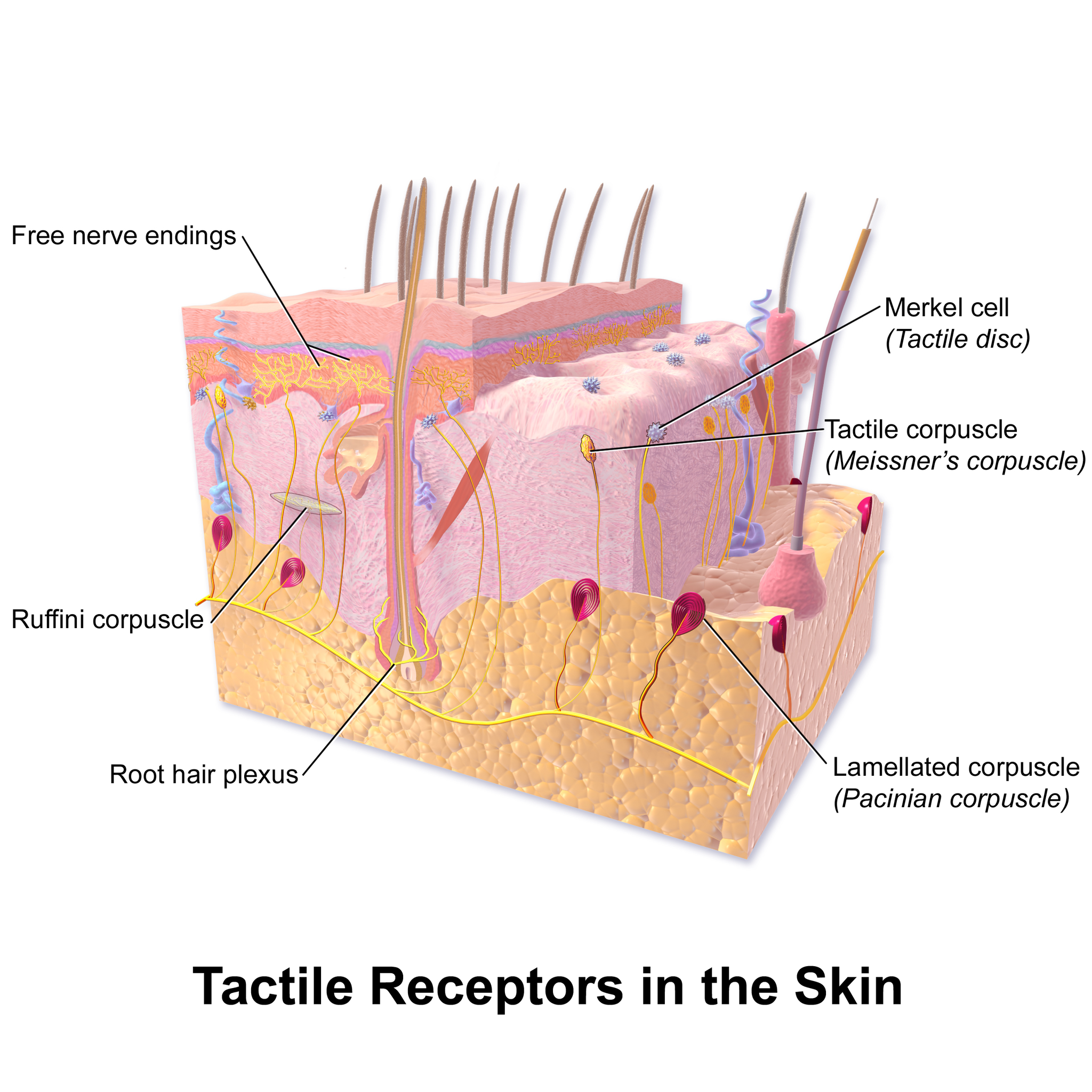
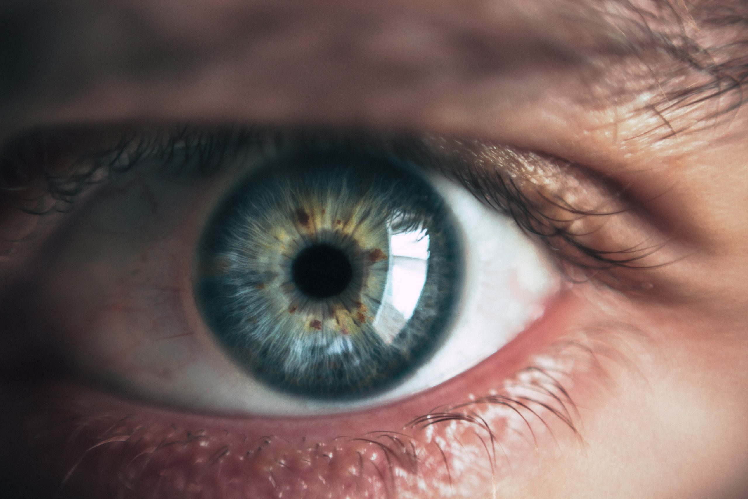
Vision
Vision (or sight) is the ability to sense light and see. The eye is the special sensory organ that collects and focuses light and forms images. The eye, however, is not sufficient for us to see. The brain also plays a necessary role in vision. Vision is our primary sense and more than 50 per cent of the cerebral cortex is devoted to processing visual information. A person with normal colour vision can differentiate between hundreds of thousands of different colours, hues, and shades.
How the Eye Works
Figure 8.7.4 (below) shows the anatomy of the human eye in cross-section. The eye gathers and focuses light to form an image, and then changes the image to nerve impulses that travel to the brain. The eye's functions are summarized in the following steps.
- Light passes first through the cornea, which is a clear outer layer that protects the eye and helps to focus the light by refracting (or bending) it.
- Next, light enters the interior of the eye through an opening called the pupil. The size of this opening is controlled by the coloured part of the eye (called the iris), which adjusts the size based on the brightness of the light. The iris causes the pupil to narrow in bright light and widen in dim light. Filling the space between the cornea and the iris is a semi-gelatinous fluid called aqueous humor and functions to maintain the shape of the eye.
- The light then passes through the lens, which refracts the light even more and focuses it on the retina at the back of the eye, as an inverted image. Sitting behind the lens is a gelatinous fluid called vitreous humor, which functions to maintain the shape of the eye.
- The retina contains two types of photoreceptors: rod and cone cells . Rod cells, which are found mainly in all areas of the retina other than the very center, are particularly sensitive to low levels of light. Cone cells, which are found mainly in the center of the retina, are sensitive to light of different colours, and allow colour vision. The rods and cones convert the light that strikes them to nerve impulses.
- The nerve impulses from the rods and cones travel to the optic nerve via the optic disc (also known as the optic nerve), which is a circular area at the back of the eye where the optic nerve connects to the retina.
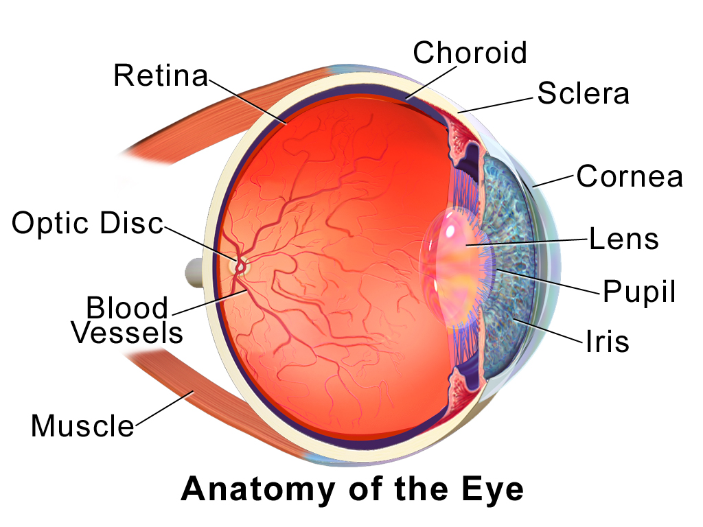
Colour Vision
Humans have colour vision because we have three types of cone cells: blue, green and red. Each of these types of cone cell detects a specific wavelength of light, for which they are named. The combined stimulus is then perceived as a specific colour, based on the ratio of the amount stimulus coming from each of the three types of cone cells. Do you know what else uses these same three pieces of information to communicate colour? Your computer monitor! When working in a creative program, such as Paint, these three reference points of red (R), green (G), and blue (B), can be used to create any of the million colours the human eye can perceive, as illustrated in Figure 8.7.5. Take a look at each of the numerical values for red, green, and blue and what colour their combined values create:
Figure 8.7.5 RGB colours.
Role of the Brain in Vision
The optic nerves from both eyes meet and cross just below the bottom of the hypothalamus in the brain. The information from both eyes is sent to the visual cortex in the occipital lobe of the cerebrum, which is part of the cerebral cortex. The visual cortex is the largest system in the human brain, and is responsible for processing visual images. It interprets messages from both eyes and “tells” us what we are seeing.
Vision Problems

Vision problems are very common. Two of the most common are myopia and hyperopia, and they often start in childhood or adolescence. Another common problem, called presbyopia, occurs in most people, beginning in middle adulthood. In all three conditions, the eyes fail to focus images correctly on the retina, resulting in blurred vision.
Myopia
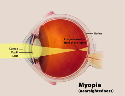
Myopia (or nearsightedness) occurs when the light that comes into the eye does not directly focus on the retina, but in front of it, as shown in Figure 8.7.7. As a result, distant objects may appear out of focus, but the focus of close objects is not affected. Myopia may occur because the eyeball is elongated from front to back, or because the cornea is too curved. Myopia can be corrected with the use of corrective lenses, either eyeglasses or contact lenses. Myopia can also be corrected by refractive surgery performed with a laser.
Hyperopia
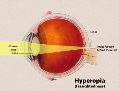
Hyperopia (or farsightedness) happens when the light coming into the eye does not directly focus on the retina but behind it, as shown in Figure 8.7.8. This causes close objects to appear out of focus, but does not affect the focus of distant objects. Hyperopia may occur because the eyeball is too short from front to back, or because the lens is not curved enough. Hyperopia can be corrected through the use of corrective lenses or laser surgery.
Presbyopia
Presbyopia is a vision problem associated with aging, in which the eye gradually loses its ability to focus on close objects. The precise origin of presbyopia is not known for certain, but evidence suggests that the lens may become less elastic with age, causing the muscles that control the lens to lose power as people grow older. The first signs of presbyopia — eyestrain, difficulty seeing in dim light, problems focusing on small objects and fine print — are usually first noticed between the ages of 40 and 50. Most older people with this problem use corrective lenses to focus on close objects, because surgical procedures to correct presbyopia have not been as successful as those for myopia and hyperopia.
Hearing
Hearing is the ability to sense sound waves, and the ear is the organ that senses sound. Sound waves enter the ear through the ear canal and travel to the eardrum (see the diagram of the ear Figure 8.7.9). The sound waves strike the eardrum, and make it vibrate. The vibrations then travel through the three tiny bones (incus, malleus and stapes) of the middle ear, which amplify the vibrations. From the middle ear, the vibrations pass to the cochlea in the inner ear. The cochlea is a coiled tube filled with liquid. The liquid moves in response to the vibrations, causing tiny hair cells(which are mechanoreceptors) lining the cochlea to bend. In response, the hair cells send nerve impulses to the auditory nerve, which carries the impulses to the brain. The brain interprets the impulses and “tells” us what we are hearing.
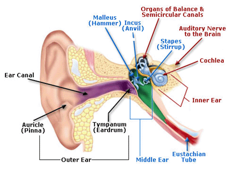
Balance
The ears are also responsible for the sense of balance. Balance is the ability to sense and maintain an appropriate body position. The semicircular canals inside the ear (see the figure above) contain fluid that moves when the head changes position. Tiny hairs lining the semicircular canals sense movement of the fluid. In response, they send nerve impulses to the vestibular nerve, which carries the impulses to the brain. The brain interprets the impulses and sends messages to the peripheral nervous system, which triggers contractions of skeletal muscles as needed to maintain balance.
Taste and Smell
Taste and smell are both abilities to sense chemicals, so both taste and olfactory (odor) receptors are chemoreceptors. Both types of chemoreceptors send nerve impulses to the brain along sensory nerves, and the brain “tells” us what we are tasting or smelling.
Taste receptors are found in tiny bumps on the tongue called taste buds.You can see a diagram of a taste receptor cell and related structures in Figure 8.7.10. Taste receptor cells make contact with chemicals in food through tiny openings called taste pores. When certain chemicals bind with taste receptor cells, it generates nerve impulses that travel through afferent nerves to the CNS. There are separate taste receptors for sweet, salty, sour, bitter, and meaty tastes. The meaty — or savory — taste is called umami.
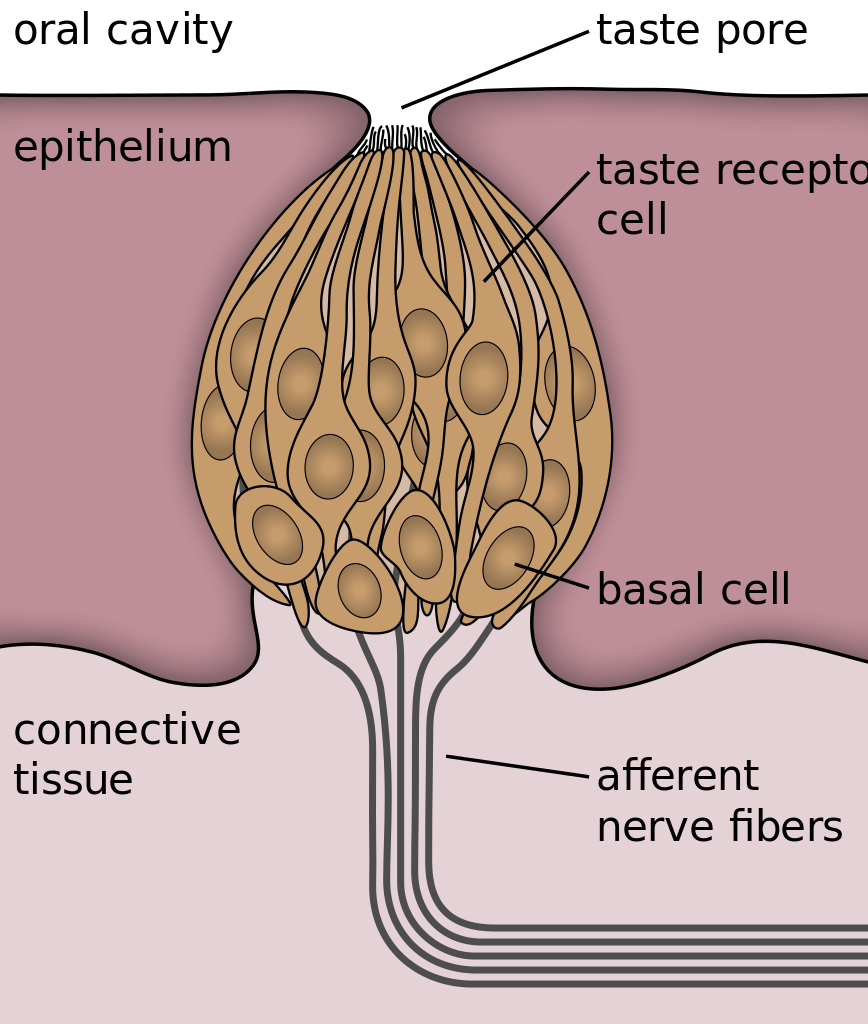
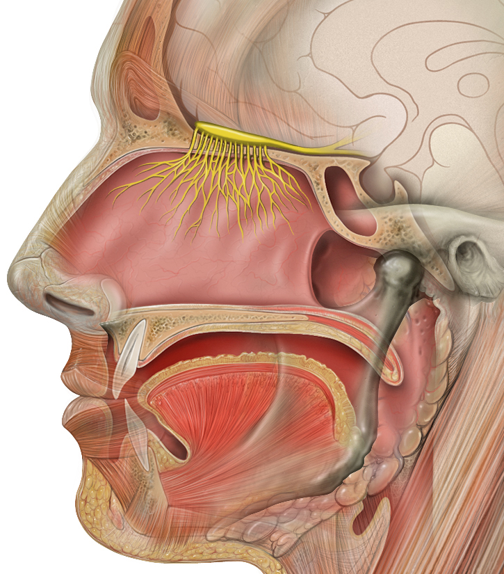
Feature: Human Biology in the News
The most common cause of blindness in the Western hemisphere is age-related macular degeneration (AMD). Approximately 1.4 million people in Canada have this type of blindness, and 196 million people are affected worldwide and is expected to increase to 288 millions people by the year 2040. At present, there is no cure for AMD. The disease occurs with the death of a layer of cells called retinal pigment epithelium, which normally provides nutrients and other support to the macula of the eye. The macula is an oval-shaped pigmented area near the center of the retina that is specialized for high visual acuity and has the retina’s greatest concentration of cones. When the epithelial cells die and the macula is no longer supported or nourished, the macula also starts to die. Patients experience a black spot in the center of their vision, and as the disease progresses, the black spot grows outward. Patients eventually lose the ability to read and even to recognize familiar faces before developing total blindness.
In 2016, a landmark surgery was performed as a trial on a patient with severe AMD. In the first ever operation of its kind, Dr. Pete Coffey of the University of London implanted a tiny patch of cells behind the retina in each of the patient’s eyes. The cells were retinal pigmented epithelial cells that had been grown in a lab from stem cells, which are undifferentiated cells that can develop into other cell types. Within six months of the operation, the new cells were still surviving, and the doctor was hopeful that the patient’s vision loss would stop and even be reversed. At that point, several other operations had already been planned to test the new procedure. If these cases are a success, Dr. Coffey predicts that the surgery will become as routine as cataract surgery, and that it will prevent millions of patients from losing their vision.
8.7 Summary
- The human body has two major types of senses: special senses and general senses. Special senses have specialized sense organs and include vision (eyes), hearing (ears), balance (ears), taste (tongue), and smell (nasal passages). General senses are all associated with touch and lack special sense organs. Touch receptors are found throughout the body, but particularly in the skin.
- All senses depend on sensory receptor cells to detect sensory stimuli and transform them into nerve impulses. Types of sensory receptors include mechanoreceptors (mechanical forces), thermoreceptors (temperature), nociceptors (pain), photoreceptors (light), and chemoreceptors (chemicals).
- Touch is the ability to sense pressure, vibration, temperature, pain, and other tactile stimuli. The skin includes several different types of touch receptor cells.
- Vision is the ability to sense light and see. The eye is the special sensory organ that collects and focuses light, forms images, and changes them to nerve impulses. Optic nerves send information from the eyes to the brain, which processes the visual information and “tells” us what we are seeing.
- Common vision problems include myopia (nearsightedness), hyperopia (farsightedness), and presbyopia (age-related decline in close vision). Vision problems can be corrected with lenses (eyeglasses or contacts) or — in many cases — with laser surgery.
- Hearing is the ability to sense sound waves, and the ear is the organ that senses sound. It changes sound waves to vibrations that trigger nerve impulses, which travel to the brain through the auditory nerve. The brain processes the information and “tells” us what we are hearing.
- The ear is also the organ responsible for the sense of balance, which is the ability to sense and maintain an appropriate body position. The ears send impulses about head position to the brain, which sends messages to skeletal muscles via the peripheral nervous system. The muscles respond by contracting to maintain balance.
- Taste and smell are both abilities to sense chemicals. Taste receptors in taste buds on the tongue sense chemicals in food, while olfactory receptors in the nasal passages sense chemicals in the air. Sense of smell contributes significantly to sense of taste.
8.7 Review Questions
-
- Compare and contrast special senses and general senses.
- What are sensory receptors?
-
- Describe the range of tactile stimuli detected in the sense of touch.
- Explain how the eye collects and focuses light to form an image, and how it converts it to nerve impulses.
- Identify two common vision problems,along with their causes and their effects on vision.
-
- Explain how structures of the ear collect and amplify sound waves and transform them to nerve impulses.
- What role does the ear play in balance? Which structures of the ear are involved in balance?
- Describe two ways that the body senses chemicals. What are the special sense organs involved in these senses?
- Explain why your skin can detect different types of stimuli, such as pressure and temperature.
- Is sensory information sent to the central nervous system via efferent or afferent nerves?
- Identify a mechanoreceptor used in two different human senses. Describe the type of mechanical stimuli that each detects.
- If a person is blind, but their retina is functioning properly, where do you think the damage might be? Explain your answer.
- When you see colours, what receptor cells are activated? Where are these receptors located? What lobe of the brain is primarily used to process visual information?
- The auditory nerve carries _______________.
-
-
- smell information
- taste information
- balance information
- sound information
-
8.7 Explore More
https://www.youtube.com/watch?time_continue=4&v=rkRbebvoYqI&feature=emb_logo
What color is Tuesday? Exploring synesthesia - Richard E. Cytowic, TED-Ed, 2013.
https://www.youtube.com/watch?v=UL8YSLhqa5U&feature=emb_logo
What Is Vertigo & Why Do We Get It?, Seeker, 2016.
https://www.youtube.com/watch?v=t3CjTU7TaNA
How do animals see in the dark? - Anna Stöckl, TED-Ed, 2016.
https://youtu.be/Y6e_m9iq-4Q
What are those floaty things in your eye? - Michael Mauser, TED-Ed, 2014.
Attributions
Figure 8.7.1
Bee Stereogram by Be Mosaic on Flickr is used under a CC BY-NC-SA 2.0 (https://creativecommons.org/licenses/by-nc-sa/2.0/) license.
Figure 8.7.2
Skin_TactileReceptors by BruceBlaus on Wikimedia Commons is used under a CC BY 3.0 (https://creativecommons.org/licenses/by/3.0) license.
Figure 8.7.3
Macro shot photograph of someone's right eye [photo] by Jordan Whitfield on Unsplash is used under the Unsplash License (https://unsplash.com/license).
Figure 8.7.4
EyeAnatomy_01 by BruceBlaus on Wikimedia Commons is used under a CC BY 3.0 (https://creativecommons.org/licenses/by/3.0) license.
Figure 8.7.5
RGB colours [screenshots] from Microsoft Paint.
Figure 8.7.6
Through the reading glasses [photo] by Dmitry Ratushny on Unsplash is used under the Unsplash License (https://unsplash.com/license).
Figure 8.7.7
Myopia_Diagram by National Eye Institute/ National Institutes of Health on Wikimedia Commons is used under a CC BY 2.0 (https://creativecommons.org/licenses/by/2.0) license.
Figure 8.7.8
Hyperopia by National Institute of Health/NIH on Wikimedia Commons is in the public domain (https://en.wikipedia.org/wiki/Public_domain).
Figure 8.7.9
AnatomyHumanEar by unknown author from Occupational Safety & Health Administration on Wikimedia Commons is in the public domain (https://en.wikipedia.org/wiki/Public_domain).
Figure 8.7.10
Taste_bud_2_eng.svg by Jonas Töle on Wikimedia Commons is used under a CC0 1.0 Universal Public Domain Dedication license (https://creativecommons.org/publicdomain/zero/1.0/deed.en).
Figure 8.7.11
Head_olfactory_nerve by Patrick.lynch, medical illustrator on Wikimedia Commons is used under a CC BY 2.5 (https://creativecommons.org/licenses/by/2.5/deed.en) license.
References
Age-Related Macular Degeneration. (n.d.). WebMD. https://www.webmd.com/eye-health/macular-degeneration/age-related-macular-degeneration-overview#3 (Reviewed by Alan Kozarsky, MD on October 26, 2019)
Blausen.com Staff. (2014). Medical gallery of Blausen Medical 2014. WikiJournal of Medicine 1 (2). DOI:10.15347/wjm/2014.010. ISSN 2002-4436.
da Cruz, L., Fynes, K., Georgiadis, O. et al. (2018, March 19). Phase 1 clinical study of an embryonic stem cell–derived retinal pigment epithelium patch in age-related macular degeneration. Natural Biotechnology, 36, 328–337. https://doi.org/10.1038/nbt.4114
File:Eye Diagram without text.gif. (2018, February 9). Wikimedia Commons. https://commons.wikimedia.org/w/index.php?title=File:Eye_Diagram_without_text.gif&oldid=286008241 (original image from National Eye Institute - modified by User:Nordelch) [public domain (https://en.wikipedia.org/wiki/Public_domain)]
Occupational Health and Safety Administration. (n.d.). Figure 7. Anatomy of the human ear [diagram]. In OSHA Technical Manual (Section III, Chapter 5 - Noise). United States Department of Labour [online]. https://www.osha.gov/dts/osta/otm/new_noise/
Seeker. (2016, March 18). What is vertigo & why do we get it? YouTube. https://www.youtube.com/watch?v=UL8YSLhqa5U&feature=youtu.be
TED-Ed. (2013, June 10). What color is Tuesday? Exploring synesthesia - Richard E. Cytowic. YouTube. https://www.youtube.com/watch?v=rkRbebvoYqI&feature=youtu.be
TED-Ed. (2014, December 1). What are those floaty things in your eye? - Michael Mauser. YouTube. https://www.youtube.com/watch?v=Y6e_m9iq-4Q&feature=youtu.be
TED-Ed. (2016, August 25). How do animals see in the dark? - Anna Stöckl. YouTube. https://www.youtube.com/watch?v=t3CjTU7TaNA&feature=youtu.be
Created by CK-12 Foundation/Adapted by Christine Miller
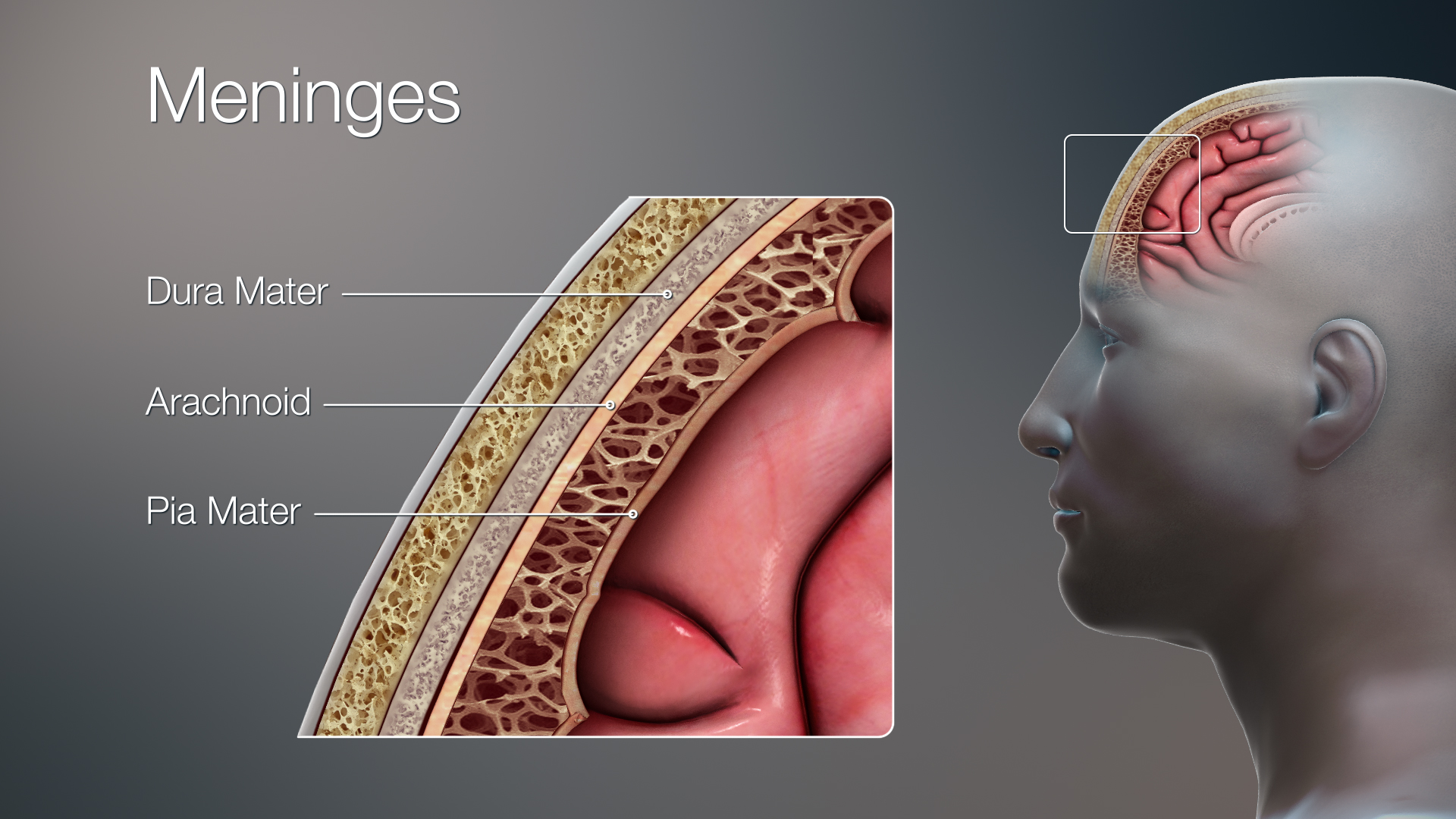
As you learned in this chapter, the human body consists of many complex systems that normally work together efficiently — like a well-oiled machine — to carry out life’s functions. For example, the image above (Figure 7.9.1) illustrates how the brain and spinal cord are protected by layers of membrane called meninges and fluid that flows between the meninges and in spaces called ventricles inside the brain. This fluid is called cerebrospinal fluid, and as you have learned, one of its important functions is to cushion and protect the brain and spinal cord, which make up most of the central nervous system (CNS). Additionally, cerebrospinal fluid circulates nutrients and removes waste products from the CNS. Cerebrospinal fluid is produced continually in the ventricles, circulates throughout the CNS, and is then reabsorbed by the bloodstream. If too much cerebrospinal fluid is produced, its flow is blocked, or not enough is reabsorbed, the system becomes out of balance and it can build up in the ventricles. This causes an enlargement of the ventricles called hydrocephalus that can put pressure on the brain, resulting in the types of neurological problems that former professional football player Jayson, described in the beginning of this chapter, is suffering from.
Recall that Jayson’s symptoms included loss of bladder control, memory loss, and difficulty walking. The cause of his symptoms was not immediately clear, although his doctors suspected that it related to the nervous system, since the nervous system acts as the control centre of the body, controlling and regulating many other organ systems. Jayson’s memory loss directly implicated the brain's involvement, since that is the site of thoughts and memory. The urinary system is also controlled in part by the nervous system, so the inability to hold urine appropriately can also be a sign of a neurological issue. Jayson’s trouble walking involved the muscular system, which works alongside the skeletal system to enable movement of the limbs. In turn, the contraction of muscles is regulated by the nervous system. You can see why a problem in the nervous system can cause a variety of different symptoms by affecting multiple organ systems in the human body.
To try to find the exact cause of Jayson’s symptoms, his doctors performed a lumbar puncture (or spinal tap), which is the removal of some cerebrospinal fluid through a needle inserted into the lower part of the spinal canal. They then analyzed Jayson’s cerebrospinal fluid for the presence of pathogens (such as bacteria) to determine whether an infection was the cause of his neurological symptoms. When no evidence of infection was found, they used an MRI to observe the structures of his brain. This is when they discovered his enlarged ventricles, which are a hallmark of hydrocephalus.
To treat Jayson’s hydrocephalus, a surgeon implanted a device called a shunt in his brain to remove the excess fluid. An illustration of a brain shunt is shown in Figure 9.7.2 . One side of the shunt consists of a small tube, called a catheter, which was inserted into Jayson’s ventricles. Excess cerebrospinal fluid is then drained through a one-way valve to the other end of the shunt, which was threaded under his skin to his abdominal cavity, where the fluid is released and can be reabsorbed by the bloodstream.
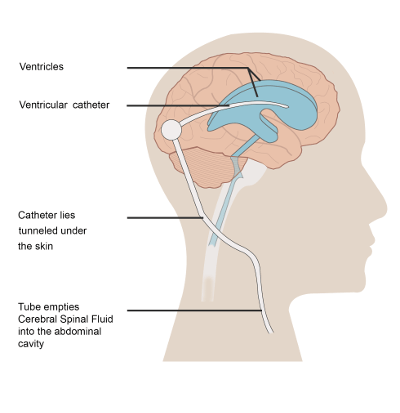
Implantation of a shunt is the most common way to treat hydrocephalus, and for some people, it can allow them to recover almost completely. However, there can be complications associated with a brain shunt. The shunt can have mechanical problems or cause an infection. Also, the rate of draining must be carefully monitored and adjusted to balance the rate of cerebrospinal fluid removal with the rate of its production. If it is drained too fast, it is called overdraining, and if it is drained too slowly, it is called underdraining. In the case of underdraining, the pressure on the brain and associated neurological symptoms will persist. In the case of overdraining, the ventricles can collapse, which can cause serious problems, such as the tearing of blood vessels and hemorrhaging. To avoid these problems, some shunts have an adjustable pressure valve, where the rate of draining can be adjusted by placing a special magnet over the scalp. You can see how the proper balance between cerebrospinal fluid production and removal is so critical – both in the causes of hydrocephalus and in its treatment.
In what other ways does your body regulate balance, or maintain a state of homeostasis? In this chapter you learned about the feedback loops that keep body temperature and blood glucose within normal ranges. Other important examples of homeostasis in the human body are the regulation of the pH in the blood and the balance of water in the body. You will learn more about homeostasis in different body systems in the coming chapters.
Thanks to Jayson’s shunt, his symptoms are starting to improve, but he has not fully recovered. Time may tell whether the removal of the excess cerebrospinal fluid from his ventricles will eventually allow him to recover normal functioning or whether permanent damage to his nervous system has already been done. The flow of cerebrospinal fluid might seem simple, but when it gets out of balance, it can easily wreak havoc on multiple organ systems because of the intricate interconnectedness of the systems within the human “machine."
To learn more about hydrocephalus and its treatment, watch this video from Boston Children's Hospital:
https://www.youtube.com/watch?v=bHD8zYImKqA
Hydrocephalus and its treatment | Boston Children’s Hospital, 2011.
Chapter 7 Summary
This chapter provided an overview of the organization and functioning of the human body. You learned that:
- The human body consists of multiple parts that function together to maintain life. The biology of the human body incorporates the body’s structure — or anatomy — and the body’s functioning, or physiology.
- The organization of the human body is a hierarchy of increasing size and complexity, starting at the level of atoms and molecules and ending at the level of the entire organism.
- Cells are the level of organization above atoms and molecules, and they are the basic units of structure and function of the human body. Each cell carries out basic life functions, as well as other specific roles. Cells of the human body show a lot of variation.
-
- Variations in cell function are generally reflected in variations in cell structure.
- Some cells are unattached to other cells and can move freely. Others are attached to each other and cannot move freely. Some cells can divide readily and form new cells, and others can divide only under exceptional circumstances. Many cells are specialized to produce and secrete particular substances.
- All the different cell types within an individual have the same genes. Cells can vary because different genes are expressed depending on the cell type.
- Many common types of human cells consist of several subtypes of cells, each of which has a special structure and function. For example, subtypes of bone cells include osteocytes, osteoblasts, osteogenic cells, and osteoclasts.
- A tissue is a group of connected cells that have a similar function. There are four basic types of human tissues that make up all the organs of the human body: epithelial, muscle, nervous, and connective tissues.
-
- Connective tissues, such as bone, tendons and blood, are made up of a scattering of living cells that are separated by non-living material, called extracellular matrix.
- Epithelial tissues, such as skin and mucous membranes, protect the body and its internal organs and secrete or absorb substances.
- Muscular tissues are made up of cells that have the unique ability to contract. They include skeletal, smooth, and cardiac muscle tissues.
- Nervous tissues are made up of neurons, which transmit messages, and neuroglia of various types, which play supporting roles.
- An organ is a structure that consists of two or more types of tissues that work together to do the same job. The brain and the heart are two examples.
-
- Many organs are composed of a major tissue that performs the organ’s main function, as well as other tissues that play supporting roles.
- The human body contains five organs that are considered vital for survival: the heart, brain, kidneys, liver, and lungs. If any of these five organs stops functioning, death of the organism is imminent without medical intervention.
- An organ system is a group of organs that work together to carry out a complex overall function. For example, the skeletal system provides structure to the body and protects internal organs.
-
- There are 11 major organ systems in the human organism. They are the integumentary, skeletal, muscular, nervous, endocrine, cardiovascular, lymphatic, respiratory, digestive, urinary, and reproductive systems. Only the reproductive system varies significantly between males and females.
- The human body is divided into a number of body cavities. A body cavity is a fluid-filled space in the body that holds and protects internal organs. The two largest human body cavities are the ventral cavity and dorsal cavity.
-
- The ventral cavity is at the anterior (or front) of the trunk. It is subdivided into the thoracic cavity, abdominal cavity and the pelvic cavity.
- The dorsal cavity is at the posterior (or back) of the body, and includes the head and the back of the trunk. It is subdivided into the cranial cavity and spinal cavity.
- Organ systems of the human body must work together to keep the body alive and functioning normally. This requires communication among organ systems. This is controlled by the autonomic nervous system and endocrine system. The autonomic nervous system controls involuntary body functions, such as heart rate and digestion. The endocrine system secretes hormones into the blood that travel to body cells and influence their activities.
-
- Cellular respiration is a good example of organ system interactions, because it is a basic life process that occurs in all living cells. It is the intracellular process that breaks down glucose with oxygen to produce carbon dioxide and energy. Cellular respiration requires the interaction of the digestive, cardiovascular, and respiratory systems.
- The fight-or-flight response is a good example of how the nervous and endocrine systems control other organ system responses. It is triggered by a message from the brain to the endocrine system and prepares the body for flight or a fight. Many organ systems are stimulated to respond, including the cardiovascular, respiratory, and digestive systems.
- Playing softball or doing other voluntary physical activities may involve the interaction of nervous, muscular, skeletal, respiratory, and cardiovascular systems.
- Homeostasis is the condition in which a system such as the human body is maintained in a more or less steady state. It is the job of cells, tissues, organs, and organ systems throughout the body to maintain homeostasis.
-
- For any given variable (such as body temperature), there is a particular set point that is the physiological optimum value. The spread of values around the set point that is considered insignificant is called the normal range.
- Homeostasis is generally maintained by a negative feedback loop that includes a stimulus, sensor, control centre, and effector. Negative feedback serves to reduce an excessive response and to keep a variable within the normal range. Negative feedback loops control body temperature and the blood glucose level.
- Sometimes homeostatic mechanisms fail, resulting in homeostatic imbalance. Diabetes is an example of a disease caused by homeostatic imbalance. Aging can bring about a reduction in the efficiency of the body’s control system, making the elderly more susceptible to disease.
- Positive feedback loops are not common in biological systems. Positive feedback serves to intensify a response until an end point is reached. Positive feedback loops control blood clotting and childbirth.
The severe and broad impact of hydrocephalus on the body’s systems highlights the importance of the nervous system and its role as the master control system of the body. In the next chapter, you will learn much more about the structures and functioning of this fascinating and important system.
Chapter 7 Review
-
- Compare and contrast tissues and organs.
-
-
- Which type of tissue lines the inner and outer surfaces of the body?
- What is a vital organ? What happens if a vital organ stops working?
- Name three organ systems that transport or remove wastes from the body.
- Name two types of tissue in the digestive system.
-
- Describe one way in which the integumentary and cardiovascular systems work together to regulate homeostasis in the human body.
-
- True or False: Body cavities are filled with air.
- In which organ system is the pituitary gland? Describe how the pituitary gland increases metabolism.
- When the level of thyroid hormone in the body gets too high, it acts on other cells to reduce production of more thyroid hormone. What type of feedback loop does this represent?
- Hypothetical organ A is the control centre in a feedback loop that helps maintain homeostasis. It secretes molecule A1 which reaches organ B, causing organ B to secrete molecule B1. B1 negatively feeds back onto organ A, reducing the production of A1 when the level of B1 gets too high.
- What is the stimulus in this feedback loop?
- If the level of B1 falls significantly below the set point, what do you think happens to the production of A1? Why?
- What is the effector in this feedback loop?
- If organs A and B are part of the endocrine system, what type of molecules do you think A1 and B1 are likely to be?
- What are the two main systems that allow various organ systems to communicate with each other?
- What are two functions of the hypothalamus?
Attributions
Figure 7.9.1
3D Medical Illustration Meninges Details by Scientific Animations on Wikimedia Commons is used under a CC BY-SA 4.0 (https://creativecommons.org/licenses/by-sa/4.0/deed.en) license.
Figure 7.9.2
Hydrocephalus with Shunt from CK-12 Foundation is used under a CC BY-NC 3.0 (https://creativecommons.org/licenses/by-nc/3.0/) license.
 ©CK-12 Foundation Licensed under
 ![]() • Terms of Use • Attribution
• Terms of Use • Attribution
References
Betts, J. G., Young, K.A., Wise, J.A., Johnson, E., Poe, B., Kruse, D.H., Korol, O., Johnson, J.E., Womble, M., DeSaix, P. (2013, April 25). Figure 1.3 Levels of structural organization of the human body [digital image]. In Anatomy and Physiology (Section 1.2). OpenStax. https://openstax.org/books/anatomy-and-physiology/pages/1-2-structural-organization-of-the-human-body
Betts, J. G., Young, K.A., Wise, J.A., Johnson, E., Poe, B., Kruse, D.H., Korol, O., Johnson, J.E., Womble, M., DeSaix, P. (2013, April 25). Figure 1.4 Organ systems of the human body [digital image]. In Anatomy and Physiology (Section 1.2). OpenStax. https://openstax.org/books/anatomy-and-physiology/pages/1-2-structural-organization-of-the-human-body
Betts, J. G., Young, K.A., Wise, J.A., Johnson, E., Poe, B., Kruse, D.H., Korol, O., Johnson, J.E., Womble, M., DeSaix, P. (2013, April 25). Figure 1.15 Dorsal and ventral body cavities [digital image]. In Anatomy and Physiology (Section 1.2). OpenStax. https://openstax.org/books/anatomy-and-physiology/pages/1-6-anatomical-terminology
Boston Children's Hospital. (2011, ). Hydrocephalus and its treatment | Boston Children’s Hospital. YouTube. https://www.youtube.com/watch?v=bHD8zYImKqA&feature=youtu.be
Brainard, J/ CK-12 Foundation. (2016). Figure 2 An illustration of a brain shunt [digital image]. In CK-12 College Human Biology (Section 9.8) [online Flexbook]. CK12.org. https://www.ck12.org/book/ck-12-college-human-biology/section/9.8/
File:Body cavities lateral view labeled.jpg. (2018, January 4). Wikimedia Commons. https://commons.wikimedia.org/w/index.php?title=File:Body_Cavities_Lateral_view_labeled.jpg&oldid=276851269. (Original image: Figure 1.15 Dorsal and ventral body cavities, from OpenStax, Anatomy and Physiology.)
File:Body cavities lateral view labeled.jpg. (2018, January 4). Wikimedia Commons. https://commons.wikimedia.org/w/index.php?title=File:Body_Cavities_Lateral_view_labeled.jpg&oldid=276851269. (Original image: OpenStax [Version 8.25 from the textbook OpenStax Anatomy and Physiology] adapted for Review questions by Christine Miller].
Created by CK-12 Foundation/ Adapted by Christine Miller
In the Blink of an Eye
As you drive into a parking lot, a boy on a skateboard suddenly flies in front of your car across your field of vision. You see the boy in the nick of time and react immediately. You slam on the brakes and steer sharply to the right — all in the blink of an eye. You avoid a collision, but just barely. You’re shaken up, but thankful that no one was hurt. How did you respond so quickly? Rapid responses like this are controlled by your nervous system.
Overview of the Nervous System
The nervous system, illustrated in the sketch below, is the human organ system that coordinates all of the body’s voluntary and involuntary actions, by transmitting electrical signals to and from different parts of the body. Specifically, the nervous system extracts information from the internal and external environments, using sensory receptors. Usually, it then sends signals encoding this information to the brain, which processes the information to determine an appropriate response. Finally, the brain sends signals to muscles, organs, or glands to bring about the response. In the example above, your eyes detected the boy, the information traveled to your brain, and your brain told your body to act so as to avoid a collision.
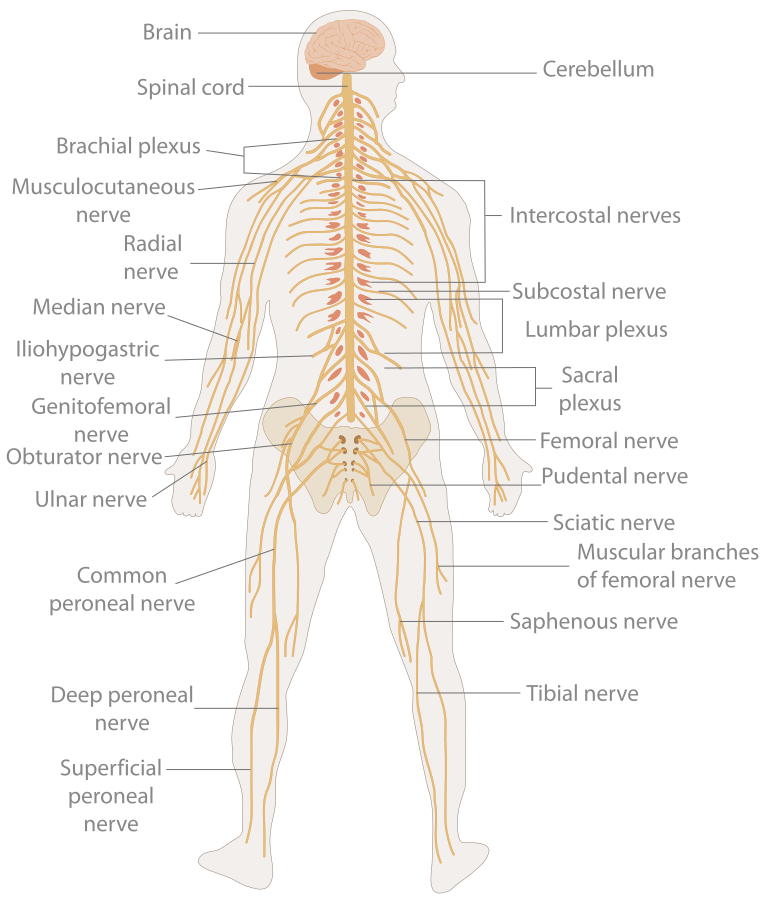
Signals of the Nervous System
The signals sent by the nervous system are electrical signals called nerve impulses, and they are transmitted by special nervous system cells called neurons (or nerve cells), like the one in Figure 8.2.3. Long projections (called axons) from neurons carry nerve impulses directly to specific target cells. A cell that receives nerve impulses from a neuron (typically a muscle or a gland) may be excited to perform a function, inhibited from carrying out an action, or otherwise controlled. In this way, the information transmitted by the nervous system is specific to particular cells and is transmitted very rapidly. In fact, the fastest nerve impulses travel at speeds greater than 100 metres per second! Compare this to the chemical messages carried by the hormones that are secreted into the blood by endocrine glands. These hormonal messages are “broadcast” to all the cells of the body, and they can travel only as quickly as the blood flows through the cardiovascular system.
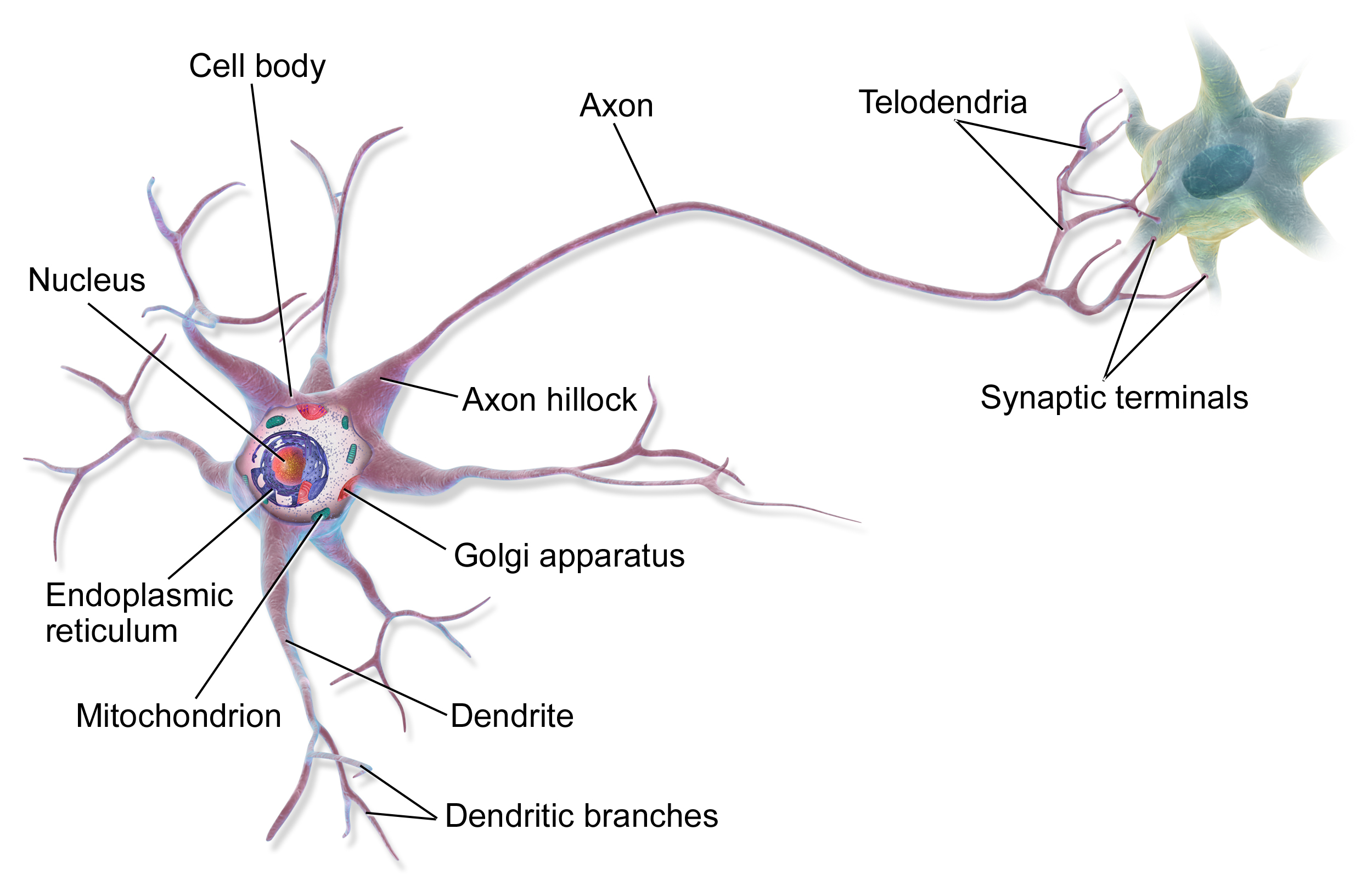
This simple model of a nerve cell shows part of its long axon which carries nerve impulses to other cells. The multiple shorter projections are called dendrites, and they receive nerve impulses from other cells.
Organization of the Nervous System
As you might predict, the human nervous system is very complex. It has multiple divisions, beginning with its two main parts, the central nervous system (CNS) and the peripheral nervous system (PNS), as shown in the diagram below (Figure 8.2.4). The CNS includes the brain and spinal cord, and the PNS consists mainly of nerves, which are bundles of axons from neurons. The nerves of the PNS connect the CNS to the rest of the body.
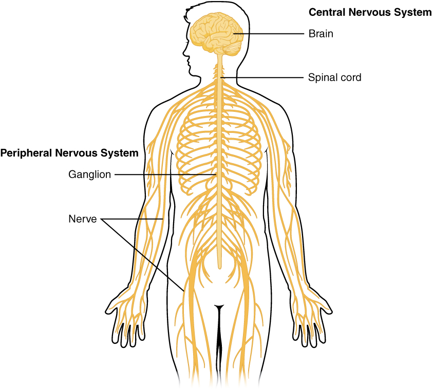
The PNS can be further subdivided into two divisions, known as the autonomic and somatic nervous systems (Figure 8.2.5). These divisions control different types of functions, and they often interact with the CNS to carry out these functions. The somatic nervous system controls activities that are under voluntary control, such as turning a steering wheel. The autonomic nervous system controls activities that are not under voluntary control, such as digesting a meal. The autonomic nervous system has three main divisions: the sympathetic division (which controls the fight-or-flight response during emergencies), the parasympathetic division (which controls the routine “housekeeping” functions of the body at other times), and the enteric division (which provides local control of the digestive system).
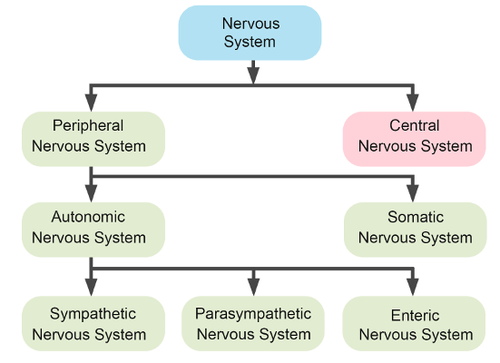
8.2 Summary
- The nervous system is the human organ system that coordinates all of the body’s voluntary and involuntary actions, by transmitting signals to and from different parts of the body.
- The nervous system has two major divisions, called the central nervous system (CNS) and the peripheral nervous system (PNS). The CNS includes the brain and spinal cord, and the PNS consists mainly of nerves that connect the CNS with the rest of the body.
- The PNS can be subdivided into two major divisions: the somatic nervous system and the autonomic nervous system.The somatic system controls activities that are under voluntary control. The autonomic system controls activities that are not under voluntary control. The autonomic nervous system is further divided into the sympathetic division (which controls the fight-or-flight response), the parasympathetic division (which controls most routine involuntary responses), and the enteric division (which provides local control of the digestive system).
- Electrical signals sent by the nervous system are called nerve impulses. They are transmitted by special cells called neurons. Nerve impulses can travel to specific target cells very rapidly.
8.2 Review Questions
- List the general steps through which the nervous system generates an appropriate response to information from the internal and external environments.
- What are neurons?
- Compare and contrast the central and peripheral nervous systems.
-
- Which major division of the peripheral nervous system allows you to walk to class? Which major division of the peripheral nervous system controls your heart rate?
- Identify the functions of the three main divisions of the autonomic nervous system.
-
- What is an axon, and what is its function?
- Define nerve impulses.
- Explain generally how the brain and spinal cord can interact with and control the rest of the body.
- How are nerves and neurons related?
- What type of information from the outside environment do you think is detected by sensory receptors in your ears?
8.2 Explore More
https://www.youtube.com/watch?v=qPix_X-9t7E
The Nervous System, Part 1: Crash Course A&P #8, CrashCourse, 2015.
https://www.youtube.com/watch?v=Nsxw5_Iz7mY&feature=emb_logo
Engineering the Human Nervous System: Megan Moynahan at TEDxBrussels,
TEDx Talks, 2013.
Attributions
Figure 8.2.1
Skateboard_1613 by Autoria propia on Wikimedia Commons is released into the public domain (https://en.wikipedia.org/wiki/Public_domain) (Derivative work of this file: SkateboardinDog.jpg)
Figure 8.2.2
Nervous_system_diagram.svg by The Emirr on Wikimedia Commons is used under a CC BY 3.0 (https://creativecommons.org/licenses/by/3.0/deed.en) license.
Figure 8.2.3
MultipolarNeuron by BruceBlaus on Wikimedia Commons is used under a CC BY 3.0 (https://creativecommons.org/licenses/by/3.0/deed.en) license.
Figure 8.2.4
Overview_of_Nervous_System by OpenStax on Wikimedia Commons is used under a CC BY 4.0 (https://creativecommons.org/licenses/by/4.0/deed.en) license.
Figure 8.2.5
Divisions of the Nervous System by CK-12 Foundation is used under the CC BY-NC 3.0 (https://creativecommons.org/licenses/by-nc/3.0/) license.
References
Betts, J. G., Young, K.A., Wise, J.A., Johnson, E., Poe, B., Kruse, D.H., Korol, O., Johnson, J.E., Womble, M., DeSaix, P. (2013, April 25). Figure 12.2 Central and peripheral nervous system [digital image]. In Anatomy and Physiology (Section 12.1). OpenStax. https://openstax.org/books/anatomy-and-physiology/pages/12-1-basic-structure-and-function-of-the-nervous-system
Brainard, J/ CK-12 Foundation. (2016). Figure 5 [digital image]. In CK-12 College Human Biology (Section 10.2) [online Flexbook]. CK12.org. https://www.ck12.org/book/ck-12-college-human-biology/section/10.2/
CrashCourse. (2015, February 23). The nervous system, Part 1: Crash Course A&P #8. YouTube. https://www.youtube.com/watch?v=qPix_X-9t7E&feature=youtu.be
TEDx Talks. (2013, November 3). Engineering the human nervous system: Megan Moynahan at TEDxBrussels. YouTube. https://www.youtube.com/watch?v=Nsxw5_Iz7mY&feature=youtu.be
Component of a homeostatic control mechanism that monitors a variable and sends signals to the effector as needed to keep the variable in homeostasis.
A component of a homeostatic control mechanism, such as a gland or an organ, that acts on a signal from the control center to move the variable back toward the set point.
Created by CK-12 Foundation/Adapted by Christine Miller

Feel the Burn
The person in Figure 10.3.1 is no doubt feeling the burn — sunburn, that is. Sunburn occurs when the outer layer of the skin is damaged by UV light from the sun or tanning lamps. Some people deliberately allow UV light to burn their skin, because after the redness subsides, they are left with a tan. A tan may look healthy, but it is actually a sign of skin damage. People who experience one or more serious sunburns are significantly more likely to develop skin cancer. Natural pigment molecules in the skin help protect it from UV light damage. These pigment molecules are found in the layer of the skin called the epidermis.
What is the Epidermis?
The epidermis is the outer of the two main layers of the skin. The inner layer is the dermis. It averages about 0.10 mm thick, and is much thinner than the dermis. The epidermis is thinnest on the eyelids (0.05 mm) and thickest on the palms of the hands and soles of the feet (1.50 mm). The epidermis covers almost the entire body surface. It is continuous with — but structurally distinct from — the mucous membranes that line the mouth, anus, urethra, and vagina.
Structure of the Epidermis
There are no blood vessels and very few nerve cells in the epidermis. Without blood to bring epidermal cells oxygen and nutrients, the cells must absorb oxygen directly from the air and obtain nutrients via diffusion of fluids from the dermis below. However, as thin as it is, the epidermis still has a complex structure. It has a variety of cell types and multiple layers.
Cells of the Epidermis
There are several different types of cells in the epidermis. All of the cells are necessary for the important functions of the epidermis.
- The epidermis consists mainly of stacks of keratin-producing epithelial cells called keratinocytes. These cells make up at least 90 per cent of the epidermis. Near the top of the epidermis, these cells are also called squamous cells.
- Another eight per cent of epidermal cells are melanocytes. These cells produce the pigment melanin that protects the dermis from UV light.
- About one per cent of epidermal cells are Langerhans cells. These are immune system cells that detect and fight pathogens entering the skin.
- Less than one per cent of epidermal cells are Merkel cells, which respond to light touch and connect to nerve endings in the dermis.
Layers of the Epidermis
The epidermis in most parts of the body consists of four distinct layers. A fifth layer occurs in the palms of the hands and soles of the feet, where the epidermis is thicker than in the rest of the body. The layers of the epidermis are shown in Figure 10.3.2, and described in the following text.

Stratum Basale
The stratum basale is the innermost (or deepest) layer of the epidermis. It is separated from the dermis by a membrane called the basement membrane. The stratum basale contains stem cells — called basal cells — which divide to form all the keratinocytes of the epidermis. When keratinocytes first form, they are cube-shaped and contain almost no keratin. As more keratinocytes are produced, previously formed cells are pushed up through the stratum basale. Melanocytes and Merkel cells are also found in the stratum basale. The Merkel cells are especially numerous in touch-sensitive areas, such as the fingertips and lips.
Stratum Spinosum
Just above the stratum basale is the stratum spinosum. This is the thickest of the four epidermal layers. The keratinocytes in this layer have begun to accumulate keratin, and they have become tougher and flatter. Spiny cellular projections form between the keratinocytes and hold them together. In addition to keratinocytes, the stratum spinosum contains the immunologically active Langerhans cells.
Stratum Granulosum
The next layer above the stratum spinosum is the stratum granulosum. In this layer, keratinocytes have become nearly filled with keratin, giving their cytoplasm a granular appearance. Lipids are released by keratinocytes in this layer to form a lipid barrier in the epidermis. Cells in this layer have also started to die, because they are becoming too far removed from blood vessels in the dermis to receive nutrients. Each dying cell digests its own nucleus and organelles, leaving behind only a tough, keratin-filled shell.
Stratum Lucidum
Only on the palms of the hands and soles of the feet, the next layer above the stratum granulosum is the stratum lucidum. This is a layer consisting of stacks of translucent, dead keratinocytes that provide extra protection to the underlying layers.
Stratum Corneum
The uppermost layer of the epidermis everywhere on the body is the stratum corneum. This layer is made of flat, hard, tightly packed dead keratinocytes that form a waterproof keratin barrier to protect the underlying layers of the epidermis. Dead cells from this layer are constantly shed from the surface of the body. The shed cells are continually replaced by cells moving up from lower layers of the epidermis. It takes a period of about 48 days for newly formed keratinocytes in the stratum basale to make their way to the top of the stratum corneum to replace shed cells.
Functions of the Epidermis
The epidermis has several crucial functions in the body. These functions include protection, water retention, and vitamin D synthesis.
Protective Functions
The epidermis provides protection to underlying tissues from physical damage, pathogens, and UV light.
Protection from Physical Damage
Most of the physical protection of the epidermis is provided by its tough outer layer, the stratum corneum. Because of this layer, minor scrapes and scratches generally do not cause significant damage to the skin or underlying tissues. Sharp objects and rough surfaces have difficulty penetrating or removing the tough, dead, keratin-filled cells of the stratum corneum. If cells in this layer are pierced or scraped off, they are quickly replaced by new cells moving up to the surface from lower skin layers.
Protection from Pathogens

When pathogens such as viruses and bacteria try to enter the body, it is virtually impossible for them to enter through intact epidermal layers. Generally, pathogens can enter the skin only if the epidermis has been breached, for example by a cut, puncture, or scrape (like the one pictured in Figure 10.3.3). That’s why it is important to clean and cover even a minor wound in the epidermis. This helps ensure that pathogens do not use the wound to enter the body. Protection from pathogens is also provided by conditions at or near the skin surface. These include relatively high acidity (pH of about 5.0), low amounts of water, the presence of antimicrobial substances produced by epidermal cells, and competition with non-pathogenic microorganisms that normally live on the epidermis.
Protection from UV Light
UV light that penetrates the epidermis can damage epidermal cells. In particular, it can cause mutations in DNA that lead to the development of skin cancer, in which epidermal cells grow out of control. UV light can also destroy vitamin B9 (in forms such as folate or folic acid), which is needed for good health and successful reproduction. In a person with light skin, just an hour of exposure to intense sunlight can reduce the body’s vitamin B9 level by 50 per cent.
Melanocytes in the stratum basale of the epidermis contain small organelles called melanosomes, which produce, store, and transport the dark brown pigment melanin. As melanosomes become full of melanin, they move into thin extensions of the melanocytes. From there, the melanosomes are transferred to keratinocytes in the epidermis, where they absorb UV light that strikes the skin. This prevents the light from penetrating deeper into the skin, where it can cause damage. The more melanin there is in the skin, the more UV light can be absorbed.
Water Retention
Skin's ability to hold water and not lose it to the surrounding environment is due mainly to the stratum corneum. Lipids arranged in an organized way among the cells of the stratum corneum form a barrier to water loss from the epidermis. This is critical for maintaining healthy skin and preserving proper water balance in the body.
Although the skin is impermeable to water, it is not impermeable to all substances. Instead, the skin is selectively permeable, allowing certain fat-soluble substances to pass through the epidermis. The selective permeability of the epidermis is both a benefit and a risk.
- Selective permeability allows certain medications to enter the bloodstream through the capillaries in the dermis. This is the basis of medications that are delivered using topical ointments, or patches (see Figure 10.3.4) that are applied to the skin. These include steroid hormones, such as estrogen (for hormone replacement therapy), scopolamine (for motion sickness), nitroglycerin (for heart problems), and nicotine (for people trying to quit smoking).
- Selective permeability of the epidermis also allows certain harmful substances to enter the body through the skin. Examples include the heavy metal lead, as well as many pesticides.

Vitamin D Synthesis
Vitamin D is a nutrient that is needed in the human body for the absorption of calcium from food. Molecules of a lipid compound named 7-dehydrocholesterol are precursors of vitamin D. These molecules are present in the stratum basale and stratum spinosum layers of the epidermis. When UV light strikes the molecules, it changes them to vitamin D3. In the kidneys, vitamin D3 is converted to calcitriol, which is the form of vitamin D that is active in the body.
What Gives Skin Its Colour?
Melanin in the epidermis is the main substance that determines the colour of human skin. It explains most of the variation in skin colour in people around the world. Two other substances also contribute to skin colour, however, especially in light-skinned people: carotene and hemoglobin.
- The pigment carotene is present in the epidermis and gives skin a yellowish tint, especially in skin with low levels of melanin.
- Hemoglobin is a red pigment found in red blood cells. It is visible through skin as a pinkish tint, mainly in skin with low levels of melanin. The pink colour is most visible when capillaries in the underlying dermis dilate, allowing greater blood flow near the surface.
Hear what Bill Nye has to say about the subject of skin colour in the video here.
Bacteria on Skin

The surface of the human skin normally provides a home to countless numbers of bacteria. Just one square inch of skin normally has an average of about 50 million bacteria. These generally harmless bacteria represent roughly one thousand bacterial species (including the one in Figure 10.3.5) from 19 different bacterial phyla. Typical variations in the moistness and oiliness of the skin produce a variety of rich and diverse habitats for these microorganisms. For example, the skin in the armpits is warm and moist and often hairy, whereas the skin on the forearms is smooth and dry. These two areas of the human body are as diverse to microorganisms as rainforests and deserts are to larger organisms. The density of bacterial populations on the skin depends largely on the region of the skin and its ecological characteristics. For example, oily surfaces, such as the face, may contain over 500 million bacteria per square inch. Despite the huge number of individual microorganisms living on the skin, their total volume is only about the size of a pea.
In general, the normal microorganisms living on the skin keep one another in check, and thereby play an important role in keeping the skin healthy. If the balance of microorganisms is disturbed, however, there may be an overgrowth of certain species, and this may result in an infection. For example, when a patient is prescribed antibiotics, it may kill off normal bacteria and allow an overgrowth of single-celled yeast. Even if skin is disinfected, no amount of cleaning can remove all of the microorganisms it contains. Disinfected areas are also quickly recolonized by bacteria residing in deeper areas (such as hair follicles) and in adjacent areas of the skin.
Feature: Myth vs. Reality
Because of the negative health effects of excessive UV light exposure, it is important to know the facts about protecting the skin from UV light.
Myth |
Reality |
| "Sunblock and sunscreen are just different names for the same type of product. They both work the same way and are equally effective." | Sunscreens and sunblocks are different types of products that protect the skin from UV light in different ways. They are not equally effective. Sunblocks are opaque, so they do not let light pass through. They prevent most of the rays of UV light from penetrating to the skin surface. Sunblocks are generally stronger and more effective than sunscreens. Sunblocks also do not need to be reapplied as often as sunscreens. Sunscreens, in contrast, are transparent once they are applied the skin. Although they can prevent most UV light from penetrating the skin when first applied, the active ingredients in sunscreens tend to break down when exposed to UV light. Sunscreens, therefore, must be reapplied often to remain effective. |
| "The skin needs to be protected from UV light only on sunny days. When the sky is cloudy, UV light cannot penetrate to the ground and harm the skin." | Even on cloudy days, a significant amount of UV radiation penetrates the atmosphere to strike Earth’s surface. Therefore, using sunscreens or sunblocks to protect exposed skin is important even when there are clouds in the sky. |
| "People who have dark skin, such as African Americans, do not need to worry about skin damage from UV light." | No matter what colour skin you have, your skin can be damaged by too much exposure to UV light. Therefore, even dark-skinned people should use sunscreens or sunblocks to protect exposed skin from UV light. |
| "Sunscreens with an SPF (sun protection factor) of 15 are adequate to fully protect the skin from UV light." | Most dermatologists recommend using sunscreens with an SPF of at least 35 for adequate protection from UV light. They also recommend applying sunscreens at least 20 minutes before sun exposure and reapplying sunscreens often, especially if you are sweating or spending time in the water. |
| "Using tanning beds is safer than tanning outside in natural sunlight." | The light in tanning beds is UV light, and it can do the same damage to the skin as the natural UV light in sunlight. This is evidenced by the fact that people who regularly use tanning beds have significantly higher rates of skin cancer than people who do not. It is also the reason that the use of tanning beds is prohibited in many places in people who are under the age of 18, just as youth are prohibited from using harmful substances, such as tobacco and alcohol. |
10.3 Summary
- The epidermis is the outer of the two main layers of the skin. It is very thin, but has a complex structure.
- Cell types in the epidermis include keratinocytes that produce keratin and make up 90 per cent of epidermal cells, melanocytes that produce melanin, Langerhans cells that fight pathogens in the skin, and Merkel cells that respond to light touch.
- The epidermis in most parts of the body consists of four distinct layers. A fifth layer occurs only in the epidermis of the palms of the hands and soles of the feet.
- The innermost layer of the epidermis is the stratum basale, which contains stem cells that divide to form new keratinocytes. The next layer is the stratum spinosum, which is the thickest layer and contains Langerhans cells and spiny keratinocytes. This is followed by the stratum granulosum, in which keratinocytes are filling with keratin and starting to die. The stratum lucidum is next, but only on the palms and soles. It consists of translucent dead keratinocytes. The outermost layer is the stratum corneum, which consists of flat, dead, tightly packed keratinocytes that form a tough, waterproof barrier for the rest of the epidermis.
- Functions of the epidermis include protecting underlying tissues from physical damage and pathogens. Melanin in the epidermis absorbs and protects underlying tissues from UV light. The epidermis also prevents loss of water from the body and synthesizes vitamin D.
- Melanin is the main pigment that determines the colour of human skin. The pigments carotene and hemoglobin, however, also contribute to skin colour, especially in skin with low levels of melanin.
- The surface of healthy skin normally is covered by vast numbers of bacteria representing about one thousand species from 19 phyla. Different areas of the body provide diverse habitats for skin microorganisms. Usually, microorganisms on the skin keep each other in check unless their balance is disturbed.
10.3 Review Questions
- What is the epidermis?
- Identify the types of cells in the epidermis.
- Describe the layers of the epidermis.
-
- State one function of each of the four epidermal layers found all over the body.
- Explain three ways the epidermis protects the body.
- What makes the skin waterproof?
- Why is the selective permeability of the epidermis both a benefit and a risk?
- How is vitamin D synthesized in the epidermis?
- Identify three pigments that impart colour to skin.
- Describe bacteria that normally reside on the skin, and explain why they do not usually cause infections.
- Explain why the keratinocytes at the surface of the epidermis are dead, while keratinocytes located deeper in the epidermis are still alive.
- Which layer of the epidermis contains keratinocytes that have begun to die?
-
- Explain why our skin is not permanently damaged if we rub off some of the surface layer by using a rough washcloth.
10.3 Explore More
https://www.youtube.com/watch?v=27lMmdmy-b8
Jonathan Eisen: Meet your microbes, TED, 2015.
https://www.youtube.com/watch?v=9AcQXnOscQ8
Why Do We Blush?, SciShow, 2014.
https://www.youtube.com/watch?v=_r4c2NT4naQ
The science of skin colour - Angela Koine Flynn, TED-Ed, 2016.
Attributions
Figure 10.3.1
Sunburn by QuinnHK at English Wikipedia on Wikimedia Commons is released into the public domain (https://en.wikipedia.org/wiki/Public_domain).
Figure 10.3.2
Blausen_0353_Epidermis by BruceBlaus on Wikimedia Commons is used under a CC BY 3.0 (https://creativecommons.org/licenses/by/3.0) license.
Figure 10.3.3
Isaac's scraped knee close-up by Alpha on Flickr is used under a CC BY-NC-SA 2.0 (https://creativecommons.org/licenses/by-nc-sa/2.0/) license.
Figure 10.3.4
Nicoderm by RegBarc on Wikimedia Commons is used under a CC BY-SA 3.0 (http://creativecommons.org/licenses/by-sa/3.0/) license. (No machine-readable author provided for original.)
Figure 10.3.5
Staphylococcus aureus bacteria, MRSA by Microbe World on Flickr is used under a CC BY-NC-SA 2.0 (https://creativecommons.org/licenses/by-nc-sa/2.0/) license.
References
Blausen.com staff. (2014). Medical gallery of Blausen Medical 2014. WikiJournal of Medicine 1 (2). DOI:10.15347/wjm/2014.010. ISSN 2002-4436.
Jeff Bone 'n' Pookie. (2020, July 19). Bill Nye the science guy explains we have different skin color. Youtube. https://www.youtube.com/watch?v=zOkj5jgC4sM&feature=youtu.be
SciShow. (2014, July 15). Why do we blush? YouTube. https://www.youtube.com/watch?v=9AcQXnOscQ8
TED. (2015, July 17). Jonathan Eisen: Meet your microbes. YouTube. https://www.youtube.com/watch?v=27lMmdmy-b8
TED-Ed. (2016, February 16). The science of skin color - Angela Koine Flynn. YouTube. https://youtu.be/_r4c2NT4naQ
Created by CK-12 Foundation/Adapted by Christine Miller
Milk on Demand
This adorable nursing infant (Figure 9.4.1) is part of a positive feedback loop. When he suckles on the nipple, it sends nerve impulses to his mother’s hypothalamus. Those nerve impulses “tell” her pituitary gland to release the hormone prolactin into her bloodstream. Prolactin travels to the mammary glands in the breasts and stimulates milk production, which motivates the infant to keep suckling.
What Is the Pituitary Gland?
The pituitary gland is the master gland of the endocrine system, which is the system of glands that secrete hormones into the bloodstream. Endocrine hormones control virtually all physiological processes. They control growth, sexual maturation, reproduction, body temperature, blood pressure, and metabolism. The pituitary gland is considered the master gland of the endocrine system, because it controls the rest of the endocrine system. Many pituitary hormones either promote or inhibit hormone secretion by other endocrine glands.
Structure and Function of the Pituitary Gland
The pituitary gland is about the size of a pea. It protrudes from the bottom of the hypothalamus at the base of the inner brain (see Figure 9.4.2). The pituitary is connected to the hypothalamus by a thin stalk (called the infundibulum). Blood vessels and nerves in the stalk allow direct connections between the hypothalamus and pituitary gland.
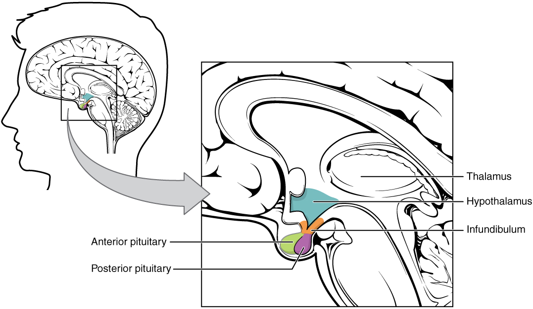
Anterior Lobe
The anterior pituitary is the lobe is at the front of the pituitary gland. It synthesizes and releases hormones into the blood. Table 9.4.1 shows some of the endocrine hormones released by the anterior pituitary, including their targets and effects.
Table 9.4.1
Endocrine Hormones Released by the Anterior Pituitary, and Their Targets and Effects.
| Anterior Pituitary Hormone | Target | Effect |
| Adrenocorticotropic hormone (ACTH) | Adrenal glands | Stimulates the cortex of each adrenal gland to secrete its hormones. |
| Thyroid-stimulating hormone (TSH) | Thyroid gland | Stimulates the thyroid gland to secrete thyroid hormone. |
| Growth hormone (GH) | Body cells | Stimulates body cells to synthesize proteins and grow. |
| Follicle-stimulating hormone (FSH) | Ovaries, testes | Stimulates the ovaries to develop mature eggs. stimulates the testes to produce sperm. |
| Luteinizing hormone (LH) | Ovaries, testes | Stimulates the ovaries and testes to secrete sex hormones; stimulates the ovaries to release eggs. |
| Prolactin (PRL) | Mammary glands | Stimulates the mammary glands to produce milk. |
The anterior pituitary gland is regulated mainly by hormones from the hypothalamus. The hypothalamus secretes hormones (called releasing hormones and inhibiting hormones) that travel through capillaries directly to the anterior lobe of the pituitary gland. The hormones stimulate the anterior pituitary to either release or stop releasing particular pituitary hormones. Several of these hypothalamic hormones and their effects on the anterior pituitary are shown in the table below.
Table 9.4.2
Hypothalamic Hormones and Their Effects on the Anterior Pituitary
| Hypothalamic Hormone | Effect on Anterior Pituitary |
| Thyrotropin releasing hormone (TRH) | Release of thyroid stimulating hormone (TSH) |
| Corticotropin releasing hormone (CRH) | Release of adrenocorticotropic hormone (ACTH) |
| Gonadotropin releasing hormone (GnRH) | Release of follicle-stimulating hormone (FSH) and luteinizing hormone (LH) |
| Growth hormone releasing hormone (GHRH) | Release of growth hormone (GH) |
| Growth hormone inhibiting hormone (GHIH) (Somatostatin) | Stopping of growth hormone release |
| Prolactin releasing hormone (PRH) | Release of prolactin |
| Prolactin inhibiting hormone (PIH) (Dopamine) | Stopping of prolactin release |
Posterior Lobe
The posterior pituitary is the lobe is at the back of the pituitary gland. This lobe does not synthesize any hormones. Instead, the posterior lobe stores hormones that come from the hypothalamus along the axons of nerves connecting the two structures (also shown in Figure 9.4.2). The posterior pituitary then secretes the hormones into the bloodstream as needed. Hypothalamic hormones secreted by the posterior pituitary include vasopressin and oxytocin.
- Vasopressin (also called antidiuretic hormone, or ADH) helps maintain homeostasis in body water. It stimulates the kidneys to conserve water by producing more concentrated urine. Specifically, vasopressin targets ducts in the kidneys and makes them more permeable to water. This allows more water to be resorbed by the body, rather than excreted in urine.
- Oxytocin (OXY) targets cells in the uterus to stimulate uterine contractions, as in childbirth. It also targets cells in the breasts of a nursing mother to stimulate the letdown of milk.
9.4 Summary
- The pituitary gland is the master gland of the endocrine system, because most of its hormones control other endocrine glands.
- The pituitary gland is at the base of the brain, where it is connected to the hypothalamus by nerves and capillaries. It has an anterior (front) lobe that synthesizes and secretes pituitary hormones and a posterior (back) lobe that stores and secretes hormones from the hypothalamus.
- Hormones synthesized and secreted by the anterior pituitary include growth hormone, which stimulates cell growth throughout the body, and thyroid stimulating hormone (TSH), which stimulates the thyroid gland to secrete its hormones.
- Hypothalamic hormones stored and secreted by the posterior pituitary gland include vasopressin, which helps maintain homeostasis in body water, and oxytocin, which stimulates uterine contractions during birth, as well as the letdown of milk during lactation.
9.4 Review Questions
-
-
- Explain why the pituitary gland is called the master gland of the endocrine system.
- Compare and contrast the two lobes of the pituitary gland and their general functions.
- Identify two hormones released by the anterior pituitary, their targets, and their effects.
- Explain how the hypothalamus influences the output of hormones by the anterior lobe of the pituitary gland.
- Name and give the function of two hypothalamic hormones released by the posterior pituitary gland.
- Answer the following questions about prolactin releasing hormone (PRH) and prolactin inhibiting hormone (PIH).
- Where are these hormones produced?
- Where are their target cells located?
- What are their effects on their target cells?
- What are their ultimate effects on milk production? Explain your answer.
- When a baby nurses, which of these hormones is most likely released in the mother? Explain your answer.
- For each of the following hormones, state whether it is synthesized in the pituitary or the hypothalamus.
- gonadotropin releasing hormone (GnRH)
- growth hormone (GH)
- oxytocin
- adrenocorticotropic hormone (ACTH)
9.4 Explore More
https://www.youtube.com/watch?v=jUKQFkmBuww&feature=emb_logo
Common Pituitary Diseases, Swedish, 2012.
https://www.youtube.com/watch?v=v41AJGP-XmI&feature=emb_logo
Diagnosing and Treating Pituitary Tumors - California Center for Pituitary Disorders at UCSF, UCSF Neurosurgery, 2015.
Attributions
Figure 9.4.1
Breastfeeding by Petr Kratochvil on Wikimedia Commons is used under a CC0 1.0 Universal
Public Domain Dedication (https://creativecommons.org/publicdomain/zero/1.0/deed.en) license.
Figure 9.4.2
The_Hypothalamus-Pituitary_Complex by OpenStax College on Wikimedia Commons is used under a CC BY 3.0 (https://creativecommons.org/licenses/by/3.0) license.
References
Betts, J. G., Young, K.A., Wise, J.A., Johnson, E., Poe, B., Kruse, D.H., Korol, O., Johnson, J.E., Womble, M., DeSaix, P. (2013, June 19). Figure 17.7 Hypothalamus–pituitary complex [digital image]. In Anatomy and Physiology (Section 17.3). OpenStax. https://openstax.org/books/anatomy-and-physiology/pages/17-3-the-pituitary-gland-and-hypothalamus
Swedish. (2012, April 19). Common pituitary diseases. YouTube. https://www.youtube.com/watch?v=jUKQFkmBuww&feature=youtu.be
UCSF Neurosurgery. (2015, May 13). Diagnosing and treating pituitary tumors - California Center for Pituitary Disorders at UCSF. YouTube. https://www.youtube.com/watch?v=v41AJGP-XmI&feature=youtu.be

