7.7 Interactions of Organ Systems
Figure 7.7.1 Everyone on a baseball team has a special job.
Teamwork
Every player on a baseball team has a special job. In the Figure 7.7.1 collage, each player has their part of the infield or outfield covered in case the ball comes their way. Other players on the team cover different parts of the field, or they pitch or catch the ball. Playing baseball clearly requires teamwork. In that regard, the human body is like a baseball team. All of the organ systems of the human body must work together as a team to keep the body alive and well. Teamwork within the body begins with communication.
Communication Among Organ Systems
Communication among organ systems is vital if they are to work together as a team. They must be able to respond to each other and change their responses as needed to keep the body in balance. Communication among organ systems is controlled mainly by the autonomic nervous system and the endocrine system.
The autonomic nervous system is the part of the nervous system that controls involuntary functions. The autonomic nervous system, for example, controls heart rate, blood flow, and digestion. You don’t have to tell your heart to beat faster or to consciously squeeze muscles to push food through the digestive system. You don’t have to even think about these functions at all! The autonomic nervous system orchestrates all the signals needed to control them. It sends messages between parts of the nervous system, as well as between the nervous system and other organ systems via chemical messengers called neurotransmitters.
The endocrine system is the system of glands that secrete hormones directly into the bloodstream. Once in the blood, endocrine hormones circulate to cells everywhere in the body. The endocrine system itself is under control of the nervous system via a part of the brain called the hypothalamus. The hypothalamus secretes hormones that travel directly to cells of the pituitary gland, which is located beneath it. The pituitary gland is the master gland of the endocrine system. Most of its hormones either turn on or turn off other endocrine glands. For example, if the pituitary gland secretes thyroid-stimulating hormone, the hormone travels through the circulation to the thyroid gland, which is stimulated to secrete thyroid hormone. Thyroid hormone then travels to cells throughout the body, where it increases their metabolism.
Examples of Organ System Interactions
An increase in cellular metabolism requires more cellular respiration. Cellular respiration is a good example of organ system interactions, because it is a basic life process that occurs in all living cells.
Cellular Respiration
Cellular respiration is the intracellular process that breaks down glucose with oxygen to produce carbon dioxide and energy in the form of ATP molecules. It is the process by which cells obtain usable energy to power other cellular processes. Which organ systems are involved in cellular respiration? The glucose needed for cellular respiration comes from the digestive system via the cardiovascular system. The oxygen needed for cellular respiration comes from the respiratory system also via the cardiovascular system. The carbon dioxide produced in cellular respiration leaves the body by the opposite route. In short, cellular respiration requires — at a minimum — the digestive, cardiovascular, and respiratory systems.
Fight-or-Flight Response
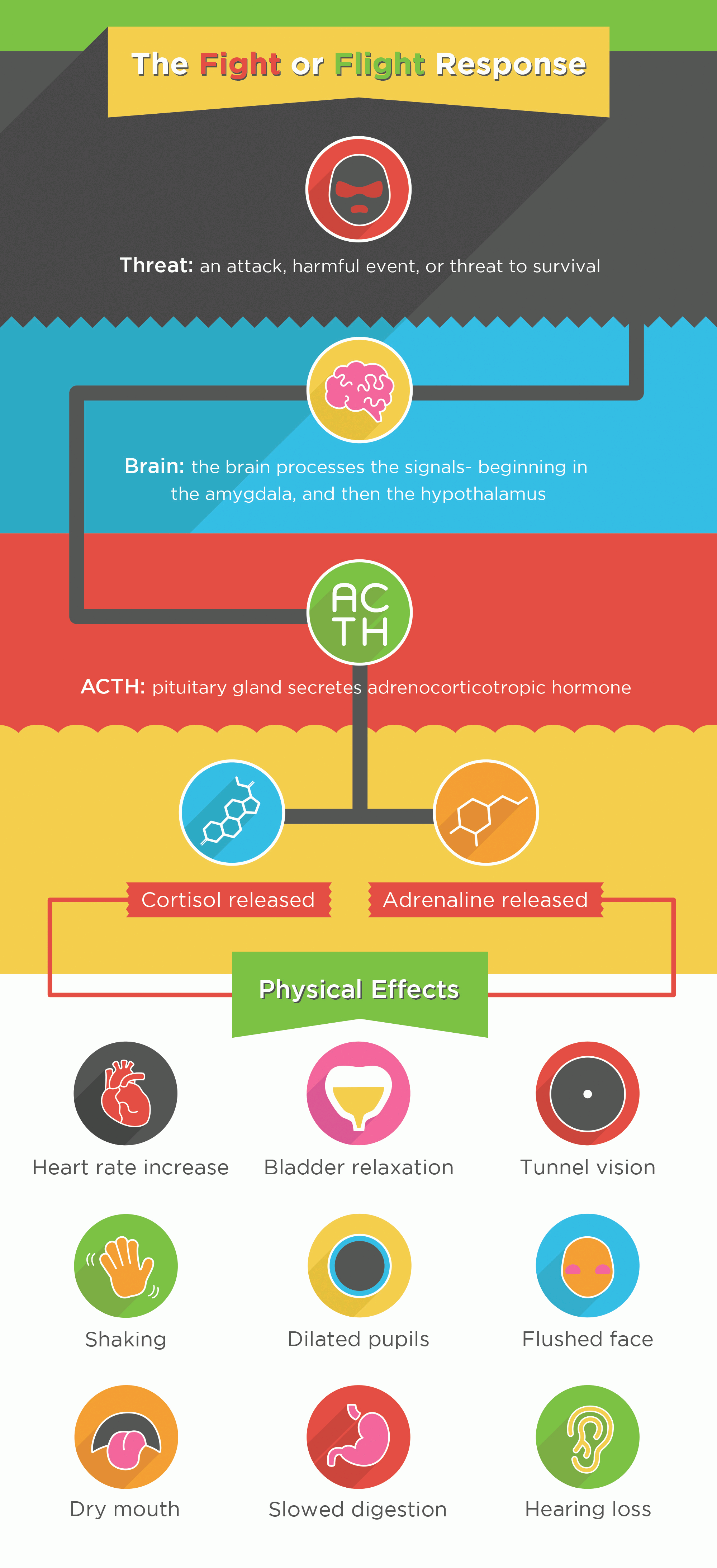
The well-known fight-or-flight response is a good example of how the nervous and endocrine systems control other organ system responses. The fight-or-flight response begins when the nervous system perceives sudden danger, as shown in the Figure 7.7.2 diagram. The brain sends a message to the endocrine system (via the pituitary gland) for the adrenal glands to secrete the hormones cortisol and adrenaline. These hormones flood the circulation and affect other organ systems throughout the body, including the cardiovascular, urinary, sensory, and digestive systems. Specific responses include increased heart rate, bladder relaxation, tunnel vision, and a shunting of blood away from the digestive system and toward the muscles, brain, and other vital organs needed to fight or flee.
Playing Baseball
The people playing baseball in the opening collage (Figure 7.7.1) are using multiple organ systems in this voluntary activity. Their nervous systems are focused on observing and preparing to respond to the next play. Their other systems are being controlled by the autonomic nervous system. The players are using the muscular, skeletal, respiratory, and cardiovascular systems. Can you explain how each of these organ systems is involved in playing baseball?
Feature: Reliable Sources
Teamwork among organ systems allows the human organism to work like a finely tuned machine — at least, it does until one of the organ systems fails. When that happens, other organ systems interacting in the same overall process will also be affected. This is especially likely if the affected system plays a controlling role in the process. An example is type 1 diabetes. This disorder occurs when the pancreas does not secrete the endocrine hormone insulin. Insulin normally is secreted in response to an increasing level of glucose in the blood, and it brings the level of glucose back to normal by stimulating body cells to take up insulin from the blood.
Learn more about type 1 diabetes. Use several reliable Internet sources to answer the following questions:
- In type 1 diabetes, what causes the endocrine system to fail to produce insulin?
- If type 1 diabetes is not controlled, which organ systems are affected by high blood glucose levels? What are some of the specific effects?
- How can blood glucose levels be controlled in patients with type 1 diabetes?
7.7 Summary
- The human body’s organ systems must work together to keep the body alive and functioning normally, which requires communication among systems. This communication is controlled by the autonomic nervous system and endocrine system. The autonomic nervous system controls involuntary body functions, such as heart rate and digestion. The endocrine system secretes hormones into the blood that travel to body cells and influence their activities.
- Cellular respiration is a good example of organ system interactions, because it is a basic life process that happens in all living cells. It is the intracellular process that breaks down glucose with oxygen to produce carbon dioxide and energy. Cellular respiration requires the interaction of the digestive, cardiovascular, and respiratory systems.
- The fight-or-flight response is a good example of how the nervous and endocrine systems control other organ system responses. It is triggered by a message from the brain to the endocrine system and prepares the body for flight or a fight. Many organ systems are stimulated to respond, including the cardiovascular, respiratory, and digestive systems.
- Playing baseball — or doing other voluntary physical activities — may involve the interaction of nervous, muscular, skeletal, respiratory, and cardiovascular systems.
7.7 Review Questions
- What is the autonomic nervous system?
- How do the autonomic nervous system and endocrine system communicate with other organ systems so the systems can interact?
- Explain how the brain communicates with the endocrine system.
- What is the role of the pituitary gland in the endocrine system?
- Identify the organ systems that play a role in cellular respiration.
- How does the hormone adrenaline prepare the body to fight or flee? What specific physiological changes does it bring about?
- Explain the role of the muscular system in digesting food.
- Describe how three different organ systems are involved when a player makes a particular play in baseball, such as catching a fly ball.
-
- What are two types of molecules that the body uses to communicate between organ systems?
- Explain why hormones can have such a wide variety of effects on the body.
7.7 Explore More
3D Medical Animation – Peristalsis in Large Intestine/Bowel ||
©Animated Biomedical Productions (ABP), 2013.
Adrenaline: Fight or Flight Response, Henk van ‘t Klooster, 2013.
Fight or Flight Response, Bozeman Science, 2012.
Attributions
Figure 7.7.1
- Baseball positions by Michael J on Wikimedia Commons is used under a CC BY-SA 4.0 (https://creativecommons.org/licenses/by-sa/4.0/deed.de) license.
- US Navy 040229-N-8629D-070 photo by US Navy‘s Photographer’s Mate 2nd Class Brett A. Dawson on Wikimedia Commons is released into the public domain (https://en.wikipedia.org/wiki/Public_domain).
- David Ortiz batter’s box by Albert Yau/ SecondPrint Productions on Flickr is used under a CC BY 2.0 (https://creativecommons.org/licenses/by/2.0/deed.es) license.
- Fenway-from Legend’s Box by Jared Vincent on Wikipedia is used under a CC BY 2.0 (https://creativecommons.org/licenses/by/2.0/deed.en) license.
Figure 7.7.2
The_Fight_or_Flight_Response by Jvnkfood (original), converted to PNG and reduced to 8-bit by Pokéfan95 on Wikimedia Commons is used under a CC BY-SA 4.0 (https://creativecommons.org/licenses/by-sa/4.0/deed.en) license.
References
Animated Biomedical Productions. (2013, January 30). 3D Medical animation – Peristalsis in large intestine/bowel || ©ABP. YouTube. https://www.youtube.com/watch?v=Ujr0UAbyPS4&feature=youtu.be
Bozeman Science. (2012, January 9). Fight or flight response. YouTube. https://www.youtube.com/watch?v=m2GywoS77qc&feature=youtu.be
Henk van ‘t Klooster. (2013). Adrenaline: Fight or flight response. YouTube. https://www.youtube.com/watch?v=FBnBTkcr6No&t=4s
Mayo Clinic Staff. (n.d.). Type 1 diabetes. MayoClinic.org. https://www.mayoclinic.org/diseases-conditions/type-1-diabetes/symptoms-causes/syc-20353011
Wikipedia contributors. (2020, July 22). Thyroid-stimulating hormone. In Wikipedia. https://en.wikipedia.org/w/index.php?title=Thyroid-stimulating_hormone&oldid=968942540
Created by CK-12 Foundation/Adapted by Christine Miller
Case Study Conclusion: Fading Memory
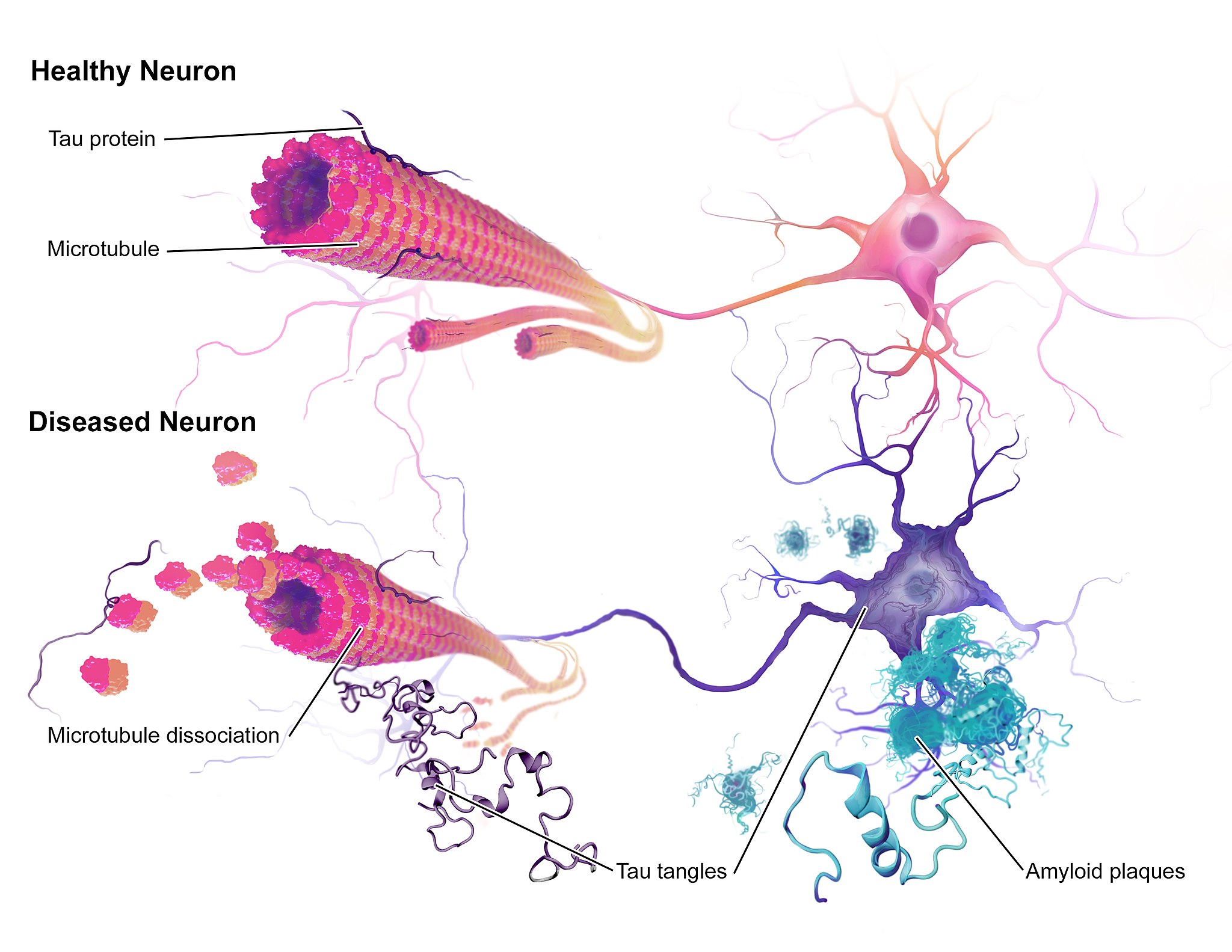
The illustration above (Figure 8.9.1) shows some of the molecular and cellular changes that occur in Alzheimer’s disease (AD). Rosa was diagnosed with AD at the beginning of this chapter after experiencing memory problems and other changes in her cognitive functioning, mood, and personality. These abnormal changes in the brain include the development of amyloid plaques between brain cells and neurofibrillary tangles inside of neurons. These hallmark characteristics of AD are associated with the loss of synapses between neurons, and ultimately the death of neurons.
After reading this chapter, you should have a good appreciation for the importance of keeping neurons alive and communicating with each other at synapses. The nervous system coordinates all of the body’s voluntary and involuntary activities. It interprets information from the outside world through sensory systems, and makes appropriate responses through the motor system, through communication between the PNS and CNS. The brain directs the rest of the nervous system and controls everything from basic vital functions (such as heart rate and breathing) to high-level functions (such as problem solving and abstract thought). The nervous system can perform these important functions by generating action potentials in neurons in response to stimulation and sending messages between cells at synapses, typically using chemical neurotransmitter molecules. When neurons are not functioning properly, lose their synapses, or die, they cannot carry out the signaling essential for the proper functioning of the nervous system.
AD is a progressive neurodegenerative disease, meaning that the damage to the brain becomes more extensive as time goes on. The picture in Figure 8.9.2 illustrates how the damage progresses from before AD is diagnosed (preclinical AD), to mild and moderate AD, to severe AD.
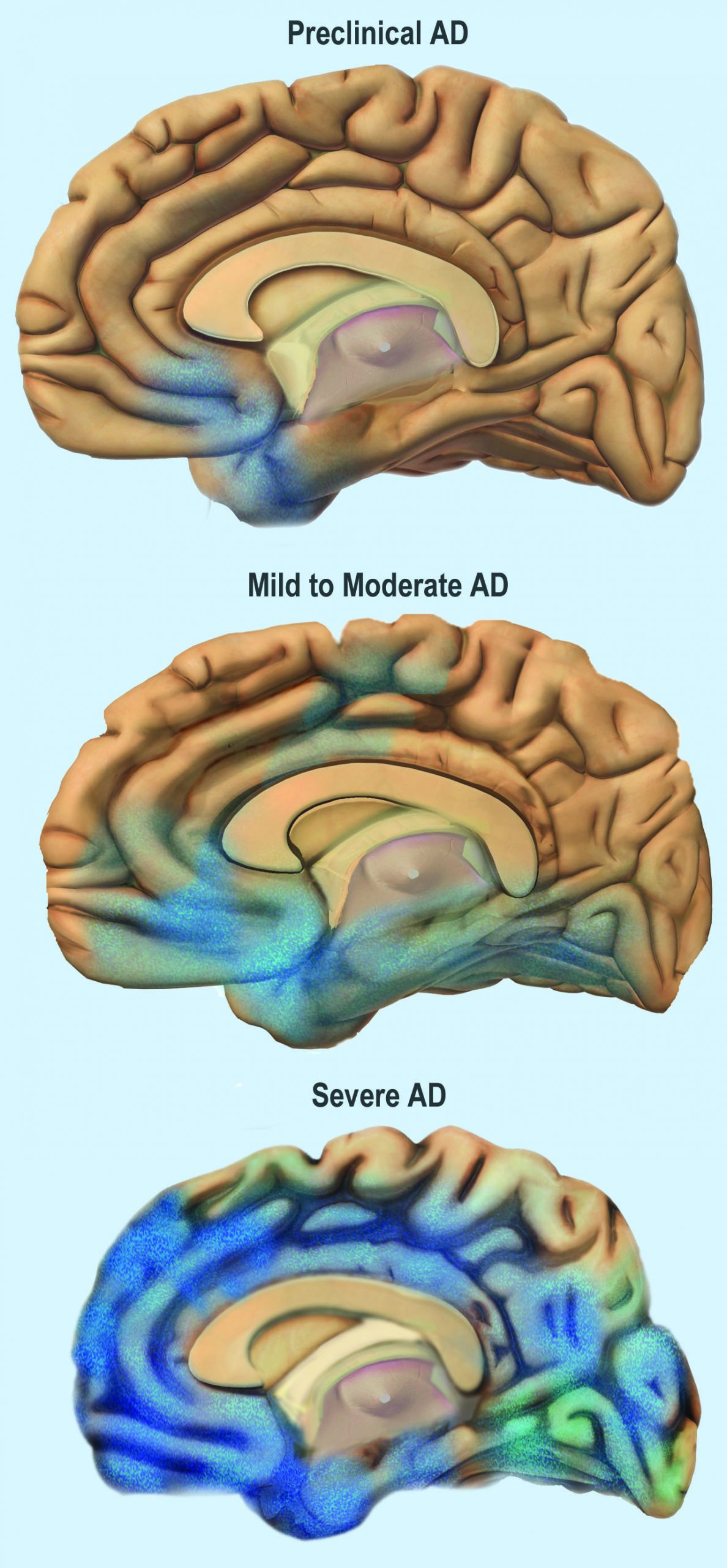
You can see that the damage starts in a relatively small location toward the bottom of the brain. One of the earliest brain areas to be affected by AD is the hippocampus. As you have learned, the hippocampus is important for learning and memory, which explains why many of Rosa’s symptoms of mild AD involve deficits in memory, such as trouble remembering where she placed objects, recent conversations, and appointments.
As AD progresses, more of the brain is affected, including areas involved in emotional regulation, social behavior, planning, language, spatial navigation, and higher-level thought. Rosa is beginning to show signs of problems in these areas, including irritability, lashing out at family members, getting lost in her neighborhood, problems finding the right words, putting objects in unusual locations, and difficulty in managing her finances. You can see that as AD progresses, damage spreads further across the cerebrum, which you now know controls conscious functions like reasoning, language, and interpretation of sensory stimuli. You can also see how the frontal lobe — which controls executive functions such as planning, self-control, and abstract thought — becomes increasingly damaged.
Increasing damage to the brain causes corresponding deficits in functioning. In moderate AD, patients have increased memory, language, and cognitive deficits, compared to mild AD. They may not recognize their own family members, and may wander and get lost, engage in inappropriate behaviors, become easily agitated, and have trouble carrying out daily activities such as dressing. In severe AD, much of the brain is affected. Patients usually cannot recognize family members or communicate, and they are often fully dependent on others for their care. They begin to lose the ability to control their basic functions, such as bladder control, bowel control, and proper swallowing. Eventually, AD causes death, usually as a result of this loss of basic functions.
For now, Rosa only has mild AD and is still able to function relatively well with care from her family. The medication her doctor gave her has helped improve some of her symptoms. It is a cholinesterase inhibitor, which blocks an enzyme that normally degrades the neurotransmitter acetylcholine. With more of the neurotransmitter available, more of it can bind to neurotransmitter receptors on postsynaptic cells. Therefore, this drug acts as an agonist for acetylcholine, which enhances communication between neurons in Rosa’s brain. This increase in neuronal communication can help restore some of the functions lost in early Alzheimer’s disease and may slow the progression of symptoms.
But medication such as this is only a short-term measure, and does not halt the progression of the underlying disease. Ideally, the damaged or dead neurons would be replaced by new, functioning neurons. Why does this not happen automatically in the body? As you have learned, neurogenesis is very limited in adult humans, so once neurons in the brain die, they are not normally replaced to any significant extent. Scientists, however, are studying the ways in which neurogenesis might be increased in cases of disease or injury to the brain. They are also investigating the possibility of using stem cell transplants to replace damaged or dead neurons with new neurons. But this research is in very early stages and is not currently a treatment for AD.
One promising area of research is in the development of methods to allow earlier detection and treatment of AD, given that the changes in the brain may actually start ten to 20 years before diagnosis of AD. A radiolabeled chemical called Pittsburgh Compound B (PiB) binds to amyloid plaques in the brain, and in the future, it may be used in conjunction with brain imaging techniques to detect early signs of AD. Scientists are also looking for biomarkers in bodily fluids (such as blood and cerebrospinal fluid) that might indicate the presence of AD before symptoms appear. Finally, researchers are also investigating possible early and subtle symptoms (such as changes in how people move or a loss of smell) to see whether they can be used to identify people who will go on to develop AD. This research is in the early stages, but the hope is that patients can be identified earlier, allowing for earlier and more effective treatment, as well as more planning time for families.
Scientists are also still trying to fully understand the causes of AD, which affects more than five million Americans. Some genetic mutations have been identified as contributors, but environmental factors also appear to be important. With more research into the causes and mechanisms of AD, hopefully a cure can be found, and people like Rosa can live a longer and better life.
Chapter 8 Summary
In this chapter, you learned about the human nervous system. Specifically, you learned that:
- The nervous system is the organ system that coordinates all of the body’s voluntary and involuntary actions by transmitting signals to and from different parts of the body. It has two major divisions: the central nervous system (CNS) and the peripheral nervous system (PNS).
- The CNS includes the brain and spinal cord.
- The PNS consists mainly of nerves that connect the CNS with the rest of the body. It has two major divisions: the somatic nervous system and the autonomic nervous system. These divisions control different types of functions, and often interact with the CNS to carry out these functions. The somatic system controls activities that are under voluntary control. The autonomic system controls activities that are involuntary.
- The autonomic nervous system is further divided into the sympathetic division (which controls the fight-or-flight response), the parasympathetic division (which controls most routine involuntary responses), and the enteric division (which provides local control for digestive processes).
- Signals sent by the nervous system are electrical signals called nerve impulses. They are transmitted by special, electrically excitable cells called neurons, which are one of two major types of cells in the nervous system.
- Neuroglia are the other major type of nervous system cells. There are many types of glial cells, and they have many specific functions. In general, neuroglia function to support, protect, and nourish neurons.
- The main parts of a neuron include the cell body, dendrites, and axon. The cell body contains the nucleus. Dendrites receive nerve impulses from other cells, and the axon transmits nerve impulses to other cells at axon terminals. A synapse is a complex membrane junction at the end of an axon terminal that transmits signals to another cell.
- Axons are often wrapped in an electrically-insulating myelin sheath, which is produced by oligodendrocytes or schwann cells, both of which are types of neuroglia. Electrical impulses called action potentials occur at gaps in the myelin sheath, called nodes of Ranvier, which speeds the conduction of nerve impulses down the axon.
- Neurogenesis, or the formation of new neurons by cell division, may occur in a mature human brain — but only to a limited extent.
- The nervous tissue in the brain and spinal cord consists of gray matter — which contains mainly unmyelinated cell bodies and dendrites of neurons — and white matter, which contains mainly myelinated axons of neurons. Nerves of the peripheral nervous system consist of long bundles of myelinated axons that extend throughout the body.
- There are hundreds of types of neurons in the human nervous system, but many can be classified on the basis of the direction in which they carry nerve impulses. Sensory neurons carry nerve impulses away from the body and toward the central nervous system, motor neurons carry them away from the central nervous system and toward the body, and interneurons often carry them between sensory and motor neurons.
- A nerve impulse is an electrical phenomenon that occurs because of a difference in electrical charge across the plasma membrane of a neuron.
- The sodium-potassium pump maintains an electrical gradient across the plasma membrane of a neuron when it is not actively transmitting a nerve impulse. This gradient is called the resting potential of the neuron.
- An action potential is a sudden reversal of the electrical gradient across the plasma membrane of a resting neuron. It begins when the neuron receives a chemical signal from another cell or some other type of stimulus. The action potential travels rapidly down the neuron’s axon as an electric current.
- A nerve impulse is transmitted to another cell at either an electrical or a chemical synapse. At a chemical synapse, neurotransmitter chemicals are released from the presynaptic cell into the synaptic cleft between cells. The chemicals travel across the cleft to the postsynaptic cell and bind to receptors embedded in its membrane.
- There are many different types of neurotransmitters. Their effects on the postsynaptic cell generally depend on the type of receptor they bind to. The effects may be excitatory, inhibitory, or modulatory in more complex ways. Both physical and mental disorders may occur if there are problems with neurotransmitters or their receptors.
- The CNS includes the brain and spinal cord. It is physically protected by bones, meninges, and cerebrospinal fluid. It is chemically protected by the blood-brain barrier.
- The brain is the control center of the nervous system and of the entire organism. The brain uses a relatively large proportion of the body’s energy, primarily in the form of glucose.
-
- The brain is divided into three major parts, each with different functions: the forebrain, the midbrain and the hindbrain.
- The forebrain includes the cerebrum, the thalamus, the hypothalamus, the hippocampus and the amygdala. The cerebrum is further divided into left and right hemispheres. Each hemisphere has four lobes: frontal, parietal, temporal, and occipital. Each lobe is associated with specific senses or other functions. The cerebrum has a thin outer layer called the cerebral cortex. Its many folds give it a large surface area. This is where most information processing takes place.
- The thalamus, hypothalamus, hippocampus and amygdala are all part of the limbic system which helps regulate memories, coordination and attention
- The brain is divided into three major parts, each with different functions: the forebrain, the midbrain and the hindbrain.
- The spinal cord is a tubular bundle of nervous tissues that extends from the head down the middle of the back to the pelvis. It functions mainly to connect the brain with the PNS. It also controls certain rapid responses called reflexes without input from the brain.
- A spinal cord injury may lead to paralysis (loss of sensation and movement) of the body below the level of the injury, because nerve impulses can no longer travel up and down the spinal cord beyond that point.
- The PNS consists of all the nervous tissue that lies outside of the CNS. Its main function is to connect the CNS to the rest of the organism.
- The tissues that make up the PNS are nerves and ganglia. Nerves are bundles of axons and ganglia are groups of cell bodies. Nerves are classified as sensory, motor, or a mix of the two.
- The PNS is not as well protected physically or chemically as the CNS, so it is more prone to injury and disease. PNS problems include injury from diabetes, shingles, and heavy metal poisoning. Two disorders of the PNS are Guillain-Barre syndrome and Charcot-Marie-Tooth disease.
- The human body has two major types of senses: special senses and general senses. Special senses have specialized sense organs and include vision (eyes), hearing (ears), balance (ears), taste (tongue), and smell (nasal passages). General senses are all associated with touch and lack special sense organs. Touch receptors are found throughout the body but particularly in the skin.
- All senses depend on sensory receptor cells to detect sensory stimuli and transform them into nerve impulses. Types of sensory receptors include mechanoreceptors (mechanical forces), thermoreceptors (temperature), nociceptors (pain), photoreceptors (light), and chemoreceptors (chemicals).
- Touch includes the ability to sense pressure, vibration, temperature, pain, and other tactile stimuli. The skin includes several different types of touch receptor cells.
- Vision is the ability to sense light and see. The eye is the special sensory organ that collects and focuses light, forms images, and changes them to nerve impulses. Optic nerves send information from the eyes to the brain, which processes the visual information and “tells” us what we are seeing.
- Common vision problems include myopia (nearsightedness), hyperopia (farsightedness), and presbyopia (age-related decline in close vision).
- Hearing is the ability to sense sound waves, and the ear is the organ that senses sound. It changes sound waves to vibrations that trigger nerve impulses, which travel to the brain through the auditory nerve. The brain processes the information and “tells” us what we are hearing.
- The ear is also the organ responsible for the sense of balance, which is the ability to sense and maintain an appropriate body position. The ears send impulses on head position to the brain, which sends messages to skeletal muscle via the peripheral nervous system. The muscles respond by contracting to maintain balance.
- Taste and smell are both abilities to sense chemicals. Taste receptors in taste buds on the tongue sense chemicals in food, and olfactory receptors in the nasal passages sense chemicals in the air. The sense of smell contributes significantly to the sense of taste.
- Psychoactive drugs are substances that change the function of the brain and result in alterations of mood, thinking, perception, and behavior. They include prescription medications (such as opioid painkillers), legal substances (such as nicotine and alcohol), and illegal drugs (such as LSD and heroin).
- Psychoactive drugs are divided into different classes according to their pharmacological effects. They include stimulants, depressants, anxiolytics, euphoriants, hallucinogens, and empathogens. Many psychoactive drugs have multiple effects, so they may be placed in more than one class.
- Psychoactive drugs generally produce their effects by affecting brain chemistry. Generally, they act either as agonists, which enhance the activity of particular neurotransmitters, or as antagonists, which decrease the activity of particular neurotransmitters.
- Psychoactive drugs are used for medical, ritual, and recreational purposes.
- Misuse of psychoactive drugs may lead to addiction, which is the compulsive use of a drug, despite its negative consequences. Sustained use of an addictive drug may produce physical or psychological dependence on the drug. Rehabilitation typically involves psychotherapy, and sometimes the temporary use of other psychoactive drugs.
In addition to the nervous system, there is another system of the body that is important for coordinating and regulating many different functions – the endocrine system. You will learn about the endocrine system in the next chapter.
Chapter 8 Review
- Imagine that you decide to make a movement. To carry out this decision, a neuron in the cerebral cortex of your brain (neuron A) fires a nerve impulse that is sent to a neuron in your spinal cord (neuron B). Neuron B then sends the signal to a muscle cell, causing it to contract, resulting in movement. Answer the following questions about this pathway.
- Which part of the brain is neuron A located in — the cerebellum, cerebrum, or brain stem? Explain how you know.
- The cell body of neuron A is located in a lobe of the brain that is involved in abstract thought, problem solving, and planning. Which lobe is this?
- Part of neuron A travels all the way down to the spinal cord to meet neuron B. Which part of neuron A travels to the spinal cord?
- Neuron A forms a chemical synapse with neuron B in the spinal cord. How is the signal from neuron A transmitted to neuron B?
- Is neuron A in the central nervous system (CNS) or peripheral nervous system (PNS)?
- The axon of neuron B travels in a nerve to a skeletal muscle cell. Is the nerve part of the CNS or PNS? Is this an afferent nerve or an efferent nerve?
- What part of the PNS is involved in this pathway — the autonomic nervous system or the somatic nervous system? Explain your answer.
- What are the differences between a neurotransmitter receptor and a sensory receptor?
-
- If a person has a stroke and then has trouble using language correctly, which hemisphere of their brain was most likely damaged? Explain your answer.
- Electrical gradients are responsible for the resting potential and action potential in neurons. Answer the following questions about the electrical characteristics of neurons.
- Define an electrical gradient, in the context of a cell.
- What is responsible for maintaining the electrical gradient that results in the resting potential?
- Compare and contrast the resting potential and the action potential.
- Where along a myelinated axon does the action potential occur? Why does it happen here?
- What does it mean that the action potential is “all-or-none?”
- Compare and contrast Schwann cells and oligodendrocytes.
- For the senses of smell and hearing, name their respective sensory receptor cells, what type of receptor cells they are, and what stimuli they detect.
- Nicotine is a psychoactive drug that binds to and activates a receptor for the neurotransmitter acetylcholine. Is nicotine an agonist or an antagonist for acetylcholine? Explain your answer.
Attributions
Figure 8.9.1
Alzheimers_Disease by BruceBlaus on Wikimedia Commons is used under a CC BY-SA 4.0 (https://creativecommons.org/licenses/by-sa/4.0/deed.en) license.
Figure 8.9.2
Alzheimer’s Disease stagess by NIH Image Gallery on Flickr is in the public domain (https://en.wikipedia.org/wiki/Public_domain).
Created by: CK-12/Adapted by Christine Miller
Jumping for Joy!
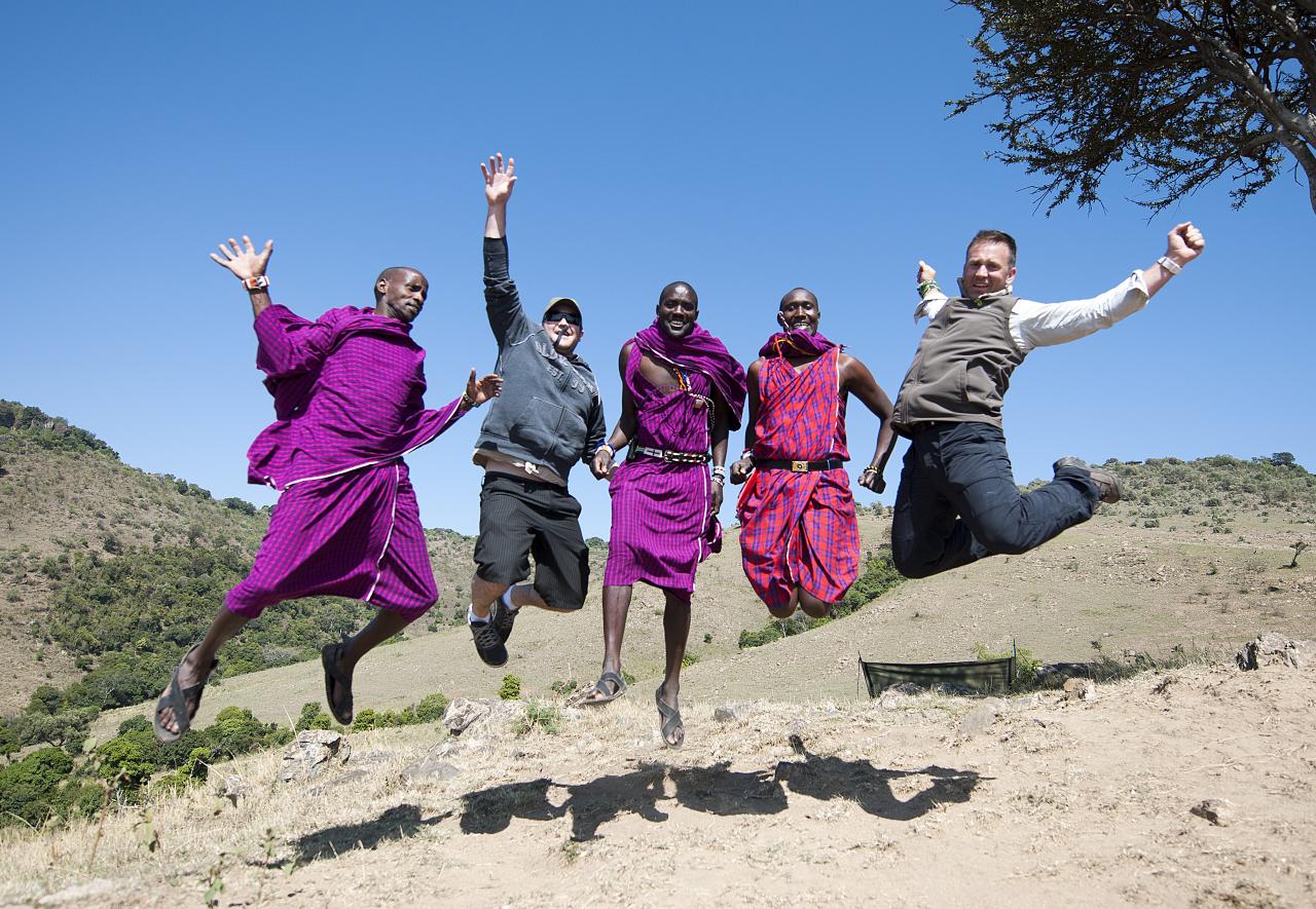
The people in Figure 6.2.1 illustrate some of the great phenotypic variation displayed in modern Homo sapiens. The lighter-skinned men in the photo are Euro-American tourists in Kenya (East Africa). The darker-skinned men are native Kenyans who belong to a tribal group named the Maasai. These men come from populations on different continents on opposite sides of the globe. Their populations have unique histories, environments, and cultures. Besides differences in skin colour, the men have different hair and eye colours, facial features, and body builds. Based on such obvious physical differences, you might think that our species is characterized by a high degree of genetic variation. In fact, there is much less genetic variation in the human species than there is in many other mammalian species, including our closest relatives — the chimpanzees.
Overview of Human Genetic Variation
No two human individuals are genetically identical unless they are monozygotic (identical) twins. Between any two people, DNA differs, on average, at about one in one thousand nucleotide base pairs. We each have a total of about three billion base pairs, so any two people differ by an average of about three million base pairs. That may sound like a lot, but it's only 0.1% of our total genetic makeup. This means that two people chosen at random are likely to be 99.9 per cent identical genetically, no matter where in the world they come from.
At an individual level, most human genetic variation is not very important biologically, because it has no apparent adaptive significance. It neither enhances nor detracts from individual fitness. Only a small percentage of DNA variations actually occur in coding regions of DNA — which are sequences that are translated into proteins — or in regulatory regions, which are sequences that control gene expression. Differences that occur in other regions of DNA have no impact on phenotype. Even variations in coding regions of DNA may or may not affect phenotype. Some DNA variations may alter the amino acid sequence of a protein, but not affect how the protein functions. Other DNA variations do not even change the amino acid sequence of the encoded protein.
At a population level, genetic variation is crucial if evolution is to occur. Genetically-based differences in fitness among individuals are the key to evolution by natural selection. Without genetic variation within populations, there can be no differential fitness by genotype, and natural selection cannot occur.
Patterns of Human Genetic Variation
Data comparing DNA sequences from around the world show that only about ten per cent of our total genetic variation occurs between people from different continents, like the American tourists and African Maasai pictured in Figure 6.1.1. The other 90 per cent of genetic variation occurs between people within continental populations, such as between North Americans or between Africans. Within any human population, many genes have two or more normal alleles that contribute to genetic differences among individuals. The case in which a gene has two or more alleles in a population at frequencies greater than one per cent is called a polymorphism. A single nucleotide polymorphism (SNP) involves variation in just one nucleotide in a DNA sequence. SNPs account for most of our genetic differences. Other types of variations (such as deletions and insertions of nucleotides in DNA sequences) account for a much smaller proportion of our overall genetic variation.
Different populations may have different allele frequencies for polymorphic genes. However, the distribution of allele frequencies in different populations around the world tends not to be discrete or distinct. Instead, the pattern is more often one of gradual geographic variations, or clines, in allele frequencies. You can see an example of a clinal distribution of allele frequencies in the map (Figure 6.2.2) below. Clinal distributions like this may be a reflection of natural selection pressures varying continuously over geographic space, or they may reflect a combination of genetic drift and gene flow of neutral alleles.
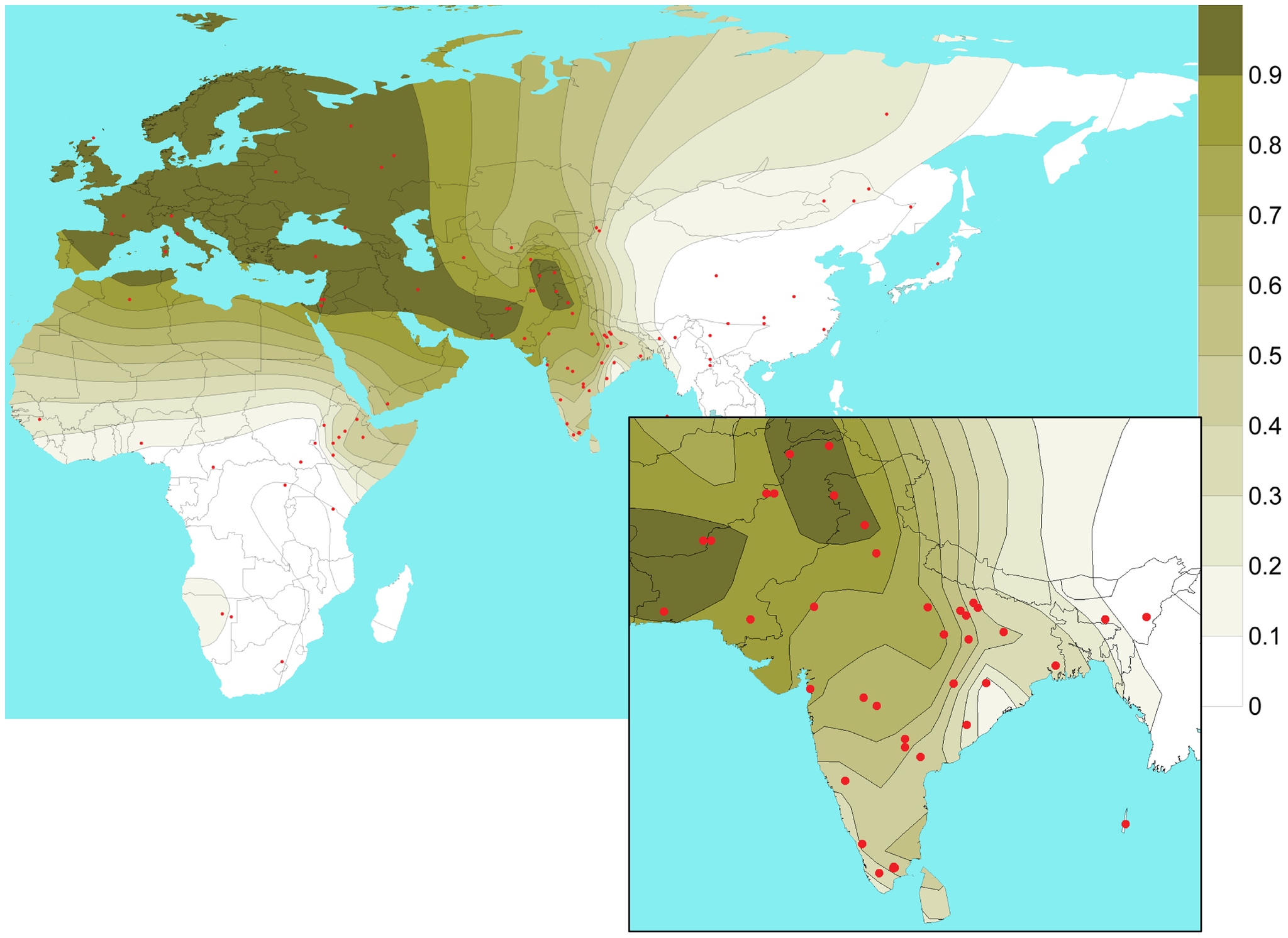
Although most genetic variation occurs within rather than between populations, certain alleles do seem to cluster in particular geographic areas. One example happens with the Duffy gene. Variations in this gene are the basis of the Duffy blood group, which is determined by the presence or absence of a red blood cell antigen, similar to the more familiar ABO blood group antigens. The genotype for having no antigen for the Duffy blood group is far higher in African populations and in people who have African ancestry than it is in non-African people, as indicated in the following table. Genes (such as the Duffy gene) may be useful as genetic markers to establish the ancestral populations of individuals.
Table 6.2.1
Population Frequencies for No Antigen in the Duffy Blood Group
| Population | Per cent of Population Lacking Duffy Antigen |
| African | 88-100 |
| African American | 68 |
| non-African American | <1 |
The reason for the different population frequencies for the Duffy antigen appears to be natural selection. People who lack the Duffy antigen are relatively resistant to malaria, which is one of the oldest and most devastating human diseases. Malaria has been a persistent and widespread disease in sub-Saharan Africa for tens of thousands of years. DNA analyses suggest that the allele associated with lack of the Duffy antigen evolved at least twice in Africa and was strongly selected for, causing it to increase in frequency. The Duffy gene is just one of many genes that have polymorphic alleles, because one of the alleles protects against malaria. In fact, a greater number of known genetic polymorphisms may be attributed to selection because of malaria than any other single selective agent.
Factors Influencing the Level of Human Genetic Variation
The age and size of a population increases the genetic variation within that population. You would expect an older, larger population to have more genetic variation. The older a population is, the longer it has been accumulating mutations. The larger a population is, the more people there are in which mutations can occur. Anatomically modern humans evolved less than a quarter million years ago, which is a relatively short period of time for mutations to accumulate. Our population was also quite small at some point in the past, perhaps consisting of no more than ten thousand adults, which reduced genetic variation even more. These factors explain why humans are relatively homogeneous genetically as a species.
What We Can Learn From Knowledge of Human Genetic Variation
Knowledge of genetic variation can help us understand our similarities and differences, our origins, and our evolutionary past. It can also help us understand human diseases and — hopefully — find new ways to treat them.
Human Origins
The data on human genetic variation generally supports the out-of-Africa hypothesis for human origins. According to this hypothesis, the common ancestor of all modern humans evolved in Africa around 200 thousand years ago. Then, starting no later than about 60 thousand years ago, part of the African population left Africa and migrated to Europe and Asia. As the migrants spread throughout the Old World, they replaced (and/or absorbed) the populations of archaic humans they encountered.
Most studies of human genetic variation find there is greater genetic diversity in African than non-African populations. This is consistent with the older age of the African population proposed by the out-of-Africa hypothesis. In addition, most of the genetic variation in non-African populations is a subset of the variation in African populations. This is consistent with the idea that part of the African population left Africa much later and migrated to other places in the Old World.
Recent comparisons of modern human and archaic human (including Neanderthal and Denisovan) DNA show that interbreeding occurred between their populations, but to differing degrees. The result of new DNA sequences entering a population’s gene pool through interbreeding is called admixture. There is greater admixture with archaic humans in modern European, Asian, and Oceanic populations than in modern African populations. Populations with the greatest admixture are those in Melanesia. About eight per cent of their DNA came from archaic Denisovans in East Asia.
Human Population History
Patterns of human genetic variation can be used to reconstruct population history. That history is literally recorded in our DNA. Any major population event (such as a significant reduction in population size or a high rate of migration) leaves a mark on a population’s genetic variation.
- Going through a dramatic size reduction decreases intra-population genetic variation (variation occurring within a population). As a case in point, DNA analyses suggest that there may have been drastic size reductions in the human populations that colonized the New World between 15 thousand and 20 thousand years ago. There were also size reductions in the human populations that first left Africa at least 60 thousand years ago, which helps explain the lower genetic diversity of modern non-African populations.
- A high rate of migration between populations may lead to gene flow, and this changes genetic variation in two ways. Gene flow decreases inter-population genetic variation (variation occurring between populations), while it increases intra-population variation. Gene flow — primarily between nearby populations — may contribute to the formation of clines in allele frequencies, as on the map in Figure 6.2.2.
Human Genetic Variation and Disease
An important benefit of studying human genetic variation is that we can learn more about the genetic basis of human diseases. The more we understand the causes of diseases, the more likely it is that we will be able to find effective treatments and cures for them.
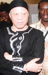
Some disorders are caused by mutations in a single gene. Most of these disorders are generally rare, but some of them occur at significantly higher frequencies in certain populations. For example, Ellis-van Creveld syndrome has an unusually high frequency in Pennsylvania Amish populations, and Tay-Sachs disease has a relatively high frequency in Ashkenazi Jewish populations. Albinism is another single-gene disorder that has a variable frequency. In North America and Europe, rates of albinism are approximately 1:18,000. In Africa, in contrast, the rates range from 1:5,000 to 1:15,000. Some African populations have estimated albinism rates as high as 1:1000. The photo below (Figure 6.2.3) shows an African albino man from Mali, where there is a relatively high rate of albinism. High population-specific frequencies of single-gene disorders like these may be attributable to a variety of factors, such as small founding populations and a relative lack of gene flow.
It is likely that the majority of human diseases are caused by a complex mix of multiple genes (polygenic) and environmental factors (multifactorial). Examples of polygenic, multifactorial diseases are type II diabetes and heart disease. We do not typically think of these diseases as genetic diseases, because our genes do not predetermine whether we develop them. Our genes, however, do influence our chances of developing the diseases under certain environmental conditions. Even our chances of developing some infectious diseases are influenced by our genes. For example, a variant allele for a gene called CCR5 seems to confer resistance to infection with HIV, the virus that causes AIDS.
6.2 Summary
- No two human individuals are genetically identical (except for monozygotic twins), but the human species as a whole exhibits relatively little genetic diversity, relative to other mammalian species. Genetically, two people chosen at random are likely to be 99.9 per cent identical.
- Of the total genetic variation in humans, about 90 per cent occurs between people within continental populations. Only about 10 per cent occurs between people from different continents. Older, larger populations tend to have greater genetic variation, because there is more time and there are more people in which to accumulate mutations.
- Single nucleotide polymorphisms account for most human genetic differences. Allele frequencies for polymorphic genes generally have a clinal (rather than discrete) distribution. A minority of alleles seem to cluster in particular geographic areas, such as the allele for no antigen in the Duffy blood group. Such alleles may be useful as genetic markers to establish the ancestry of individuals.
- Knowledge of genetic variation can help us understand our similarities and differences. It can also help us reconstruct our evolutionary origins and history as a species. For example, the distribution of modern human genetic variation is consistent with the out-of-Africa hypothesis for the origin of modern humans.
- An important benefit of studying human genetic variation is learning more about the genetic basis of human diseases. This, in turn, should help us find more effective treatments and cures.
6.2 Review Questions
- Compare and contrast the significance of genetic variation at the individual and population levels.
- Describe genetic variation within and between human populations on different continents.
- Explain why allele frequencies for the Duffy gene may be used as a genetic marker for African ancestry.
- Identify factors that increase the level of genetic variation within populations.
-
- Discuss genetic evidence that supports the out-of-Africa hypothesis of modern human origins.
- What evidence suggests that modern humans interbred with archaic human populations after modern humans left Africa?
- How do population size reductions and gene flow impact the genetic variation of populations?
- Describe the role of genetic variation in human disease.
- Explain the reasons why variation in a DNA sequence can have no effect on the fitness of an individual.
- Explain why migration between populations decreases inter-population genetic variation. How does this relate to the development of clines in allele frequency?
- The amount of mixing of modern human DNA and archaic human DNA is an example of _________ .
6.2 Explore More
https://www.youtube.com/watch?v=RGtaq3PiIoU
The Journey of Your Past | National Geographic, National Geographic, 2013.
https://www.youtube.com/watch?v=kU0ei9ApmsY
Svante Pääbo: DNA clues to our inner neanderthal, TED, 2011.
https://www.youtube.com/watch?time_continue=2&v=cHRM2S_fBOk&feature=emb_logo
Why Are Some People Albino?, Seeker, 2015.
Attributions
Figure 6.2.1
Maasai_men_and_tourists_jumping by Christopher Michel on Wikimedia Commons is used under a CC BY 2.0 (https://creativecommons.org/licenses/by/2.0/deed.en) (license.
Figure 6.2.2
Geospatial_distribution_of_SNP_rs1426654-A_allele by Basu Mallick C, Iliescu FM, Möls M, Hill S, Tamang R, Chaubey G, et al. on Wikimedia Commons is used under a CC BY 2.5 (https://creativecommons.org/licenses/by/2.5/deed.en) license.
Figure 6.2.3
Mali_Salif_Keita2_400 [cropped] by unknown from The Department of State, Washington, DC. on Wikimedia Commons is in the public domain (https://en.wikipedia.org/wiki/Public_domain).
References
Basu Mallick C., Iliescu, F.M., Möls, M., Hill, S., Tamang, R., Chaubey, G., et al. (2013). The light skin allele of SLC24A5 in South Asians and Europeans shares identity by descent: Figure 2. Isofrequency map illustrating the geospatial distribution of SNP rs1426654-A allele across the world. PLoS Genetics, 9(11): e1003912. doi:10.1371/journal.pgen.1003912 http://journals.plos.org/plosgenetics/article?id=10.1371/journal.pgen.1003912
HealthLinkBC. (2019, November 5). Health topics: Malaria [online article]. BC Government (gov.bc.ca). https://www.healthlinkbc.ca/health-topics/hw119119
Mayo Clinic Staff. (n.d.). Albinism [online article]. MayoClinic.org. https://www.mayoclinic.org/diseases-conditions/albinism/symptoms-causes/syc-20369184
Mayo Clinic Staff. (n.d.). Heart disease [online article]. MayoClinic.org. https://www.mayoclinic.org/diseases-conditions/heart-disease/symptoms-causes/syc-20353118
Mayo Clinic Staff. (n.d.). HIV/AIDS [online article]. MayoClinic.org. https://www.mayoclinic.org/diseases-conditions/hiv-aids/symptoms-causes/syc-20373524
Mayo Clinic Staff. (n.d.). Type 2 diabetes [online article]. MayoClinic.org. https://www.mayoclinic.org/diseases-conditions/type-2-diabetes/symptoms-causes/syc-20351193
National Geographic. (2013, March 13). The journey of your past | National Geographic. YouTube. https://www.youtube.com/watch?v=RGtaq3PiIoU&feature=youtu.be
National Institutes of Health/ National Library of Medicine. (n.d.). Genes: CCR5 gene - C-C motif chemokine receptor 5 [online article]. US Government. https://ghr.nlm.nih.gov/gene/CCR5
National Organization for Rare Disorders (NORD). (2012). Ellis Van Creveld syndrome [online article]. RareDiseases.org. https://rarediseases.org/rare-diseases/ellis-van-creveld-syndrome/
National Organization for Rare Disorders (NORD). (2017). Tay Sachs disease [online article]. RareDiseases.org. https://rarediseases.org/rare-diseases/tay-sachs-disease/
Seeker. (2015, July 25). Why are some people albino?. YouTube. https://www.youtube.com/watch?v=cHRM2S_fBOk&feature=youtu.be
TED. (2011, August 30). Svante Pääbo: DNA clues to our inner neanderthal. YouTube. https://www.youtube.com/watch?v=kU0ei9ApmsY&feature=youtu.be
Wikipedia contributors. (2020, June 18). Melanesia. In Wikipedia. https://en.wikipedia.org/w/index.php?title=Melanesia&oldid=963224885
Wikipedia contributors. (2020, June 4). Old world. In Wikipedia. https://en.wikipedia.org/w/index.php?title=Old_World&oldid=960713597
The body system which acts as a chemical messenger system comprising feedback loops of the hormones released by internal glands of an organism directly into the circulatory system, regulating distant target organs. In humans, the major endocrine glands are the thyroid gland and the adrenal glands.
Created by: CK-12/Adapted by Christine Miller
Ribosome Review
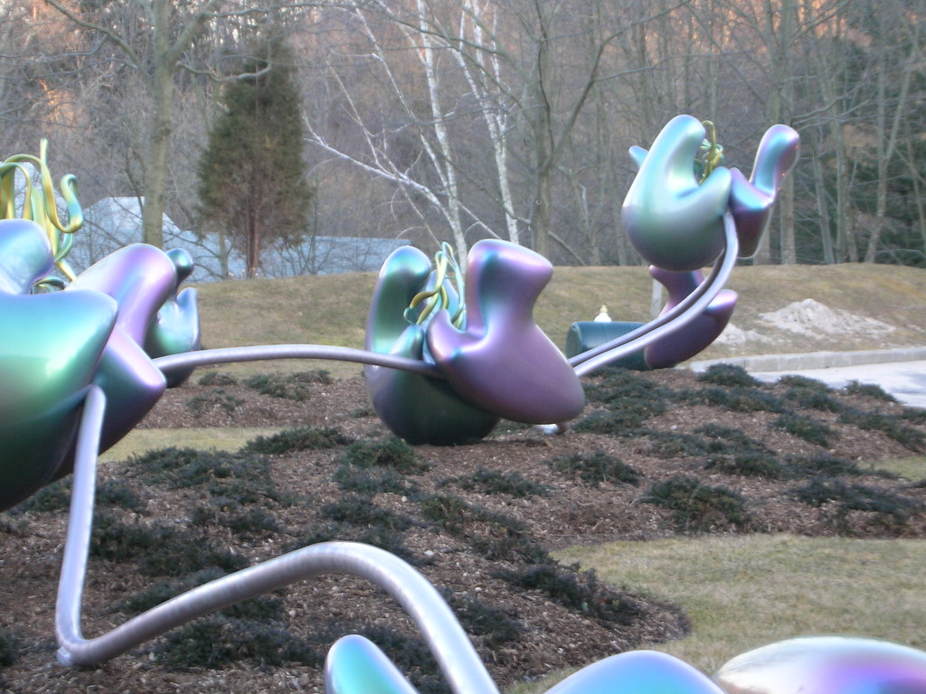
The 25-metre long sculpture shown in Figure 4.6.1 is a recognition of the beauty of one of the metabolic functions that takes place in the cells in your body. This artwork brings to life an important structure in living cells: the ribosome, the cell structure where proteins are synthesized. The slender silver strand is the messenger RNA(mRNA) bringing the code for a protein out into the cytoplasm. The purple and green structures are ribosomal subunits (which together form a single ribosome), which can "read" the code on the mRNA and direct the bonding of the correct sequence of amino acids to create a protein. All living cells — whether they are prokaryotic or eukaryotic — contain ribosomes, but only eukaryotic cells also contain a nucleus and several other types of organelles.
What Are Organelles?
An organelle is a structure within the cytoplasm of a eukaryotic cell that is enclosed within a membrane and performs a specific job. Organelles are involved in many vital cell functions. Organelles in animal cells include the nucleus, mitochondria, endoplasmic reticulum, Golgi apparatus, vesicles, and vacuoles. Ribosomes are not enclosed within a membrane, but they are still commonly referred to as organelles in eukaryotic cells.
The Nucleus
The nucleus is the largest organelle in a eukaryotic cell, and it's considered the cell’s control center. It contains most of the cell’s DNA(which makes up chromosomes), and it is encoded with the genetic instructions for making proteins. The function of the nucleus is to regulate gene expression, including controlling which proteins the cell makes. In addition to DNA, the nucleus contains a thick liquid called nucleoplasm, which is similar in composition to the cytosol found in the cytoplasm outside the nucleus. Most eukaryotic cells contain just a single nucleus, but some types of cells (such as red blood cells) contain no nucleus and a few other types of cells (such as muscle cells) contain multiple nuclei.
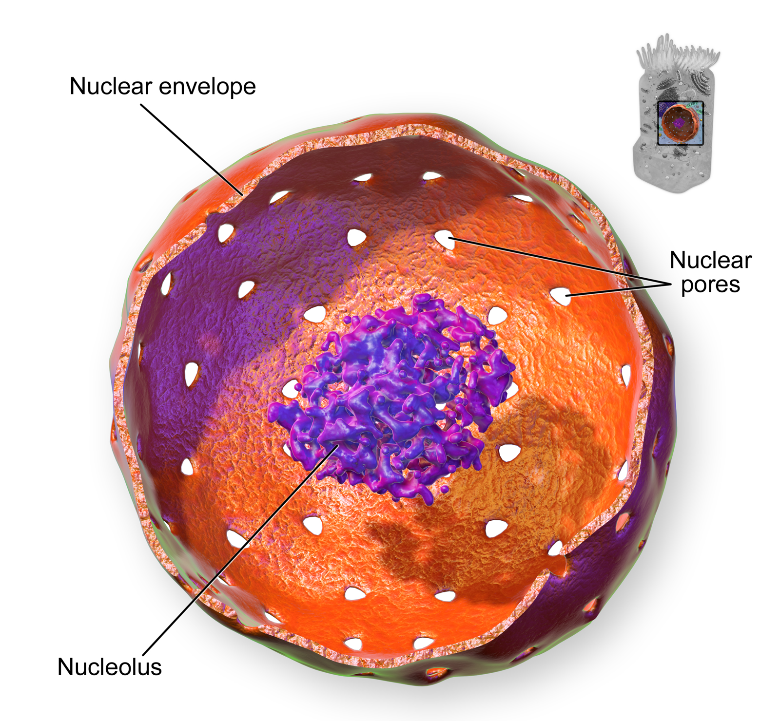
As you can see in the model pictured in Figure 4.6.2, the membrane enclosing the nucleus is called the nuclear envelope. This is actually a double membrane that encloses the entire organelle and isolates its contents from the cellular cytoplasm. Tiny holes called nuclear pores allow large molecules to pass through the nuclear envelope, with the help of special proteins. Large proteins and RNA molecules must be able to pass through the nuclear envelope so proteins can be synthesized in the cytoplasm and the genetic material can be maintained inside the nucleus. The nucleolus shown in the model below is mainly involved in the assembly of ribosomes. After being produced in the nucleolus, ribosomes are exported to the cytoplasm, where they are involved in the synthesis of proteins.
Mitochondria
The mitochondrion (plural, mitochondria) is an organelle that makes energy available to the cell. This is why mitochondria are sometimes referred to as the "power plants of the cell." They use energy from organic compounds (such as glucose) to make molecules of ATP (adenosine triphosphate), an energy-carrying molecule that is used almost universally inside cells for energy.
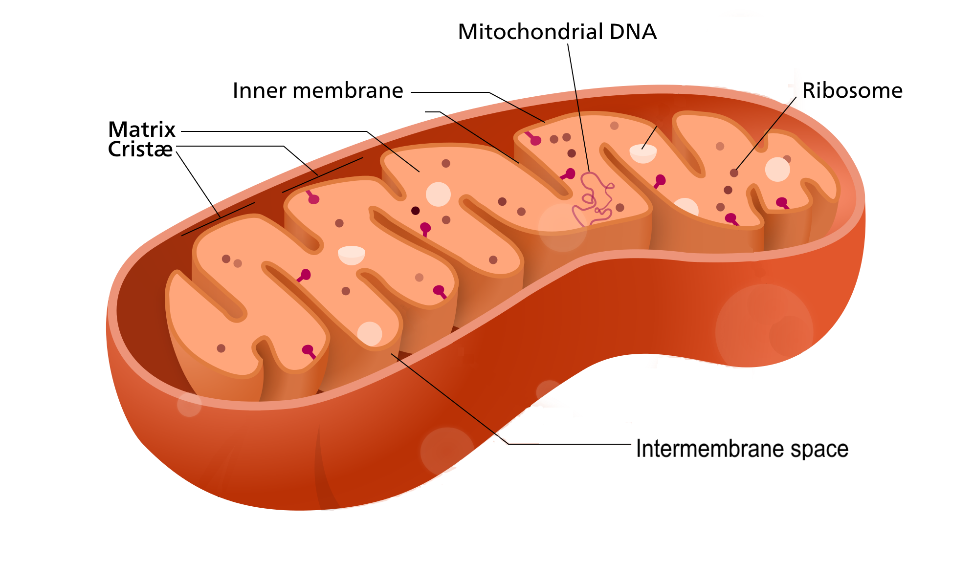
Mitochondria (as in the Figure 4.6.3 diagram) have a complex structure including an inner and out membrane. In addition, mitochondria have their own DNA, ribosomes, and a version of cytoplasm, called matrix. Does this sound similar to the requirements to be considered a cell? That's because they are!
Scientists think that mitochondria were once free-living organisms because they contain their own DNA. They theorize that ancient prokaryotes infected (or were engulfed by) larger prokaryotic cells, and the two organisms evolved a symbiotic relationship that benefited both of them. The larger cells provided the smaller prokaryotes with a place to live. In return, the larger cells got extra energy from the smaller prokaryotes. Eventually, the smaller prokaryotes became permanent guests of the larger cells, as organelles inside them. This theory is called endosymbiotic theory, and it is widely accepted by biologists today. (See the video in section 4.3 to learn all about endosymbiotic theory.)
Endoplasmic Reticulum
The endoplasmic reticulum (ER) is an organelle that helps make and transport proteins and lipids. There are two types of endoplasmic reticulum: rough endoplasmic reticulum (rER) and smooth endoplasmic reticulum (sER). Both types are shown in Figure 4.6.4.
- rER looks rough because it is studded with ribosomes. It provides a framework for the ribosomes, which make proteins. Bits of its membrane pinch off to form tiny sacs called vesicles, which carry proteins away from the ER.
- sER looks smooth because it does not have ribosomes. sER makes lipids, stores substances, and plays other roles.
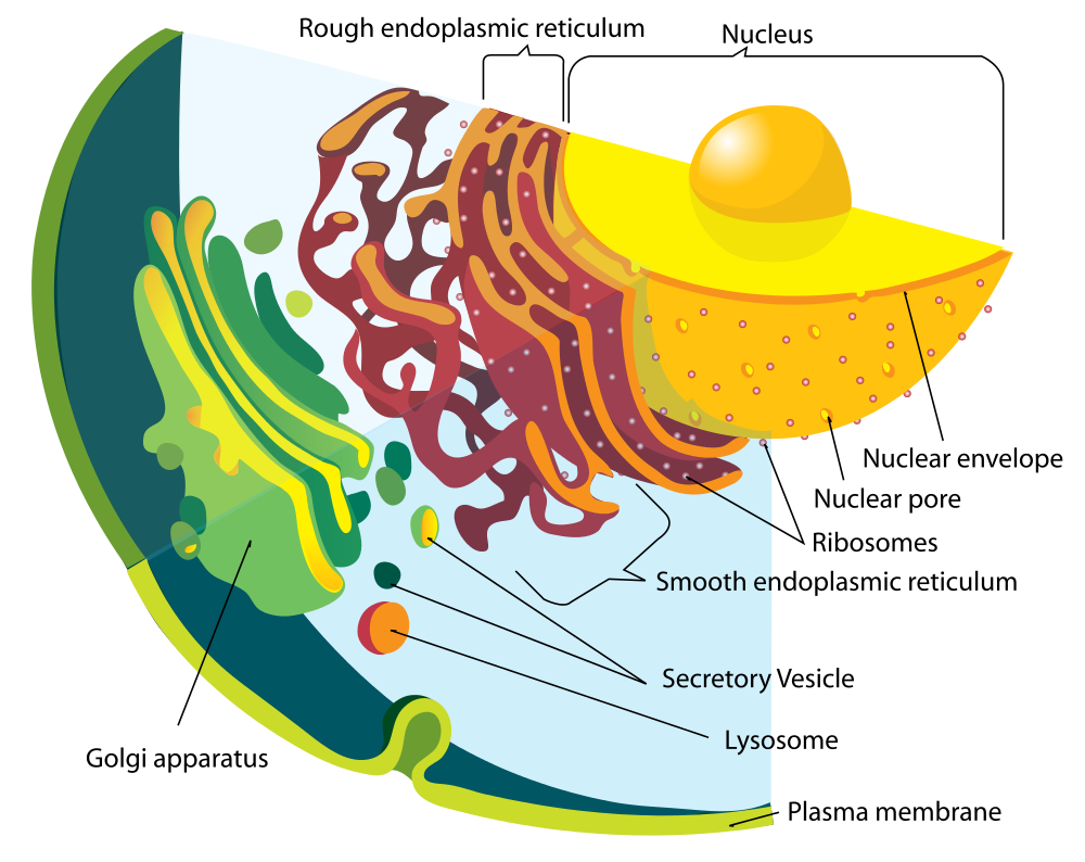
The Figure 4.6.4 drawing includes the nucleus, rER, sER, and Golgi apparatus. From the drawing, you can see how all these organelles work together to make and transport proteins.
Golgi Apparatus
The Golgi apparatus (shown in the Figure 4.6.4 diagram) is a large organelle that processes proteins and prepares them for use both inside and outside the cell. You can see the Golgi apparatus in the figure above. The Golgi apparatus is something like a post office. It receives items (proteins from the ER), then packages and labels them before sending them on to their destinations (to different parts of the cell or to the cell membrane for transport out of the cell). The Golgi apparatus is also involved in the transport of lipids around the cell.
Vesicles and Vacuoles
Both vesicles and vacuoles are sac-like organelles made of phospholipid bilayer that store and transport materials in the cell. Vesicles are much smaller than vacuoles and have a variety of functions. The vesicles that pinch off from the membranes of the ER and Golgi apparatus store and transport protein and lipid molecules. You can see an example of this type of transport vesicle in the Figure 4.6.4. Some vesicles are used as chambers for biochemical reactions.
There are some vesicles which are specialized to carry out specific functions. Lysosomes, which use enzymes to break down foreign matter and dead cells, have a double membrane to make sure their contents don't leak into the rest of the cell. Peroxisomes are another type of specialized vesicle with the main function of breaking down fatty acids and some toxins.
Centrioles
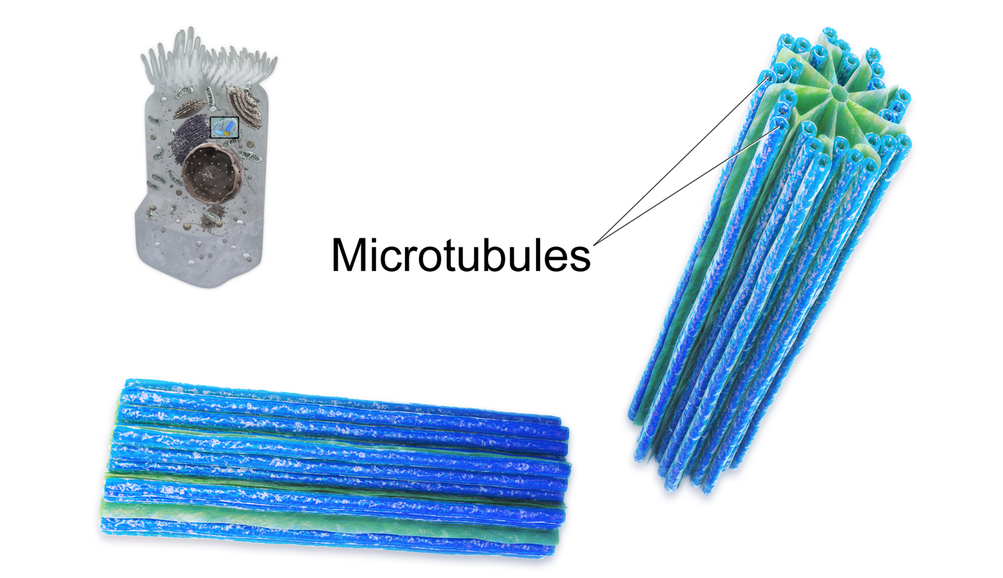
Centrioles are organelles involved in cell division. The function of centrioles is to help organize the chromosomes before cell division occurs so that each daughter cell has the correct number of chromosomes after the cell divides. Centrioles are found only in animal cells, and are located near the nucleus. Each centriole is made mainly of a protein named tubulin. The centriole is cylindrical in shape and consists of many microtubules, as shown in the model pictured in Figure 4.6.5.
Ribosomes
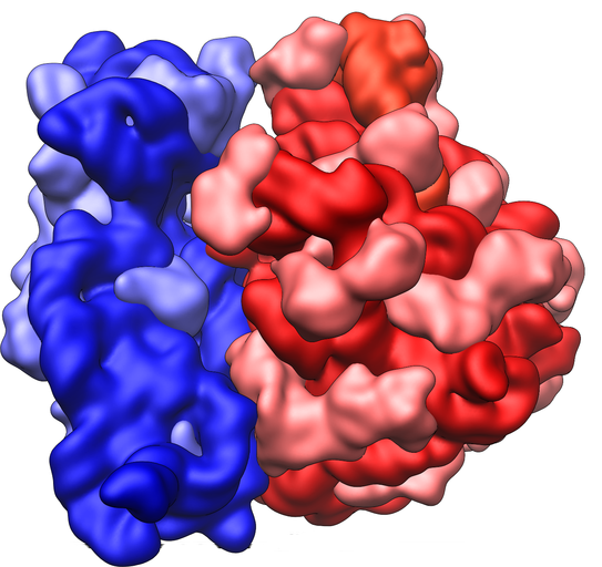
Ribosomes are small structures where proteins are made. Although they are not enclosed within a membrane, they are frequently considered organelles. Each ribosome is formed of two subunits, like the ones pictured at the beginning of this section (Figure 4.6.1) and in Figure 4.6.6. Both subunits consist of proteins and RNA. mRNA from the nucleus carries the genetic code, copied from DNA, which remains in the nucleus. At the ribosome, the genetic code in mRNA is used to assemble and join together amino acids to make proteins. Ribosomes can be found alone or in groups within the cytoplasm, as well as on the rER.
4.6 Summary
- An organelle is a structure within the cytoplasm of a eukaryotic cell that is enclosed within a membrane and performs a specific job. Although ribosomes are not enclosed within a membrane, they are still commonly referred to as organelles in eukaryotic cells.
- The nucleus is the largest organelle in a eukaryotic cell, and it is considered to be the cell's control center. It controls gene expression, including controlling which proteins the cell makes.
- The mitochondrion (plural, mitochondria) is an organelle that makes energy available to the cells. It is like the power plant of the cell. According to the widely accepted endosymbiotic theory, mitochondria evolved from prokaryotic cells that were once free-living organisms that infected or were engulfed by larger prokaryotic cells.
- The endoplasmic reticulum (ER) is an organelle that helps make and transport proteins and lipids. Rough endoplasmic reticulum (rER) is studded with ribosomes. Smooth endoplasmic reticulum (sER) has no ribosomes.
- The Golgi apparatus is a large organelle that processes proteins and prepares them for use both inside and outside the cell. It is also involved in the transport of lipids around the cell.
- Both vesicles and vacuoles are sac-like organelles that may be used to store and transport materials in the cell or as chambers for biochemical reactions. Lysosomes and peroxisomes are special types of vesicles that break down foreign matter, dead cells, or poisons.
- Centrioles are organelles located near the nucleus that help organize the chromosomes before cell division so each daughter cell receives the correct number of chromosomes.
- Ribosomes are small structures where proteins are made. They are found in both prokaryotic and eukaryotic cells. They may be found alone or in groups within the cytoplasm or on the rER.
4.6 Review Questions
- What is an organelle?
- Describe the structure and function of the nucleus.
- Explain how the nucleus, ribosomes, rough endoplasmic reticulum, and Golgi apparatus work together to make and transport proteins.
- Why are mitochondria referred to as the "power plants of the cell"?
- What roles are played by vesicles and vacuoles?
- Why do all cells need ribosomes — even prokaryotic cells that lack a nucleus and other cell organelles?
- Explain endosymbiotic theory as it relates to mitochondria. What is one piece of evidence that supports this theory?
-
4.6 Explore More
https://www.youtube.com/watch?v=URUJD5NEXC8&t=121s
Biology: Cell Structure I Nucleus Medical Media, Nucleus Medical Media, 2015.
https://www.youtube.com/watch?v=Id2rZS59xSE&feature=youtu.be
David Bolinsky: Visualizing the wonder of a living cell, TED, 2007.
Attributes
Figure 4.6.1
Ribosomes at Work by Pedrik on Flickr is used under a CC BY-NC-SA 2.0 (https://creativecommons.org/licenses/by-nc-sa/2.0/) license.
Figure 4.6.2
Nucleus by BruceBlaus on Wikimedia Commons is used under a CC BY 3.0 (https://creativecommons.org/licenses/by/3.0) license.
Figure 4.6.3
Mitochondrion_structure.svg by Kelvinsong; modified by Sowlos on Wikimedia Commons is used and adapted by Christine Miller under a CC BY-SA 3.0 (https://creativecommons.org/licenses/by-sa/3.0) license.
Figure 4.6.4
Endomembrane_system_diagram_en.svg by Mariana Ruiz [LadyofHats] on Wikimedia Commons is released into the public domain (https://en.wikipedia.org/wiki/Public_domain).
Figure 4.6.5
Centrioles by BruceBlaus on Wikimedia Commons is used under a CC BY 3.0 (https://creativecommons.org/licenses/by/3.0) license.
Figure 4.6.6
Ribosome_shape by Vossman on Wikimedia Commons is used and adapted by Christine Miller under a CC BY-SA 3.0 (https://creativecommons.org/licenses/by-sa/3.0) license.
References
Blausen.com staff. (2014). Nucleus - Medical gallery of Blausen Medical 2014. WikiJournal of Medicine 1 (2). DOI:10.15347/wjm/2014.010. ISSN 2002-4436. https://en.wikiversity.org/wiki/WikiJournal_of_Medicine/Medical_gallery_of_Blausen_Medical_2014
Blausen.com staff (2014). Centrioles - Medical gallery of Blausen Medical 2014. WikiJournal of Medicine 1 (2). DOI:10.15347/wjm/2014.010. ISSN 2002-4436.https://en.wikiversity.org/wiki/WikiJournal_of_Medicine/Medical_gallery_of_Blausen_Medical_2014
Nucleus Medical Media. (2015, March 18). Biology: Cell structure I Nucleus Medical Media. YouTube. https://www.youtube.com/watch?v=URUJD5NEXC8&feature=youtu.be
TED. (2007, July 24). David Bolinsky: Visualizing the wonder of a living cell. YouTube. https://www.youtube.com/watch?v=Id2rZS59xSE&feature=youtu.be
Created by: CK-12/Adapted by Christine Miller
Figure 6.3.1 How would you classify these people?
Why Classify?
What do you see when you look at Figure 6.3.1? Did you sort individuals into categories based in gender, age, body type, facial features, skin colour or other characteristics? As humans, we seem to have a penchant for classifying and labeling people and things. It helps us establish a sense of order in the world around us. The 18th century taxonomist Carl Linnaeus, for example, classified virtually all known living things into different species, genera, families, and other taxonomic categories. His classifications were based on observable phenotypic characteristics, such as skin colour. Modern biological classifications of living things are usually based on phylogenetic relationships. Phylogenies reflect evolutionary history and group together living things that are related by descent from a common ancestor.
Starting with Linnaeus and continuing to the present, scientists and others have attempted to classify human variation. There are three basic approaches to classification: typological, populational, and clinal.
Typological Approach
The typological approach involves creating a typology, which is a system of discrete types, or categories. This approach was widely used by scientists up through the early 20th century. Racial classifications are typological classifications. They place people into a small number of discrete categories, or races, based on a few readily observable traits, such as skin colour, hair texture, facial features, and body build.
Racial Classifications and Racism
Racial classifications of humans probably go back as long as people distinguished “us” from “them.” An early “scientific” classification of humans into races is Linnaeus’ 1735 classification. He divided Homo sapiens into continental races, which he named europaeus, asiaticus, americanus, and afer. Linnaeus described these races in terms of observable physical traits. He also associated, inaccurately, each race with different personality qualities and behaviors. For example, he described Homo sapiens europaeus as active and adventurous and Homo sapiens afer as lazy and careless. In 1795, the German naturalist Johann Friedrich Blumenbach proposed five major races of Homo sapiens, which he named the caucasoid, mongoloid, negroid, American Indian, and Malayan races. Blumenbach thought that the caucasoid race was the original race, and that the other races arose in a process of “degeneration” from the caucasoids.
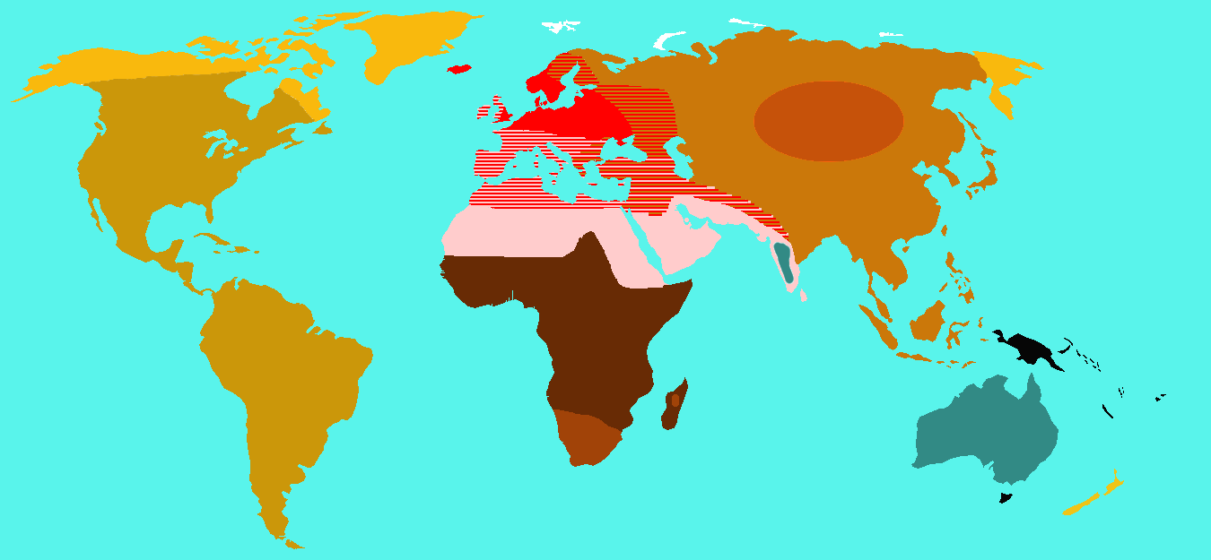
In 1870, the English biologist Thomas Huxley classified Homo sapiens into nine races which were distributed geographically. The map in Figure 6.3.2 shows how Huxley thought the races were distributed worldwide. Each colour represents one of Huxley’s proposed races. These categories included “Australoid,” “Xanthochroi,” “Melanochori,” “Negroes,” and “Mongoloids,” and they are not used today. It should be noted that Huxley did not hold such strong negative stereotypes about non-European (or non-caucasoid) races as did his intellectual forebears. Huxley, however, still attributed different behaviors to racial groups that had nothing to do with the colour of their skin or continent of origin.
By the early 20th century, so-called scientific racism was a popular ideology. This was the idea that race is a biological concept and that human behavior is partly determined by race. At around 1950, in a series of groundbreaking studies of skeletal anatomy, anthropologist Franz Boas showed that cranial (skull) shape and size were highly malleable, depending on environmental factors (such as health and nutrition). He contrasted this with racial anthropologists' claims that head shape is a stable racial trait. In this way, Boas demonstrated that this commonly used racial trait was determined by the environment, and not just genes. Boas also worked to demonstrate that differences in human behavior are not determined primarily by innate biological dispositions, but are largely the result of cultural differences acquired through social learning.
Unfortunately, racism still persists today — in society at large, if not in science. This is the association of racial traits (such as skin colour) with unrelated traits (such as intelligence), often leading to prejudice and discrimination against people based only on how they look. The concept of human race is real, not in a biological sense, but in a social sense. Racial stereotypes and racism are deeply ingrained in our history and culture, and they have real material effects on human lives.
Additional Problems with Typological Classification
Besides the problem of racism, there are other problems with typological approaches to the biological classification of Homo sapiens. One problem is that most human biological traits are not either present or absent, but instead vary on a continuum. This type of distribution cannot be adequately represented by discrete categories, such as races. The typological approach also results in groupings of people that may be similar in terms of some traits, but not others. How people are grouped together depends on which traits are chosen. In addition, the number of groups that are needed to classify people depends on the number of traits that are used. The greater the number of traits, the greater the number of racial categories there must be. If racial categories depend on the traits chosen to define them, it is clear that the racial classifications are arbitrary and do not reflect biological reality.
Another problem with typological classifications is that they lead to the mistaken belief that people within typological categories are more similar to each other than they are to people in other categories. There is actually more variation within than between typological groups. An estimated 90 per cent of human genetic variation occurs between people within races, and only 10 per cent occurs between races. Clearly, races are far from homogenous in terms of their genetic composition. In short, we are all more alike than we are different.
Populational Approach
By the middle of the 20th century, scientists started advocating a populational approach to classifying Homo sapiens. This approach is based on the idea that the breeding population is the only biologically meaningful group. The breeding population is the unit of evolution, and it includes people who have mated and produced offspring together for many generations. As a result, members of the same breeding population should share many genetic traits. You would also expect them to have many of the same phenotypic traits, because of their similar genetic makeup.
While the populational approach makes sense in theory, in reality, it can rarely be applied, because most human populations are not closed breeding populations. Some people have always selected mates from outside their local population (even mating with archaic humans such as Neanderthals). This tendency has increased dramatically in recent centuries with the advent of efficient means of traveling long distances. As a consequence, there are very few remaining distinct breeding populations within the human species.
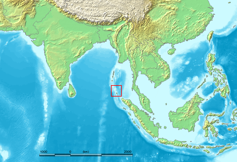
An example of one such population is the Sentinelese, a small population of hunter-gatherers who live alone on a small island in the Andaman Islands (see the map). The Sentinelese are thought to be direct descendants of the first modern humans to leave Africa, and they may have lived in the Andaman Islands for as long as 60 thousand years. The Sentinelese are also one of the most isolated human populations on Earth. The fact that their language is distinctly different from other Andaman Islands languages is evidence that they have had little contact with other people for thousands of years. Although closed breeding populations (such as the Sentinelese) may be useful for investigating questions about evolutionary processes, they are not useful for classifying most of humanity.
Clinal Approach
By the 1960s, scientists began to use a clinal approach to classify human variation. This approach maps variation in traits over geographic regions (such as continents) or even worldwide. Clinal models are a useful way of describing human variation that does not lead to discrete races or other categories of people.
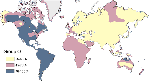
In Figure 6.3.4 you can see a worldwide clinal map for type O blood in the human ABO blood group system. The frequency of this trait is shown for the indigenous populations of various regions. It is lowest throughout Asia and highest in Native American populations in both North and South America. This geographic distribution results from the complex interaction of a variety of factors, including natural selection, genetic drift, and gene flow. You can read more about geographic variation in blood types in the concept Variation in Blood Types.
Clinal maps for many genetic traits show variation that changes gradually from one geographic area to another, which may happen because of the nature of gene flow. Gene flow occurs when mating takes place between people in different populations. The likelihood of mating with others depends on their distance from us. You may not marry the boy or girl next door, but your mate is more likely to be someone in the same state or country than someone on another continent.
Natural selection has a major impact on the clinal distribution of some traits, because variation in the traits tracks variation in selective pressures. For example, the environmental stressor of malaria varies throughout Africa with climate, as you can see in the left-hand map below (Figure 6.3.5). The sickle cell trait that protects from malaria has a similar distribution, as shown in the right-hand map.
Figure 6.3.5
6.3 Summary
- Humans seem to have a need to classify and label people based on their similarities and differences. Three approaches to classifying human variation include typological, populational, and clinal approaches.
- The typological approach involves creating a typology, which is a system of discrete categories, or races. This approach was widely used by scientists until the early 20th century. Racial categories are based on observable phenotypic traits (such as skin colour), but other traits and behaviors are often assumed to apply to racial groups, as well. The use of racial classifications often leads to racism.
- By the mid-20th century, scientists started advocating a population approach. This assumes that the breeding population, which is the unit of evolution, is the only biologically meaningful group. While this approach makes sense in theory, in reality, it can rarely be applied to actual human populations. With few exceptions, most human populations are not closed breeding populations.
- By the 1960s, scientists began to use a clinal approach to classify human variation. This approach maps variation in the frequency of traits or alleles over geographic regions or worldwide. Clinal maps for many genetic traits show variation that changes gradually from one geographic area to another. This type of distribution may result from gene flow and/or natural selection.
6.3 Review Questions
- Name the 18th century taxonomist that classified virtually all known living things.
- Describe the typological approach to classifying human variation.
- Discuss why typological classifications of Homo sapiens are associated with racism.
- Why is the breeding population considered to be the most meaningful biological group?
- Explain why it is generally unrealistic to apply a populational approach to classifying the human species.
- What does a clinal map show?
- Explain how gene flow and natural selection can result in a gradual change in the frequency of a trait over geographic space.
- Most human traits vary on a continuum. Explain why this presents a problem for the typological classification approach.
-
6.3 Explore More
https://www.youtube.com/watch?v=ntimKsWDUpA&feature=emb_logo
The Biology of Race in the Absence of Biological Races,
Centre for Genetic Medicine, 2015.
https://www.youtube.com/watch?v=QOSPNVunyFQ
Nina Jablonski breaks the illusion of skin color, TED, 2009.
https://www.youtube.com/watch?v=_r4c2NT4naQ
The science of skin color - Angela Koine Flynn, TED-Ed, 2016.
Attributions
Figure 6.3.1
- Three women sitting by flowers and laughing by Priscilla Du Preez on Unsplash is used under the Unsplash License (https://unsplash.com/license).
- Two women sitting on sofa by AllGo on Unsplash is used under the Unsplash License (https://unsplash.com/license).
- Young people in conversation by Alexis Brown on Unsplash is used under the Unsplash License (https://unsplash.com/license).
- Men talking in the cold by Anna Vander Stel on Unsplash is used under the Unsplash License (https://unsplash.com/license).
- Laughing by the tracks by Priscilla Du Preez on Unsplash is used under the Unsplash License (https://unsplash.com/license).
Figure 6.3.2
Huxley_races by Wobble on Wikimedia Commons is released into the public domain (https://en.wikipedia.org/wiki/Public_domain).
Figure 6.3.3
Nicobar_Islands is edited by M.Minderhoud on Wikimedia Commons, and was released into the public domain by its original author, www.demis.nl. (See also approval email on de.wp and its clarification.)
Figure 6.3.4
Map_of_Group_O/ (Percent of Native population that has the O blood type) by Ephert on Wikimedia Commons is used under a CC BY 3.0 (https://creativecommons.org/licenses/by/3.0/deed.en) license. (Original Spanish edition by Maulucioni)
Figure 6.3.5
- Malaria distribution by Muntuwandi at English Wikipedia on Wikimedia Commons is used under a CC BY-SA 3.0 (https://creativecommons.org/licenses/by-sa/3.0/deed.en) license.
- Sickle cell distribution by Muntuwandi at English Wikipedia on Wikimedia Commons is used under a CC BY-SA 3.0 (https://creativecommons.org/licenses/by-sa/3.0/deed.en) license.
References
Centre for Genetic Medicine. (2015, July 14). The biology of race in the absence of biological races. YouTube. https://www.youtube.com/watch?v=ntimKsWDUpA&feature=youtu.be
TED. (2009, August 7). Nina Jablonski breaks the illusion of skin color. YouTube. https://www.youtube.com/watch?v=QOSPNVunyFQ&feature=youtu.be
TED-Ed. (2016, February 16). The science of skin color - Angela Koine Flynn. YouTube. https://www.youtube.com/watch?v=_r4c2NT4naQ&feature=youtu.be
Wikipedia contributors. (2020, June 27). Carl Linnaeus. In Wikipedia. https://en.wikipedia.org/w/index.php?title=Carl_Linnaeus&oldid=964690855
Wikipedia contributors. (2020, May 18). Franz Boas. In Wikipedia. https://en.wikipedia.org/w/index.php?title=Franz_Boas&oldid=957282443
Wikipedia contributors. (2020, July 5). Johann Friedrich Blumenbach. In Wikipedia. https://en.wikipedia.org/w/index.php?title=Johann_Friedrich_Blumenbach&oldid=966196943
Wikipedia contributors. (2020, July 11). Sentinelese. In Wikipedia. https://en.wikipedia.org/w/index.php?title=Sentinelese&oldid=967121254
Wikipedia contributors. (2020, July 14). Thomas Henry Huxley. In Wikipedia. https://en.wikipedia.org/w/index.php?title=Thomas_Henry_Huxley&oldid=967701553
A hormone is a signaling molecule produced by glands in multicellular organisms that target distant organs to regulate physiology and behavior.
Structures containing neuronal cell bodies in the peripheral nervous system.
Created by: CK-12/Adapted by Christine Miller
Oh, the Agony!
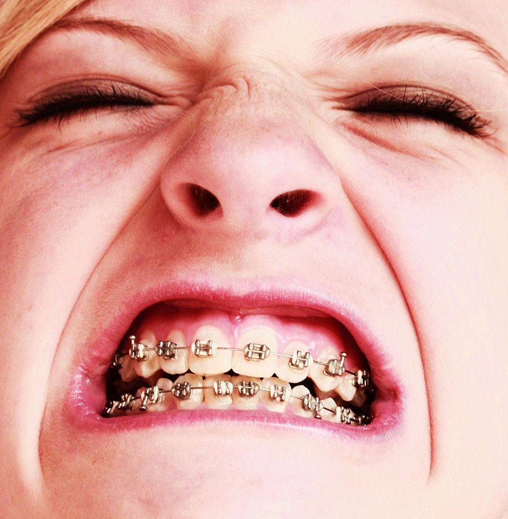
Wearing braces can be very uncomfortable, but it is usually worth it. Braces and other orthodontic treatments can re-align the teeth and jaws to improve bite and appearance. Braces can change the position of the teeth and the shape of the jaws because the human body is malleable. Many phenotypic traits — even those that have a strong genetic basis — can be molded by the environment. Changing the phenotype in response to the environment is just one of several ways we respond to environmental stress.
Types of Responses to Environmental Stress
There are four different types of responses that humans may make to cope with environmental stress:
- Adaptation
- Developmental adjustment
- Acclimatization
- Cultural responses
The first three types of responses are biological in nature, and the fourth type is cultural. Only adaptation involves genetic change and occurs at the level of the population or species. The other three responses do not require genetic change, and they occur at the individual level.
Adaptation
An adaptation is a genetically-based trait that has evolved because it helps living things survive and reproduce in a given environment. Adaptations generally evolve in a population over many generations in response to stresses that last for a long period of time. Adaptations come about through natural selection. Those individuals who inherit a trait that confers an advantage in coping with an environmental stress are likely to live longer and reproduce more. As a result, more of their genes pass on to the next generation. Over many generations, the genes and the trait they control become more frequent in the population.
A Classic Example: Hemoglobin S and Malaria
Probably the most frequently-cited example of a genetic adaptation to an environmental stress is sickle cell trait. As you read in the previous section, people with sickle cell trait have one abnormal allele (S) and one normal allele (A) for hemoglobin, the red blood cell protein that carries oxygen in the blood. Sickle cell trait is an adaptation to the environmental stress of malaria, because people with the trait have resistance to this parasitic disease. In areas where malaria is endemic (present year-round), the sickle cell trait and its allele have evolved to relatively high frequencies. It is a classic example of natural selection favoring heterozygotes for a gene with two alleles. This type of selection keeps both alleles at relatively high frequencies in a population.
To Taste or Not to Taste
Another example of an adaptation in humans is the ability to taste bitter compounds. Plants produce a variety of toxic compounds in order to protect themselves from being eaten, and these toxic compounds often have a bitter taste. The ability to taste bitter compounds is thought to have evolved as an adaptation, because it prevented people from eating poisonous plants. Humans have many different genes that code for bitter taste receptors, allowing us to taste a wide variety of bitter compounds.
A harmless bitter compound called phenylthiocarbamide (PTC) is not found naturally in plants, but it is similar to toxic bitter compounds that are found in plants. Humans' ability to taste this harmless substance has been tested in many different populations. In virtually every population studied, there are some people who can taste PTC (called tasters), and some people who cannot taste PTC, (called nontasters). The ratio of tasters to non-tasters varies among populations, but on average, 75 per cent of people can taste PTC and 25 per cent cannot.
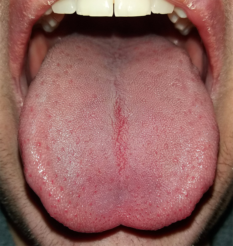
Like many scientific discoveries, human variation in PTC-taster status was discovered by chance. Around 1930, a chemist named Arthur Fox was working with powdered PTC in his lab. Some of the powder accidentally blew into the air. Another lab worker noticed that the powdered PTC tasted bitter, but Fox couldn't detect any taste at all. Fox wondered how to explain this difference in PTC-tasting ability. Geneticists soon determined that PTC-taster status is controlled by a single gene with two common alleles, usually represented by the letters T and t. The T allele encodes a chemical receptor protein (found in taste buds on the tongue, as illustrated in Figure 6.4.2) that can strongly bind to PTC. The other allele, t, encodes a version of the receptor protein that cannot bind as strongly to PTC. The particular combination of these two alleles that a person inherits determines whether the person finds PTC to taste very bitter (TT), somewhat bitter (Tt), or not bitter at all (tt).
If the ability to taste bitter compounds is advantageous, why does every human population studied contain a significant percentage of people who are nontasters? Why has the nontasting allele been preserved in human populations at all? Some scientists hypothesize that the nontaster allele actually confers the ability to taste some other, yet-to-be identified, bitter compound in plants. People who inherit both alleles would presumably be able to taste a wider range of bitter compounds, so they would have the greatest ability to avoid plant toxins. In other words, the heterozygote genotype for the taster gene would be the most fit and favored by natural selection.
Most people no longer have to worry whether the plants they eat contain toxins. The produce you grow in your garden or buy at the supermarket consists of known varieties that are safe to eat. However, natural selection may still be at work in human populations for the PTC-taster gene, because PTC tasters may be more sensitive than nontasters to bitter compounds in tobacco and vegetables in the cabbage family (that is, cruciferous vegetables, such as the broccoli, cauliflower, and cabbage pictured in Figure 6.4.3).
- People who find PTC to taste very bitter are less likely to smoke tobacco, presumably because tobacco smoke has a stronger bitter taste to these individuals. In this case, selection would favor taster genotypes, because tasters would be more likely to avoid smoking and its serious health risks.
- Strong tasters find cruciferous vegetables to taste bitter. As a result, they may avoid eating these vegetables (and perhaps other foods, as well), presumably resulting in a diet that is less varied and nutritious. In this scenario, natural selection might work against taster genotypes.
Figure 6.4.3 Cruciferous vegetables.
Developmental Adjustment
It takes a relatively long time for genetic change in response to environmental stress to produce a population with adaptations. Fortunately, we can adjust to some environmental stresses more quickly by changing in nongenetic ways. One type of nongenetic response to stress is developmental adjustment. This refers to phenotypic change that occurs during development in infancy or childhood, and that may persist into adulthood. This type of change may be irreversible by adulthood.
Phenotypic Plasticity
Developmental adjustment is possible because humans have a high degree of phenotypic plasticity, which is the ability to alter the phenotype in response to changes in the environment. Phenotypic plasticity allows us to respond to changes that occur within our lifetime, and it is particularly important for species (like our own) that have a long generation time. With long generations, evolution of genetic adaptations may occur too slowly to keep up with changing environmental stresses.
Developmental Adjustment and Cultural Practices
Developmental adjustment may be the result of naturally occurring environmental stresses or cultural practices, including medical or dental treatments. Like our example at the beginning of this section, using braces to change the shape of the jaw and the position of the teeth is an example of a dental practice that brings about a developmental adjustment. Another example of developmental adjustment is the use of a back brace to treat scoliosis (see images in Figure 6.4.4). Scoliosis is an abnormal curvature from side to side in the spine. If the problem is not too severe, a brace, if worn correctly, should prevent the curvature from worsening as a child grows, although it cannot straighten a curve that is already present. Surgery may be required to do that.
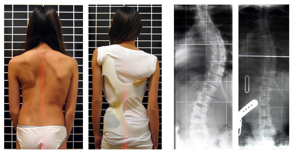
Developmental Adjustment and Nutritional Stress
An important example of developmental adjustment that results from a naturally occurring environmental stress is the cessation of physical growth that occurs in children who are under nutritional stress. Children who lack adequate food to fuel both growth and basic metabolic processes are likely to slow down in their growth rate — or even to stop growing entirely. Shunting all available calories and nutrients into essential life functions may keep the child alive at the expense of increasing body size.
Table 6.4.1 shows the effects of inadequate diet on children's' growth in several countries worldwide. For each country, the table gives the prevalence of stunting in children under the age of five. Children are considered stunted if their height is at least two standard deviations below the median height for their age in an international reference population.
Table 6.4.1
Percentage of Stunting in Young Children in Selected Countries (2011-2015)
| Percentage of Stunting in Young Children in Selected Countries (2011-2015) | |
| Country | Per cent of Children Under Age 5 with Stunting |
| United States | 2.1 |
| Turkey | 9.5 |
| Mexico | 13.6 |
| Thailand | 16.3 |
| Iraq | 22.6 |
| Philippines | 33.6 |
| Pakistan | 45.0 |
| Papua New Guinea | 49.5 |
After a growth slow-down occurs and if adequate food becomes available, a child may be able to make up the loss of growth. If food is plentiful, the child may grow more rapidly than normal until the original, genetically-determined growth trajectory is reached. If the inadequate diet persists, however, the failure of growth may become chronic, and the child may never reach his or her full potential adult size.
Phenotypic plasticity of body size in response to dietary change has been observed in successive generations within populations. For example, children in Japan were taller, on average, in each successive generation after the end of World War II. Boys aged 14-15 years old in 1986 were an average of about 18 cm (7 in.) taller than boys of the same age in 1959, a generation earlier. This is a highly significant difference, and it occurred too quickly to be accounted for by genetic change. Instead, the increase in height is a developmental adjustment, thought to be largely attributable to changes in the Japanese diet since World War II. During this period, there was an increase in the amount of animal protein and fat, as well as in the total calories consumed.
Acclimatization
Other responses to environmental stress are reversible and not permanent, whether they occur in childhood or adulthood. The development of reversible changes to environmental stress is called acclimatization. Acclimatization generally develops over a relatively short period of time. It may take just a few days or weeks to attain a maximum response to a stress. When the stress is no longer present, the acclimatized state declines, and the body returns to its normal baseline state. Generally, the shorter the time for acclimatization to occur, the more quickly the condition is reversed when the environmental stress is removed.
Acclimatization to UV Light
A common example of acclimatization is tanning of the skin (see Figure 6.4.5). This occurs in many people in response to exposure to ultraviolet radiation from the sun. Special pigment cells in the skin, called melanocytes, produce more of the brown pigment melanin when exposed to sunlight. The melanin collects near the surface of the skin where it absorbs UV radiation so it cannot penetrate and potentially damage deeper skin structures. Tanning is a reversible change in the phenotype that helps the body deal temporarily with the environmental stress of high levels of UV radiation. When the skin is no longer exposed to the sun’s rays, the tan fades, generally over a period of a few weeks or months.
Figure 6.4.5 Tanning of the skin occurs in many people in response to exposure to ultraviolet radiation from the sun.
Acclimatization to Heat
Another common example of acclimatization occurs in response to heat. Changes that occur with heat acclimatization include increased sweat output and earlier onset of sweat production, which helps the body stay cool because evaporation of sweat takes heat from the body’s surface in a process called evaporative cooling. It generally takes a couple of weeks for maximum heat acclimatization to come about by gradually working out harder and longer at high air temperatures. The changes that occur with acclimatization just as quickly subside when the body is no longer exposed to excessive heat.
Acclimatization to High Altitude
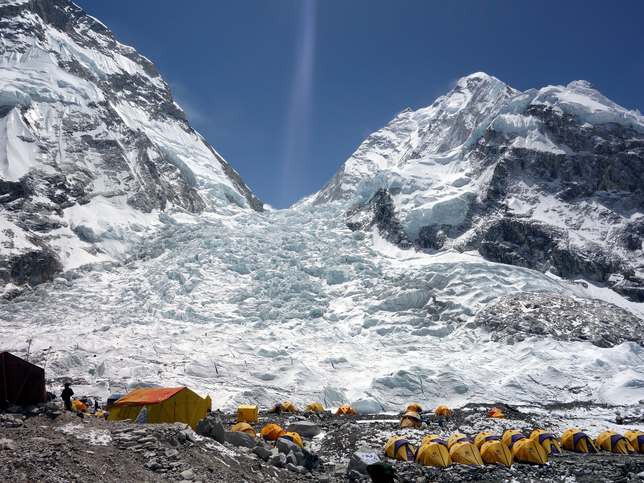
Short term acclimatization to high altitude occurs as a response to low levels of oxygen in the blood. This reduced level of oxygen is detected by carotid bodies, which will trigger in increase in breathing and heart rate. Over a period of weeks the body will compensate by increasing red blood cell production, thereby improving the oxygen-carrying capacity of the blood. This is why mountaineers wishing to climb to the peak of Mount Everest must complete the full climb in portions; it is recommended that climbers spend 2-3 days acclimatizing for every 600 metres of elevation increase. In addition, the higher to altitude, the longer it make take to acclimatize; climbers are advised to spend 4-5 days acclimatizing at base camp (whether the base camp in Nepal or China) before completing the final leg of the climb to the peak. The concentration of red blood cells gradually decreases to normal levels once a climber returns to their normal elevation.
Cultural Responses
More than any other species, humans respond to environmental stresses with learned behaviors and technology. These cultural responses allow us to change our environments to control stresses, rather than changing our bodies genetically or physiologically to cope with the stresses. Even archaic humans responded to some environmental stresses in this way. For example, Neanderthals used shelters, fires, and animal hides as clothing to stay warm in the cold climate in Europe during the last ice age. Today, we use more sophisticated technologies to stay warm in cold climates while retaining our essentially tropical-animal anatomy and physiology. We also use technology (such as furnaces and air conditioners) to avoid temperature stress and stay comfortable in hot or cold climates.
6.4 Summary
- Humans may respond to environmental stress in four different ways: adaptation, developmental adjustment, acclimatization, and cultural responses.
- An adaptation is a genetically based trait that has evolved because it helps living things survive and reproduce in a given environment. Adaptations evolve by natural selection in populations over a relatively long period to time. Examples of adaptations include sickle cell trait as an adaptation to the stress of endemic malaria and the ability to taste bitter compounds as an adaptation to the stress of bitter-tasting toxins in plants.
- A developmental adjustment is a non-genetic response to stress that occurs during infancy or childhood, and that may persist into adulthood. This type of change may be irreversible. Developmental adjustment is possible because humans have a high degree of phenotypic plasticity. It may be the result of environmental stresses (such as inadequate food), which may stunt growth, or cultural practices (such as orthodontic treatments), which re-align the teeth and jaws.
- Acclimatization is the development of reversible changes to environmental stress that develop over a relatively short period of time. The changes revert to the normal baseline state after the stress is removed. Examples of acclimatization include tanning of the skin and physiological changes (such as increased sweating) that occur with heat acclimatization.
- More than any other species, humans respond to environmental stress with learned behaviors and technology, which are cultural responses. These responses allow us to change our environment to control stress, rather than changing our bodies genetically or physiologically to cope with stress. Examples include using shelter, fire, and clothing to cope with a cold climate.
6.4 Review Questions
- List four different types of responses that humans may make to cope with environmental stress.
- Define adaptation.
-
- Explain how natural selection may have resulted in most human populations having people who can and people who cannot taste PTC.
- What is a developmental adjustment?
- Define phenotypic plasticity.
- Explain why phenotypic plasticity may be particularly important in a species with a long generation time.
- Why may stunting of growth occur in children who have an inadequate diet? Why is stunting preferable to the alternative?
- What is acclimatization?
- How does acclimatization to heat come about, and what are two physiological changes that occur in heat acclimatization?
- Give an example of a cultural response to heat stress.
- Which is more likely to be reversible — a change due to acclimatization, or a change due to developmental adjustment? Explain your answer.
6.4 Explore More
https://www.youtube.com/watch?v=upp9-w6GPhU
Could we survive prolonged space travel? - Lisa Nip, TED-Ed, 2016.
https://www.youtube.com/watch?v=hRnrIpUMyZQ&t=182s
How this disease changes the shape of your cells - Amber M. Yates, TED-Ed, 2019.
Attributions
Figure 6.4.1
Free_Awesome_Girl_With_Braces_Close_Up by D. Sharon Pruitt from Hill Air Force Base, Utah, USA on Wikimedia Commons is used under a CC BY 2.0 (https://creativecommons.org/licenses/by/2.0/deed.en) license.
Figure 6.4.2
Tongue by Mahdiabbasinv on Wikimedia Commons is used under a CC BY-SA 4.0 (https://creativecommons.org/licenses/by-sa/4.0/deed.en) license.
Figure 6.4.3
- White cauliflower on brown wooden chopping board by Louis Hansel @shotsoflouis on Unsplash is used under the Unsplash License (https://unsplash.com/license).
- Broccoli on wooden chopping board by Louis Hansel @shotsoflouis on Unsplash is used under the Unsplash License (https://unsplash.com/license).
- Green cabbage close up by Craig Dimmick on Unsplash is used under the Unsplash License (https://unsplash.com/license).
- Cabbage hybrid/ brussel sprouts by Solstice Hannan on Unsplash is used under the Unsplash License (https://unsplash.com/license).
- Kale by Laura Johnston on Unsplash is used under the Unsplash License (https://unsplash.com/license).
- Tiny bok choy at the Asian market by Jodie Morgan on Unsplash is used under the Unsplash License (https://unsplash.com/license).
Figure 6.4.4
Scoliosis_patient_in_cheneau_brace_correcting_from_56_to_27_deg by Weiss H.R. from Scoliosis Journal/BioMed Central Ltd. on Wikimedia Commons is used under a CC BY 2.0 (https://creativecommons.org/licenses/by/2.0) license.
Figure 6.4.5
- Tan Lines by k.steudel on Flickr is used under a CC BY 2.0 (https://creativecommons.org/licenses/by/2.0/) license.
- Twin tan lines (all sizes) by Quinn Dombrowski on Flickr is used under a CC BY-SA 2.0 (https://creativecommons.org/licenses/by-sa/2.0/) license.
- Wedding ring tan line by Quinn Dombrowski on Flickr is used under a CC BY-SA 2.0 (https://creativecommons.org/licenses/by-sa/2.0/) license.
- Tan by Evil Erin on Flickr is used under a CC BY 2.0 (https://creativecommons.org/licenses/by/2.0/) license.
Figure 6.4.6
Nepalese base camp by Mark Horrell on Flickr is used under a CC BY-NC-SA 2.0 (https://creativecommons.org/licenses/by-nc-sa/2.0/) license.
References
TED-Ed. (2016, October 4). Could we survive prolonged space travel? - Lisa Nip. YouTube. https://www.youtube.com/watch?v=upp9-w6GPhU&feature=youtu.be
TED-Ed. (2019, May 6). How this disease changes the shape of your cells - Amber M. Yates. YouTube. https://www.youtube.com/watch?v=hRnrIpUMyZQ&feature=youtu.be
Weiss, H. (2007). Is there a body of evidence for the treatment of patients with Adolescent Idiopathic Scoliosis (AIS)? [Figure 2 - digital photograph], Scoliosis, 2(19). https://doi.org/10.1186/1748-7161-2-19
Created by: CK-12/Adapted by Christine Miller
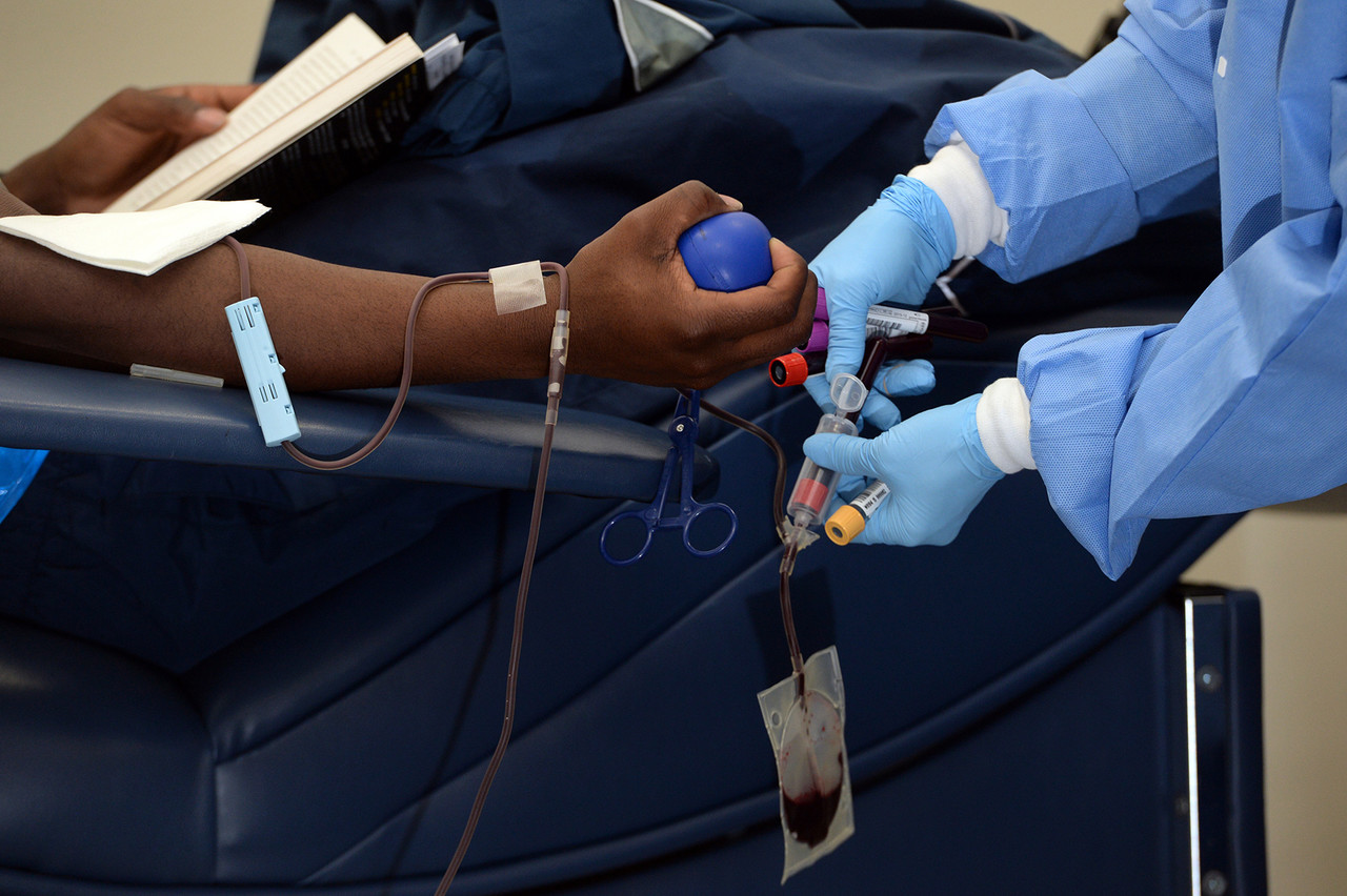
Giving the Gift of Life
Did you ever donate blood? If you did, then you probably know that your blood type is an important factor in blood transfusions. People vary in the type of blood they inherit, and this determines which type(s) of blood they can safely receive in a transfusion. Do you know your blood type?
What Are Blood Types?
Blood is composed of cells suspended in a liquid called plasma. There are three types of cells in blood: red blood cells, which carry oxygen; white blood cells, which fight infections and other threats; and platelets, which are cell fragments that help blood clot. Blood type (or blood group) is a genetic characteristic associated with the presence or absence of certain molecules, called antigens, on the surface of red blood cells. These molecules may help maintain the integrity of the cell membrane, act as receptors, or have other biological functions. A blood group system refers to all of the gene(s), alleles, and possible genotypes and phenotypes that exist for a particular set of blood type antigens. Human blood group systems include the well-known ABO and Rhesus (Rh) systems, as well as at least 33 others that are less well known.
Antigens and Antibodies
Antigens — such as those on the red blood cells — are molecules that the immune system identifies as either self (produced by your own body) or non-self (not produced by your own body). Blood group antigens may be proteins, carbohydrates, glycoproteins (proteins attached to chains of sugars), or glycolipids (lipids attached to chains of sugars), depending on the particular blood group system. If antigens are identified as non-self, the immune system responds by forming antibodies that are specific to the non-self antigens. Antibodies are large, Y-shaped proteins produced by the immune system that recognize and bind to non-self antigens. The analogy of a lock and key is often used to represent how an antibody and antigen fit together, as shown in the illustration below (Figure 6.5.2). When antibodies bind to antigens, it marks them for destruction by other immune system cells. Non-self antigens may enter your body on pathogens (such as bacteria or viruses), on foods, or on red blood cells in a blood transfusion from someone with a different blood type than your own. The last way is virtually impossible nowadays because of effective blood typing and screening protocols.
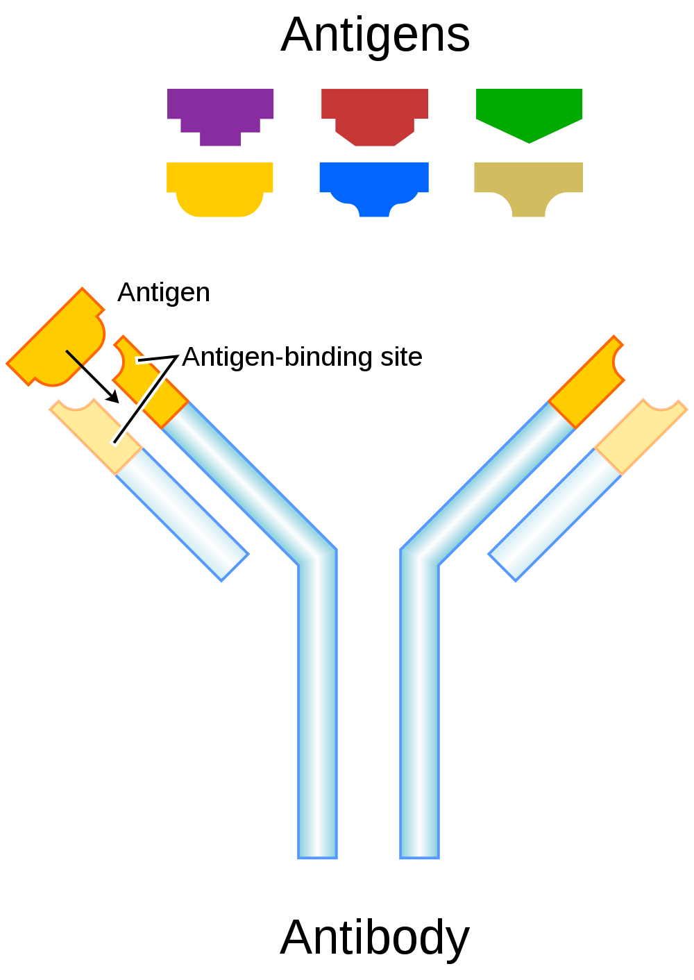
Genetics of Blood Type
An individual’s blood type depends on which alleles for a blood group system were inherited from their parents. Generally, blood type is controlled by alleles for a single gene, or for two or more very closely linked genes. Closely linked genes are almost always inherited together, because there is little or no recombination between them. Like other genetic traits, a person’s blood type is generally fixed for life, but there are rare instances in which blood type can change. This could happen, for example, if an individual receives a bone marrow transplant to treat a disease, such as leukemia. If the bone marrow comes from a donor who has a different blood type, the patient’s blood type may eventually convert to the donor’s blood type, because red blood cells are produced in bone marrow.
ABO Blood Group System
The ABO blood group system is the best known human blood group system. Antigens in this system are glycoproteins. These antigens are shown in the list below. There are four common blood types for the ABO system:
- Type A, in which only the A antigen is present.
- Type B, in which only the B antigen is present.
- Type AB, in which both the A and B antigens are present.
- Type O, in which neither the A nor the B antigen is present.
Genetics of the ABO System
The ABO blood group system is controlled by a single gene on chromosome 9. There are three common alleles for the gene, often represented by the letters A , B , and O. With three alleles, there are six possible genotypes for ABO blood group. Alleles A and B, however, are both dominant to allele O and codominant to each other. This results in just four possible phenotypes (blood types) for the ABO system. These genotypes and phenotypes are shown in Table 6.5.1.
Table 6.5.1
ABO Blood Group System: Genotypes and Phenotypes
| ABO Blood Group System | |
| Genotype | Phenotype (Blood Type, or Group) |
| AA | A |
| AO | A |
| BB | B |
| BO | B |
| OO | O |
| AB | AB |
The diagram below (Figure 6.5.3) shows an example of how ABO blood type is inherited. In this particular example, the father has blood type A (genotype AO) and the mother has blood type B (genotype BO). This mating type can produce children with each of the four possible ABO phenotypes, although in any given family, not all phenotypes may be present in the children.
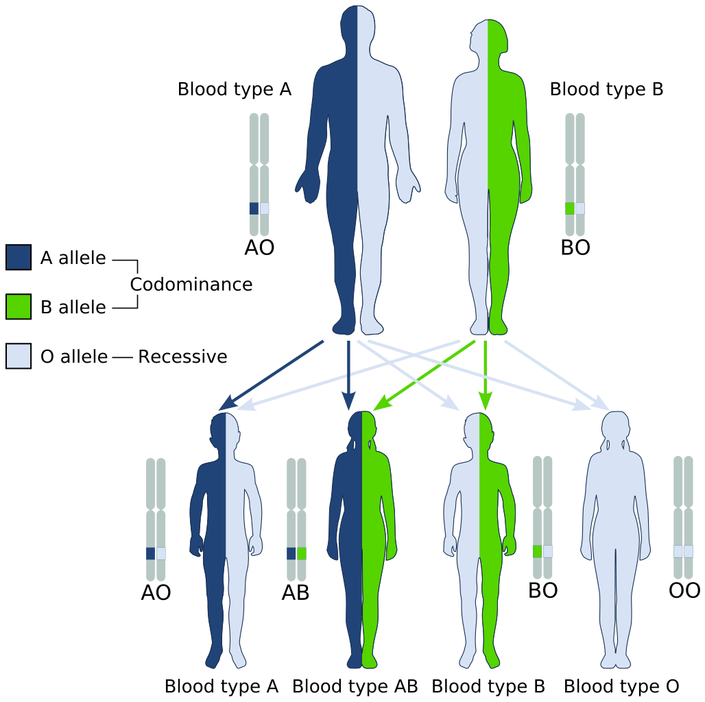
Medical Significance of ABO Blood Type
The ABO system is the most important blood group system in blood transfusions. If red blood cells containing a particular ABO antigen are transfused into a person who lacks that antigen, the person’s immune system will recognize the antigen on the red blood cells as non-self. Antibodies specific to that antigen will attack the red blood cells, causing them to agglutinate (or clump) and break apart. If a unit of incompatible blood were to be accidentally transfused into a patient, a severe reaction (called acute hemolytic transfusion reaction) is likely to occur, in which many red blood cells are destroyed. This may result in kidney failure, shock, and even death. Fortunately, such medical accidents virtually never occur today.
These antibodies are often spontaneously produced in the first years of life, after exposure to common microorganisms in the environment that have antigens similar to blood antigens. Specifically, a person with type A blood will produce anti-B antibodies, while a person with type B blood will produce anti-A antibodies. A person with type AB blood does not produce either antibody, while a person with type O blood produces both anti-A and anti-B antibodies. Once the antibodies have been produced, they circulate in the plasma. The relationship between ABO red blood cell antigens and plasma antibodies is shown in Figure 6.5.4.
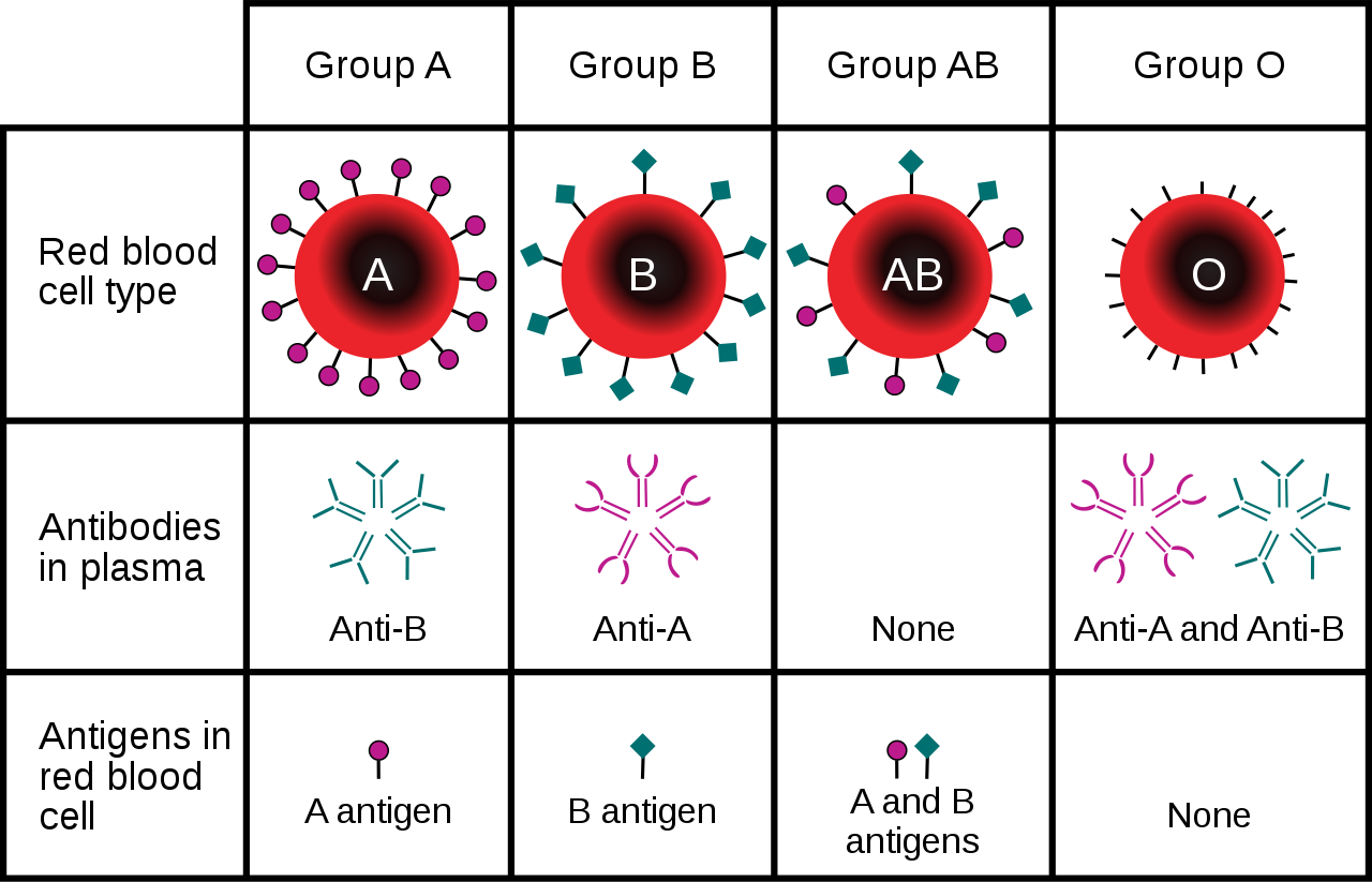
The antibodies that circulate in the plasma are for different antigens than those on red blood cells, which are recognized as self antigens.
Which blood types are compatible and which are not? Type O blood contains both anti-A and anti-B antibodies, so people with type O blood can only receive type O blood. However, they can donate blood to people of any ABO blood type, which is why individuals with type O blood are called universal donors. Type AB blood contains neither anti-A nor anti-B antibodies, so people with type AB blood can receive blood from people of any ABO blood type. That’s why individuals with type AB blood are called universal recipients. They can donate blood, however, only to people who also have type AB blood. These and other relationships between blood types of donors and recipients are summarized in the simple diagram to the right.
Geographic Distribution of ABO Blood Groups
The frequencies of blood groups for the ABO system vary around the world. You can see how the A and B alleles and the blood group O are distributed geographically on the maps in Figure 6.5.6.
- Worldwide, B is the rarest ABO allele, so type B blood is the least common ABO blood type. Only about 16 per cent of all people have the B allele. Its highest frequency is in Asia. Its lowest frequency is among the indigenous people of Australia and the Americas.
- The A allele is somewhat more common around the world than the B allele, so type A blood is also more common than type B blood. The highest frequencies of the A allele are in Australian Aborigines, the Lapps (Sami) of Northern Scandinavia, and Blackfoot Native Americans in North America. The allele is nearly absent among Native Americans in Central and South America.
- The O allele is the most common ABO allele around the world, and type O blood is the most common ABO blood type. Almost two-thirds of people have at least one copy of the O allele. It is especially common in Native Americans in Central and South America, where it reaches frequencies close to 100 per cent. It also has relatively high frequencies in Australian Aborigines and Western Europeans. Its frequencies are lowest in Eastern Europe and Central Asia.
Figure 6.5.6 Maps of populations that have the A, B and O alleles.
Evolution of the ABO Blood Group System
The geographic distribution of ABO blood type alleles provides indirect evidence for the evolutionary history of these alleles. Evolutionary biologists hypothesize that the allele for blood type A evolved first, followed by the allele for blood type O, and then by the allele for blood type B. This chronology accounts for the percentages of people worldwide with each blood group, and is also consistent with known patterns of early population movements.
The evolutionary forces of founder effect and genetic drift have no doubt played a significant role in the current distribution of ABO blood types worldwide. Geographic variation in ABO blood groups is also likely to be influenced by natural selection, because different blood types are thought to vary in their susceptibility to certain diseases. For example:
- People with type O blood may be more susceptible to cholera and plague. They are also more likely to develop gastrointestinal ulcers.
- People with type A blood may be more susceptible to smallpox and more likely to develop certain cancers.
- People with types A, B, and AB blood appear to be less likely to form blood clots that can cause strokes. However, early in our history, the ability of blood to form clots — which appears greater in people with type O blood — may have been a survival advantage.
- Perhaps the greatest natural selective force associated with ABO blood types is malaria. There is considerable evidence to suggest that people with type O blood are somewhat resistant to malaria, giving them a selective advantage where malaria is endemic.
Rhesus Blood Group System
Another well-known blood group system is the Rhesus (Rh) blood group system. The Rhesus system has dozens of different antigens, but only five main antigens (called D, C, c, E, and e). The major Rhesus antigen is the D antigen. People with the D antigen are called Rh positive (Rh+), and people who lack the D antigen are called Rh negative (Rh-). Rhesus antigens are thought to play a role in transporting ions across cell membranes by acting as channel proteins.
The Rhesus blood group system is controlled by two linked genes on chromosome 1. One gene, called RHD, produces a single antigen, antigen D. The other gene, called RHCE, produces the other four relatively common Rhesus antigens (C, c, E, and e), depending on which alleles for this gene are inherited.
Rhesus Blood Group and Transfusions
After the ABO system, the Rhesus system is the second most important blood group system in blood transfusions. The D antigen is the one most likely to provoke an immune response in people who lack the antigen. People who have the D antigen (Rh+) can be safely transfused with either Rh+ or Rh- blood, whereas people who lack the D antigen (Rh-) can be safely transfused only with Rh- blood.
Unlike anti-A and anti-B antibodies to ABO antigens, anti-D antibodies for the Rhesus system are not usually produced by sensitization to environmental substances. People who lack the D antigen (Rh-), however, may produce anti-D antibodies if exposed to Rh+ blood. This may happen accidentally in a blood transfusion, although this is extremely unlikely today. It may also happen during pregnancy with an Rh+ fetus if some of the fetal blood cells pass into the mother’s blood circulation.
Hemolytic Disease of the Newborn
If a woman who is Rh- is carrying an Rh+ fetus, the fetus may be at risk. This is especially likely if the mother has formed anti-D antibodies during a prior pregnancy because of a mixing of maternal and fetal blood during childbirth. Unlike antibodies against ABO antigens, antibodies against the Rhesus D antigen can cross the placenta and enter the blood of the fetus. This may cause hemolytic disease of the newborn (HDN), also called erythroblastosis fetalis, an illness in which fetal red blood cells are destroyed by maternal antibodies, causing anemia. This illness may range from mild to severe. If it is severe, it may cause brain damage and is sometimes fatal for the fetus or newborn. Fortunately, HDN can be prevented by preventing the formation of anti-D antibodies in the Rh- mother. This is achieved by injecting the mother with a medication called Rho(D) immune globulin.
Geographic Distribution of Rhesus Blood Types
The majority of people worldwide are Rh+, but there is regional variation in this blood group system, as there is with the ABO system. The aboriginal inhabitants of the Americas and Australia originally had very close to 100 per cent Rh+ blood. The frequency of the Rh+ blood type is also very high in African populations, at about 97 to 99 per cent. In East Asia, the frequency of Rh+ is slightly lower, at about 93 to 99 per cent. Europeans have the lowest frequency of the Rh+ blood type at about 83 to 85 per cent.
What explains the population variation in Rhesus blood types? Prior to the advent of modern medicine, Rh+ positive children conceived by Rh- women were at risk of fetal or newborn death or impairment from HDN. This was an enigma, because presumably, natural selection would work to remove the rarer phenotype (Rh-) from populations. However, the frequency of this phenotype is relatively high in many populations.
Recent studies have found evidence that natural selection may actually favor heterozygotes for the Rhesus D antigen. The selective agent in this case is thought to be toxoplasmosis, a parasitic disease caused by the protozoan Toxoplasma gondii, which is very common worldwide. You can see a life cycle diagram of the parasite in Figure 6.5.7. Infection by this parasite often causes no symptoms at all, or it may cause flu-like symptoms for a few days or weeks. Exposure to the parasite has been linked, however, to increased risk of mental disorders (such as schizophrenia), neurological disorders (such as Alzheimer’s), and other neurological problems, including delayed reaction times. One study found that people who tested positive for antibodies to the parasite were more than twice as likely to be involved in traffic accidents.
People who are heterozygous for the D antigen appear less likely to develop the negative neurological and mental effects of Toxoplasma gondii infection. This could help explain why both phenotypes (Rh+ and Rh-) are maintained in most populations. There are also striking geographic differences in the prevalence of toxoplasmosis worldwide, ranging from zero to 95 per cent in different regions. This could explain geographic variation in the D antigen worldwide, because its strength as a selective agent would vary with its prevalence.
Feature: Myth vs. Reality
Myth |
Reality |
| "Your nutritional needs can be determined by your ABO blood type. Knowing your blood type allows you to choose the appropriate foods that will help you lose weight, increase your energy, and live a longer, healthier life." | This idea was proposed in 1996 in a New York Times bestseller Eat Right for Your Type, by Peter D’Adamo, a naturopath. Naturopathy is a method of treating disorders that involves the use of herbs, sunlight, fresh air, and other natural substances. Some medical doctors consider naturopathy a pseudoscience. A major scientific review of the blood type diet could find no evidence to support it. In one study, adults eating the diet designed for blood type A showed improved health — but this occurred in everyone, regardless of their blood type. Because the blood type diet is based solely on blood type, it fails to account for other factors that might require dietary adjustments or restrictions. For example, people with diabetes — but different blood types — would follow different diets, and one or both of the diets might conflict with standard diabetes dietary recommendations and be dangerous. |
| "ABO blood type is associated with certain personality traits. People with blood type A, for example, are patient and responsible, but may also be stubborn and tense, whereas people with blood type B are energetic and creative, but may also be irresponsible and unforgiving. In selecting a spouse, both your own and your potential mate’s blood type should be taken into account to ensure compatibility of your personalities." | The belief that blood type is correlated with personality is widely held in Japan and other East Asian countries. The idea was originally introduced in the 1920s in a study commissioned by the Japanese government, but it was later shown to have no scientific support. The idea was revived in the 1970s by a Japanese broadcaster, who wrote popular books about it. There is no scientific basis for the idea, and it is generally dismissed as pseudoscience by the scientific community. Nonetheless, it remains popular in East Asian countries, just as astrology is popular in many other countries. |
6.5 Summary
- Blood type (or blood group) is a genetic characteristic associated with the presence or absence of antigens on the surface of red blood cells. A blood group system refers to all of the gene(s), alleles, and possible genotypes and phenotypes that exist for a particular set of blood type antigens.
- Antigens are molecules that the immune system identifies as either self or non-self. If antigens are identified as non-self, the immune system responds by forming antibodies that are specific to the non-self antigens, leading to the destruction of cells bearing the antigens.
- The ABO blood group system is a system of red blood cell antigens controlled by a single gene with three common alleles on chromosome 9. There are four possible ABO blood types: A, B, AB, and O. The ABO system is the most important blood group system in blood transfusions. People with type O blood are universal donors, and people with type AB blood are universal recipients.
- The frequencies of ABO blood type alleles and blood groups vary around the world. The allele for the B antigen is least common, and blood type O is the most common. The evolutionary forces of founder effect, genetic drift, and natural selection are responsible for the geographic distribution of ABO alleles and blood types. People with type O blood, for example, may be somewhat resistant to malaria, possibly giving them a selective advantage where malaria is endemic.
- The Rhesus blood group system is a system of red blood cell antigens controlled by two genes with many alleles on chromosome 1. There are five common Rhesus antigens, of which antigen D is most significant. Individuals who have antigen D are called Rh+, and individuals who lack antigen D are called Rh-. Rh- mothers of Rh+ fetuses may produce antibodies against the D antigen in the fetal blood, causing hemolytic disease of the newborn (HDN).
- The majority of people worldwide are Rh+, but there is regional variation in this blood group system. This variation may be explained by natural selection that favors heterozygotes for the D antigen, because this genotype seems to be protected against some of the neurological consequences of the common parasitic infection toxoplasmosis.
6.5 Review Questions
- Define blood type and blood group system.
- Explain the relationship between antigens and antibodies.
- Identify the alleles, genotypes, and phenotypes in the ABO blood group system.
- Discuss the medical significance of the ABO blood group system.
- Compare the relative worldwide frequencies of the three ABO alleles.
- Give examples of how different ABO blood types vary in their susceptibility to diseases.
- Describe the Rhesus blood group system.
- Relate Rhesus blood groups to blood transfusions.
- What causes hemolytic disease of the newborn?
- Describe how toxoplasmosis may explain the persistence of the Rh- blood type in human populations.
- A woman is blood type O and Rh-, and her husband is blood type AB and Rh+. Answer the following questions about this couple and their offspring.
- What are the possible genotypes of their offspring in terms of ABO blood group?
- What are the possible phenotypes of their offspring in terms of ABO blood group?
- Can the woman donate blood to her husband? Explain your answer.
- Can the man donate blood to his wife? Explain your answer.
- Type O blood is characterized by the presence of O antigens — explain why this statement is false.
- Explain why newborn hemolytic disease may be more likely to occur in a second pregnancy than in a first.
6.5 Explore More
https://www.youtube.com/watch?v=xfZhb6lmxjk
Why do blood types matter? - Natalie S. Hodge, TED-Ed, 2015.
https://www.youtube.com/watch?v=qcZKbjYyOfE
How do blood transfusions work? - Bill Schutt, TED-Ed, 2020.
Attributes
Figure 6.5.1
Following the Blood Donation Trail by EJ Hersom/ USA Department of Defense is in the public domain. [Disclaimer: The appearance of U.S. Department of Defense (DoD) visual information does not imply or constitute DoD endorsement.]
Figure 6.5.2
Antibody by Fvasconcellos on Wikimedia Commons is released into the public domain (https://en.wikipedia.org/wiki/Public_domain).
Figure 6.5.3
ABO system codominance.svg, adapted by YassineMrabet (original "Codominant" image from US National Library of Medicine) on Wikimedia Commons is in the public domain (https://en.wikipedia.org/wiki/Public_domain).
Figure 6.5.4
ABO_blood_type.svg by InvictaHOG on Wikimedia Commons is released into the public domain (https://en.wikipedia.org/wiki/Public_domain).
Figure 6.5.5
Blood Donor and recipient ABO by CK-12 Foundation is used under a CC BY-NC 3.0 (https://creativecommons.org/licenses/by-nc/3.0/) license.
Figure 6.5.6
- Map of Blood Group A by Muntuwandi at en.wikipedia on Wikimedia Commons is used under a CC BY-SA 3.0 (https://creativecommons.org/licenses/by-sa/3.0/) license.
- Map of Blood Group B by Muntuwandi at en.wikipedia on Wikimedia Commons is used under a CC BY-SA 3.0 (https://creativecommons.org/licenses/by-sa/3.0/) license.
- Map of Blood Group O by anthro palomar at en.wikipedia on Wikimedia Commons is used under a CC BY-SA 3.0 (https://creativecommons.org/licenses/by-sa/3.0/) license.
Figure 6.5.7
Toxoplasma_gondii_Life_cycle_PHIL_3421_lores by Alexander J. da Silva, PhD/Melanie Moser, Centers for Disease Control and Prevention's Public Health Image Library (PHIL#3421) on Wikimedia Commons is in the public domain (https://en.wikipedia.org/wiki/Public_domain).
Table 6.5.1
ABO Blood Group System: Genotypes and Phenotypes was created by Christine Miller.
References
Dean, L. (2005). Chapter 4 Hemolytic disease of the newborn. In Blood Groups and Red Cell Antigens [Internet]. National Center for Biotechnology Information (US). https://www.ncbi.nlm.nih.gov/books/NBK2266/
Mayo Clinic Staff. (n.d.). Toxoplasmosis [online article]. MayoClinic.org. https://www.mayoclinic.org/diseases-conditions/toxoplasmosis/symptoms-causes/syc-20356249
MedlinePlus. (2019, January 29). Hemolytic transfusion reaction [online article]. U.S. National Library of Medicine. https://en.wikipedia.org/w/index.php?title=Chromosome_9&oldid=946440619
TED-Ed. (2015, June 29). Why do blood types matter? - Natalie S. Hodge. YouTube. https://www.youtube.com/watch?v=xfZhb6lmxjk&feature=youtu.be
TED-Ed. (2020, February 18). How do blood transfusions work? - Bill Schutt. YouTube. https://www.youtube.com/watch?v=qcZKbjYyOfE&feature=youtu.be
Wikipedia contributors. (2020, May 10). Chromosome 1. In Wikipedia. https://en.wikipedia.org/w/index.php?title=Chromosome_1&oldid=955942444
Wikipedia contributors. (2020, March 20). Chromosome 9. In Wikipedia. https://en.wikipedia.org/w/index.php?title=Chromosome_9&oldid=946440619
Any gland of the endocrine system, which is the system of glands that releases hormones directly into the blood.
A set of metabolic reactions and processes that take place in the cells of organisms to convert biochemical energy from nutrients into adenosine triphosphate (ATP).
The smallest unit of life, consisting of at least a membrane, cytoplasm, and genetic material.
A complex organic chemical that provides energy to drive many processes in living cells, e.g. muscle contraction, nerve impulse propagation, and chemical synthesis. Found in all forms of life, ATP is often referred to as the "molecular unit of currency" of intracellular energy transfer.
Glucose (also called dextrose) is a simple sugar with the molecular formula C6H12O6. Glucose is the most abundant monosaccharide, a subcategory of carbohydrates. Glucose is mainly made by plants and most algae during photosynthesis from water and carbon dioxide, using energy from sunlight.
A body system including a series of hollow organs joined in a long, twisting tube from the mouth to the anus. The hollow organs that make up the GI tract are the mouth, esophagus, stomach, small intestine, large intestine, and anus. The liver, pancreas, and gallbladder are the solid organs of the digestive system.
Refers to the body system consisting of the heart, blood vessels and the blood. Blood contains oxygen and other nutrients which your body needs to survive. The body takes these essential nutrients from the blood.
Created by: CK-12/Adapted by Christine Miller
Mush!
These beautiful sled dogs are a metabolic marvel. While running up to 160 kilometres (about 99 miles) a day, they will each consume and burn about 12 thousand calories — about 240 calories per pound per day, which is the equivalent of about 24 Big Macs! A human endurance athlete, in contrast, typically burns only about 100 calories per pound (0.45 kg) each day. Scientists are intrigued by the amazing metabolism of sled dogs, although they still haven't determined how they use up so much energy. But one thing is certain: all living things need energy for everything they do, whether it's running a race or blinking an eye. In fact, every cell of your body constantly needs energy just to carry out basic life processes. You probably know that you get energy from the food you eat, but where does food come from? How does it come to contain energy? And how do your cells get the energy from food?
What Is Energy?
In the scientific world, energy is defined as the ability to do work. You can often see energy at work in living things — a bird flies through the air, a firefly glows in the dark, a dog wags its tail. These are obvious ways that living things use energy, but living things constantly use energy in less obvious ways, as well.
Why Living Things Need Energy
Inside every cell of all living things, energy is needed to carry out life processes. Energy is required to break down and build up molecules, and to transport many molecules across plasma membranes. All of life’s work needs energy. A lot of energy is also simply lost to the environment as heat. The story of life is a story of energy flow — its capture, its change of form, its use for work, and its loss as heat. Energy (unlike matter) cannot be recycled, so organisms require a constant input of energy. Life runs on chemical energy. Where do living organisms get this chemical energy?
How Organisms Get Energy
The chemical energy that organisms need comes from food. Food consists of organic molecules that store energy in their chemical bonds. In terms of obtaining food for energy, there are two types of organisms: autotrophs and heterotrophs.
Autotrophs
Autotrophs are organisms that capture energy from nonliving sources and transfer that energy into the living part of the ecosystem. They are also able to make their own food. Most autotrophs use the energy in sunlight to make food in the process of photosynthesis. Only certain organisms — such as plants, algae, and some bacteria — can make food through photosynthesis. Some photosynthetic organisms are shown in Figure 4.9.2.
 |
 |
 |
| Figure 4.9.2 Photosynthetic autotrophs, which make food using the energy in sunlight, include plants (left), algae (middle), and certain bacteria (right). |
Autotrophs are also called producers. They produce food not only for themselves, but for all other living things (known as consumers), as well. This is why autotrophs form the basis of food chains, such as the food chain shown In Figure 4.9.3.
A food chain shows how energy and matter flow from producers to consumers. Matter is recycled, but energy must keep flowing into the system. Where does this energy come from?
Watch the video "The simple story of photosynthesis and food - Amanda Ooten" from TED-Ed to learn more about photosynthesis:
https://www.youtube.com/watch?time_continue=39&v=eo5XndJaz-Y
The simple story of photosynthesis and food - Amanda Ooten, TED-Ed, 2013.
Heterotrophs
Heterotrophs are living things that cannot make their own food. Instead, they get their food by consuming other organisms, which is why they are also called consumers. They may consume autotrophs or other heterotrophs. Heterotrophs include all animals and fungi, as well as many single-celled organisms. In Figure 4.9.3, all of the organisms are consumers except for the grasses and phytoplankton. What do you think would happen to consumers if all producers were to vanish from Earth?
Energy Molecules: Glucose and ATP
Organisms mainly use two types of molecules for chemical energy: glucose and ATP. Both molecules are used as fuels throughout the living world. Both molecules are also key players in the process of photosynthesis.
Glucose
Glucose is a simple carbohydrate with the chemical formula C6H12O6. It stores chemical energy in a concentrated, stable form. In your body, glucose is the form of energy that is carried in your blood and taken up by each of your trillions of cells. Glucose is the end product of photosynthesis, and it is the nearly universal food for life. In Figure 4.9.4, you can see how photosynthesis stores energy from the sun in the glucose molecule and then how cellular respiration breaks the bonds in glucose to retrieve the energy.
ATP
If you remember from section 3.7 Nucleic Acids, ATP (adenosine triphosphate) is the energy-carrying molecule that cells use to power most cellular processes (nerve impulse conduction, protein synthesis and active transport are good examples of cell processes that rely on ATP as their energy source). ATP is made during the first half of photosynthesis and then used for energy during the second half of photosynthesis, when glucose is made. ATP releases energy when it gives up one of its three phosphate groups (Pi) and changes to ADP (adenosine diphosphate, which has two phosphate groups), as shown in Figure 4.9.5. Thus, the breakdown of ATP into ADP + Pi is a catabolic reaction that releases energy (exothermic). ATP is made from the combination of ADP and Pi, an anabolic reaction that takes in energy (endothermic).
Why Organisms Need Both Glucose and ATP
Why do living things need glucose if ATP is the molecule that cells use for energy? Why don’t autotrophs just make ATP and be done with it? The answer is in the “packaging.” A molecule of glucose contains more chemical energy in a smaller “package” than a molecule of ATP. Glucose is also more stable than ATP. Therefore, glucose is better for storing and transporting energy. Glucose, however, is too powerful for cells to use. ATP, on the other hand, contains just the right amount of energy to power life processes within cells. For these reasons, both glucose and ATP are needed by living things.
How Energy Flows Through Living Things
The flow of energy through living organisms begins with photosynthesis. This process stores energy from sunlight in the chemical bonds of glucose. By breaking the chemical bonds in glucose, cells release the stored energy and make the ATP they need. The process in which glucose is broken down and ATP is made is called cellular respiration.
Photosynthesis and cellular respiration are like two sides of the same coin. This is apparent in Figure 4.9.6. The products of one process are the reactants of the other. Together, the two processes store and release energy in living organisms. The two processes also work together to recycle oxygen in the Earth’s atmosphere.
4.9 Summary
- Energy is the ability to do work. It is needed by all living things and every living cell to carry out life processes, such as breaking down and building up molecules, and transporting many molecules across cell membranes.
- The form of energy that living things need for these processes is chemical energy, and it comes from food. Food consists of organic molecules that store energy in their chemical bonds.
- Autotrophs make their own food. Plants, for example, make food by photosynthesis. Autotrophs are also called producers.
- Heterotrophss obtain food by eating other organisms. Heterotrophs are also known as consumers.
- Organisms mainly use the molecules glucose and ATP for energy. Glucose is a compact, stable form of energy that is carried in the blood and taken up by cells. ATP contains less energy and is used to power cell processes.
- The flow of energy through living things begins with photosynthesis, which creates glucose. In a process called cellular respiration, organisms' cells break down glucose and make the ATP they need.
4.9 Review Questions
- Define energy.
- Why do living things need energy?
-
- Compare and contrast the two basic ways that organisms get energy.
- Describe the roles and relationships of the energy molecules glucose and ATP.
- Summarize how energy flows through living things.
- Why does the transformation of ATP to ADP release energy?
4.9 Explore More
https://www.youtube.com/watch?v=eDalQv7d2cs
Learn Biology: Autotrophs vs. Heterotrophs, Mahalodotcom, 2011.
https://www.youtube.com/watch?v=0glkXIj1DgE&feature=emb_logo
Energy Transfer in Trophic Levels, Teacher's Pet, 2015.
Attributions
Figure 4.9.1
Three Airmen participate in dog-sled expedition by U.S. Air Force photo by Tech. Sgt. Dan Rea is released into the public domain (https://en.wikipedia.org/wiki/Public_domain).
Figure 4.9.2
- Plant [photo] by Ren Ran on Unsplash is used under the Unsplash License (https://unsplash.com/license).
- Green Algae by Tristan Schmurr on Flickr is used under a CC BY 2.0 (https://creativecommons.org/licenses/by/2.0/) license.
- Cyanobacteria by Argon National Laboratory on Flickr is used under a CC BY-NC-SA 2.0 (https://creativecommons.org/licenses/by-nc-sa/2.0/) license.
Figure 4.9.3
Biomass_Pyramid by Swiggity.Swag.YOLO.Bro on Wikipedia is used and adapted by Christine Miller under a CC BY-SA 4.0 (https://creativecommons.org/licenses/by-sa/4.0/deed.en) license.
Figure 4.9.4
Photosynthesis and respiration by Christine Miller is used under a CC BY 4.0 (https://creativecommons.org/licenses/by/4.0/) license.
Figure 4.9.5
Photo synthesis and cellular respiration by Lady of Hats/ CK-12 Foundation is used under a CC BY-NC 3.0 (https://creativecommons.org/licenses/by-nc/3.0/) license.
References
LadyofHats/CK-12 Foundation. (2016, August 15). Figure 5: Photosynthesis and cellular respiration [digital image]. In Brainard, J., and Henderson, R., CK-12's College Human Biology FlexBook® (Section 4.9). CK-12 Foundation. https://www.ck12.org/book/ck-12-college-human-biology/section/4.9/
Mahalodotcom. (2011, January 14). Learn biology: Autotrophs vs. heterotrophs. YouTube. https://www.youtube.com/watch?v=eDalQv7d2cs
Teacher's Pet. (2015, March 23). Energy transfer in trophic levels. YouTube. https://www.youtube.com/watch?v=0glkXIj1DgE&feature=emb_logo
TED-Ed. (2013, March 5). The simple story of photosynthesis and food - Amanda Ooten. YouTube. https://www.youtube.com/watch?v=eo5XndJaz-Y&feature=youtu.be
An involuntary human body response mediated by the nervous and endocrine systems that prepares the body to fight or flee from perceived danger.
division of the peripheral nervous system that controls involuntary activities
Created by: CK-12/Adapted by Christine Miller
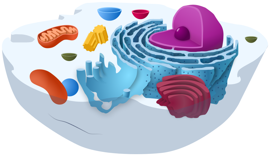

A Bag Full of Jell-O
The simple cut-away model of an animal cell (Figure 4.4.1) shows that a cell resembles a plastic bag full of Jell-O. Its basic structure is a plasma membrane filled with cytoplasm. Like Jell-O containing mixed fruit (Figure 4.4.2), the cytoplasm of the cell also contains various structures, including a nucleus and other organelles. Your body is composed of trillions of cells, but all of them perform the same basic life functions. They all obtain and use energy, respond to the environment, and reproduce. How do your cells carry out these basic functions and keep themselves — and you — alive? To answer these questions, you need to know more about the structures that make up cells, starting with the plasma membrane.
What is the Plasma Membrane?
The plasma membrane is a structure that forms a barrier between the cytoplasm inside the cell and the environment outside the cell. Without the plasma membrane, there would be no cell. Although it is very thin and flexible, the plasma membrane protects and supports the cell by controlling everything that enters and leaves it. It allows only certain substances to pass through, while keeping others in or out. To understand how the plasma membrane controls what passes into or out of the cell, you need to know its basic structure.
Phospholipid Bilayer
The plasma membrane is composed mainly of phospholipids, which consist of fatty acids and alcohol. The phospholipids in the plasma membrane are arranged in two layers, called a phospholipid bilayer. As shown in the simplified diagram in Figure 4.4.3, each individual phospholipid molecule has a phosphate group head (in red) and two fatty acid tails (in yellow). The head “loves” water (hydrophilic) and the tails “hate” water (hydrophobic). The water-hating tails are on the interior of the membrane, whereas the water-loving heads point outward, toward either the cytoplasm (intracellular) or the fluid that surrounds the cell (extracellular).
Hydrophobic molecules can easily pass through the plasma membrane if they are small enough, because they are water-hating like the interior of the membrane. Hydrophilic molecules, on the other hand, cannot pass through the plasma membrane — at least not without help — because they are water-loving like the exterior of the membrane.
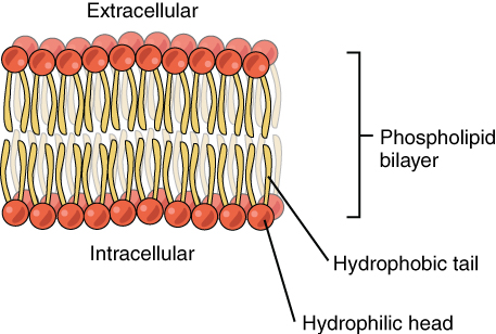
Other Molecules in the Plasma Membrane
The plasma membrane also contains other molecules, primarily other lipids and proteins. The yellow molecules in the diagram here, for example, are the lipid cholesterol. Molecules of the steroid lipid cholesterol help the plasma membrane keep its shape. Proteins in the plasma membrane (shown blue in Figure 4.4.4) include: transport proteins that assist other substances in crossing the cell membrane, receptors that allow the cell to respond to chemical signals in its environment, and cell-identity markers that indicate what type of cell it is and whether it belongs in the body.
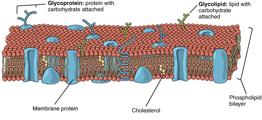
Additional Functions of the Plasma Membrane
The plasma membrane may have extensions, such as whip-like flagella (singular flagellum) or brush-like cilia (singular cilium), shown below (Figure 4.4.5), that give it other functions. In single-celled organisms, these membrane extensions may help the organisms move. In multicellular organisms, the extensions have different functions. For example, the cilia on human lung cells sweep foreign particles and mucus toward the mouth and nose, while the flagellum on a human sperm cell allows it to swim.
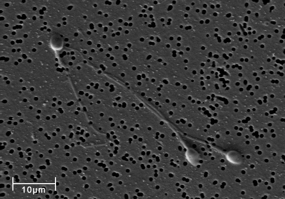
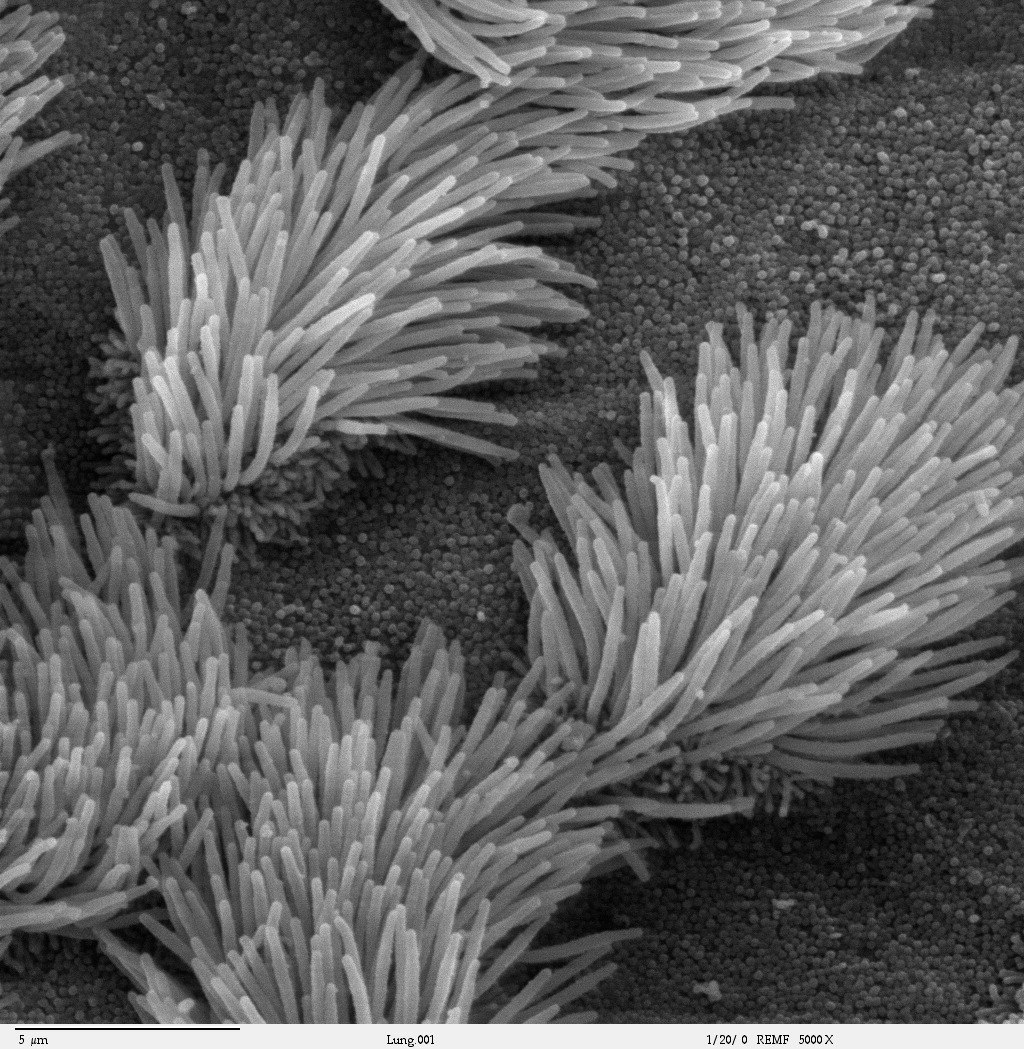
Feature: My Human Body
If you smoke or use e-cigarettes (vaping) and need another reason to quit, here's a good one. We usually think of lung cancer as the major disease caused by smoking. But smoking and vaping can have devastating effects on the body's ability to protect itself from repeated, serious respiratory infections, such as bronchitis and pneumonia.
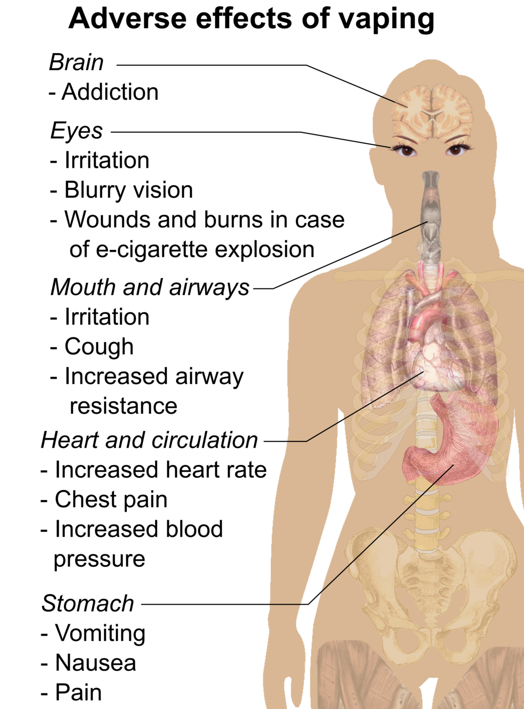
Cilia are microscopic, hair-like projects on cells that line the respiratory, reproductive, and digestive systems. Cilia in the respiratory system line most of your airways, where they have the job of trapping and removing dust, germs, and other foreign particles before they can make you sick. Cilia secrete mucus that traps particles, and they move in a continuous wave-like motion that sweeps the mucus and particles upward toward the throat, where they can be expelled from the body. When you are sick and cough up phlegm, that's what you are doing.
Smoking prevents cilia from performing these important functions. Chemicals in tobacco smoke paralyze the cilia so they can't sweep mucus out of the airways. Those chemicals also inhibit the cilia from producing mucus. Fortunately, these effects start to wear off soon after the most recent exposure to tobacco smoke. If you stop smoking, your cilia will return to normal. Even if prolonged smoking has destroyed cilia, they will regrow and resume functioning in a matter of months after you stop smoking.
4.4 Summary
- The plasma membrane is a structure that forms a barrier between the cytoplasm inside the cell and the environment outside the cell. It allows only certain substances to pass in or out of the cell.
- The plasma membrane is composed mainly of a bilayer of phospholipid molecules. It also contains other molecules, such as the steroid cholesterol, which helps the membrane keep its shape, and transport proteins, which help substances pass through the membrane.
- The plasma membranes of some cells have extensions that have other functions, like flagella to help sperm move, or cilia to help keep our airways clear.
4.4 Review Questions
- What are the general functions of the plasma membrane?
- Describe the phospholipid bilayer of the plasma membrane.
- Identify other molecules in the plasma membrane. State their functions.
- Why do some cells have plasma membrane extensions, like flagella and cilia?
- Explain why hydrophilic molecules cannot easily pass through the cell membrane. What type of molecule in the cell membrane might help hydrophilic molecules pass through it?
- Which part of a phospholipid molecule in the plasma membrane is made of fatty acid chains? Is this part hydrophobic or hydrophilic?
- The two layers of phospholipids in the plasma membrane are called a phospholipid ____________.
-
- Steroid hormones can pass directly through cell membranes. Why do you think this is the case?
- Some antibiotics work by making holes in the plasma membrane of bacterial cells. How do you think this kills the cells?
- What is the name of the long, whip-like extensions of the plasma membrane that helps some single-celled organisms move?
4.4 Explore More
https://www.youtube.com/watch?v=yAXnYcUjn5k&feature=emb_logo
Insights into cell membranes via dish detergent - Ethan Perlstein, TED-Ed, 2013.
https://www.youtube.com/watch?v=qBCVVszQQNs
Inside the cell membrane, by The Amoeba Sisters, 2018.
Attributions
Figure 4.4.1
Animal Cell Unannotated, by Kelvin Song on Wikimedia Commons is used under a CC0 1.0 (https://creativecommons.org/publicdomain/zero/1.0/deed.en) public domain dedication license.
Figure 4.4.2
Jello mold at the mexican bakery photo by Aimée Knight on Flickr is used under a CC BY 2.0 (https://creativecommons.org/licenses/by/2.0/) license.
Figure 4.4.3
Phospholipid_Bilayer by OpenStax on Wikimedia Commons is used under a CC BY 4.0 (https://creativecommons.org/licenses/by/4.0) license.
Figure 4.4.4
Lipid bilayer by OpenStax on Wikimedia Commons is used under a CC BY 4.0 (https://creativecommons.org/licenses/by/4.0) license.
Figure 4.4.5
Spermatozoa-human-3140x by No specific author on Wikimedia Commons is released into the public domain (https://en.wikipedia.org/wiki/Public_domain).
Figure 4.4.6
Cilia/ Bronchiolar epithelium 3 - SEM by Charles Daghlian on Wikimedia Commons is released into the public domain (https://en.wikipedia.org/wiki/Public_domain).
Figure 4.4.7
Adverse effects of vaping (raster) by Mikael Häggström on Wikimedia Commons is released into the public domain (https://en.wikipedia.org/wiki/Public_domain).
References
Amoeba Sisters. (2018, February 27). Inside the cell membrane. YouTube. https://www.youtube.com/watch?v=qBCVVszQQNs&feature=youtu.be
Betts, J.G., Young, K.A., Wise, J.A., Johnson, E., Poe, B., Kruse, D.H., Korol, O., Johnson, J.E.. Womble, M., DeSaix. P. (2013, April 25). Figure 3.3 Phospolipid Bilayer [digital image]. In Anatomy and Physiology. OpenStax. https://openstax.org/books/anatomy-and-physiology/pages/3-1-the-cell-membrane
Betts, J.G., Young, K.A., Wise, J.A., Johnson, E., Poe, B., Kruse, D.H., Korol, O., Johnson, J.E.. Womble, M., DeSaix. P. (2013, April 25). Figure 3.4 Cell Membrane [digital image]. In Anatomy and Physiology. OpenStax. https://openstax.org/books/anatomy-and-physiology/pages/3-1-the-cell-membrane
Ghosh, A., Coakley, R. C., Mascenik, T., Rowell, T. R., Davis, E. S., Rogers, K., Webster, M. J., Dang, H., Herring, L. E., Sassano, M. F., Livraghi-Butrico, A., Van Buren, S. K., Graves, L. M., Herman, M. A., Randell, S. H., Alexis, N. E., & Tarran, R. (n.d.). Chronic E-Cigarette Exposure Alters the Human Bronchial Epithelial Proteome. American Journal of Respiratory and Critical /Care Medicine, 198(1), 67–76. https://doi-org.ezproxy.tru.ca/10.1164/rccm.201710-2033OC
TED-Ed. (2013, February 26). Insights into cell membranes via dish detergent - Ethan Perlstein. YouTube. https://www.youtube.com/watch?v=yAXnYcUjn5k&feature=youtu.be
Created by: CK-12/Adapted by Christine Miller
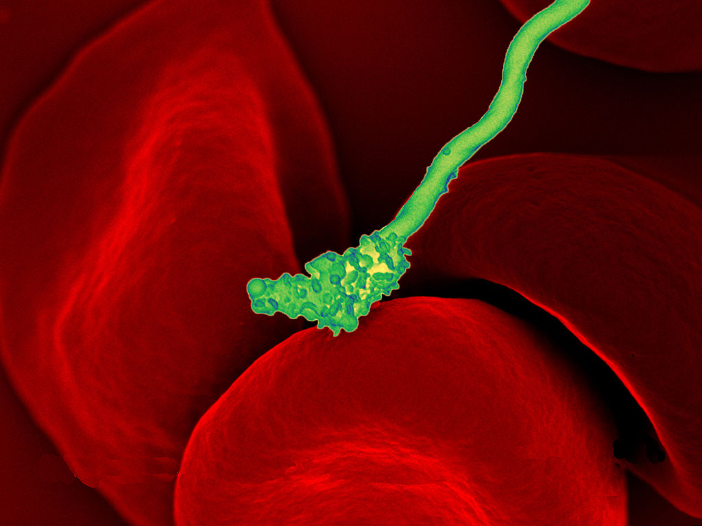
Bacteria Attack!
The colourful image in Figure 4.3.1 shows a bacterial cell (in green) attacking human red blood cells. The bacterium causes a disease called relapsing fever. The bacterial and human cells look very different in size and shape. Although all living cells have certain things in common — such as a plasma membrane and cytoplasm — different types of cells, even within the same organism, may have their own unique structures and functions. Cells with different functions generally have different shapes that suit them for their particular job. Cells vary not only in shape, but also in size, as this example shows. In most organisms, however, even the largest cells are no bigger than the period at the end of this sentence. Why are cells so small?
Explaining Cell Size
Most organisms, even very large ones, have microscopic cells. Why don't cells get bigger instead of remaining tiny and multiplying? Why aren't you one giant cell rolling around school? What limits cell size?
Once you know how a cell functions, the answers to these questions are clear. To carry out life processes, a cell must be able to quickly pass substances in and out of the cell. For example, it must be able to pass nutrients and oxygen into the cell and waste products out of the cell. Anything that enters or leaves a cell must cross its outer surface. The size of a cell is limited by its need to pass substances across that outer surface.
Look at the three cubes in Figure 4.3.2. A larger cube has less surface area relative to its volume than a smaller cube. This relationship also applies to cells — a larger cell has less surface area relative to its volume than a smaller cell. A cell with a larger volume also needs more nutrients and oxygen, and produces more waste. Because all of these substances must pass through the surface of the cell, a cell with a large volume will not have enough surface area to allow it to meet its needs. The larger the cell is, the smaller its ratio of surface area to volume, and the more difficult it will be for the cell to get rid of its waste and take in necessary substances. This is what limits the size of the cell.
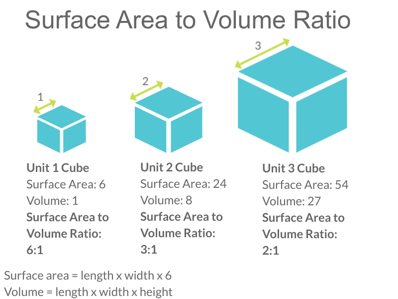
Cell Form and Function
Cells with different functions often have varying shapes. The cells pictured below (Figure 4.3.3) are just a few examples of the many different shapes that human cells may have. Each type of cell has characteristics that help it do its job. The job of the nerve cell, for example, is to carry messages to other cells. The nerve cell has many long extensions that reach out in all directions, allowing it to pass messages to many other cells at once. Do you see the tail of each tiny sperm cell? Its tail helps a sperm cell "swim" through fluids in the female reproductive tract in order to reach an egg cell. The white blood cell has the job of destroying bacteria and other pathogens. It is a large cell that can engulf foreign invaders.
Figure 4.3.3 Human cells may have many different shapes that help them to do their jobs.
Cells With and Without a Nucleus
The nucleus is a basic cell structure present in many — but not all — living cells. The nucleus of a cell is a structure in the cytoplasm that is surrounded by a membrane (the nuclear membrane) and contains DNA. Based on whether or not they have a nucleus, there are two basic types of cells: prokaryotic cells and eukaryotic cells.
Prokaryotic Cells
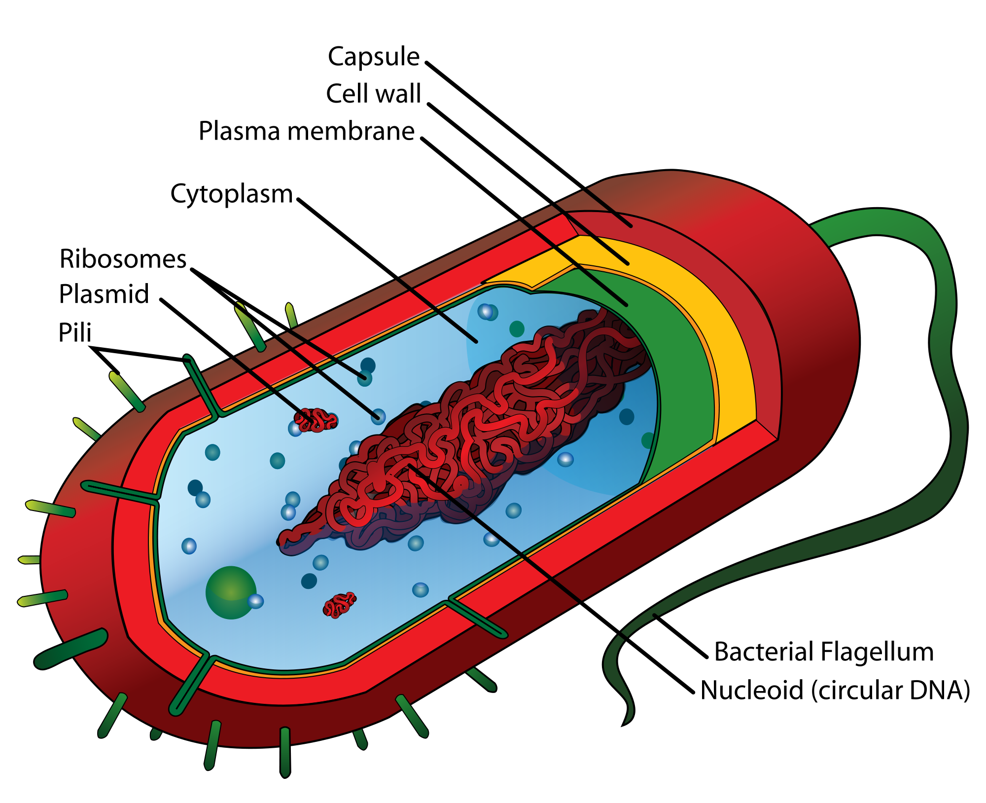
Prokaryotic cells are cells without a nucleus. The DNA in prokaryotic cells is in the cytoplasm, rather than enclosed within a nuclear membrane. In addition, these cells are typically smaller than eukaryotic cells and contain fewer organelles. Prokaryotic cells are found in single-celled organisms, such as the bacterium represented by the model in Figure 4.3.3. Organisms with prokaryotic cells are called prokaryotes. They were the first type of organisms to evolve, and they are still the most common organisms today.
Eukaryotic Cells
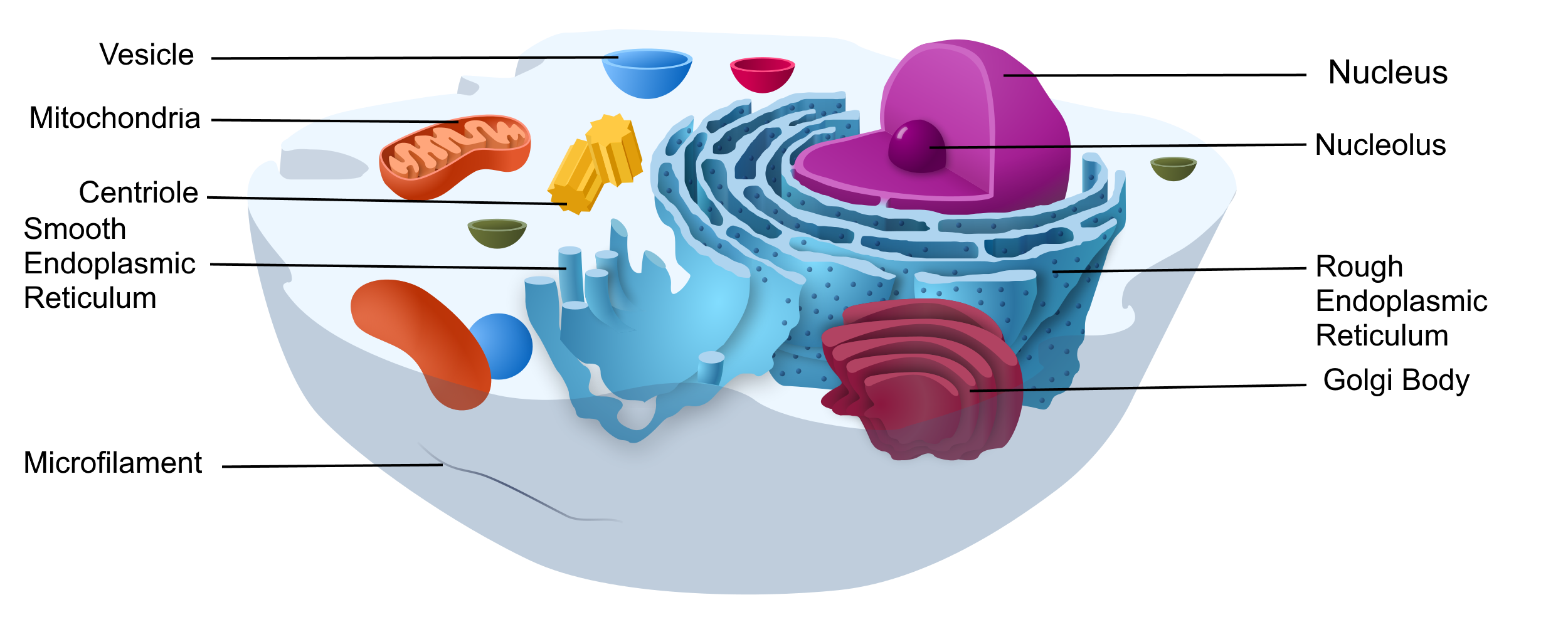
Eukaryotic cells are cells that contain a nucleus. A typical eukaryotic cell is represented by the model in Figure 4.3.4. Eukaryotic cells are usually larger than prokaryotic cells. They are found in some single-celled and all multicellular organisms. Organisms with eukaryotic cells are called eukaryotes, and they range from fungi to humans.
Besides a nucleus, eukaryotic cells also contain other organelles. An organelle is a structure within the cytoplasm that performs a specific job in the cell. Organelles called mitochondria, for example, provide energy to the cell, and organelles called vesicles store substances in the cell. Organelles allow eukaryotic cells to carry out more functions than prokaryotic cells can.
Interestingly, scientists think that mitochondria were once free-living prokaryotes that infected (or were engulfed by) larger cells. The two organisms developed a symbiotic relationship that was beneficial to both of them, resulting in the smaller prokaryote becoming an organelle within the larger cell. This is called endosymbiotic theory, and it is supported by a lot of evidence, including the fact that mitochondria have their own DNA separate from the DNA in the nucleus of the eukaryotic cell. Endosymbiotic theory will be described in more detail in later sections, and it's also discussed in the video below.
https://www.youtube.com/watch?v=FGnS-Xk0ZqU
Endosymbiotic Theory, Amoeba Sisters, 2017.
4.3 Summary
- Cells must be very small so they have a large enough surface area-to-volume ratio to maintain normal cell processes.
- Cells with different functions often have different shapes.
- Prokaryotic cells do not have a nucleus. Eukaryotic cells do have a nucleus, along with other organelles.
4.3 Review Questions
- Explain why most cells are very small.
- Discuss variations in the form and function of cells.
-
-
- Do human cells have organelles? Explain your answer.
- Which are usually larger – prokaryotic or eukaryotic cells? What do you think this means for their relative ability to take in needed substances and release wastes? Discuss your answer.
- DNA in eukaryotes is enclosed within the _______ ________.
- Name three different types of cells in humans.
- Which organelle provides energy in eukaryotic cells?
- What is a function of a vesicle in a cell?
4.3 Explore More
https://www.youtube.com/watch?time_continue=1&v=9i7kAt97XYU&feature=emb_logo
How we think complex cells evolved - Adam Jacobson, TED-Ed, 2015.
https://www.youtube.com/watch?v=Pxujitlv8wc
Prokaryotic vs. Eukaryotic Cells (updated), Amoeba Sisters, 2018.
Attributions
Figure 4.3.1
Borrelia_hermsii_Bacteria_(13758011613) by NAID on Wikimedia Commons is released into the public domain (https://en.wikipedia.org/wiki/Public_domain).
Figure 4.3.2
Cell Size by Christine Miller is released into the Public Domain (https://creativecommons.org/publicdomain/mark/1.0/).
Figure 4.3.3
- Chondrocyte. BioTek-Wikipedia-Image by BioTek Instruments, Inc. on Wikimedia Commons is used under a CC BY-SA 3.0 (https://creativecommons.org/licenses/by-sa/3.0/deed.en) license.
- Neutrophil with anthrax copy by Volker Brinkmann from PLOS Pathogens on Wikimedia Commons is used under a CC BY 2.5 (https://creativecommons.org/licenses/by/2.5/deed.en) license.
- PLoSBiol4.e126.Fig6fNeuron by Lee, et al. from PLOS Biology on Wikimedia Commons is used under a CC BY 2.5 (https://creativecommons.org/licenses/by/2.5/deed.en) license.
- Sperm (265 33) human by Doc. RNDr. Josef Reischig, CSc. on Wikimedia Commons is used under a CC BY-SA 3.0 (https://creativecommons.org/licenses/by-sa/3.0) license.
Figure 4.3.4
Model of a prokaryotic cell: bacterium by Mariana Ruiz Villarreal [LadyofHats] on Wikimedia Commons is released into the public domain (https://en.wikipedia.org/wiki/Public_domain).
Figure 4.3.5
Animal Cell adapted by Christine Miller is used under a CC0 1.0 (https://creativecommons.org/publicdomain/zero/1.0/deed.en) public domain dedication license. (Original image, Animal Cell Unannotated, is by Kelvin Song on Wikimedia Commons.)
References
Amoeba Sisters. (2017, May 3). Endosymbiotic theory. YouTube. https://www.youtube.com/watch?v=FGnS-Xk0ZqU&feature=youtu.be
Amoeba Sisters. (2018, July 30). Prokaryotic vs. eukaryotic cells (updated). YouTube. https://www.youtube.com/watch?v=Pxujitlv8wc&feature=youtu.be
Brinkmann, V. (November 2005). Neutrophil engulfing Bacillus anthracis. PLoS Pathogens 1 (3): Cover page [digital image]. DOI:10.1371. https://journals.plos.org/plospathogens/issue?id=10.1371/issue.ppat.v01.i03
Lee, W.C.A., Huang, H., Feng, G., Sanes, J.R., Brown, E.N., et al. (2005, December 27) Figure 6f, slightly altered (plus scalebar, minus letter "f".) [digital image]. Dynamic Remodeling of Dendritic Arbors in GABAergic Interneurons of Adult Visual Cortex. PLoS Biology, 4(2), e29. doi:10.1371/journal.pbio.0040029. https://journals.plos.org/plosbiology/article?id=10.1371/journal.pbio.0040029
TED-Ed. (2015, February 17). How we think complex cells evolved - Adam Jacobson. https://www.youtube.com/watch?v=9i7kAt97XYU&feature=youtu.be
Created by CK-12 Foundation/Adapted by Christine Miller
Case Study: Flight Risk
Nineteen-year-old Malcolm is about to take his first plane flight. Shortly after he boards the plane and sits down, a man in his late sixties sits next to him in the aisle seat. About half an hour after the plane takes off, the pilot announces that she is turning the seat belt light off, and that it is safe to move around the cabin.
The man in the aisle seat — who has introduced himself to Malcolm as Willie — immediately unbuckles his seat belt and paces up and down the aisle a few times before returning to his seat. After about 45 minutes, Willie gets up again, walks some more, then sits back down and does some foot and leg exercises. After the third time Willie gets up and paces the aisles, Malcolm asks him whether he is walking so much to accumulate steps on a pedometer or fitness tracking device. Willie laughs and says no. He is actually trying to do something even more important for his health — prevent a blood clot from forming in his legs.
Willie explains that he has a chronic condition: heart failure. Although it sounds scary, his condition is currently well-managed, and he is able to lead a relatively normal lifestyle. However, it does put him at risk of developing other serious health conditions, such as deep vein thrombosis (DVT), which is when a blood clot occurs in the deep veins, usually in the legs. Air travel — and other situations where a person has to sit for a long period of time — increases the risk of DVT. Willie’s doctor said that he is healthy enough to fly, but that he should walk frequently and do leg exercises to help avoid a blood clot.
As you read this chapter, you will learn about the heart, blood vessels, and blood that make up the cardiovascular system, as well as disorders of the cardiovascular system, such as heart failure. At the end of the chapter you will learn more about why DVT occurs, why Willie has to take extra precautions when he flies, and what can be done to lower the risk of DVT and its potentially deadly consequences.
Chapter Overview: Cardiovascular System
In this chapter, you will learn about the cardiovascular system, which transports substances throughout the body. Specifically, you will learn about:
- The major components of the cardiovascular system: the heart, blood vessels, and blood.
- The functions of the cardiovascular system, including transporting needed substances (such as oxygen and nutrients) to the cells of the body, and picking up waste products.
- How blood is oxygenated through the pulmonary circulation, which transports blood between the heart and lungs.
- How blood is circulated throughout the body through the systemic circulation.
- The components of blood — including plasma, red blood cells, white blood cells, and platelets — and their specific functions.
- Types of blood vessels — including arteries, veins, and capillaries — and their functions, similarities, and differences.
- The structure of the heart, how it pumps blood, and how contractions of the heart are controlled.
- What blood pressure is and how it is regulated.
- Blood disorders, including anemia, HIV, and leukemia.
- Cardiovascular diseases (including heart attack, stroke, and angina), and the risk factors and precursors — such as high blood pressure and atherosclerosis — that contribute to them.
As you read the chapter, think about the following questions:
- What is heart failure?Why do you think it increases the risk of DVT?
- What is a blood clot? What are possible health consequences of blood clots?
- Why do you think sitting for long periods of time increases the risk of DVT? Why does walking and exercising the legs help reduce this risk?
Attribution
Figure 14.1.1
aircraft-1583871_1920 [photo] by olivier89 from Pixabay is used under the Pixabay License (https://pixabay.com/de/service/license/).


