5.9 Regulation of Gene Expression
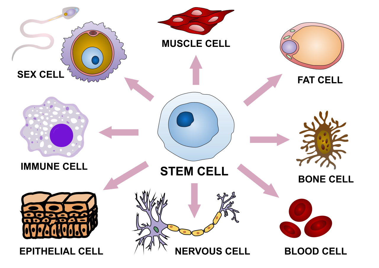
Express Yourself
This sketch illustrates some of the variability in human cells. The shape and other characteristics that make each type of cell unique depend mainly on the specific proteins that particular cell type makes. Proteins are encoded in genes. All the cells in an organism have the same genes, so they all have genetic instructions for the same proteins. Obviously, different types of cells must use (or express) different genes to make different proteins.
What Is Gene Expression?
Using a gene to make a protein is called gene expression. It includes the synthesis of the protein by the processes of transcription of DNA into mRNA, and translation of mRNA into a protein. It may also include further processing of the protein after synthesis.
Gene expression is regulated to ensure that the correct proteins are made when and where they are needed. Regulation may occur at any point in the expression of a gene, from the start of the transcription phase of protein synthesis to the processing of a protein after synthesis occurs. The regulation of transcription is one of the most complicated parts of gene regulation in eukaryotic cells, and it is the focus of this concept.
Regulation of Transcription
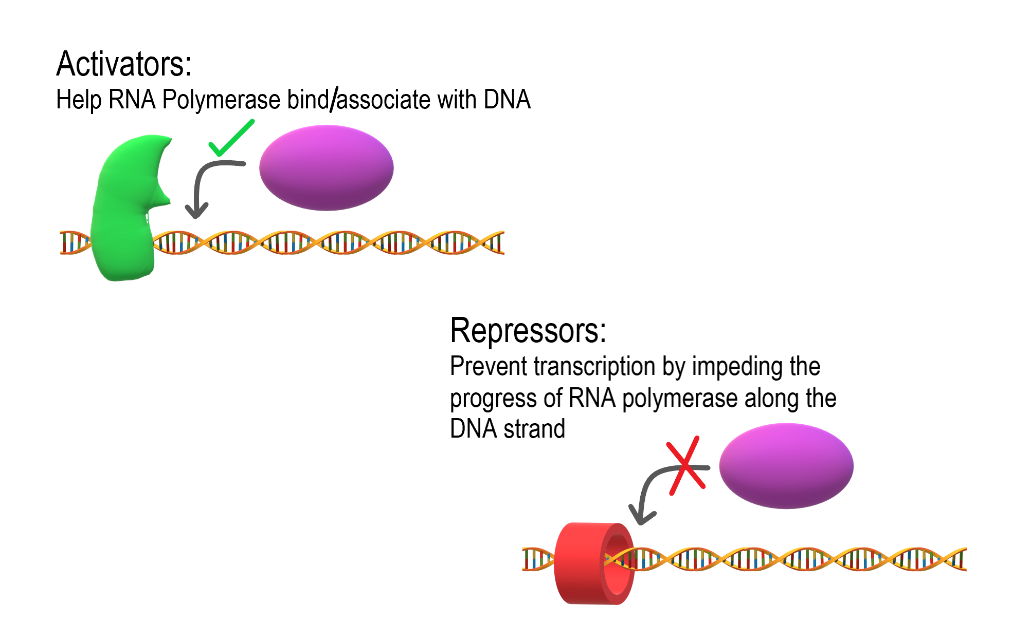
As shown in Figure 5.9.2, transcription is controlled by regulatory proteins. These proteins bind to regions of DNA, called regulatory elements, which are located near promoters. The promoter is the region of a gene where RNA polymerase binds to initiate transcription of the DNA to mRNA. After regulatory proteins bind to regulatory elements, the proteins can interact with RNA polymerase. Regulatory proteins are typically either activators or repressors. Activators are regulatory proteins that promote transcription by enhancing the interaction of RNA polymerase with the promoter. Repressors are regulatory proteins that prevent transcription by impeding the progress of RNA polymerase along the DNA strand, so the DNA cannot be transcribed to mRNA.
Enhancers
Although regulatory proteins and elements are typically the key players in the regulation of transcription, other factors may also be involved. Regulation of transcription may also involve enhancers. Enhancers are distant regions of DNA that can loop back to interact with a gene’s promoter. They can also increase the likelihood that transcription of the gene will occur.Enhancers
The TATA Box
Different types of cells have unique patterns of regulatory elements that result in only the necessary genes being transcribed. That’s why a blood cell and nerve cell, for example, are so different from each other. Some regulatory elements, however, are common to virtually all genes, regardless of the cells in which they occur. An example is the TATA box, which is a regulatory element that is part of the promoter of almost every eukaryotic gene. A number of regulatory proteins bind to the TATA box, forming a multi-protein complex. It is only when all of the appropriate proteins are bound to the TATA box that RNA polymerase recognizes the complex and binds to the promoter so transcription can begin.

Regulation During Development
The regulation of gene expression is extremely important in an organism’s early development. Regulatory proteins must “turn on” certain genes in particular cells at just the right time, so the individual develops normal organs and organ systems. Homeobox genes are important genes that regulate development.
Homeobox genes are a large group of similar genes that direct the formation of many body structures during the embryonic stage. In humans, there are an estimated 235 functional homeobox genes. They are present on every chromosome and generally grouped in clusters. Homeobox genes contain instructions for making chains of 60 amino acids, called homeodomains. Proteins containing homeodomains are transcription factors that bind to and control the activities of other genes. The homeodomain is the part of the protein that binds to the target gene and controls its expression.
Gene Expression and Cancer
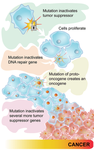
Some types of cancer occur because of mutations in the genes that control the cell cycle. Cancer-causing mutations most often occur in two types of regulatory genes: proto-oncogenes and tumor-suppressor genes. Both are shown in Figure 5.9.4.
- Proto-oncogenes are genes that normally help cells divide. When a proto-oncogene mutates to become an oncogene, it is continuously expressed, even when it is not supposed to be. This is like a car’s accelerator pedal being stuck at full throttle. The car keeps racing at top speed. A cell, in this case, keeps dividing out of control, which can lead to cancer.
- Tumor suppressor genes are genes that normally slow down or stop cell division. When a mutation occurs in a tumor suppressor gene, it can no longer control cell division. This is like a car without brakes. The car can’t be slowed or stopped. A cell, in this case, keeps dividing out of control, which can lead to cancer.
5.9 Summary
- Using a gene to make a protein is called gene expression. Gene expression is regulated to ensure that the correct proteins are made when and where they are needed. Regulation may occur at any stage of protein synthesis or processing.
- The regulation of transcription is controlled by regulatory proteins that bind to regions of DNA called regulatory elements, which are usually located near promoters. Most regulatory proteins are either activators that promote transcription, or repressors that impede transcription.
- A regulatory element common to almost all eukaryotic genes is the TATA box. A number of regulatory proteins must bind to the TATA box in the promoter before transcription can proceed.
- Regulation of gene expression is extremely important during an organism’s early development. Homeobox genes — which encode for chains of amino acids called homeodomains — are important genes that regulate development.
- Some types of cancer occur because of mutations in the genes that control the cell cycle. Cancer-causing mutations most often occur in two types of regulatory genes: tumor-suppressor genes and proto-oncogenes.
5.9 Review Questions
- Define gene expression.
- Why must gene expression be regulated?
- Explain how regulatory proteins may activate or repress transcription.
-
- What is the TATA box, and how does it work?
- Describe homeobox genes and their role in an organism’s development.
- Discuss the role of regulatory gene mutations in cancer.
- Explain the relationship between proto-oncogenes and oncogenes.
- If a newly fertilized egg contained a mutation in a homeobox gene, how do you think this would affect the developing embryo? Explain your answer.
- Compare and contrast enhancers and activators.
5.9 Explore More
Regulated Transcription, ndsuvirtualcell, 2008.
How do cancer cells behave differently from healthy ones? – George Zaidan,
TED-Ed, 2012.
What is leukemia? – Danilo Allegra and Dania Puggioni, 2015.
Attributions
Figure 5.9.1
Stem_cell_differentiation.svg by Haileyfournier on Wikimedia Commons is used under a CC BY-SA 4.0 (https://creativecommons.org/licenses/by-sa/4.0) license.
Figure 5.9.2
Activators and Repressors by Christine Miller is used under a CC BY-SA 4.0 (https://creativecommons.org/licenses/by-sa/4.0) license.
Figure 5.9.3
TATA_box_description by Luttysar on Wikimedia Commons is used under a CC BY-SA 4.0 (https://creativecommons.org/licenses/by-sa/4.0) license.
Figure 5.9.4
Pathways to cancer by CK-12 Foundation is used under a CC BY-NC 3.0 (http://creativecommons.org/licenses/by-nc/3.0/) license.
 ©CK-12 Foundation Licensed under
 ![]() • Terms of Use • Attribution
• Terms of Use • Attribution
References
Brainard, J/ CK-12 Foundation. (2012). Figure 3 Flow chart (series of mutations leading to cancer) [digital image]. In CK-12 College Human Biology (Section 5.8) [online Flexbook]. CK12.org. https://www.ck12.org/c/physical-science/concentration/?referrer=crossref
ndsuvirtualcell.(2008). Regulated transcription. YouTube. https://www.youtube.com/watch?v=vi-zWoobt_Q&feature=youtu.be
TED-Ed. (2012, December 5). How do cancer cells behave differently from healthy ones? – George Zaidan. YouTube. https://www.youtube.com/watch?v=BmFEoCFDi-w&feature=youtu.be
TED-Ed. (2015, April 15). What is leukemia? – Danilo Allegra and Dania Puggioni. YouTube. https://www.youtube.com/watch?v=Z3B-AaqjyjE&feature=youtu.be
The smallest unit of life, consisting of at least a membrane, cytoplasm, and genetic material.
A sequence of nucleotides in DNA or RNA that codes for a molecule that has a function.
A class of biological molecule consisting of linked monomers of amino acids and which are the most versatile macromolecules in living systems and serve crucial functions in essentially all biological processes.
Image shows a labelled diagram highlighting the location of the uterus in pregnancy. The developing fetus, amniotic fluid and placenta are all housed in the uterus, which stretches to many times its regular size to accommodate pregnancy.
Long chains of hydrocarbons with a carboxyl group and a methyl group at opposite ends. Can be either saturated, containing mostly single bonds between adjacent carbons, or unsaturated, containing many double bonds between adjacent carbons.
Attracted to water.
Created by CK-12 Foundation/Adapted by Christine Miller
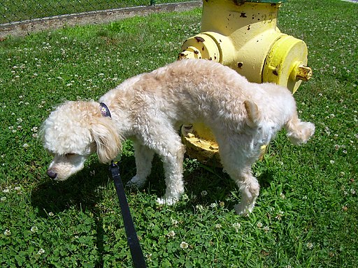
Communicating with Urine
Why do dogs pee on fire hydrants? Besides “having to go,” they are marking their territory with chemicals in their urine called pheromones. It’s a form of communication, in which they are “saying” with odors that the yard is theirs and other dogs should stay away. In addition to fire hydrants, dogs may urinate on fence posts, trees, car tires, and many other objects. Urination in dogs, as in people, is usually a voluntary process controlled by the brain. The process of forming urine — which occurs in the kidneys — occurs constantly, and is not under voluntary control. What happens to all the urine that forms in the kidneys? It passes from the kidneys through the other organs of the urinary system, starting with the ureters.
Ureters
As shown in Figure 16.5.2, ureters are tube-like structures that connect the kidneys with the urinary bladder. They are paired structures, with one ureter for each kidney. In adults, ureters are between 25 and 30 cm (about 10–12 in) long and about 3 to 4 mm in diameter.
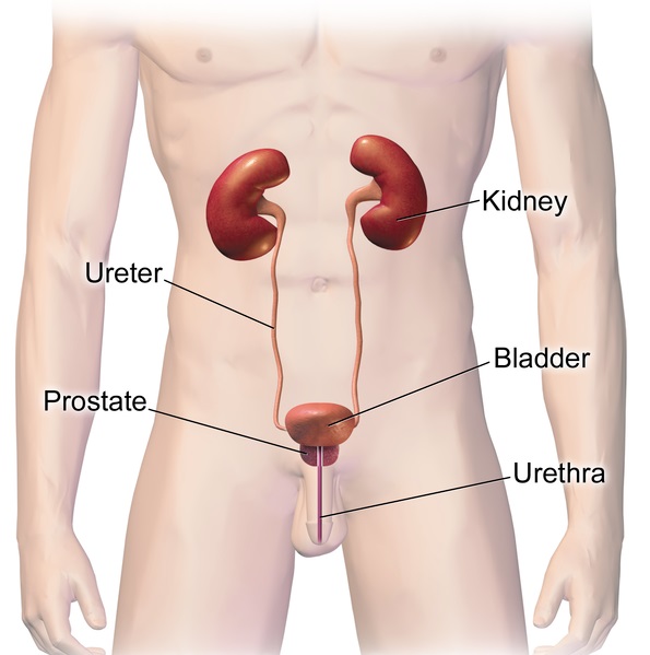
Each ureter arises in the pelvis of a kidney (the renal pelvis in Figure 16.5.3). It then passes down the side of the kidney, and finally enters the back of the bladder. At the entrance to the bladder, the ureters have sphincters that prevent the backflow of urine.
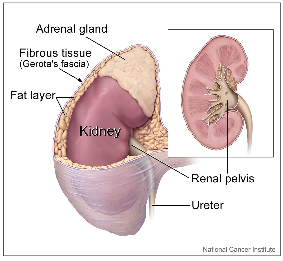
The walls of the ureters are composed of multiple layers of different types of tissues. The innermost layer is a special type of epithelium, called transitional epithelium. Unlike the epithelium lining most organs, transitional epithelium is capable of stretching and does not produce mucus. It lines much of the urinary system, including the renal pelvis, bladder, and much of the urethra, in addition to the ureters. Transitional epithelium allows these organs to stretch and expand as they fill with urine or allow urine to pass through. The next layer of the ureter walls is made up of loose connective tissue containing elastic fibres, nerves, and blood and lymphatic vessels. After this layer are two layers of smooth muscles, an inner circular layer, and an outer longitudinal layer. The smooth muscle layers can contract in waves of peristalsis to propel urine down the ureters from the kidneys to the urinary bladder. The outermost layer of the ureter walls consists of fibrous tissue.
Urinary Bladder
The urinary bladder is a hollow, muscular, and stretchy organ that rests on the pelvic floor. It collects and stores urine from the kidneys before the urine is eliminated through urination. As shown in Figure 16.5.4, urine enters the urinary bladder from the ureters through two ureteral openings on either side of the back wall of the bladder. Urine leaves the bladder through a sphincter called the internal urethral sphincter. When the sphincter relaxes and opens, it allows urine to flow out of the bladder and into the urethra.
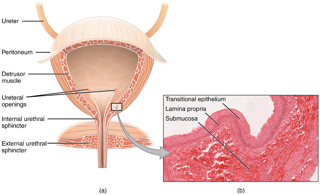
Like the ureters, the bladder is lined with transitional epithelium, which can flatten out and stretch as needed as the bladder fills with urine. The next layer (lamina propria) is a layer of loose connective tissue, nerves, and blood and lymphatic vessels. This is followed by a submucosa layer, which connects the lining of the bladder with the detrusor muscle in the walls of the bladder. The outer covering of the bladder is peritoneum, which is a smooth layer of epithelial cells that lines the abdominal cavity and covers most abdominal organs.
The detrusor muscle in the wall of the bladder is made of smooth muscle fibres controlled by both the autonomic and somatic nervous systems. As the bladder fills, the detrusor muscle automatically relaxes to allow it to hold more urine. When the bladder is about half full, the stretching of the walls triggers the sensation of needing to urinate. When the individual is ready to void, conscious nervous signals cause the detrusor muscle to contract, and the internal urethral sphincter to relax and open. As a result, urine is forcefully expelled out of the bladder and into the urethra.
Urethra
The urethra is a tube that connects the urinary bladder to the external urethral orifice, which is the opening of the urethra on the surface of the body. As shown in Figure 16.5.5, the urethra in males travels through the penis, so it is much longer than the urethra in females. In males, the urethra averages about 20 cm (about 7.8 in) long, whereas in females, it averages only about 4.8 cm (about 1.9 in) long. In males, the urethra carries semen (as well as urine), but in females, it carries only urine. In addition, in males, the urethra passes through the prostate gland (part of the reproductive system) which is absent in women.
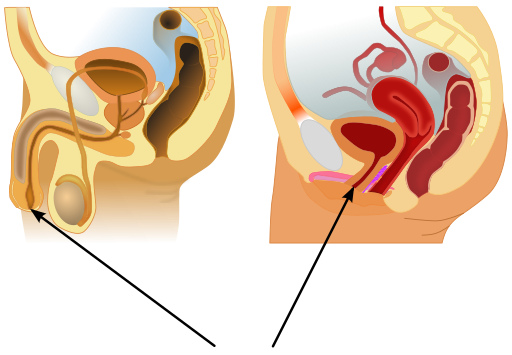
Like the ureters and bladder, the proximal (closer to the bladder) two-thirds of the urethra are lined with transitional epithelium. The distal (farther from the bladder) third of the urethra is lined with mucus-secreting epithelium. The mucus helps protect the epithelium from urine, which is corrosive. Below the epithelium is loose connective tissue, and below that are layers of smooth muscle that are continuous with the muscle layers of the urinary bladder. When the bladder contracts to forcefully expel urine, the smooth muscle of the urethra relaxes to allow the urine to pass through.
In order for urine to leave the body through the external urethral orifice, the external urethral sphincter must relax and open. This sphincter is a striated muscle that is controlled by the somatic nervous system, so it is under conscious, voluntary control in most people (exceptions are infants, some elderly people, and patients with certain injuries or disorders). The muscle can be held in a contracted state and hold in the urine until the person is ready to urinate. Following urination, the smooth muscle lining the urethra automatically contracts to re-establish muscle tone, and the individual consciously contracts the external urethral sphincter to close the external urethral opening.
16.5 Summary
- Ureters are tube-like structures that connect the kidneys with the urinary bladder. Each ureter arises at the renal pelvis of a kidney and travels down through the abdomen to the urinary bladder. The walls of the ureter contain smooth muscle that can contract to push urine through the ureter by peristalsis. The walls are lined with transitional epithelium that can expand and stretch.
- The urinary bladder is a hollow, muscular organ that rests on the pelvic floor. It is also lined with transitional epithelium. The function of the bladder is to collect and store urine from the kidneys before the urine is eliminated through urination. Filling of the bladder triggers the sensation of needing to urinate. When a conscious decision to urinate is made, the detrusor muscle in the bladder wall contracts and forces urine out of the bladder and into the urethra.
- The urethra is a tube that connects the urinary bladder to the external urethral orifice. Somatic nerves control the sphincter at the distal end of the urethra. This allows the opening of the sphincter for urination to be under voluntary control.
16.5 Review Questions
- What are ureters? Describe the location of the ureters relative to other urinary tract organs.
- Identify layers in the walls of a ureter. How do they contribute to the ureter’s function?
- Describe the urinary bladder. What is the function of the urinary bladder?
-
- How does the nervous system control the urinary bladder?
- What is the urethra?
- How does the nervous system control urination?
- Identify the sphincters that are located along the pathway from the ureters to the external urethral orifice.
- What are two differences between the male and female urethra?
- When the bladder muscle contracts, the smooth muscle in the walls of the urethra _________ .
16.5 Explore More
https://youtu.be/2Brajdazp1o
The taboo secret to better health | Molly Winter, TED. 2016.
https://youtu.be/dg4_deyHLvQ
What Happens When You Hold Your Pee? SciShow, 2016.
Attributions
Figure 16.5.1
Cliche by Jackie on Wikimedia Common s is used under a CC BY 2.0 (https://creativecommons.org/licenses/by/2.0) license.
Figure 16.5.2
Urinary System Male by BruceBlaus on Wikimedia Commons is used under a CC BY-SA 4.0 (https://creativecommons.org/licenses/by-sa/4.0) license.
Figure 16.5.3
Adrenal glands on Kidney by NCI Public Domain by Alan Hoofring (Illustrator) /National Cancer Institute (photo ID 4355) on Wikimedia Commons is in the public domain (https://en.wikipedia.org/wiki/Public_domain).
Figure 16.5.4
2605_The_Bladder by OpenStax College on Wikimedia Commons is used under a CC BY 3.0 (https://creativecommons.org/licenses/by/3.0) license. (Micrograph originally provided by the Regents of the University of Michigan Medical School © 2012.)
Figure 16.5.5
512px-Male_and_female_urethral_openings.svg by andrybak (derivative work) on Wikimedia Commons is used under a CC BY-SA 3.0 (https://creativecommons.org/licenses/by-sa/3.0) license. (Original: Male anatomy blank.svg: alt.sex FAQ, derivative work: Tsaitgaist Female anatomy with g-spot.svg: Tsaitgaist.)
References
Betts, J. G., Young, K.A., Wise, J.A., Johnson, E., Poe, B., Kruse, D.H., Korol, O., Johnson, J.E., Womble, M., DeSaix, P. (2013, June 19). Figure 25.4 Bladder (a) Anterior cross section of the bladder. (b) The detrusor muscle of the bladder (source: monkey tissue) LM × 448 [digital image]. In Anatomy and Physiology (Section 7.3). OpenStax. https://openstax.org/books/anatomy-and-physiology/pages/25-2-gross-anatomy-of-urine-transport
SciShow. (2016, January 22). What happens when you hold your pee? YouTube. https://www.youtube.com/watch?v=dg4_deyHLvQ&feature=youtu.be
TED. (2016, September 2). The taboo secret to better health | Molly Winter. YouTube. https://www.youtube.com/watch?v=2Brajdazp1o&feature=youtu.be
A central organelle containing hereditary material.
image shows a labelled diagram of the respiratory system. If you follow path of air into the body, the diagram shows the structures of the upper respiratory tract: nasal cavity, pharynx, larynx ; and the lower respiratory tract: trachea, primary bronchi, lungs.
A central organelle containing hereditary material.
A microorganism which causes disease.
A microorganism which causes disease.
A tree diagram used to show the hypothesized evolutionary relationships between groups of organisms.
A tree diagram used to show the hypothesized evolutionary relationships between groups of organisms.
A large molecule, or macromolecule, composed of many repeated subunits.
An early stage of development of a multicellular organism. In general, in organisms that reproduce sexually, embryonic development refers to the portion of the life cycle that begins just after fertilization and continues through the formation of body structures, such as tissues and organs.
Amino acids are organic compounds that combine to form proteins.
As per caption.
A group of diseases involving abnormal cell growth with the potential to invade or spread to other parts of the body.
The process by which information from a gene is used in the synthesis of a functional protein.
A biomolecule consisting of carbon (C), hydrogen (H) and oxygen (O) atoms, usually with a hydrogen–oxygen atom ratio of 2:1. Complex carbohydrates are polymers made from monomers of simple carbohydrates, also termed monosaccharides.
A molecule that can undergo polymerization, creating macromolecules. Large numbers of monomers combine to form polymers in a process called polymerization.
A cycle of growth and division that cells go through. It includes interphase (G1, S, and G2) and the mitotic phase.

