5.7 Protein Synthesis
The Art of Protein Synthesis
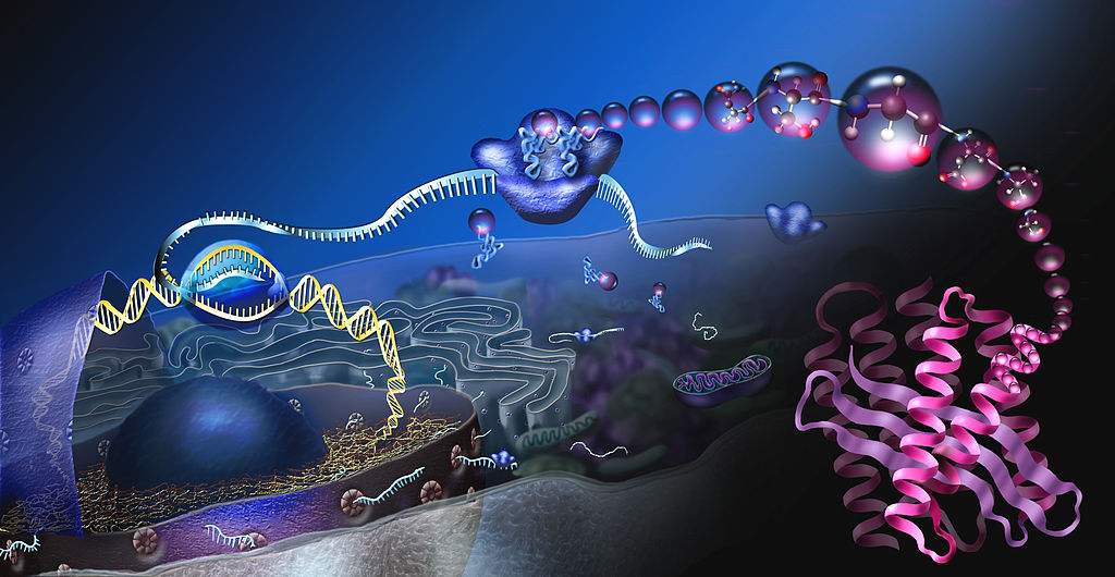
This amazing artwork (Figure 5.7.1) shows a process that takes place in the cells of all living things: the production of proteins. This process is called protein synthesis, and it actually consists of two processes — transcription and translation. In eukaryotic cells, transcription takes place in the nucleus. During transcription, DNA is used as a template to make a molecule of messenger RNA (mRNA). The molecule of mRNA then leaves the nucleus and goes to a ribosome in the cytoplasm, where translation occurs. During translation, the genetic code in mRNA is read and used to make a polypeptide. These two processes are summed up by the central dogma of molecular biology: DNA → RNA → Protein.
Transcription
Transcription is the first part of the central dogma of molecular biology: DNA → RNA. It is the transfer of genetic instructions in DNA to mRNA. During transcription, a strand of mRNA is made to complement a strand of DNA. You can see how this happens in Figure 5.7.2.
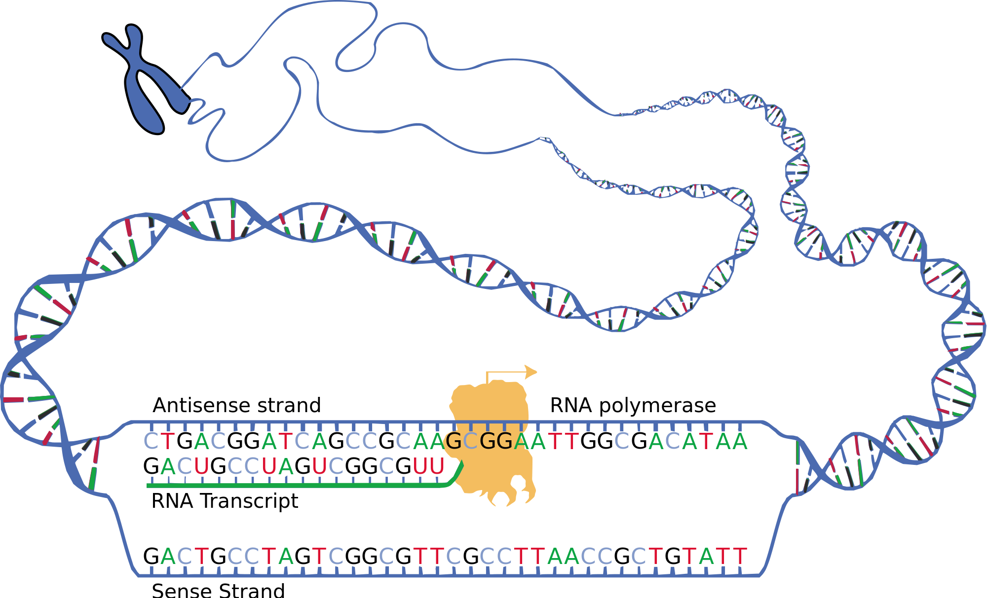
Transcription begins when the enzyme RNA polymerase binds to a region of a gene called the promoter sequence. This signals the DNA to unwind so the enzyme can “read” the bases of DNA. The two strands of DNA are named based on whether they will be used as a template for RNA or not. The strand that is used as a template is called the template strand, or can also be called the antisense strand. The sequence of bases on the opposite strand of DNA is called the non-coding or sense strand. Once the DNA has opened, and RNA polymerase has attached, the RNA polymerase moves along the DNA, adding RNA nucleotides to the growing mRNA strand. The template strand of DNA is used as to create mRNA through complementary base pairing. Once the mRNA strand is complete, and it detaches from DNA. The result is a strand of mRNA that is nearly identical to the coding strand DNA – the only difference being that DNA uses the base thymine, and the mRNA uses uracil in the place of thymine
Processing mRNA
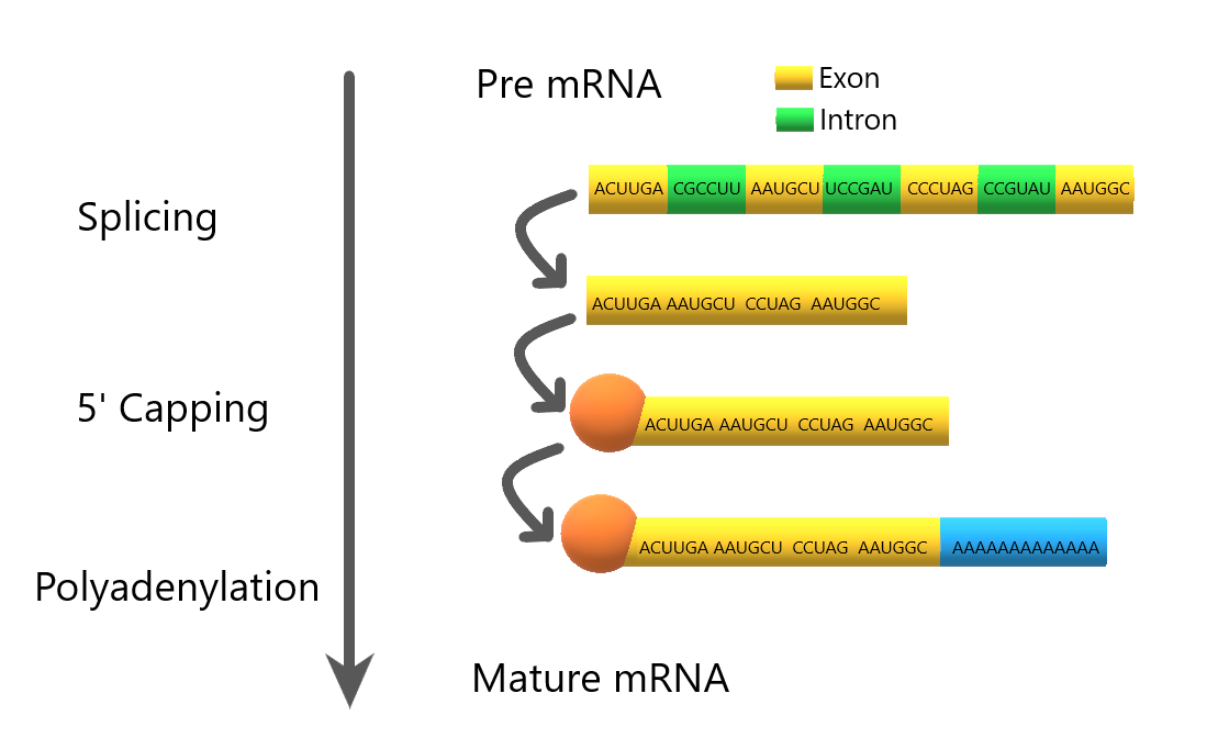
In eukaryotes, the new mRNA is not yet ready for translation. At this stage, it is called pre-mRNA, and it must go through more processing before it leaves the nucleus as mature mRNA. The processing may include splicing, editing, and polyadenylation. These processes modify the mRNA in various ways. Such modifications allow a single gene to be used to make more than one protein.
- Splicing removes introns from mRNA, as shown in Figure 5.7.3. Introns are regions that do not code for the protein. The remaining mRNA consists only of regions called exons that do code for the protein. The ribonucleoproteins in the diagram are small proteins in the nucleus that contain RNA and are needed for the splicing process.
- Editing changes some of the nucleotides in mRNA. For example, a human protein called APOB, which helps transport lipids in the blood, has two different forms because of editing. One form is smaller than the other because editing adds an earlier stop signal in mRNA.
- 5′ Capping adds a methylated cap to the “head” of the mRNA. This cap protects the mRNA from breaking down, and helps the ribosomes know where to bind to the mRNA
- Polyadenylation adds a “tail” to the mRNA. The tail consists of a string of As (adenine bases). It signals the end of mRNA. It is also involved in exporting mRNA from the nucleus, and it protects mRNA from enzymes that might break it down.
Translation
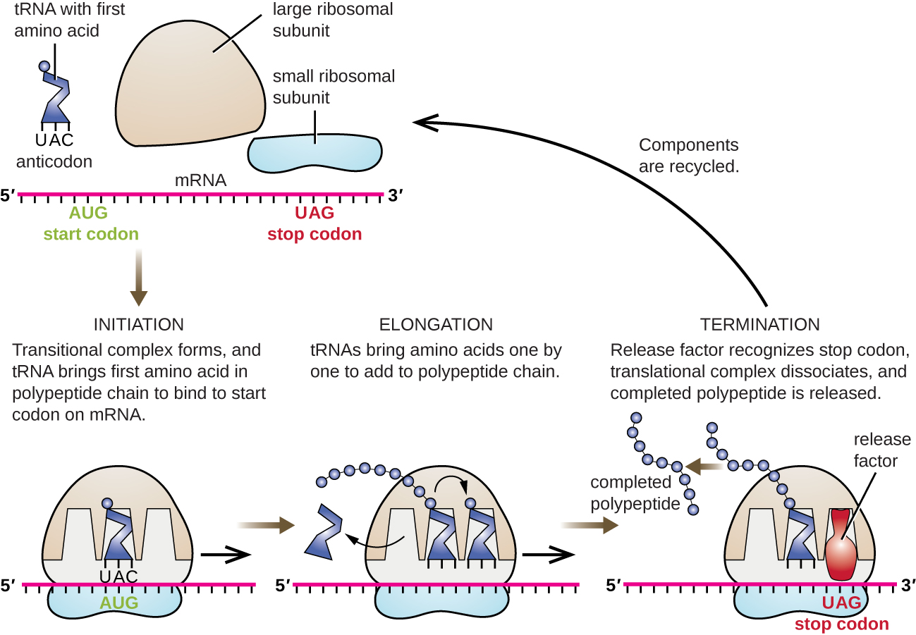
Translation is the second part of the central dogma of molecular biology: RNA → Protein. It is the process in which the genetic code in mRNA is read to make a protein. Translation is illustrated in Figure 5.7.4. After mRNA leaves the nucleus, it moves to a ribosome, which consists of rRNA and proteins. The ribosome reads the sequence of codons in mRNA, and molecules of tRNA bring amino acids to the ribosome in the correct sequence.
Translation occurs in three stages: Initiation, Elongation and Termination.
Initiation:
After transcription in the nucleus, the mRNA exits through a nuclear pore and enters the cytoplasm. At the region on the mRNA containing the methylated cap and the start codon, the small and large subunits of the ribosome bind to the mRNA. These are then joined by a tRNA which contains the anticodons matching the start codon on the mRNA. This group of molecues (mRNA, ribosome, tRNA) is called an initiation complex.
Elongation:
tRNA keep bringing amino acids to the growing polypeptide according to complementary base pairing between the codons on the mRNA and the anticodons on the tRNA. As a tRNA moves into the ribosome, its amino acid is transferred to the growing polypeptide. Once this transfer is complete, the tRNA leaves the ribosome, the ribosome moves one codon length down the mRNA, and a new tRNA enters with its corresponding amino acid. This process repeats and the polypeptide grows.
Termination:
At the end of the mRNA coding is a stop codon which will end the elongation stage. The stop codon doesn’t call for a tRNA, but instead for a type of protein called a release factor, which will cause the entire complex (mRNA, ribosome, tRNA, and polypeptide) to break apart, releasing all of the components.
Watch this video “Protein Synthesis (Updated) with the Amoeba Sisters” to see this process in action:
Protein Synthesis (Updated), Amoeba Sisters, 2018.
What Happens Next?
After a polypeptide chain is synthesized, it may undergo additional processes. For example, it may assume a folded shape due to interactions between its amino acids. It may also bind with other polypeptides or with different types of molecules, such as lipids or carbohydrates. Many proteins travel to the Golgi apparatus within the cytoplasm to be modified for the specific job they will do.7 Summary
5.7 Summary
- Protein synthesis is the process in which cells make proteins. It occurs in two stages: transcription and translation.
- Transcription is the transfer of genetic instructions in DNA to mRNA in the nucleus. It includes three steps: initiation, elongation, and termination. After the mRNA is processed, it carries the instructions to a ribosome in the cytoplasm.
- Translation occurs at the ribosome, which consists of rRNA and proteins. In translation, the instructions in mRNA are read, and tRNA brings the correct sequence of amino acids to the ribosome. Then, rRNA helps bonds form between the amino acids, producing a polypeptide chain.
- After a polypeptide chain is synthesized, it may undergo additional processing to form the finished protein.
5.7 Review Questions
- Relate protein synthesis and its two major phases to the central dogma of molecular biology.
- Explain how mRNA is processed before it leaves the nucleus.
- What additional processes might a polypeptide chain undergo after it is synthesized?
- Where does transcription take place in eukaryotes?
- Where does translation take place?
-
5.7 Explore More
Protein Synthesis, Teacher’s Pet, 2014.
Attributions
Figure 5.7.1
How proteins are made by Nicolle Rager, National Science Foundation on Wikimedia Commons is released into the public domain (https://en.wikipedia.org/wiki/Public_domain).
Figure 5.7.2
Transcription by National Human Genome Research Institute, (reworked and vectorized by Sulai) on Wikimedia Commons is released into the public domain (https://en.wikipedia.org/wiki/Public_domain).
Figure 5.7.3
Pre mRNA processing by Christine Miller is used under a CC BY-NC-SA 4.0 (https://creativecommons.org/licenses/by-nc-sa/4.0/) license.
Figure 5.7.4
Translation by CNX OpenStax on Wikimedia Commons is used under a CC BY 4.0 (https://creativecommons.org/licenses/by/4.0) license.
References
Amoeba Sisters. (2018, January 18) Protein synthesis (Updated). YouTube. https://www.youtube.com/watch?v=oefAI2x2CQM&feature=youtu.be
Parker, N., Schneegurt, M., Thi Tu, A-H., Lister, P., Forster, B.M. (2016, November 1). Microbiology [online]. Figure 11.15 Translation in bacteria begins with the formation of the initiation complex. In Microbiology (Section 11-4). OpenStax. https://openstax.org/books/microbiology/pages/11-4-protein-synthesis-translation
Teacher’s Pet. (2014, December 7). Protein synthesis. YouTube. https://www.youtube.com/watch?v=2zAGAmTkZNY&feature=youtu.be
A biomolecule consisting of carbon (C), hydrogen (H) and oxygen (O) atoms, usually with a hydrogen–oxygen atom ratio of 2:1. Complex carbohydrates are polymers made from monomers of simple carbohydrates, also termed monosaccharides.
Image shows a labelled diagram highlighting the location of the uterus in pregnancy. The developing fetus, amniotic fluid and placenta are all housed in the uterus, which stretches to many times its regular size to accommodate pregnancy.
Attracted to water.
Created by CK-12 Foundation/Adapted by Christine Miller
Communicating with Urine
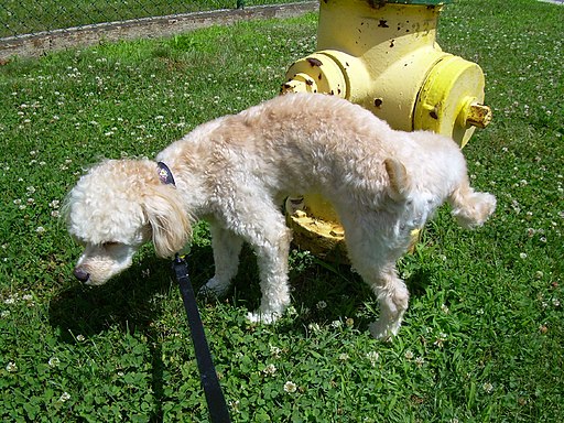
Why do dogs pee on fire hydrants? Besides “having to go,” they are marking their territory with chemicals in their urine called pheromones. It’s a form of communication, in which they are “saying” with odors that the yard is theirs and other dogs should stay away. In addition to fire hydrants, dogs may urinate on fence posts, trees, car tires, and many other objects. Urination in dogs, as in people, is usually a voluntary process controlled by the brain. The process of forming urine — which occurs in the kidneys — occurs constantly, and is not under voluntary control. What happens to all the urine that forms in the kidneys? It passes from the kidneys through the other organs of the urinary system, starting with the ureters.
Ureters
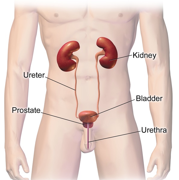
As shown in Figure 16.5.2, ureters are tube-like structures that connect the kidneys with the urinary bladder. They are paired structures, with one ureter for each kidney. In adults, ureters are between 25 and 30 cm (about 10–12 in) long and about 3 to 4 mm in diameter.
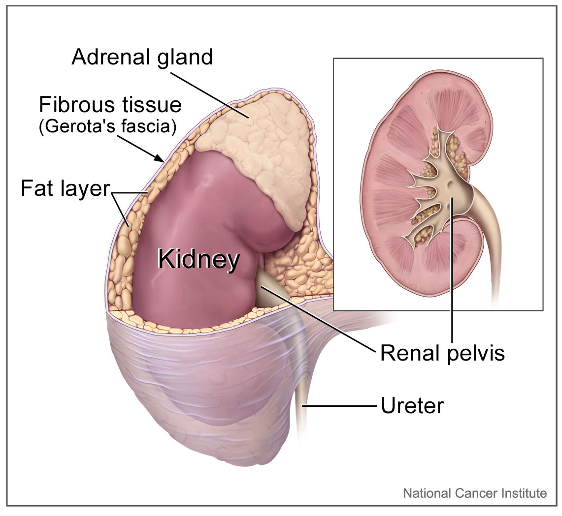
Each ureter arises in the pelvis of a kidney (the renal pelvis in Figure 16.5.3). It then passes down the side of the kidney, and finally enters the back of the bladder. At the entrance to the bladder, the ureters have sphincters that prevent the backflow of urine.
The walls of the ureters are composed of multiple layers of different types of tissues. The innermost layer is a special type of epithelium, called transitional epithelium. Unlike the epithelium lining most organs, transitional epithelium is capable of stretching and does not produce mucus. It lines much of the urinary system, including the renal pelvis, bladder, and much of the urethra, in addition to the ureters. Transitional epithelium allows these organs to stretch and expand as they fill with urine or allow urine to pass through. The next layer of the ureter walls is made up of loose connective tissue containing elastic fibres, nerves, and blood and lymphatic vessels. After this layer are two layers of smooth muscles, an inner circular layer, and an outer longitudinal layer. The smooth muscle layers can contract in waves of peristalsis to propel urine down the ureters from the kidneys to the urinary bladder. The outermost layer of the ureter walls consists of fibrous tissue.
Urinary Bladder
The urinary bladder is a hollow, muscular, and stretchy organ that rests on the pelvic floor. It collects and stores urine from the kidneys before the urine is eliminated through urination. As shown in Figure 16.5.4, urine enters the urinary bladder from the ureters through two ureteral openings on either side of the back wall of the bladder. Urine leaves the bladder through a sphincter called the internal urethral sphincter. When the sphincter relaxes and opens, it allows urine to flow out of the bladder and into the urethra.
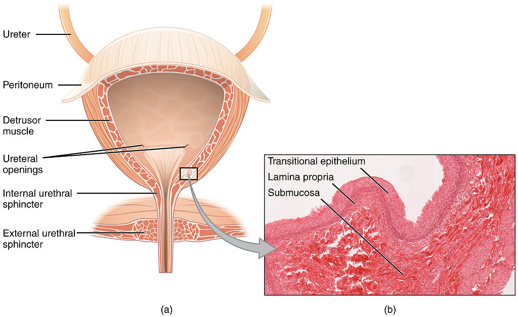
Like the ureters, the bladder is lined with transitional epithelium, which can flatten out and stretch as needed as the bladder fills with urine. The next layer (lamina propria) is a layer of loose connective tissue, nerves, and blood and lymphatic vessels. This is followed by a submucosa layer, which connects the lining of the bladder with the detrusor muscle in the walls of the bladder. The outer covering of the bladder is peritoneum, which is a smooth layer of epithelial cells that lines the abdominal cavity and covers most abdominal organs.
The detrusor muscle in the wall of the bladder is made of smooth muscle fibres controlled by both the autonomic and somatic nervous systems. As the bladder fills, the detrusor muscle automatically relaxes to allow it to hold more urine. When the bladder is about half full, the stretching of the walls triggers the sensation of needing to urinate. When the individual is ready to void, conscious nervous signals cause the detrusor muscle to contract, and the internal urethral sphincter to relax and open. As a result, urine is forcefully expelled out of the bladder and into the urethra.
Urethra
The urethra is a tube that connects the urinary bladder to the external urethral orifice, which is the opening of the urethra on the surface of the body. As shown in Figure 16.5.5, the urethra in males travels through the penis, so it is much longer than the urethra in females. In males, the urethra averages about 20 cm (about 7.8 in) long, whereas in females, it averages only about 4.8 cm (about 1.9 in) long. In males, the urethra carries semen (as well as urine), but in females, it carries only urine. In addition, in males, the urethra passes through the prostate gland (part of the reproductive system) which is absent in women.
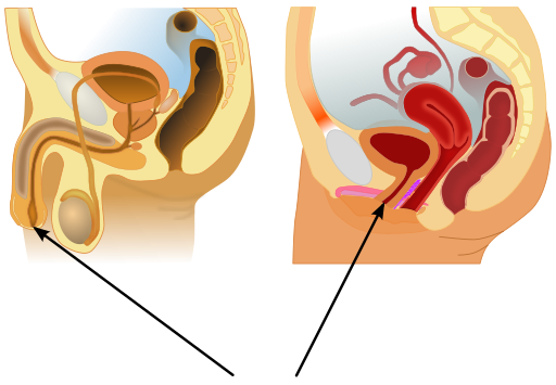
Like the ureters and bladder, the proximal (closer to the bladder) two-thirds of the urethra are lined with transitional epithelium. The distal (farther from the bladder) third of the urethra is lined with mucus-secreting epithelium. The mucus helps protect the epithelium from urine, which is corrosive. Below the epithelium is loose connective tissue, and below that are layers of smooth muscle that are continuous with the muscle layers of the urinary bladder. When the bladder contracts to forcefully expel urine, the smooth muscle of the urethra relaxes to allow the urine to pass through.
In order for urine to leave the body through the external urethral orifice, the external urethral sphincter must relax and open. This sphincter is a striated muscle that is controlled by the somatic nervous system, so it is under conscious, voluntary control in most people (exceptions are infants, some elderly people, and patients with certain injuries or disorders). The muscle can be held in a contracted state and hold in the urine until the person is ready to urinate. Following urination, the smooth muscle lining the urethra automatically contracts to re-establish muscle tone, and the individual consciously contracts the external urethral sphincter to close the external urethral opening.
16.5 Summary
- Ureters are tube-like structures that connect the kidneys with the urinary bladder. Each ureter arises at the renal pelvis of a kidney and travels down through the abdomen to the urinary bladder. The walls of the ureter contain smooth muscle that can contract to push urine through the ureter by peristalsis. The walls are lined with transitional epithelium that can expand and stretch.
- The urinary bladder is a hollow, muscular organ that rests on the pelvic floor. It is also lined with transitional epithelium. The function of the bladder is to collect and store urine from the kidneys before the urine is eliminated through urination. Filling of the bladder triggers the sensation of needing to urinate. When a conscious decision to urinate is made, the detrusor muscle in the bladder wall contracts and forces urine out of the bladder and into the urethra.
- The urethra is a tube that connects the urinary bladder to the external urethral orifice. Somatic nerves control the sphincter at the distal end of the urethra. This allows the opening of the sphincter for urination to be under voluntary control.
16.5 Review Questions
- What are ureters? Describe the location of the ureters relative to other urinary tract organs.
- Identify layers in the walls of a ureter. How do they contribute to the ureter’s function?
- Describe the urinary bladder. What is the function of the urinary bladder?
-
- How does the nervous system control the urinary bladder?
- What is the urethra?
- How does the nervous system control urination?
- Identify the sphincters that are located along the pathway from the ureters to the external urethral orifice.
- What are two differences between the male and female urethra?
- When the bladder muscle contracts, the smooth muscle in the walls of the urethra _________ .
16.5 Explore More
https://youtu.be/2Brajdazp1o
The taboo secret to better health | Molly Winter, TED. 2016.
https://youtu.be/dg4_deyHLvQ
What Happens When You Hold Your Pee? SciShow, 2016.
Attributions
Figure 16.5.1
Cliche by Jackie on Wikimedia Common s is used under a CC BY 2.0 (https://creativecommons.org/licenses/by/2.0) license.
Figure 16.5.2
Urinary System Male by BruceBlaus on Wikimedia Commons is used under a CC BY-SA 4.0 (https://creativecommons.org/licenses/by-sa/4.0) license.
Figure 16.5.3
Adrenal glands on Kidney by NCI Public Domain by Alan Hoofring (Illustrator) /National Cancer Institute (photo ID 4355) on Wikimedia Commons is in the public domain (https://en.wikipedia.org/wiki/Public_domain).
Figure 16.5.4
2605_The_Bladder by OpenStax College on Wikimedia Commons is used under a CC BY 3.0 (https://creativecommons.org/licenses/by/3.0) license. (Micrograph originally provided by the Regents of the University of Michigan Medical School © 2012.)
Figure 16.5.5
512px-Male_and_female_urethral_openings.svg by andrybak (derivative work) on Wikimedia Commons is used under a CC BY-SA 3.0 (https://creativecommons.org/licenses/by-sa/3.0) license. (Original: Male anatomy blank.svg: alt.sex FAQ, derivative work: Tsaitgaist Female anatomy with g-spot.svg: Tsaitgaist.)
References
Betts, J. G., Young, K.A., Wise, J.A., Johnson, E., Poe, B., Kruse, D.H., Korol, O., Johnson, J.E., Womble, M., DeSaix, P. (2013, June 19). Figure 25.4 Bladder (a) Anterior cross section of the bladder. (b) The detrusor muscle of the bladder (source: monkey tissue) LM × 448 [digital image]. In Anatomy and Physiology (Section 7.3). OpenStax. https://openstax.org/books/anatomy-and-physiology/pages/25-2-gross-anatomy-of-urine-transport
SciShow. (2016, January 22). What happens when you hold your pee? YouTube. https://www.youtube.com/watch?v=dg4_deyHLvQ&feature=youtu.be
TED. (2016, September 2). The taboo secret to better health | Molly Winter. YouTube. https://www.youtube.com/watch?v=2Brajdazp1o&feature=youtu.be
A central organelle containing hereditary material.
Long chains of hydrocarbons with a carboxyl group and a methyl group at opposite ends. Can be either saturated, containing mostly single bonds between adjacent carbons, or unsaturated, containing many double bonds between adjacent carbons.
A large complex of RNA and protein which acts as the site of RNA translation, building proteins from amino acids using messenger RNA as a template.
The jellylike material that makes up much of a cell inside the cell membrane, and, in eukaryotic cells, surrounds the nucleus. The organelles of eukaryotic cells, such as mitochondria, the endoplasmic reticulum, and (in green plants) chloroplasts, are contained in the cytoplasm.
A class of biological molecule consisting of linked monomers of amino acids and which are the most versatile macromolecules in living systems and serve crucial functions in essentially all biological processes.
A sequence of 3 DNA or RNA nucleotides that corresponds with a specific amino acid or stop signal during protein synthesis.
A sequence of 3 DNA or RNA nucleotides that corresponds with a specific amino acid or stop signal during protein synthesis.
An antibody, also known as an immunoglobulin, is a large, Y-shaped protein produced mainly by plasma cells that is used by the immune system to neutralize pathogens such as pathogenic bacteria and viruses.
Amino acids are organic compounds that combine to form proteins.
A substance that is insoluble in water. Examples include fats, oils and cholesterol. Lipids are made from monomers such as glycerol and fatty acids.
A biomolecule consisting of carbon (C), hydrogen (H) and oxygen (O) atoms, usually with a hydrogen–oxygen atom ratio of 2:1. Complex carbohydrates are polymers made from monomers of simple carbohydrates, also termed monosaccharides.
A membrane-bound organelle found in eukaryotic cells made up of a series of flattened stacked pouches with the purpose of collecting and dispatching protein and lipid products received from the endoplasmic reticulum (ER). Also referred to as the Golgi complex or the Golgi body.

