5.5 RNA
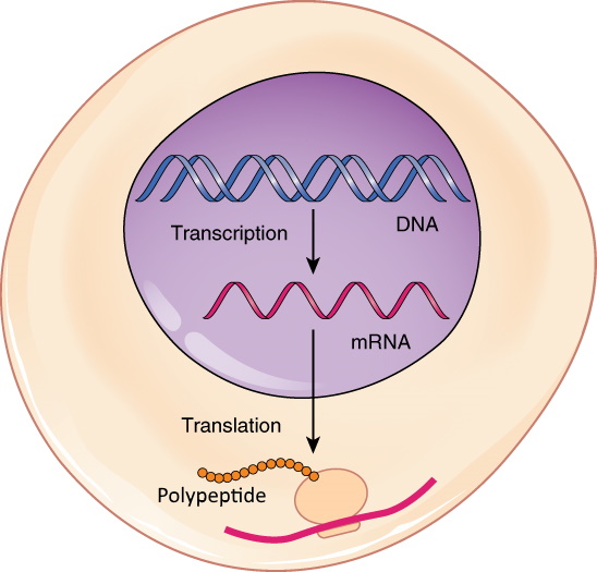
A Deceptively Simple Model
This simple model sums up one of the most important ideas in biology, which is called the central dogma of molecular biology (you’ll read more about it below). You probably recognize the spiral-shaped structure in the nucleus. It represents a molecule of DNA, the biochemical molecule that stores genetic information in most living cells. The yellow chain represents a newly formed polypeptide — the beginning stage of creating a protein. Proteins are the class of biochemical molecules that carry out virtually all life processes. What is the structure in the center of the model? It appears to resemble DNA, but it is smaller and simpler. This molecule is the key to the central dogma, and it may have been the first type of biochemical molecule to evolve.
Central Dogma of Molecular Biology
DNA is found in chromosomes. In eukaryotic cells, chromosomes always remain in the nucleus, but proteins are made at ribosomes in the cytoplasm. How do the instructions in DNA get to the site of protein synthesis outside the nucleus?
Another type of nucleic acid is responsible. This nucleic acid is RNA, or ribonucleic acid. RNA is a small molecule that can squeeze through pores in the nuclear membrane. It carries the information from DNA in the nucleus to a ribosome in the cytoplasm and then helps assemble the protein. In short:
DNA → RNA → Protein
This expresses in words what the diagram in Figure 5.5.1 shows. The genetic instructions encoded in DNA in the nucleus are transcribed to RNA. Then, RNA carries the instructions to a ribosome in the cytoplasm, where they are translated into a protein. Discovering this sequence of events was a major milestone in molecular biology. It’s called the central dogma of molecular biology.
Introducing RNA
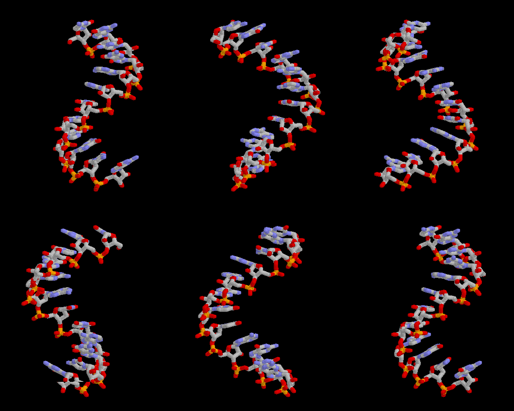
DNA alone cannot “tell” your cells how to make proteins. It needs the help of RNA, the other main player in the central dogma of molecular biology. Like DNA, RNA is a nucleic acid, so it consists of repeating nucleotides bonded together to form a polynucleotide chain. RNA differs from DNA in several ways: it exists as a single stranded molecule, contains the sugar ribose (as opposed to deoxyribose) and uses the base uracil instead of thymine.
Functions of RNA
The main function of RNA is to help make proteins. There are three main types of RNA involved in protein synthesis:
-
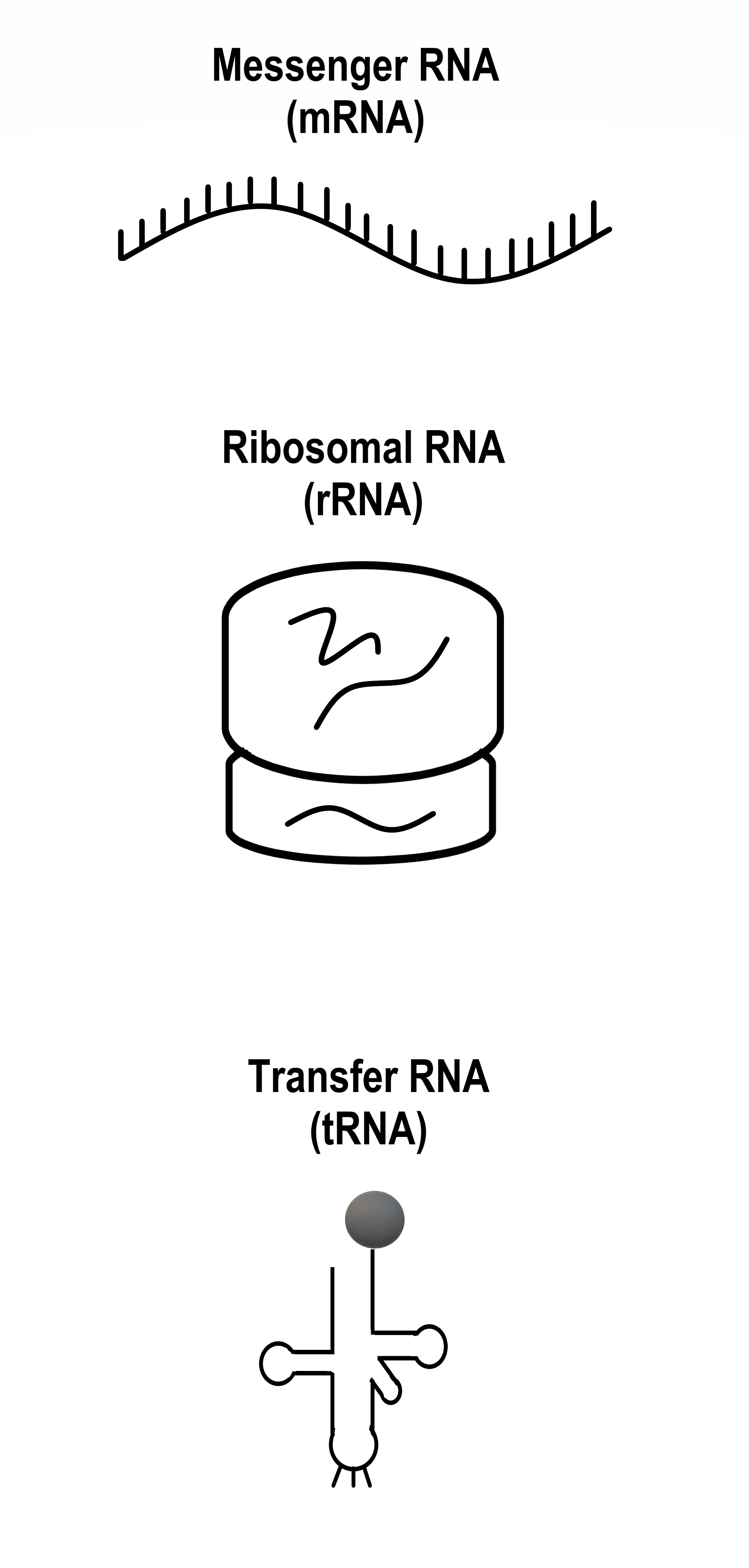
Figure 5.5.3 The three types of RNA take very different forms. Messenger RNA (mRNA) copies (or transcribes) the genetic instructions from DNA in the nucleus and carries them to the cytoplasm.
- Ribosomal RNA (rRNA) helps form ribosomes, where proteins are assembled. Ribosomes also contain proteins.
- Transfer RNA (tRNA) brings amino acids to ribosomes, where rRNA catalyzes the formation of chemical bonds between them to form a protein.
In section 5.7 Protein Synthesis, you can read in detail about how these three types of RNA build primary structure of proteins.
RNA is a very versatile molecule which plays multiple roles in living things. In addition to helping to make proteins, for example, there are RNA molecules that regulate the expression of genes, and RNA molecules that catalyze other biochemical reactions needed to sustain life. Because of the diversity of roles that RNA molecules play, they have been called the Swiss Army knives of the cellular world.
It’s an RNA World
The function of DNA is to store genetic information inside cells. It does this job well, but that’s about all it can do. DNA can’t act as an enzyme, for example, to catalyze biochemical reactions that are needed to keep us alive. Proteins are needed for this and many other life functions. Proteins work exceptionally well to keep us alive, but they are unable to store genetic information. Proteins need DNA for that. Without DNA, proteins could not exist. On the other hand, without proteins, DNA could not survive. This poses a chicken-and-egg sort of problem: Which evolved first? DNA or proteins?
Some scientists think that the answer is neither. They speculate instead that RNA was the first biochemical to evolve. The reason? RNA can do more than one job. It can store information as DNA does, but it can also perform various jobs (such as catalysis) to keep cells alive, as proteins do. The idea that RNA was the first biochemical to evolve, predating both DNA and proteins, is called the RNA world hypothesis. According to this hypothesis, billions of years ago, RNA molecules evolved that could both survive and make copies of themselves. According to the hypothesis, early RNA molecules eventually evolved the ability to make proteins, and at some point RNA mutated to form DNA.
Feature: Reliable Sources
The RNA world hypothesis has not gained enough support in the scientific community to be accepted as a scientific theory. In fact, there are probably as many detractors as supporters of the hypothesis. Do a web search to learn more about the RNA world hypothesis and the evidence and arguments for and against it. When weighing the information you gather, consider the likely reliability of the different websites you visit. Based on what you determine are the most reliable sources and the most convincing arguments, form your own opinion about the hypothesis. You may decide to accept or reject the hypothesis. Alternatively, you may decide to reserve judgement until — or if — more evidence or arguments are forthcoming.
5.5 Summary
- The central dogma of molecular biology can be summed up as: DNA → RNA → Protein. This means that the genetic instructions encoded in DNA are first transcribed to RNA, and then from RNA they are translated into a protein.
- Like DNA, RNA is a nucleic acid. Unlike DNA, RNA consists of just one polynucleotide chain instead of two, contains the base uracil instead of thymine, and contains the sugar ribose instead of deoxyribose.
- The main function of RNA is helping to make proteins. There are three main types of RNA involved in protein synthesis: messenger RNA (mRNA), ribosomal RNA (rRNA), and transfer RNA (tRNA). RNA has additional functions, including regulating gene expression and catalyzing other biochemical reactions.
- According to the RNA world hypothesis, RNA was the first type of biochemical molecule to evolve, predating both DNA and proteins. The hypothesis is based mainly on the multiple functions of RNA, which can store genetic information like DNA and carry out life processes (like proteins).
5.5 Review Questions
- State the central dogma of molecular biology.
- Drag and drop to compare the structure and function of DNA and RNA:
3.
4.
5.5 Explore More
The RNA Origin of Life, NOVA PBS Official, 2014.
DNA vs RNA (Updated), Amoeba Sisters, 2019.
Attributions
Figure 5.5.1
From DNA to Protein: Transcription through Translation by OpenStax College on Wikimedia Commons is used under a CC BY 4.0 (https://creativecommons.org/licenses/by/4.0/) license.
Figure 5.5.2
Molbio-Header by Squidonius on Wikimedia Commons is released into the public domain (https://en.wikipedia.org/wiki/Public_domain).
Figure 5.5.2
ARNm-Rasmol by Corentin Le Reun on Wikimedia Commons is is released into the public domain (https://en.wikipedia.org/wiki/Public_domain).ublic domain.
References
Amoeba Sisters. (2019, August 29). DNA vs RNA (Updated). YouTube. https://www.youtube.com/watch?v=JQByjprj_mA&feature=youtu.be
Betts, J. G., Young, K.A., Wise, J.A., Johnson, E., Poe, B., Kruse, D.H., Korol, O., Johnson, J.E., Womble, M., DeSaix, P. (2013, April 25). Figure 3.29 From DNA to Protein: Transcription through Translation [digital image]. In Anatomy and Physiology. OpenStax. https://openstax.org/books/anatomy-and-physiology/pages/3-4-protein-synthesis#fig-ch03_04_05
NOVA PBS Official. (2014, April 23). The RNA origin of life. YouTube. https://www.youtube.com/watch?v=VYQQD0KNOis&feature=youtu.be
Wikipedia contributors. (2020, June 28). RNA world. In Wikipedia. https://en.wikipedia.org/w/index.php?title=RNA_world&oldid=964998696
A central organelle containing hereditary material.
A class of biological molecule consisting of linked monomers of amino acids and which are the most versatile macromolecules in living systems and serve crucial functions in essentially all biological processes.
A threadlike structure of nucleic acids and protein found in the nucleus of most living cells, carrying genetic information in the form of genes.
Created by CK-12 Foundation/Adapted by Christine Miller
Communicating with Urine
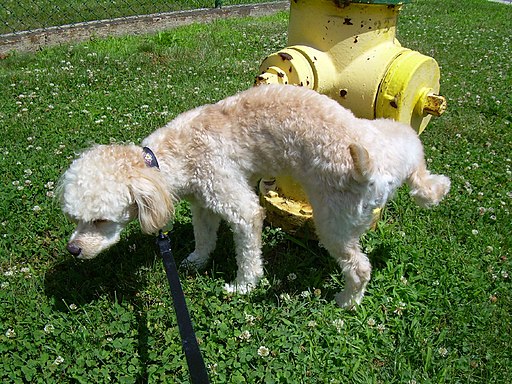
Why do dogs pee on fire hydrants? Besides “having to go,” they are marking their territory with chemicals in their urine called pheromones. It’s a form of communication, in which they are “saying” with odors that the yard is theirs and other dogs should stay away. In addition to fire hydrants, dogs may urinate on fence posts, trees, car tires, and many other objects. Urination in dogs, as in people, is usually a voluntary process controlled by the brain. The process of forming urine — which occurs in the kidneys — occurs constantly, and is not under voluntary control. What happens to all the urine that forms in the kidneys? It passes from the kidneys through the other organs of the urinary system, starting with the ureters.
Ureters
As shown in Figure 16.5.2, ureters are tube-like structures that connect the kidneys with the urinary bladder. They are paired structures, with one ureter for each kidney. In adults, ureters are between 25 and 30 cm (about 10–12 in) long and about 3 to 4 mm in diameter.
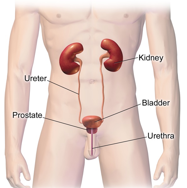
Each ureter arises in the pelvis of a kidney (the renal pelvis in Figure 16.5.3). It then passes down the side of the kidney, and finally enters the back of the bladder. At the entrance to the bladder, the ureters have sphincters that prevent the backflow of urine.
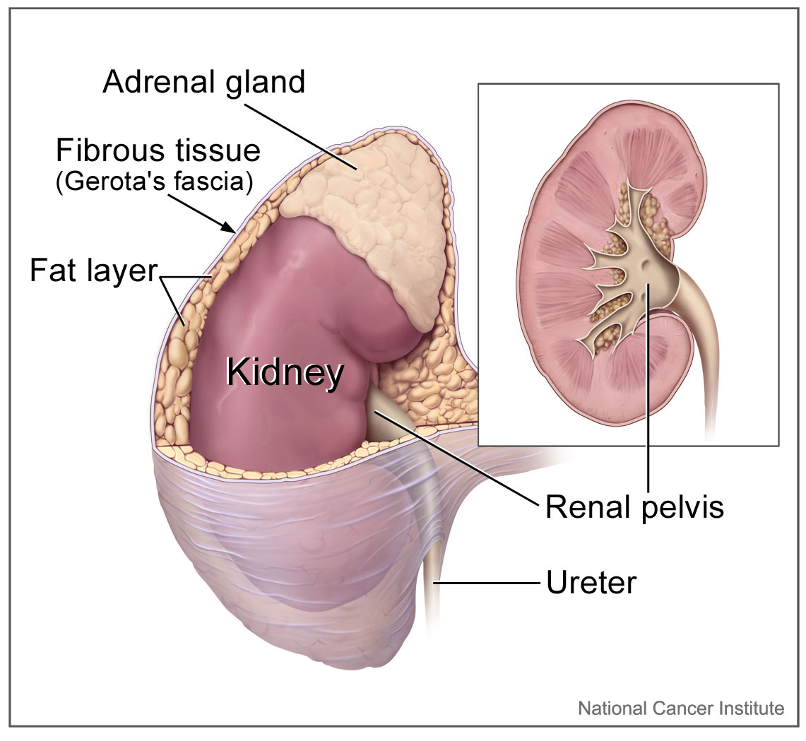
The walls of the ureters are composed of multiple layers of different types of tissues. The innermost layer is a special type of epithelium, called transitional epithelium. Unlike the epithelium lining most organs, transitional epithelium is capable of stretching and does not produce mucus. It lines much of the urinary system, including the renal pelvis, bladder, and much of the urethra, in addition to the ureters. Transitional epithelium allows these organs to stretch and expand as they fill with urine or allow urine to pass through. The next layer of the ureter walls is made up of loose connective tissue containing elastic fibres, nerves, and blood and lymphatic vessels. After this layer are two layers of smooth muscles, an inner circular layer, and an outer longitudinal layer. The smooth muscle layers can contract in waves of peristalsis to propel urine down the ureters from the kidneys to the urinary bladder. The outermost layer of the ureter walls consists of fibrous tissue.
Urinary Bladder
The urinary bladder is a hollow, muscular, and stretchy organ that rests on the pelvic floor. It collects and stores urine from the kidneys before the urine is eliminated through urination. As shown in Figure 16.5.4, urine enters the urinary bladder from the ureters through two ureteral openings on either side of the back wall of the bladder. Urine leaves the bladder through a sphincter called the internal urethral sphincter. When the sphincter relaxes and opens, it allows urine to flow out of the bladder and into the urethra.
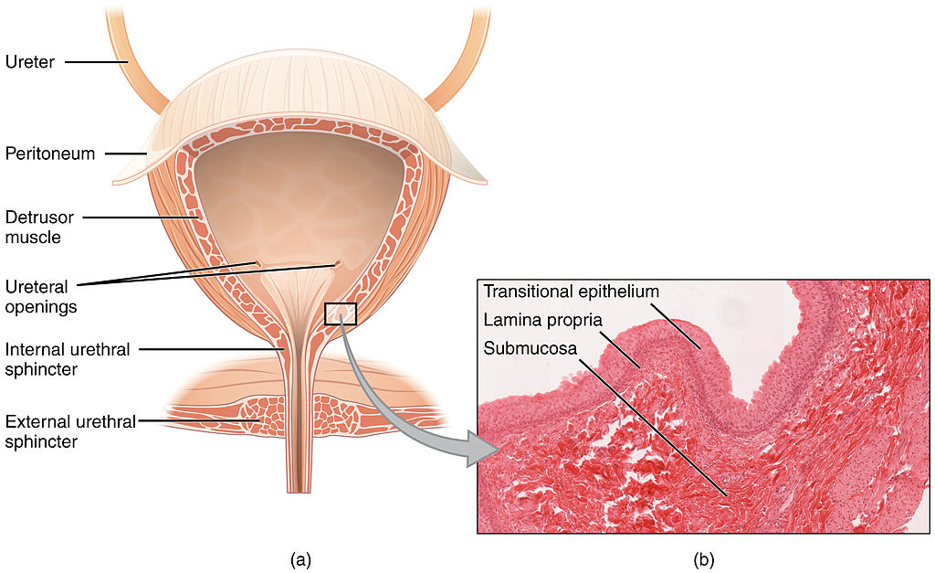
Like the ureters, the bladder is lined with transitional epithelium, which can flatten out and stretch as needed as the bladder fills with urine. The next layer (lamina propria) is a layer of loose connective tissue, nerves, and blood and lymphatic vessels. This is followed by a submucosa layer, which connects the lining of the bladder with the detrusor muscle in the walls of the bladder. The outer covering of the bladder is peritoneum, which is a smooth layer of epithelial cells that lines the abdominal cavity and covers most abdominal organs.
The detrusor muscle in the wall of the bladder is made of smooth muscle fibres controlled by both the autonomic and somatic nervous systems. As the bladder fills, the detrusor muscle automatically relaxes to allow it to hold more urine. When the bladder is about half full, the stretching of the walls triggers the sensation of needing to urinate. When the individual is ready to void, conscious nervous signals cause the detrusor muscle to contract, and the internal urethral sphincter to relax and open. As a result, urine is forcefully expelled out of the bladder and into the urethra.
Urethra
The urethra is a tube that connects the urinary bladder to the external urethral orifice, which is the opening of the urethra on the surface of the body. As shown in Figure 16.5.5, the urethra in males travels through the penis, so it is much longer than the urethra in females. In males, the urethra averages about 20 cm (about 7.8 in) long, whereas in females, it averages only about 4.8 cm (about 1.9 in) long. In males, the urethra carries semen (as well as urine), but in females, it carries only urine. In addition, in males, the urethra passes through the prostate gland (part of the reproductive system) which is absent in women.
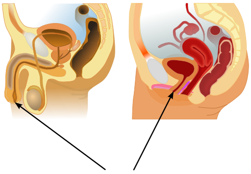
Like the ureters and bladder, the proximal (closer to the bladder) two-thirds of the urethra are lined with transitional epithelium. The distal (farther from the bladder) third of the urethra is lined with mucus-secreting epithelium. The mucus helps protect the epithelium from urine, which is corrosive. Below the epithelium is loose connective tissue, and below that are layers of smooth muscle that are continuous with the muscle layers of the urinary bladder. When the bladder contracts to forcefully expel urine, the smooth muscle of the urethra relaxes to allow the urine to pass through.
In order for urine to leave the body through the external urethral orifice, the external urethral sphincter must relax and open. This sphincter is a striated muscle that is controlled by the somatic nervous system, so it is under conscious, voluntary control in most people (exceptions are infants, some elderly people, and patients with certain injuries or disorders). The muscle can be held in a contracted state and hold in the urine until the person is ready to urinate. Following urination, the smooth muscle lining the urethra automatically contracts to re-establish muscle tone, and the individual consciously contracts the external urethral sphincter to close the external urethral opening.
16.5 Summary
- Ureters are tube-like structures that connect the kidneys with the urinary bladder. Each ureter arises at the renal pelvis of a kidney and travels down through the abdomen to the urinary bladder. The walls of the ureter contain smooth muscle that can contract to push urine through the ureter by peristalsis. The walls are lined with transitional epithelium that can expand and stretch.
- The urinary bladder is a hollow, muscular organ that rests on the pelvic floor. It is also lined with transitional epithelium. The function of the bladder is to collect and store urine from the kidneys before the urine is eliminated through urination. Filling of the bladder triggers the sensation of needing to urinate. When a conscious decision to urinate is made, the detrusor muscle in the bladder wall contracts and forces urine out of the bladder and into the urethra.
- The urethra is a tube that connects the urinary bladder to the external urethral orifice. Somatic nerves control the sphincter at the distal end of the urethra. This allows the opening of the sphincter for urination to be under voluntary control.
16.5 Review Questions
- What are ureters? Describe the location of the ureters relative to other urinary tract organs.
- Identify layers in the walls of a ureter. How do they contribute to the ureter’s function?
- Describe the urinary bladder. What is the function of the urinary bladder?
-
- How does the nervous system control the urinary bladder?
- What is the urethra?
- How does the nervous system control urination?
- Identify the sphincters that are located along the pathway from the ureters to the external urethral orifice.
- What are two differences between the male and female urethra?
- When the bladder muscle contracts, the smooth muscle in the walls of the urethra _________ .
16.5 Explore More
https://youtu.be/2Brajdazp1o
The taboo secret to better health | Molly Winter, TED. 2016.
https://youtu.be/dg4_deyHLvQ
What Happens When You Hold Your Pee? SciShow, 2016.
Attributions
Figure 16.5.1
Cliche by Jackie on Wikimedia Common s is used under a CC BY 2.0 (https://creativecommons.org/licenses/by/2.0) license.
Figure 16.5.2
Urinary System Male by BruceBlaus on Wikimedia Commons is used under a CC BY-SA 4.0 (https://creativecommons.org/licenses/by-sa/4.0) license.
Figure 16.5.3
Adrenal glands on Kidney by NCI Public Domain by Alan Hoofring (Illustrator) /National Cancer Institute (photo ID 4355) on Wikimedia Commons is in the public domain (https://en.wikipedia.org/wiki/Public_domain).
Figure 16.5.4
2605_The_Bladder by OpenStax College on Wikimedia Commons is used under a CC BY 3.0 (https://creativecommons.org/licenses/by/3.0) license. (Micrograph originally provided by the Regents of the University of Michigan Medical School © 2012.)
Figure 16.5.5
512px-Male_and_female_urethral_openings.svg by andrybak (derivative work) on Wikimedia Commons is used under a CC BY-SA 3.0 (https://creativecommons.org/licenses/by-sa/3.0) license. (Original: Male anatomy blank.svg: alt.sex FAQ, derivative work: Tsaitgaist Female anatomy with g-spot.svg: Tsaitgaist.)
References
Betts, J. G., Young, K.A., Wise, J.A., Johnson, E., Poe, B., Kruse, D.H., Korol, O., Johnson, J.E., Womble, M., DeSaix, P. (2013, June 19). Figure 25.4 Bladder (a) Anterior cross section of the bladder. (b) The detrusor muscle of the bladder (source: monkey tissue) LM × 448 [digital image]. In Anatomy and Physiology (Section 7.3). OpenStax. https://openstax.org/books/anatomy-and-physiology/pages/25-2-gross-anatomy-of-urine-transport
SciShow. (2016, January 22). What happens when you hold your pee? YouTube. https://www.youtube.com/watch?v=dg4_deyHLvQ&feature=youtu.be
TED. (2016, September 2). The taboo secret to better health | Molly Winter. YouTube. https://www.youtube.com/watch?v=2Brajdazp1o&feature=youtu.be
A large complex of RNA and protein which acts as the site of RNA translation, building proteins from amino acids using messenger RNA as a template.
A biomolecule consisting of carbon (C), hydrogen (H) and oxygen (O) atoms, usually with a hydrogen–oxygen atom ratio of 2:1. Complex carbohydrates are polymers made from monomers of simple carbohydrates, also termed monosaccharides.
A complex organic substance present in living cells, especially DNA or RNA, whose molecules consist of many nucleotides linked in a long chain.
A scientific law is a statement based on repeated experimental observations that describes some aspect of the world.
To arrange a group of living things into classes or categories according to shared qualities or characteristics.
To arrange a group of living things into classes or categories according to shared qualities or characteristics.
A biomolecule consisting of carbon (C), hydrogen (H) and oxygen (O) atoms, usually with a hydrogen–oxygen atom ratio of 2:1. Complex carbohydrates are polymers made from monomers of simple carbohydrates, also termed monosaccharides.
Long chains of hydrocarbons with a carboxyl group and a methyl group at opposite ends. Can be either saturated, containing mostly single bonds between adjacent carbons, or unsaturated, containing many double bonds between adjacent carbons.
Created by CK-12 Foundation/Adapted by Christine Miller
Marvelous Muscles
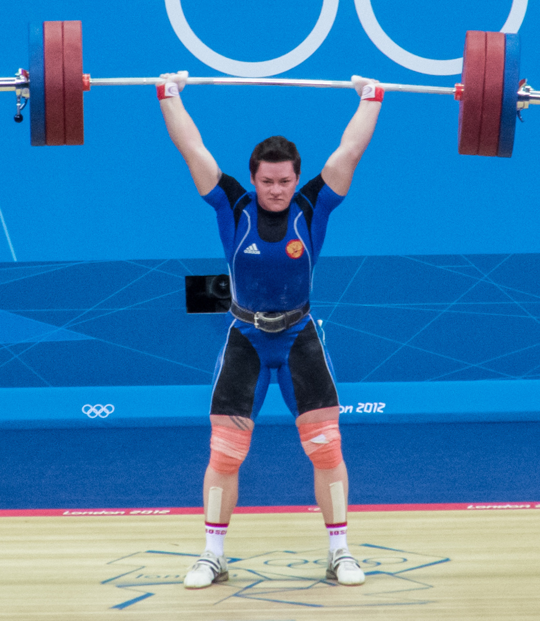
Does the word muscle make you think of the well-developed muscles of a weightlifter, like the woman in Figure 12.2.1? Her name is Natalia Zabolotnaya, and she’s a Russian Olympian. The muscles that are used to lift weights are easy to feel and see, but they aren’t the only muscles in the human body. Many muscles are deep within the body, where they form the walls of internal organs and other structures. You can flex your biceps at will, but you can’t control internal muscles like these. It’s a good thing that these internal muscles work without any conscious effort on your part, because movement of these muscles is essential for survival. Muscles are the organs of the muscular system.
What Is the Muscular System?
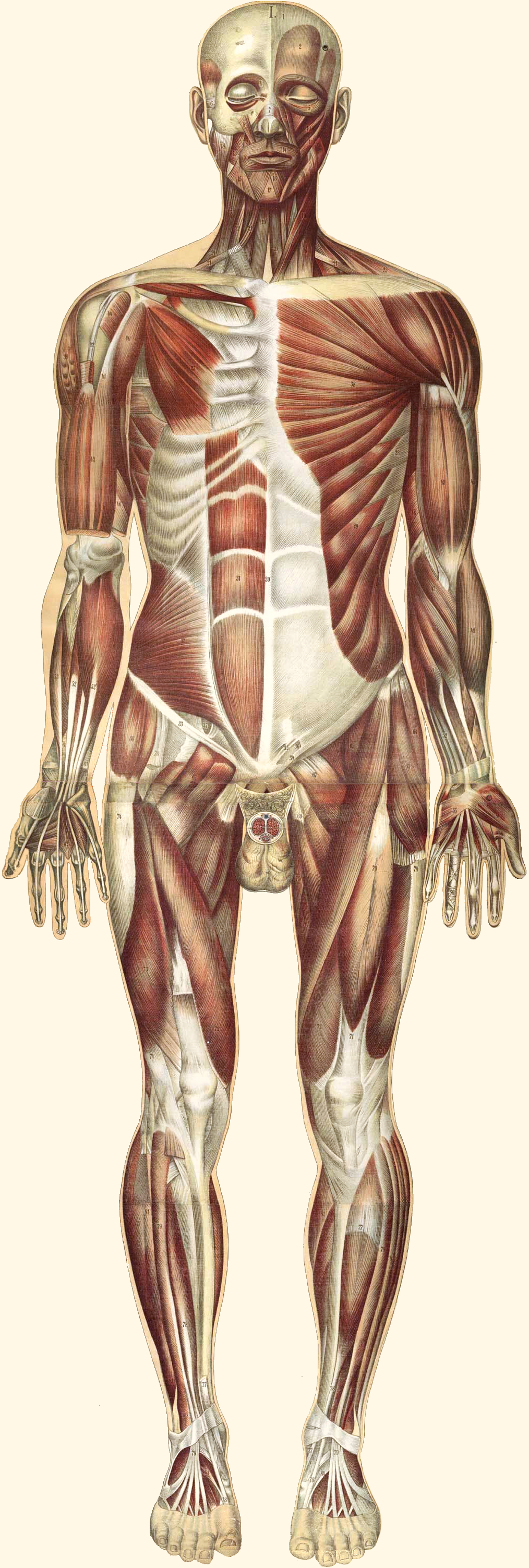
The muscular system consists of all the muscles of the body. The largest percentage of muscles in the muscular system consists of skeletal muscles, which are attached to bones and enable voluntary body movements (shown in Figure 12.2.2). There are almost 650 skeletal muscles in the human body, many of them shown in Figure 12.2.2. Besides skeletal muscles, the muscular system also includes cardiac muscle, which makes up the walls of the heart, and smooth muscles, which control movement in other internal organs and structures.
Muscle Structure and Function
Muscles are organs composed mainly of muscle cells, which are also called muscle fibres (mainly in skeletal and cardiac muscle) or myocytes (mainly in smooth muscle). Muscle cells are long, thin cells that are specialized for the function of contracting. They contain protein filaments that slide over one another using energy in ATP. The sliding filaments increase the tension in — or shorten the length of — muscle cells, causing a contraction. Muscle contractions are responsible for virtually all the movements of the body, both inside and out.
Skeletal muscles are attached to bones of the skeleton. When these muscles contract, they move the body. They allow us to use our limbs in a variety of ways, from walking to turning cartwheels. Skeletal muscles also maintain posture and help us to keep balance.
Smooth muscles in the walls of blood vessels contract to cause vasoconstriction, which may help conserve body heat. Relaxation of these muscles causes vasodilation, which may help the body lose heat. In the organs of the digestive system, smooth muscles squeeze food through the gastrointestinal tract by contracting in sequence to form a wave of muscle contractions called peristalsis. Think of squirting toothpaste through a tube by applying pressure in sequence from the bottom of the tube to the top, and you have a good idea of how food is moved by muscles through the digestive system. Peristalsis of smooth muscles also moves urine through the urinary tract.
Cardiac muscle tissue is found only in the walls of the heart. When cardiac muscle contracts, it makes the heart beat. The pumping action of the beating heart keeps blood flowing through the cardiovascular system.
Muscle Hypertrophy and Atrophy
Muscles can grow larger, or hypertrophy. This generally occurs through increased use, although hormonal or other influences can also play a role. The increase in testosterone that occurs in males during puberty, for example, causes a significant increase in muscle size. Physical exercise that involves weight bearing or resistance training can increase the size of skeletal muscles in virtually everyone. Exercises (such as running) that increase the heart rate may also increase the size and strength of cardiac muscle. The size of muscle, in turn, is the main determinant of muscle strength, which may be measured by the amount of force a muscle can exert.
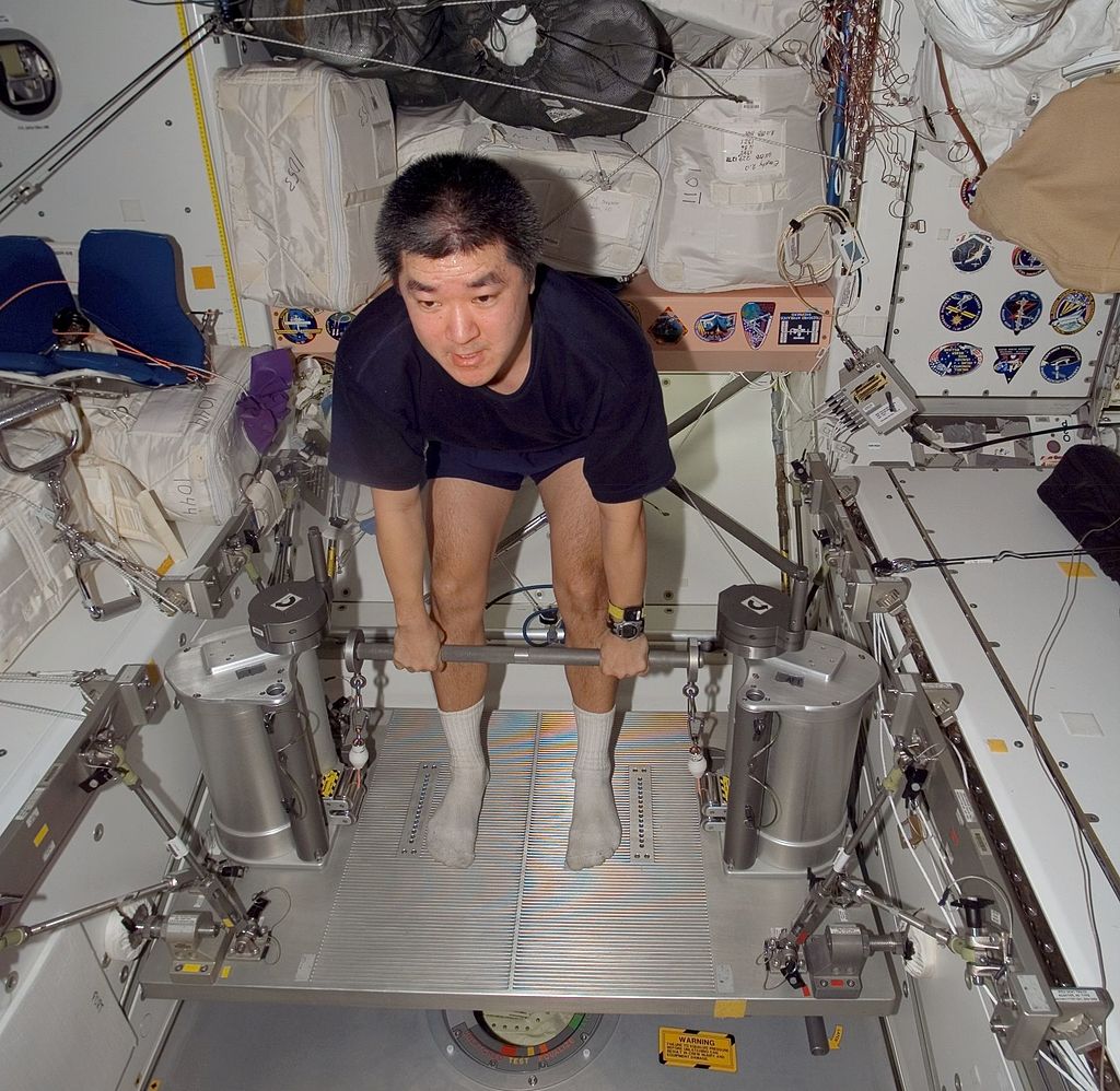
Muscles can also grow smaller, or atrophy, which can occur through lack of physical activity or from starvation. People who are immobilized for any length of time — for example, because of a broken bone or surgery — lose muscle mass relatively quickly. People in concentration or famine camps may be so malnourished that they lose much of their muscle mass, becoming almost literally just “skin and bones.” Astronauts on the International Space Station may also lose significant muscle mass because of weightlessness in space (see Figure 12.2.3).
Many diseases, including cancer and AIDS, are often associated with muscle atrophy. Atrophy of muscles also happens with age. As people grow older, there is a gradual decrease in the ability to maintain skeletal muscle mass, known as sarcopenia. The exact cause of sarcopenia is not known, but one possible cause is a decrease in sensitivity to growth factors that are needed to maintain muscle mass. Because muscle size determines strength, muscle atrophy causes a corresponding decline in muscle strength.
In both hypertrophy and atrophy, the number of muscle fibres does not change. What changes is the size of the muscle fibres. When muscles hypertrophy, the individual fibres become wider. When muscles atrophy, the fibres become narrower.
Interactions with Other Body Systems
Muscles cannot contract on their own. Skeletal muscles need stimulation from motor neurons in order to contract. The point where a motor neuron attaches to a muscle is called a neuromuscular junction. Let’s say you decide to raise your hand in class. Your brain sends electrical messages through motor neurons to your arm and shoulder. The motor neurons, in turn, stimulate muscle fibres in your arm and shoulder to contract, causing your arm to rise.
Involuntary contractions of smooth and cardiac muscles are also controlled by electrical impulses, but in the case of these muscles, the impulses come from the autonomic nervous system (smooth muscle) or specialized cells in the heart (cardiac muscle). Hormones and some other factors also influence involuntary contractions of cardiac and smooth muscles. For example, the fight-or-flight hormone adrenaline increases the rate at which cardiac muscle contracts, thereby speeding up the heartbeat.
Muscles cannot move the body on their own. They need the skeletal system to act upon. The two systems together are often referred to as the musculoskeletal system. Skeletal muscles are attached to the skeleton by tough connective tissues called tendons. Many skeletal muscles are attached to the ends of bones that meet at a joint. The muscles span the joint and connect the bones. When the muscles contract, they pull on the bones, causing them to move. The skeletal system provides a system of levers that allow body movement. The muscular system provides the force that moves the levers.
12.2 Summary
- The muscular system consists of all the muscles of the body. There are three types of muscle: skeletal muscle (which is attached to bones and enables voluntary body movements), cardiac muscle (which makes up the walls of the heart and makes it beat), and smooth muscle (which is found in the walls of internal organs and other internal structures and controls their movements).
- Muscles are organs composed mainly of muscle cells, which may also be called muscle fibres or myocytes. Muscle cells are specialized for the function of contracting, which occurs when protein filaments inside the cells slide over one another using energy in ATP.
- Muscles can grow larger, or hypertrophy. This generally occurs through increased use (physical exercise), although hormonal or other influences can also play a role. Muscles can also grow smaller, or atrophy. This may occur through lack of use, starvation, certain diseases, or aging. In both hypertrophy and atrophy, the size — but not the number — of muscle fibres changes. The size of muscles is the main determinant of muscle strength.
- Skeletal muscles need the stimulus of motor neurons to contract, and to move the body, they need the skeletal system to act upon. Involuntary contractions of cardiac and smooth muscles are controlled by special cells in the heart, nerves of the autonomic nervous system, hormones, or other factors.
12.2 Review Questions
- What is the muscular system?
- Describe muscle cells and their function.
- Identify three types of muscle tissue and where each type is found.
- Define muscle hypertrophy and muscle atrophy.
- What are some possible causes of muscle hypertrophy?
- Give three reasons that muscle atrophy may occur.
- How do muscles change when they increase or decrease in size?
- How do changes in muscle size affect strength?
- Explain why astronauts can easily lose muscle mass in space.
- Describe how the terms muscle cells, muscle fibres, and myocytes relate to each other.
-
- Name two systems in the body that work together with the muscular system to carry out movements.
- Describe one way in which the muscular system is involved in regulating body temperature.
12.2 Explore More
https://www.youtube.com/watch?v=VVL-8zr2hk4
How your muscular system works - Emma Bryce, TED-Ed, 2017.
https://www.youtube.com/watch?v=Ujr0UAbyPS4&feature=emb_logo
3D Medical Animation - Peristalsis in Large Intestine/Bowel || ABP ©, AnimatedBiomedical, 2013.
https://www.youtube.com/watch?v=LkXwfTsqQgQ&feature=emb_logo
Muscle matters: Dr Brendan Egan at TEDxUCD, TEDx Talks, 2014.
Attributions
Figure 12.2.1
Natalia_Zabolotnaya_2012b by Simon Q on Wikimedia Commons is used under a CC BY 2.0 (https://creativecommons.org/licenses/by/2.0/deed.en) license.
Figure 12.2.2
Bougle_whole2_retouched by Bouglé, Julien from the National LIbrary of Medicine (NLM) on Wikimedia Commons is in the public domain (https://en.wikipedia.org/wiki/Public_domain).
Figure 12.2.3
Daniel_Tani_iss016e027910 by NASA/ International Space Station Imagery on Wikimedia Commons is in the public domain (https://en.wikipedia.org/wiki/Public_domain).
References
AnimatedBiomedical. (2013, January 30). 3D Medical animation - Peristalsis in large intestine/bowel || ABP ©. YouTube. https://www.youtube.com/watch?v=Ujr0UAbyPS4&feature=youtu.be
Bouglé, J. (1899). Le corps humain en grandeur naturelle : planches coloriées et superposées, avec texte explicatif. J. B. Baillière et fils. In Historical Anatomies on the Web. http://www.nlm.nih.gov/exhibition/historicalanatomies/bougle_home.html
TED-Ed. (2017, October 26). How your muscular system works - Emma Bryce. YouTube. https://www.youtube.com/watch?v=VVL-8zr2hk4&feature=youtu.be
TEDx Talks. (2014, June 27). Muscle matters: Dr Brendan Egan at TEDxUCD. YouTube. https://www.youtube.com/watch?v=LkXwfTsqQgQ&feature=youtu.be
Wikipedia contributors. (2020, June 15). Natalya Zabolotnaya. In Wikipedia. https://en.wikipedia.org/w/index.php?title=Natalya_Zabolotnaya&oldid=962630409
Long chains of hydrocarbons with a carboxyl group and a methyl group at opposite ends. Can be either saturated, containing mostly single bonds between adjacent carbons, or unsaturated, containing many double bonds between adjacent carbons.
An antibody, also known as an immunoglobulin, is a large, Y-shaped protein produced mainly by plasma cells that is used by the immune system to neutralize pathogens such as pathogenic bacteria and viruses.
The transformation of one molecule to a different molecule inside a cell.

