4.5 Cytoplasm and Cytoskeleton
A Peek Inside the Cell
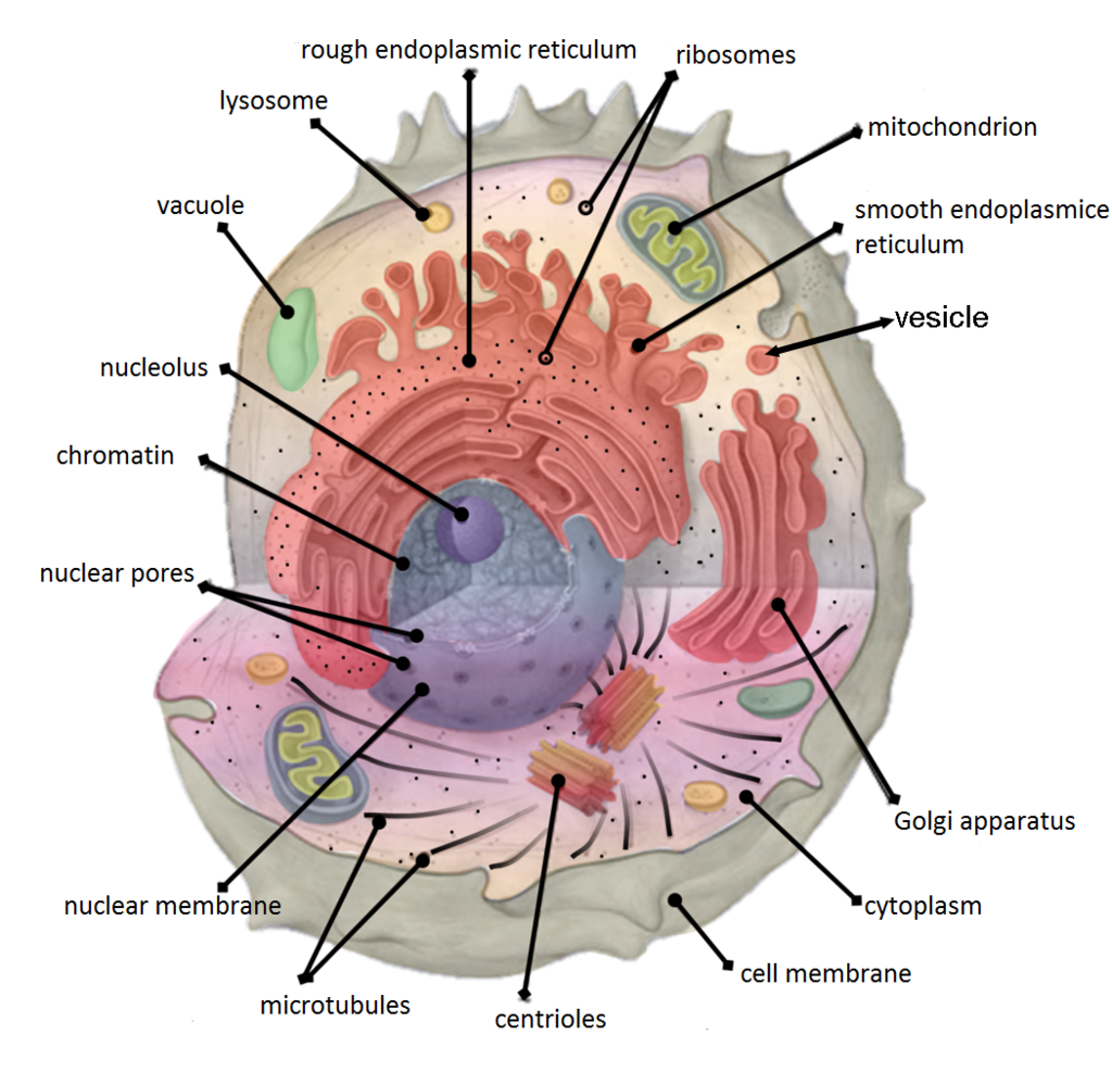
Figure 4.5.1 is an artist’s representation of what you might see if you could take a peek inside one of these basic building blocks of living things. A cell’s interior is obviously a crowded and busy space. It contains cytoplasm, dissolved substances, and many structures. It’s a hive of countless biochemical activities all going on at once.
Cytoplasm
Cytoplasm is a thick, usually colourless solution that fills each cell and is enclosed by the cell membrane. Cytoplasm presses against the cell membrane, filling out the cell and giving it its shape. Sometimes, cytoplasm acts like a watery solution, and sometimes, it takes on a more gel-like consistency. In eukaryotic cells, the cytoplasm includes all of the material inside the cell but outside of the nucleus, which contains its own watery substance called the nucleoplasm. All of the organelles in eukaryotic cells (such as the endoplasmic reticulum and mitochondria) are located in the cytoplasm. The cytoplasm helps to keep them in place. It is also the site of most metabolic activities in the cell, and it allows materials to pass easily throughout the cell.
The portion of the cytoplasm surrounding organelles is called cytosol. Cytosol is the liquid part of cytoplasm. It is composed of about 80 per cent water, and it contains dissolved salts, fatty acids, sugars, amino acids, and proteins (such as enzymes). These dissolved substances are needed to keep the cell alive and carry out metabolic processes. Enzymes dissolved in cytosol, for example, break down larger molecules into smaller products that can then be used by organelles of the cell. Waste products are also dissolved in the cytosol before they are taken in by vacuoles or expelled from the cell.
Cytoskeleton
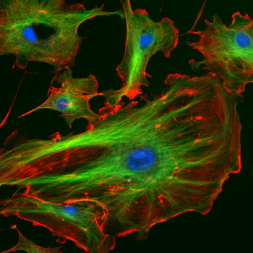
Although cytoplasm may appear to have no form or structure, it is actually highly organized. A framework of protein scaffolds called the cytoskeleton provides the cytoplasm and the cell with structure. The cytoskeleton consists of thread-like microfilaments, intermediate filaments, and microtubules that criss-cross the cytoplasm. You can see these filaments and tubules in the cells in Figure 4.5.2. As its name suggests, the cytoskeleton is like a cellular “skeleton.” It helps the cell maintain its shape and also helps to hold cell structures (like organelles) in place within the cytoplasm.
Feature: Human Biology in the News
News about an important study of the cytoplasm of eukaryotic cells came out in early 2016. Researchers in Dresden, Germany discovered that when cells are deprived of adequate nutrients, they may essentially shut down and become dormant. Specifically, when cells do not get enough nutrients, they shut down their metabolism, their energy level drops, and the pH of their cytoplasm decreases. Their normally liquid cytoplasm also assumes a solid state. The cells appear dead, as though a kind of rigor mortis has set in. The researchers think that these changes protect the sensitive structures inside the cells and allow the cells to survive difficult conditions. If nutrients are returned to the cells, they can emerge from their dormant state unharmed. They will continue to grow and multiply when conditions improve.
This important basic science research was executed on a nonhuman organism: one-celled fungi called yeasts. Nonetheless, it may have important implications for humans, because yeasts have eukaryotic cells with many of the same structures as human cells. Yeast cells appear to be able to “trick” death by shutting down all life processes in a controlled way. Through continued study, researchers hope to learn whether human cells can be taught this “trick” as well.
4.5 Summary
- Cytoplasm is a thick solution that fills a cell and is enclosed by the cell membrane. It has many functions. It helps give the cell shape, holds organelles, and provides a site for many of the biochemical reactions inside the cell.
- The liquid part of the cytoplasm is called cytosol. It is mainly water, and it contains many dissolved substances. The cytoplasm of a eukaryotic cell also contains a membrane-enclosed nucleus and other organelles.
- The cytoskeleton is a highly organized framework of protein filaments and tubules that criss-cross the cytoplasm of a cell. It gives the cell structure and helps hold cell structures (such as organelles) in place within the cytoplasm.
4.5 Review Questions
- Describe the composition of cytoplasm. Draw a picture of a cell, including the basic components required to be considered a cell, and the organelles you have learned about in this section.
- What are some of the functions of cytoplasm?
- Outline the structure and functions of the cytoskeleton.
- Is the cytoplasm made of cells? Why or why not?
- Name two types of cytoskeletal structures.
- In the picture of the different cytoskeletal structures above (Figure 4.5.2), what do you notice about these different structures?
- Describe one example of a metabolic process that happens in the cytosol.
- In eukaryotic cells, all of the material inside of the cell, but outside of the nucleus is called the ___________ .
- What is the liquid part of cytoplasm called?
- What chemical substance composes most of the cytosol?
- When yeast cells deprived of nutrients go dormant, their cytoplasm assumes a solid state. What effect do you think a solid cytoplasm would have on normal cellular processes? Explain your answer.
4.5 Explore More
Cytoskeleton Structure and Function, National Center for Case Study
Teaching in Science, 2015.
Attributions
Figure 4.5.1
Cell-organelles-labeled by Koswac on Wikimedia Commons is used under a CC BY-SA 4.0 (https://creativecommons.org/licenses/by-sa/4.0) license.
Figure 4.5.2
Cytoskeleton/ Fluorescent Cells by National Institute of Health (NIH) on Wikimedia Commons is released into the public domain (https://en.wikipedia.org/wiki/Public_domain).
National Center for Case Study Teaching in Science. (2015, August 3). Cytoskeleton structure and function. YouTube. https://www.youtube.com/watch?v=YTv9ItGd050&feature=youtu.be
The smallest unit of life, consisting of at least a membrane, cytoplasm, and genetic material.
The jellylike material that makes up much of a cell inside the cell membrane, and, in eukaryotic cells, surrounds the nucleus. The organelles of eukaryotic cells, such as mitochondria, the endoplasmic reticulum, and (in green plants) chloroplasts, are contained in the cytoplasm.
The semipermeable membrane surrounding the cytoplasm of a cell.
Created by CK-12 Foundation/Adapted by Christine Miller
Communicating with Urine
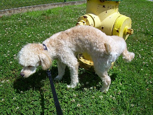
Why do dogs pee on fire hydrants? Besides “having to go,” they are marking their territory with chemicals in their urine called pheromones. It’s a form of communication, in which they are “saying” with odors that the yard is theirs and other dogs should stay away. In addition to fire hydrants, dogs may urinate on fence posts, trees, car tires, and many other objects. Urination in dogs, as in people, is usually a voluntary process controlled by the brain. The process of forming urine — which occurs in the kidneys — occurs constantly, and is not under voluntary control. What happens to all the urine that forms in the kidneys? It passes from the kidneys through the other organs of the urinary system, starting with the ureters.
Ureters
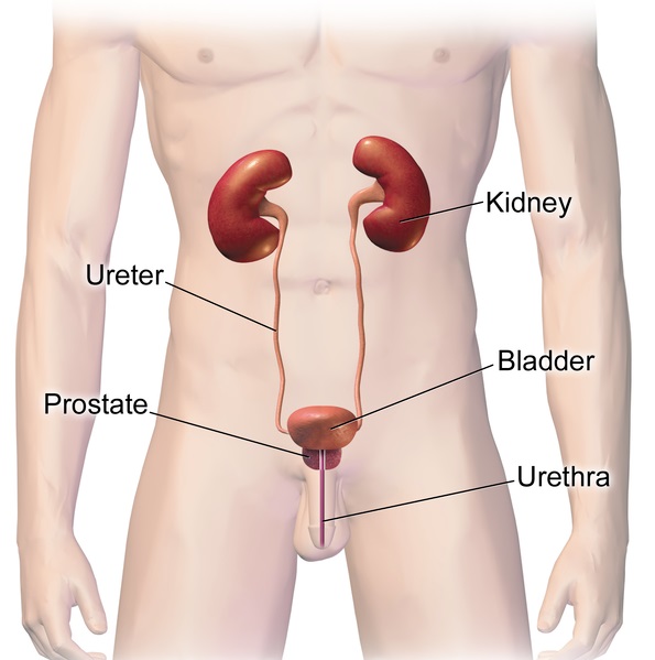
As shown in Figure 16.5.2, ureters are tube-like structures that connect the kidneys with the urinary bladder. They are paired structures, with one ureter for each kidney. In adults, ureters are between 25 and 30 cm (about 10–12 in) long and about 3 to 4 mm in diameter.
Each ureter arises in the pelvis of a kidney (the renal pelvis in Figure 16.5.3). It then passes down the side of the kidney, and finally enters the back of the bladder. At the entrance to the bladder, the ureters have sphincters that prevent the backflow of urine.
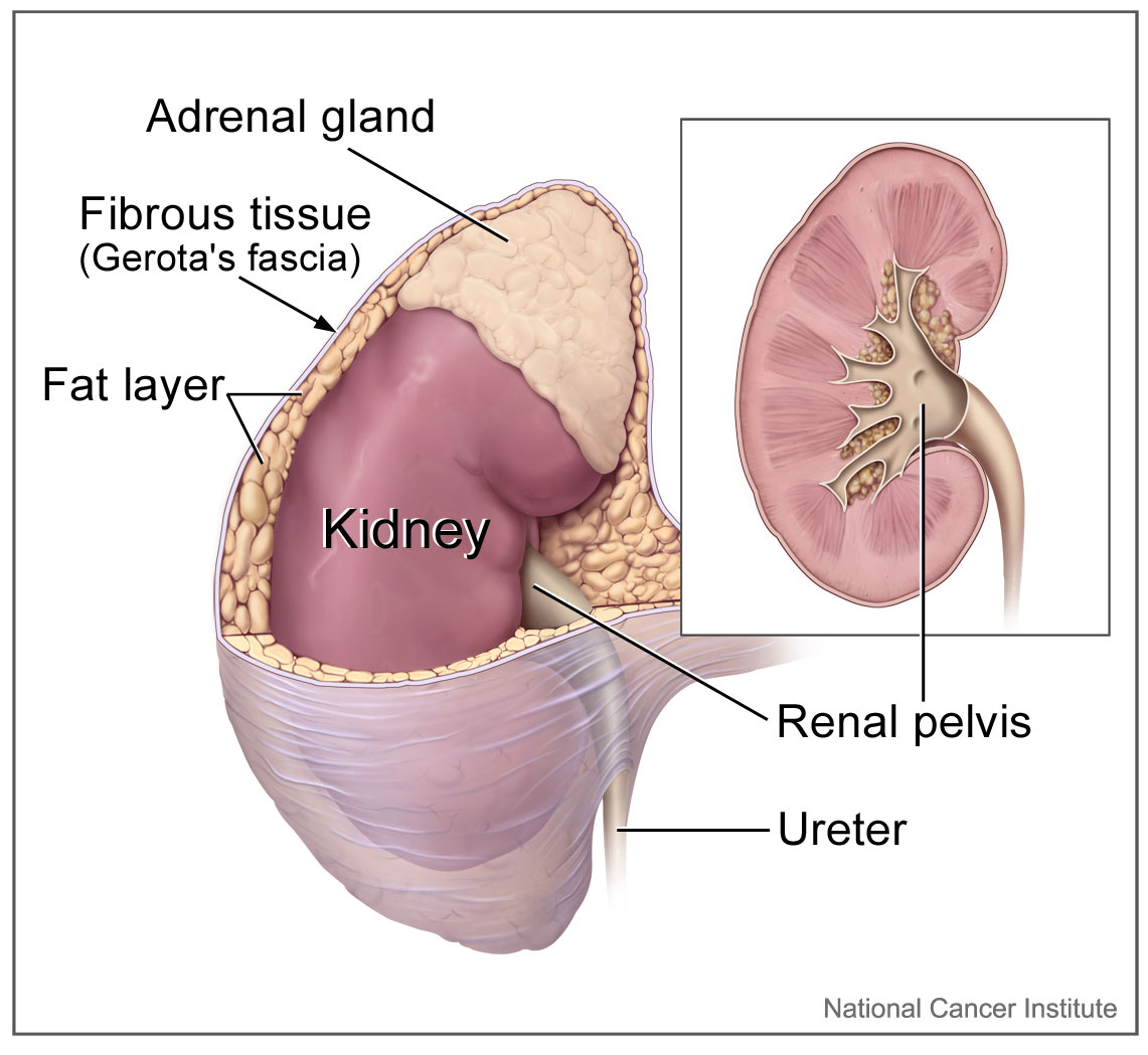
The walls of the ureters are composed of multiple layers of different types of tissues. The innermost layer is a special type of epithelium, called transitional epithelium. Unlike the epithelium lining most organs, transitional epithelium is capable of stretching and does not produce mucus. It lines much of the urinary system, including the renal pelvis, bladder, and much of the urethra, in addition to the ureters. Transitional epithelium allows these organs to stretch and expand as they fill with urine or allow urine to pass through. The next layer of the ureter walls is made up of loose connective tissue containing elastic fibres, nerves, and blood and lymphatic vessels. After this layer are two layers of smooth muscles, an inner circular layer, and an outer longitudinal layer. The smooth muscle layers can contract in waves of peristalsis to propel urine down the ureters from the kidneys to the urinary bladder. The outermost layer of the ureter walls consists of fibrous tissue.
Urinary Bladder
The urinary bladder is a hollow, muscular, and stretchy organ that rests on the pelvic floor. It collects and stores urine from the kidneys before the urine is eliminated through urination. As shown in Figure 16.5.4, urine enters the urinary bladder from the ureters through two ureteral openings on either side of the back wall of the bladder. Urine leaves the bladder through a sphincter called the internal urethral sphincter. When the sphincter relaxes and opens, it allows urine to flow out of the bladder and into the urethra.
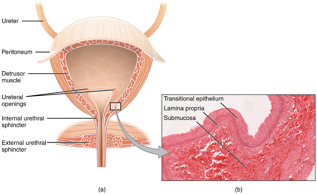
Like the ureters, the bladder is lined with transitional epithelium, which can flatten out and stretch as needed as the bladder fills with urine. The next layer (lamina propria) is a layer of loose connective tissue, nerves, and blood and lymphatic vessels. This is followed by a submucosa layer, which connects the lining of the bladder with the detrusor muscle in the walls of the bladder. The outer covering of the bladder is peritoneum, which is a smooth layer of epithelial cells that lines the abdominal cavity and covers most abdominal organs.
The detrusor muscle in the wall of the bladder is made of smooth muscle fibres controlled by both the autonomic and somatic nervous systems. As the bladder fills, the detrusor muscle automatically relaxes to allow it to hold more urine. When the bladder is about half full, the stretching of the walls triggers the sensation of needing to urinate. When the individual is ready to void, conscious nervous signals cause the detrusor muscle to contract, and the internal urethral sphincter to relax and open. As a result, urine is forcefully expelled out of the bladder and into the urethra.
Urethra
The urethra is a tube that connects the urinary bladder to the external urethral orifice, which is the opening of the urethra on the surface of the body. As shown in Figure 16.5.5, the urethra in males travels through the penis, so it is much longer than the urethra in females. In males, the urethra averages about 20 cm (about 7.8 in) long, whereas in females, it averages only about 4.8 cm (about 1.9 in) long. In males, the urethra carries semen (as well as urine), but in females, it carries only urine. In addition, in males, the urethra passes through the prostate gland (part of the reproductive system) which is absent in women.
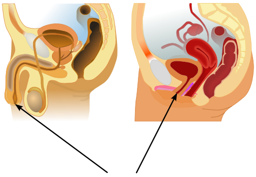
Like the ureters and bladder, the proximal (closer to the bladder) two-thirds of the urethra are lined with transitional epithelium. The distal (farther from the bladder) third of the urethra is lined with mucus-secreting epithelium. The mucus helps protect the epithelium from urine, which is corrosive. Below the epithelium is loose connective tissue, and below that are layers of smooth muscle that are continuous with the muscle layers of the urinary bladder. When the bladder contracts to forcefully expel urine, the smooth muscle of the urethra relaxes to allow the urine to pass through.
In order for urine to leave the body through the external urethral orifice, the external urethral sphincter must relax and open. This sphincter is a striated muscle that is controlled by the somatic nervous system, so it is under conscious, voluntary control in most people (exceptions are infants, some elderly people, and patients with certain injuries or disorders). The muscle can be held in a contracted state and hold in the urine until the person is ready to urinate. Following urination, the smooth muscle lining the urethra automatically contracts to re-establish muscle tone, and the individual consciously contracts the external urethral sphincter to close the external urethral opening.
16.5 Summary
- Ureters are tube-like structures that connect the kidneys with the urinary bladder. Each ureter arises at the renal pelvis of a kidney and travels down through the abdomen to the urinary bladder. The walls of the ureter contain smooth muscle that can contract to push urine through the ureter by peristalsis. The walls are lined with transitional epithelium that can expand and stretch.
- The urinary bladder is a hollow, muscular organ that rests on the pelvic floor. It is also lined with transitional epithelium. The function of the bladder is to collect and store urine from the kidneys before the urine is eliminated through urination. Filling of the bladder triggers the sensation of needing to urinate. When a conscious decision to urinate is made, the detrusor muscle in the bladder wall contracts and forces urine out of the bladder and into the urethra.
- The urethra is a tube that connects the urinary bladder to the external urethral orifice. Somatic nerves control the sphincter at the distal end of the urethra. This allows the opening of the sphincter for urination to be under voluntary control.
16.5 Review Questions
- What are ureters? Describe the location of the ureters relative to other urinary tract organs.
- Identify layers in the walls of a ureter. How do they contribute to the ureter’s function?
- Describe the urinary bladder. What is the function of the urinary bladder?
-
- How does the nervous system control the urinary bladder?
- What is the urethra?
- How does the nervous system control urination?
- Identify the sphincters that are located along the pathway from the ureters to the external urethral orifice.
- What are two differences between the male and female urethra?
- When the bladder muscle contracts, the smooth muscle in the walls of the urethra _________ .
16.5 Explore More
https://youtu.be/2Brajdazp1o
The taboo secret to better health | Molly Winter, TED. 2016.
https://youtu.be/dg4_deyHLvQ
What Happens When You Hold Your Pee? SciShow, 2016.
Attributions
Figure 16.5.1
Cliche by Jackie on Wikimedia Common s is used under a CC BY 2.0 (https://creativecommons.org/licenses/by/2.0) license.
Figure 16.5.2
Urinary System Male by BruceBlaus on Wikimedia Commons is used under a CC BY-SA 4.0 (https://creativecommons.org/licenses/by-sa/4.0) license.
Figure 16.5.3
Adrenal glands on Kidney by NCI Public Domain by Alan Hoofring (Illustrator) /National Cancer Institute (photo ID 4355) on Wikimedia Commons is in the public domain (https://en.wikipedia.org/wiki/Public_domain).
Figure 16.5.4
2605_The_Bladder by OpenStax College on Wikimedia Commons is used under a CC BY 3.0 (https://creativecommons.org/licenses/by/3.0) license. (Micrograph originally provided by the Regents of the University of Michigan Medical School © 2012.)
Figure 16.5.5
512px-Male_and_female_urethral_openings.svg by andrybak (derivative work) on Wikimedia Commons is used under a CC BY-SA 3.0 (https://creativecommons.org/licenses/by-sa/3.0) license. (Original: Male anatomy blank.svg: alt.sex FAQ, derivative work: Tsaitgaist Female anatomy with g-spot.svg: Tsaitgaist.)
References
Betts, J. G., Young, K.A., Wise, J.A., Johnson, E., Poe, B., Kruse, D.H., Korol, O., Johnson, J.E., Womble, M., DeSaix, P. (2013, June 19). Figure 25.4 Bladder (a) Anterior cross section of the bladder. (b) The detrusor muscle of the bladder (source: monkey tissue) LM × 448 [digital image]. In Anatomy and Physiology (Section 7.3). OpenStax. https://openstax.org/books/anatomy-and-physiology/pages/25-2-gross-anatomy-of-urine-transport
SciShow. (2016, January 22). What happens when you hold your pee? YouTube. https://www.youtube.com/watch?v=dg4_deyHLvQ&feature=youtu.be
TED. (2016, September 2). The taboo secret to better health | Molly Winter. YouTube. https://www.youtube.com/watch?v=2Brajdazp1o&feature=youtu.be
A central organelle containing hereditary material.
A solution, similar to the cytoplasm of a cell, enveloped by the nuclear envelope and surrounding the chromosomes and nucleolus.
A tiny cellular structure that performs specific functions within a cell.
The aqueous component of the cytoplasm of a cell, within which various organelles and particles are suspended.
Long chains of hydrocarbons with a carboxyl group and a methyl group at opposite ends. Can be either saturated, containing mostly single bonds between adjacent carbons, or unsaturated, containing many double bonds between adjacent carbons.
Amino acids are organic compounds that combine to form proteins.
A class of biological molecule consisting of linked monomers of amino acids and which are the most versatile macromolecules in living systems and serve crucial functions in essentially all biological processes.
Biological molecules that lower amount the energy required for a reaction to occur.
A complex network of interlinking protein filaments that extends from the cell nucleus to the cell membrane, gives the cell its shape and help organize the cell's parts.

