4.14 Case Study Conclusion: More Than Just Tired
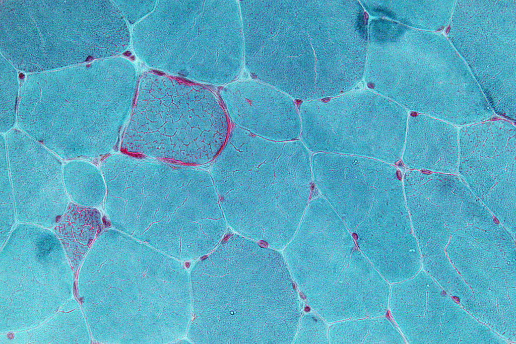
Jasmin discovered that her extreme fatigue, muscle pain, vision problems, and vomiting were due to problems in her mitochondria, like the damaged mitochondria shown in red in Figure 4.14.1. Mitochondria are small, membrane-bound organelles found in eukaryotic cells that provide energy for the cells of the body. They do this by carrying out the final two steps of aerobic cellular respiration: the Krebs cycle and electron transport. This is the major way that the human body breaks down the sugar glucose from food into a form of energy cells can use, namely the molecule ATP.
Because mitochondria provide energy for cells, you can understand why Jasmin was experiencing extreme fatigue, particularly after running. Her damaged mitochondria could not keep up with her need for energy, particularly after intense exercise, which requires a lot of additional energy. What is perhaps not so obvious are the reasons for her other symptoms, such as blurry vision, muscle spasms, and vomiting. All of the cells in the body require energy in order to function properly. Mitochondrial diseases can cause problems in mitochondria in any cell of the body, including muscle cells and cells of the nervous system, which includes the brain and nerves. The nervous system and muscles work together to control vision and digestive system functions, such as vomiting, so when they are not functioning properly, a variety of symptoms can emerge. This also explains why Jasmin’s niece, who has a similar mitochondrial disease, has symptoms related to brain function, such as seizures and learning disabilities. Our cells are microscopic, and mitochondria are even tinier — but they are essential for the proper functioning of our bodies. When they are damaged, serious health effects can occur.

One seemingly confusing aspect of mitochondrial diseases is that the type of symptoms, severity of symptoms, and age of onset can vary wildly between people — even within the same family! In Jasmin’s case, she did not notice symptoms until adulthood, while her niece had more severe symptoms starting at a much younger age. This makes sense when you know more about how mitochondrial diseases work.
Inherited mitochondrial diseases can be due to damage in either the DNA in the nucleus of cells or in the DNA in the mitochondria themselves. Recall that mitochondria are thought to have evolved from prokaryotic organisms that were once free-living, but were then infected or engulfed by larger cells. One of the pieces of evidence that supports this endosymbiotic theory is that mitochondria have their own, separate DNA. When the mitochondrial DNA is damaged (or mutated) it can result in some types of mitochondrial diseases. However, these mutations do not typically affect all of the mitochondria in a cell. During cell division, organelles such as mitochondria are replicated and passed down to the new daughter cells. If some of the mitochondria are damaged, and others are not, the daughter cells can have different amounts of damaged mitochondria. This helps explain the wide range of symptoms in people with mitochondrial diseases — even ones in the same family — because different cells in their bodies are affected in varying degrees. Jasmin’s niece was affected strongly and her symptoms were noticed early, while Jasmin’s symptoms were more mild and did not become apparent until adulthood.
There is still much more that needs to be discovered about the different types of mitochondrial diseases. But by learning about cells, their organelles, how they obtain energy, and how they divide, you should now have a better understanding of the biology behind these diseases.
Apply your understanding of cells to your own life. Can you think of other diseases that affect cellular structures or functions. Do they affect people you know? Since your entire body is made of cells, when cells are damaged or not functioning properly, it can cause a wide variety of health problems.
Chapter 4 Summary
Type your learning objectives here.
In this chapter you learned many facts about cells. Specifically, you learned that:
- Cells are the basic units of structure and function of living things.
- The first cells were observed from cork by Hooke in the 1600s. Soon after, van Leeuwenhoek observed other living cells.
- In the early 1800s, Schwann and Schleiden theorized that cells are the basic building blocks of all living things. Around 1850, Virchow saw cells dividing, and added his own theory that living cells arise only from other living cells. These ideas led to cell theory, which states that all organisms are made of cells, all life functions occur in cells, and all cells come from other cells.
- The invention of the electron microscope in the 1950s allowed scientists to see organelles and other structures inside cells for the first time.
- There is variation in cells, but all cells have a plasma membrane, cytoplasm, ribosomes, and DNA.
-
- The plasma membrane is composed mainly of a bilayer of phospholipid molecules and forms a barrier between the cytoplasm inside the cell and the environment outside the cell. It allows only certain substances to pass in or out of the cell. Some cells have extensions of their plasma membrane with other functions, such as flagella or cilia.
- Cytoplasm is a thick solution that fills a cell and is enclosed by the plasma membrane. It helps give the cell shape, holds organelles, and provides a site for many of the biochemical reactions inside the cell. The liquid part of the cytoplasm is called cytosol.
- Ribosomes are small structures where proteins are made.
- Cells are usually very small, so they have a large enough surface area-to-volume ratio to maintain normal cell processes. Cells with different functions often have different shapes.
- Prokaryotic cells do not have a nucleus. Eukaryotic cells have a nucleus, as well as other organelles. An organelle is a structure within the cytoplasm of a cell that is enclosed within a membrane and performs a specific job.
- The cytoskeleton is a highly organized framework of protein filaments and tubules that criss-cross the cytoplasm of a cell. It gives the cell shape and helps to hold cell structures (such as organelles) in place.
- The nucleus is the largest organelle in a eukaryotic cell. It is considered to be the cell’s control center, and it contains DNA and controls gene expression, including which proteins the cell makes.
- The mitochondrion is an organelle that makes energy available to cells. According to the widely accepted endosymbiotic theory, mitochondria evolved from prokaryotic cells that were once free-living organisms that infected or were engulfed by larger prokaryotic cells.
- The endoplasmic reticulum (ER) is an organelle that helps make and transport proteins and lipids. Rough endoplasmic reticulum (RER) is studded with ribosomes. Smooth endoplasmic reticulum (SER) has no ribosomes.
- The Golgi apparatus is a large organelle that processes proteins and prepares them for use both inside and outside the cell. It is also involved in the transport of lipids around the cell.
- Vesicles and vacuoles are sac-like organelles that may be used to store and transport materials in the cell or as chambers for biochemical reactions. Lysosomes and peroxisomes are vesicles that break down foreign matter, dead cells, or poisons.
- Centrioles are organelles located near the nucleus that help organize the chromosomes before cell division so each daughter cell receives the correct number of chromosomes.
- There are two basic ways that substances can cross the cell’s plasma membrane: passive transport (which requires no energy expenditure by the cell) and active transport (which requires energy).
- No energy is needed from the cell for passive transport because it occurs when substances move naturally from an area of higher concentration to an area of lower concentration. Types of passive transport in cells include:
-
- Simple diffusion, which is the movement of a substance due to differences in concentration without any help from other molecules. This is how very small, hydrophobic molecules, such as oxygen and carbon dioxide, enter and leave the cell.
- Osmosis, which is the diffusion of water molecules across the membrane.
- Facilitated diffusion, which is the movement of a substance across a membrane due to differences in concentration, but only with the help of transport proteins in the membrane (such as channel proteins or carrier proteins). This is how large or hydrophilic molecules and charged ions enter and leave the cell.
- Active transport requires energy to move substances across the plasma membrane, often because the substances are moving from an area of lower concentration to an area of higher concentration or because of their large size. Two examples of active transport are the sodium-potassium pump and vesicle transport.
-
- The sodium-potassium pump moves sodium ions out of the cell and potassium ions into the cell, both against a concentration gradient, in order to maintain the proper concentrations of both ions inside and outside the cell and to thereby control membrane potential.
- Vesicle transport uses vesicles to move large molecules into or out of cells.
- Energy is the ability to do work. It is needed by every living cell to carry out life processes.
- The form of energy that living things need is chemical energy, and it comes from food. Food consists of organic molecules that store energy in their chemical bonds.
- Autotrophs (producers) make their own food. Think of plants that make food by photosynthesis. Heterotrophs (consumers) obtain food by eating other organisms.
- Organisms mainly use the molecules glucose and ATP for energy. Glucose is the compact, stable form of energy that is carried in the blood and taken up by cells. ATP contains less energy and is used to power cell processes.
- The flow of energy through living things begins with photosynthesis, which creates glucose. The cells of organisms break down glucose and make ATP.
- Cellular respiration is the aerobic process by which living cells break down glucose molecules, release energy, and form molecules of ATP. Overall, this three-stage process involves glucose and oxygen reacting to form carbon dioxide and water.
-
- Glycolysis, the first stage of cellular respiration, takes place in the cytoplasm. In this step, enzymes split a molecule of glucose into two molecules of pyruvate, which releases energy that is transferred to ATP.
- Transition Reaction takes place between glycolysis and Krebs Cycle. It is a very short reaction in which the pyruvate molecules from glycolysis are converted into Acetyl CoA in order to enter the Krebs Cycle.
- Krebs Cycle, the second stage of cellular respiration, takes place in the matrix of a mitochondrion. During this stage, two turns through the cycle result in all of the carbon atoms from the two pyruvate molecules forming carbon dioxide and the energy from their chemical bonds being stored in a total of 16 energy-carrying molecules (including four from glycolysis).
- The Electron Transport System, he third stage of cellular respiration, takes place on the inner membrane of the mitochondrion. Electrons are transported from molecule to molecule down an electron-transport chain. Some of the energy from the electrons is used to pump hydrogen ions across the membrane, creating an electrochemical gradient that drives the synthesis of many more molecules of ATP.
- In all three stages of aerobic cellular respiration combined, as many as 38 molecules of ATP are produced from just one molecule of glucose.
- Some organisms can produce ATP from glucose by anaerobic respiration, which does not require oxygen. Fermentation is an important type of anaerobic process. There are two types: alcoholic fermentation and lactic acid fermentation. Both start with glycolysis.
-
- Alcoholic fermentation is carried out by single-celled organisms, including yeasts and some bacteria. We use alcoholic fermentation in these organisms to make biofuels, bread, and wine.
- Lactic acid fermentation is undertaken by certain bacteria, including the bacteria in yogurt, and also by our muscle cells when they are worked hard and fast.
- Anaerobic respiration produces far less ATP (typically produces 2 ATP) than does aerobic cellular respiration, but it has the advantage of being much faster.
- The cell cycle is a repeating series of events that includes growth, DNA synthesis, and cell division.
- In a eukaryotic cell, the cell cycle has two major phases: interphase and mitotic phase. During interphase, the cell grows, performs routine life processes, and prepares to divide. During mitotic phase, first the nucleus divides (mitosis) and then the cytoplasm divides (cytokinesis), which produces two daughter cells.
-
- Until a eukaryotic cell divides, its nuclear DNA exists as a grainy material called chromatin. After DNA replicates and the cell is about to divide, the DNA condenses and coils into the X-shaped form of a chromosome. Each chromosome consists of two sister chromatids, which are joined together at a centromere.
- During mitosis, sister chromatids separate from each other and move to opposite poles of the cell. This happens in four phases: prophase, metaphase, anaphase, and telophase.
- The cell cycle is controlled mainly by regulatory proteins that signal the cell to either start or delay the next phase of the cycle at key checkpoints.
- Cancer is a disease that occurs when the cell cycle is no longer regulated, often because the cell’s DNA has become damaged. Cancerous cells grow out of control and may form a mass of abnormal cells called a tumor.
In this chapter, you learned about cells and some of their functions, as well as how they pass genetic material in the form of DNA to their daughter cells. In the next chapter, you will learn how DNA is passed down to offspring, which causes traits to be inherited. These traits may be innocuous (such as eye colour) or detrimental (such as mutations that cause disease). The study of how genes are passed down to offspring is called genetics, and as you will learn in the next chapter, this is an interesting topic that is highly relevant to human health.
Chapter 4 Review
- Sequence:
- Drag and Drop:
- True or False:
- Multiple Choice:
- Briefly explain how the energy in the food you eat gets there, and how it provides energy for your neurons in the form necessary to power this process.
- Explain why the inside of the plasma membrane — the side that faces the cytoplasm of the cell — must be hydrophilic.
- Explain the relationships between interphase, mitosis, and cytokinesis.
Attributions
Figure 4.14.1
Mitochondrial Disease muscle sample by Nephron is used under a CC BY-SA 3.0 (https://creativecommons.org/licenses/by-sa/3.0) license.
Figure 4.14.2
Aunt and Niece by Tatiana Rodriguez on Unsplash is used under the Unsplash License (https://unsplash.com/license).
Reference
Wikipedia contributors. (2020, June 6). Mitochondrial disease. In Wikipedia. https://en.wikipedia.org/w/index.php?title=Mitochondrial_disease&oldid=961126371
A double-membrane-bound organelle found in most eukaryotic organisms. Mitochondria convert oxygen and nutrients into adenosine triphosphate (ATP). ATP is the chemical energy "currency" of the cell that powers the cell's metabolic activities.
Created by CK-12 Foundation/Adapted by Christine Miller
Communicating with Urine
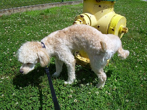
Why do dogs pee on fire hydrants? Besides “having to go,” they are marking their territory with chemicals in their urine called pheromones. It’s a form of communication, in which they are “saying” with odors that the yard is theirs and other dogs should stay away. In addition to fire hydrants, dogs may urinate on fence posts, trees, car tires, and many other objects. Urination in dogs, as in people, is usually a voluntary process controlled by the brain. The process of forming urine — which occurs in the kidneys — occurs constantly, and is not under voluntary control. What happens to all the urine that forms in the kidneys? It passes from the kidneys through the other organs of the urinary system, starting with the ureters.
Ureters
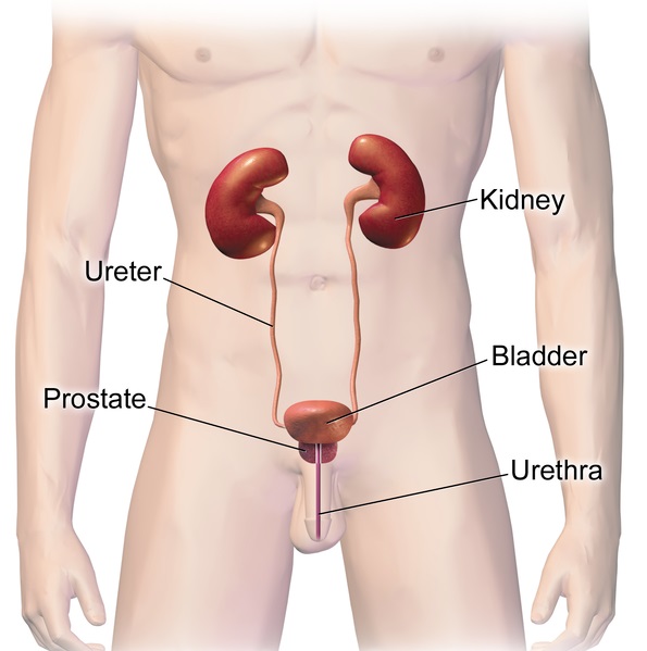
As shown in Figure 16.5.2, ureters are tube-like structures that connect the kidneys with the urinary bladder. They are paired structures, with one ureter for each kidney. In adults, ureters are between 25 and 30 cm (about 10–12 in) long and about 3 to 4 mm in diameter.
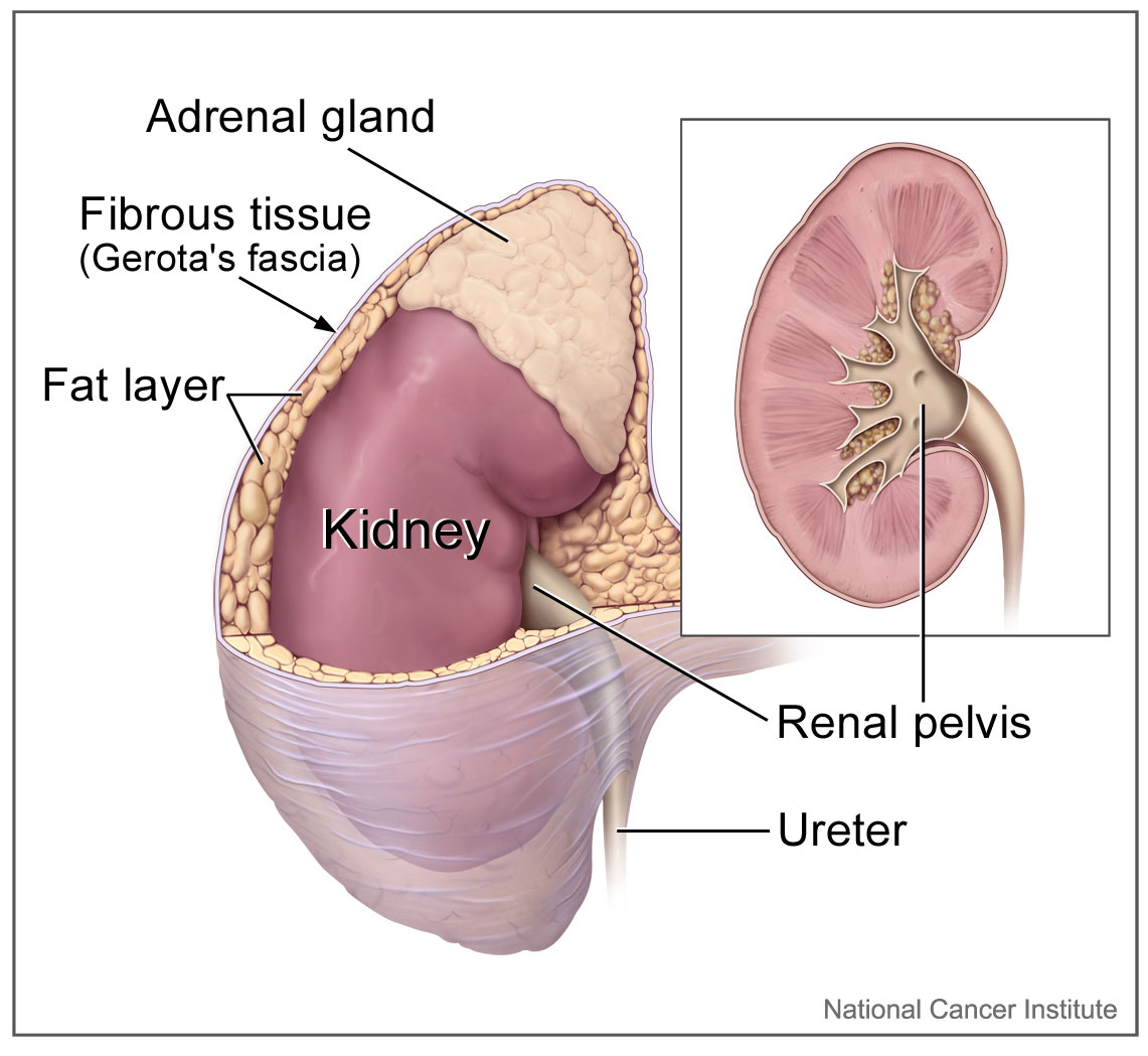
Each ureter arises in the pelvis of a kidney (the renal pelvis in Figure 16.5.3). It then passes down the side of the kidney, and finally enters the back of the bladder. At the entrance to the bladder, the ureters have sphincters that prevent the backflow of urine.
The walls of the ureters are composed of multiple layers of different types of tissues. The innermost layer is a special type of epithelium, called transitional epithelium. Unlike the epithelium lining most organs, transitional epithelium is capable of stretching and does not produce mucus. It lines much of the urinary system, including the renal pelvis, bladder, and much of the urethra, in addition to the ureters. Transitional epithelium allows these organs to stretch and expand as they fill with urine or allow urine to pass through. The next layer of the ureter walls is made up of loose connective tissue containing elastic fibres, nerves, and blood and lymphatic vessels. After this layer are two layers of smooth muscles, an inner circular layer, and an outer longitudinal layer. The smooth muscle layers can contract in waves of peristalsis to propel urine down the ureters from the kidneys to the urinary bladder. The outermost layer of the ureter walls consists of fibrous tissue.
Urinary Bladder
The urinary bladder is a hollow, muscular, and stretchy organ that rests on the pelvic floor. It collects and stores urine from the kidneys before the urine is eliminated through urination. As shown in Figure 16.5.4, urine enters the urinary bladder from the ureters through two ureteral openings on either side of the back wall of the bladder. Urine leaves the bladder through a sphincter called the internal urethral sphincter. When the sphincter relaxes and opens, it allows urine to flow out of the bladder and into the urethra.
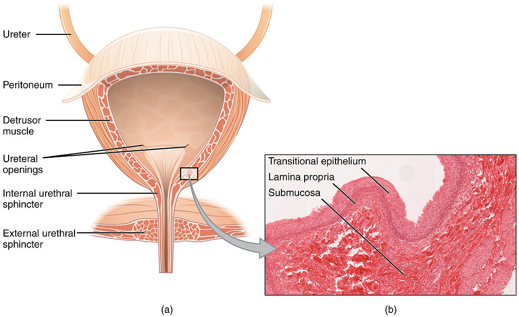
Like the ureters, the bladder is lined with transitional epithelium, which can flatten out and stretch as needed as the bladder fills with urine. The next layer (lamina propria) is a layer of loose connective tissue, nerves, and blood and lymphatic vessels. This is followed by a submucosa layer, which connects the lining of the bladder with the detrusor muscle in the walls of the bladder. The outer covering of the bladder is peritoneum, which is a smooth layer of epithelial cells that lines the abdominal cavity and covers most abdominal organs.
The detrusor muscle in the wall of the bladder is made of smooth muscle fibres controlled by both the autonomic and somatic nervous systems. As the bladder fills, the detrusor muscle automatically relaxes to allow it to hold more urine. When the bladder is about half full, the stretching of the walls triggers the sensation of needing to urinate. When the individual is ready to void, conscious nervous signals cause the detrusor muscle to contract, and the internal urethral sphincter to relax and open. As a result, urine is forcefully expelled out of the bladder and into the urethra.
Urethra
The urethra is a tube that connects the urinary bladder to the external urethral orifice, which is the opening of the urethra on the surface of the body. As shown in Figure 16.5.5, the urethra in males travels through the penis, so it is much longer than the urethra in females. In males, the urethra averages about 20 cm (about 7.8 in) long, whereas in females, it averages only about 4.8 cm (about 1.9 in) long. In males, the urethra carries semen (as well as urine), but in females, it carries only urine. In addition, in males, the urethra passes through the prostate gland (part of the reproductive system) which is absent in women.
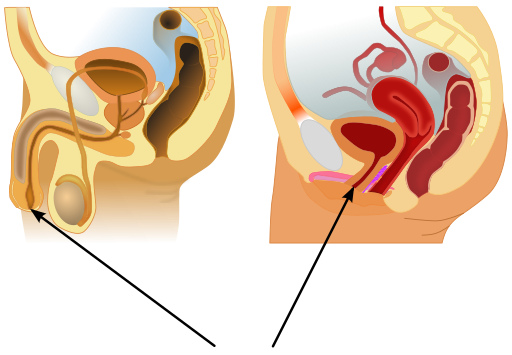
Like the ureters and bladder, the proximal (closer to the bladder) two-thirds of the urethra are lined with transitional epithelium. The distal (farther from the bladder) third of the urethra is lined with mucus-secreting epithelium. The mucus helps protect the epithelium from urine, which is corrosive. Below the epithelium is loose connective tissue, and below that are layers of smooth muscle that are continuous with the muscle layers of the urinary bladder. When the bladder contracts to forcefully expel urine, the smooth muscle of the urethra relaxes to allow the urine to pass through.
In order for urine to leave the body through the external urethral orifice, the external urethral sphincter must relax and open. This sphincter is a striated muscle that is controlled by the somatic nervous system, so it is under conscious, voluntary control in most people (exceptions are infants, some elderly people, and patients with certain injuries or disorders). The muscle can be held in a contracted state and hold in the urine until the person is ready to urinate. Following urination, the smooth muscle lining the urethra automatically contracts to re-establish muscle tone, and the individual consciously contracts the external urethral sphincter to close the external urethral opening.
16.5 Summary
- Ureters are tube-like structures that connect the kidneys with the urinary bladder. Each ureter arises at the renal pelvis of a kidney and travels down through the abdomen to the urinary bladder. The walls of the ureter contain smooth muscle that can contract to push urine through the ureter by peristalsis. The walls are lined with transitional epithelium that can expand and stretch.
- The urinary bladder is a hollow, muscular organ that rests on the pelvic floor. It is also lined with transitional epithelium. The function of the bladder is to collect and store urine from the kidneys before the urine is eliminated through urination. Filling of the bladder triggers the sensation of needing to urinate. When a conscious decision to urinate is made, the detrusor muscle in the bladder wall contracts and forces urine out of the bladder and into the urethra.
- The urethra is a tube that connects the urinary bladder to the external urethral orifice. Somatic nerves control the sphincter at the distal end of the urethra. This allows the opening of the sphincter for urination to be under voluntary control.
16.5 Review Questions
- What are ureters? Describe the location of the ureters relative to other urinary tract organs.
- Identify layers in the walls of a ureter. How do they contribute to the ureter’s function?
- Describe the urinary bladder. What is the function of the urinary bladder?
-
- How does the nervous system control the urinary bladder?
- What is the urethra?
- How does the nervous system control urination?
- Identify the sphincters that are located along the pathway from the ureters to the external urethral orifice.
- What are two differences between the male and female urethra?
- When the bladder muscle contracts, the smooth muscle in the walls of the urethra _________ .
16.5 Explore More
https://youtu.be/2Brajdazp1o
The taboo secret to better health | Molly Winter, TED. 2016.
https://youtu.be/dg4_deyHLvQ
What Happens When You Hold Your Pee? SciShow, 2016.
Attributions
Figure 16.5.1
Cliche by Jackie on Wikimedia Common s is used under a CC BY 2.0 (https://creativecommons.org/licenses/by/2.0) license.
Figure 16.5.2
Urinary System Male by BruceBlaus on Wikimedia Commons is used under a CC BY-SA 4.0 (https://creativecommons.org/licenses/by-sa/4.0) license.
Figure 16.5.3
Adrenal glands on Kidney by NCI Public Domain by Alan Hoofring (Illustrator) /National Cancer Institute (photo ID 4355) on Wikimedia Commons is in the public domain (https://en.wikipedia.org/wiki/Public_domain).
Figure 16.5.4
2605_The_Bladder by OpenStax College on Wikimedia Commons is used under a CC BY 3.0 (https://creativecommons.org/licenses/by/3.0) license. (Micrograph originally provided by the Regents of the University of Michigan Medical School © 2012.)
Figure 16.5.5
512px-Male_and_female_urethral_openings.svg by andrybak (derivative work) on Wikimedia Commons is used under a CC BY-SA 3.0 (https://creativecommons.org/licenses/by-sa/3.0) license. (Original: Male anatomy blank.svg: alt.sex FAQ, derivative work: Tsaitgaist Female anatomy with g-spot.svg: Tsaitgaist.)
References
Betts, J. G., Young, K.A., Wise, J.A., Johnson, E., Poe, B., Kruse, D.H., Korol, O., Johnson, J.E., Womble, M., DeSaix, P. (2013, June 19). Figure 25.4 Bladder (a) Anterior cross section of the bladder. (b) The detrusor muscle of the bladder (source: monkey tissue) LM × 448 [digital image]. In Anatomy and Physiology (Section 7.3). OpenStax. https://openstax.org/books/anatomy-and-physiology/pages/25-2-gross-anatomy-of-urine-transport
SciShow. (2016, January 22). What happens when you hold your pee? YouTube. https://www.youtube.com/watch?v=dg4_deyHLvQ&feature=youtu.be
TED. (2016, September 2). The taboo secret to better health | Molly Winter. YouTube. https://www.youtube.com/watch?v=2Brajdazp1o&feature=youtu.be
Image shows a four-tier diagram in a 28 day timeline of the menstruation cycle. The first tier shows basal body temperature (BBT) over the 28 days. BBT is about 1 degree celcius lower before ovulation than after. The next tier shows relative levels of follicle stimulating hormone (FSH), leutenizing hormone (LH), estrogen and progesterone. At the time of ovulation, levels of FSH, LH and estrogen spike and then drop off. Progesterone increases after ovulation and then drops off close to day 25.
In the third tier is a representation of changes to the follicle and corpus luteum during the ovarian cycle. In the first 14 days of the cycle, the follicle is growing. On day 14, the follicle ejects the ovum and starts converting to a corpus lutem, which persists from day 15-28, at which point it has degenerated.
In the fourth tier, the uterine cycle is illustrated, showing relative thickness of the endometrium. On days 1-5 menses rids the body of broken down endometrial tissue from the last cycle. Day 6-14 the endometrium thickens and develops, and then day 15 the endometrium is maintained. If fertilization and implantation do no occur, the endometrium begins to break down towards the end of the secretory phase.
A series of electron transporters embedded in the inner mitochondrial membrane that shuttles electrons from NADH and FADH2 to molecular oxygen. In the process, protons are pumped from the mitochondrial matrix to the intermembrane space, and oxygen is reduced to form water.
A complex organic chemical that provides energy to drive many processes in living cells, e.g. muscle contraction, nerve impulse propagation, and chemical synthesis. Found in all forms of life, ATP is often referred to as the "molecular unit of currency" of intracellular energy transfer.
An evolutionary theory of the origin of eukaryotic cells from prokaryotic organisms.
A semi-permeable lipid bilayer that separates the interior of all cells from their surroundings.
The jellylike material that makes up much of a cell inside the cell membrane, and, in eukaryotic cells, surrounds the nucleus. The organelles of eukaryotic cells, such as mitochondria, the endoplasmic reticulum, and (in green plants) chloroplasts, are contained in the cytoplasm.
A large complex of RNA and protein which acts as the site of RNA translation, building proteins from amino acids using messenger RNA as a template.
A whip-like structure that allows a cell to move.
Image shows an operating room. There are several surgeons in gowns, masks and gloves. They are operating on a patient.
Created by CK-12 Foundation/Adapted by Christine Miller

Communicating with Urine
Why do dogs pee on fire hydrants? Besides “having to go,” they are marking their territory with chemicals in their urine called pheromones. It’s a form of communication, in which they are “saying” with odors that the yard is theirs and other dogs should stay away. In addition to fire hydrants, dogs may urinate on fence posts, trees, car tires, and many other objects. Urination in dogs, as in people, is usually a voluntary process controlled by the brain. The process of forming urine — which occurs in the kidneys — occurs constantly, and is not under voluntary control. What happens to all the urine that forms in the kidneys? It passes from the kidneys through the other organs of the urinary system, starting with the ureters.
Ureters
As shown in Figure 16.5.2, ureters are tube-like structures that connect the kidneys with the urinary bladder. They are paired structures, with one ureter for each kidney. In adults, ureters are between 25 and 30 cm (about 10–12 in) long and about 3 to 4 mm in diameter.

Each ureter arises in the pelvis of a kidney (the renal pelvis in Figure 16.5.3). It then passes down the side of the kidney, and finally enters the back of the bladder. At the entrance to the bladder, the ureters have sphincters that prevent the backflow of urine.

The walls of the ureters are composed of multiple layers of different types of tissues. The innermost layer is a special type of epithelium, called transitional epithelium. Unlike the epithelium lining most organs, transitional epithelium is capable of stretching and does not produce mucus. It lines much of the urinary system, including the renal pelvis, bladder, and much of the urethra, in addition to the ureters. Transitional epithelium allows these organs to stretch and expand as they fill with urine or allow urine to pass through. The next layer of the ureter walls is made up of loose connective tissue containing elastic fibres, nerves, and blood and lymphatic vessels. After this layer are two layers of smooth muscles, an inner circular layer, and an outer longitudinal layer. The smooth muscle layers can contract in waves of peristalsis to propel urine down the ureters from the kidneys to the urinary bladder. The outermost layer of the ureter walls consists of fibrous tissue.
Urinary Bladder
The urinary bladder is a hollow, muscular, and stretchy organ that rests on the pelvic floor. It collects and stores urine from the kidneys before the urine is eliminated through urination. As shown in Figure 16.5.4, urine enters the urinary bladder from the ureters through two ureteral openings on either side of the back wall of the bladder. Urine leaves the bladder through a sphincter called the internal urethral sphincter. When the sphincter relaxes and opens, it allows urine to flow out of the bladder and into the urethra.

Like the ureters, the bladder is lined with transitional epithelium, which can flatten out and stretch as needed as the bladder fills with urine. The next layer (lamina propria) is a layer of loose connective tissue, nerves, and blood and lymphatic vessels. This is followed by a submucosa layer, which connects the lining of the bladder with the detrusor muscle in the walls of the bladder. The outer covering of the bladder is peritoneum, which is a smooth layer of epithelial cells that lines the abdominal cavity and covers most abdominal organs.
The detrusor muscle in the wall of the bladder is made of smooth muscle fibres controlled by both the autonomic and somatic nervous systems. As the bladder fills, the detrusor muscle automatically relaxes to allow it to hold more urine. When the bladder is about half full, the stretching of the walls triggers the sensation of needing to urinate. When the individual is ready to void, conscious nervous signals cause the detrusor muscle to contract, and the internal urethral sphincter to relax and open. As a result, urine is forcefully expelled out of the bladder and into the urethra.
Urethra
The urethra is a tube that connects the urinary bladder to the external urethral orifice, which is the opening of the urethra on the surface of the body. As shown in Figure 16.5.5, the urethra in males travels through the penis, so it is much longer than the urethra in females. In males, the urethra averages about 20 cm (about 7.8 in) long, whereas in females, it averages only about 4.8 cm (about 1.9 in) long. In males, the urethra carries semen (as well as urine), but in females, it carries only urine. In addition, in males, the urethra passes through the prostate gland (part of the reproductive system) which is absent in women.

Like the ureters and bladder, the proximal (closer to the bladder) two-thirds of the urethra are lined with transitional epithelium. The distal (farther from the bladder) third of the urethra is lined with mucus-secreting epithelium. The mucus helps protect the epithelium from urine, which is corrosive. Below the epithelium is loose connective tissue, and below that are layers of smooth muscle that are continuous with the muscle layers of the urinary bladder. When the bladder contracts to forcefully expel urine, the smooth muscle of the urethra relaxes to allow the urine to pass through.
In order for urine to leave the body through the external urethral orifice, the external urethral sphincter must relax and open. This sphincter is a striated muscle that is controlled by the somatic nervous system, so it is under conscious, voluntary control in most people (exceptions are infants, some elderly people, and patients with certain injuries or disorders). The muscle can be held in a contracted state and hold in the urine until the person is ready to urinate. Following urination, the smooth muscle lining the urethra automatically contracts to re-establish muscle tone, and the individual consciously contracts the external urethral sphincter to close the external urethral opening.
16.5 Summary
- Ureters are tube-like structures that connect the kidneys with the urinary bladder. Each ureter arises at the renal pelvis of a kidney and travels down through the abdomen to the urinary bladder. The walls of the ureter contain smooth muscle that can contract to push urine through the ureter by peristalsis. The walls are lined with transitional epithelium that can expand and stretch.
- The urinary bladder is a hollow, muscular organ that rests on the pelvic floor. It is also lined with transitional epithelium. The function of the bladder is to collect and store urine from the kidneys before the urine is eliminated through urination. Filling of the bladder triggers the sensation of needing to urinate. When a conscious decision to urinate is made, the detrusor muscle in the bladder wall contracts and forces urine out of the bladder and into the urethra.
- The urethra is a tube that connects the urinary bladder to the external urethral orifice. Somatic nerves control the sphincter at the distal end of the urethra. This allows the opening of the sphincter for urination to be under voluntary control.
16.5 Review Questions
- What are ureters? Describe the location of the ureters relative to other urinary tract organs.
- Identify layers in the walls of a ureter. How do they contribute to the ureter’s function?
- Describe the urinary bladder. What is the function of the urinary bladder?
-
- How does the nervous system control the urinary bladder?
- What is the urethra?
- How does the nervous system control urination?
- Identify the sphincters that are located along the pathway from the ureters to the external urethral orifice.
- What are two differences between the male and female urethra?
- When the bladder muscle contracts, the smooth muscle in the walls of the urethra _________ .
16.5 Explore More
https://youtu.be/2Brajdazp1o
The taboo secret to better health | Molly Winter, TED. 2016.
https://youtu.be/dg4_deyHLvQ
What Happens When You Hold Your Pee? SciShow, 2016.
Attributions
Figure 16.5.1
Cliche by Jackie on Wikimedia Common s is used under a CC BY 2.0 (https://creativecommons.org/licenses/by/2.0) license.
Figure 16.5.2
Urinary System Male by BruceBlaus on Wikimedia Commons is used under a CC BY-SA 4.0 (https://creativecommons.org/licenses/by-sa/4.0) license.
Figure 16.5.3
Adrenal glands on Kidney by NCI Public Domain by Alan Hoofring (Illustrator) /National Cancer Institute (photo ID 4355) on Wikimedia Commons is in the public domain (https://en.wikipedia.org/wiki/Public_domain).
Figure 16.5.4
2605_The_Bladder by OpenStax College on Wikimedia Commons is used under a CC BY 3.0 (https://creativecommons.org/licenses/by/3.0) license. (Micrograph originally provided by the Regents of the University of Michigan Medical School © 2012.)
Figure 16.5.5
512px-Male_and_female_urethral_openings.svg by andrybak (derivative work) on Wikimedia Commons is used under a CC BY-SA 3.0 (https://creativecommons.org/licenses/by-sa/3.0) license. (Original: Male anatomy blank.svg: alt.sex FAQ, derivative work: Tsaitgaist Female anatomy with g-spot.svg: Tsaitgaist.)
References
Betts, J. G., Young, K.A., Wise, J.A., Johnson, E., Poe, B., Kruse, D.H., Korol, O., Johnson, J.E., Womble, M., DeSaix, P. (2013, June 19). Figure 25.4 Bladder (a) Anterior cross section of the bladder. (b) The detrusor muscle of the bladder (source: monkey tissue) LM × 448 [digital image]. In Anatomy and Physiology (Section 7.3). OpenStax. https://openstax.org/books/anatomy-and-physiology/pages/25-2-gross-anatomy-of-urine-transport
SciShow. (2016, January 22). What happens when you hold your pee? YouTube. https://www.youtube.com/watch?v=dg4_deyHLvQ&feature=youtu.be
TED. (2016, September 2). The taboo secret to better health | Molly Winter. YouTube. https://www.youtube.com/watch?v=2Brajdazp1o&feature=youtu.be
A central organelle containing hereditary material.
A complex network of interlinking protein filaments that extends from the cell nucleus to the cell membrane, gives the cell its shape and help organize the cell's parts.
An organelle found in eukaryotic cells. Its main function is to produce proteins. It is a portion of the endoplasmic reticulum which is studded with attached ribosomes.
An organelle found in eukaryotic cells with the function of making cellular products such as hormones and lipids. The smooth endoplasmic reticulum is a part of the endoplasmic reticulum that does not have attached ribosomes.
A membrane-bound organelle found in eukaryotic cells made up of a series of flattened stacked pouches with the purpose of collecting and dispatching protein and lipid products received from the endoplasmic reticulum (ER). Also referred to as the Golgi complex or the Golgi body.
A structure within a cell, consisting of lipid bilayer. Vesicles form naturally during the processes of secretion, uptake and transport of materials within the plasma membrane.
A membrane-bound organelle which is present in all plant and fungal cells and some protist, animal and bacterial cells. It's function is storage of substances and to maintain the rigidity of plant cells.
A cylindrical organelle composed of microtubules located near the nucleus in animal cells, occurring in pairs and involved in the development of spindle fibers in cell division.
a type of movement of substances across the cell membrane which does not require energy because the substances are moving with the concentration gradient (from high to low concentration).
The movement of ions or molecules across a cell membrane into a region of higher concentration, assisted by enzymes and requiring energy.
Image shows the pathway of events in the activation of T Cells. This includes: 1) T Cells are activated when they encounter a foreign antigen on an MHC from an antigen-presenting cell. 2) Cytokines help the T cell to mature. Some T Cells become helper T cells and continue to produce cytokines. 3) Some T Cells become Killer T Cells and search out and destroy infected or cancerous cells.
The movement of water or other solvent through a plasma membrane from a region of low solute concentration to a region of high solute concentration.
The passive movement of molecules across the cell membrane with the aid of a membrane protein.
Created by CK-12 Foundation/Adapted by Christine Miller
Case Study: Trying to Conceive
Alicia, 28, and Victor, 30, have been married for three years. A year ago, they decided they wanted to have a baby, and they stopped using birth control. At first, they did not pay attention to the timing of their sexual activity in relation to Alicia’s menstrual cycle, but after six months passed without Alicia becoming pregnant, they decided to try to maximize their efforts.
They knew that in order for a woman to become pregnant, the man’s sperm must encounter the woman’s egg, which is typically released once a month through a process called ovulation. They also had heard that for the average woman, ovulation occurs around day 14 of the menstrual cycle. To maximize their chances of conception, they tried to have sexual intercourse on day 14 of Alicia’s menstrual cycle each month.
After several months of trying this method, Alicia is still not pregnant. She is concerned that she may not be ovulating on a regular basis, because her menstrual cycles are irregular and often longer than the average 28 days. Victor is also concerned about his own fertility. He had some injuries to his testicles (testes) when he was younger, and wonders if that may have caused a problem with his sperm.
Alicia calls her doctor for advice. Dr. Bashir recommends that she try taking her temperature each morning before she gets out of bed. This temperature is called basal body temperature (BBT), and recording BBT throughout a woman’s menstrual cycle can sometimes help identify if and when she is ovulating. Additionally, Dr. Bashir recommends she try using a home ovulation predictor kit, which predicts ovulation by measuring the level of luteinizing hormone (LH) in urine. In the meantime, Dr. Bashir sets up an appointment for Victor to give a semen sample, so that his sperm may be examined with a microscope.

As you read this chapter, you will learn about the male and female reproductive systems, how sperm and eggs are produced, and how they meet each other to ultimately produce a baby. You will learn how these complex processes are regulated, and how they can be susceptible to problems along the way. Problems in either the male or female reproductive systems can result in infertility, or difficulty in achieving a successful pregnancy. As you read the chapter, you will understand exactly how BBT and LH relate to ovulation, why Dr. Bashir recommended that Alicia monitor these variables, and the types of problems she will look for in Victor’s semen. At the end of the chapter, you will find out the results of Alicia and Victor’s fertility assessments, steps they can take to increase their chances of conception, and whether they are ultimately able to get pregnant.
Chapter Overview: Reproductive System
In this chapter you will learn about the male and female reproductive systems. Specifically, you will learn about:
- The functions of the reproductive system, which includes the production and fertilization of gametes (eggs and sperm), the production of sex hormones by the gonads (testes and ovaries), and, in females, the carrying of a fetus.
- How the male and female reproductive systems differentiate in the embryo and fetus, and how they mature during puberty.
- The structures of the male reproductive system, including the testes, epididymis, vas deferens, ejaculatory ducts, seminal vesicles, prostate gland, bulbourethral glands, and the penis.
- How sperm are produced, how they mature, how they are stored, and how they are deposited into the female.
- The fluids in semen that protect and nourish sperm, and where those fluids are produced.
- Disorders of the male reproductive system, including erectile dysfunction, epididymitis, prostate cancer, and testicular cancer — some of which predominantly affect younger men.
- The structures of the female reproductive system, including the ovaries, fallopian tubes, uterus, cervix, vagina, and external structures of the vulva.
- How eggs are produced in the female fetus, and how they then mature after puberty through the process of ovulation.
- The menstrual cycle, its purpose, and the hormones that control it.
- How fertilization and implantation occur, the stages of pregnancy and childbirth, and how the mother’s body produces milk to feed the baby.
- Disorders of the female reproductive system, including cervical cancer, endometriosis, and vaginitis (which includes yeast infections).
- Some causes and treatments of male and female infertility.
- Forms of contraception (birth control), including barrier methods (such as condoms), hormonal methods (such as the birth control pill), behavioural methods, intrauterine devices, and sterilization.
As you read the chapter, think about the following questions:
- Why might sexual intercourse on day 14 of Alicia’s menstrual cycle not necessarily be optimal timing to achieve a pregnancy?
- Why is Alicia concerned about her irregular and long menstrual cycles? How could tracking her BBT and LH level help identify if she is ovulating and when?
- Why do you think Victor is concerned about past injuries to his testes? How might analysis of his semen help assess whether he has a fertility issue and, if so, the type of issue?
Attributions
Figure 18.1.1
Couple by Md saad andalib on Flickr is used under a CC BY 2.0 (https://creativecommons.org/licenses/by/2.0/) license.
Figure 18.1.2
Basal_Body_Temperature by BruceBlaus on Wikimedia Commons is used under a CC BY SA 4.0 (https://creativecommons.org/licenses/by-sa/4.0) license.
Image shows a labelled diagram of the human reproductive system. This includes components that hang below the pelvic cavity including the testes and epididymes, which are enclosed in the scrotum. The vas deferens is a tube than runs up into the pelvic cavity from each testes. These two vas deferens merge and empty into the urinary urethra, which runs along the length of the penis.
Glucose (also called dextrose) is a simple sugar with the molecular formula C6H12O6. Glucose is the most abundant monosaccharide, a subcategory of carbohydrates. Glucose is mainly made by plants and most algae during photosynthesis from water and carbon dioxide, using energy from sunlight.
Photosynthesis is a process used by plants and other organisms to convert light energy into chemical energy that can later be released to fuel the organisms' activities.
A set of metabolic reactions and processes that take place in the cells of organisms to convert biochemical energy from nutrients into adenosine triphosphate (ATP).
Respiration using electron acceptors other than molecular oxygen. Although oxygen is not the final electron acceptor, the process still uses a respiratory electron transport chain.
A metabolic process that produces chemical changes in organic substrates through the action of enzymes. In biochemistry, it is narrowly defined as the extraction of energy from carbohydrates in the absence of oxygen.
A biological process which converts sugars such as glucose, fructose, and sucrose into cellular energy, producing ethanol and carbon dioxide as by-products.
A cycle of growth and division that cells go through. It includes interphase (G1, S, and G2) and the mitotic phase.
A group of diseases involving abnormal cell growth with the potential to invade or spread to other parts of the body.

