18.6 Structures of the Female Reproductive System
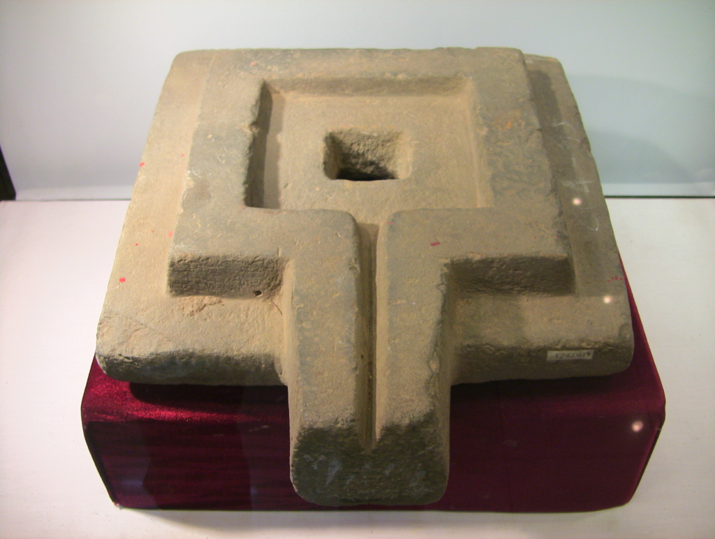
Fertility Symbol
The geometric design on the ancient stone carving in Figure 18.6.1 represents a powerful symbol of female fertility: the vagina. The symbol is called yoni in Hindu, and it reflects the value placed by Hindu culture on the ability of females to give birth. The vagina is one of several organs in the female reproductive system.
Female Reproductive Organs
The female reproductive system is made up of internal and external organs that function to produce haploid female gametes called ova (or oocytes), secrete female sex hormones (such as estrogen), and carry and give birth to a fetus. The internal female reproductive organs include the vagina, uterus, oviducts, and ovaries. The external organs — collectively called the vulva — include the clitoris and labia.
The vagina is an elastic, muscular canal leading from its opening in the vulva to the neck of the uterus, called the cervix. It is about 7.5 cm (about 3 in) long at the front, and about 9 cm (3.5 in) long at the back. The vagina accommodates the penis and is the site where sperm are usually ejaculated during sexual intercourse. In the context of pregnancy and natural (vaginal) childbirth, the vagina is referred to as the birth canal. In addition, it channels the flow of menstrual blood from the uterus.
Structure of the Vagina
Muscles and ligaments support the vagina within the pelvic cavity. The vagina itself is made up of several layers of fibrous and muscular tissues and lined with mucous membranes. Folds in the mucosa provide the vagina with extra surface area so it can stretch in both length and width during intercourse or childbirth. The elasticity of the vagina and the extra mucosa allow it to stretch to many times its normal diameter in order to deliver a baby.
Bacteria and pH in the Vagina
A healthy vagina is home to many symbiotic bacteria that help prevent pathogens (such as yeast) from colonizing the vagina. The pH in the vagina is normally between 3.8 and 4.5, and this acidity also helps keep pathogenic microorganisms from colonizing it. The vagina constantly sheds its epithelium, so it is considered self-cleaning. As a consequence, there is no need for douching to clean it. Physicians actually discourage the practice, as it may upset the normal bacterial and pH balance in the vagina, although washing the vulva with a mild soap is good practice.
Uterus
The uterus (commonly called the womb) is a pear-shaped, muscular organ that is about 7.6 cm (about 3 in) long. It is located above the vagina and behind the bladder in the centre of the pelvis. The position of the uterus in the pelvis is stabilized by several ligaments and bands of supportive tissue. The uterus is where a fetus develops during gestation, and the organ provides mechanical protection and support for the developing offspring. Contractions of the muscular wall of the uterus are responsible for pushing the fetus out of the uterus during childbirth.
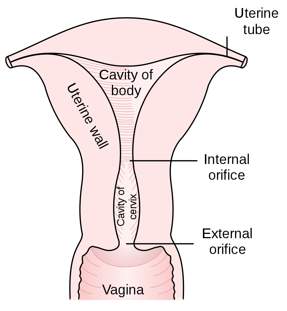
Parts of the Uterus
As shown in Figure 18.6.2, the lower end of the uterus forms the cervix, which is also called the neck of the uterus. The cervix is about 2.5 cm (almost 1 in) long and protrudes downward into the vagina. A small canal runs the length of the cervix, connecting the uterine cavity with the lumen of the vagina. This allows semen deposited in the vagina to enter the uterus, and a baby to pass from the uterus into the vagina during birth. Glands in the cervix secrete mucus that varies in water content and thickness, so it can function either as a barrier to keep microorganisms out of the uterus during pregnancy, or as a transport medium to help sperm enter the uterus around the time of ovulation. The rest of the uterus above the cervix is called the body of the uterus. The upper end of the uterus is connected with the two oviducts.
Tissues of the Uterus
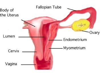
As indicated in Figure 18.6.3, the uterus consists of three tissue layers, called the endometrium, myometrium, and perimetrium.
- The endometrium is the innermost tissue layer of the uterus. It consists of epithelial tissue, including mucous membranes. This layer thickens during each menstrual cycle and, unless an egg is fertilized, sloughs off during the following menstrual period. If an ovum is fertilized, the thickened endometrium is maintained by hormones and provides nourishment to the embryo. As gestation progresses, the endometrium develops into the maternal portion of the placenta.
- The placenta is a temporary organ that consists of a mass of maternal and fetal blood vessels through which the mother’s and fetus’s blood exchange substances.
- The myometrium is the middle layer of the uterus. It consists mostly of a thick layer of smooth muscle tissue. Powerful contractions of the smooth muscle allow the uterus to contract and expel an infant during childbirth.
- The perimetrium is the outermost layer of the uterus. It covers the outer surface of the uterus. This layer actually consists of two layers of epithelium that secrete fluid into the space between them. The fluid allows for small movements of the uterus within the pelvis, without causing it to rub against other organs.
Oviducts
The oviducts (often referred to as Fallopian tubes) are two thin tubes that lie between the ovaries and the uterus. The oviducts are not attached to the ovaries, but their broad upper ends — called infundibula — lie very close to the ovaries. The infundibula also have fringe-like extensions called fimbriae that move in a waving motion to help guide eggs from the ovaries into the oviducts. The lower ends of the oviducts are attached to the upper part of the body of the uterus on either side of the body. They open into the uterus.
The oviducts are made up of multiple tissue layers. The innermost layer consists of mucosal epithelium. The epithelium is covered with cilia, which can move in a sweeping motion to help ova move through the tube from the ovary to the uterus. In between the ciliated cells of the epithelium are cells that secrete a fluid called tubular fluid. This fluid contains nutrients for sperm, ova, and zygotes. The secretions in tubular fluid also remove certain molecules from the plasma membrane of sperm so they are better able to penetrate an egg. Other layers of the oviducts consist of connective tissue and smooth muscle. Contractions of the smooth muscle allow peristalsis to help move eggs through the tubes.
Ovaries
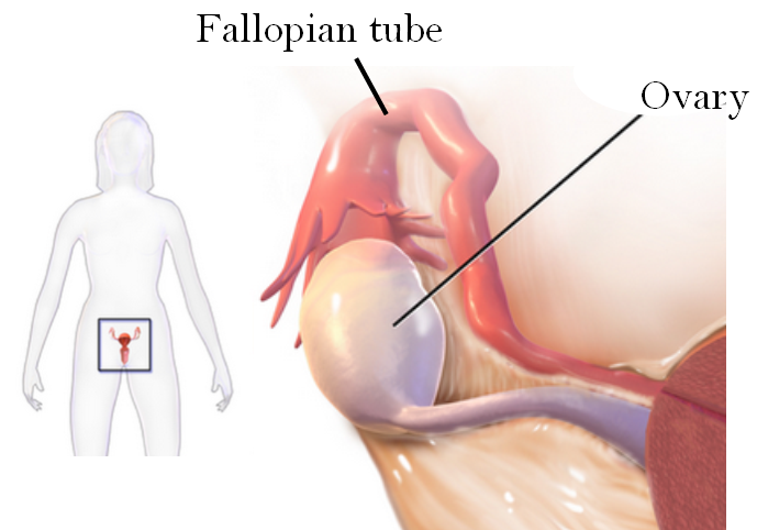
Like the testes in males, the ovaries in females are gonads that produce gametes and secrete sex hormones. The gametes produced by the ovaries are called ova, or oocytes. The main sex hormone secreted by the ovaries is estrogen. The position of the paired ovaries relative to the other reproductive system organs is shown in Figure 18.6.4. Each ovary lies along one side of the uterus and is about 4 cm (a little more than 1.5 in) long. Fibrous ligaments attach one end of each ovary to its nearby oviduct and the other and to its side of the uterus. These ligaments keep the ovaries in place within the pelvis.
Ovarian Follicles
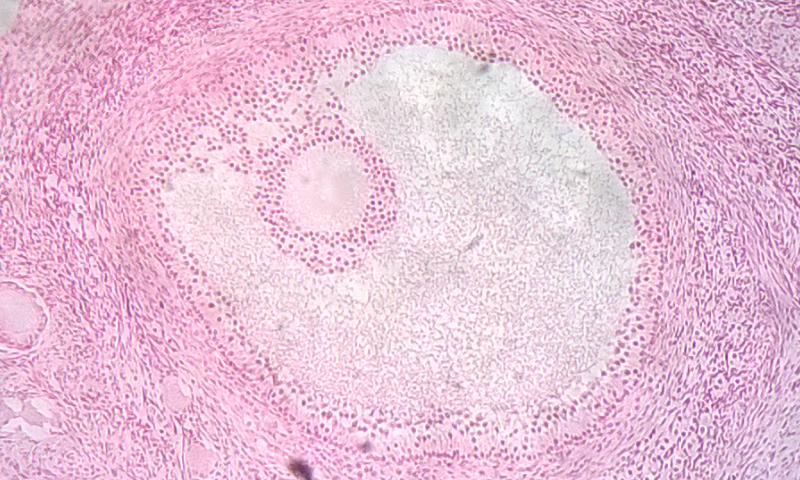
The ovary consists of two main layers, called the ovarian medulla (the inner layer) and the ovarian cortex (the outer layer). The ovary also contains blood and lymphatic vessels. The ovarian cortex consists primarily of the functional units of the ovaries, which are called ovarian follicles. The follicles are nests of epithelial cells, within each of which is an ovum. The photomicrograph in Figure 18.6.5 shows an ovarian follicle and the developing ovum inside it. If an ovum and follicle complete maturation, the follicle ruptures and the ovum is released from the ovary. This event is called ovulation.
Ova in the Ovaries
Whereas the male testes produce sperm continuously after puberty, the female ovary already contains all the ova it will ever produce by the time a female is born. At birth, a baby girl’s ovaries contain at least a million eggs, each of which is contained within a follicle. Only about 500 of these eggs will eventually mature and be ovulated. This process starts at puberty and typically continues at monthly intervals until menopause occurs around age 52. The remaining eggs never mature, and their number declines as the woman ages. By menopause, a woman’s reserve of eggs is nearly depleted, and ovulation no longer occurs.
Vulva
The vulva is a general term for all of the external female reproductive organs. The vulva includes the clitoris, labia, and external openings for the urethra and vagina.
Labia
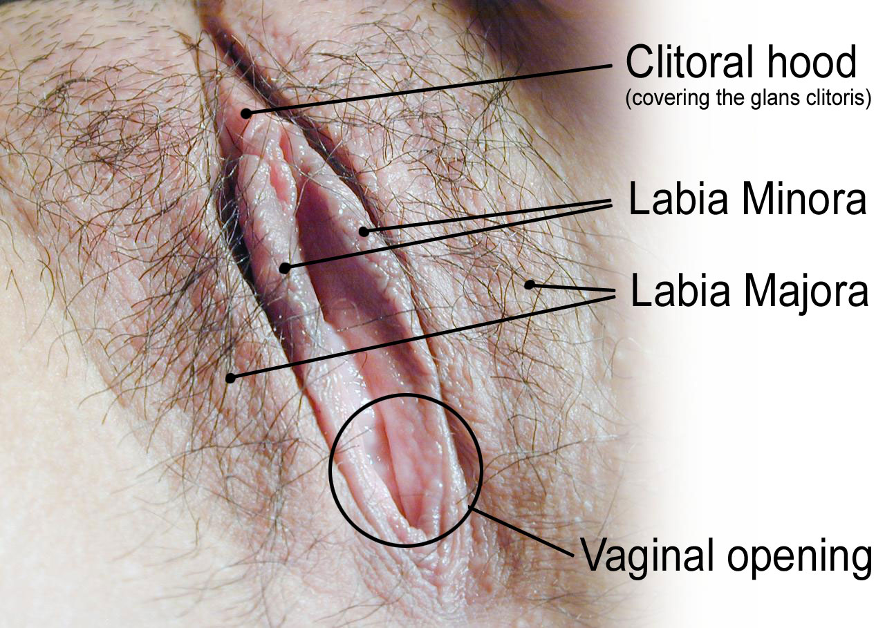
The labia (singular, labium) refer to the “lips” of the vulva, which are folds of tissue that contain and protect the other, more delicate structures of the vulva (as shown in Figure 18.6.6). There are two pairs of labia: the outer and larger labia majora, and the inner and smaller labia minora. The labia minora contain numerous sebaceous glands. These glands release secretions that help lubricate the labia and vulvar area.
Clitoris
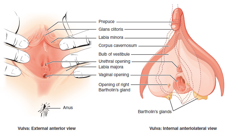
The clitoris, is located at the front of the vulva where the labia minora meet. The visible portion of the clitoris is called the glans clitoris. It is roughly the size and shape of a pea. It is highly sensitive, because it contains many nerve endings. A hood of tissue called the clitoral hood (shown in Figure 18.6.5 above), or prepuce, normally covers and protects the clitoris. The clitoris is the homologue to the male penis, and they both contain spongy tissue. Stimulation of the glans clitoris during sexual activity generally results in sexual arousal in females, and may lead to orgasm. The glans clitoris is the only part of the overall clitoris visible externally, but this spongy tissue extends down either side of the openings to the urethra and vagina, as seen in Figure 18.6.7.
Other Vulvar Structures
The area between the two labia minora is called the vestibule of the vulva. Both the urethra and vagina have openings to the outside of the body in the vestibule. As you can see in Figure 18.6.7 above, the urethral opening (or meatus) is located just in front of, and is much smaller than, the vaginal opening. Both openings are protected by the labia. Two glands — called Bartholin’s glands — open on either side of the vaginal opening. These glands secrete mucus and a vaginal and vulvar lubricant.
Breasts
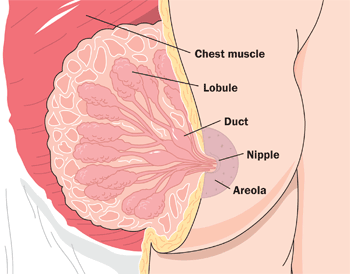
The breasts are not directly involved in reproduction, but because they contain mammary glands, they can provide nourishment to an infant after birth. The breasts overlay major muscles in the chest from which they project outward in a conical shape. The two main types of tissues in the breast are adipose (fat) tissue and glandular tissue that produces milk. As shown in Figure 18.6.8, each mature breast contains many lobules, where milk is produced and stored during pregnancy. During breastfeeding (or lactation), the milk drains into ducts and sacs, which in turn converge at the nipple. Milk exits the breast through the nipple in response to the suckling action of an infant and is regulated by a positive feedback loop. The nipple is surrounded by a more darkly pigmented area called the areola. The areola contains glands that secrete an oily fluid, which lubricates and protects the nipple during breastfeeding.
18.6 Summary
- The female reproductive system is made up of internal and external organs that function to produce haploid female gametes called ova, secrete female sex hormones (such as estrogen), and carry and give birth to a fetus.
- The vagina is an elastic, muscular canal that can accommodate the penis. It is also where sperm are usually ejaculated during sexual intercourse. The vagina is the birth canal, and it channels the flow of menstrual blood from the uterus. A healthy vagina has a balance of symbiotic bacteria and an acidic pH.
- The uterus is a muscular organ above the vagina where a fetus develops. Its muscular walls contract to push out the fetus during childbirth. The cervix is the neck of the uterus that extends down into the vagina. It contains a canal connecting the vagina and uterus for sperm, or for an infant to pass through. The innermost layer of the uterus — the endometrium — thickens each month in preparation for an embryo, but is shed in the following menstrual period if fertilization does not occur.
- The oviducts extend from the uterus to the ovaries. Waving fimbriae at the ovary ends of the oviducts guide ovulated eggs into the tubes where fertilization may occur as the ova travel to the uterus. Cilia and peristalsis help ova move through the tubes. Tubular fluid helps nourish sperm as they swim up the tubes toward ova.
- The ovaries are gonads that produce ova and secrete sex hormones, including estrogen. Nests of cells called follicles in the ovarian cortex are the functional units of ovaries. Each follicle surrounds an immature ovum. At birth, a baby girl’s ovaries contain at least a million ova, and they will not produce any more during her lifetime. During a woman’s reproductive years, one ova typically matures and is ovulated each month.
- The vulva is a general term for external female reproductive organs. The vulva includes the clitoris, two pairs of labia, and openings for the urethra and vagina. Secretions from mucosal glands near the vaginal opening lubricate the vulva.
- The breasts are not technically reproductive organs, but their mammary glands produce milk to feed an infant after birth. Milk drains through ducts and sacs, and out through the nipple when a baby sucks during breastfeeding.
18.6 Review Questions
-
- State the general functions of the female reproductive system.
- Describe the vagina and its reproductive functions.
- Outline the structure and basic functions of the uterus.
- What is the endometrium? How does it change during the monthly cycle?
- Why are breasts included in discussions of reproduction, if they are not organs of the female reproductive system?
- What is the function of the folds in the mucous membrane lining of the vagina?
- What are two ways in which the female reproductive system protects itself from pathogens?
18.6 Explore More
The uncomplicated truth about women’s sexuality | Sarah Barmak, TED, 2019.
Human Physiology – Functional Anatomy of the Female Reproductive System, Janux, 2015.
Attributes
Figure 18.6.1
1024px-Cattien_stone_yoni by Binh Giang on Wikimedia Commons is in the public domain (https://en.wikipedia.org/wiki/en:public_domain).
Figure 18.6.2
1000px-Gray1167.svg by Henry Vandyke Carter (1831-1897) on Wikimedia Commons is in the public domain (https://en.wikipedia.org/wiki/public_domain). (Bartleby.com: Gray’s Anatomy, Plate 1167).
Figure 18.6.3
Uterine_anatomy. from Uterine Stem cells by The Stem Cell Research Community, StemBook on Wikimedia Commons is used under a CC BY 3.0 (https://creativecommons.org/licenses/by/3.0) license.
Figure 18.6.4
Sites_of_tubo_ovarian_abscess by Bfpage on Wikimedia Commons is used under a CC BY-SA 4.0 (https://creativecommons.org/licenses/by-sa/4.0) license.
Figure 18.6.5
Ovarian_follicle by TiagoLubiana on Wikimedia Commons is used under a CC BY 4.0 (https://creativecommons.org/licenses/by-sa/4.0) license.
Figure 18.6.6
HumanVulva-NewText-PhiloViv by Amphis (edited) on Wikimedia Commons is used under a CC BY-SA 3.0 (https://creativecommons.org/licenses/by-sa/3.0/deed.en) license. (Original en:Image:HumanVulva-NoText-PhiloVivero.jpg by en:user:PhiloVivero)
Figure 18.6.7
Vulva by OpenStax College on Wikimedia Commons is used under a CC BY 3.0 (https://creativecommons.org/licenses/by/3.0) license.
Figure 18.6.8
Breast-Diagram by Women’s Health (NCI/ NIH) on Wikimedia Commons is in the public domain (https://en.wikipedia.org/wiki/en:Public_domain).
References
Betts, J. G., Young, K.A., Wise, J.A., Johnson, E., Poe, B., Kruse, D.H., Korol, O., Johnson, J.E., Womble, M., DeSaix, P. (2013, June 19). Figure 27.10 The vulva [digital image]. In Anatomy and Physiology (Section 27.2). OpenStax. https://openstax.org/books/anatomy-and-physiology/pages/27-2-anatomy-and-physiology-of-the-female-reproductive-system
Janux. (2015, January 10). Human physiology – Functional anatomy of the female reproductive system. YouTube. https://www.youtube.com/watch?v=9rs2gNchQig&feature=youtu.be
TED. (2019, March 22). The uncomplicated truth about women’s sexuality | Sarah Barmak. YouTube. https://www.youtube.com/watch?v=SkB4gG8ke7Q&feature=youtu.be
Teixeira, J., Rueda, B.R., and Pru, J.K. (September 30, 2008). Figure 1 Uterine anatomy. In Uterine Stem Cells (StemBook, ed.). The Stem Cell Research Community, StemBook, doi/10.3824/stembook.1.16.1, http://www.stembook.org
Case Study: Please Don’t Pass the Bread
Angela and Saloni are college students who met in physics class. They decide to study together for their upcoming midterm, but first, they want to grab some lunch. Angela says there is a particular restaurant she would like to go to, because they are able to accommodate her dietary restrictions. Saloni agrees and they head to the restaurant.
At lunch, Saloni asks Angela what is special about her diet. Angela tells her that she can’t eat gluten. Saloni says, “My cousin did that for a while because she heard that gluten is bad for you. But it was too hard for her to not eat bread and pasta, so she gave it up.” Angela tells Saloni that avoiding gluten isn’t optional for her — she has celiac disease. Eating even very small amounts of gluten could damage her digestive system. It can be difficult for people living with celiac disease to find foods when eating out.
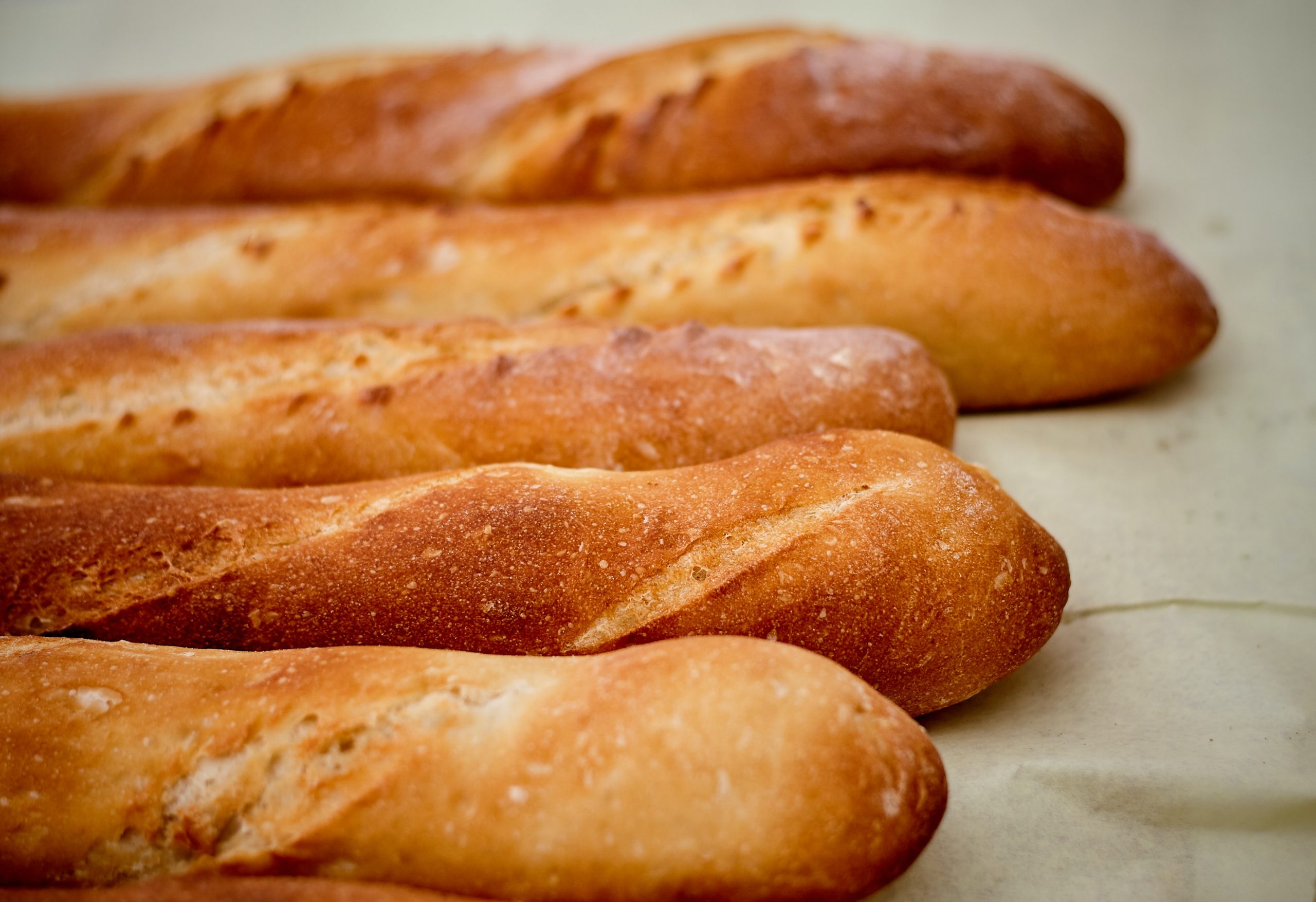
You have probably heard of gluten, but what is it, and why is it harmful to people with celiac disease? Gluten is a protein present in wheat and some other grains (such as barley, rye, and oats), so it is commonly found in foods like bread, pasta, baked goods, and many packaged foods, like the ones pictured in Figure 15.1.2.
Figure 15.1.2 Gluten is a protein present in foods like bread, pasta, and baked goods.
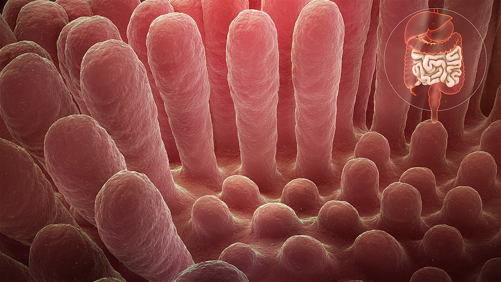
For people with celiac disease, eating gluten causes an autoimmune reaction that results in damage to the small, finger-like villi lining the small intestine, causing them to become inflamed and flattened (see Figure 15.1.3). This damage interferes with the digestive process, which can result in a wide variety of symptoms including diarrhea, anemia, skin rash, bone pain, depression, and anxiety, among others. The degree of damage to the villi can vary from mild to severe, with more severe damage generally resulting in more significant symptoms and complications. Celiac disease can have serious long-term consequences, such as osteoporosis, problems in the nervous and reproductive systems, and the development of certain types of cancers.
Why does celiac disease cause so many different types of symptoms and have such significant negative health consequences? As you read this chapter and learn about how the digestive system works, you will see just how important the villi of the small intestine are to the body as a whole. At the end of the chapter, you will learn more about celiac disease, why it can be so serious, and whether it is worth avoiding gluten for people who do not have a diagnosed medical issue with it.
Chapter Overview: Digestive System
In this chapter, you will learn about the digestive system, which processes food so that our bodies can obtain nutrients. Specifically, you will learn about:
- The structures and organs of the gastrointestinal (GI) tract through which food directly passes. This includes the mouth, pharynx, esophagus, stomach, small intestine, and large intestine.
- The functions of the GI tract, including mechanical and chemical digestion, absorption of nutrients, and the elimination of solid waste.
- The accessory organs of digestion — the liver, gallbladder, and pancreas — which secrete substances needed for digestion into the GI tract, in addition to performing other important functions.
- Specializations of the tissues of the digestive system that allow it to carry out its functions.
- How different types of nutrients (such as carbohydrates, proteins, and fats) are digested and absorbed by the body.
- Beneficial bacteria that live in the GI tract and help us digest food, produce vitamins, and protect us from harmful pathogens and toxic substances.
- Disorders of the digestive system, including inflammatory bowel diseases, ulcers, diverticulitis, and gastroenteritis (commonly known as “stomach flu”).
As you read this chapter, think about the following questions related to celiac disease:
- What are the general functions of the small intestine? What do the villi in the small intestine do?
- Why do you think celiac disease causes so many different types of symptoms and potentially serious complications?
- What are some other autoimmune diseases that involve the body attacking its own digestive system?
Attributions
Figure 15.1.1
Bread [photo] by Sergio Arze on Unsplash is used under the Unsplash License (https://unsplash.com/license).
Figure 15.1.2
- Paste cu sos de roșii by Sestrjevitovschii Ina on Unsplash is used under the Unsplash License (https://unsplash.com/license).
- Cookies and More by Sarah Shaffer on Unsplash is used under the Unsplash License (https://unsplash.com/license).
- Raspberry waffles by Izabelle Acheson on Unsplash is used under the Unsplash License (https://unsplash.com/license).
- Homemade croissant & pain au chocolat by Cristiano Pinto on Unsplash is used under the Unsplash License (https://unsplash.com/license).
Figure 15.1.3
Inflammed_mucous_layer_of_the_intestinal_villi_depicting_Celiac_disease by www.scientificanimations.com (image 140/191) on Wikimedia Commons is used under a CC BY-SA 4.0 (https://creativecommons.org/licenses/by-sa/4.0) license.
The female sex hormone secreted mainly by the ovaries.
As per caption.
Image shows a diagram labeling the major arteries of the body. Some of these include the carotid artery which provides blood to the neck and head, the brachiocephalic artery which supplies blood to the arms and head, the renal artery supplying blood to the kidneys, the mesenteric arteries supplying blood to the intestines, the femoral arteries supplying blood to the legs.
A testable proposed explanation for a phenomenon.

We All Scream for Ice Cream
If you’re an ice cream lover, then just the sight of this yummy ice cream cone may make your mouth water. The “water” in your mouth is actually saliva, a fluid released by glands that are part of the digestive system. Saliva contains digestive enzymes, among other substances important for digestion. When your mouth waters at the sight of a tasty treat, it’s a sign that your digestive system is preparing to digest food.
What Is the Digestive System?
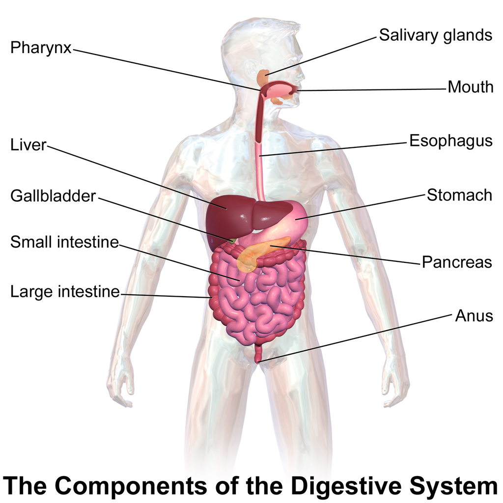
The digestive system consists of organs that break down food, absorb its nutrients, and expel any remaining waste. Organs of the digestive system are shown in Figure 15.2.2. Most of these organs make up the gastrointestinal (GI) tract, through which food actually passes. The rest of the organs of the digestive system are called accessory organs. These organs secrete enzymes and other substances into the GI tract, but food does not actually pass through them.
Functions of the Digestive System
The digestive system has three main functions relating to food: digestion of food, absorption of nutrients from food, and elimination of solid food waste. Digestion is the process of breaking down food into components the body can absorb. It consists of two types of processes: mechanical digestion and chemical digestion. Mechanical digestion is the physical breakdown of chunks of food into smaller pieces, and it takes place mainly in the mouth and stomach. Chemical digestion is the chemical breakdown of large, complex food molecules into smaller, simpler nutrient molecules that can be absorbed by body fluids (blood or lymph). This type of digestion begins in the mouth and continues in the stomach, but occurs mainly in the small intestine.
After food is digested, the resulting nutrients are absorbed. Absorption is the process in which substances pass into the bloodstream or lymph system to circulate throughout the body. Absorption of nutrients occurs mainly in the small intestine. Any remaining matter from food that is not digested and absorbed passes out of the body through the anus in the process of elimination.
Gastrointestinal Tract
The gastrointestinal (GI) tract is basically a long, continuous tube that connects the mouth with the anus. If it were fully extended, it would be about nine metres long in adults. It includes the mouth, pharynx, esophagus, stomach, and small and large intestines. Food enters the mouth, and then passes through the other organs of the GI tract, where it is digested and/or absorbed. Finally, any remaining food waste leaves the body through the anus at the end of the large intestine. It takes up to 50 hours for food or food waste to make the complete trip through the GI tract.
Tissues of the GI Tract
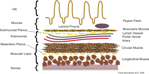
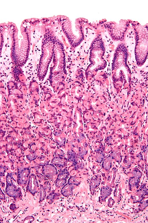
The walls of the organs of the GI tract consist of four different tissue layers, which are illustrated in Figure 15.2.3: mucosa, submucosa, muscularis externa, and serosa.
- The mucosa is the innermost layer surrounding the lumen (open space within the organs of the GI tract). This layer consists mainly of epithelium with the capacity to secrete and absorb substances. The epithelium can secret digestive enzymes and mucus, and it can absorb nutrients and water.
- The submucosa layer consists of connective tissue that contains blood and lymph vessels, as well as nerves. The vessels are needed to absorb and carry away nutrients after food is digested, and nerves help control the muscles of the GI tract organs.
- The muscularis externa layer contains two types of smooth muscle: longitudinal muscle and circular muscle. Longitudinal muscle runs the length of the GI tract organs, and circular muscle encircles the organs. Both types of muscles contract to keep food moving through the tract by the process of peristalsis, which is described below.
- The serosa layer is the outermost layer of the walls of GI tract organs. This is a thin layer that consists of connective tissue and separates the organs from surrounding cavities and tissues.
 |
 |
Peristalisis in the GI Tract
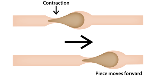
The muscles in the walls of GI tract organs enable peristalsis, which is illustrated in Figure 15.2.5. Peristalsis is a continuous sequence of involuntary muscle contraction and relaxation that moves rapidly along an organ like a wave, similar to the way a wave moves through a spring toy. Peristalsis in organs of the GI tract propels food through the tract.
Watch the video "What is peristalsis?" by Mister Science to see peristalsis in action:
https://youtu.be/kVjeNZA5pi4
What is peristalsis?, Mister Science, 2018.
Immune Function of the GI Tract
The GI tract plays an important role in protecting the body from pathogens. The surface area of the GI tract is estimated to be about 32 square metres (105 square feet), or about half the area of a badminton court. This is more than three times the area of the exposed skin of the body, and it provides a lot of area for pathogens to invade the tissues of the body. The innermost mucosal layer of the walls of the GI tract provides a barrier to pathogens so they are less likely to enter the blood or lymph circulations. The mucus produced by the mucosal layer, for example, contains antibodies that mark many pathogenic microorganisms for destruction. Enzymes in some of the secretions of the GI tract also destroy pathogens. In addition, stomach acids have a very low pH that is fatal for many microorganisms that enter the stomach.
Divisions of the GI Tract
The GI tract is often divided into an upper GI tract and a lower GI tract. For medical purposes, the upper GI tract is typically considered to include all the organs from the mouth through the first part of the small intestine, called the duodenum. For our instructional purposes, it makes more sense to include the mouth through the stomach in the upper GI tract, and all of the small intestine — as well as the large intestine — in the lower GI tract.
Upper GI Tract
The mouth is the first digestive organ that food enters. The sight, smell, or taste of food stimulates the release of digestive enzymes and other secretions by salivary glands inside the mouth. The major salivary gland enzyme is amylase. It begins the chemical digestion of carbohydrates by breaking down starches into sugar. The mouth also begins the mechanical digestion of food. When you chew, your teeth break, crush, and grind food into increasingly smaller pieces. Your tongue helps mix the food with saliva and also helps you swallow.
A lump of swallowed food is called a bolus. The bolus passes from the mouth into the pharynx, and from the pharynx into the esophagus. The esophagus is a long, narrow tube that carries food from the pharynx to the stomach. It has no other digestive functions. Peristalsis starts at the top of the esophagus when food is swallowed and continues down the esophagus in a single wave, pushing the bolus of food ahead of it.
From the esophagus, food passes into the stomach, where both mechanical and chemical digestion continue. The muscular walls of the stomach churn and mix the food, thus completing mechanical digestion, as well as mixing the food with digestive fluids secreted by the stomach. One of these fluids is hydrochloric acid (HCl). In addition to killing pathogens in food, it gives the stomach the low pH needed by digestive enzymes that work in the stomach. One of these enzymes is pepsin, which chemically digests proteins. The stomach stores the partially digested food until the small intestine is ready to receive it. Food that enters the small intestine from the stomach is in the form of a thick slurry (semi-liquid) called chyme.
Lower GI Tract
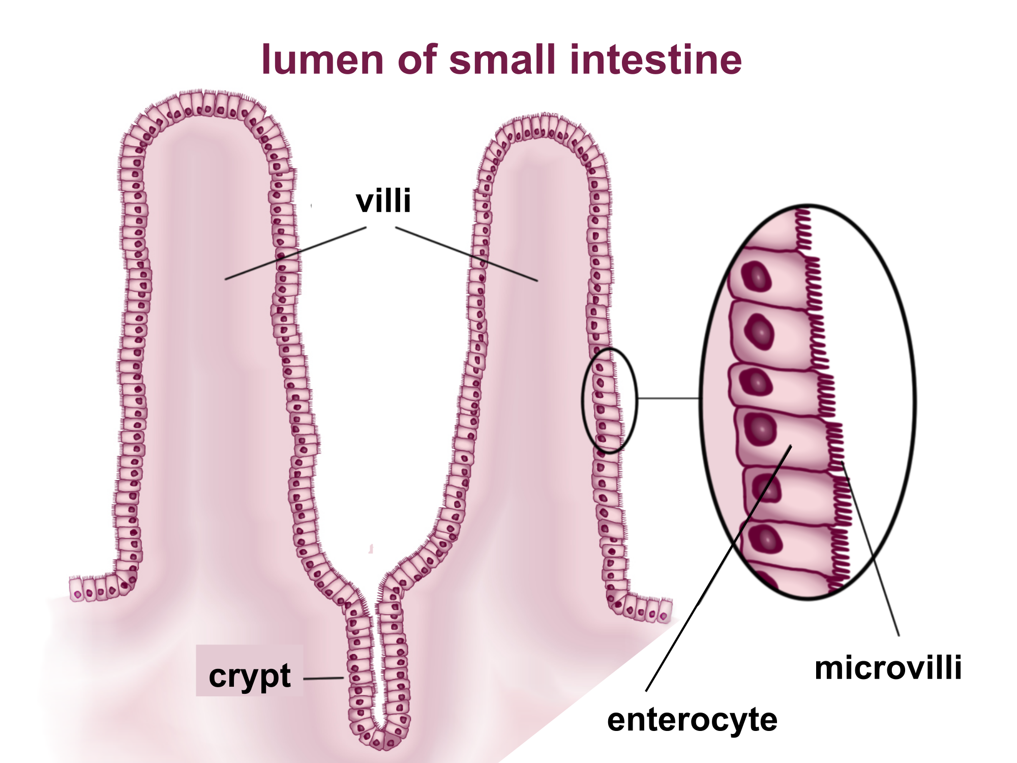
The small intestine is a narrow, but very long tubular organ. It may be almost seven metres long in adults. It is the site of most chemical digestion and virtually all absorption of nutrients. Many digestive enzymes are active in the small intestine, some of which are produced by the small intestine itself, and some of which are produced by the pancreas, an accessory organ of the digestive system. Much of the inner lining of the small intestine is covered by tiny finger-like projections called villi, each of which is covered by even tinier projections called microvilli. These projections, shown in the drawing below (Figure 15.2.6), greatly increase the surface area through which nutrients can be absorbed from the small intestine.
From the small intestine, any remaining nutrients and food waste pass into the large intestine. The large intestine is another tubular organ, but it is wider and shorter than the small intestine. It connects the small intestine and the anus. Waste that enters the large intestine is in a liquid state. As it passes through the large intestine, excess water is absorbed from it. The remaining solid waste — called feces — is eventually eliminated from the body through the anus.
Accessory Organs of the Digestive System
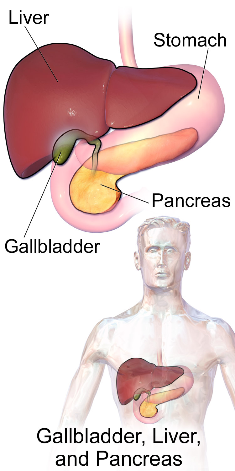
Accessory organs of the digestive system are not part of the GI tract, so they are not sites where digestion or absorption take place. Instead, these organs secrete or store substances needed for the chemical digestion of food. The accessory organs include the liver, gallbladder, and pancreas. They are shown in Figure 15.2.7 and described in the text that follows.
- The liver is an organ with multitude of functions. Its main digestive function is producing and secreting a fluid called bile, which reaches the small intestine through a duct. Bile breaks down large globules of lipids into smaller ones that are easier for enzymes to chemically digest. Bile is also needed to reduce the acidity of food entering the small intestine from the highly acidic stomach, because enzymes in the small intestine require a less acidic environment in order to work.
- The gallbladder is a small sac below the liver that stores some of the bile from the liver. The gallbladder also concentrates the bile by removing some of the water from it. It then secretes the concentrated bile into the small intestine as needed for fat digestion following a meal.
- The pancreas secretes many digestive enzymes, and releases them into the small intestine for the chemical digestion of carbohydrates, proteins, and lipids. The pancreas also helps lessen the acidity of the small intestine by secreting bicarbonate, a basic substance that neutralizes acid.
15.2 Summary
- The digestive system consists of organs that break down food, absorb its nutrients, and expel any remaining food waste.
- Digestion is the process of breaking down food into components that the body can absorb. It includes mechanical digestion and chemical digestion. Absorption is the process of taking up nutrients from food by body fluids for circulation to the rest of the body. Elimination is the process of excreting any remaining food waste after digestion and absorption are finished.
- Most digestive organs form a long, continuous tube called the gastrointestinal (GI) tract. It starts at the mouth, which is followed by the pharynx, esophagus, stomach, small intestine, and large intestine. The upper GI tract consists of the mouth through the stomach, while the lower GI tract consists of the small and large intestines.
- Digestion and/or absorption take place in most of the organs of the GI tract. Organs of the GI tract have walls that consist of several tissue layers that enable them to carry out these functions. The inner mucosa has cells that secrete digestive enzymes and other digestive substances, as well as cells that absorb nutrients. The muscle layer of the organs enables them to contract and relax in waves of peristalsis to move food through the GI tract.
- Three digestive organs — the liver, gallbladder, and pancreas — are accessory organs of digestion. They secrete substances needed for chemical digestion into the small intestine.
15.2 Review Questions
- What is the digestive system?
- What are the three main functions of the digestive system? Define each function.
-
- Relate the tissues in the walls of GI tract organs to the functions the organs perform.
15.2 Explore More
https://youtu.be/Og5xAdC8EUI
How your digestive system works - Emma Bryce, TED-Ed, 2017.
https://youtu.be/YVfyYrEmzgM
How does your body know you're full? - Hilary Coller, TED-Ed, 2017.
Attributions
Figure 15.2.1
Ice Cream [photo] by Mark Cruz on Unsplash is used under the Unsplash License (https://unsplash.com/license).
Figure 15.2.2
Blausen_0316_DigestiveSystem by BruceBlaus on Wikimedia Commons is used under a CC BY 3.0 (https://creativecommons.org/licenses/by/3.0) license.
Figure 15.2.3
Intestinal_layers by Boumphreyfr on Wikimedia Commons is used under a CC BY-SA 3.0 (https://creativecommons.org/licenses/by-sa/3.0) license.
Figure 15.2.4
512px-Normal_gastric_mucosa_intermed_mag by Nephron on Wikimedia Commons is used under a CC BY-SA 3.0 (https://creativecommons.org/licenses/by-sa/3.0) license.
Figure 15.2.5
Peristalsis pushes food through the GI tract by CK-12 Foundation is used under a CC BY NC 3.0 (https://creativecommons.org/licenses/by-nc/3.0/) license.
Figure 15.2.6
Villi_&_microvilli_of_small_intestine.svg by BallenaBlanca on Wikimedia Commons is used under a CC BY-SA 4.0 (https://creativecommons.org/licenses/by-sa/4.0) license.
Figure 15.2.7
Blausen_0428_Gallbladder-Liver-Pancreas_Location by BruceBlaus on Wikimedia Commons is used under a CC BY 3.0 (https://creativecommons.org/licenses/by/3.0) license.
References
Blausen.com Staff. (2014). Medical gallery of Blausen Medical 2014. WikiJournal of Medicine 1 (2). DOI:10.15347/wjm/2014.010. ISSN 2002-4436.
Brainard, J/ CK-12 Foundation. (2016). Figure 4 Peristalsis pushes food through the GI tract. [digital image]. In CK-12 College Human Biology (Section 17.2) [online Flexbook]. CK12.org. https://www.ck12.org/book/ck-12-college-human-biology/section/17.2/
Mister Science. (2018). What is peristalsis? YouTube. https://www.youtube.com/channel/UCxTlkZfjArUobBAeVwzJjYg/videos
TED-Ed. (2017, November 13). How does your body know you're full? - Hilary Coller. YouTube. https://www.youtube.com/watch?v=YVfyYrEmzgM&feature=youtu.be
TED-Ed. (2017, December 14). How your digestive system works - Emma Bryce. YouTube. https://www.youtube.com/watch?v=Og5xAdC8EUI&feature=youtu.be
Created by CK-12 Foundation/Adapted by Christine Miller

We All Scream for Ice Cream
If you’re an ice cream lover, then just the sight of this yummy ice cream cone may make your mouth water. The “water” in your mouth is actually saliva, a fluid released by glands that are part of the digestive system. Saliva contains digestive enzymes, among other substances important for digestion. When your mouth waters at the sight of a tasty treat, it’s a sign that your digestive system is preparing to digest food.
What Is the Digestive System?
The digestive system consists of organs that break down food, absorb its nutrients, and expel any remaining waste. Organs of the digestive system are shown in Figure 15.2.2. Most of these organs make up the gastrointestinal (GI) tract, through which food actually passes. The rest of the organs of the digestive system are called accessory organs. These organs secrete enzymes and other substances into the GI tract, but food does not actually pass through them.

Functions of the Digestive System
The digestive system has three main functions relating to food: digestion of food, absorption of nutrients from food, and elimination of solid food waste. Digestion is the process of breaking down food into components the body can absorb. It consists of two types of processes: mechanical digestion and chemical digestion. Mechanical digestion is the physical breakdown of chunks of food into smaller pieces, and it takes place mainly in the mouth and stomach. Chemical digestion is the chemical breakdown of large, complex food molecules into smaller, simpler nutrient molecules that can be absorbed by body fluids (blood or lymph). This type of digestion begins in the mouth and continues in the stomach, but occurs mainly in the small intestine.
After food is digested, the resulting nutrients are absorbed. Absorption is the process in which substances pass into the bloodstream or lymph system to circulate throughout the body. Absorption of nutrients occurs mainly in the small intestine. Any remaining matter from food that is not digested and absorbed passes out of the body through the anus in the process of elimination.
Gastrointestinal Tract
The gastrointestinal (GI) tract is basically a long, continuous tube that connects the mouth with the anus. If it were fully extended, it would be about nine metres long in adults. It includes the mouth, pharynx, esophagus, stomach, and small and large intestines. Food enters the mouth, and then passes through the other organs of the GI tract, where it is digested and/or absorbed. Finally, any remaining food waste leaves the body through the anus at the end of the large intestine. It takes up to 50 hours for food or food waste to make the complete trip through the GI tract.
Tissues of the GI Tract
The walls of the organs of the GI tract consist of four different tissue layers, which are illustrated in Figure 15.2.3: mucosa, submucosa, muscularis externa, and serosa.
- The mucosa is the innermost layer surrounding the lumen (open space within the organs of the GI tract). This layer consists mainly of epithelium with the capacity to secrete and absorb substances. The epithelium can secret digestive enzymes and mucus, and it can absorb nutrients and water.
- The submucosa layer consists of connective tissue that contains blood and lymph vessels, as well as nerves. The vessels are needed to absorb and carry away nutrients after food is digested, and nerves help control the muscles of the GI tract organs.
- The muscularis externa layer contains two types of smooth muscle: longitudinal muscle and circular muscle. Longitudinal muscle runs the length of the GI tract organs, and circular muscle encircles the organs. Both types of muscles contract to keep food moving through the tract by the process of peristalsis, which is described below.
- The serosa layer is the outermost layer of the walls of GI tract organs. This is a thin layer that consists of connective tissue and separates the organs from surrounding cavities and tissues.
 |
 |
Peristalisis in the GI Tract
The muscles in the walls of GI tract organs enable peristalsis, which is illustrated in Figure 15.2.5. Peristalsis is a continuous sequence of involuntary muscle contraction and relaxation that moves rapidly along an organ like a wave, similar to the way a wave moves through a spring toy. Peristalsis in organs of the GI tract propels food through the tract.

Watch the video "What is peristalsis?" by Mister Science to see peristalsis in action:
https://youtu.be/kVjeNZA5pi4
What is peristalsis?, Mister Science, 2018.
Immune Function of the GI Tract
The GI tract plays an important role in protecting the body from pathogens. The surface area of the GI tract is estimated to be about 32 square metres (105 square feet), or about half the area of a badminton court. This is more than three times the area of the exposed skin of the body, and it provides a lot of area for pathogens to invade the tissues of the body. The innermost mucosal layer of the walls of the GI tract provides a barrier to pathogens so they are less likely to enter the blood or lymph circulations. The mucus produced by the mucosal layer, for example, contains antibodies that mark many pathogenic microorganisms for destruction. Enzymes in some of the secretions of the GI tract also destroy pathogens. In addition, stomach acids have a very low pH that is fatal for many microorganisms that enter the stomach.
Divisions of the GI Tract
The GI tract is often divided into an upper GI tract and a lower GI tract. For medical purposes, the upper GI tract is typically considered to include all the organs from the mouth through the first part of the small intestine, called the duodenum. For our instructional purposes, it makes more sense to include the mouth through the stomach in the upper GI tract, and all of the small intestine — as well as the large intestine — in the lower GI tract.
Upper GI Tract
The mouth is the first digestive organ that food enters. The sight, smell, or taste of food stimulates the release of digestive enzymes and other secretions by salivary glands inside the mouth. The major salivary gland enzyme is amylase. It begins the chemical digestion of carbohydrates by breaking down starches into sugar. The mouth also begins the mechanical digestion of food. When you chew, your teeth break, crush, and grind food into increasingly smaller pieces. Your tongue helps mix the food with saliva and also helps you swallow.
A lump of swallowed food is called a bolus. The bolus passes from the mouth into the pharynx, and from the pharynx into the esophagus. The esophagus is a long, narrow tube that carries food from the pharynx to the stomach. It has no other digestive functions. Peristalsis starts at the top of the esophagus when food is swallowed and continues down the esophagus in a single wave, pushing the bolus of food ahead of it.
From the esophagus, food passes into the stomach, where both mechanical and chemical digestion continue. The muscular walls of the stomach churn and mix the food, thus completing mechanical digestion, as well as mixing the food with digestive fluids secreted by the stomach. One of these fluids is hydrochloric acid (HCl). In addition to killing pathogens in food, it gives the stomach the low pH needed by digestive enzymes that work in the stomach. One of these enzymes is pepsin, which chemically digests proteins. The stomach stores the partially digested food until the small intestine is ready to receive it. Food that enters the small intestine from the stomach is in the form of a thick slurry (semi-liquid) called chyme.
Lower GI Tract
The small intestine is a narrow, but very long tubular organ. It may be almost seven metres long in adults. It is the site of most chemical digestion and virtually all absorption of nutrients. Many digestive enzymes are active in the small intestine, some of which are produced by the small intestine itself, and some of which are produced by the pancreas, an accessory organ of the digestive system. Much of the inner lining of the small intestine is covered by tiny finger-like projections called villi, each of which is covered by even tinier projections called microvilli. These projections, shown in the drawing below (Figure 15.2.6), greatly increase the surface area through which nutrients can be absorbed from the small intestine.

From the small intestine, any remaining nutrients and food waste pass into the large intestine. The large intestine is another tubular organ, but it is wider and shorter than the small intestine. It connects the small intestine and the anus. Waste that enters the large intestine is in a liquid state. As it passes through the large intestine, excess water is absorbed from it. The remaining solid waste — called feces — is eventually eliminated from the body through the anus.
Accessory Organs of the Digestive System

Accessory organs of the digestive system are not part of the GI tract, so they are not sites where digestion or absorption take place. Instead, these organs secrete or store substances needed for the chemical digestion of food. The accessory organs include the liver, gallbladder, and pancreas. They are shown in Figure 15.2.7 and described in the text that follows.
- The liver is an organ with multitude of functions. Its main digestive function is producing and secreting a fluid called bile, which reaches the small intestine through a duct. Bile breaks down large globules of lipids into smaller ones that are easier for enzymes to chemically digest. Bile is also needed to reduce the acidity of food entering the small intestine from the highly acidic stomach, because enzymes in the small intestine require a less acidic environment in order to work.
- The gallbladder is a small sac below the liver that stores some of the bile from the liver. The gallbladder also concentrates the bile by removing some of the water from it. It then secretes the concentrated bile into the small intestine as needed for fat digestion following a meal.
- The pancreas secretes many digestive enzymes, and releases them into the small intestine for the chemical digestion of carbohydrates, proteins, and lipids. The pancreas also helps lessen the acidity of the small intestine by secreting bicarbonate, a basic substance that neutralizes acid.
15.2 Summary
- The digestive system consists of organs that break down food, absorb its nutrients, and expel any remaining food waste.
- Digestion is the process of breaking down food into components that the body can absorb. It includes mechanical digestion and chemical digestion. Absorption is the process of taking up nutrients from food by body fluids for circulation to the rest of the body. Elimination is the process of excreting any remaining food waste after digestion and absorption are finished.
- Most digestive organs form a long, continuous tube called the gastrointestinal (GI) tract. It starts at the mouth, which is followed by the pharynx, esophagus, stomach, small intestine, and large intestine. The upper GI tract consists of the mouth through the stomach, while the lower GI tract consists of the small and large intestines.
- Digestion and/or absorption take place in most of the organs of the GI tract. Organs of the GI tract have walls that consist of several tissue layers that enable them to carry out these functions. The inner mucosa has cells that secrete digestive enzymes and other digestive substances, as well as cells that absorb nutrients. The muscle layer of the organs enables them to contract and relax in waves of peristalsis to move food through the GI tract.
- Three digestive organs — the liver, gallbladder, and pancreas — are accessory organs of digestion. They secrete substances needed for chemical digestion into the small intestine.
15.2 Review Questions
- What is the digestive system?
- What are the three main functions of the digestive system? Define each function.
-
- Relate the tissues in the walls of GI tract organs to the functions the organs perform.
15.2 Explore More
https://youtu.be/Og5xAdC8EUI
How your digestive system works - Emma Bryce, TED-Ed, 2017.
https://youtu.be/YVfyYrEmzgM
How does your body know you're full? - Hilary Coller, TED-Ed, 2017.
Attributions
Figure 15.2.1
Ice Cream [photo] by Mark Cruz on Unsplash is used under the Unsplash License (https://unsplash.com/license).
Figure 15.2.2
Blausen_0316_DigestiveSystem by BruceBlaus on Wikimedia Commons is used under a CC BY 3.0 (https://creativecommons.org/licenses/by/3.0) license.
Figure 15.2.3
Intestinal_layers by Boumphreyfr on Wikimedia Commons is used under a CC BY-SA 3.0 (https://creativecommons.org/licenses/by-sa/3.0) license.
Figure 15.2.4
512px-Normal_gastric_mucosa_intermed_mag by Nephron on Wikimedia Commons is used under a CC BY-SA 3.0 (https://creativecommons.org/licenses/by-sa/3.0) license.
Figure 15.2.5
Peristalsis pushes food through the GI tract by CK-12 Foundation is used under a CC BY NC 3.0 (https://creativecommons.org/licenses/by-nc/3.0/) license.
Figure 15.2.6
Villi_&_microvilli_of_small_intestine.svg by BallenaBlanca on Wikimedia Commons is used under a CC BY-SA 4.0 (https://creativecommons.org/licenses/by-sa/4.0) license.
Figure 15.2.7
Blausen_0428_Gallbladder-Liver-Pancreas_Location by BruceBlaus on Wikimedia Commons is used under a CC BY 3.0 (https://creativecommons.org/licenses/by/3.0) license.
References
Blausen.com Staff. (2014). Medical gallery of Blausen Medical 2014. WikiJournal of Medicine 1 (2). DOI:10.15347/wjm/2014.010. ISSN 2002-4436.
Brainard, J/ CK-12 Foundation. (2016). Figure 4 Peristalsis pushes food through the GI tract. [digital image]. In CK-12 College Human Biology (Section 17.2) [online Flexbook]. CK12.org. https://www.ck12.org/book/ck-12-college-human-biology/section/17.2/
Mister Science. (2018). What is peristalsis? YouTube. https://www.youtube.com/channel/UCxTlkZfjArUobBAeVwzJjYg/videos
TED-Ed. (2017, November 13). How does your body know you're full? - Hilary Coller. YouTube. https://www.youtube.com/watch?v=YVfyYrEmzgM&feature=youtu.be
TED-Ed. (2017, December 14). How your digestive system works - Emma Bryce. YouTube. https://www.youtube.com/watch?v=Og5xAdC8EUI&feature=youtu.be
Image shows a man participating in a hot-dog eating contest. His mouth is so full of hot dog that he can't close his lips.
Image shows a side view diagram of the male and female pelvis. The male urethra is much longer because it extends through the penis, and in women it exits through the pelvic floor.
A biological process which converts sugars such as glucose, fructose, and sucrose into cellular energy, producing ethanol and carbon dioxide as by-products.
Created by CK-12 Foundation/Adapted by Christine Miller
Case Study Conclusion: Cough That Won't Quit
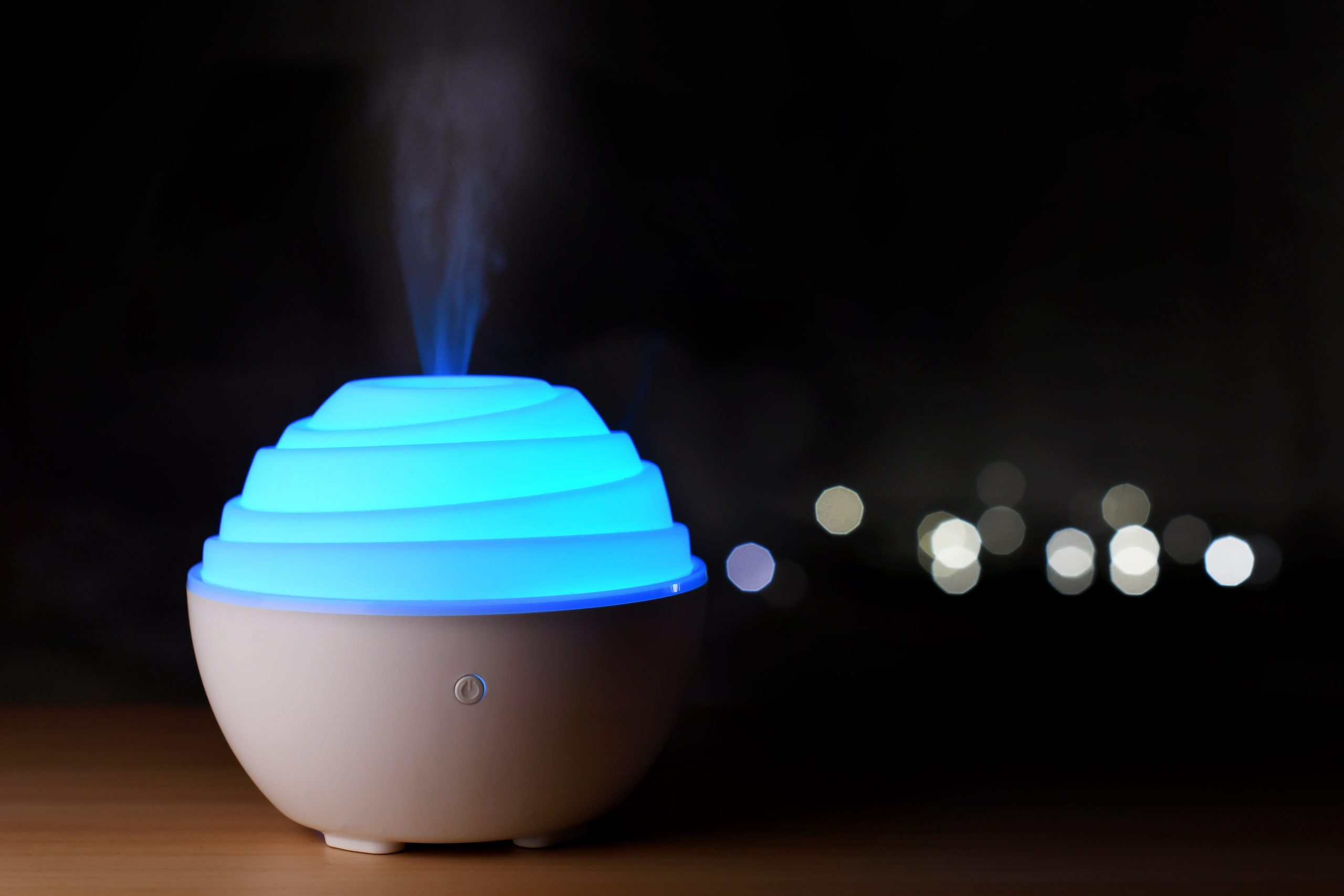
Inhaling the moist air from a humidifier or steamy shower can feel particularly good if you have a respiratory system infection, such as bronchitis. The moist air helps to loosen and thin mucus in the respiratory system, allowing you to breathe easier.
In the beginning of this chapter, you learned about Erica, who developed acute bronchitis after getting a cold. She had a worsening cough, a sore throat due to coughing, and chest congestion. She was also coughing up thick mucus.
Acute bronchitis usually occurs after a cold or flu, usually due to the same viruses that cause cold or flu. Because bronchitis is not usually caused by bacteria (although it can be), in most cases, antibiotics are not an effective treatment.
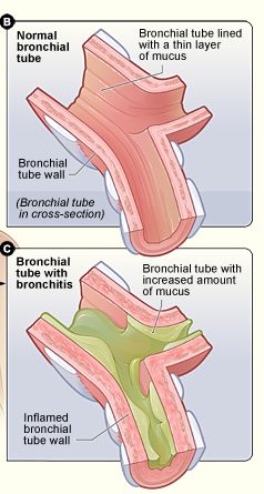
Bronchitis affects the bronchial tubes, which, as you have learned, are air passages in the lower respiratory tract. The main bronchi branch off of the trachea and then branch into smaller bronchi, and then bronchioles. In bronchitis, the walls of the bronchi become inflamed, which makes them narrower. There is also excessive production of mucus in the bronchi, which further narrows the pathway where air can flow through. Figure 13.7.2, shows how bronchitis affects the bronchial tubes.
The treatment for most cases of bronchitis involves thinning and loosening the mucus so that it can be effectively coughed out of the airways. This can be done by drinking plenty of fluids, using humidifiers or steam, and — in some cases — using over-the-counter medications (such as expectorants). Dr. Choo recommended some of these treatments to Erica, and also warned against using cough suppressants. Cough suppressants work on the nervous system to suppress the cough reflex. When a patient has a “productive” cough (which means they are coughing up mucus), doctors generally advise them not to take cough suppressants, so that they can cough the mucus out of their bodies.
When Dr. Choo was examining Erica, she used a pulse oximeter to measure the oxygen level in her blood. Why did she do this? As you have learned, the bronchial tubes branch into bronchioles, which ultimately branch into the alveoli of the lungs. The alveoli are where gas exchange occurs between the air and the blood to take in oxygen and remove carbon dioxide and other wastes. By checking Erica’s blood oxygen level, Dr. Choo was making sure that her clogged airways were not impacting her level of much-needed oxygen.
Erica has acute bronchitis, but you may recall that chronic bronchitis was discussed earlier in this chapter (Section 13.5) as a term that describes the symptoms of chronic obstructive pulmonary disease (COPD). COPD is often due to tobacco smoking, and it causes damage to the walls of the alveoli. Acute bronchitis, on the other hand, typically occurs after a cold or flu, and involves inflammation and mucus build-up in the bronchial tubes. As implied by the difference in their names, chronic bronchitis is an ongoing, long-term condition, while acute bronchitis is likely to resolve relatively quickly with proper rest and treatment.
Erica uses e-cigarettes (vaping), so she is more likely to develop chronic respiratory conditions, such as COPD. As you have learned, smoking damages the respiratory system, along with many other systems of the body. Smoking and vaping increases the risk of respiratory infections, including bronchitis and flu, due to its damaging effects on the respiratory and immune systems. Dr. Choo strongly encouraged Erica to quit vaping, not only so that her acute bronchitis resolves, but so that she can avoid future infections and other negative health outcomes associated with vaping and smoking, including COPD and lung cancer.
As you have learned in this chapter, the respiratory system is critical to carry out the gas exchange necessary for life’s functions, and to protect the body from pathogens and other potentially harmful substances in the air. But this ability to interface with the outside air has a cost. The respiratory system is prone to infections, as well as damage and other negative effects from allergens, mold, air pollution, cigarette smoke and vaping. While exposure to most of these things cannot be avoided, not smoking is an important step you can take to protect this organ system — as well as many other systems of your body.
Chapter 13 Summary
In this chapter, you learned about the respiratory system. Specifically, you learned that:
- Respiration is the process in which oxygen moves from the outside air into the body, and carbon dioxide and other waste gases move from inside the body to the outside air. It involves two subsidiary processes: ventilation and gas exchange.
- The organs of the respiratory system form a continuous system of passages, called the respiratory tract. It has two major divisions: the upper respiratory tract and the lower respiratory tract.
-
- The upper respiratory tract includes the nasal cavity, pharynx, and larynx. All of these organs are involved in conduction, or the movement of air into and out of the body. Incoming air is also cleaned, humidified, and warmed as it passes through the upper respiratory tract. The larynx is also called the voice box, because it contains the vocal cords, which are needed to produce vocal sounds.
- The lower respiratory tract includes the trachea, bronchi and bronchioles, and the lungs. The trachea, bronchi, and bronchioles are involved in conduction. Gas exchange takes place only in the lungs, which are the largest organs of the respiratory tract. Lung tissue consists mainly of tiny air sacs called alveoli, which is where gas exchange takes place between air in the alveoli and the blood in capillaries surrounding them.
- The respiratory system protects itself from potentially harmful substances in the air by the mucociliary escalator. This includes mucus-producing cells, which trap particles and pathogens in incoming air. It also includes tiny hair-like cilia that continually move to sweep the mucus and trapped debris away from the lungs and toward the outside of the body.
- The level of carbon dioxide in the blood is monitored by cells in the brain. If the level becomes too high, it triggers a faster rate of breathing, which lowers the level to the normal range. The opposite occurs if the level becomes too low. The respiratory system exchanges gases with the outside air, but it needs the cardiovascular system to carry the gases to and from cells throughout the body.
- Breathing, or ventilation, is the two-step process of drawing air into the lungs (inhalation) and letting air out of the lungs (exhalation). Inhaling is an active process that results mainly from contraction of a muscle called the diaphragm. Exhaling is typically a passive process that occurs mainly due to the elasticity of the lungs when the diaphragm relaxes.
-
- Breathing is one of the few vital bodily functions that can be controlled consciously, as well as unconsciously. Conscious control of breathing is common in many activities, including swimming and singing. However, there are limits on the conscious control of breathing. If you try to hold your breath, for example, you will soon have an irrepressible urge to breathe.
- Unconscious breathing is controlled by respiratory centers in the medulla and pons of the brainstem. They respond to variations in blood pH by either increasing or decreasing the rate of breathing as needed to return the pH level to the normal range.
- Nasal breathing is generally considered to be superior to mouth breathing, because it does a better job of filtering, warming, and moistening incoming air. It also results in slower emptying of the lungs, which allows more oxygen to be extracted from the air.
- Gas exchange is the biological process through which gases are transferred across cell membranes to either enter or leave the blood. Gas exchange takes place continuously between the blood and cells throughout the body, and also between the blood and the air inside the lungs.
-
- Gas exchange in the lungs takes place in alveoli. The pulmonary artery carries deoxygenated blood from the heart to the lungs, where it travels through pulmonary capillaries, picking up oxygen and releasing carbon dioxide. The oxygenated blood then leaves the lungs through pulmonary veins.
- Gas exchange occurs by diffusion across cell membranes. Gas molecules naturally move down a concentration gradient from an area of higher concentration to an area of lower concentration. This is a passive process that requires no energy.
- Gas exchange by diffusion depends on the large surface area provided by the hundreds of millions of alveoli in the lungs. It also depends on a steep concentration gradient for oxygen and carbon dioxide. This gradient is maintained by continuous blood flow and constant breathing.
- Asthma is a chronic inflammatory disease of the airways in the lungs, in which the airways periodically become inflamed. This causes swelling and narrowing of the airways, often with excessive mucus production, leading to difficulty breathing and other symptoms. Asthma is thought to be caused by a combination of genetic and environmental factors. Asthma attacks are triggered by allergens, air pollution, or other factors.
- Pneumonia is a common inflammatory disease of the respiratory tract in which inflammation affects primarily the alveoli, which become filled with fluid that inhibits gas exchange. Most cases of pneumonia are caused by viral or bacterial infections. Vaccines are available to prevent pneumonia. Treatment often includes prescription antibiotics.
- Chronic obstructive pulmonary disease (COPD) is a lung disease characterized by chronic poor airflow, which causes shortness of breath and a productive cough. It is caused most often by tobacco smoking, which leads to breakdown of connective tissues in the lungs. Alveoli are reduced in number and elasticity, making it impossible to fully exhale air from the lungs. There is no cure for COPD, but stopping smoking may reduce the rate at which COPD worsens.
- Lung cancer is a malignant tumor characterized by uncontrolled cell growth in tissues of the lung. It results from accumulated DNA damage, most often caused by tobacco smoking. Lung cancer is typically diagnosed late, so most cases cannot be cured. It may be treated with surgery, chemotherapy, and/or radiation therapy.
- Smoking is the single greatest cause of preventable death worldwide. It has adverse effects on just about every body system and organ. Tobacco smoke affects not only smokers, but also non-smokers who are exposed to secondhand smoke. The nicotine in tobacco is highly addictive, making it very difficult to quit smoking.
-
- A major health risk of smoking is lung cancer. Smoking also increases the risk of many other types of cancer. Tobacco smoke contains dozens of chemicals that are known carcinogens.
- Smoking is the primary cause of COPD. Chemicals — such as carbon monoxide and cyanide in tobacco smoke — reduce the elasticity of alveoli so the lungs can no longer fully exhale air.
- Smoking and/or vaping damages the cardiovascular system and increases the risk of high blood pressure, blood clots, heart attack, and stroke. Smoking also has a negative impact on blood lipid levels.
- A wide diversity of additional adverse health effects — such as erectile dysfunction, female infertility, and slow wound healing — are attributable to smoking.
As you have learned, the respiratory system brings in oxygen to the body and removes waste gases to the atmosphere — but these molecules wouldn’t get to where they need to go without the cardiovascular system to transport them via the bloodstream. Read the next chapter to learn about how the cardiovascular system carries out these critical functions.
Chapter 13 Review
-
- Describe the relationship between the bronchi, secondary bronchi, tertiary bronchi, and bronchioles.
- Deoxygenated and oxygenated blood both travel to the lungs. Describe what happens to that blood when it gets to the lungs.
- Explain the difference between ventilation and gas exchange.
- Which way do oxygen and carbon dioxide flow during gas exchange in the lungs, and why? Which way do oxygen and carbon dioxide flow during gas exchange between the blood and the body’s cells, and why?
- Why does the body require oxygen, and why does it emit carbon dioxide as a waste product?
- What do coughing and sneezing have in common?
- COPD can cause too much carbon dioxide in the blood. Answer the following questions about this:
- How does COPD cause there to be too much carbon dioxide in the blood?
- What does this do to the blood pH?
- How does the body respond to this change in blood pH?
- What are three different types of things that can enter the respiratory system and cause illness or injury? Describe the negative health effects of each in your answer.
- Where are the respiratory centers of the brain located? What is the main function of the respiratory centers of the brain?
- Smoking increases the risk of getting influenza, commonly known as the flu. Explain why this could lead to a greater risk of pneumonia.
- If a person has a gene that caused them to get asthma, could changes to their environment (such as more frequent cleaning) help their asthma? Why or why not?
- Explain why nasal breathing generally stops particles from entering the body at an earlier stage than mouth breathing does.
Attributions
Figure 13.7.1
Tags: Essential Oils Aroma Diffuser Diffuse Led by asundermeier on Pixabay is used under the Pixabay License (https://unsplash.com/license).
Figure 13.7.2
Bronchitis by National Heart Lung and Blood Institute on Wikimedia Commons is in the public domain (https://en.wikipedia.org/wiki/Public_domain).
The space occurring between two or more membranes. In cell biology, it's most commonly described as the region between the inner membrane and the outer membrane of a mitochondrion or a chloroplast.
Any type of a close and long-term biological interaction between two different biological organisms.
Created by CK-12 Foundation/Adapted by Christine Miller

Case Study: Flight Risk
Nineteen-year-old Malcolm is about to take his first plane flight. Shortly after he boards the plane and sits down, a man in his late sixties sits next to him in the aisle seat. About half an hour after the plane takes off, the pilot announces that she is turning the seat belt light off, and that it is safe to move around the cabin.
The man in the aisle seat — who has introduced himself to Malcolm as Willie — immediately unbuckles his seat belt and paces up and down the aisle a few times before returning to his seat. After about 45 minutes, Willie gets up again, walks some more, then sits back down and does some foot and leg exercises. After the third time Willie gets up and paces the aisles, Malcolm asks him whether he is walking so much to accumulate steps on a pedometer or fitness tracking device. Willie laughs and says no. He is actually trying to do something even more important for his health — prevent a blood clot from forming in his legs.
Willie explains that he has a chronic condition: heart failure. Although it sounds scary, his condition is currently well-managed, and he is able to lead a relatively normal lifestyle. However, it does put him at risk of developing other serious health conditions, such as deep vein thrombosis (DVT), which is when a blood clot occurs in the deep veins, usually in the legs. Air travel — and other situations where a person has to sit for a long period of time — increases the risk of DVT. Willie’s doctor said that he is healthy enough to fly, but that he should walk frequently and do leg exercises to help avoid a blood clot.
As you read this chapter, you will learn about the heart, blood vessels, and blood that make up the cardiovascular system, as well as disorders of the cardiovascular system, such as heart failure. At the end of the chapter you will learn more about why DVT occurs, why Willie has to take extra precautions when he flies, and what can be done to lower the risk of DVT and its potentially deadly consequences.
Chapter Overview: Cardiovascular System
In this chapter, you will learn about the cardiovascular system, which transports substances throughout the body. Specifically, you will learn about:
- The major components of the cardiovascular system: the heart, blood vessels, and blood.
- The functions of the cardiovascular system, including transporting needed substances (such as oxygen and nutrients) to the cells of the body, and picking up waste products.
- How blood is oxygenated through the pulmonary circulation, which transports blood between the heart and lungs.
- How blood is circulated throughout the body through the systemic circulation.
- The components of blood — including plasma, red blood cells, white blood cells, and platelets — and their specific functions.
- Types of blood vessels — including arteries, veins, and capillaries — and their functions, similarities, and differences.
- The structure of the heart, how it pumps blood, and how contractions of the heart are controlled.
- What blood pressure is and how it is regulated.
- Blood disorders, including anemia, HIV, and leukemia.
- Cardiovascular diseases (including heart attack, stroke, and angina), and the risk factors and precursors — such as high blood pressure and atherosclerosis — that contribute to them.
As you read the chapter, think about the following questions:
- What is heart failure?Why do you think it increases the risk of DVT?
- What is a blood clot? What are possible health consequences of blood clots?
- Why do you think sitting for long periods of time increases the risk of DVT? Why does walking and exercising the legs help reduce this risk?
Attribution
Figure 14.1.1
aircraft-1583871_1920 [photo] by olivier89 from Pixabay is used under the Pixabay License (https://pixabay.com/de/service/license/).
What Are You Made of?

Your entire body is made of cells and cells are made of molecules.If you look at your hand, what do you see? Of course, you see skin, which consists of cells. But what are skin cells made of? Like all living cells, they are made of matter. In fact, all things are made of matter. Matter is anything that takes up space and has mass. Matter, in turn, is made up of chemical substances. A chemical substance is matter that has a definite composition that is consistent throughout. A chemical substance may be either an element or a compound.
Elements and Atoms
An element is a pure substance. It cannot be broken down into other types of substances. Each element is made up of just one type of atom.
Structure of an Atom
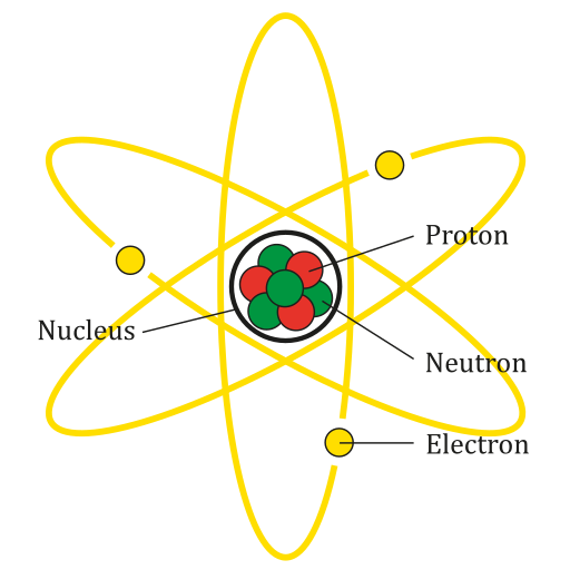
An atom is the smallest particle of an element that still has the properties of that element. Every substance is composed of atoms. Atoms are extremely small, typically about a ten-billionth of a metre in diametre. However, atoms do not have well-defined boundaries, as suggested by the atomic model shown below.
Every atom is composed of a central area — called the nucleus — and one or more subatomic particles called electrons, which move around the nucleus. The nucleus also consists of subatomic particles. It contains one or more protons and typically a similar number of neutrons. The number of protons in the nucleus determines the type of element an atom represents. An atom of hydrogen, for example, contains just one proton. Atoms of the same element may have different numbers of neutrons in the nucleus. Atoms of the same element with the same number of protons — but different numbers of neutrons — are called isotopes.
Protons have a positive electric charge and neutrons have no electric charge. Virtually all of an atom's mass is in the protons and neutrons in the nucleus. Electrons surrounding the nucleus have almost no mass, as well as a negative electric charge. If the number of protons and electrons in an atom are equal, then an atom is electrically neutral, because the positive and negative charges cancel each other out. If an atom has more or fewer electrons than protons, then it has an overall negative or positive charge, respectively, and it is called an ion.
The negatively-charged electrons of an atom are attracted to the positively-charged protons in the nucleus by a force called electromagnetic force, for which opposite charges attract. Electromagnetic force between protons in the nucleus causes these subatomic particles to repel each other, because they have the same charge. However, the protons and neutrons in the nucleus are attracted to each other by a different force, called nuclear force, which is usually stronger than the electromagnetic force. Nuclear force repels the positively-charged protons from each other.
Periodic Table of the Elements
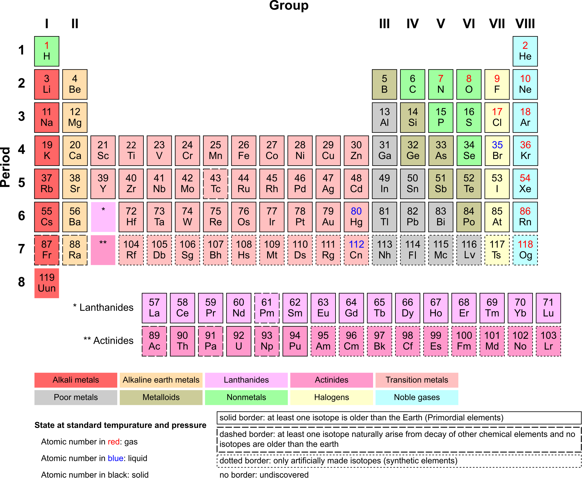
There are almost 120 known elements. As you can see in the Periodic Table of the Elements shown below, the majority of elements are metals. Examples of metals are iron (Fe) and copper (Cu). Metals are shiny and good conductors of electricity and heat. Nonmetal elements are far fewer in number. They include hydrogen (H) and oxygen (O). They lack the properties of metals.
The periodic table of the elements arranges elements in groups based on their properties. The element most important to life is carbon (C). Find carbon in the table. What type of element is it: metal or nonmetal?
Compounds and Molecules
A compound is a unique substance that consists of two or more elements combined in fixed proportions. This means that the composition of a compound is always the same. The smallest particle of most compounds in living things is called a molecule.
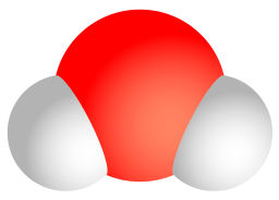
Consider water as an example. A molecule of water always contains one atom of oxygen and two atoms of hydrogen. The composition of water is expressed by the chemical formula H2O. A model of a water molecule is shown in Figure 3.2.4.
What causes the atoms of a water molecule to “stick” together? The answer is chemical bonds. A chemical bond is a force that holds together the atoms of molecules. Bonds in molecules involve the sharing of electrons among atoms. New chemical bonds form when substances react with one another. A chemical reaction is a process that changes some chemical substances into others. A chemical reaction is needed to form a compound, and another chemical reaction is needed to separate the substances in that compound.
3.2 Summary
- All matter consists of chemical substances. A chemical substance has a definite composition which is consistent throughout. A chemical substance may be either an element or a compound.
- An element is a pure substance that cannot be broken down into other types of substances.
- An atom is the smallest particle of an element that still has the properties of that element. Atoms, in turn, are composed of subatomic particles, including negative electrons, positive protons, and neutral neutrons. The number of protons in an atom determines the element it represents.
- Atoms have equal numbers of electrons and protons, so they have no charge. Ions are atoms that have lost or gained electrons, and as a result have either a positive or negative charge. Atoms with the same number of protons — but different numbers of neutrons — are called isotopes.
- There are almost 120 known elements. The majority of elements are metals. A smaller number are nonmetals. The latter include carbon, hydrogen, and oxygen.
- A compound is a substance that consists of two or more elements in a unique composition. The smallest particle of a compound is called a molecule. Chemical bonds hold together the atoms of molecules. Compounds can form only in chemical reactions, and they can break down only in other chemical reactions.
3.2 Review Questions
-
- What is an element? Give three examples.
- Define compound. Explain how compounds form.
- Compare and contrast atoms and molecules.
- The compound called water can be broken down into its constituent elements by applying an electric current to it. What ratio of elements is produced in this process?
- Relate ions and isotopes to elements and atoms.
- What is the most important element to life?
- Iron oxide is often known as rust — the reddish substance you might find on corroded metal. The chemical formula for this type of iron oxide is Fe2O3. Answer the following questions about iron oxide and briefly explain each answer.
- Is iron oxide an element or a compound?
- Would one particle of iron oxide be considered a molecule or an atom?
- Describe the relative proportion of atoms in iron oxide.
- What causes the Fe and O to stick together in iron oxide?
- Is iron oxide made of metal atoms, metalloid atoms, nonmetal atoms, or a combination of any of these?
- 14C is an isotope of carbon used in the radiocarbon dating of organic material. The most common isotope of carbon is 12C. Do you think 14C and 12C have different numbers of neutrons or protons? Explain your answer.
- Explain why ions have a positive or negative charge.
- Name the three subatomic particles described in this section.
3.2 Explore More
https://www.youtube.com/watch?v=yQP4UJhNn0I&feature=emb_logo
Just how small is an atom? TED-Ed, 2012
Attributions
Figure 3.2.1
Man Sitting, by Gregory Culmer, on Unsplash, is used under the Unsplash license (https://unsplash.com/license).
Figure 3.2.2
Lithium Atom diagram, by AG Caesar, is used under a CC BY-SA 4.0 (https://creativecommons.org/licenses/by-sa/4.0/deed.en)
Figure 3.2.3
Periodic Table Armtuk3, by Armtuk, is used under a CC BY-SA 3.0 (https://creativecommons.org/licenses/by-sa/3.0/) license.
Figure 3.2.4
Water molecule, by Sakurambo, is released into the public domain (https://en.wikipedia.org/wiki/Public_domain).
References
TED-Ed. (2012, April 16). Just how small is an atom. YouTube. https://www.youtube.com/watch?v=yQP4UJhNn0I&feature=youtu.be
Image shows a diagram of the negative feedback loops that maintain homeostasis of body temperature. When body temperature falls, blood vessels constrict so that heat is conserved, sweat glands do not secrete fluid, and shivering generates body heat, which warms the body. When body temperature rises, blood vessels dilate, resulting in heat loss to the environment, sweat glands release fluid and as the fluid evaporates, heat is lost from the body.
An organisms that is so small it is invisible to the human eye.
Image shows a diagram comparing a healthy nephron and its blood supply and one with diabetic nephropathy. The diseased one has blood vessels that look deformed and fragile.
Image shows a diagram of the process of hemodialysis. Blood is removed from the patient from a location on the arm. Blood enters the hemodialysis apparatus and is run through dialyser to clean wastes from the blood. There are mechanisms to maintain blood tonicity and pressure, to prevent clotting, and ensure no air enters the bloodstream. The cleaned blood is returned to the patient in their arm, proximal to the place where the blood was first removed.
Image shows a photograph of a kidney stone. It has a crystal structure, with irregular sharp edges and points.
Image shows a diagram of the kidney with several kidney stones, which have accumulated in the renal pelvis and are blocking the exit to the ureter.
As per caption
Image shows a photograph of several wine bottles on a shelf. The image has been deliberately blurred to simulate the effects of drunkeness.
Image shows an operating room. There are several surgeons in gowns, masks and gloves. They are operating on a patient.
A semi-permeable lipid bilayer that separates the interior of all cells from their surroundings.
Created by CK-12 Foundation/Adapted by Christine Miller

Work Those Eye Muscles!
Imagine the man in Figure 12.3.1 turns his eyes in your direction. This is a very small movement, considering the conspicuously large and strong external eye muscles that control eyeball movements. These muscles have been called the strongest muscles in the human body relative to the work they do. However, the external eye muscles actually do a surprising amount of work. Eye movements occur almost constantly during waking hours, especially when we are scanning faces or reading. Eye muscles are also exercised nightly during the phase of sleep called rapid eye movement sleep. External eye muscles can move the eyes because they are made mainly of muscle tissue.
What is Muscle Tissue?
Muscle tissue is a soft tissue that makes up most of the tissues in the muscles of the human muscular system. Other tissues in muscles are connective tissues, such as tendons that attach skeletal muscles to bones and sheaths of connective tissues that cover or line muscle tissues. Only muscle tissue per se, has cells with the ability to contract.
There are three major types of muscle tissues in the human body: skeletal, smooth, and cardiac muscle tissues. Figure 12.3.2 shows how the three types of muscle tissues appear under magnification. When you read about each type below, you will learn why the three types appear as they do.
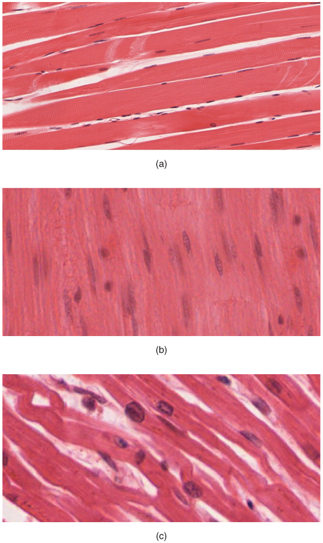
Skeletal Muscle Tissue
Skeletal muscle is muscle tissue that is attached to bones by tendons, which are bundles of collagen fibres. Whether you are moving your eyes or running a marathon, you are using skeletal muscles. Contractions of skeletal muscles are voluntary, or under conscious control of the central nervous system via the somatic nervous system. Skeletal muscle tissue is the most common type of muscle tissue in the human body. By weight, an average adult male is about 42% skeletal muscles, and the average adult female is about 36% skeletal muscles. Some of the major skeletal muscles in the human body are labeled in Figure 12.3.3 below.
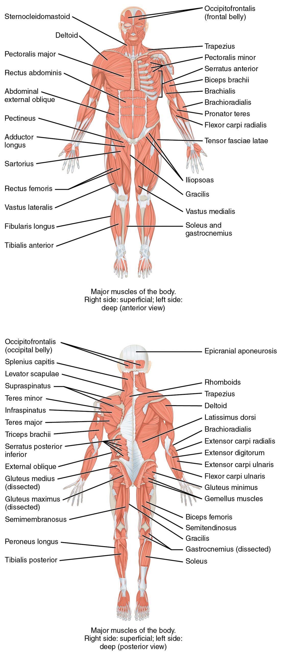
Skeletal Muscle Pairs
To move bones in opposite directions, skeletal muscles often consist of muscle pairs that work in opposition to one another, also called antagonistic muscle pairs. For example, when the biceps muscle (on the front of the upper arm) contracts, it can cause the elbow joint to flex or bend the arm, as shown in Figure 12.3.4. When the triceps muscle (on the back of the upper arm) contracts, it can cause the elbow to extend or straighten the arm. The biceps and triceps muscles, also shown in Figure 12.3.4, are an example of a muscle pair where the muscles work in opposition to each other.
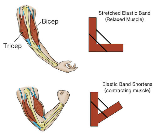
Skeletal Muscle Structure
Each skeletal muscle consists of hundreds — or even thousands — of skeletal muscle fibres, which are long, string-like cells. As shown in Figure 12.3.5 below, skeletal muscle fibres are individually wrapped in connective tissue called endomysium. The skeletal muscle fibres are bundled together in units called muscle fascicles, which are surrounded by sheaths of connective tissue called perimysium. Each fascicle contains between ten and 100 (or even more!) skeletal muscle fibres. Fascicles, in turn, are bundled together to form individual skeletal muscles, which are wrapped in connective tissue called epimysium. The connective tissues in skeletal muscles have a variety of functions. They support and protect muscle fibres, allowing them to withstand the forces of contraction by distributing the forces applied to the muscle. They also provide pathways for nerves and blood vessels to reach the muscles. In addition, the epimysium anchors the muscles to tendons.
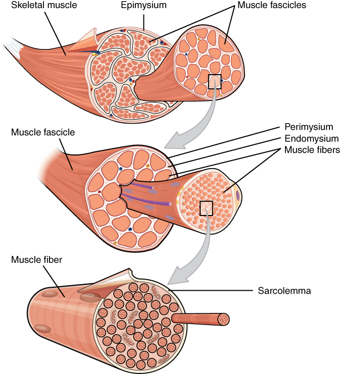
The same bundles-within-bundles structure is replicated within each muscle fibre. As shown in Figure 12.3.6, a muscle fibre consists of a bundle of myofibrils, which are themselves bundles of protein filaments. These protein filaments consist of thin filaments of the protein actin, which are anchored to structures called Z discs, and thick filaments of the protein myosin. The filaments are arranged together within a myofibril in repeating units called sarcomeres, which run from one Z disc to the next. The sarcomere is the basic functional unit of skeletal and cardiac muscles. It contracts as actin and myosin filaments slide over one another. Skeletal muscle tissue is said to be striated, because it appears striped. It has this appearance because of the regular, alternating A (dark) and I (light) bands of filaments arranged in sarcomeres inside the muscle fibres. Other components of a skeletal muscle fibre include multiple nuclei and mitochondria.
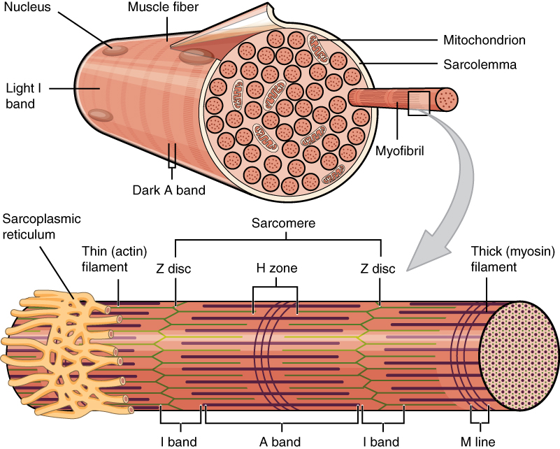
Slow- and Fast-Twitch Skeletal Muscle Fibres
Skeletal muscle fibres can be divided into two types, called slow-twitch (or type I) muscle fibres and fast-twitch (or type II) muscle fibres.
- Slow-twitch muscle fibres are dense with capillaries and rich in mitochondria and myoglobin, which is a protein that stores oxygen until needed for muscle activity. Relative to fast-twitch fibres, slow-twitch fibres can carry more oxygen and sustain aerobic (oxygen-using) activity. Slow-twitch fibres can contract for long periods of time, but not with very much force. They are relied upon primarily in endurance events, such as distance running or cycling.
- Fast-twitch muscle fibres contain fewer capillaries and mitochondria and less myoglobin. This type of muscle fibre can contract rapidly and powerfully, but it fatigues very quickly. Fast-twitch fibres can sustain only short, anaerobic (non-oxygen-using) bursts of activity. Relative to slow-twitch fibres, fast-twitch fibres contribute more to muscle strength and have a greater potential for increasing in mass. They are relied upon primarily in short, strenuous events, such as sprinting or weightlifting.
Proportions of fibre types vary considerably from muscle to muscle and from person to person. Individuals may be genetically predisposed to have a larger percentage of one type of muscle fibre than the other. Generally, an individual who has more slow-twitch fibres is better suited for activities requiring endurance, whereas an individual who has more fast-twitch fibres is better suited for activities requiring short bursts of power.
Smooth Muscle
Smooth muscle is muscle tissue in the walls of internal organs and other internal structures such as blood vessels. When smooth muscles contract, they help the organs and vessels carry out their functions. When smooth muscles in the stomach wall contract, for example, they squeeze the food inside the stomach, helping to mix and churn the food and break it into smaller pieces. This is an important part of digestion. Contractions of smooth muscles are involuntary, so they are not under conscious control. Instead, they are controlled by the autonomic nervous system, hormones, neurotransmitters, and other physiological factors.
Structure of Smooth Muscle
The cells that make up smooth muscle are generally called myocytes. Unlike the muscle fibres of striated muscle tissue, the myocytes of smooth muscle tissue do not have their filaments arranged in sarcomeres. Therefore, smooth tissue is not striated. However, the myocytes of smooth muscle do contain myofibrils, which in turn contain bundles of myosin and actin filaments. The filaments cause contractions when they slide over each other, as shown in Figure 12.3.7.

Functions of Smooth Muscle
Unlike striated muscle, smooth muscle can sustain very long-term contractions. Smooth muscle can also stretch and still maintain its contractile function, which striated muscle cannot. The elasticity of smooth muscle is enhanced by an extracellular matrix secreted by myocytes. The matrix consists of elastin, collagen, and other stretchy fibres. The ability to stretch and still contract is an important attribute of smooth muscle in organs such as the stomach and uterus (see Figures 12.3.8 and 12.3.9), both of which must stretch considerably as they perform their normal functions.
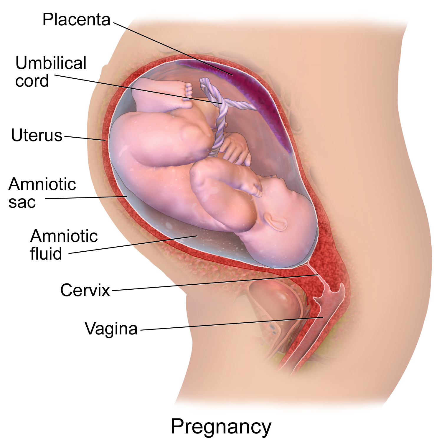 |
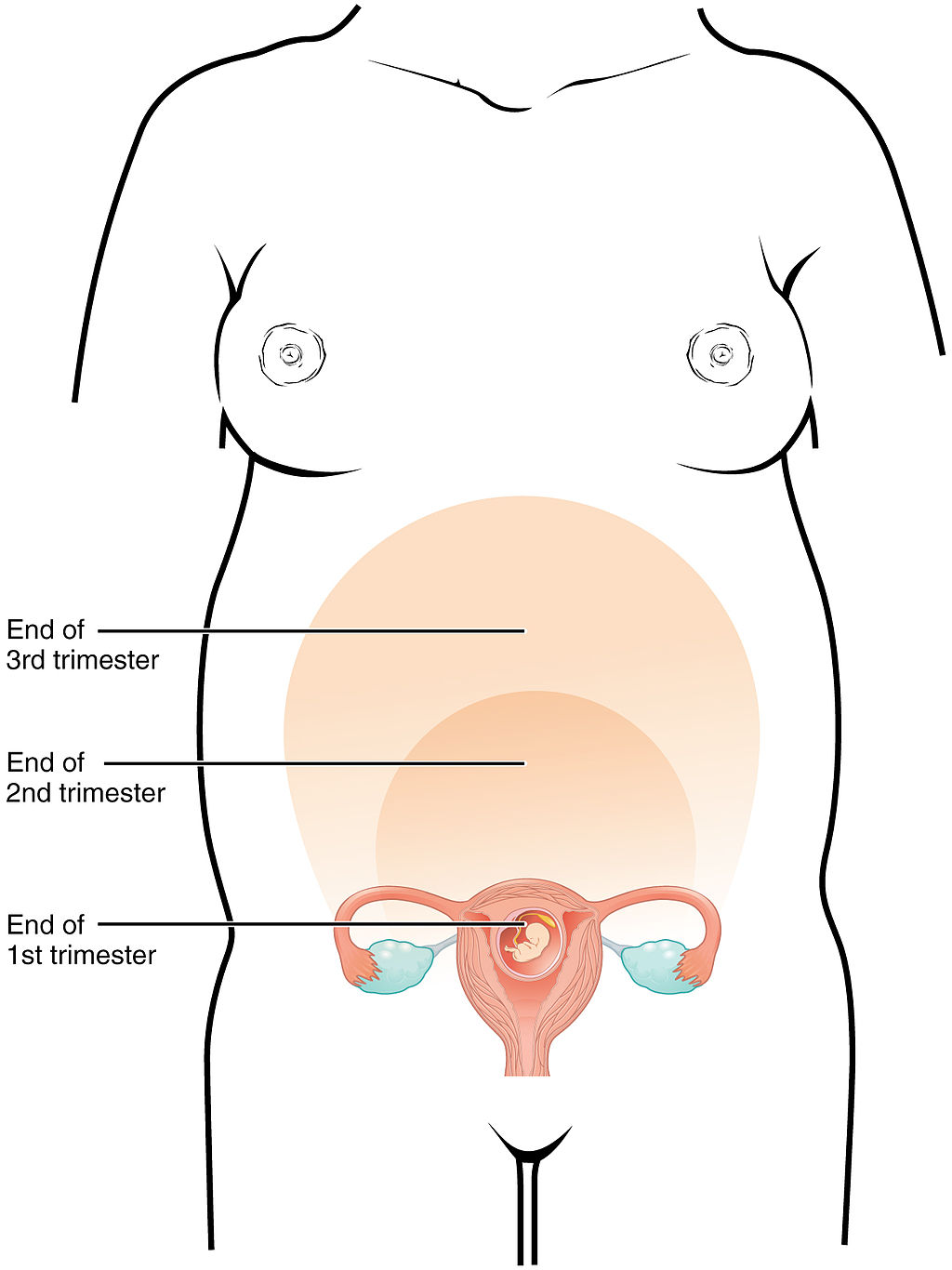 |
The following list indicates where many smooth muscles are found, along with some of their specific functions.
- Walls of organs of the gastrointestinal tract (such as the esophagus, stomach, and intestines), moving food through the tract by peristalsis
- Walls of air passages of the respiratory tract (such as the bronchi), controlling the diameter of the passages and the volume of air that can pass through them
- Walls of organs of the male and female reproductive tracts; in the uterus, for example, pushing a baby out of the uterus and into the birth canal
- Walls of structures of the urinary system, including the urinary bladder, allowing the bladder to expand so it can hold more urine, and then contract as urine is released
- Walls of blood vessels, controlling the diameter of the vessels and thereby affecting blood flow and blood pressure
- Walls of lymphatic vessels, squeezing the fluid called lymph through the vessels
- Iris of the eyes, controlling the size of the pupils and thereby the amount of light entering the eyes
- Arrector pili in the skin, raising hairs in hair follicles in the dermis
Cardiac Muscle
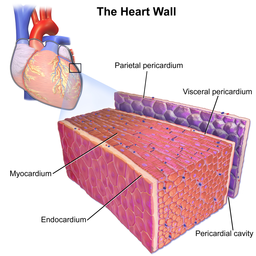
Cardiac muscle is found only in the wall of the heart. It is also called myocardium. As shown in Figure 12.3.10, myocardium is enclosed within connective tissues, including the endocardium on the inside of the heart and pericardium on the outside of the heart. When cardiac muscle contracts, the heart beats and pumps blood. Contractions of cardiac muscle are involuntary, like those of smooth muscles. They are controlled by electrical impulses from specialized cardiac muscle cells in an area of the heart muscle called the sinoatrial node.
Like skeletal muscle, cardiac muscle is striated because its filaments are arranged in sarcomeres inside the muscle fibres. However, in cardiac muscle, the myofibrils are branched at irregular angles rather than arranged in parallel rows (as they are in skeletal muscle). This explains why cardiac and skeletal muscle tissues look different from one another.
The cells of cardiac muscle tissue are arranged in interconnected networks. This arrangement allows rapid transmission of electrical impulses, which stimulate virtually simultaneous contractions of the cells. This enables the cells to coordinate contractions of the heart muscle.
The heart is the muscle that performs the greatest amount of physical work in the course of a lifetime. Although the power output of the heart is much less than the maximum power output of some other muscles in the human body, the heart does its work continuously over an entire lifetime without rest. Cardiac muscle contains a great many mitochondria, which produce ATP for energy and help the heart resist fatigue.
Feature: Human Biology in the News
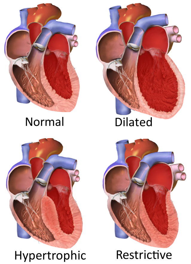
Cardiomyopathy is a disease in which the muscles of the heart are no longer able to effectively pump blood to the body — extreme forms of this disease can lead to heart failure. There are four main types of cardiomyopathy (also illustrated in Figure 12.3.11):
- Dilated (congestive) cardiomyopathy: the left ventricle (the chamber itself) of the heart becomes enlarged and can't pump blood our to the body. This is normally related to coronary artery disease and/or heart attack
- Hypertrophic cardiomyopathy: abnormal thickening of the muscular walls of the left ventricle make the chamber less able to work properly. This condition is more common in patients with a family history of the disease.
- Restrictive cardiomyopathy: the myocardium becomes abnormally rigid and inelastic and is unable to expand in between heartbeats to refill with blood. Restrictive cardiomyopathy typically affects older people.
- Arrhythmogenic right ventricular cardiomyopathy: the right ventricular muscle is replaced by adipose or scar tissue, reducing elasticity and interfering with normal heartbeat and rhythm. This disease is often caused by genetic mutations.
Cardiomyopathy is typically diagnosed with a physical exam supplemented by medical and family history, an angiogram, blood tests, chest x-rays and electrocardiograms. In some cases your doctor would also requisition a CT scan and/or genetic testing.
When treating cardiomyopathy, the goal is to reduce symptoms that affect everyday life. Certain medications can help regularize and slow heart rate, decrease chances of blood clots and cause vasodilation in the coronary arteries. If medication is not sufficient to manage symptoms, a pacemaker or even a heart transplant may be the best option. Lifestyle can also help manage the symptoms of cardiomyopathy — people living with this disease are encouraged to avoid drug and alcohol use, control high blood pressure, eat a healthy diet, get ample rest and exercise, as well as reduce stress levels.
12.3 Summary
- Muscle tissue is a soft tissue that makes up most of the tissues in the muscles of the human muscular system. It is the only type of tissue that has cells with the ability to contract.
- Skeletal muscle tissue is attached to bones by tendons. It allows voluntary body movements.
- Skeletal muscle is the most common type of muscle tissue in the human body. To move bones in opposite directions, skeletal muscles often consist of pairs of muscles that work in opposition to one another to move bones in different directions at joints.
- Skeletal muscle fibres are bundled together in units called muscle fascicles, which are bundled together to form individual skeletal muscles. Skeletal muscles also have connective tissue supporting and protecting the muscle tissue.
- Each skeletal muscle fibre consists of a bundle of myofibrils, which are bundles of protein filaments. The filaments are arranged in repeating units called sarcomeres, which are the basic functional units of skeletal muscles. Skeletal muscle tissue is striated because of the pattern of sarcomeres in its fibres.
- Skeletal muscle fibres can be divided into two types, called slow-twitch and fast-twitch fibres. Slow-twitch fibres are used mainly in aerobic endurance activities, such as long-distance running. Fast-twitch fibres are used mainly for non-aerobic, strenuous activities, such as sprinting. Proportions of the two types of fibres vary from muscle to muscle and person to person.
- Smooth muscle tissue is found in the walls of internal organs and vessels. When smooth muscles contract, they help the organs and vessels carry out their functions. Contractions of smooth muscles are involuntary and controlled by the autonomic nervous system, hormones, and other substances.
- Cells of smooth muscle tissue are not striated because they lack sarcomeres, but the cells contract in the same basic way as striated muscle cells. Unlike striated muscle, smooth muscle can sustain very long-term contractions and maintain its contractile function, even when stretched.
- Cardiac muscle tissue is found only in the wall of the heart. When cardiac muscle contracts, the heart beats and pumps blood. Contractions of cardiac muscle are involuntary, like those of smooth muscles. They are controlled by electrical impulses from specialized cardiac cells.
- Like skeletal muscle, cardiac muscle is striated because its filaments are arranged in sarcomeres inside the muscle fibres. However, the myofibrils are branched instead of arranged in parallel rows, making cardiac and skeletal muscle tissues look different from one another.
- The heart is the muscle that performs the greatest amount of physical work in the course of a lifetime. Its cells contain a great many mitochondria to produce ATP for energy and help the heart resist fatigue.
12.3 Review Questions
- What is muscle tissue?
-
-
- Where is skeletal muscle found, and what is its general function?
- Why do many skeletal muscles work in pairs?
- Describe the structure of a skeletal muscle.
- Relate muscle fibre structure to the functional units of muscles.
- Why is skeletal muscle tissue striated?
- Where is smooth muscle found? What controls the contraction of smooth muscle?
- Where is cardiac muscle found? What controls its contractions?
- The heart muscle is smaller and less powerful than some other muscles in the body. Why is the heart the muscle that performs the greatest amount of physical work in the course of a lifetime? How does the heart resist fatigue?
- Give one example of connective tissue that is found in muscles. Describe one of its functions.
12.3 Explore More
https://www.youtube.com/watch?v=3_PYnWVoUzM
What happens during a heart attack? - Krishna Sudhir, TED-Ed, 2017.
https://www.youtube.com/watch?v=bwOE1MEginA&feature=emb_logo
Three types of muscle | Circulatory system physiology | NCLEX-RN | KhanAcademyMedicine, 2012.
Attributions
Figure 12.3.1
Look by ali-yahya-155huuQwGvA [photo] by Ali Yahya on Unsplash is used under the Unsplash License (https://unsplash.com/license).
Figure 12.3.2
Skeletal_Smooth_Cardiac by OpenStax College on Wikimedia Commons is used under a CC BY 3.0 (https://creativecommons.org/licenses/by/3.0) license.
Figure 12.3.3
Anterior_and_Posterior_Views_of_Muscles by OpenStax on Wikimedia Commons is used under a CC BY 4.0 (https://creativecommons.org/licenses/by/4.0) license.
Figure 12.3.4
Antagonistic Muscle Pair by Laura Guerin at CK-12 Foundation on Wikimedia Commons is used under a CC BY-NC 3.0 (https://creativecommons.org/licenses/by-nc/3.0/) license.
Figure 12.3.5
Muscle_Fibes_(large) by OpenStax on Wikimedia Commons is used under a CC BY 4.0 (https://creativecommons.org/licenses/by/4.0) license.
Figure 12.3.6
Muscle_Fibers_(small) by OpenStax on Wikimedia Commons is used under a CC BY 4.0 (https://creativecommons.org/licenses/by/4.0) license.
Figure 12.3.7
Smooth_Muscle_Contraction by OpenStax on Wikimedia Commons is used under a CC BY 4.0 (https://creativecommons.org/licenses/by/4.0) license.
Figure 12.3.8
Blausen_0747_Pregnancy by BruceBlaus on Wikimedia Commons is used under a CC BY 3.0 (https://creativecommons.org/licenses/by/3.0) license.
Figure 12.3.9
Size_of_Uterus_Throughout_Pregnancy-02 by OpenStax College on Wikimedia Commons is used under a CC BY 3.0 (https://creativecommons.org/licenses/by/3.0) license.
Figure 12.3.10
1024px-Blausen_0470_HeartWall by BruceBlaus on Wikimedia Commons is used under a CC BY 3.0 (https://creativecommons.org/licenses/by/3.0) license.
Figure 12.3.11
Tipet_e_kardiomiopative by Npatchett at English Wikipedia on Wikimedia Commons is used under a CC BY-SA 3.0 (https://creativecommons.org/licenses/by-sa/3.0) license. (Work derived from Blausen 0165 Cardiomyopathy Dilated by BruceBlaus)
References
Betts, J. G., Young, K.A., Wise, J.A., Johnson, E., Poe, B., Kruse, D.H., Korol, O., Johnson, J.E., Womble, M., DeSaix, P. (2013, June 19). Figure 4.18 Muscle tissue [digital image]. In Anatomy and Physiology (Section 4.4). OpenStax. https://openstax.org/books/anatomy-and-physiology/pages/4-4-muscle-tissue-and-motion
Betts, J. G., Young, K.A., Wise, J.A., Johnson, E., Poe, B., Kruse, D.H., Korol, O., Johnson, J.E., Womble, M., DeSaix, P. (2013, June 19). Figure 28.18 Size of uterus throughout pregnancy [digital image]. In Anatomy and Physiology (Section 28.4). OpenStax. https://openstax.org/books/anatomy-and-physiology/pages/28-4-maternal-changes-during-pregnancy-labor-and-birth
Betts, J. G., Young, K.A., Wise, J.A., Johnson, E., Poe, B., Kruse, D.H., Korol, O., Johnson, J.E., Womble, M., DeSaix, P. (2016, May 18). Figure 10.3 The three connective tissue layers [digital image]. In Anatomy and Physiology (Section 10.2). OpenStax. https://openstax.org/books/anatomy-and-physiology/pages/10-2-skeletal-muscle
Betts, J. G., Young, K.A., Wise, J.A., Johnson, E., Poe, B., Kruse, D.H., Korol, O., Johnson, J.E., Womble, M., DeSaix, P. (2016, May 18). Figure 10.4 Muscle fiber [digital image]. In Anatomy and Physiology (Section 10.2). OpenStax. https://openstax.org/books/anatomy-and-physiology/pages/10-2-skeletal-muscle
Betts, J. G., Young, K.A., Wise, J.A., Johnson, E., Poe, B., Kruse, D.H., Korol, O., Johnson, J.E., Womble, M., DeSaix, P. (2016, May 18). Figure 10.24 Muscle contraction [digital image]. In Anatomy and Physiology (Section 10.8). OpenStax. https://openstax.org/books/anatomy-and-physiology/pages/10-8-smooth-muscle
Betts, J. G., Young, K.A., Wise, J.A., Johnson, E., Poe, B., Kruse, D.H., Korol, O., Johnson, J.E., Womble, M., DeSaix, P. (2016, May 18). Figure 11.5 Overview of the muscular system [digital image]. In Anatomy and Physiology (Section 11.2). OpenStax. https://openstax.org/books/anatomy-and-physiology/pages/11-2-naming-skeletal-muscles
Blausen.com staff. (2014). Medical gallery of Blausen Medical 2014. WikiJournal of Medicine 1 (2). DOI:10.15347/wjm/2014.010. ISSN 2002-4436.
Brainard, J/ CK-12 Foundation. (2012). Figure 5 Triceps and biceps muscles in the upper arm are opposing muscles. [digital image]. In CK-12 Biology (Section 21.3) [online Flexbook]. CK12.org. https://www.ck12.org/book/ck-12-biology/section/21.3/ (Last modified August 11, 2017.)
khanacademymedicine. (2012, October 19). Three types of muscle | Circulatory system physiology | NCLEX-RN | Khan Academy. YouTube.
TED-Ed. (2017, February 14). What happens during a heart attack? - Krishna Sudhir. YouTube. https://www.youtube.com/watch?v=3_PYnWVoUzM&feature=youtu.be
Created by CK-12/Adapted by Christine Miller
As you read in the beginning of this chapter, new parents Samantha and Aki left their pediatrician’s office still unsure whether or not to vaccinate baby James. Dr. Rodriguez gave them a list of reputable sources where they could look up information about the safety of vaccines, including the Centers for Disease Control and Prevention (CDC). Samantha and Aki read that the consensus within the scientific community is that there is no link between vaccines and autism. They find a long list of studies published in peer-reviewed scientific journals that disprove any link. Additionally, some of the studies are “meta-analyses” that analyzed the findings from many individual studies. The new parents are reassured by the fact that many different researchers, using a large number of subjects in numerous well-controlled and well-reviewed studies, all came to the same conclusion.

Samantha also went back to the web page that originally scared her about the safety of vaccines. She found that the author was not a medical doctor or scientific researcher, but rather a self-proclaimed “child wellness expert.” He sold books and advertising on his site, some of which were related to claims of vaccine injury. She realized that he was both an unqualified and potentially biased source of information.
Samantha also realized that some of his arguments were based on correlations between autism and vaccines, but, as the saying goes, “correlation does not imply causation.” For instance, the recent rise in autism rates may have occurred during the same time period as an increase in the number of vaccines given in childhood, but Samantha could think of many other environmental and social factors that have also changed during this time period. There are just too many variables to come to the conclusion that vaccines, or anything else, are the cause of the rise in autism rates based on that type of argument alone. Also, she learned that the age of onset of autism symptoms happens to typically be around the time that the MMR vaccine is first given, so the apparent association in the timing may just be a coincidence.
Finally, Samantha came across news about a measles outbreak in Vancouver, British Columbia in the winter of 2019. Measles wasn’t just a disease of the past! She learned that measles and whooping cough, which had previously been rare thanks to widespread vaccinations, are now on the rise, and that people choosing not to vaccinate their children seems to be one of the contributing factors. She realized that it is important to vaccinate her baby against these diseases, not only to protect him from their potentially deadly effects, but also to protect others in the population.
In their reading, Samantha and Aki learn that scientists do not yet know the causes of autism, but they feels reassured by the abundance of data that disproves any link with vaccines. Both parents think that the potential benefits of protecting their baby’s health against deadly diseases outweighs any unsubstantiated claims about vaccines. They will be making an appointment to get baby James his shots soon.
Chapter 1 Summary
In this chapter, you learned about some of the same concepts that helped Samantha and Aki make an informed decision. Specifically:
- Science is a distinctive way of gaining knowledge about the natural world that is based on the use of evidence to logically test ideas. As such, science is a process, as well as a body of knowledge.
- A scientific theory, such as the germ theory of disease, is the highest level of explanation in science. A theory is a broad explanation for many phenomena that is widely accepted because it is supported by a great deal of evidence.
- The scientific investigation is the cornerstone of science as a process. A scientific investigation is a systematic approach to answering questions about the physical and natural world. An investigation may be observational or experimental.
- A scientific experiment is a type of scientific investigation in which the researcher manipulates variables under controlled conditions to test expected outcomes. Experiments are the gold standard for scientific investigations and can establish causation between variables.
- Nonexperimental scientific investigations such as observational studies and modeling may be undertaken when experiments are impractical, unethical, or impossible. Observational studies generally can establish correlation — but not causation — between variables.
- A pseudoscience, such as astrology, is a field that is presented as scientific but that does not adhere to scientific standards and methods. Other misuses of science include deliberate hoaxes, frauds, and fallacies made by researchers.
- Strict guidelines must be followed when using human subjects in scientific research. Among the most important protections is the requirement for informed consent.
Now that you know about the nature and process of science, you can apply these concepts in the next chapter to the study of human biology.
Chapter 1 Review
-
- Why does a good hypothesis have to be falsifiable?
- Name one scientific law.
- Name one scientific theory.
- Give an example of a scientific idea that was later discredited.
- A statistical measurement called a P-value is often used in science to determine whether or not a difference between two groups is actually significant or simply due to chance. A P-value of 0.03 means that there is a 3% chance that the difference is due to chance alone. Do you think a P-value of 0.03 would indicate that the difference is likely to be significant? Why or why not?
- Why is it important that scientists communicate their findings to others? How do they usually do this?
- What is a “control group” in science?
- In a scientific experiment, why is it important to only change one variable at a time?
- Which is the dependent variable – the variable that is manipulated or the variable that is being affected by the change?
- You see an ad for a “miracle supplement” called NQP3 that claims the supplement will reduce belly fat. They say it works by reducing the hormone cortisol and by providing your body with missing unspecified “nutrients”, but they do not cite any peer-reviewed clinical studies. They show photographs of three people who appear slimmer after taking the product. A board-certified plastic surgeon endorses the product on television. Answer the following questions about this product.
a. Do you think that because a doctor endorsed the product, it really works? Explain your answer.
b. What are two signs that these claims could actually be pseudoscience instead of true science?
c. Do you think the photographs are good evidence that the product works? Why or why not?
d. If you wanted to do a strong scientific study of whether this supplement does what it claims, what would you do? Be specific about the subjects, data collected, how you would control variables, and how you would analyze the data.
e. What are some ways that you would ensure that the subjects in your experiment in part d are treated ethically and according to human subjects protections regulations?
Attribution
Figure 1.8.1
[Photo of person sitting in front of personal computer] by Avel Chuklanov on Unsplash is used under the Unsplash License (https://unsplash.com/license).
a hormonal disorder common among women of reproductive age. Women with PCOS may have infrequent or prolonged menstrual periods or excess male hormone (androgen) levels. The ovaries may develop numerous small collections of fluid (follicles) and fail to regularly release eggs.
A type of disease in which cells of the central nervous system stop working or die. Neurodegenerative disorders usually get worse over time and have no cure. They may be genetic or be caused by a tumor or stroke.
A mature haploid male or female germ cell which is able to unite with another of the opposite sex in sexual reproduction to form a zygote.
A hormone is a signaling molecule produced by glands in multicellular organisms that target distant organs to regulate physiology and behavior.
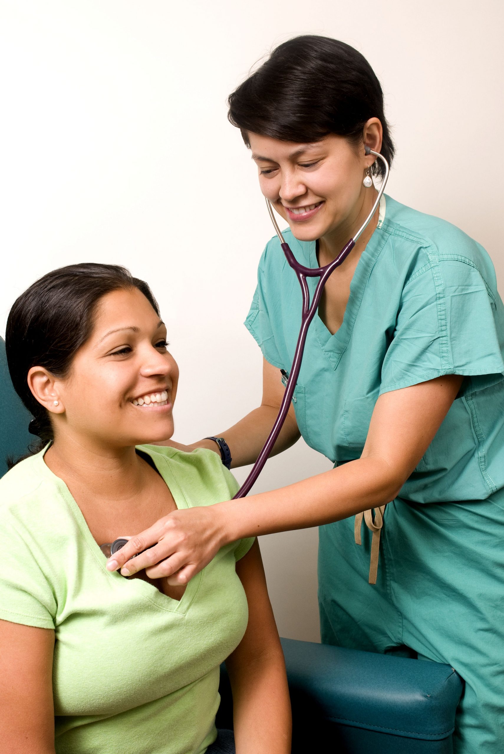
http://humanbiology.pressbooks.tru.ca/wp-content/uploads/sites/6/2019/06/human-heartbeat-daniel_simon.mp3
Lub, Dub
Lub dub, lub dub, lub dub... That’s how the sound of a beating heart is typically described. Those are also the only two sounds that should be audible when listening to a normal, healthy heart through a stethoscope, as in Figure 14.3.1. If a doctor hears something different from the normal lub dub sounds, it’s a sign of a possible heart abnormality. What causes the heart to produce the characteristic lub dub sounds? Read on to find out.
Introduction to the Heart
The heart is a muscular organ behind the sternum (breastbone), slightly to the left of the center of the chest. A normal adult heart is about the size of a fist. The function of the heart is to pump blood through blood vessels of the cardiovascular system. The continuous flow of blood through the system is necessary to provide all the cells of the body with oxygen and nutrients, and to remove their metabolic wastes.
Structure of the Heart
The heart has a thick muscular wall that consists of several layers of tissue. Internally, the heart is divided into four chambers through which blood flows. Because of heart valves, blood flows in just one direction through the chambers.
Heart Wall
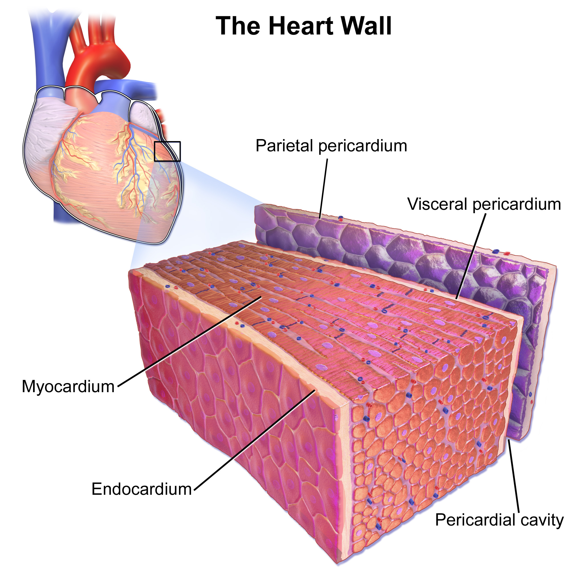
As shown in Figure 14.3.2, the wall of the heart is made up of three layers, called the endocardium, myocardium, and pericardium.
- The endocardium is the innermost layer of the heart wall. It is made up primarily of simple epithelial cells. It covers the heart chambers and valves. A thin layer of connective tissue joins the endocardium to the myocardium.
- The myocardium is the middle and thickest layer of the heart wall. It consists of cardiac muscle surrounded by a framework of collagen. There are two types of cardiac muscle cells in the myocardium: cardiomyocytes — which have the ability to contract easily — and pacemaker cells, which conduct electrical impulses that cause the cardiomyocytes to contract. About 99 per cent of cardiac muscle cells are cardiomyocytes, and the remaining one per cent is pacemaker cells. The myocardium is supplied with blood vessels and nerve fibres via the pericardium.
- The pericardium is a protective sac that encloses and protects the heart. The pericardium consists of two membranes (visceral pericardium and parietal pericardium), between which there is a fluid-filled cavity. The fluid helps to cushion the heart, and also lubricates its outer surface.
Heart Chambers
As shown in Figure 14.3.3 the four chambers of the heart include two upper chambers called atria (singular, atrium), and two lower chambers called ventricles. The atria are also referred to as receiving chambers, because blood coming into the heart first enters these two chambers. The right atrium receives deoxygenated blood from the upper and lower body through the superior vena cava and inferior vena cava, respectively. The left atrium receives oxygenated blood from the lungs through the pulmonary veins. The ventricles are also referred to as discharging chambers, because blood leaving the heart passes out through these two chambers. The right ventricle discharges blood to the lungs through the pulmonary artery, and the left ventricle discharges blood to the rest of the body through the aorta. The four chambers are separated from each other by dense connective tissue consisting mainly of collagen.
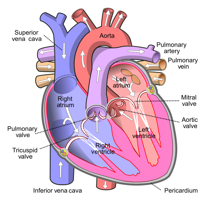
Heart Valves
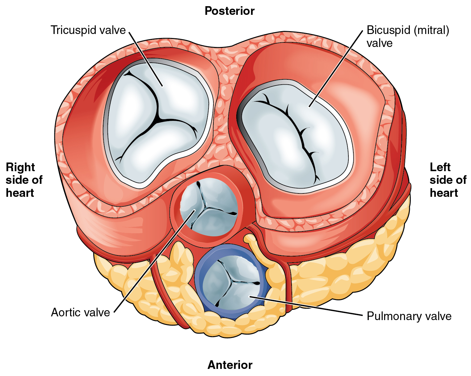
Figure 14.3.4 shows the location of the heart's four valves in a top-down view, looking down at the heart as if the arteries and veins feeding into and out of the heart were removed. The heart valves allow blood to flow from the atria to the ventricles, and from the ventricles to the pulmonary artery and aorta. The valves are constructed in such a way that blood can flow through them in only one direction, thus preventing the backflow of blood. Figure 14.3.5 shows how valves open to let blood into the appropriate chamber, and then close to prevent blood from moving in the wrong direction and the next chamber contracts. The four valves are the:
- Tricuspid atrioventricular valve, (can be shortened to tricuspid AV valve) which allows blood to flow from the right atrium to the right ventricle.
- Bicuspid atrioventricular valve (also know as the mitral valve), which allows blood to flow from the left atrium to the left ventricle.
- Pulmonary semilunar valve, which allows blood to flow from the right ventricle to the pulmonary artery.
- Aortic semilunar valve, which allows blood to flow from the left ventricle to the aorta.
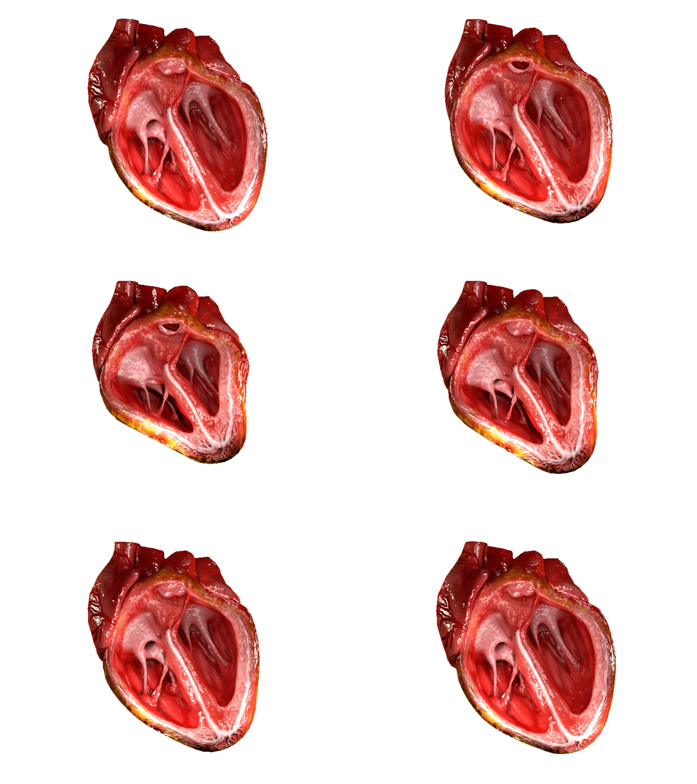
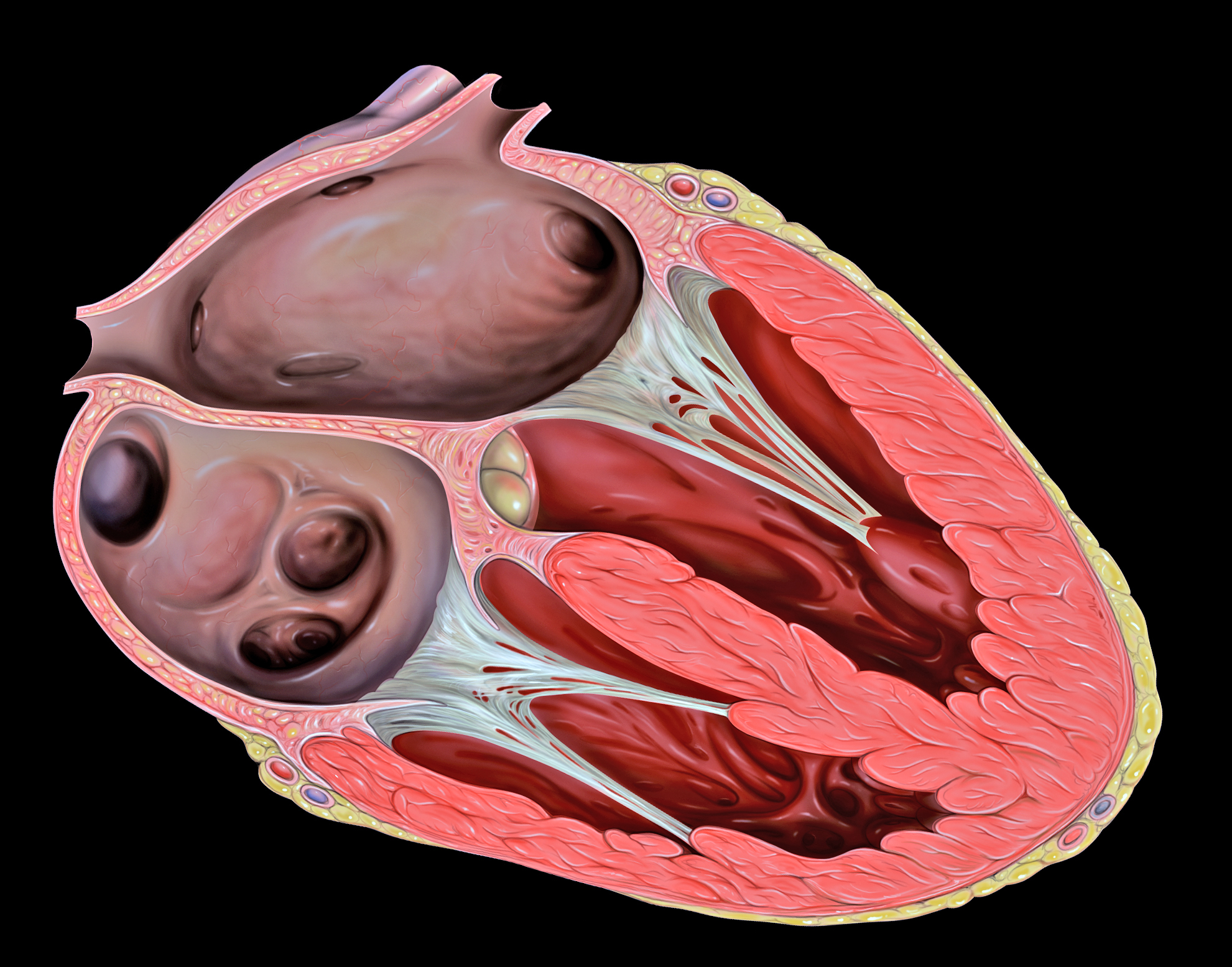
The two atrioventricular (AV) valves prevent backflow when the ventricles are contracting, while the semilunar valves prevent backflow from vessels. This means that the AV valves must withstand much more pressure than do the semilunar valves. In order to withstand the force of the ventricles contracting (to prevent blood from backflowing into the atria), the AV valves are reinforced with structures called chordae tendineae — tendon-like cords of connective tissue which anchor the valve and prevent it from prolapse. Figure 14.3.6 shows the structure and location of the chordae tendoneae.
The chordae tendoneae are under such force that they need special attachments to the interior of the ventricles where they anchor. Papillary muscles are specialized muscles in the interior of the ventricle that provide a strong anchor point for the chordae tendineae.
Coronary Circulation
The cardiomyocytes of the muscular walls of the heart are very active cells, because they are responsible for the constant beating of the heart. These cells need a continuous supply of oxygen and nutrients. The carbon dioxide and waste products they produce also must be continuously removed. The blood vessels that carry blood to and from the heart muscle cells make up the coronary circulation. Note that the blood vessels of the coronary circulation supply heart tissues with blood, and are different from the blood vessels that carry blood to and from the chambers of the heart as part of the general circulation. Coronary arteries supply oxygen-rich blood to the heart muscle cells. Coronary veins remove deoxygenated blood from the heart muscles cells.
- There are two coronary arteries — a right coronary artery that supplies the right side of the heart, and a left coronary artery that supplies the left side of the heart. These arteries branch repeatedly into smaller and smaller arteries and finally into capillaries, which exchange gases, nutrients, and waste products with cardiomyocytes.
- At the back of the heart, small cardiac veins drain into larger veins, and finally into the great cardiac vein, which empties into the right atrium. At the front of the heart, small cardiac veins drain directly into the right atrium.
Blood Circulation Through the Heart
Figure 14.3.7 shows how blood circulates through the chambers of the heart. The right atrium collects blood from two large veins, the superior vena cava (from the upper body) and the inferior vena cava (from the lower body). The blood that collects in the right atrium is pumped through the tricuspid valve into the right ventricle. From the right ventricle, the blood is pumped through the pulmonary valve into the pulmonary artery. The pulmonary artery carries the blood to the lungs, where it enters the pulmonary circulation, gives up carbon dioxide, and picks up oxygen. The oxygenated blood travels back from the lungs through the pulmonary veins (of which there are four), and enters the left atrium of the heart. From the left atrium, the blood is pumped through the mitral valve into the left ventricle. From the left ventricle, the blood is pumped through the aortic valve into the aorta, which subsequently branches into smaller arteries that carry the blood throughout the rest of the body. After passing through capillaries and exchanging substances with cells, the blood returns to the right atrium via the superior vena cava and inferior vena cava, and the process begins anew.
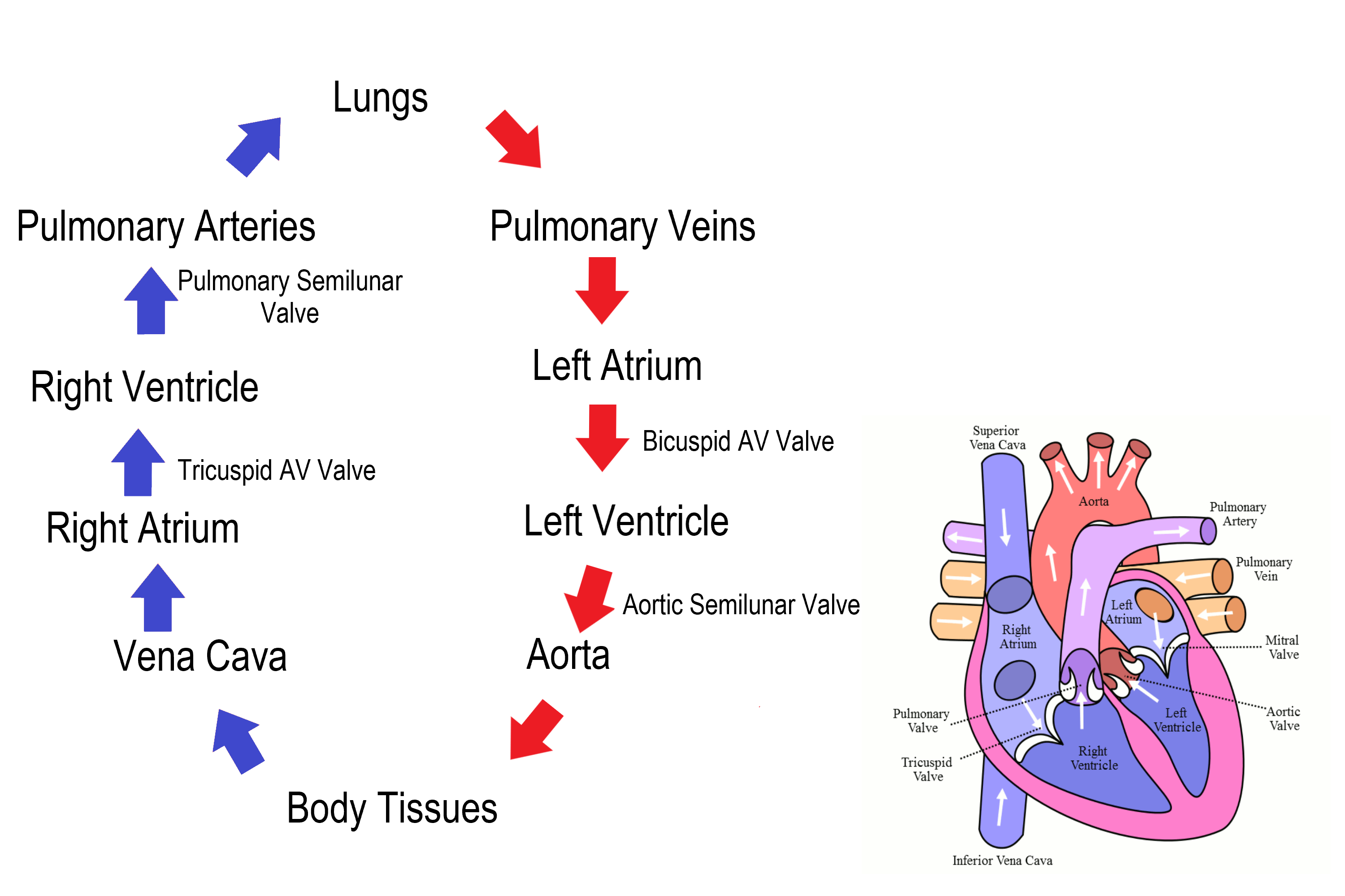
Cardiac Cycle
The cardiac cycle refers to a single complete heartbeat, which includes one iteration of the lub and dub sounds heard through a stethoscope. During the cardiac cycle, the atria and ventricles work in a coordinated fashion so that blood is pumped efficiently through and out of the heart. The cardiac cycle includes two parts, called diastole and systole, which are illustrated in the diagrams in Figure 14.3.8.
- During diastole, the atria contract and pump blood into the ventricles, while the ventricles relax and fill with blood from the atria.
- During systole, the atria relax and collect blood from the lungs and body, while the ventricles contract and pump blood out of the heart.
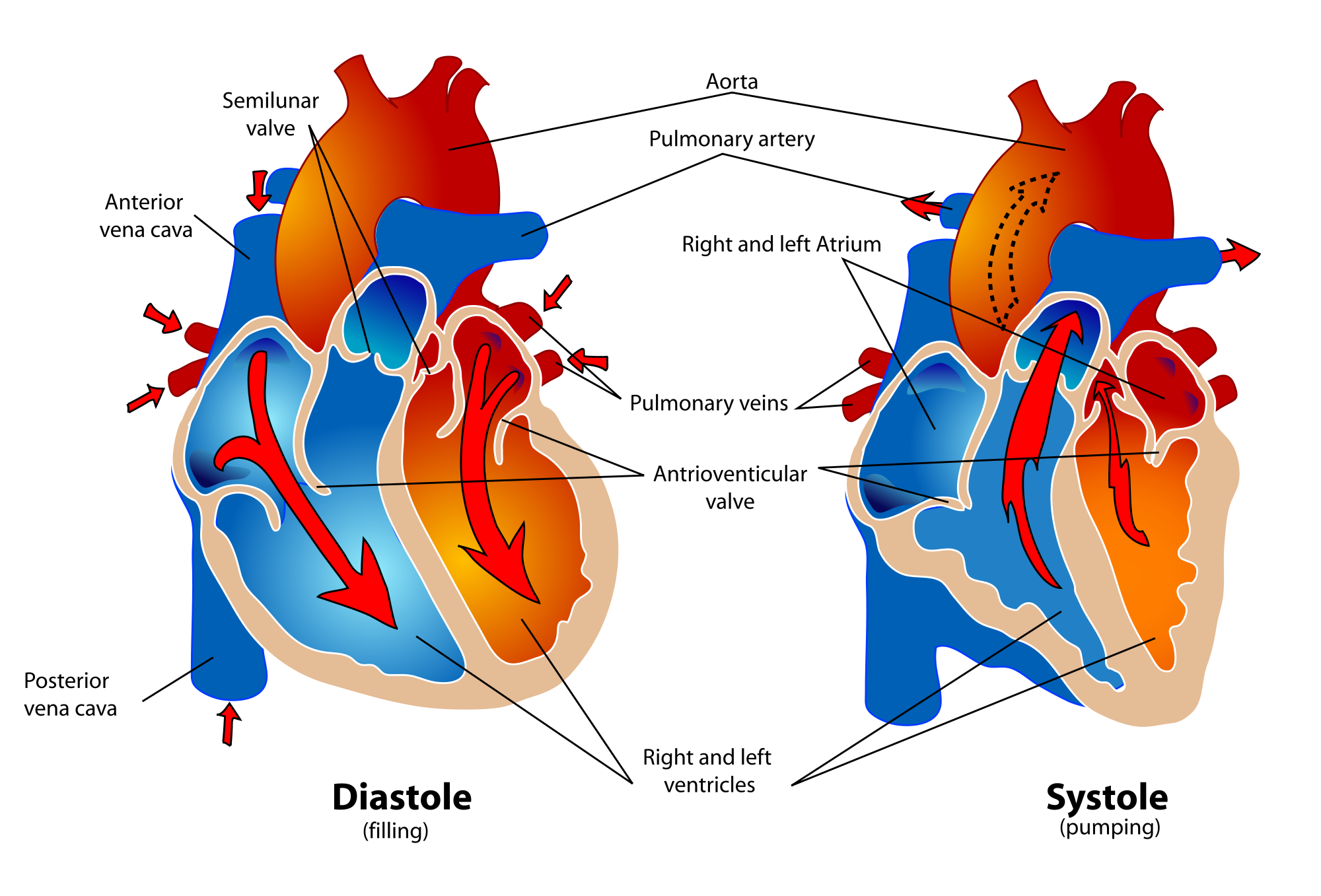
Electrical Stimulation of the Heart
The normal, rhythmical beating of the heart is called sinus rhythm. It is established by the heart’s pacemaker cells, which are located in an area of the heart called the sinoatrial node (shown in Figure 14.3.9). The pacemaker cells create electrical signals with the movement of electrolytes (sodium, potassium, and calcium ions) into and out of the cells. For each cardiac cycle, an electrical signal rapidly travels first from the sinoatrial node, to the right and left atria so they contract together. Then, the signal travels to another node, called the atrioventricular node (Figure 14.3.9), and from there to the right and left ventricles (which also contract together), just a split second after the atria contract.
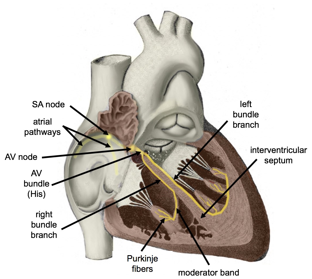
The normal sinus rhythm of the heart is influenced by the autonomic nervous system through sympathetic and parasympathetic nerves. These nerves arise from two paired cardiovascular centers in the medulla of the brainstem. The parasympathetic nerves act to decrease the heart rate, and the sympathetic nerves act to increase the heart rate. Parasympathetic input normally predominates. Without it, the pacemaker cells of the heart would generate a resting heart rate of about 100 beats per minute, instead of a normal resting heart rate of about 72 beats per minute. The cardiovascular centers receive input from receptors throughout the body, and act through the sympathetic nerves to increase the heart rate, as needed. Increased physical activity, for example, is detected by receptors in muscles, joints, and tendons. These receptors send nerve impulses to the cardiovascular centers, causing sympathetic nerves to increase the heart rate, and allowing more blood to flow to the muscles.
Besides the autonomic nervous system, other factors can also affect the heart rate. For example, thyroid hormones and adrenal hormones (such as epinephrine) can stimulate the heart to beat faster. The heart rate also increases when blood pressure drops or the body is dehydrated or overheated. On the other hand, cooling of the body and relaxation — among other factors — can contribute to a decrease in the heart rate.
Feature: Human Biology in the News
When a patient’s heart is too diseased or damaged to sustain life, a heart transplant is likely to be the only long-term solution. The first successful heart transplant was undertaken in South Africa in 1967. There are over 2,200 Canadians walking around today because of life-saving heart transplant surgery. Approximately 180 heart transplant surgeries are performed each year, but there are still so many Canadians on the transplant list that some die while waiting for a heart. The problem is that far too few hearts are available for transplant — there is more demand (people waiting for a heart transplant) than supply (organ donors). Sometimes, recipient hopefuls will receive a device called a Total Artificial Heart (see Figure 14.3.10), which can buy them some time until a donor heart becomes available.
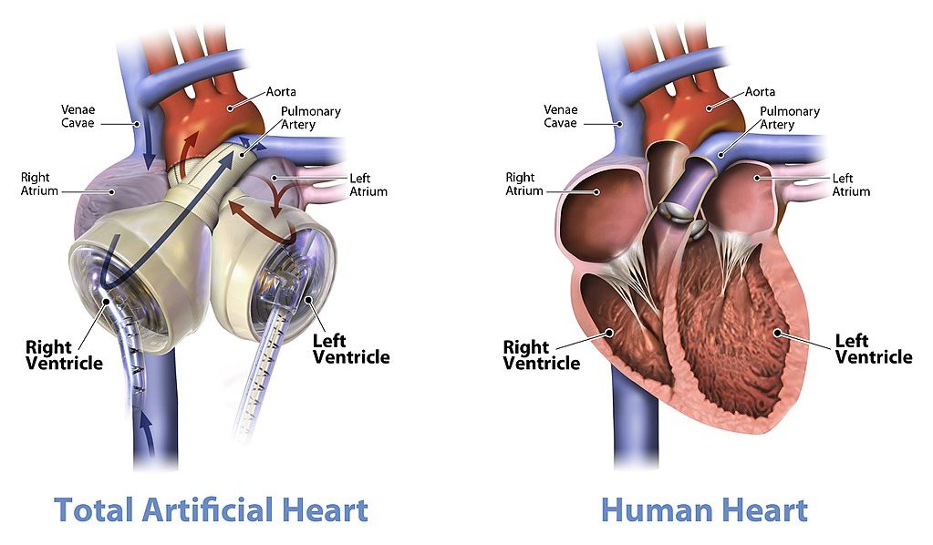
Watch the video below "Total artificial heart option..." from Stanford Health Care to see how it works:
https://youtu.be/1PtxaxcPnGc
Total artificial heart option at Stanford (Includes surgical graphic footage), Stanford Health Care, 2014.
14.3 Summary
- The heart is a muscular organ behind the sternum and slightly to the left of the center of the chest. Its function is to pump blood through the blood vessels of the cardiovascular system.
- The wall of the heart consists of three layers. The middle layer, the myocardium, is the thickest layer and consists mainly of cardiac muscle. The interior of the heart consists of four chambers, with an upper atrium and lower ventricle on each side of the heart. Blood enters the heart through the atria, which pump it to the ventricles. Then the ventricles pump blood out of the heart. Four valves in the heart keep blood flowing in the correct direction and prevent backflow.
- The coronary circulation consists of blood vessels that carry blood to and from the heart muscle cells, and is different from the general circulation of blood through the heart chambers. There are two coronary arteries that supply the two sides of the heart with oxygenated blood. Cardiac veins drain deoxygenated blood back into the heart.
- Deoxygenated blood flows into the right atrium through veins from the upper and lower body (superior and inferior vena cava, respectively), and oxygenated blood flows into the left atrium through four pulmonary veins from the lungs. Each atrium pumps the blood to the ventricle below it. From the right ventricle, deoxygenated blood is pumped to the lungs through the two pulmonary arteries. From the left ventricle, oxygenated blood is pumped to the rest of the body through the aorta.
- The cardiac cycle refers to a single complete heartbeat. It includes diastole — when the atria contract — and systole, when the ventricles contract.
- The normal, rhythmic beating of the heart is called sinus rhythm. It is established by the heart’s pacemaker cells in the sinoatrial node. Electrical signals from the pacemaker cells travel to the atria, and cause them to contract. Then, the signals travel to the atrioventricular node and from there to the ventricles, causing them to contract. Electrical stimulation from the autonomic nervous system and hormones from the endocrine system can also influence heartbeat.
14.3 Review Questions
- What is the heart, where is located, and what is its function?
-
- Describe the coronary circulation.
- Summarize how blood flows into, through, and out of the heart.
- Explain what controls the beating of the heart.
- What are the two types of cardiac muscle cells in the myocardium? What are the differences between these two types of cells?
- Explain why the blood from the cardiac veins empties into the right atrium of the heart. Focus on function (rather than anatomy) in your answer.
14.3 Explore More
https://www.youtube.com/watch?v=1bnzVjOJ6NM
Noel Bairey Merz: The single biggest health threat women face, TED, 2012.
https://www.youtube.com/watch?v=jJm7zBcN6-M
Watch a Transcatheter Aortic Valve Replacement (TAVR) Procedure at St. Luke's in Cedar Rapids, Iowa, UnityPoint Health - Cedar Rapids, 2018.
https://www.youtube.com/watch?v=zU6mmix04PI
A Change of Heart: My Transplant Experience | Thomas Volk | TEDxUWLaCrosse, TEDx Talks, 2018.
https://www.youtube.com/watch?v=biGuwQhuAsk
Heart Transplant Recipient Meets Donor Family For The First Time, WMC Health, 2018.
Attributions
Figure 14.3.1
- Female clinician dressed in scrubs using a stethoscope by Amanda Mills, USCDCP, on Pixnio is used under a CC0 public domain certification license (https://creativecommons.org/licenses/publicdomain/).
- Human heart beating loud and strong (audio) by Daniel Simion on Soundbible.com is used under a CC BY 3.0 (https://creativecommons.org/licenses/by/3.0) license.
Figure 14.3.2
Blausen_0470_HeartWall by BruceBlaus on Wikimedia Commons is used under a CC BY 3.0 (https://creativecommons.org/licenses/by/3.0) license.
Figure 14.3.3
Diagram_of_the_human_heart_(cropped).svg by Wapcaplet on Wikimedia Commons is used under a CC BY-SA 3.0 (http://creativecommons.org/licenses/by-sa/3.0/) license.
Figure 14.3.4
Heart_Valves by OpenStax College on Wikimedia Commons is used under a CC BY 3.0 (https://creativecommons.org/licenses/by/3.0) license.
Figure 14.3.5
CG_Heart Valve Animation by DrJanaOfficial on Wikimedia Commons is used under a CC BY-SA 4.0 (https://creativecommons.org/licenses/by-sa/4.0) license.
Figure 14.3.6
Heart_tee_four_chamber_view by Patrick J. Lynch, medical illustrator from Yale University School of Medicine, on Wikimedia Commons is used under a CC BY 2.5 (https://creativecommons.org/licenses/by/2.5) license.
Figure 14.3.7
Circulation of blood through the heart by Christinelmiller on Wikimedia Commons is used under a CC BY-SA 4.0 (https://creativecommons.org/licenses/by-sa/4.0) license. [Original image in the bottom right is by Wapcaplet / CC BY-SA 3.0 (https://creativecommons.org/licenses/by-sa/3.0/)]
Figure 14.3.8
Human_healthy_pumping_heart_en.svg by Mariana Ruiz Villarreal [LadyofHats] on Wikimedia Common is released into the public domain (https://en.wikipedia.org/wiki/Public_domain).
Figure 14.3.9
Cardiac_Conduction_System by Cypressvine on Wikimedia Commons is used under a CC BY-SA 4.0 (https://creativecommons.org/licenses/by-sa/4.0) license.
References
Betts, J. G., Young, K.A., Wise, J.A., Johnson, E., Poe, B., Kruse, D.H., Korol, O., Johnson, J.E., Womble, M., DeSaix, P. (2013, June 19). Figure 19.12 Heart valves with the atria and major vessels removed [digital image]. In Anatomy and Physiology (Section 19.1). OpenStax. https://openstax.org/books/anatomy-and-physiology/pages/19-1-heart-anatomy#fig-ch20_01_04
Blausen.com Staff. (2014). Medical gallery of Blausen Medical 2014. WikiJournal of Medicine 1 (2). DOI:10.15347/wjm/2014.010. ISSN 2002-4436.
Heart and Stroke Foundation of Canada. (n.d.). https://www.heartandstroke.ca/
Sliwa, K., Zilla, P. (2017, December 7). 50th anniversary of the first human heart transplant—How is it seen today? European Heart Journal, 38(46):3402–3404. https://doi.org/10.1093/eurheartj/ehx695
Stanford Health Care. (2014, December 3). Total artificial heart option at Stanford (Includes surgical graphic footage). YouTube. https://www.youtube.com/watch?v=1PtxaxcPnGc&feature=youtu.be
TED. (2012, March 21). Noel Bairey Merz: The single biggest health threat women face. YouTube. https://www.youtube.com/watch?v=1bnzVjOJ6NM&feature=youtu.be
TEDx Talks. (2018, April 18). A change of heart: My transplant experience | Thomas Volk | TEDxUWLaCrosse. YouTube. https://www.youtube.com/watch?v=zU6mmix04PI&feature=youtu.be
UMagazine. (2015, Fall). The cutting edge: Patient first to bridge from experimental total artificial heart to transplant. UCLA Health. https://www.uclahealth.org/u-magazine/patient-first-to-bridge-from-experimental-total-artificial-heart-to-transplant
UnityPoint Health - Cedar Rapids. (2018, February 7). Watch a transcatheter aortic valve replacement (TAVR) Procedure at St. Luke's in Cedar Rapids, Iowa. YouTube. https://www.youtube.com/watch?v=jJm7zBcN6-M&feature=youtu.be
WMC Health. (2018, September 13). Heart transplant recipient meets donor family for the first time. YouTube. https://www.youtube.com/watch?v=biGuwQhuAsk&feature=youtu.be
A hollow, tube-like structure through which blood flows in the cardiovascular system; vein, artery, or capillary.
Image shows a photo of a young man.
Image shows a diagram of all the locations that chemical and mechanical digestion take place along the GI tract. In the mouth and pharynx, mechanical digestion includes chewing and swallowing and chemical digestion of carbohydrates and fats occurs. In the stomach, mechanical digestion includes peristaltic mixing and propulsion, and the chemical digestion of proteins and fats occurs. In the small intestine, mechanical digestion includes mixing and propulsion, and chemical digestion of carbohydrates, fats, polypeptides and nucleic acids takes place.
Case Study: Defending Your Defenses

Twenty-six-year-old Hakeem wasn’t feeling well. He was more tired than usual, dragging through his workdays despite going to bed earlier, and napping on the weekends. He didn’t have much of an appetite, and had started losing weight. When he pressed on the side of his neck, like the doctor is doing in Figure 17.1.1, he noticed an unusual lump.
Hakeem went to his doctor, who performed a physical exam and determined that the lump was a swollen lymph node. Lymph nodes are part of the immune system, and they will often become enlarged when the body is fighting off an infection. Dr. Hayes thinks that the swollen lymph node and fatigue could be signs of a viral or bacterial infection, although he is concerned about Hakeem’s lack of appetite and weight loss. All of those symptoms combined can indicate a type of cancer called lymphoma. An infection, however, is a more likely cause, particularly in a young person like Hakeem. Dr. Hayes prescribes an antibiotic in case Hakeem has a bacterial infection, and advises him to return in a few weeks if his lymph node does not shrink, or if he is not feeling better.
Hakeem returns a few weeks later. He is not feeling better and his lymph node is still enlarged. Dr. Hayes is concerned, and orders a biopsy of the enlarged lymph node. A lymph node biopsy for suspected lymphoma often involves the surgical removal of all or part of a lymph node. This helps to determine whether the tissue contains cancerous cells.
The initial results of the biopsy indicate that Hakeem does have lymphoma. Although lymphoma is more common in older people, young adults and even children can get this disease. There are many types of lymphoma, with the two main types being Hodgkin's lymphoma and non-Hodgkin's lymphoma. Non-Hodgkin lymphoma (NHL), in turn, has many subtypes. The subtype depends on several factors, including which cell types are affected. Some subtypes of NHL, for example, affect immune system cells called B cells, while others affect different immune system cells called T cells.
Dr. Hayes explains to Hakeem that it is important to determine which type of lymphoma he has, in order to choose the best course of treatment. Hakeem’s biopsied tissue will be further examined and tested to see which cell types are affected, as well as which specific cell-surface proteins — called antigens — are present. This should help identify his specific type of lymphoma.
As you read this chapter, you will learn about the functions of the immune system, and the specific roles that its cells and organs — such as B and T cells and lymph nodes — play in defending the body. At the end of this chapter, you will learn what type of lymphoma Hakeem has and what some of his treatment options are, including treatments that make use of the biochemistry of the immune system to fight cancer with the immune system itself.
Chapter Overview: Immune System
In this chapter, you will learn about the immune system — the system that defends the body against infections and other causes of disease, such as cancerous cells. Specifically, you will learn about:
- How the immune system identifies normal cells of the body as “self” and pathogens and damaged cells as “non-self.”
- The two major subsystems of the general immune system: the innate immune system — which provides a quick, but non-specific response — and the adaptive immune system, which is slower, but provides a specific response that often results in long-lasting immunity.
- The specialized immune system that protects the brain and spinal cord, called the neuroimmune system.
- The organs, cells, and responses of the innate immune system, which includes physical barriers (such as skin and mucus), chemical and biological barriers, inflammation, activation of the complement system of molecules, and non-specific cellular responses (such as phagocytosis).
- The lymphatic system — which includes white blood cells called lymphocytes, lymphatic vessels (which transport a fluid called lymph), and organs (such as the spleen, tonsils, and lymph nodes) — and its important role in the adaptive immune system.
- Specific cells of the immune system and their functions, including B cells, T cells, plasma cells, and natural killer cells.
- How the adaptive immune system can generate specific and often long-lasting immunity against pathogens through the production of antibodies.
- How vaccines work to generate immunity.
- How cells in the immune system detect and kill cancerous cells.
- Some strategies that pathogens employ to evade the immune system.
- Disorders of the immune system, including allergies, autoimmune diseases (such as diabetes and multiple sclerosis), and immunodeficiency resulting from conditions such as HIV infection.
As you read the chapter, think about the following questions:
- What are the functions of lymph nodes?
- What are B and T cells? How do they relate to lymph nodes?
- What are cell-surface antigens? How do they relate to the immune system and to cancer?
Attributions
Figure 17.1.1
Lymph nodes/Is it a Cold or the Flu by Lee Health on Vimeo is used under Vimeo's Terms of Service (https://vimeo.com/terms#licenses).
Figure 17.1.2
mitchell-luo-ymo_yC_N_2o-unsplash [photo] by Mitchell Luo on Unsplash is used under the Unsplash License (https://unsplash.com/license).
Figure 17.1.3
Lymph node biopsy by US Army Africa on Flickr is used under a CC BY 2.0 (https://creativecommons.org/licenses/by/2.0/) license.
References
Mayo Clinic Staff. (n.d.). Hodgkin's lymphoma [online article]. MayoClinic.org. https://www.mayoclinic.org/diseases-conditions/hodgkins-lymphoma/symptoms-causes/syc-20352646
Mayo Clinic Staff. (n.d.). Non-Hodgkin's lymphoma [online article]. MayoClinic.org. https://www.mayoclinic.org/diseases-conditions/non-hodgkins-lymphoma/symptoms-causes/syc-20375680
As described in the caption.
Genes causing a trait or disorder which are present on the X sex determining chromosome.
Created by CK-12 Foundation/Adapted by Christine Miller
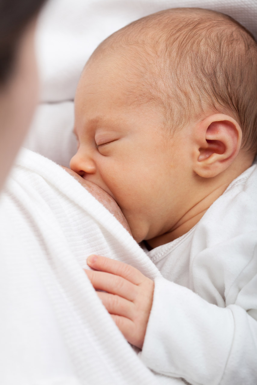
Milk on Demand
This adorable nursing infant (Figure 9.4.1) is part of a positive feedback loop. When he suckles on the nipple, it sends nerve impulses to his mother’s hypothalamus. Those nerve impulses “tell” her pituitary gland to release the hormone prolactin into her bloodstream. Prolactin travels to the mammary glands in the breasts and stimulates milk production, which motivates the infant to keep suckling.
What Is the Pituitary Gland?
The pituitary gland is the master gland of the endocrine system, which is the system of glands that secrete hormones into the bloodstream. Endocrine hormones control virtually all physiological processes. They control growth, sexual maturation, reproduction, body temperature, blood pressure, and metabolism. The pituitary gland is considered the master gland of the endocrine system, because it controls the rest of the endocrine system. Many pituitary hormones either promote or inhibit hormone secretion by other endocrine glands.
Structure and Function of the Pituitary Gland
The pituitary gland is about the size of a pea. It protrudes from the bottom of the hypothalamus at the base of the inner brain (see Figure 9.4.2). The pituitary is connected to the hypothalamus by a thin stalk (called the infundibulum). Blood vessels and nerves in the stalk allow direct connections between the hypothalamus and pituitary gland.
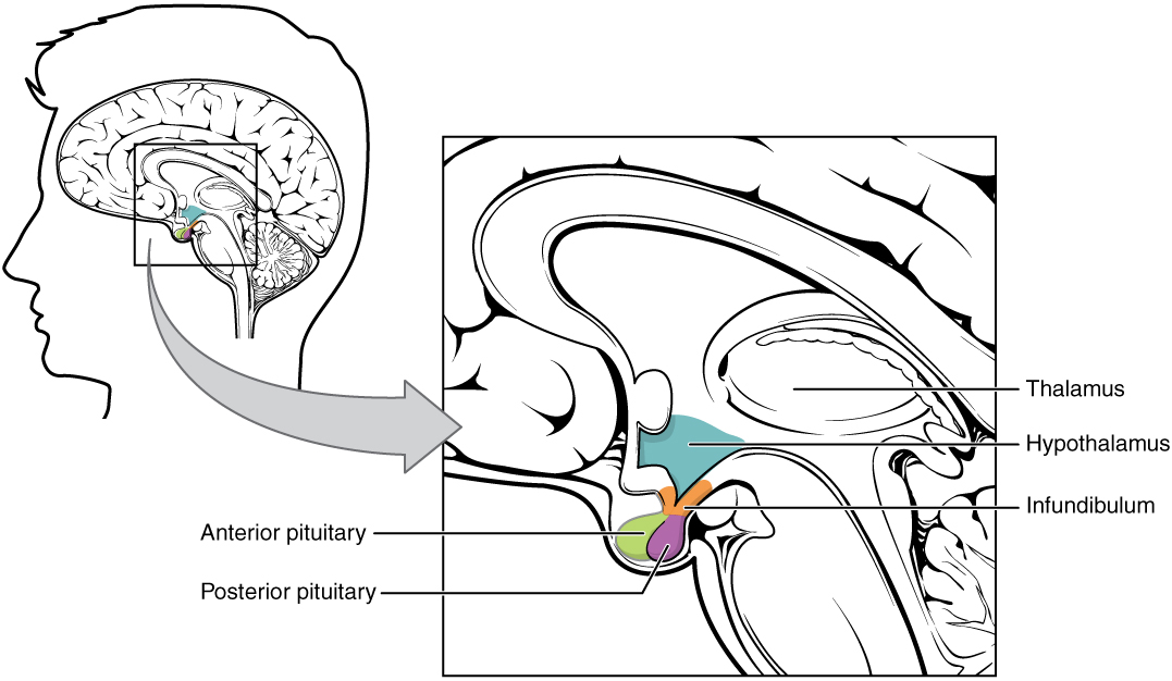
Anterior Lobe
The anterior pituitary is the lobe is at the front of the pituitary gland. It synthesizes and releases hormones into the blood. Table 9.4.1 shows some of the endocrine hormones released by the anterior pituitary, including their targets and effects.
Table 9.4.1
Endocrine Hormones Released by the Anterior Pituitary, and Their Targets and Effects.
| Anterior Pituitary Hormone | Target | Effect |
| Adrenocorticotropic hormone (ACTH) | Adrenal glands | Stimulates the cortex of each adrenal gland to secrete its hormones. |
| Thyroid-stimulating hormone (TSH) | Thyroid gland | Stimulates the thyroid gland to secrete thyroid hormone. |
| Growth hormone (GH) | Body cells | Stimulates body cells to synthesize proteins and grow. |
| Follicle-stimulating hormone (FSH) | Ovaries, testes | Stimulates the ovaries to develop mature eggs. stimulates the testes to produce sperm. |
| Luteinizing hormone (LH) | Ovaries, testes | Stimulates the ovaries and testes to secrete sex hormones; stimulates the ovaries to release eggs. |
| Prolactin (PRL) | Mammary glands | Stimulates the mammary glands to produce milk. |
The anterior pituitary gland is regulated mainly by hormones from the hypothalamus. The hypothalamus secretes hormones (called releasing hormones and inhibiting hormones) that travel through capillaries directly to the anterior lobe of the pituitary gland. The hormones stimulate the anterior pituitary to either release or stop releasing particular pituitary hormones. Several of these hypothalamic hormones and their effects on the anterior pituitary are shown in the table below.
Table 9.4.2
Hypothalamic Hormones and Their Effects on the Anterior Pituitary
| Hypothalamic Hormone | Effect on Anterior Pituitary |
| Thyrotropin releasing hormone (TRH) | Release of thyroid stimulating hormone (TSH) |
| Corticotropin releasing hormone (CRH) | Release of adrenocorticotropic hormone (ACTH) |
| Gonadotropin releasing hormone (GnRH) | Release of follicle-stimulating hormone (FSH) and luteinizing hormone (LH) |
| Growth hormone releasing hormone (GHRH) | Release of growth hormone (GH) |
| Growth hormone inhibiting hormone (GHIH) (Somatostatin) | Stopping of growth hormone release |
| Prolactin releasing hormone (PRH) | Release of prolactin |
| Prolactin inhibiting hormone (PIH) (Dopamine) | Stopping of prolactin release |
Posterior Lobe
The posterior pituitary is the lobe is at the back of the pituitary gland. This lobe does not synthesize any hormones. Instead, the posterior lobe stores hormones that come from the hypothalamus along the axons of nerves connecting the two structures (also shown in Figure 9.4.2). The posterior pituitary then secretes the hormones into the bloodstream as needed. Hypothalamic hormones secreted by the posterior pituitary include vasopressin and oxytocin.
- Vasopressin (also called antidiuretic hormone, or ADH) helps maintain homeostasis in body water. It stimulates the kidneys to conserve water by producing more concentrated urine. Specifically, vasopressin targets ducts in the kidneys and makes them more permeable to water. This allows more water to be resorbed by the body, rather than excreted in urine.
- Oxytocin (OXY) targets cells in the uterus to stimulate uterine contractions, as in childbirth. It also targets cells in the breasts of a nursing mother to stimulate the letdown of milk.
9.4 Summary
- The pituitary gland is the master gland of the endocrine system, because most of its hormones control other endocrine glands.
- The pituitary gland is at the base of the brain, where it is connected to the hypothalamus by nerves and capillaries. It has an anterior (front) lobe that synthesizes and secretes pituitary hormones and a posterior (back) lobe that stores and secretes hormones from the hypothalamus.
- Hormones synthesized and secreted by the anterior pituitary include growth hormone, which stimulates cell growth throughout the body, and thyroid stimulating hormone (TSH), which stimulates the thyroid gland to secrete its hormones.
- Hypothalamic hormones stored and secreted by the posterior pituitary gland include vasopressin, which helps maintain homeostasis in body water, and oxytocin, which stimulates uterine contractions during birth, as well as the letdown of milk during lactation.
9.4 Review Questions
-
-
- Explain why the pituitary gland is called the master gland of the endocrine system.
- Compare and contrast the two lobes of the pituitary gland and their general functions.
- Identify two hormones released by the anterior pituitary, their targets, and their effects.
- Explain how the hypothalamus influences the output of hormones by the anterior lobe of the pituitary gland.
- Name and give the function of two hypothalamic hormones released by the posterior pituitary gland.
- Answer the following questions about prolactin releasing hormone (PRH) and prolactin inhibiting hormone (PIH).
- Where are these hormones produced?
- Where are their target cells located?
- What are their effects on their target cells?
- What are their ultimate effects on milk production? Explain your answer.
- When a baby nurses, which of these hormones is most likely released in the mother? Explain your answer.
- For each of the following hormones, state whether it is synthesized in the pituitary or the hypothalamus.
- gonadotropin releasing hormone (GnRH)
- growth hormone (GH)
- oxytocin
- adrenocorticotropic hormone (ACTH)
9.4 Explore More
https://www.youtube.com/watch?v=jUKQFkmBuww&feature=emb_logo
Common Pituitary Diseases, Swedish, 2012.
https://www.youtube.com/watch?v=v41AJGP-XmI&feature=emb_logo
Diagnosing and Treating Pituitary Tumors - California Center for Pituitary Disorders at UCSF, UCSF Neurosurgery, 2015.
Attributions
Figure 9.4.1
Breastfeeding by Petr Kratochvil on Wikimedia Commons is used under a CC0 1.0 Universal
Public Domain Dedication (https://creativecommons.org/publicdomain/zero/1.0/deed.en) license.
Figure 9.4.2
The_Hypothalamus-Pituitary_Complex by OpenStax College on Wikimedia Commons is used under a CC BY 3.0 (https://creativecommons.org/licenses/by/3.0) license.
References
Betts, J. G., Young, K.A., Wise, J.A., Johnson, E., Poe, B., Kruse, D.H., Korol, O., Johnson, J.E., Womble, M., DeSaix, P. (2013, June 19). Figure 17.7 Hypothalamus–pituitary complex [digital image]. In Anatomy and Physiology (Section 17.3). OpenStax. https://openstax.org/books/anatomy-and-physiology/pages/17-3-the-pituitary-gland-and-hypothalamus
Swedish. (2012, April 19). Common pituitary diseases. YouTube. https://www.youtube.com/watch?v=jUKQFkmBuww&feature=youtu.be
UCSF Neurosurgery. (2015, May 13). Diagnosing and treating pituitary tumors - California Center for Pituitary Disorders at UCSF. YouTube. https://www.youtube.com/watch?v=v41AJGP-XmI&feature=youtu.be
Image shows a man at an oxygen bar. There are several erlenmeyer flasks of differently scented oxygen with tubes coming out of the top of each. The man is wearing a nasal cannula (tube that blows air into your nostrils).
Image shows a diagram of the heart with all chambers and major vessels labelled. Arrows indicate blood flow. Deoxygenated blood is brought the the right atrium by the superior and inferior vena cava. The right atrium moves blood into the right ventricle, which then sends blood to the lungs via the pulmonary arteries. Oxygenated blood returns to the heart from the lungs via the pulmonary veins and enters the left atrium. From there, blood is pumped into the left ventricle and then into the aorta for distribution to the body.
Image shows a diagram illustrating how peristalsis pushes food through the digestive tract by squeezing just behind the food, pushing it forward.
Having a higher proportion of hydronium ions than hydroxide ions; having the properties of an acid; having a pH below 7.
Image shows a diagram of the four steps of a white blood cell phagocytizing a bacterium. In the first stage, the white blood cell extends its plasma membrane to surround the bacteria and trap it inside a vesicle, very similar to endocytosis. In the next stage, a lysosome merges with the vesicle containing the bacterium; the digestive enzymes break down the bacterium, and the white blood cell absorbs the resulting nutrients.
The fusion of haploid gametes, egg and sperm, to form the diploid zygote.


