18.3 Structures of the Male Reproductive System

Rocky Mountain Oysters
First, they are peeled and pounded flat. Then, they are coated in flour, seasoned with salt and pepper, and deep fried. What are they? They are often called Rocky Mountain oysters, but they don’t come from the sea. They may also be known as Montana tendergroin, cowboy caviar, or swinging beef — all names that hint at their origins. Here’s another hint: they are harvested only from male animals, such as bulls or sheep. What are they? In a word: testes.
Testes and Scrotum
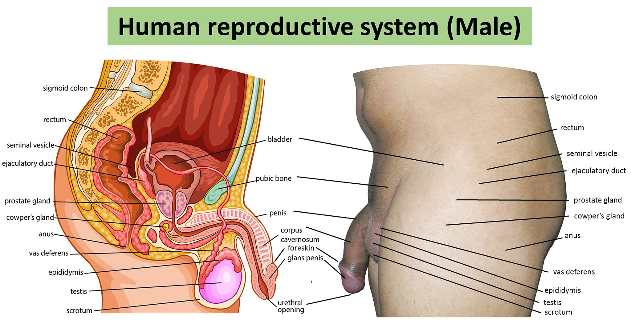
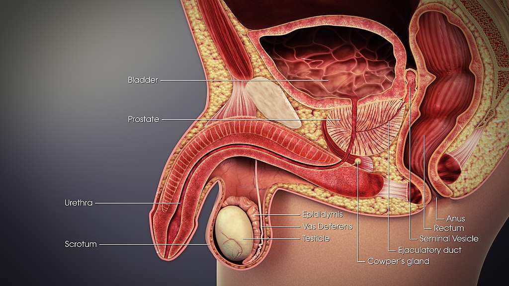
The two testes (singular, testis) are sperm– and testosterone-producing gonads in male mammals, including male humans. These and other organs of the human male reproductive system are shown in Figure 18.3.2. The testes are contained within the scrotum, a pouch made of skin and smooth muscle that hangs down behind the penis.
Testes Structure
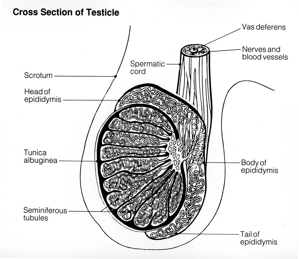
The testes are filled with hundreds of tiny tubes, called seminiferous tubules, which are the functional units of the testes. As shown in the longitudinal-section drawing of a testis in Figure 18.3.3, the seminiferous tubules are coiled and tightly packed within divisions of the testis called lobules. Lobules are separated from one another by internal walls (or septa).
Tunica
The multi-layered covering of each testis, called the tunica, protects the organ, ensures its blood supply, and separates the testis into lobules. There are three layers of the tunica: the tunica vasculosa, tunica albuginea, and tunica vaginalis. The latter two layers are labeled in the drawing above (Figure 18.3.3).
- The tunica vasculosa is the innermost layer of the tunica. It consists of connective tissue and contains arteries and veins that carry blood to and from the testis.
- The tunica albuginea is the middle layer of the tunica. It is a dense layer of fibrous tissue that encases the testis. It also extends into the testis, creating the septa between lobules.
- The tunica vaginalis is the outmost layer of the tunica. It actually consists of two layers of tissue separated by a thin fluid layer. The fluid reduces friction between the testes and the scrotum.
Seminiferous Tubules
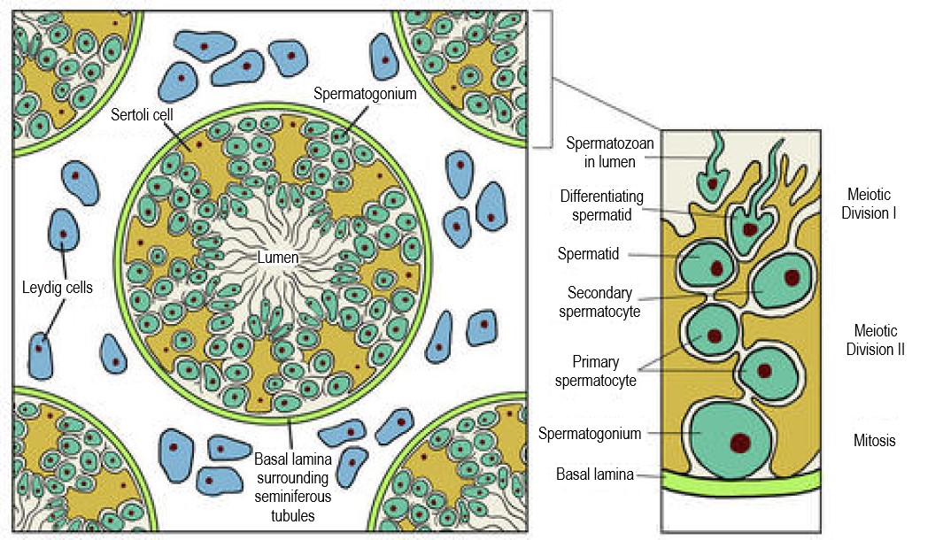
One or more seminiferous tubules are tightly coiled within each of the hundreds of lobules in the testis. A single testis normally contains a total of about 30 metres of these tightly packed tubules! As shown in the cross-sectional drawing of a seminiferous tubule in Figure 18.3.4, the tubule contains sperm in several different stages of development (spermatogonia, spermatocytes, spermatids, and spermatozoa). The seminiferous tubule is also lined with epithelial cells called Sertoli cells. These cells release a hormone (inhibin) that helps regulate sperm production. Adjacent Sertoli cells are closely spaced so large molecules cannot pass from the blood into the tubules. This prevents the male’s immune system from reacting against the developing sperm, which may be antigenically different from his own self tissues. Cells of another type, called Leydig cells, are found between the seminiferous tubules. Leydig cells produce and secrete testosterone.
Other Scrotal Structures
Besides the two testes, the scrotum also contains a pair of organs called epididymes (singular, epididymis) and part of each of the paired vas deferens (or ducti deferens). Both structures play important functions in the production or transport of sperm.
Epididymis
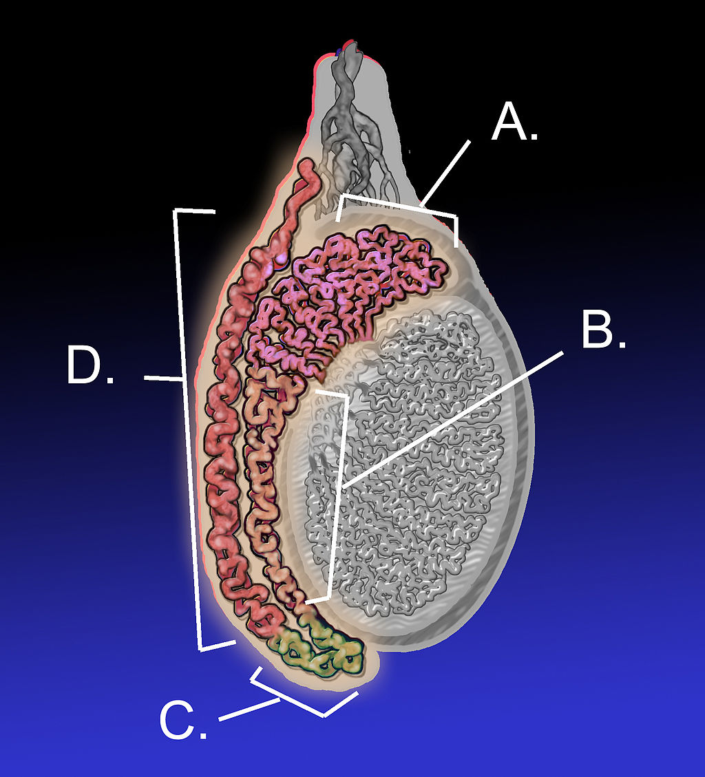
The seminiferous tubules within each testis join together to form ducts (called efferent ducts) that transport immature sperm to the epididymis associated with that testis. Each epididymis (plural, epididymes) consists of a tightly coiled tubule with a total length of about 6 metres. As shown in Figure 18.3.5, the epididymis is generally divided into three parts: the head (which rests on top of the testis), the body (which drapes down the side of the testis), and the tail (which joins with the vas deferens near the bottom of the testis). The functions of the two epididymes are to mature sperm, and then to store that mature sperm until they leave the body during an ejaculation, when they pass the sperm on to the vas deferens.
Vas Deferens
The vas deferens, also known as sperm ducts, are a pair of thin tubes, each about 30 cm (almost 12 in) long, which begin at the epididymes in the scrotum, and continue up into the pelvic cavity. They are composed of ciliated epithelium and smooth muscle. These structures help the vas deferens fulfill their function of transporting sperm from the epididymes to the ejaculatory ducts, which are accessory structures of the male reproductive system.
Accessory Structures
In addition to the structures within the scrotum, the male reproductive system includes several internal accessory structures that are shown in the detailed drawing in Figure 18.3.6. They include the ejaculatory ducts, seminal vesicles, and the prostate and bulbourethral (Cowper’s) glands.
Seminal Vesicles
The seminal vesicles are a pair of exocrine glands that each consist of a single tube, which is folded and coiled upon itself. Each vesicle is about 5 cm (almost 2 in) long and has an excretory duct that merges with the vas deferens to form one of the two ejaculatory ducts. Fluid secreted by the seminal vesicles into the ducts makes up about 70% of the total volume of semen, which is the sperm-containing fluid that leaves the penis during an ejaculation. The fluid from the seminal vesicles is alkaline, so it gives semen a basic pH that helps prolong the lifespan of sperm after it enters the acidic secretions inside the female vagina. Fluid from the seminal vesicles also contains proteins, fructose (a simple sugar), and other substances that help nourish sperm.
Ejaculatory Ducts
The ejaculatory ducts form where the vas deferens join with the ducts of the seminal vesicles in the prostate gland. They connect the vas deferens with the urethra. The ejaculatory ducts carry sperm from the vas deferens, as well as secretions from the seminal vesicles and prostate gland that together form semen. The substances secreted into semen by the glands as it passes through the ejaculatory ducts control its pH and provide nutrients to sperm, among other functions. The fluid itself provides sperm with a medium in which to “swim.”
Prostate Gland
The prostate gland is located just below the seminal vesicles. It is a walnut-sized organ that surrounds the urethra and its junction with the two ejaculatory ducts. The function of the prostate gland is to secrete a slightly alkaline fluid that constitutes close to 30% of the total volume of semen. Prostate fluid contains small quantities of proteins, such as enzymes. In addition, it has a very high concentration of zinc, which is an important nutrient for maintaining sperm quality and motility.
Bulbourethral Glands
Also called Cowper’s glands, the two bulbourethral glands are each about the size of a pea and located just below the prostate gland. The bulbourethral glands secrete a clear, alkaline fluid that is rich in proteins. Each of the glands has a short duct that carries the secretions into the urethra, where they make up a tiny percentage of the total volume of semen. The function of the bulbourethral secretions is to help lubricate the urethra and neutralize any urine (which is acidic) that may remain in the urethra.
Figure 18.3.7 Male reproductive system.
Penis
The penis is the external male organ that has the reproductive function of delivering sperm to the female reproductive tract. This function is called intromission. The penis also serves as the organ that excretes urine.
Structure of the Penis
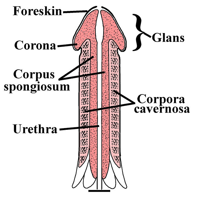
The structure of the penis and its location relative to other reproductive organs are shown in Figure 18.3.8. The part of the penis that is located inside the body and out of sight is called the root of the penis. The shaft of the penis is the part of the penis that is outside the body. The enlarged, bulbous end of the shaft is called the glans penis.
Urethra
The urethra passes through the penis to carry urine from the bladder — or semen from the ejaculatory ducts — through the penis and out of the body. After leaving the urinary bladder, the urethra passes through the prostate gland, where the urethra is joined by the ejaculatory ducts. From there, the urethra passes through the penis to its external opening at the tip of the glans penis. Called the external urethral orifice, this opening provides a way for urine or semen to leave the body.
Tissues of the Penis
The penis is covered with skin (epithelium) that is unattached and free to move over the body of the penis. In an uncircumcised male, the glans penis is also mainly covered by epithelium, which (in this location) is called the foreskin, and below which is a layer of mucous membrane. The foreskin is attached to the penis at an area on the underside of the penis called the frenulum.
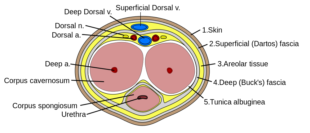
As shown in the Figure 18.3.9, the interior of the penis consists of three columns of spongy tissue that can fill with blood and swell in size, allowing the penis to become erect. This spongy tissue is called corpus cavernosum (plural, corpora cavernosa). Two columns of this tissue run side by side along the top of the shaft, and one column runs along the bottom of the shaft. The urethra runs through this bottom column of spongy tissue, which is sometimes called corpus spongiosum. The glans penis also consists mostly of spongy erectile tissue. Veins and arteries run along the top of the penis, allowing blood circulation through the spongy tissues.
Feature: Human Biology in the News
Lung, heart, kidney, and other organ transplants have become relatively commonplace, so when they occur, they are unlikely to make the news. However, when the nation’s first penis transplant took place, it was considered very newsworthy.
In 2016, Massachusetts General Hospital in Boston announced that a team of its surgeons had performed the first penis transplant in the United States. The patient who received the donated penis was a 64-year-old cancer patient. During the 15-hour procedure, the intricate network of nerves and blood vessels of the donor penis were connected with those of the penis recipient. The surgery went well, but doctors reported it would be a few weeks until they would know if normal urination would be possible, and even longer before they would know if sexual functioning would be possible. At the time that news of the surgery was reported in the media, the patient had not shown any signs of rejecting the donated organ. Within 6 months, the patient was able to urinate properly and was beginning to regain sexual function. The surgeons also reported they were hopeful that such transplants would become relatively common, and that patient populations would expand to include wounded warriors and transgender males seeking to transition.
The 2016 Massachusetts operation was not the first penis transplant ever undertaken. The world’s first successful penis transplant was actually performed in 2014 in Cape Town, South Africa. A young man who had lost his penis from complications of a botched circumcision at age 18 was given a donor penis three years later. That surgery lasted nine hours and was highly successful. The young man made a full recovery and regained both urinary and sexual functions in the transplanted organ.
In 2005, a man in China also received a donated penis in a technically successful operation. However, the patient asked doctors to reverse the procedure just two weeks later, because of psychological problems associated with the transplanted organ for both himself and his wife.
18.3 Summary
- The two testes are sperm– and testosterone-producing male gonads. They are contained within the scrotum, a pouch that hangs down behind the penis. The testes are filled with hundreds of tiny, tightly coiled seminiferous tubules, where sperm are produced. The tubules contain sperm in different stages of development and also Sertoli cells, which secrete substances needed for sperm production. Between the tubules are Leydig cells, which secrete testosterone.
- Also contained within the scrotum are the two epididymes. Each epididymis is a tightly coiled tubule where sperm mature and are stored until they leave the body during an ejaculation.
- The two vas deferens are long, thin tubes that run from the scrotum up into the pelvic cavity. During ejaculation, each vas deferens carries sperm from one of the two epididymes to one of the pair of ejaculatory ducts.
- The two seminal vesicles are glands within the pelvis that secrete fluid through ducts into the junction of each vas deferens and ejaculatory duct. This alkaline fluid makes up about 70% of semen, the sperm-containing fluid that leaves the penis during ejaculation. Semen contains alkaline substances and nutrients that sperm need to survive and “swim” in the female reproductive tract.
- The paired ejaculatory ducts form where the vas deferens joins with the ducts of the seminal vesicles in the prostate gland. They connect the vas deferens with the urethra. The ejaculatory ducts carry sperm from the vas deferens, as well as secretions from the seminal vesicles and prostate gland that together form semen.
- The prostate gland is located just below the seminal vesicles, and it surrounds the urethra and its junction with the ejaculatory ducts. The prostate secretes an alkaline fluid that makes close to 30% of semen. Prostate fluid contains a high concentration of zinc, which sperm need to be healthy and motile.
- The paired bulbourethral glands are located just below the prostate gland. They secrete a tiny amount of fluid into semen. The secretions help lubricate the urethra and neutralize any acidic urine it may contain.
- The penis is the external male organ that has the reproductive function of intromission, which is delivering sperm to the female reproductive tract. The penis also serves as the organ that excretes urine. The urethra passes through the penis and carries urine or semen out of the body. Internally, the penis consists largely of columns of spongy tissue that can fill with blood and make the penis stiff and erect. This is necessary for sexual intercourse so intromission can occur.
18.3 Review Questions
-
-
- Describe the structure of a testis.
- Which parts of the male reproductive system are connected by the ejaculatory ducts? What fluids enter and leave the ejaculatory ducts?
- A vasectomy is a form of birth control for men that is performed by surgically cutting or blocking the vas deferens so that sperm cannot be ejaculated out of the body. Do you think men who have a vasectomy emit semen when they ejaculate? Why or why not?
-
18.3 Explore More
Human Physiology – Functional Anatomy of the Male Reproductive System (Updated), Janux, 2015.
The Science of ‘Morning Wood’, AsapSCIENCE, 2012.
I Had One Of The World’s First Penis Transplants – Thomas Manning | This Morning, 2016.
Attributions
Figure 18.3.1
Lamb_fries by Paul Lowry on Wikimedia Commons is used under a CC BY 2.0 (https://creativecommons.org/licenses/by/2.0) license.
Figure 18.3.2
Human_reproductive_system_(Male) by Baresh25 on Wikimedia Commons is used under a CC BY-SA 4.0 (https://creativecommons.org/licenses/by-sa/4.0) license.
Figure 18.3.3
Testicle by Unknown Illustrator from National Cancer Institute, of the National Institutes of Health, Visuals Online, ID 1769 is in the public domain (https://en.wikipedia.org/wiki/en:public_domain).
Figure 18.3.4
Testis-cross-section by Laura Guerin from CK-12 Foundation is used under a
CC BY-NC 3.0 (https://creativecommons.org/licenses/by-nc/3.0/) license.
Figure 18.3.5
Epididymis-KDS by KDS444 on Wikimedia Commons is used under a CC BY-SA 3.0 (https://creativecommons.org/licenses/by-sa/3.0) license.
Figure 18.3.6
3D_Medical_Animation_Vas_Deferens by https://www.scientificanimations.com/wiki-images (image 26 of 191) on Wikimedia Commons is used under a CC BY-SA 4.0 (https://creativecommons.org/licenses/by-sa/4.0) license.
Figure 18.3.7
Male anatomy blank [adapted] by Tsaitgaist on Wikimedia Commons is used and adapted by Christine Miller under a CC BY-SA 3.0 (http://creativecommons.org/licenses/by-sa/3.0/) license. (Original: Male anatomy.png)
Figure 18.3.8
Penile-Clitoral_Structure by Esseh on Wikimedia Commons is used under a CC BY-SA 3.0 (http://creativecommons.org/licenses/by-sa/3.0/) license.
Figure 18.3.9
Penis_cross_section.svg by Mcstrother on Wikimedia Commons is used under a CC BY 3.0 (https://creativecommons.org/licenses/by/3.0) license.
References
AsapSCIENCE, (2012, November 14). The science of ‘morning wood’. YouTube. https://www.youtube.com/watch?v=D1et5NgT6bQ&feature=youtu.be
Associated Press. (2016, May 17). Man receives new penis in 15-hour operation, the first transplant of its kind in U.S. history [online article]. Canada.com. http://www.canada.com/health/receives+penis+hour+operation+first+transplant+kind+history/11922832/story.html
Brainard, J/ CK-12 Foundation. (2012). Figure 3 Cross section of a testis and seminiferous tubules [digital image]. In CK-12 Biology (Section 25.1) [online Flexbook]. CK12.org. https://www.ck12.org/book/ck-12-biology/section/25.1/
Gallagher, J. (2015, March 13). South Africans perform first ‘successful’ penis transplant (online article). BBC News. https://www.bbc.com/news/health-31876219
Grady, D. (2016, May 16). Cancer survivor receives first penis transplant in the United States [online article]. New York Times. https://www.nytimes.com/2016/05/17/health/thomas-manning-first-penis-transplant-in-us.html
Janux. (2015, August 16). Human physiology – Functional anatomy of the male reproductive system (Updated). YouTube. https://www.youtube.com/watch?v=k60M1h-DKVY&feature=youtu.be
This Morning. (2016, June 15). I had one of the world’s first penis transplants – Thomas Manning | This Morning. YouTube. https://www.youtube.com/watch?v=Ot7CYjm9B7U&feature=youtu.be
a hormonal disorder common among women of reproductive age. Women with PCOS may have infrequent or prolonged menstrual periods or excess male hormone (androgen) levels. The ovaries may develop numerous small collections of fluid (follicles) and fail to regularly release eggs.
A biological process which converts sugars such as glucose, fructose, and sucrose into cellular energy, producing ethanol and carbon dioxide as by-products.
A substance that takes part in and undergoes change during a chemical reaction.
Image shows a female clinician dressed in scrubs listening to the heartbeat of a patient.
Image shows a diagram of the heart with all chambers and major vessels labelled. Arrows indicate blood flow. Deoxygenated blood is brought the the right atrium by the superior and inferior vena cava. The right atrium moves blood into the right ventricle, which then sends blood to the lungs via the pulmonary arteries. Oxygenated blood returns to the heart from the lungs via the pulmonary veins and enters the left atrium. From there, blood is pumped into the left ventricle and then into the aorta for distribution to the body.
Image shows a scanning electroflourescent pictomicrograph. It shows the villi of the small intestine, with a layer of mucus, and then a multitude of smaller bacterial cells above the mucous.
Competitive Eating
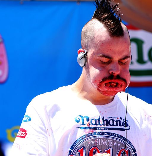
This man is on his way to coming in third in an international hot dog eating contest (Figure 15.3.1). It may look as though he is regurgitating his hot dogs, but in fact, he is trying to get them into his mouth and down his throat as quickly as he can. In order to eat as many hot dogs as possible in the allotted time, he pushes several into his mouth at once, and doesn’t bother doing much chewing. Chewing is normally the first step in the process of digestion.
Digestion
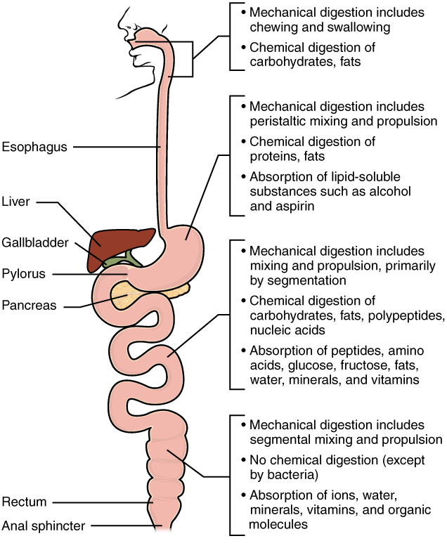
Digestion of food is a form of catabolism, in which the food is broken down into small molecules that the body can absorb and use for energy, growth, and repair. Digestion occurs when food is moved through the digestive system. This process begins in the mouth and ends in the small intestine. The final products of digestion are absorbed from the digestive tract, primarily in the small intestine. There are two different types of digestion that occur in the digestive system: mechanical digestion and chemical digestion. Figure 15.3.2 summarizes the roles played by different digestive organs in mechanical and chemical digestion, both of which are described in detail below.
Mechanical Digestion

Mechanical digestion is a physical process in which food is broken into smaller pieces without becoming changed chemically. It begins with your first bite of food (see Figure 15.3.3) and continues as you chew food with your teeth into smaller pieces. The process of mechanical digestion continues in the stomach. This muscular organ churns and mixes the food it contains, an action that breaks any solid food into still smaller pieces.
Although some mechanical digestion also occurs in the small intestine, it is mostly completed by the time food leaves the stomach. At that stage, food in the GI tract has been changed to the thick semi-fluid called chyme. Mechanical digestion is necessary so that chemical digestion can be effective. Mechanical digestion tremendously increases the surface area of food particles so they can be acted upon more effectively by digestive enzymes.
Chemical Digestion
Chemical digestion is the biochemical process in which macromolecules in food are changed into smaller molecules that can be absorbed into body fluids and transported to cells throughout the body. Substances in food that must be chemically digested include carbohydrates, proteins, lipids, and nucleic acids. Carbohydrates must be broken down into simple sugars, proteins into amino acids, lipids into fatty acids and glycerol, and nucleic acids into nitrogen bases and sugars. Some chemical digestion takes place in the mouth and stomach, but most of it occurs in the first part of the small intestine (duodenum).
Digestive Enzymes
Chemical digestion could not occur without the help of many different digestive enzymes. Enzymes are proteins that catalyze, or speed up, biochemical reactions. Digestive enzymes are secreted by exocrine glands or by the mucosal layer of epithelium lining the gastrointestinal tract. In the mouth, digestive enzymes are secreted by salivary glands. The lining of the stomach secretes enzymes, as does the lining of the small intestine. Many more digestive enzymes are secreted by exocrine cells in the pancreas and carried by ducts to the small intestine. The following table lists several important digestive enzymes, the organs and/or glands that secrete them, the compounds they digest, and the pH necessary for optimal functioning. You can read more about them below.
| Digestive Enzyme | Source Organ | Site of Action | Reactant and Product | Optimal pH |
|---|---|---|---|---|
| Salivary Amylase | Salivary Glands | Mouth | starch + water ⇒ maltose | Neutral |
| Pepsin | Stomach | Stomach | protein + water ⇒ peptides | Acidic |
| Pancreatic Amylase | Pancreas | Duodenum | starch + water ⇒ maltose | Basic |
| Maltase | Small intestine | Small intestine | maltose + water ⇒ glucose | Basic |
| Sucrase | Small intestine | Small intestine | sucrose + water ⇒ glucose + fructose | Basic |
| Lactase | Small intestine | Small intestine | lactose + water ⇒ glucose + galactose | Basic |
| Lipase | Pancreas | Duodenum | fat droplet and water ⇒ glycerol and fatty acids | Basic |
| Trypsin | Pancreas | Duodenum | protein + water ⇒ peptides | Basic |
| Chymotrypsin | Pancreas | Duodenum | protein + water ⇒ peptides | Basic |
| Peptidases | Small intestine | Small intestine | peptides + water ⇒ | Basic |
| Deoxyribonuclease | Pancreas | Duodenum | DNA + water ⇒ nucleotide fragments | Basic |
| Ribonuclease | Pancreas | Duodenum | RNA + water ⇒ nucleotide fragments | Basic |
| Nuclease | Small intestine | Small intestine | nucleic acids + water ⇒ nucleotide fragments | Basic |
| Nucleosidases | Small intestine | Small intestine | nucleotides + water ⇒ nitrogen base + phosphate sugar | Basic |
Chemical Digestion of Carbohydrates
About 80% of digestible carbohydrates in a typical Western diet are in the form of the plant polysaccharide amylose, which consists mainly of long chains of glucose and is one of two major components of starch. Additional dietary carbohydrates include the animal polysaccharide glycogen, along with some sugars, which are mainly disaccharides.
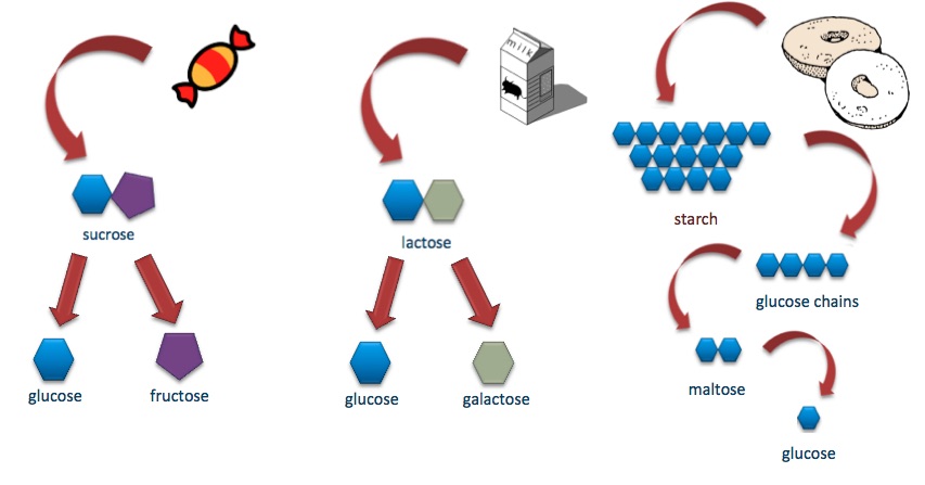
The process of chemical digestion for some carbohydrates is illustrated Figure 15.3.4. To chemically digest amylose and glycogen, the enzyme amylase is required. The chemical digestion of these polysaccharides begins in the mouth, aided by amylase in saliva. Saliva also contains mucus — which lubricates the food — and hydrogen carbonate, which provides the ideal alkaline conditions for amylase to work. Carbohydrate digestion is completed in the small intestine, with the help of amylase secreted by the pancreas. In the digestive process, polysaccharides are reduced in length by the breaking of bonds between glucose monomers. The macromolecules are broken down to shorter polysaccharides and disaccharides, resulting in progressively shorter chains of glucose. The end result is molecules of the simple sugars glucose and maltose (which consists of two glucose molecules), both of which can be absorbed by the small intestine.
Other sugars are digested with the help of different enzymes produced by the small intestine. Sucrose (or table sugar), for example, is a disaccharide that is broken down by the enzyme sucrase to form glucose and fructose, which are readily absorbed by the small intestine. Digestion of the sugar lactose, which is found in milk, requires the enzyme lactase, which breaks down lactose into glucose and galactose. Glucose and galactose are then absorbed by the small intestine. Fewer than half of all adults produce sufficient lactase to be able to digest lactose. Those who cannot are said to be lactose intolerant.
Chemical Digestion of Proteins
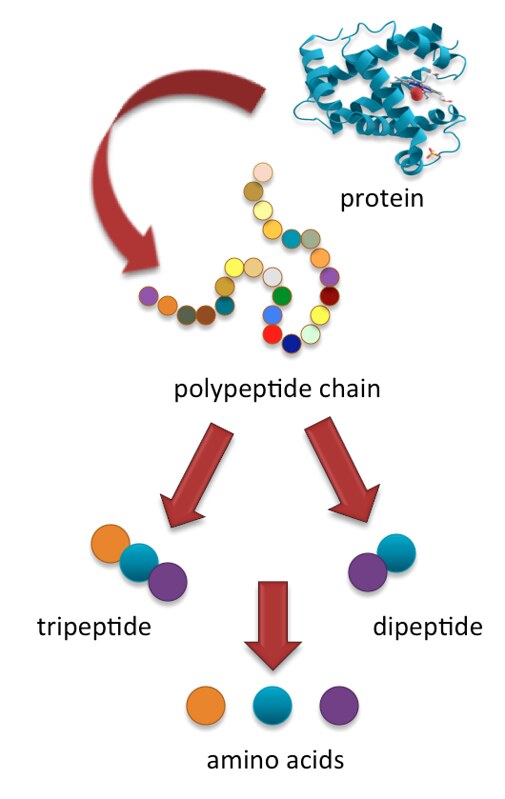
Proteins consist of polypeptides, which must be broken down into their constituent amino acids before they can be absorbed. An overview of this process is shown in Figure 15.3.5. Protein digestion occurs in the stomach and small intestine through the action of three primary enzymes: pepsin (secreted by the stomach), and trypsin and chymotrypsin (secreted by the pancreas). The stomach also secretes hydrochloric acid (HCl), making the contents highly acidic, which is a required condition for pepsin to work. Trypsin and chymotrypsin in the small intestine require an alkaline (basic) environment to work. Bile from the liver and bicarbonate from the pancreas neutralize the acidic chyme as it empties into the small intestine. After pepsin, trypsin, and chymotrypsin break down proteins into peptides, these are further broken down into amino acids by other enzymes called peptidases, also secreted by the pancreas.
Chemical Digestion of Lipids
The chemical digestion of lipids begins in the mouth. The salivary glands secrete the digestive enzyme lipase, which breaks down short-chain lipids into molecules consisting of two fatty acids. A tiny amount of lipid digestion may take place in the stomach, but most lipid digestion occurs in the small intestine.
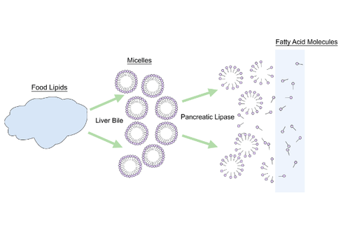
Digestion of lipids in the small intestine occurs with the help of another lipase enzyme from the pancreas, as well as bile secreted by the liver. As shown in the diagram below (Figure 15.3.6), bile is required for the digestion of lipids, because lipids are oily and do not dissolve in the watery chyme. Bile emulsifies (or breaks up) large globules of food lipids into much smaller ones, called micelles, much as dish detergent breaks up grease. The micelles provide a great deal more surface area to be acted upon by lipase, and also point the hydrophilic (“water-loving”) heads of the fatty acids outward into the watery chyme. Lipase can then access and break down the micelles into individual fatty acid molecules.
Chemical Digestion of Nucleic Acids
Nucleic acids (DNA and RNA) in foods are digested in the small intestine with the help of both pancreatic enzymes and enzymes produced by the small intestine itself. Pancreatic enzymes called ribonuclease and deoxyribonuclease break down RNA and DNA, respectively, into smaller nucleic acids. These, in turn, are further broken down into nitrogen bases and sugars by small intestine enzymes called nucleases.
Bacteria in the Digestive System
Your large intestine is not just made up of cells. It is also an ecosystem, home to trillions of bacteria known as the "gut flora" (Figure 15.3.7). But don't worry, most of these bacteria are helpful. Friendly bacteria live mostly in the large intestine and part of the small intestine. The acidic environment of the stomach does not allow bacterial growth.
Gut bacteria have several roles in the body. For example, intestinal bacteria:
- Produce vitamin B12 and vitamin K.
- Control the growth of harmful bacteria.
- Break down poisons in the large intestine.
- Break down some substances in food that cannot be digested, such as fibre and some starches and sugars. Bacteria produce enzymes that digest carbohydrates in plant cell walls. Most of the nutritional value of plant material would be wasted without these bacteria. These help us digest plant foods like spinach.
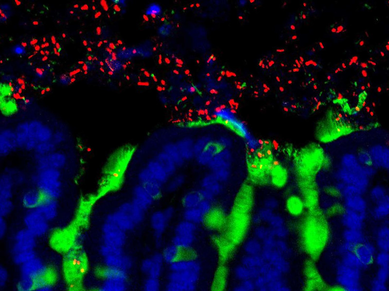
A wide range of friendly bacteria live in the gut. Bacteria begin to populate the human digestive system right after birth. Gut bacteria include Lactobacillus, the bacteria commonly used in probiotic foods such as yogurt, and E. coli bacteria. About a third of all bacteria in the gut are members of the Bacteroides species. Bacteroides are key in helping us digest plant food.
It is estimated that 100 trillion bacteria live in the gut. This is more than the human cells that make up you. It has also been estimated that there are more bacteria in your mouth than people on the planet — there are over 7 billion people on the planet!
The bacteria in your digestive system are from anywhere between 300 and 1,000 species. As these bacteria are helpful, your body does not attack them. They actually appear to the body's immune system as cells of the digestive system, not foreign invaders. The bacteria actually cover themselves with sugar molecules removed from the actual cells of the digestive system. This disguises the bacteria and protects them from the immune system.
As the bacteria that live in the human gut are beneficial to us, and as the bacteria enjoy a safe environment to live, the relationship that we have with these tiny organisms is described as mutualism, a type of symbiotic relationship.
Lastly, keep in mind the small size of bacteria. Together, all the bacteria in your gut may weigh just about two pounds.
Control of the Digestive Process
The process of digestion is controlled by both hormones and nerves. Hormonal control is mainly by endocrine hormones secreted by cells in the lining of the stomach and small intestine. These hormones stimulate the production of digestive enzymes, bicarbonate, and bile. The hormone secretin, for example, is produced by endocrine cells lining the duodenum of the small intestine. Acidic chyme entering the duodenum from the stomach triggers the release of secretin into the bloodstream. When the secretin returns via the circulation to the digestive system, it signals the release of bicarbonate from the pancreas. The bicarbonate neutralizes the acidic chyme. See Table 15.3.2 for a summary of the major hormones governing the process of chemical digestion.
| Hormone | Source Organ | Target Organ | Trigger | Result |
|---|---|---|---|---|
| Gastrin | Stomach walls | Stomach | High protein intake | HCL and pepsin release, stomach churning |
| Secretin | Duodenum | Pancreas
Gallbladder |
Acidic chyme entering the duodenum | Release sodium bicarbonate, release bile |
| Cholecystokinin (CCK) | Duodenum | Pancreas
Gallbladder |
Partially digested fat and protein in duodenum | Release lipase, trypsin, release bile |
Nerves involved in digestion include those that connect digestive organs to the central nervous system, as well as nerves inside the walls of the digestive organs. Nerves connecting the digestive organs to the central nervous system cause smooth muscles in the walls of digestive organs to contract or relax as needed, depending on whether or not there is food to be digested. Nerves within digestive organs are stimulated when food enters the organs and stretches their walls. These nerves trigger the release of substances that speed up or slow down the movement of food through the GI tract and the secretion of digestive enzymes.
Absorption
When digestion is finished, it results in many simple nutrient molecules that must go through the process of absorption from the lumen of the GI tract to blood or lymph vessels, so they can be transported to and used by cells throughout the body. A few substances are absorbed in the stomach and large intestine. Water is absorbed in both of these organs, and some minerals and vitamins are also absorbed in the large intestine, but about 95% of nutrient molecules are absorbed in the small intestine. Absorption of the majority of these molecules takes place in the second part of the small intestine, called the jejunum. There are, however, a few exceptions — for example, iron is absorbed in the duodenum, and vitamin B12 is absorbed in the last part of the small intestine, called the ileum. After being absorbed in the small intestine, nutrient molecules are transported to other parts of the body for storage or further chemical modification. Amino acids, for instance, are transported to the liver to be used for protein synthesis.
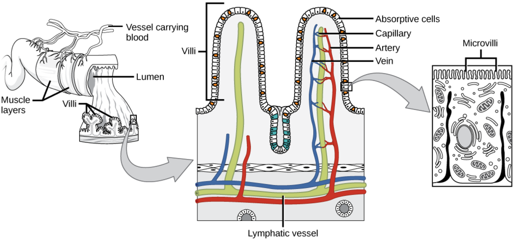
The epithelial tissue lining the small intestine is specialized for absorption. It is highly enfolded and is covered with villi and microvilli, creating an enormous surface area for absorption. As shown in Figure 15.3.8, each villus also has a network of blood capillaries and fine lymphatic vessels called lacteals close to its surface. The thin surface layer of epithelial cells of the villi transports nutrients from the lumen of the small intestine into these capillaries and lacteals. Blood in the capillaries absorbs most of the molecules, including simple sugars, amino acids, glycerol, salts, and water-soluble vitamins (vitamin C and the many B vitamins). Lymph in the lacteals absorbs fatty acids and fat-soluble vitamins (vitamins A, D, E, and K).
Feature: My Human Body
The process of digestion does not always go as it should. Many people suffer from indigestion, or dyspepsia, a condition of impaired digestion. Symptoms may include upper abdominal fullness or pain, heartburn, nausea, belching, or some combination of these symptoms. The majority of cases of indigestion occur without evidence of an organic disease that is likely to explain the symptoms. Anxiety or certain foods or medications (such as aspirin) may be contributing factors in these cases. In other cases, indigestion is a symptom of an organic disease, most often gastroesophageal reflux disease (GERD) or gastritis. In a small minority of cases, indigestion is a symptom of a peptic ulcer of the stomach or duodenum, usually caused by a bacterial infection. Very rarely, indigestion is a sign of cancer.
An occasional bout of indigestion is usually nothing to worry about, especially in people less than 55 years of age. However, if you suffer frequent or chronic indigestion, it’s a good idea to see a doctor. If an underlying disorder such as GERD or an ulcer is causing the indigestion, this can and should be treated. If no organic disease is discovered, the doctor can recommend lifestyle changes or treatments to help prevent or soothe the symptoms of acute indigestion. Lifestyle changes might include modifications in eating habits, such as eating more slowly, eating smaller meals, or avoiding fatty foods. You also might be advised to refrain from taking certain medications, especially on an empty stomach. The use of antacids or other medications to relieve symptoms may also be recommended.
15.3 Summary
- Digestion is a form of catabolism, in which food is broken down into small molecules that the body can absorb and use for energy, growth, and repair. Digestion occurs when food moves through the gastrointestinal (GI) tract. The digestive process is controlled by both hormones and nerves.
- Mechanical digestion is a physical process in which food is broken into smaller pieces without becoming chemically changed. It occurs mainly in the mouth and stomach.
- Chemical digestion is a chemical process in which macromolecules — including carbohydrates, proteins, lipids, and nucleic acids — in food are changed into simple nutrient molecules that can be absorbed into body fluids. Carbohydrates are chemically digested to sugars, proteins to amino acids, lipids to fatty acids, and nucleic acids to individual nucleotides. Chemical digestion requires digestive enzymes. Gut flora carry out additional chemical digestion.
- Absorption occurs when the simple nutrient molecules that result from digestion are absorbed into blood or lymph.
15.3 Review Questions
- Define digestion. Where does it occur?
-
- Identify two organ systems that control the process of digestion by the digestive system.
- What is mechanical digestion? Where does it occur?
- Describe chemical digestion.
- What is the role of enzymes in chemical digestion?
- What is absorption? When does it occur?
- Where does most absorption occur in the digestive system? Why does most of the absorption occur in this organ, and not earlier in the GI tract?
15.3 Explore More
https://youtu.be/awtmTJW9ic8
Food for thought: How your belly controls your brain | Ruairi Robertson | TEDxFulbrightSantaMonica, TEDx Talks, 2015.
https://youtu.be/1sISguPDlhY
How the food you eat affects your gut - Shilpa Ravella, TED-Ed, 2017.
https://youtu.be/jP-9AD0wMOk
What causes heartburn? - Rusha Modi, TED-Ed, 2018.
Attributions
Figure 15.3.1
Patrick_Bertoletti_eating_hot_dogs by Michael on Wikimedia Commons is used under a CC BY 2.0 (https://creativecommons.org/licenses/by/2.0) license.
Figure 15.3.2
2426_Mechanical_and_Chemical_DigestionN by OpenStax College on Wikimedia Commons is used under a CC BY 3.0 (https://creativecommons.org/licenses/by/3.0) license.
Figure 15.3.3
Eating tacos [photo] by DeMorris Byrd on Unsplash is used under the Unsplash License (https://unsplash.com/license).
Figure 15.3.4
Carbohydrate digestion by Nutritional Doublethink on Flickr is used under a CC BY-SA 2.0 (https://creativecommons.org/licenses/by-sa/2.0/) license.
Figure 15.3.5
Peptide Digestion by Nutritional Doublethink on Flickr is used under a CC BY 2.0 (https://creativecommons.org/licenses/by/2.0/) license.
Figure 15.3.6
Bile from the liver and lipase from the pancreas help digest lipids in small intestine by CK-12 Foundation is used under a CC BY NC 3.0 (https://creativecommons.org/licenses/by-nc/3.0/) license.
Figure 15.3.7
Gut Flora by NIH Image Gallery on Flickr by NIH Image Gallery on Flickr is used under a CC BY-NC-SA 2.0 (https://creativecommons.org/licenses/by-nc-sa/2.0/) license.
Figure 15.3.8
Figure_34_01_11f by CNX OpenStax on Wikimedia Commons is used under a CC BY 4.0 (https://creativecommons.org/licenses/by/4.0) license.
References
Betts, J. G., Young, K.A., Wise, J.A., Johnson, E., Poe, B., Kruse, D.H., Korol, O., Johnson, J.E., Womble, M., DeSaix, P. (2013, June 19). Figure 23.28 Digestion and absorption [digital image]. In Anatomy and Physiology (Section 23.7). OpenStax. https://openstax.org/books/anatomy-and-physiology/pages/23-7-chemical-digestion-and-absorption-a-closer-look
Brainard, J/ CK-12 Foundation. (2016). Figure 6 Both bile from the liver and lipase from the pancreas help digest lipids in the small intestine [digital image]. In CK-12 College Human Biology (Section 17.3) [online Flexbook]. CK12.org. https://www.ck12.org/book/ck-12-college-human-biology/section/17.3/
OpenStax. (2016, May 27) Figure 11 Villi are folds on the small intestine lining that increase the surface area to facilitate the absorption of nutrients. [digital image]. In OpenStax, Biology (Section 34.1). OpenStax CNX. https://cnx.org/contents/GFy_h8cu@10.53:Oestf0YE@6/Digestive-Systems
TED-Ed. (2017, March 23). How the food you eat affects your gut - Shilpa Ravella. YouTube. https://www.youtube.com/watch?v=1sISguPDlhY&feature=youtu.be
TED-Ed. (2018, November 1). What causes heartburn? - Rusha Modi. YouTube. https://www.youtube.com/watch?v=jP-9AD0wMOk&feature=youtu.be
TEDx Talks. (2015, December 7). Food for thought: How your belly controls your brain | Ruairi Robertson | TEDxFulbrightSantaMonica. YouTube. https://www.youtube.com/watch?v=awtmTJW9ic8&feature=youtu.be
Created by CK-12 Foundation/Adapted by Christine Miller

Competitive Eating
This man is on his way to coming in third in an international hot dog eating contest (Figure 15.3.1). It may look as though he is regurgitating his hot dogs, but in fact, he is trying to get them into his mouth and down his throat as quickly as he can. In order to eat as many hot dogs as possible in the allotted time, he pushes several into his mouth at once, and doesn’t bother doing much chewing. Chewing is normally the first step in the process of digestion.
Digestion
Digestion of food is a form of catabolism, in which the food is broken down into small molecules that the body can absorb and use for energy, growth, and repair. Digestion occurs when food is moved through the digestive system. This process begins in the mouth and ends in the small intestine. The final products of digestion are absorbed from the digestive tract, primarily in the small intestine. There are two different types of digestion that occur in the digestive system: mechanical digestion and chemical digestion. Figure 15.3.2 summarizes the roles played by different digestive organs in mechanical and chemical digestion, both of which are described in detail below.

Mechanical Digestion

Mechanical digestion is a physical process in which food is broken into smaller pieces without becoming changed chemically. It begins with your first bite of food (see Figure 15.3.3) and continues as you chew food with your teeth into smaller pieces. The process of mechanical digestion continues in the stomach. This muscular organ churns and mixes the food it contains, an action that breaks any solid food into still smaller pieces.
Although some mechanical digestion also occurs in the small intestine, it is mostly completed by the time food leaves the stomach. At that stage, food in the GI tract has been changed to the thick semi-fluid called chyme. Mechanical digestion is necessary so that chemical digestion can be effective. Mechanical digestion tremendously increases the surface area of food particles so they can be acted upon more effectively by digestive enzymes.
Chemical Digestion
Chemical digestion is the biochemical process in which macromolecules in food are changed into smaller molecules that can be absorbed into body fluids and transported to cells throughout the body. Substances in food that must be chemically digested include carbohydrates, proteins, lipids, and nucleic acids. Carbohydrates must be broken down into simple sugars, proteins into amino acids, lipids into fatty acids and glycerol, and nucleic acids into nitrogen bases and sugars. Some chemical digestion takes place in the mouth and stomach, but most of it occurs in the first part of the small intestine (duodenum).
Digestive Enzymes
Chemical digestion could not occur without the help of many different digestive enzymes. Enzymes are proteins that catalyze, or speed up, biochemical reactions. Digestive enzymes are secreted by exocrine glands or by the mucosal layer of epithelium lining the gastrointestinal tract. In the mouth, digestive enzymes are secreted by salivary glands. The lining of the stomach secretes enzymes, as does the lining of the small intestine. Many more digestive enzymes are secreted by exocrine cells in the pancreas and carried by ducts to the small intestine. The following table lists several important digestive enzymes, the organs and/or glands that secrete them, the compounds they digest, and the pH necessary for optimal functioning. You can read more about them below.
| Digestive Enzyme | Source Organ | Site of Action | Reactant and Product | Optimal pH |
|---|---|---|---|---|
| Salivary Amylase | Salivary Glands | Mouth | starch + water ⇒ maltose | Neutral |
| Pepsin | Stomach | Stomach | protein + water ⇒ peptides | Acidic |
| Pancreatic Amylase | Pancreas | Duodenum | starch + water ⇒ maltose | Basic |
| Maltase | Small intestine | Small intestine | maltose + water ⇒ glucose | Basic |
| Sucrase | Small intestine | Small intestine | sucrose + water ⇒ glucose + fructose | Basic |
| Lactase | Small intestine | Small intestine | lactose + water ⇒ glucose + galactose | Basic |
| Lipase | Pancreas | Duodenum | fat droplet and water ⇒ glycerol and fatty acids | Basic |
| Trypsin | Pancreas | Duodenum | protein + water ⇒ peptides | Basic |
| Chymotrypsin | Pancreas | Duodenum | protein + water ⇒ peptides | Basic |
| Peptidases | Small intestine | Small intestine | peptides + water ⇒ | Basic |
| Deoxyribonuclease | Pancreas | Duodenum | DNA + water ⇒ nucleotide fragments | Basic |
| Ribonuclease | Pancreas | Duodenum | RNA + water ⇒ nucleotide fragments | Basic |
| Nuclease | Small intestine | Small intestine | nucleic acids + water ⇒ nucleotide fragments | Basic |
| Nucleosidases | Small intestine | Small intestine | nucleotides + water ⇒ nitrogen base + phosphate sugar | Basic |
Chemical Digestion of Carbohydrates
About 80% of digestible carbohydrates in a typical Western diet are in the form of the plant polysaccharide amylose, which consists mainly of long chains of glucose and is one of two major components of starch. Additional dietary carbohydrates include the animal polysaccharide glycogen, along with some sugars, which are mainly disaccharides.
The process of chemical digestion for some carbohydrates is illustrated Figure 15.3.4. To chemically digest amylose and glycogen, the enzyme amylase is required. The chemical digestion of these polysaccharides begins in the mouth, aided by amylase in saliva. Saliva also contains mucus — which lubricates the food — and hydrogen carbonate, which provides the ideal alkaline conditions for amylase to work. Carbohydrate digestion is completed in the small intestine, with the help of amylase secreted by the pancreas. In the digestive process, polysaccharides are reduced in length by the breaking of bonds between glucose monomers. The macromolecules are broken down to shorter polysaccharides and disaccharides, resulting in progressively shorter chains of glucose. The end result is molecules of the simple sugars glucose and maltose (which consists of two glucose molecules), both of which can be absorbed by the small intestine.
Other sugars are digested with the help of different enzymes produced by the small intestine. Sucrose (or table sugar), for example, is a disaccharide that is broken down by the enzyme sucrase to form glucose and fructose, which are readily absorbed by the small intestine. Digestion of the sugar lactose, which is found in milk, requires the enzyme lactase, which breaks down lactose into glucose and galactose. Glucose and galactose are then absorbed by the small intestine. Fewer than half of all adults produce sufficient lactase to be able to digest lactose. Those who cannot are said to be lactose intolerant.

Chemical Digestion of Proteins
Proteins consist of polypeptides, which must be broken down into their constituent amino acids before they can be absorbed. An overview of this process is shown in Figure 15.3.5. Protein digestion occurs in the stomach and small intestine through the action of three primary enzymes: pepsin (secreted by the stomach), and trypsin and chymotrypsin (secreted by the pancreas). The stomach also secretes hydrochloric acid (HCl), making the contents highly acidic, which is a required condition for pepsin to work. Trypsin and chymotrypsin in the small intestine require an alkaline (basic) environment to work. Bile from the liver and bicarbonate from the pancreas neutralize the acidic chyme as it empties into the small intestine. After pepsin, trypsin, and chymotrypsin break down proteins into peptides, these are further broken down into amino acids by other enzymes called peptidases, also secreted by the pancreas.

Chemical Digestion of Lipids
The chemical digestion of lipids begins in the mouth. The salivary glands secrete the digestive enzyme lipase, which breaks down short-chain lipids into molecules consisting of two fatty acids. A tiny amount of lipid digestion may take place in the stomach, but most lipid digestion occurs in the small intestine.
Digestion of lipids in the small intestine occurs with the help of another lipase enzyme from the pancreas, as well as bile secreted by the liver. As shown in the diagram below (Figure 15.3.6), bile is required for the digestion of lipids, because lipids are oily and do not dissolve in the watery chyme. Bile emulsifies (or breaks up) large globules of food lipids into much smaller ones, called micelles, much as dish detergent breaks up grease. The micelles provide a great deal more surface area to be acted upon by lipase, and also point the hydrophilic (“water-loving”) heads of the fatty acids outward into the watery chyme. Lipase can then access and break down the micelles into individual fatty acid molecules.

Chemical Digestion of Nucleic Acids
Nucleic acids (DNA and RNA) in foods are digested in the small intestine with the help of both pancreatic enzymes and enzymes produced by the small intestine itself. Pancreatic enzymes called ribonuclease and deoxyribonuclease break down RNA and DNA, respectively, into smaller nucleic acids. These, in turn, are further broken down into nitrogen bases and sugars by small intestine enzymes called nucleases.
Bacteria in the Digestive System
Your large intestine is not just made up of cells. It is also an ecosystem, home to trillions of bacteria known as the "gut flora" (Figure 15.3.7). But don't worry, most of these bacteria are helpful. Friendly bacteria live mostly in the large intestine and part of the small intestine. The acidic environment of the stomach does not allow bacterial growth.
Gut bacteria have several roles in the body. For example, intestinal bacteria:
- Produce vitamin B12 and vitamin K.
- Control the growth of harmful bacteria.
- Break down poisons in the large intestine.
- Break down some substances in food that cannot be digested, such as fibre and some starches and sugars. Bacteria produce enzymes that digest carbohydrates in plant cell walls. Most of the nutritional value of plant material would be wasted without these bacteria. These help us digest plant foods like spinach.

A wide range of friendly bacteria live in the gut. Bacteria begin to populate the human digestive system right after birth. Gut bacteria include Lactobacillus, the bacteria commonly used in probiotic foods such as yogurt, and E. coli bacteria. About a third of all bacteria in the gut are members of the Bacteroides species. Bacteroides are key in helping us digest plant food.
It is estimated that 100 trillion bacteria live in the gut. This is more than the human cells that make up you. It has also been estimated that there are more bacteria in your mouth than people on the planet — there are over 7 billion people on the planet!
The bacteria in your digestive system are from anywhere between 300 and 1,000 species. As these bacteria are helpful, your body does not attack them. They actually appear to the body's immune system as cells of the digestive system, not foreign invaders. The bacteria actually cover themselves with sugar molecules removed from the actual cells of the digestive system. This disguises the bacteria and protects them from the immune system.
As the bacteria that live in the human gut are beneficial to us, and as the bacteria enjoy a safe environment to live, the relationship that we have with these tiny organisms is described as mutualism, a type of symbiotic relationship.
Lastly, keep in mind the small size of bacteria. Together, all the bacteria in your gut may weigh just about two pounds.
Control of the Digestive Process
The process of digestion is controlled by both hormones and nerves. Hormonal control is mainly by endocrine hormones secreted by cells in the lining of the stomach and small intestine. These hormones stimulate the production of digestive enzymes, bicarbonate, and bile. The hormone secretin, for example, is produced by endocrine cells lining the duodenum of the small intestine. Acidic chyme entering the duodenum from the stomach triggers the release of secretin into the bloodstream. When the secretin returns via the circulation to the digestive system, it signals the release of bicarbonate from the pancreas. The bicarbonate neutralizes the acidic chyme. See Table 15.3.2 for a summary of the major hormones governing the process of chemical digestion.
| Hormone | Source Organ | Target Organ | Trigger | Result |
|---|---|---|---|---|
| Gastrin | Stomach walls | Stomach | High protein intake | HCL and pepsin release, stomach churning |
| Secretin | Duodenum | Pancreas
Gallbladder |
Acidic chyme entering the duodenum | Release sodium bicarbonate, release bile |
| Cholecystokinin (CCK) | Duodenum | Pancreas
Gallbladder |
Partially digested fat and protein in duodenum | Release lipase, trypsin, release bile |
Nerves involved in digestion include those that connect digestive organs to the central nervous system, as well as nerves inside the walls of the digestive organs. Nerves connecting the digestive organs to the central nervous system cause smooth muscles in the walls of digestive organs to contract or relax as needed, depending on whether or not there is food to be digested. Nerves within digestive organs are stimulated when food enters the organs and stretches their walls. These nerves trigger the release of substances that speed up or slow down the movement of food through the GI tract and the secretion of digestive enzymes.
Absorption
When digestion is finished, it results in many simple nutrient molecules that must go through the process of absorption from the lumen of the GI tract to blood or lymph vessels, so they can be transported to and used by cells throughout the body. A few substances are absorbed in the stomach and large intestine. Water is absorbed in both of these organs, and some minerals and vitamins are also absorbed in the large intestine, but about 95% of nutrient molecules are absorbed in the small intestine. Absorption of the majority of these molecules takes place in the second part of the small intestine, called the jejunum. There are, however, a few exceptions — for example, iron is absorbed in the duodenum, and vitamin B12 is absorbed in the last part of the small intestine, called the ileum. After being absorbed in the small intestine, nutrient molecules are transported to other parts of the body for storage or further chemical modification. Amino acids, for instance, are transported to the liver to be used for protein synthesis.
The epithelial tissue lining the small intestine is specialized for absorption. It is highly enfolded and is covered with villi and microvilli, creating an enormous surface area for absorption. As shown in Figure 15.3.8, each villus also has a network of blood capillaries and fine lymphatic vessels called lacteals close to its surface. The thin surface layer of epithelial cells of the villi transports nutrients from the lumen of the small intestine into these capillaries and lacteals. Blood in the capillaries absorbs most of the molecules, including simple sugars, amino acids, glycerol, salts, and water-soluble vitamins (vitamin C and the many B vitamins). Lymph in the lacteals absorbs fatty acids and fat-soluble vitamins (vitamins A, D, E, and K).

Feature: My Human Body
The process of digestion does not always go as it should. Many people suffer from indigestion, or dyspepsia, a condition of impaired digestion. Symptoms may include upper abdominal fullness or pain, heartburn, nausea, belching, or some combination of these symptoms. The majority of cases of indigestion occur without evidence of an organic disease that is likely to explain the symptoms. Anxiety or certain foods or medications (such as aspirin) may be contributing factors in these cases. In other cases, indigestion is a symptom of an organic disease, most often gastroesophageal reflux disease (GERD) or gastritis. In a small minority of cases, indigestion is a symptom of a peptic ulcer of the stomach or duodenum, usually caused by a bacterial infection. Very rarely, indigestion is a sign of cancer.
An occasional bout of indigestion is usually nothing to worry about, especially in people less than 55 years of age. However, if you suffer frequent or chronic indigestion, it’s a good idea to see a doctor. If an underlying disorder such as GERD or an ulcer is causing the indigestion, this can and should be treated. If no organic disease is discovered, the doctor can recommend lifestyle changes or treatments to help prevent or soothe the symptoms of acute indigestion. Lifestyle changes might include modifications in eating habits, such as eating more slowly, eating smaller meals, or avoiding fatty foods. You also might be advised to refrain from taking certain medications, especially on an empty stomach. The use of antacids or other medications to relieve symptoms may also be recommended.
15.3 Summary
- Digestion is a form of catabolism, in which food is broken down into small molecules that the body can absorb and use for energy, growth, and repair. Digestion occurs when food moves through the gastrointestinal (GI) tract. The digestive process is controlled by both hormones and nerves.
- Mechanical digestion is a physical process in which food is broken into smaller pieces without becoming chemically changed. It occurs mainly in the mouth and stomach.
- Chemical digestion is a chemical process in which macromolecules — including carbohydrates, proteins, lipids, and nucleic acids — in food are changed into simple nutrient molecules that can be absorbed into body fluids. Carbohydrates are chemically digested to sugars, proteins to amino acids, lipids to fatty acids, and nucleic acids to individual nucleotides. Chemical digestion requires digestive enzymes. Gut flora carry out additional chemical digestion.
- Absorption occurs when the simple nutrient molecules that result from digestion are absorbed into blood or lymph.
15.3 Review Questions
- Define digestion. Where does it occur?
-
- Identify two organ systems that control the process of digestion by the digestive system.
- What is mechanical digestion? Where does it occur?
- Describe chemical digestion.
- What is the role of enzymes in chemical digestion?
- What is absorption? When does it occur?
- Where does most absorption occur in the digestive system? Why does most of the absorption occur in this organ, and not earlier in the GI tract?
15.3 Explore More
https://youtu.be/awtmTJW9ic8
Food for thought: How your belly controls your brain | Ruairi Robertson | TEDxFulbrightSantaMonica, TEDx Talks, 2015.
https://youtu.be/1sISguPDlhY
How the food you eat affects your gut - Shilpa Ravella, TED-Ed, 2017.
https://youtu.be/jP-9AD0wMOk
What causes heartburn? - Rusha Modi, TED-Ed, 2018.
Attributions
Figure 15.3.1
Patrick_Bertoletti_eating_hot_dogs by Michael on Wikimedia Commons is used under a CC BY 2.0 (https://creativecommons.org/licenses/by/2.0) license.
Figure 15.3.2
2426_Mechanical_and_Chemical_DigestionN by OpenStax College on Wikimedia Commons is used under a CC BY 3.0 (https://creativecommons.org/licenses/by/3.0) license.
Figure 15.3.3
Eating tacos [photo] by DeMorris Byrd on Unsplash is used under the Unsplash License (https://unsplash.com/license).
Figure 15.3.4
Carbohydrate digestion by Nutritional Doublethink on Flickr is used under a CC BY-SA 2.0 (https://creativecommons.org/licenses/by-sa/2.0/) license.
Figure 15.3.5
Peptide Digestion by Nutritional Doublethink on Flickr is used under a CC BY 2.0 (https://creativecommons.org/licenses/by/2.0/) license.
Figure 15.3.6
Bile from the liver and lipase from the pancreas help digest lipids in small intestine by CK-12 Foundation is used under a CC BY NC 3.0 (https://creativecommons.org/licenses/by-nc/3.0/) license.
Figure 15.3.7
Gut Flora by NIH Image Gallery on Flickr by NIH Image Gallery on Flickr is used under a CC BY-NC-SA 2.0 (https://creativecommons.org/licenses/by-nc-sa/2.0/) license.
Figure 15.3.8
Figure_34_01_11f by CNX OpenStax on Wikimedia Commons is used under a CC BY 4.0 (https://creativecommons.org/licenses/by/4.0) license.
References
Betts, J. G., Young, K.A., Wise, J.A., Johnson, E., Poe, B., Kruse, D.H., Korol, O., Johnson, J.E., Womble, M., DeSaix, P. (2013, June 19). Figure 23.28 Digestion and absorption [digital image]. In Anatomy and Physiology (Section 23.7). OpenStax. https://openstax.org/books/anatomy-and-physiology/pages/23-7-chemical-digestion-and-absorption-a-closer-look
Brainard, J/ CK-12 Foundation. (2016). Figure 6 Both bile from the liver and lipase from the pancreas help digest lipids in the small intestine [digital image]. In CK-12 College Human Biology (Section 17.3) [online Flexbook]. CK12.org. https://www.ck12.org/book/ck-12-college-human-biology/section/17.3/
OpenStax. (2016, May 27) Figure 11 Villi are folds on the small intestine lining that increase the surface area to facilitate the absorption of nutrients. [digital image]. In OpenStax, Biology (Section 34.1). OpenStax CNX. https://cnx.org/contents/GFy_h8cu@10.53:Oestf0YE@6/Digestive-Systems
TED-Ed. (2017, March 23). How the food you eat affects your gut - Shilpa Ravella. YouTube. https://www.youtube.com/watch?v=1sISguPDlhY&feature=youtu.be
TED-Ed. (2018, November 1). What causes heartburn? - Rusha Modi. YouTube. https://www.youtube.com/watch?v=jP-9AD0wMOk&feature=youtu.be
TEDx Talks. (2015, December 7). Food for thought: How your belly controls your brain | Ruairi Robertson | TEDxFulbrightSantaMonica. YouTube. https://www.youtube.com/watch?v=awtmTJW9ic8&feature=youtu.be
Image shows a woman doing a handstand.
Image is a GIF of an xray of someone swallowing. You can see the bones of the head and neck from a side view. The liquid is swallowed and flows in through the mouth, down the pharynx and down the esophagus.
Medical artwork showing the anatomical composition of the stomach in a sectional view on white background. There are three layers of muscle in the stomach wall, each layer runs in a different direction: circular, longitudinal and oblique.

Head Stand
Did you ever wonder what would happen if you tried to swallow food while standing on your head like this person in Figure 15.4.1? Many people think that food travels down the gullet from the mouth by the force of gravity. If that were the case, then food you swallowed would stay in your throat while you were standing on your head. In reality, your position doesn’t have much to do with your ability to swallow. Food will travel from your mouth to your stomach whether you are standing upright or upside down. That’s because the tube the food travels through — the esophagus — moves the food along via muscular contractions known as peristalsis. The esophagus is one of several organs that make up the upper gastrointestinal tract.
Organs of the Upper Gastrointestinal Tract
Besides the esophagus, organs of the upper gastrointestinal (GI) tract include the mouth, pharynx, and stomach. These hollow organs are all connected to form a tube through which food passes during digestion. The only role in digestion played by the pharynx and esophagus is to move food through the GI tract. The mouth and stomach, in contrast, are organs where digestion — or the breakdown of food — also occurs. In both of these organs, food is broken into smaller pieces (mechanical digestion), as well as broken down chemically (chemical digestion). It should be noted that the first part of the small intestine (duodenum) is considered in some contexts to be part of the upper GI tract, but that practice is not followed here.
Mouth
The mouth is the first organ of the GI tract. Most of the oral cavity is lined with mucous membrane. This tissue produces mucus, which helps moisten, soften, and lubricate food. Underlying the mucous membrane is a thin layer of smooth muscle to which the mucous membrane is only loosely connected. This gives the mucous membrane considerable ability to stretch as you eat food. The roof of the mouth, called the palate, separates the oral cavity from the nasal cavity. The front part is hard, consisting of mucous membrane covering a plate of bone. The back part of the palate is softer and more pliable, consisting of mucous membrane over muscle and connective tissue. The hard surface of the front of the palate allows for pressure needed in chewing and mixing food. The soft, pliable surface of the back of the palate can move to accommodate the passage of food while swallowing. Muscles at either side of the soft palate contract to create the swallowing action.
Several specific structures in the mouth are specialized for digestion. These include salivary glands, tongue, and teeth.
Salivary Glands
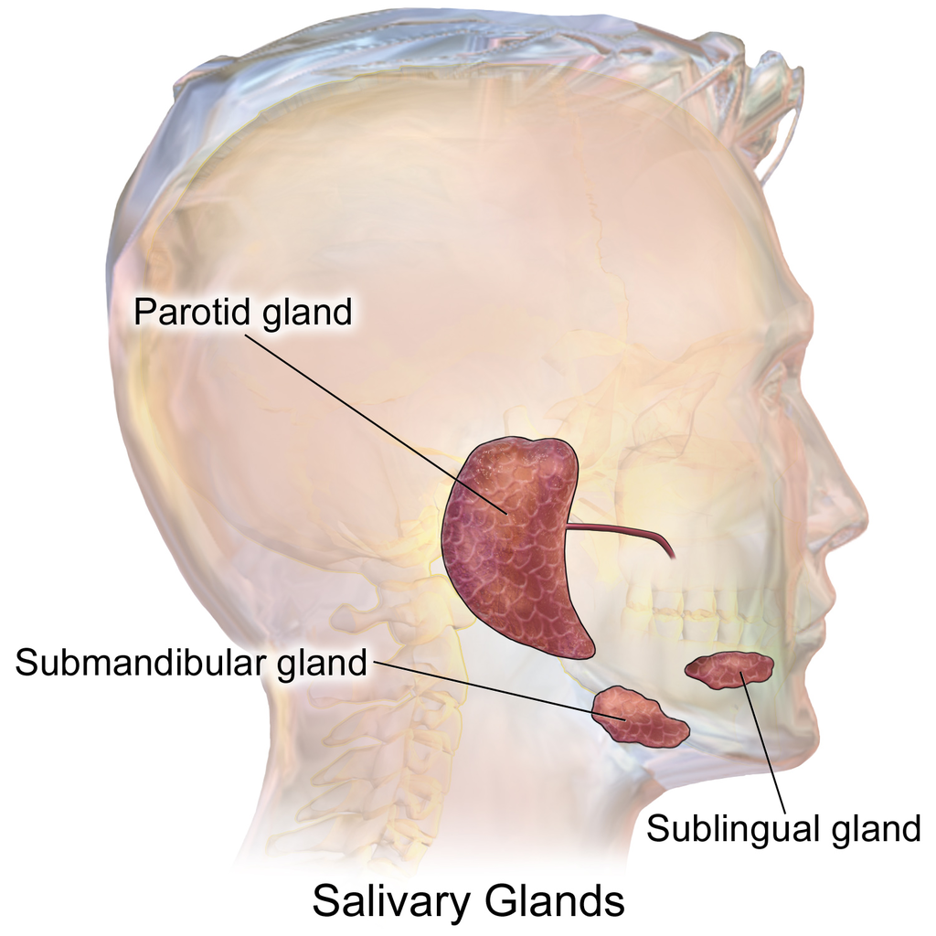
The mouth contains three pairs of major salivary glands, shown in Figure 15.4.2. These three pairs are all exocrine glands that secrete saliva into the mouth through ducts.
- The largest of the three major pairs of salivary glands are the parotid glands, which are located on either side of the mouth in front of the ears.
- The next largest pair is the submandibular glands, located beneath the lower jaw.
- The third pair is the sublingual glands, located underneath the tongue.
In addition to these three pairs of major salivary glands, there are also hundreds of minor salivary glands in the oral mucosa lining the mouth and on the tongue. Along with the major glands, most of the minor glands secrete the digestive enzyme amylase, which begins the chemical digestion of starch and glycogen (polysaccharides). However, the minor salivary glands on the tongue secrete the fat-digesting enzyme lipase, which in the mouth is called lingual lipase (to distinguish it from pancreatic lipase secreted by the pancreas).
Saliva secreted by the salivary glands mainly helps digestion, but it also plays other roles. It helps maintain dental health by cleaning the teeth, and it contains antibodies that help protect against infection. By keeping the mouth lubricated, saliva also allows the mouth movements needed for speech.
Tongue
The tongue is a fleshy, muscular organ that is attached to the floor of the mouth by a band of ligaments that gives it great mobility. This is necessary so the tongue can manipulate food for chewing and swallowing. Movements of the tongue are also necessary for speaking. The upper surface of the tongue is covered with tiny projections called papillae, which contain taste buds. The latter are collections of chemoreceptor cells (shown in Figure 15.4.3). These sensory cells sense chemicals in food and send the information to the brain via cranial nerves, thus enabling the sense of taste.
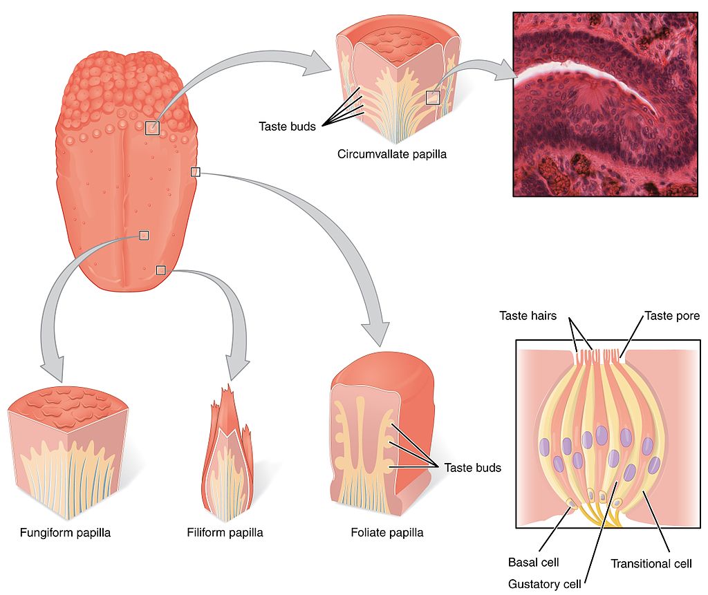
There are five basic tastes detected by the chemoreceptor cells in taste buds: saltiness, sourness, bitterness, sweetness, and umami (often described as a meaty taste). Contrary to popular belief, taste buds for the five basic tastes are not located on different parts of the tongue. Why does taste matter? The taste of food helps to stimulate the secretion of saliva from the salivary glands. It also helps us to eat foods that are good for us, instead of rotten or toxic foods. The detection of saltiness, for example, enables the control of salt intake and salt balance in the body. The detection of sourness may help us avoid spoiled foods, which often taste sour due to fermentation by bacteria. The detection of bitterness warns of poisons, because many plants defend themselves with toxins that taste bitter. The detection of sweetness guides us to foods that supply quick energy. The detection of umami may signal protein-rich foods.
Teeth
The teeth are complex structures made of a bone-like material called dentin and covered with enamel, which is the hardest tissue in the body. Adults normally have a total of 32 teeth, with 16 in each jaw. The right and left sides of each jaw are mirror images in terms of the numbers and types of teeth they contain. Teeth have different shapes to suit them for different aspects of mastication (chewing). The different types of teeth are illustrated in Figure 15.4.4.
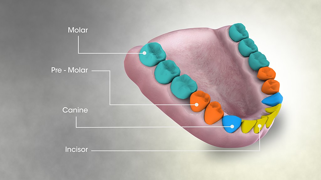
- Incisors are the sharp, blade-like teeth at the front of the mouth. They are used for cutting or biting off pieces of food. In adults, there are normally four incisors in each jaw, or eight in total.
- Canines are the pointed teeth on either side of the incisors. They are used for tearing foods that are tough or stringy. Adults normally have two canines in each jaw, or four altogether.
- Premolars and molars are cuboid teeth with cusps and grooves that are located on the sides and toward the back of the jaws. Premolars are closer to the front of the mouth. Molars are larger and have more cusps than premolars, but both are used for crushing and grinding food. Adults normally have two premolars and three molars on each side of each jaw, for a total of eight premolars and twelve molars.
Pharynx
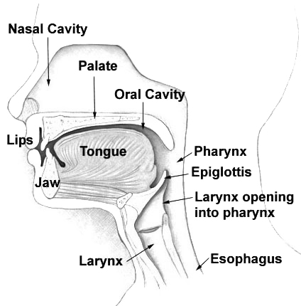
The tube-like pharynx (see Figure 15.4.5 below) plays a dual role as an organ of both respiration and digestion. As part of the respiratory system, it conducts air between the nasal cavity and larynx. As part of the digestive system, it allows swallowed food to pass from the oral cavity to the esophagus. Anything swallowed has priority over inhaled air when passing through the pharynx. During swallowing, the backward motion of the tongue causes a flap of elastic cartilage — called the epiglottis — to close over the opening to the larynx. This prevents food or drink from entering the larynx.
Esophagus
The esophagus (shown in Figure 15.4.6) is a muscular tube through which food is pushed from the pharynx to the stomach. The esophagus passes through an opening in the diaphragm (the large breathing muscle that separates the abdomen from the thorax) before reaching the stomach. In adults, the esophagus averages about 25 cm (about 9.8 inches) in length, depending on a person’s height. The inner lining of the esophagus consists of mucous membrane, which provides a smooth, slippery surface for the passage of food. The cells of this membrane are constantly being replaced as they are worn away from the frequent passage of food over them.
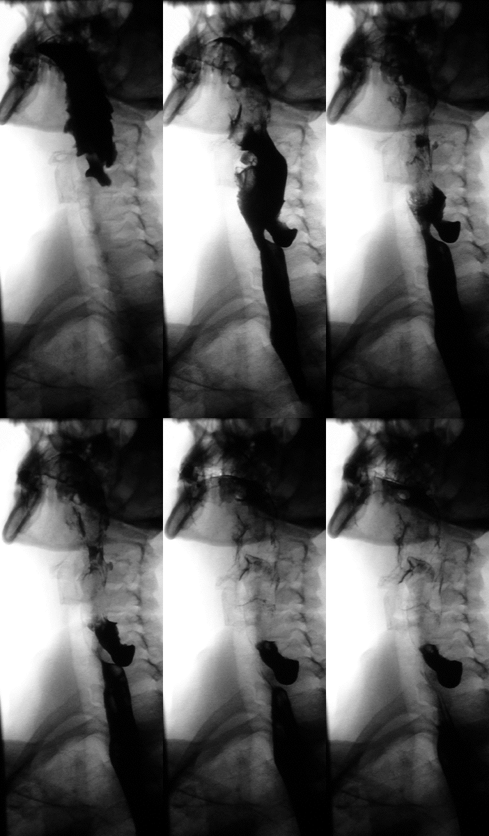
When food is not being swallowed, the esophagus is closed at both ends by upper and lower esophageal sphincters. Sphincters are rings of muscle that can contract to close off openings between structures. The upper esophageal sphincter is triggered to relax and open by the act of swallowing, allowing a bolus of food to enter the esophagus from the pharynx. Then, the esophageal sphincter closes again to prevent food from moving back into the pharynx from the esophagus.
Once in the esophagus, the food bolus travels down to the stomach, pushed along by the rhythmic contraction and relaxation of muscles (peristalsis). The lower esophageal sphincter is located at the junction between the esophagus and the stomach. This sphincter opens when the bolus reaches it, allowing the food to enter the stomach. The sphincter normally remains closed at other times to prevent the contents of the stomach from entering the esophagus. Failure of this sphincter to remain completely closed can lead to heartburn. If it happens chronically, it can lead to gastroesophageal reflux disease (GERD), in which the mucous membrane of the esophagus may become damaged by the highly acidic contents of the stomach.
See the video below to see how the parts of the upper GI tract work together to carry out swallowing:
https://youtu.be/pNcV6yAfq-g
Swallowing, uploaded by Alejandra Cork, 2012.
Stomach
The stomach is a J-shaped organ (shown in Figure 15.4.7) that is joined to the esophagus at its upper end, and to the first part of the small intestine (duodenum) at its lower end. When the stomach is empty of food, it normally has a volume of about 75 millilitres, but it can expand to hold up to about a litre of food. Waves of muscle contractions (peristalsis) passing through the muscular walls of the stomach cause the food inside to be mixed and churned. The wall of the stomach has an extra layer of muscle tissue not found in other organs of the GI tract that helps it squeeze and mix the food. These movements of the stomach wall contribute greatly to mechanical digestion by breaking the food into much smaller pieces. The churning also helps mix the food with stomach secretions that aid in its chemical digestion.
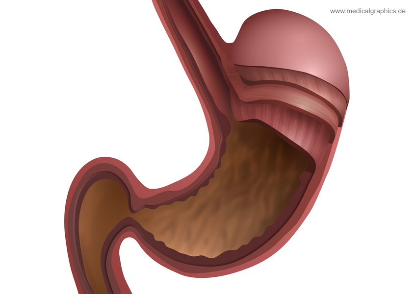
Secretions of the stomach include gastric acid, which consists mainly of hydrochloric acid (HCl). This makes the stomach contents highly acidic, which is necessary so that the enzyme pepsin — also secreted by the stomach — can begin the digestion of protein. Mucus is secreted by the lining of stomach to provide a slimy protective coating against the otherwise damaging effects of gastric acid. The fat-digesting enzyme lipase is secreted in small amounts in the stomach, but very little fat digestion occurs there.
By the time food has been in the stomach for about an hour, it has become the thick, semi-liquid chyme. When the small intestine is ready to receive chyme, a sphincter between the stomach and duodenum — called the pyloric sphincter — opens to allow the chyme to enter the small intestine for further digestion and absorption.
Feature: Reliable Sources
The ongoing epidemic of obesity in the wealthier nations of the world, including Canada, has led to the development of several different bariatric surgeries that modify the stomach to help obese patients reduce their food intake and lose weight. Go online to learn more about bariatric surgery. Find sources you judge to be reliable that answer the following questions:
- Who qualifies for bariatric surgery?
- Describe the bariatric surgeries commonly called stomach stapling, lap band, and gastric sleeve. How does each type of surgery modify the stomach? In terms of weight loss, how effective is each type?
- What are the major potential risks of bariatric surgery?
- Besides weight loss, what other benefits have been shown to result from bariatric surgery?
15.4 Summary
- Organs of the upper gastrointestinal (GI) tract include the mouth, pharynx, esophagus, and stomach.
- The mouth is the first organ of the GI tract. It has several structures that are specialized for digestion, including salivary glands, tongue, and teeth. Both mechanical digestion and chemical digestion of carbohydrates and fats begin in the mouth.
- The pharynx and esophagus move food from the mouth to the stomach, but are not involved in the process of digestion or absorption. Food moves through the esophagus by peristalsis.
- Mechanical and chemical digestion continue in the stomach. Acid and digestive enzymes secreted by the stomach start the chemical digestion of proteins. The stomach turns masticated food into a semi-fluid mixture called chyme.
15.4 Review Questions
-
- Identify structures in the mouth that are specialized for digestion.
- Describe digestion in the mouth.
- What general role do the pharynx and esophagus play in the digestion of food?
- How does food travel through the esophagus?
- Describe digestion in the stomach.
- Describe the differences between how air and food normally move past the pharynx.
- Name two structures in the mouth that contribute to mechanical digestion.
- What structure normally keeps stomach contents from backing up into the esophagus?
- Thirty minutes after you eat a meal, where is most of your food located? Explain your answer.
- What are two roles of mucus in the upper GI tract?
15.4 Explore More
https://youtu.be/zGoBFU1q4g0
What causes cavities? - Mel Rosenberg, TED-Ed, 2016.
https://youtu.be/gCrmFbgT37I
How does alcohol make you drunk? - Judy Grisel, TED-Ed, 2020.
https://youtu.be/twJBEypJDfU
Gastric Bypass Surgery: One Patient’s Journey - Mayo Clinic, 2014.
https://youtu.be/u_1sVri3b2w
Here's What Happens In Your Body When You Swallow Gum | The Human Body, Tech Insider, 2018.
Attributions
Figure 15.4.1
Handstand, Pender Island, B.C. [photo] by Jasper Garratt on Unsplash is used under the Unsplash License (https://unsplash.com/license).
Figure 15.4.2
Blausen_0780_SalivaryGlands by BruceBlaus on Wikimedia Commons is used under a CC BY 3.0 (https://creativecommons.org/licenses/by/3.0) license.
Figure 15.4.3
1402_The_Tongue by OpenStax on Wikimedia Commons is used under a CC BY 4.0 (https://creativecommons.org/licenses/by/4.0) license.
Figure 15.4.4
1024px-3D_Medical_Animation_Still_Showing_Types_of_Teeth by http://www.scientificanimations.com on Wikimedia Commons is used under a CC BY-SA 4.0 (https://creativecommons.org/licenses/by-sa/4.0) license.
Figure 15.4.5
Illu01_head_neck by Arcadian from NCI/ SEER Training Modules on Wikimedia Common is in the public domain (https://en.wikipedia.org/wiki/public_domain).
Figure 15.4.6
ZenkerSchraeg by Bernd Brägelmann Braegel on Wikimedia Commons is used under a CC BY 3.0 (https://creativecommons.org/licenses/by/3.0) license. (Courtesy of Dr. Martin Steinhoff. It is not known whether there is a possibly necessary approval from the patient.)
Figure 15.4.7
Anatomy stomach – white by www.medicalgraphics.de from MedicalGraphics is used under a CC BY-ND 4.0 (https://creativecommons.org/licenses/by-nd/4.0/) license.
References
Alejandra Cork. (2012). Swallowing. YouTube. https://www.youtube.com/watch?v=pNcV6yAfq-g&t=4s
Betts, J. G., Young, K.A., Wise, J.A., Johnson, E., Poe, B., Kruse, D.H., Korol, O., Johnson, J.E., Womble, M., DeSaix, P. (2016, May 27). Figure 14.3 The tongue [digital image]. In Anatomy and Physiology (Section 14.1). OpenStax. https://openstax.org/books/anatomy-and-physiology/pages/14-1-sensory-perception
Blausen.com Staff. (2014). Medical gallery of Blausen Medical 2014. WikiJournal of Medicine 1 (2). DOI:10.15347/wjm/2014.010. ISSN 2002-4436.
Mayo Clinic. (2014, August 26). Gastric bypass surgery: One patient’s journey - Mayo Clinic. https://www.youtube.com/watch?v=twJBEypJDfU&feature=youtu.be
Mayo Clinic Staff. (n.d.). Gastroesophageal reflux disease (GERD) [online article]. MayoClinic.org. https://www.mayoclinic.org/diseases-conditions/gerd/symptoms-causes/syc-20361940
Tech Insider. (2018, March 20). Here's what happens in your body when you swallow gum | The human body. YouTube. https://www.youtube.com/watch?v=u_1sVri3b2w&feature=youtu.be
TED-Ed. (2020, April 9). How does alcohol make you drunk? - Judy Grisel. YouTube. https://www.youtube.com/watch?v=gCrmFbgT37I&feature=youtu.be
TED-Ed. (2016, October 17). What causes cavities? - Mel Rosenberg. YouTube. https://www.youtube.com/watch?v=zGoBFU1q4g0&feature=youtu.be
Image shows a diagram of the cross sectional layering of heart tissues. As described in the preceding paragraph, the endocardium is a thin layer on the inside of the heart chambers, and myocardium is an extremely thick layer of cardiac muscle, and the pericardium consists of two membranes separated by fluid. The visceral pericardium envelopes the myocardium and the parietal pericardium envelopes the entire heart structure.
Image shows a flowchart of the path of blood through the heart. Beginning at the lungs, oxygenated blood enters the pulmonary veins, then enters the left atrium. Blood then passes through the bicuspid AV valve to enter the left ventricle, which contracts to send blood through the aortic semilunar valve into the aorta. The aorta connects to arteries of the systemic circuit and ultimately deliver oxygen to body tissues. From here, blood is deoxygenated and returns to the heart via veins which merge to form the two vena cava. The vena cava delivers blood to the right atrium, which contracts to send blood through the tricuspid AV valve into the right ventricle, which contracts to send blood through the pulmonary semilunar valve into the pulmonary arteries and to the lungs for re-oxygenation.
Created by CK-12 Foundation/Adapted by Christine Miller
Case Study Conclusion: Cough That Won't Quit
Inhaling the moist air from a humidifier or steamy shower can feel particularly good if you have a respiratory system infection, such as bronchitis. The moist air helps to loosen and thin mucus in the respiratory system, allowing you to breathe easier.
In the beginning of this chapter, you learned about Erica, who developed acute bronchitis after getting a cold. She had a worsening cough, a sore throat due to coughing, and chest congestion. She was also coughing up thick mucus.
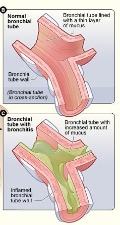
Acute bronchitis usually occurs after a cold or flu, usually due to the same viruses that cause cold or flu. Because bronchitis is not usually caused by bacteria (although it can be), in most cases, antibiotics are not an effective treatment.
Bronchitis affects the bronchial tubes, which, as you have learned, are air passages in the lower respiratory tract. The main bronchi branch off of the trachea and then branch into smaller bronchi, and then bronchioles. In bronchitis, the walls of the bronchi become inflamed, which makes them narrower. There is also excessive production of mucus in the bronchi, which further narrows the pathway where air can flow through. Figure 13.7.2, shows how bronchitis affects the bronchial tubes.
The treatment for most cases of bronchitis involves thinning and loosening the mucus so that it can be effectively coughed out of the airways. This can be done by drinking plenty of fluids, using humidifiers or steam, and — in some cases — using over-the-counter medications (such as expectorants). Dr. Choo recommended some of these treatments to Erica, and also warned against using cough suppressants. Cough suppressants work on the nervous system to suppress the cough reflex. When a patient has a “productive” cough (which means they are coughing up mucus), doctors generally advise them not to take cough suppressants, so that they can cough the mucus out of their bodies.
When Dr. Choo was examining Erica, she used a pulse oximeter to measure the oxygen level in her blood. Why did she do this? As you have learned, the bronchial tubes branch into bronchioles, which ultimately branch into the alveoli of the lungs. The alveoli are where gas exchange occurs between the air and the blood to take in oxygen and remove carbon dioxide and other wastes. By checking Erica’s blood oxygen level, Dr. Choo was making sure that her clogged airways were not impacting her level of much-needed oxygen.
Erica has acute bronchitis, but you may recall that chronic bronchitis was discussed earlier in this chapter (Section 13.5) as a term that describes the symptoms of chronic obstructive pulmonary disease (COPD). COPD is often due to tobacco smoking, and it causes damage to the walls of the alveoli. Acute bronchitis, on the other hand, typically occurs after a cold or flu, and involves inflammation and mucus build-up in the bronchial tubes. As implied by the difference in their names, chronic bronchitis is an ongoing, long-term condition, while acute bronchitis is likely to resolve relatively quickly with proper rest and treatment.
Erica uses e-cigarettes (vaping), so she is more likely to develop chronic respiratory conditions, such as COPD. As you have learned, smoking damages the respiratory system, along with many other systems of the body. Smoking and vaping increases the risk of respiratory infections, including bronchitis and flu, due to its damaging effects on the respiratory and immune systems. Dr. Choo strongly encouraged Erica to quit vaping, not only so that her acute bronchitis resolves, but so that she can avoid future infections and other negative health outcomes associated with vaping and smoking, including COPD and lung cancer.
As you have learned in this chapter, the respiratory system is critical to carry out the gas exchange necessary for life’s functions, and to protect the body from pathogens and other potentially harmful substances in the air. But this ability to interface with the outside air has a cost. The respiratory system is prone to infections, as well as damage and other negative effects from allergens, mold, air pollution, cigarette smoke and vaping. While exposure to most of these things cannot be avoided, not smoking is an important step you can take to protect this organ system — as well as many other systems of your body.
Chapter 13 Summary
In this chapter, you learned about the respiratory system. Specifically, you learned that:
- Respiration is the process in which oxygen moves from the outside air into the body, and carbon dioxide and other waste gases move from inside the body to the outside air. It involves two subsidiary processes: ventilation and gas exchange.
- The organs of the respiratory system form a continuous system of passages, called the respiratory tract. It has two major divisions: the upper respiratory tract and the lower respiratory tract.
-
- The upper respiratory tract includes the nasal cavity, pharynx, and larynx. All of these organs are involved in conduction, or the movement of air into and out of the body. Incoming air is also cleaned, humidified, and warmed as it passes through the upper respiratory tract. The larynx is also called the voice box, because it contains the vocal cords, which are needed to produce vocal sounds.
- The lower respiratory tract includes the trachea, bronchi and bronchioles, and the lungs. The trachea, bronchi, and bronchioles are involved in conduction. Gas exchange takes place only in the lungs, which are the largest organs of the respiratory tract. Lung tissue consists mainly of tiny air sacs called alveoli, which is where gas exchange takes place between air in the alveoli and the blood in capillaries surrounding them.
- The respiratory system protects itself from potentially harmful substances in the air by the mucociliary escalator. This includes mucus-producing cells, which trap particles and pathogens in incoming air. It also includes tiny hair-like cilia that continually move to sweep the mucus and trapped debris away from the lungs and toward the outside of the body.
- The level of carbon dioxide in the blood is monitored by cells in the brain. If the level becomes too high, it triggers a faster rate of breathing, which lowers the level to the normal range. The opposite occurs if the level becomes too low. The respiratory system exchanges gases with the outside air, but it needs the cardiovascular system to carry the gases to and from cells throughout the body.
- Breathing, or ventilation, is the two-step process of drawing air into the lungs (inhalation) and letting air out of the lungs (exhalation). Inhaling is an active process that results mainly from contraction of a muscle called the diaphragm. Exhaling is typically a passive process that occurs mainly due to the elasticity of the lungs when the diaphragm relaxes.
-
- Breathing is one of the few vital bodily functions that can be controlled consciously, as well as unconsciously. Conscious control of breathing is common in many activities, including swimming and singing. However, there are limits on the conscious control of breathing. If you try to hold your breath, for example, you will soon have an irrepressible urge to breathe.
- Unconscious breathing is controlled by respiratory centers in the medulla and pons of the brainstem. They respond to variations in blood pH by either increasing or decreasing the rate of breathing as needed to return the pH level to the normal range.
- Nasal breathing is generally considered to be superior to mouth breathing, because it does a better job of filtering, warming, and moistening incoming air. It also results in slower emptying of the lungs, which allows more oxygen to be extracted from the air.
- Gas exchange is the biological process through which gases are transferred across cell membranes to either enter or leave the blood. Gas exchange takes place continuously between the blood and cells throughout the body, and also between the blood and the air inside the lungs.
-
- Gas exchange in the lungs takes place in alveoli. The pulmonary artery carries deoxygenated blood from the heart to the lungs, where it travels through pulmonary capillaries, picking up oxygen and releasing carbon dioxide. The oxygenated blood then leaves the lungs through pulmonary veins.
- Gas exchange occurs by diffusion across cell membranes. Gas molecules naturally move down a concentration gradient from an area of higher concentration to an area of lower concentration. This is a passive process that requires no energy.
- Gas exchange by diffusion depends on the large surface area provided by the hundreds of millions of alveoli in the lungs. It also depends on a steep concentration gradient for oxygen and carbon dioxide. This gradient is maintained by continuous blood flow and constant breathing.
- Asthma is a chronic inflammatory disease of the airways in the lungs, in which the airways periodically become inflamed. This causes swelling and narrowing of the airways, often with excessive mucus production, leading to difficulty breathing and other symptoms. Asthma is thought to be caused by a combination of genetic and environmental factors. Asthma attacks are triggered by allergens, air pollution, or other factors.
- Pneumonia is a common inflammatory disease of the respiratory tract in which inflammation affects primarily the alveoli, which become filled with fluid that inhibits gas exchange. Most cases of pneumonia are caused by viral or bacterial infections. Vaccines are available to prevent pneumonia. Treatment often includes prescription antibiotics.
- Chronic obstructive pulmonary disease (COPD) is a lung disease characterized by chronic poor airflow, which causes shortness of breath and a productive cough. It is caused most often by tobacco smoking, which leads to breakdown of connective tissues in the lungs. Alveoli are reduced in number and elasticity, making it impossible to fully exhale air from the lungs. There is no cure for COPD, but stopping smoking may reduce the rate at which COPD worsens.
- Lung cancer is a malignant tumor characterized by uncontrolled cell growth in tissues of the lung. It results from accumulated DNA damage, most often caused by tobacco smoking. Lung cancer is typically diagnosed late, so most cases cannot be cured. It may be treated with surgery, chemotherapy, and/or radiation therapy.
- Smoking is the single greatest cause of preventable death worldwide. It has adverse effects on just about every body system and organ. Tobacco smoke affects not only smokers, but also non-smokers who are exposed to secondhand smoke. The nicotine in tobacco is highly addictive, making it very difficult to quit smoking.
-
- A major health risk of smoking is lung cancer. Smoking also increases the risk of many other types of cancer. Tobacco smoke contains dozens of chemicals that are known carcinogens.
- Smoking is the primary cause of COPD. Chemicals — such as carbon monoxide and cyanide in tobacco smoke — reduce the elasticity of alveoli so the lungs can no longer fully exhale air.
- Smoking and/or vaping damages the cardiovascular system and increases the risk of high blood pressure, blood clots, heart attack, and stroke. Smoking also has a negative impact on blood lipid levels.
- A wide diversity of additional adverse health effects — such as erectile dysfunction, female infertility, and slow wound healing — are attributable to smoking.
As you have learned, the respiratory system brings in oxygen to the body and removes waste gases to the atmosphere — but these molecules wouldn’t get to where they need to go without the cardiovascular system to transport them via the bloodstream. Read the next chapter to learn about how the cardiovascular system carries out these critical functions.
Chapter 13 Review
-
- Describe the relationship between the bronchi, secondary bronchi, tertiary bronchi, and bronchioles.
- Deoxygenated and oxygenated blood both travel to the lungs. Describe what happens to that blood when it gets to the lungs.
- Explain the difference between ventilation and gas exchange.
- Which way do oxygen and carbon dioxide flow during gas exchange in the lungs, and why? Which way do oxygen and carbon dioxide flow during gas exchange between the blood and the body’s cells, and why?
- Why does the body require oxygen, and why does it emit carbon dioxide as a waste product?
- What do coughing and sneezing have in common?
- COPD can cause too much carbon dioxide in the blood. Answer the following questions about this:
- How does COPD cause there to be too much carbon dioxide in the blood?
- What does this do to the blood pH?
- How does the body respond to this change in blood pH?
- What are three different types of things that can enter the respiratory system and cause illness or injury? Describe the negative health effects of each in your answer.
- Where are the respiratory centers of the brain located? What is the main function of the respiratory centers of the brain?
- Smoking increases the risk of getting influenza, commonly known as the flu. Explain why this could lead to a greater risk of pneumonia.
- If a person has a gene that caused them to get asthma, could changes to their environment (such as more frequent cleaning) help their asthma? Why or why not?
- Explain why nasal breathing generally stops particles from entering the body at an earlier stage than mouth breathing does.
Attributions
Figure 13.7.1
Tags: Essential Oils Aroma Diffuser Diffuse Led by asundermeier on Pixabay is used under the Pixabay License (https://unsplash.com/license).
Figure 13.7.2
Bronchitis by National Heart Lung and Blood Institute on Wikimedia Commons is in the public domain (https://en.wikipedia.org/wiki/Public_domain).
Created by CK-12 Foundation/Adapted by Christine Miller

Work Those Eye Muscles!
Imagine the man in Figure 12.3.1 turns his eyes in your direction. This is a very small movement, considering the conspicuously large and strong external eye muscles that control eyeball movements. These muscles have been called the strongest muscles in the human body relative to the work they do. However, the external eye muscles actually do a surprising amount of work. Eye movements occur almost constantly during waking hours, especially when we are scanning faces or reading. Eye muscles are also exercised nightly during the phase of sleep called rapid eye movement sleep. External eye muscles can move the eyes because they are made mainly of muscle tissue.
What is Muscle Tissue?
Muscle tissue is a soft tissue that makes up most of the tissues in the muscles of the human muscular system. Other tissues in muscles are connective tissues, such as tendons that attach skeletal muscles to bones and sheaths of connective tissues that cover or line muscle tissues. Only muscle tissue per se, has cells with the ability to contract.
There are three major types of muscle tissues in the human body: skeletal, smooth, and cardiac muscle tissues. Figure 12.3.2 shows how the three types of muscle tissues appear under magnification. When you read about each type below, you will learn why the three types appear as they do.
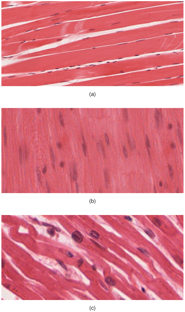
Skeletal Muscle Tissue
Skeletal muscle is muscle tissue that is attached to bones by tendons, which are bundles of collagen fibres. Whether you are moving your eyes or running a marathon, you are using skeletal muscles. Contractions of skeletal muscles are voluntary, or under conscious control of the central nervous system via the somatic nervous system. Skeletal muscle tissue is the most common type of muscle tissue in the human body. By weight, an average adult male is about 42% skeletal muscles, and the average adult female is about 36% skeletal muscles. Some of the major skeletal muscles in the human body are labeled in Figure 12.3.3 below.
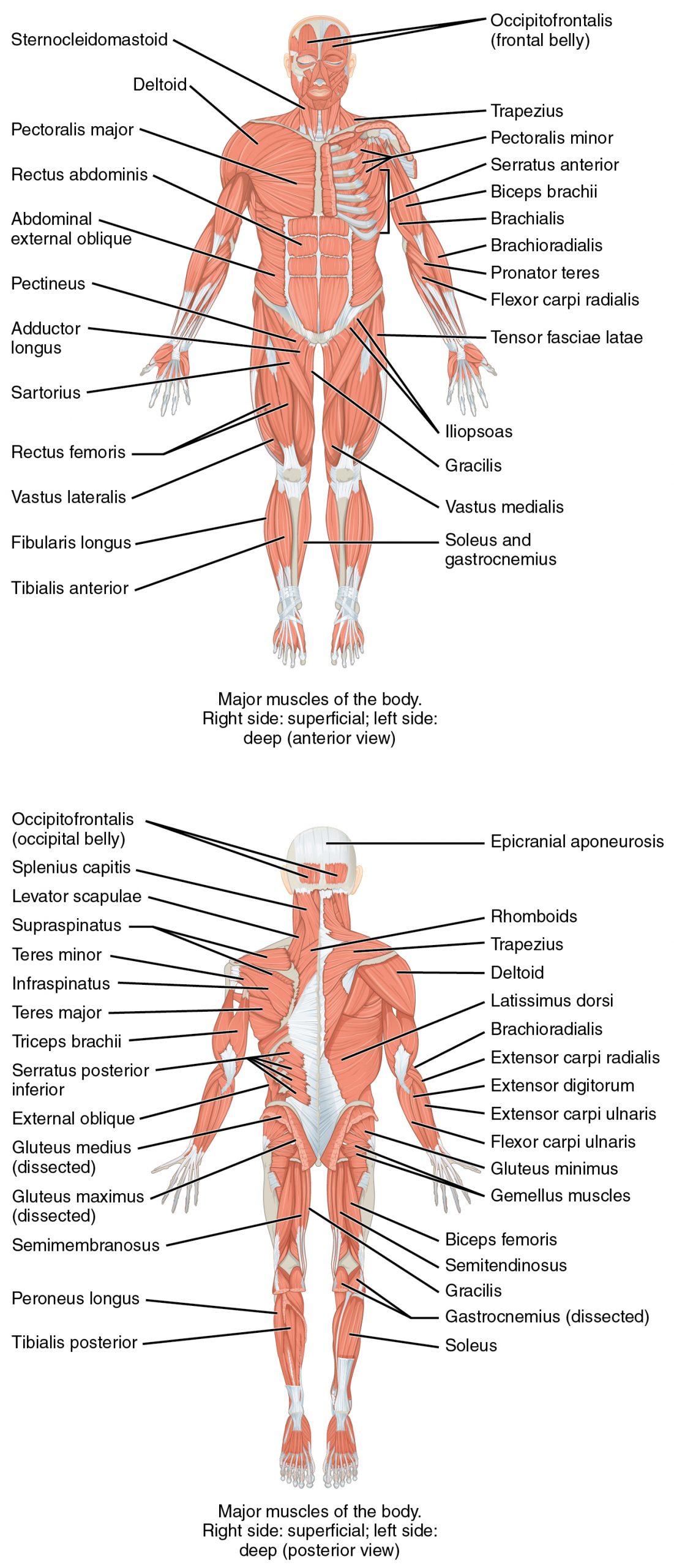
Skeletal Muscle Pairs
To move bones in opposite directions, skeletal muscles often consist of muscle pairs that work in opposition to one another, also called antagonistic muscle pairs. For example, when the biceps muscle (on the front of the upper arm) contracts, it can cause the elbow joint to flex or bend the arm, as shown in Figure 12.3.4. When the triceps muscle (on the back of the upper arm) contracts, it can cause the elbow to extend or straighten the arm. The biceps and triceps muscles, also shown in Figure 12.3.4, are an example of a muscle pair where the muscles work in opposition to each other.
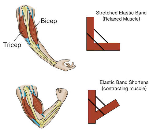
Skeletal Muscle Structure
Each skeletal muscle consists of hundreds — or even thousands — of skeletal muscle fibres, which are long, string-like cells. As shown in Figure 12.3.5 below, skeletal muscle fibres are individually wrapped in connective tissue called endomysium. The skeletal muscle fibres are bundled together in units called muscle fascicles, which are surrounded by sheaths of connective tissue called perimysium. Each fascicle contains between ten and 100 (or even more!) skeletal muscle fibres. Fascicles, in turn, are bundled together to form individual skeletal muscles, which are wrapped in connective tissue called epimysium. The connective tissues in skeletal muscles have a variety of functions. They support and protect muscle fibres, allowing them to withstand the forces of contraction by distributing the forces applied to the muscle. They also provide pathways for nerves and blood vessels to reach the muscles. In addition, the epimysium anchors the muscles to tendons.
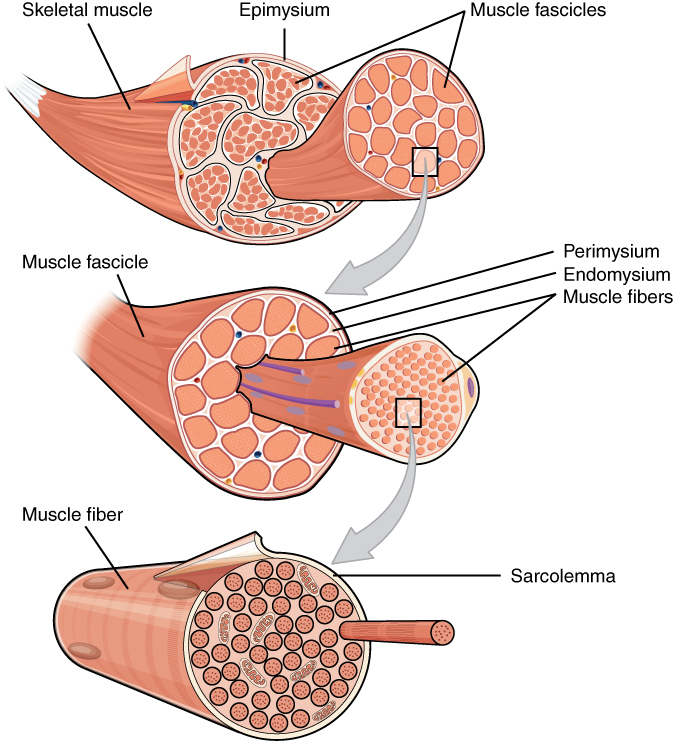
The same bundles-within-bundles structure is replicated within each muscle fibre. As shown in Figure 12.3.6, a muscle fibre consists of a bundle of myofibrils, which are themselves bundles of protein filaments. These protein filaments consist of thin filaments of the protein actin, which are anchored to structures called Z discs, and thick filaments of the protein myosin. The filaments are arranged together within a myofibril in repeating units called sarcomeres, which run from one Z disc to the next. The sarcomere is the basic functional unit of skeletal and cardiac muscles. It contracts as actin and myosin filaments slide over one another. Skeletal muscle tissue is said to be striated, because it appears striped. It has this appearance because of the regular, alternating A (dark) and I (light) bands of filaments arranged in sarcomeres inside the muscle fibres. Other components of a skeletal muscle fibre include multiple nuclei and mitochondria.
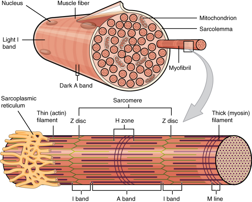
Slow- and Fast-Twitch Skeletal Muscle Fibres
Skeletal muscle fibres can be divided into two types, called slow-twitch (or type I) muscle fibres and fast-twitch (or type II) muscle fibres.
- Slow-twitch muscle fibres are dense with capillaries and rich in mitochondria and myoglobin, which is a protein that stores oxygen until needed for muscle activity. Relative to fast-twitch fibres, slow-twitch fibres can carry more oxygen and sustain aerobic (oxygen-using) activity. Slow-twitch fibres can contract for long periods of time, but not with very much force. They are relied upon primarily in endurance events, such as distance running or cycling.
- Fast-twitch muscle fibres contain fewer capillaries and mitochondria and less myoglobin. This type of muscle fibre can contract rapidly and powerfully, but it fatigues very quickly. Fast-twitch fibres can sustain only short, anaerobic (non-oxygen-using) bursts of activity. Relative to slow-twitch fibres, fast-twitch fibres contribute more to muscle strength and have a greater potential for increasing in mass. They are relied upon primarily in short, strenuous events, such as sprinting or weightlifting.
Proportions of fibre types vary considerably from muscle to muscle and from person to person. Individuals may be genetically predisposed to have a larger percentage of one type of muscle fibre than the other. Generally, an individual who has more slow-twitch fibres is better suited for activities requiring endurance, whereas an individual who has more fast-twitch fibres is better suited for activities requiring short bursts of power.
Smooth Muscle
Smooth muscle is muscle tissue in the walls of internal organs and other internal structures such as blood vessels. When smooth muscles contract, they help the organs and vessels carry out their functions. When smooth muscles in the stomach wall contract, for example, they squeeze the food inside the stomach, helping to mix and churn the food and break it into smaller pieces. This is an important part of digestion. Contractions of smooth muscles are involuntary, so they are not under conscious control. Instead, they are controlled by the autonomic nervous system, hormones, neurotransmitters, and other physiological factors.
Structure of Smooth Muscle
The cells that make up smooth muscle are generally called myocytes. Unlike the muscle fibres of striated muscle tissue, the myocytes of smooth muscle tissue do not have their filaments arranged in sarcomeres. Therefore, smooth tissue is not striated. However, the myocytes of smooth muscle do contain myofibrils, which in turn contain bundles of myosin and actin filaments. The filaments cause contractions when they slide over each other, as shown in Figure 12.3.7.

Functions of Smooth Muscle
Unlike striated muscle, smooth muscle can sustain very long-term contractions. Smooth muscle can also stretch and still maintain its contractile function, which striated muscle cannot. The elasticity of smooth muscle is enhanced by an extracellular matrix secreted by myocytes. The matrix consists of elastin, collagen, and other stretchy fibres. The ability to stretch and still contract is an important attribute of smooth muscle in organs such as the stomach and uterus (see Figures 12.3.8 and 12.3.9), both of which must stretch considerably as they perform their normal functions.
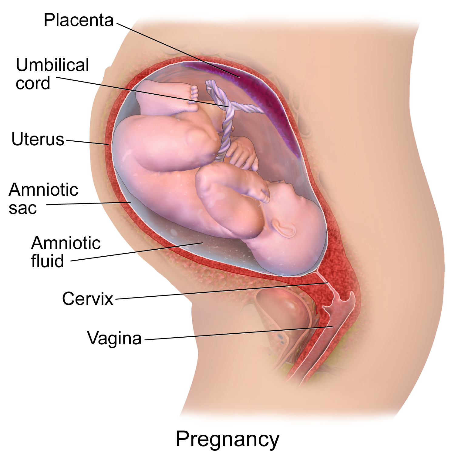 |
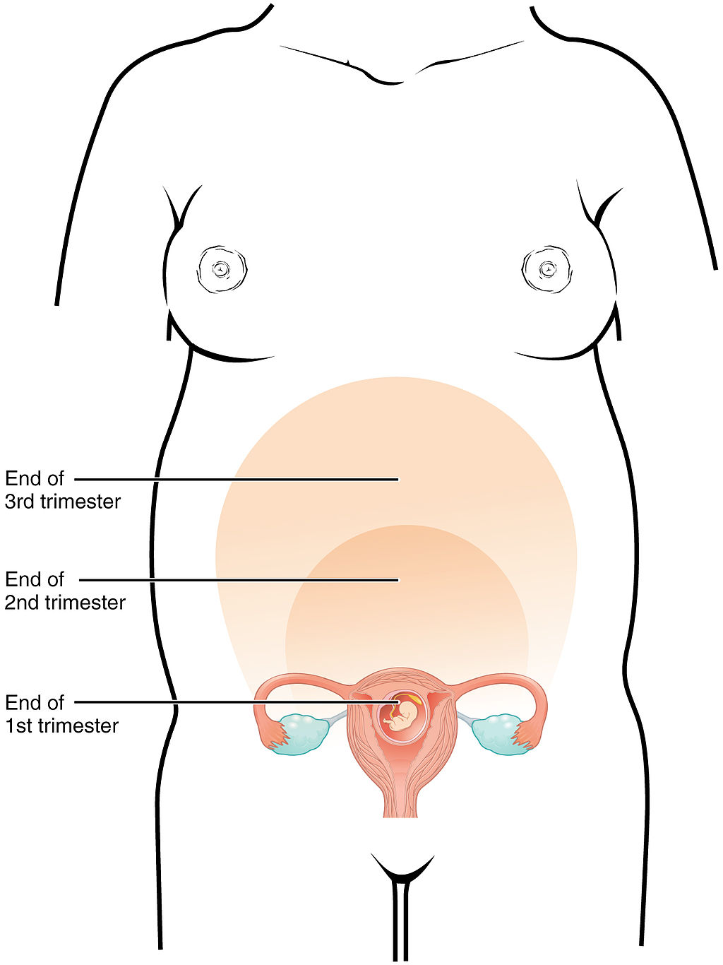 |
The following list indicates where many smooth muscles are found, along with some of their specific functions.
- Walls of organs of the gastrointestinal tract (such as the esophagus, stomach, and intestines), moving food through the tract by peristalsis
- Walls of air passages of the respiratory tract (such as the bronchi), controlling the diameter of the passages and the volume of air that can pass through them
- Walls of organs of the male and female reproductive tracts; in the uterus, for example, pushing a baby out of the uterus and into the birth canal
- Walls of structures of the urinary system, including the urinary bladder, allowing the bladder to expand so it can hold more urine, and then contract as urine is released
- Walls of blood vessels, controlling the diameter of the vessels and thereby affecting blood flow and blood pressure
- Walls of lymphatic vessels, squeezing the fluid called lymph through the vessels
- Iris of the eyes, controlling the size of the pupils and thereby the amount of light entering the eyes
- Arrector pili in the skin, raising hairs in hair follicles in the dermis
Cardiac Muscle
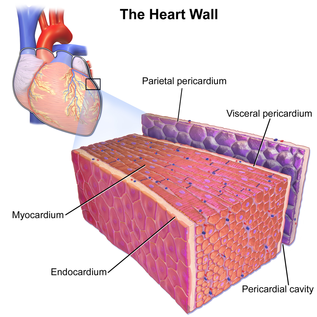
Cardiac muscle is found only in the wall of the heart. It is also called myocardium. As shown in Figure 12.3.10, myocardium is enclosed within connective tissues, including the endocardium on the inside of the heart and pericardium on the outside of the heart. When cardiac muscle contracts, the heart beats and pumps blood. Contractions of cardiac muscle are involuntary, like those of smooth muscles. They are controlled by electrical impulses from specialized cardiac muscle cells in an area of the heart muscle called the sinoatrial node.
Like skeletal muscle, cardiac muscle is striated because its filaments are arranged in sarcomeres inside the muscle fibres. However, in cardiac muscle, the myofibrils are branched at irregular angles rather than arranged in parallel rows (as they are in skeletal muscle). This explains why cardiac and skeletal muscle tissues look different from one another.
The cells of cardiac muscle tissue are arranged in interconnected networks. This arrangement allows rapid transmission of electrical impulses, which stimulate virtually simultaneous contractions of the cells. This enables the cells to coordinate contractions of the heart muscle.
The heart is the muscle that performs the greatest amount of physical work in the course of a lifetime. Although the power output of the heart is much less than the maximum power output of some other muscles in the human body, the heart does its work continuously over an entire lifetime without rest. Cardiac muscle contains a great many mitochondria, which produce ATP for energy and help the heart resist fatigue.
Feature: Human Biology in the News
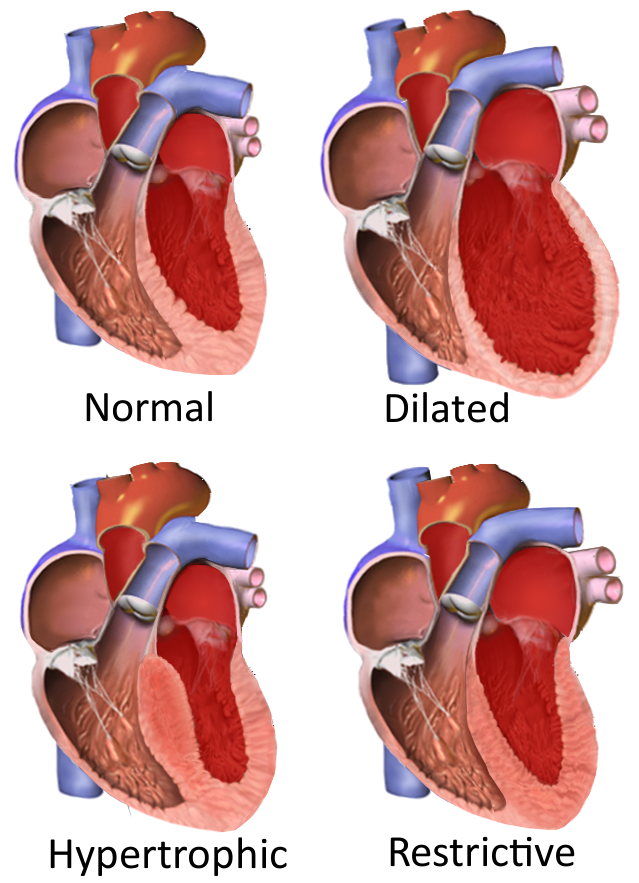
Cardiomyopathy is a disease in which the muscles of the heart are no longer able to effectively pump blood to the body — extreme forms of this disease can lead to heart failure. There are four main types of cardiomyopathy (also illustrated in Figure 12.3.11):
- Dilated (congestive) cardiomyopathy: the left ventricle (the chamber itself) of the heart becomes enlarged and can't pump blood our to the body. This is normally related to coronary artery disease and/or heart attack
- Hypertrophic cardiomyopathy: abnormal thickening of the muscular walls of the left ventricle make the chamber less able to work properly. This condition is more common in patients with a family history of the disease.
- Restrictive cardiomyopathy: the myocardium becomes abnormally rigid and inelastic and is unable to expand in between heartbeats to refill with blood. Restrictive cardiomyopathy typically affects older people.
- Arrhythmogenic right ventricular cardiomyopathy: the right ventricular muscle is replaced by adipose or scar tissue, reducing elasticity and interfering with normal heartbeat and rhythm. This disease is often caused by genetic mutations.
Cardiomyopathy is typically diagnosed with a physical exam supplemented by medical and family history, an angiogram, blood tests, chest x-rays and electrocardiograms. In some cases your doctor would also requisition a CT scan and/or genetic testing.
When treating cardiomyopathy, the goal is to reduce symptoms that affect everyday life. Certain medications can help regularize and slow heart rate, decrease chances of blood clots and cause vasodilation in the coronary arteries. If medication is not sufficient to manage symptoms, a pacemaker or even a heart transplant may be the best option. Lifestyle can also help manage the symptoms of cardiomyopathy — people living with this disease are encouraged to avoid drug and alcohol use, control high blood pressure, eat a healthy diet, get ample rest and exercise, as well as reduce stress levels.
12.3 Summary
- Muscle tissue is a soft tissue that makes up most of the tissues in the muscles of the human muscular system. It is the only type of tissue that has cells with the ability to contract.
- Skeletal muscle tissue is attached to bones by tendons. It allows voluntary body movements.
- Skeletal muscle is the most common type of muscle tissue in the human body. To move bones in opposite directions, skeletal muscles often consist of pairs of muscles that work in opposition to one another to move bones in different directions at joints.
- Skeletal muscle fibres are bundled together in units called muscle fascicles, which are bundled together to form individual skeletal muscles. Skeletal muscles also have connective tissue supporting and protecting the muscle tissue.
- Each skeletal muscle fibre consists of a bundle of myofibrils, which are bundles of protein filaments. The filaments are arranged in repeating units called sarcomeres, which are the basic functional units of skeletal muscles. Skeletal muscle tissue is striated because of the pattern of sarcomeres in its fibres.
- Skeletal muscle fibres can be divided into two types, called slow-twitch and fast-twitch fibres. Slow-twitch fibres are used mainly in aerobic endurance activities, such as long-distance running. Fast-twitch fibres are used mainly for non-aerobic, strenuous activities, such as sprinting. Proportions of the two types of fibres vary from muscle to muscle and person to person.
- Smooth muscle tissue is found in the walls of internal organs and vessels. When smooth muscles contract, they help the organs and vessels carry out their functions. Contractions of smooth muscles are involuntary and controlled by the autonomic nervous system, hormones, and other substances.
- Cells of smooth muscle tissue are not striated because they lack sarcomeres, but the cells contract in the same basic way as striated muscle cells. Unlike striated muscle, smooth muscle can sustain very long-term contractions and maintain its contractile function, even when stretched.
- Cardiac muscle tissue is found only in the wall of the heart. When cardiac muscle contracts, the heart beats and pumps blood. Contractions of cardiac muscle are involuntary, like those of smooth muscles. They are controlled by electrical impulses from specialized cardiac cells.
- Like skeletal muscle, cardiac muscle is striated because its filaments are arranged in sarcomeres inside the muscle fibres. However, the myofibrils are branched instead of arranged in parallel rows, making cardiac and skeletal muscle tissues look different from one another.
- The heart is the muscle that performs the greatest amount of physical work in the course of a lifetime. Its cells contain a great many mitochondria to produce ATP for energy and help the heart resist fatigue.
12.3 Review Questions
- What is muscle tissue?
-
-
- Where is skeletal muscle found, and what is its general function?
- Why do many skeletal muscles work in pairs?
- Describe the structure of a skeletal muscle.
- Relate muscle fibre structure to the functional units of muscles.
- Why is skeletal muscle tissue striated?
- Where is smooth muscle found? What controls the contraction of smooth muscle?
- Where is cardiac muscle found? What controls its contractions?
- The heart muscle is smaller and less powerful than some other muscles in the body. Why is the heart the muscle that performs the greatest amount of physical work in the course of a lifetime? How does the heart resist fatigue?
- Give one example of connective tissue that is found in muscles. Describe one of its functions.
12.3 Explore More
https://www.youtube.com/watch?v=3_PYnWVoUzM
What happens during a heart attack? - Krishna Sudhir, TED-Ed, 2017.
https://www.youtube.com/watch?v=bwOE1MEginA&feature=emb_logo
Three types of muscle | Circulatory system physiology | NCLEX-RN | KhanAcademyMedicine, 2012.
Attributions
Figure 12.3.1
Look by ali-yahya-155huuQwGvA [photo] by Ali Yahya on Unsplash is used under the Unsplash License (https://unsplash.com/license).
Figure 12.3.2
Skeletal_Smooth_Cardiac by OpenStax College on Wikimedia Commons is used under a CC BY 3.0 (https://creativecommons.org/licenses/by/3.0) license.
Figure 12.3.3
Anterior_and_Posterior_Views_of_Muscles by OpenStax on Wikimedia Commons is used under a CC BY 4.0 (https://creativecommons.org/licenses/by/4.0) license.
Figure 12.3.4
Antagonistic Muscle Pair by Laura Guerin at CK-12 Foundation on Wikimedia Commons is used under a CC BY-NC 3.0 (https://creativecommons.org/licenses/by-nc/3.0/) license.
Figure 12.3.5
Muscle_Fibes_(large) by OpenStax on Wikimedia Commons is used under a CC BY 4.0 (https://creativecommons.org/licenses/by/4.0) license.
Figure 12.3.6
Muscle_Fibers_(small) by OpenStax on Wikimedia Commons is used under a CC BY 4.0 (https://creativecommons.org/licenses/by/4.0) license.
Figure 12.3.7
Smooth_Muscle_Contraction by OpenStax on Wikimedia Commons is used under a CC BY 4.0 (https://creativecommons.org/licenses/by/4.0) license.
Figure 12.3.8
Blausen_0747_Pregnancy by BruceBlaus on Wikimedia Commons is used under a CC BY 3.0 (https://creativecommons.org/licenses/by/3.0) license.
Figure 12.3.9
Size_of_Uterus_Throughout_Pregnancy-02 by OpenStax College on Wikimedia Commons is used under a CC BY 3.0 (https://creativecommons.org/licenses/by/3.0) license.
Figure 12.3.10
1024px-Blausen_0470_HeartWall by BruceBlaus on Wikimedia Commons is used under a CC BY 3.0 (https://creativecommons.org/licenses/by/3.0) license.
Figure 12.3.11
Tipet_e_kardiomiopative by Npatchett at English Wikipedia on Wikimedia Commons is used under a CC BY-SA 3.0 (https://creativecommons.org/licenses/by-sa/3.0) license. (Work derived from Blausen 0165 Cardiomyopathy Dilated by BruceBlaus)
References
Betts, J. G., Young, K.A., Wise, J.A., Johnson, E., Poe, B., Kruse, D.H., Korol, O., Johnson, J.E., Womble, M., DeSaix, P. (2013, June 19). Figure 4.18 Muscle tissue [digital image]. In Anatomy and Physiology (Section 4.4). OpenStax. https://openstax.org/books/anatomy-and-physiology/pages/4-4-muscle-tissue-and-motion
Betts, J. G., Young, K.A., Wise, J.A., Johnson, E., Poe, B., Kruse, D.H., Korol, O., Johnson, J.E., Womble, M., DeSaix, P. (2013, June 19). Figure 28.18 Size of uterus throughout pregnancy [digital image]. In Anatomy and Physiology (Section 28.4). OpenStax. https://openstax.org/books/anatomy-and-physiology/pages/28-4-maternal-changes-during-pregnancy-labor-and-birth
Betts, J. G., Young, K.A., Wise, J.A., Johnson, E., Poe, B., Kruse, D.H., Korol, O., Johnson, J.E., Womble, M., DeSaix, P. (2016, May 18). Figure 10.3 The three connective tissue layers [digital image]. In Anatomy and Physiology (Section 10.2). OpenStax. https://openstax.org/books/anatomy-and-physiology/pages/10-2-skeletal-muscle
Betts, J. G., Young, K.A., Wise, J.A., Johnson, E., Poe, B., Kruse, D.H., Korol, O., Johnson, J.E., Womble, M., DeSaix, P. (2016, May 18). Figure 10.4 Muscle fiber [digital image]. In Anatomy and Physiology (Section 10.2). OpenStax. https://openstax.org/books/anatomy-and-physiology/pages/10-2-skeletal-muscle
Betts, J. G., Young, K.A., Wise, J.A., Johnson, E., Poe, B., Kruse, D.H., Korol, O., Johnson, J.E., Womble, M., DeSaix, P. (2016, May 18). Figure 10.24 Muscle contraction [digital image]. In Anatomy and Physiology (Section 10.8). OpenStax. https://openstax.org/books/anatomy-and-physiology/pages/10-8-smooth-muscle
Betts, J. G., Young, K.A., Wise, J.A., Johnson, E., Poe, B., Kruse, D.H., Korol, O., Johnson, J.E., Womble, M., DeSaix, P. (2016, May 18). Figure 11.5 Overview of the muscular system [digital image]. In Anatomy and Physiology (Section 11.2). OpenStax. https://openstax.org/books/anatomy-and-physiology/pages/11-2-naming-skeletal-muscles
Blausen.com staff. (2014). Medical gallery of Blausen Medical 2014. WikiJournal of Medicine 1 (2). DOI:10.15347/wjm/2014.010. ISSN 2002-4436.
Brainard, J/ CK-12 Foundation. (2012). Figure 5 Triceps and biceps muscles in the upper arm are opposing muscles. [digital image]. In CK-12 Biology (Section 21.3) [online Flexbook]. CK12.org. https://www.ck12.org/book/ck-12-biology/section/21.3/ (Last modified August 11, 2017.)
khanacademymedicine. (2012, October 19). Three types of muscle | Circulatory system physiology | NCLEX-RN | Khan Academy. YouTube.
TED-Ed. (2017, February 14). What happens during a heart attack? - Krishna Sudhir. YouTube. https://www.youtube.com/watch?v=3_PYnWVoUzM&feature=youtu.be
Image shows a diagram highlighting the different sections of the small intestine. The duodenum is the shortest portion and is the part of the small intestine that receives chyme directly from the stomch. Next, chyme would pass through the jejenum and then finally the ileum. The jejenum and ileum are a similar length (2.5-3 meters).
Image shows a diagram of the location of the sinoatrial and atrioventricular nodes. The SA node is located at the top right corner of the right atrium, and the AV node is in left wall of the right atrium, very close to the tricuspid AV valve.
Image shows a photograph of an Amish man. His hairstyle and beard with no mustache is evidence that he is married.
As per caption
What Are You Made of?

Your entire body is made of cells and cells are made of molecules.If you look at your hand, what do you see? Of course, you see skin, which consists of cells. But what are skin cells made of? Like all living cells, they are made of matter. In fact, all things are made of matter. Matter is anything that takes up space and has mass. Matter, in turn, is made up of chemical substances. A chemical substance is matter that has a definite composition that is consistent throughout. A chemical substance may be either an element or a compound.
Elements and Atoms
An element is a pure substance. It cannot be broken down into other types of substances. Each element is made up of just one type of atom.
Structure of an Atom
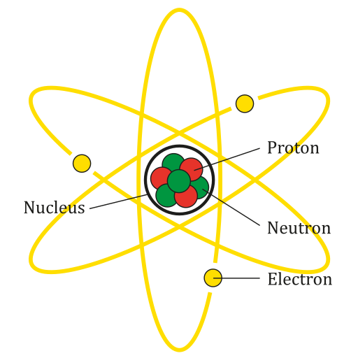
An atom is the smallest particle of an element that still has the properties of that element. Every substance is composed of atoms. Atoms are extremely small, typically about a ten-billionth of a metre in diametre. However, atoms do not have well-defined boundaries, as suggested by the atomic model shown below.
Every atom is composed of a central area — called the nucleus — and one or more subatomic particles called electrons, which move around the nucleus. The nucleus also consists of subatomic particles. It contains one or more protons and typically a similar number of neutrons. The number of protons in the nucleus determines the type of element an atom represents. An atom of hydrogen, for example, contains just one proton. Atoms of the same element may have different numbers of neutrons in the nucleus. Atoms of the same element with the same number of protons — but different numbers of neutrons — are called isotopes.
Protons have a positive electric charge and neutrons have no electric charge. Virtually all of an atom's mass is in the protons and neutrons in the nucleus. Electrons surrounding the nucleus have almost no mass, as well as a negative electric charge. If the number of protons and electrons in an atom are equal, then an atom is electrically neutral, because the positive and negative charges cancel each other out. If an atom has more or fewer electrons than protons, then it has an overall negative or positive charge, respectively, and it is called an ion.
The negatively-charged electrons of an atom are attracted to the positively-charged protons in the nucleus by a force called electromagnetic force, for which opposite charges attract. Electromagnetic force between protons in the nucleus causes these subatomic particles to repel each other, because they have the same charge. However, the protons and neutrons in the nucleus are attracted to each other by a different force, called nuclear force, which is usually stronger than the electromagnetic force. Nuclear force repels the positively-charged protons from each other.
Periodic Table of the Elements
There are almost 120 known elements. As you can see in the Periodic Table of the Elements shown below, the majority of elements are metals. Examples of metals are iron (Fe) and copper (Cu). Metals are shiny and good conductors of electricity and heat. Nonmetal elements are far fewer in number. They include hydrogen (H) and oxygen (O). They lack the properties of metals.
The periodic table of the elements arranges elements in groups based on their properties. The element most important to life is carbon (C). Find carbon in the table. What type of element is it: metal or nonmetal?
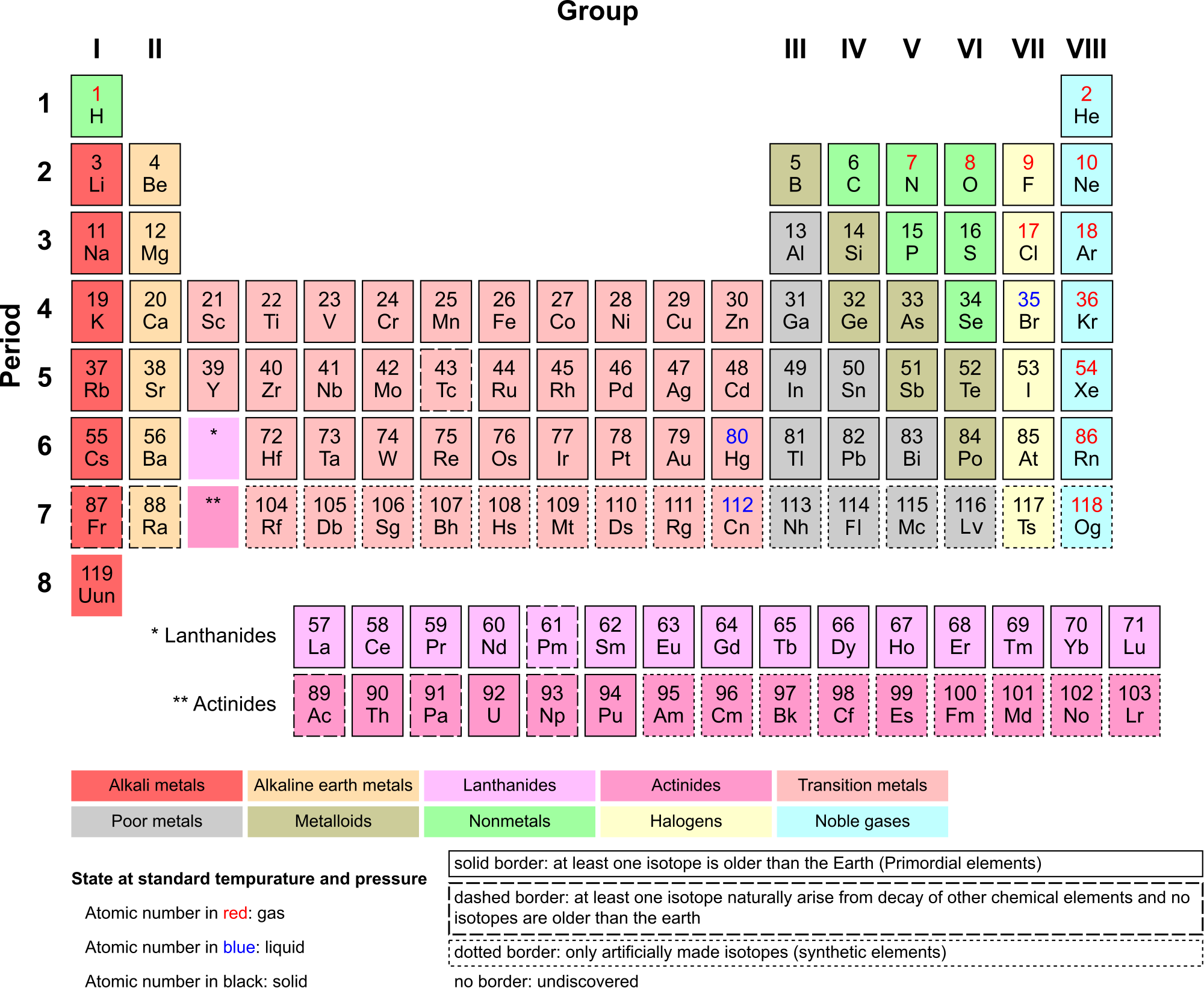
Compounds and Molecules
A compound is a unique substance that consists of two or more elements combined in fixed proportions. This means that the composition of a compound is always the same. The smallest particle of most compounds in living things is called a molecule.
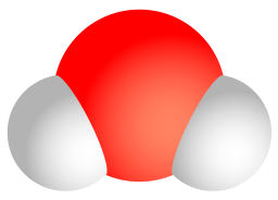
Consider water as an example. A molecule of water always contains one atom of oxygen and two atoms of hydrogen. The composition of water is expressed by the chemical formula H2O. A model of a water molecule is shown in Figure 3.2.4.
What causes the atoms of a water molecule to “stick” together? The answer is chemical bonds. A chemical bond is a force that holds together the atoms of molecules. Bonds in molecules involve the sharing of electrons among atoms. New chemical bonds form when substances react with one another. A chemical reaction is a process that changes some chemical substances into others. A chemical reaction is needed to form a compound, and another chemical reaction is needed to separate the substances in that compound.
3.2 Summary
- All matter consists of chemical substances. A chemical substance has a definite composition which is consistent throughout. A chemical substance may be either an element or a compound.
- An element is a pure substance that cannot be broken down into other types of substances.
- An atom is the smallest particle of an element that still has the properties of that element. Atoms, in turn, are composed of subatomic particles, including negative electrons, positive protons, and neutral neutrons. The number of protons in an atom determines the element it represents.
- Atoms have equal numbers of electrons and protons, so they have no charge. Ions are atoms that have lost or gained electrons, and as a result have either a positive or negative charge. Atoms with the same number of protons — but different numbers of neutrons — are called isotopes.
- There are almost 120 known elements. The majority of elements are metals. A smaller number are nonmetals. The latter include carbon, hydrogen, and oxygen.
- A compound is a substance that consists of two or more elements in a unique composition. The smallest particle of a compound is called a molecule. Chemical bonds hold together the atoms of molecules. Compounds can form only in chemical reactions, and they can break down only in other chemical reactions.
3.2 Review Questions
-
- What is an element? Give three examples.
- Define compound. Explain how compounds form.
- Compare and contrast atoms and molecules.
- The compound called water can be broken down into its constituent elements by applying an electric current to it. What ratio of elements is produced in this process?
- Relate ions and isotopes to elements and atoms.
- What is the most important element to life?
- Iron oxide is often known as rust — the reddish substance you might find on corroded metal. The chemical formula for this type of iron oxide is Fe2O3. Answer the following questions about iron oxide and briefly explain each answer.
- Is iron oxide an element or a compound?
- Would one particle of iron oxide be considered a molecule or an atom?
- Describe the relative proportion of atoms in iron oxide.
- What causes the Fe and O to stick together in iron oxide?
- Is iron oxide made of metal atoms, metalloid atoms, nonmetal atoms, or a combination of any of these?
- 14C is an isotope of carbon used in the radiocarbon dating of organic material. The most common isotope of carbon is 12C. Do you think 14C and 12C have different numbers of neutrons or protons? Explain your answer.
- Explain why ions have a positive or negative charge.
- Name the three subatomic particles described in this section.
3.2 Explore More
https://www.youtube.com/watch?v=yQP4UJhNn0I&feature=emb_logo
Just how small is an atom? TED-Ed, 2012
Attributions
Figure 3.2.1
Man Sitting, by Gregory Culmer, on Unsplash, is used under the Unsplash license (https://unsplash.com/license).
Figure 3.2.2
Lithium Atom diagram, by AG Caesar, is used under a CC BY-SA 4.0 (https://creativecommons.org/licenses/by-sa/4.0/deed.en)
Figure 3.2.3
Periodic Table Armtuk3, by Armtuk, is used under a CC BY-SA 3.0 (https://creativecommons.org/licenses/by-sa/3.0/) license.
Figure 3.2.4
Water molecule, by Sakurambo, is released into the public domain (https://en.wikipedia.org/wiki/Public_domain).
References
TED-Ed. (2012, April 16). Just how small is an atom. YouTube. https://www.youtube.com/watch?v=yQP4UJhNn0I&feature=youtu.be

Case Study: Please Don’t Pass the Bread
Angela and Saloni are college students who met in physics class. They decide to study together for their upcoming midterm, but first, they want to grab some lunch. Angela says there is a particular restaurant she would like to go to, because they are able to accommodate her dietary restrictions. Saloni agrees and they head to the restaurant.
At lunch, Saloni asks Angela what is special about her diet. Angela tells her that she can’t eat gluten. Saloni says, “My cousin did that for a while because she heard that gluten is bad for you. But it was too hard for her to not eat bread and pasta, so she gave it up.” Angela tells Saloni that avoiding gluten isn’t optional for her — she has celiac disease. Eating even very small amounts of gluten could damage her digestive system. It can be difficult for people living with celiac disease to find foods when eating out.
You have probably heard of gluten, but what is it, and why is it harmful to people with celiac disease? Gluten is a protein present in wheat and some other grains (such as barley, rye, and oats), so it is commonly found in foods like bread, pasta, baked goods, and many packaged foods, like the ones pictured in Figure 15.1.2.
Figure 15.1.2 Gluten is a protein present in foods like bread, pasta, and baked goods.
For people with celiac disease, eating gluten causes an autoimmune reaction that results in damage to the small, finger-like villi lining the small intestine, causing them to become inflamed and flattened (see Figure 15.1.3). This damage interferes with the digestive process, which can result in a wide variety of symptoms including diarrhea, anemia, skin rash, bone pain, depression, and anxiety, among others. The degree of damage to the villi can vary from mild to severe, with more severe damage generally resulting in more significant symptoms and complications. Celiac disease can have serious long-term consequences, such as osteoporosis, problems in the nervous and reproductive systems, and the development of certain types of cancers.
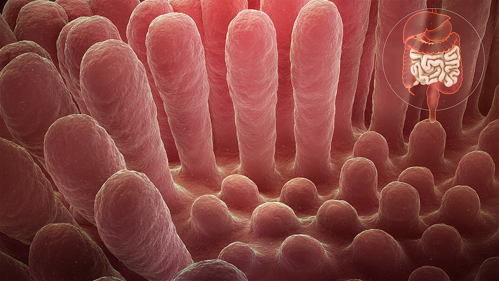
Why does celiac disease cause so many different types of symptoms and have such significant negative health consequences? As you read this chapter and learn about how the digestive system works, you will see just how important the villi of the small intestine are to the body as a whole. At the end of the chapter, you will learn more about celiac disease, why it can be so serious, and whether it is worth avoiding gluten for people who do not have a diagnosed medical issue with it.
Chapter Overview: Digestive System
In this chapter, you will learn about the digestive system, which processes food so that our bodies can obtain nutrients. Specifically, you will learn about:
- The structures and organs of the gastrointestinal (GI) tract through which food directly passes. This includes the mouth, pharynx, esophagus, stomach, small intestine, and large intestine.
- The functions of the GI tract, including mechanical and chemical digestion, absorption of nutrients, and the elimination of solid waste.
- The accessory organs of digestion — the liver, gallbladder, and pancreas — which secrete substances needed for digestion into the GI tract, in addition to performing other important functions.
- Specializations of the tissues of the digestive system that allow it to carry out its functions.
- How different types of nutrients (such as carbohydrates, proteins, and fats) are digested and absorbed by the body.
- Beneficial bacteria that live in the GI tract and help us digest food, produce vitamins, and protect us from harmful pathogens and toxic substances.
- Disorders of the digestive system, including inflammatory bowel diseases, ulcers, diverticulitis, and gastroenteritis (commonly known as “stomach flu”).
As you read this chapter, think about the following questions related to celiac disease:
- What are the general functions of the small intestine? What do the villi in the small intestine do?
- Why do you think celiac disease causes so many different types of symptoms and potentially serious complications?
- What are some other autoimmune diseases that involve the body attacking its own digestive system?
Attributions
Figure 15.1.1
Bread [photo] by Sergio Arze on Unsplash is used under the Unsplash License (https://unsplash.com/license).
Figure 15.1.2
- Paste cu sos de roșii by Sestrjevitovschii Ina on Unsplash is used under the Unsplash License (https://unsplash.com/license).
- Cookies and More by Sarah Shaffer on Unsplash is used under the Unsplash License (https://unsplash.com/license).
- Raspberry waffles by Izabelle Acheson on Unsplash is used under the Unsplash License (https://unsplash.com/license).
- Homemade croissant & pain au chocolat by Cristiano Pinto on Unsplash is used under the Unsplash License (https://unsplash.com/license).
Figure 15.1.3
Inflammed_mucous_layer_of_the_intestinal_villi_depicting_Celiac_disease by www.scientificanimations.com (image 140/191) on Wikimedia Commons is used under a CC BY-SA 4.0 (https://creativecommons.org/licenses/by-sa/4.0) license.
Image shows systole and diastole of the heart. In diastole, the ventricles of the heart are relaxed and are able to receive blood from the atria. In systole, both ventricles contract, squeezing the blood out and into the pulmonary trunk and aorta.
As described in the caption.
Image shows a diagram of the vessels going into and out of the liver. The hepatic artery bring blood from the heart to the liver and the hepatic vein returns this blood to the heart. The portal vein brings blood from the lower GI tract to the liver.
As per caption
The space occurring between two or more membranes. In cell biology, it's most commonly described as the region between the inner membrane and the outer membrane of a mitochondrion or a chloroplast.
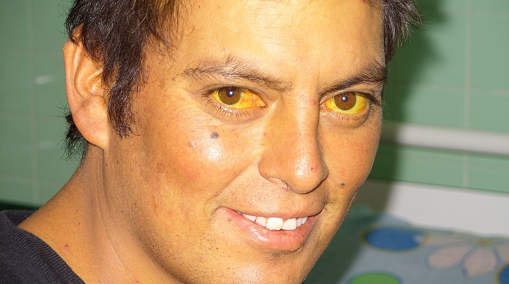
Jaundiced Eyes
Did you ever hear of a person looking at something or someone with a “jaundiced eye”? It means to take a negative view, such as envy, maliciousness, or ill will. The expression may be based on the antiquated idea that liver bile is associated with such negative emotions as these, as well as the fact that excessive liver bile causes jaundice, or yellowing of the eyes and skin. Jaundice is likely a sign of a liver disorder or blockage of the duct that carries bile away from the liver. Bile contains waste products, making the liver an organ of excretion. Bile has an important role in digestion, which makes the liver an accessory organ of digestion, too.
What Are Accessory Organs of Digestion?
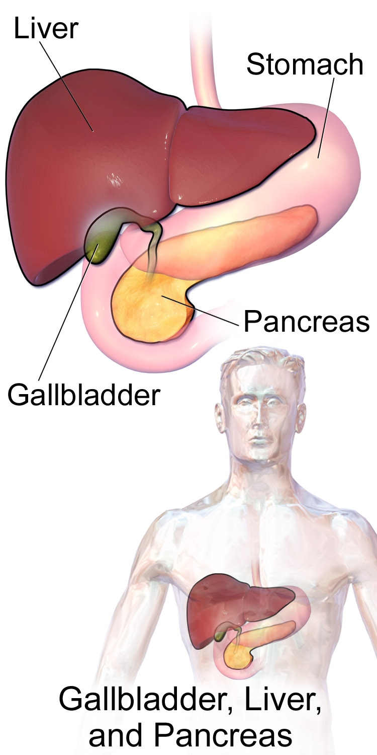
Accessory organs of digestion are organs that secrete substances needed for the chemical digestion of food, but through which food does not actually pass as it is digested. Besides the liver, the major accessory organs of digestion are the gallbladder and pancreas. These organs secrete or store substances that are needed for digestion in the first part of the small intestine — the duodenum — where most chemical digestion takes place. You can see the three organs and their locations in Figure 15.6.2.
Liver
The liver is a vital organ located in the upper right part of the abdomen. It lies just below the diaphragm, to the right of the stomach. The liver plays an important role in digestion by secreting bile, but the liver has a wide range of additional functions unrelated to digestion. In fact, some estimates put the number of functions of the liver at about 500! A few of them are described below.
Structure of the Liver
The liver is a reddish brown, wedge-shaped structure. In adults, the liver normally weighs about 1.5 kg (about 3.3 lb). It is both the heaviest internal organ and the largest gland in the human body. The liver is divided into four lobes of unequal size and shape. Each lobe, in turn, is made up of lobules, which are the functional units of the liver. Each lobule consists of millions of liver cells, called hepatic cells (or hepatocytes). They are the basic metabolic cells that carry out the various functions of the liver.
As shown in Figure 15.6.3, the liver is connected to two large blood vessels: the hepatic artery and the portal vein. The hepatic artery carries oxygen-rich blood from the aorta, whereas the portal vein carries blood that is rich in digested nutrients from the GI tract and wastes filtered from the blood by the spleen. The blood vessels subdivide into smaller arteries and capillaries, which lead into the liver lobules. The nutrients from the GI tract are used to build many vital biochemical compounds, and the wastes from the spleen are degraded and excreted.
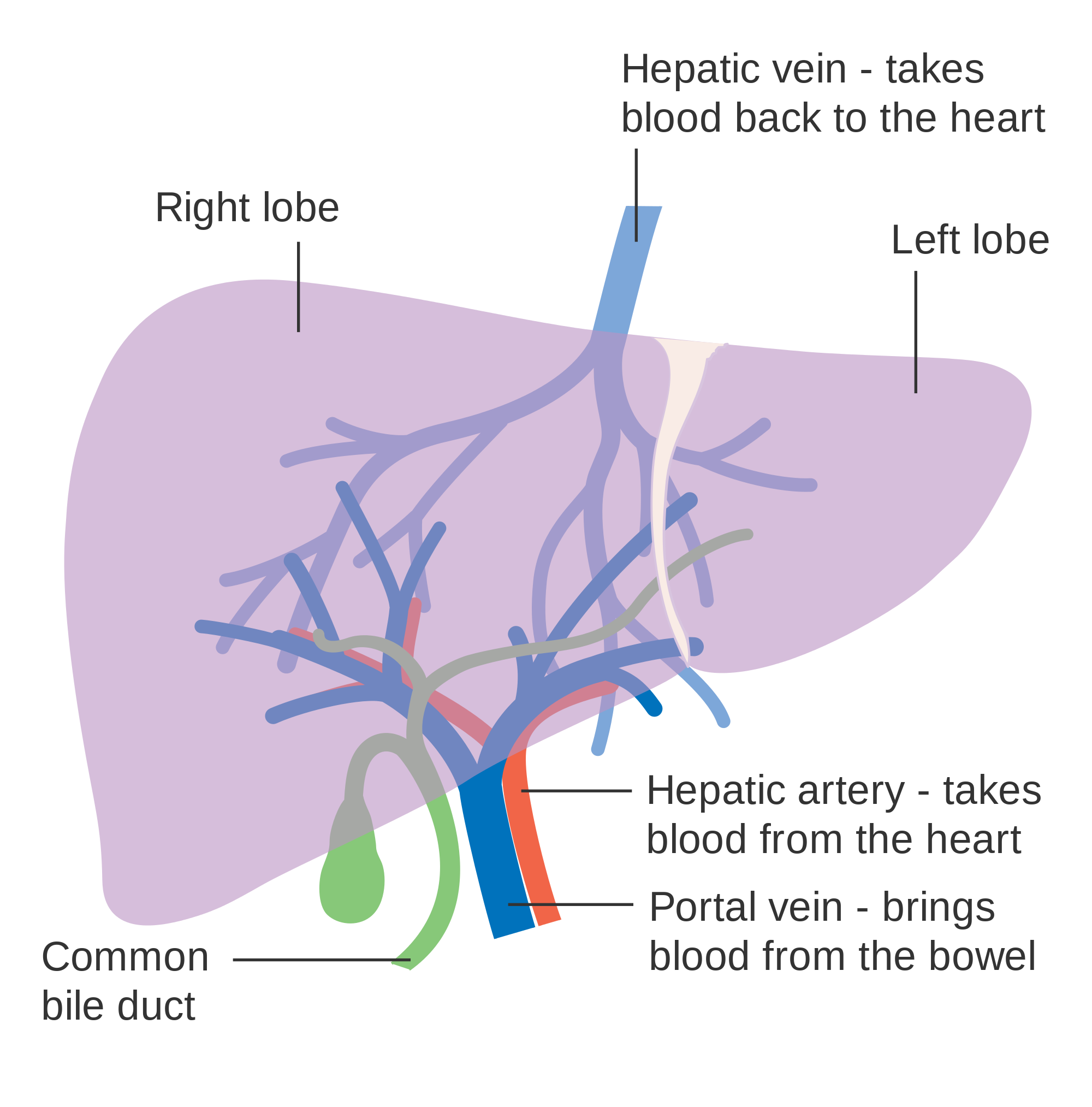
Functions of the Liver
The main digestive function of the liver is the production of bile. Bile is a yellowish alkaline liquid that consists of water, electrolytes, bile salts, and cholesterol, among other substances, many of which are waste products. Some of the components of bile are synthesized by hepatocytes. The rest are extracted from the blood.
As shown in Figure 15.6.4, bile is secreted into small ducts that join together to form larger ducts, with just one large duct carrying bile out of the liver. If bile is needed to digest a meal, it goes directly to the duodenum through the common bile duct. In the duodenum, the bile neutralizes acidic chyme from the stomach and emulsifies fat globules into smaller particles (called micelles) that are easier to digest chemically by the enzyme lipase. Bile also aids with the absorption of vitamin K. Bile that is secreted when digestion is not taking place goes to the gallbladder for storage until the next meal. In either case, the bile enters the duodenum through the common bile duct.
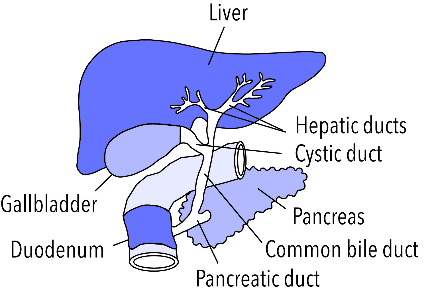
Besides its roles in digestion, the liver has many other vital functions:
- The liver synthesizes glycogen from glucose and stores the glycogen as required to help regulate blood sugar levels. It also breaks down the stored glycogen to glucose and releases it back into the blood as needed.
- The liver stores many substances in addition to glycogen, including vitamins A, D, B12, and K. It also stores the minerals iron and copper.
- The liver synthesizes numerous proteins and many of the amino acids needed to make them. These proteins have a wide range of functions. They include fibrinogen, which is needed for blood clotting; insulin-like growth factor (IGF-1), which is important for childhood growth; and albumen, which is the most abundant protein in blood serum and functions to transport fatty acids and steroid hormones in the blood.
- The liver synthesizes many important lipids, including cholesterol, triglycerides, and lipoproteins.
- The liver is responsible for the breakdown of many waste products and toxic substances. The wastes are excreted in bile or travel to the kidneys, which excrete them in urine.
The liver is clearly a vital organ that supports almost every other organ in the body. Because of its strategic location and diversity of functions, the liver is also prone to many diseases, some of which cause loss of liver function. There is currently no way to compensate for the absence of liver function in the long term, although liver dialysis techniques can be used in the short term. An artificial liver has not yet been developed, so liver transplantation may be the only option for people with liver failure.
Gallbladder
The gallbladder is a small, hollow, pouch-like organ that lies just under the right side of the liver (see Figure 15.6.5). It is about 8 cm (about 3 in) long and shaped like a tapered sac, with the open end continuous with the cystic duct. The gallbladder stores and concentrates bile from the liver until it is needed in the duodenum to help digest lipids. After the bile leaves the liver, it reaches the gallbladder through the cystic duct. At any given time, the gallbladder may store between 30 to 60 mL (1 to 2 oz) of bile. A hormone stimulated by the presence of fat in the duodenum signals the gallbladder to contract and force its contents back through the cystic duct and into the common bile duct to drain into the duodenum.
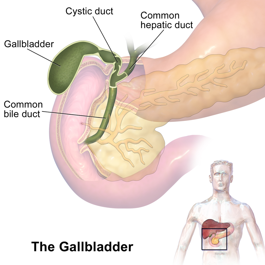
Pancreas
The pancreas is a glandular organ that is part of both the digestive system and the endocrine system. As shown in Figure 15.6.6, it is located in the abdomen behind the stomach, with the head of the pancreas surrounded by the duodenum of the small intestine. The pancreas is about 15 cm (almost 6 in) long, and it has two major ducts: the main pancreatic duct and the accessory pancreatic duct. Both of these ducts drain into the duodenum.
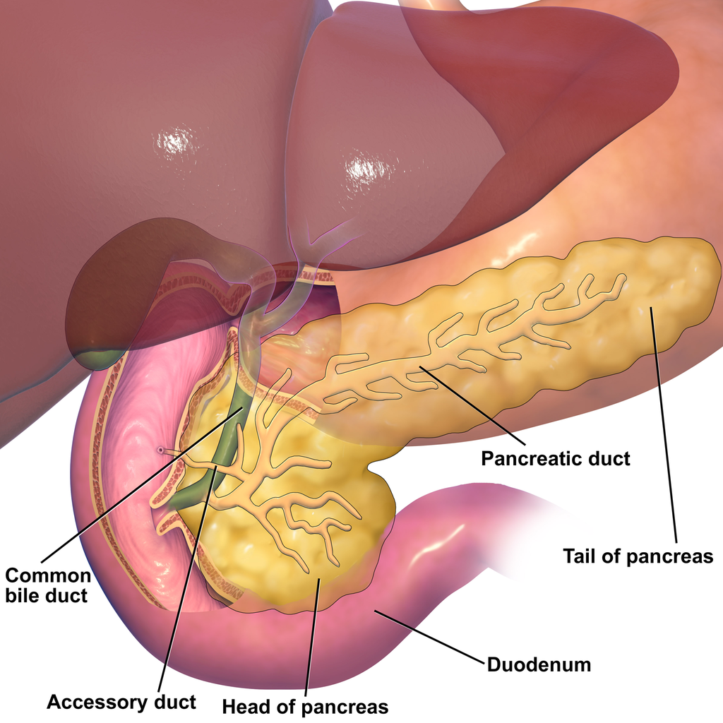
As an endocrine gland, the pancreas secretes several hormones, including insulin and glucagon, which circulate in the blood. The endocrine hormones are secreted by clusters of cells called pancreatic islets (or islets of Langerhans). As a digestive organ, the pancreas secretes many digestive enzymes and also bicarbonate, which helps neutralize acidic chyme after it enters the duodenum. The pancreas is stimulated to secrete its digestive substances when food in the stomach and duodenum triggers the release of endocrine hormones into the blood that reach the pancreas via the bloodstream. The pancreatic digestive enzymes are secreted by clusters of cells called acini, and they travel through the pancreatic ducts to the duodenum. In the duodenum, they help to chemically break down carbohydrates, proteins, lipids, and nucleic acids in chyme. The pancreatic digestive enzymes include:
- Amylase, which helps digest starch and other carbohydrates.
- Trypsin and chymotrypsin, which help digest proteins.
- Lipase, which helps digest lipids.
- Deoxyribonucleases and ribonucleases, which help digest nucleic acids.
15.6 Summary
- Accessory organs of digestion are organs that secrete substances needed for the chemical digestion of food, but through which food does not actually pass as it is digested. The accessory organs include the liver, gallbladder, and pancreas. These organs secrete or store substances that are carried to the duodenum of the small intestine as needed for digestion.
- The liver is a large organ in the abdomen that is divided into lobes and smaller lobules, which consist of metabolic cells called hepatic cells, or hepatocytes. The liver receives oxygen in blood from the aorta through the hepatic artery. It receives nutrients in blood from the GI tract and wastes in blood from the spleen through the portal vein.
- The main digestive function of the liver is the production of the alkaline liquid called bile. Bile is carried directly to the duodenum by the common bile duct or to the gallbladder first for storage. Bile neutralizes acidic chyme that enters the duodenum from the stomach, and also emulsifies fat globules into smaller particles (micelles) that are easier to digest chemically.
- Other vital functions of the liver include regulating blood sugar levels by storing excess sugar as glycogen, storing many vitamins and minerals, synthesizing numerous proteins and lipids, and breaking down waste products and toxic substances.
- The gallbladder is a small pouch-like organ near the liver. It stores and concentrates bile from the liver until it is needed in the duodenum to neutralize chyme and help digest lipids.
- The pancreas is a glandular organ that secretes both endocrine hormones and digestive enzymes. As an endocrine gland, the pancreas secretes insulin and glucagon to regulate blood sugar. As a digestive organ, the pancreas secretes digestive enzymes into the duodenum through ducts. Pancreatic digestive enzymes include amylase (starches) trypsin and chymotrypsin (proteins), lipase (lipids), and ribonucleases and deoxyribonucleases (RNA and DNA).
15.6 Review Questions
- Name three accessory organs of digestion. How do these organs differ from digestive organs that are part of the GI tract?
-
- Describe the liver and its blood supply.
- Explain the main digestive function of the liver and describe the components of bile and it's importance in the digestive process.
- What type of secretions does the pancreas release as part of each body system?
- List pancreatic enzymes that work in the duodenum, along with the substances they help digest.
- What are two substances produced by accessory organs of digestion that help neutralize chyme in the small intestine? Where are they produced?
- People who have their gallbladder removed sometimes have digestive problems after eating high-fat meals. Why do you think this happens?
- Which accessory organ of digestion synthesizes cholesterol?
15.6 Explore More
https://youtu.be/8dgoeYPoE-0
What does the pancreas do? - Emma Bryce, TED-Ed. 2015.
https://youtu.be/wbh3SjzydnQ
What does the liver do? - Emma Bryce, TED-Ed, 2014.
https://youtu.be/a0d1yvGcfzQ
Scar wars: Repairing the liver, nature video, 2018.
Attributions
Figure 15.6.1
Scleral_Icterus by Sheila J. Toro on Wikimedia Commons is used under a CC BY 4.0 (https://creativecommons.org/licenses/by/4.0) license.
Figure 15.6.2
Blausen_0428_Gallbladder-Liver-Pancreas_Location by BruceBlaus on Wikimedia Commons is used under a CC BY 3.0 (https://creativecommons.org/licenses/by/3.0) license.
Figure 15.6.3
Diagram_showing_the_two_lobes_of_the_liver_and_its_blood_supply_CRUK_376.svg by Cancer Research UK on Wikimedia Commons is used under a CC BY-SA 4.0 (https://creativecommons.org/licenses/by-sa/4.0) license.
Figure 15.6.4
Gallbladder by NIH Image Gallery on Flickr is used CC BY-NC 2.0 (https://creativecommons.org/licenses/by-nc/2.0/) license.
Figure 15.6.5
Gallbladder_(organ) (1) by BruceBlaus on Wikimedia Commons is used under a CC BY-SA 4.0 (https://creativecommons.org/licenses/by-sa/4.0) license. (See a full animation of this medical topic at blausen.com.)
Figure 15.6.6
Blausen_0698_PancreasAnatomy by BruceBlaus on Wikimedia Commons is used under a CC BY 3.0 (https://creativecommons.org/licenses/by/3.0) license.
References
Blausen.com Staff. (2014). Medical gallery of Blausen Medical 2014. WikiJournal of Medicine 1 (2). DOI:10.15347/wjm/2014.010. ISSN 2002-4436.
nature video. (2018, December 19). Scar wars: Repairing the liver. YouTube. https://www.youtube.com/watch?v=a0d1yvGcfzQ&feature=youtu.be
TED-Ed. (2014, November 25). What does the liver do? - Emma Bryce. YouTube. https://www.youtube.com/watch?v=wbh3SjzydnQ&feature=youtu.be
TED-Ed. (2015, February 19). What does the pancreas do? - Emma Bryce. YouTube. https://www.youtube.com/watch?v=8dgoeYPoE-0&feature=youtu.be
Created by CK-12 Foundation/Adapted by Christine Miller

Jaundiced Eyes
Did you ever hear of a person looking at something or someone with a “jaundiced eye”? It means to take a negative view, such as envy, maliciousness, or ill will. The expression may be based on the antiquated idea that liver bile is associated with such negative emotions as these, as well as the fact that excessive liver bile causes jaundice, or yellowing of the eyes and skin. Jaundice is likely a sign of a liver disorder or blockage of the duct that carries bile away from the liver. Bile contains waste products, making the liver an organ of excretion. Bile has an important role in digestion, which makes the liver an accessory organ of digestion, too.
What Are Accessory Organs of Digestion?

Accessory organs of digestion are organs that secrete substances needed for the chemical digestion of food, but through which food does not actually pass as it is digested. Besides the liver, the major accessory organs of digestion are the gallbladder and pancreas. These organs secrete or store substances that are needed for digestion in the first part of the small intestine — the duodenum — where most chemical digestion takes place. You can see the three organs and their locations in Figure 15.6.2.
Liver
The liver is a vital organ located in the upper right part of the abdomen. It lies just below the diaphragm, to the right of the stomach. The liver plays an important role in digestion by secreting bile, but the liver has a wide range of additional functions unrelated to digestion. In fact, some estimates put the number of functions of the liver at about 500! A few of them are described below.
Structure of the Liver
The liver is a reddish brown, wedge-shaped structure. In adults, the liver normally weighs about 1.5 kg (about 3.3 lb). It is both the heaviest internal organ and the largest gland in the human body. The liver is divided into four lobes of unequal size and shape. Each lobe, in turn, is made up of lobules, which are the functional units of the liver. Each lobule consists of millions of liver cells, called hepatic cells (or hepatocytes). They are the basic metabolic cells that carry out the various functions of the liver.
As shown in Figure 15.6.3, the liver is connected to two large blood vessels: the hepatic artery and the portal vein. The hepatic artery carries oxygen-rich blood from the aorta, whereas the portal vein carries blood that is rich in digested nutrients from the GI tract and wastes filtered from the blood by the spleen. The blood vessels subdivide into smaller arteries and capillaries, which lead into the liver lobules. The nutrients from the GI tract are used to build many vital biochemical compounds, and the wastes from the spleen are degraded and excreted.

Functions of the Liver
The main digestive function of the liver is the production of bile. Bile is a yellowish alkaline liquid that consists of water, electrolytes, bile salts, and cholesterol, among other substances, many of which are waste products. Some of the components of bile are synthesized by hepatocytes. The rest are extracted from the blood.
As shown in Figure 15.6.4, bile is secreted into small ducts that join together to form larger ducts, with just one large duct carrying bile out of the liver. If bile is needed to digest a meal, it goes directly to the duodenum through the common bile duct. In the duodenum, the bile neutralizes acidic chyme from the stomach and emulsifies fat globules into smaller particles (called micelles) that are easier to digest chemically by the enzyme lipase. Bile also aids with the absorption of vitamin K. Bile that is secreted when digestion is not taking place goes to the gallbladder for storage until the next meal. In either case, the bile enters the duodenum through the common bile duct.

Besides its roles in digestion, the liver has many other vital functions:
- The liver synthesizes glycogen from glucose and stores the glycogen as required to help regulate blood sugar levels. It also breaks down the stored glycogen to glucose and releases it back into the blood as needed.
- The liver stores many substances in addition to glycogen, including vitamins A, D, B12, and K. It also stores the minerals iron and copper.
- The liver synthesizes numerous proteins and many of the amino acids needed to make them. These proteins have a wide range of functions. They include fibrinogen, which is needed for blood clotting; insulin-like growth factor (IGF-1), which is important for childhood growth; and albumen, which is the most abundant protein in blood serum and functions to transport fatty acids and steroid hormones in the blood.
- The liver synthesizes many important lipids, including cholesterol, triglycerides, and lipoproteins.
- The liver is responsible for the breakdown of many waste products and toxic substances. The wastes are excreted in bile or travel to the kidneys, which excrete them in urine.
The liver is clearly a vital organ that supports almost every other organ in the body. Because of its strategic location and diversity of functions, the liver is also prone to many diseases, some of which cause loss of liver function. There is currently no way to compensate for the absence of liver function in the long term, although liver dialysis techniques can be used in the short term. An artificial liver has not yet been developed, so liver transplantation may be the only option for people with liver failure.
Gallbladder
The gallbladder is a small, hollow, pouch-like organ that lies just under the right side of the liver (see Figure 15.6.5). It is about 8 cm (about 3 in) long and shaped like a tapered sac, with the open end continuous with the cystic duct. The gallbladder stores and concentrates bile from the liver until it is needed in the duodenum to help digest lipids. After the bile leaves the liver, it reaches the gallbladder through the cystic duct. At any given time, the gallbladder may store between 30 to 60 mL (1 to 2 oz) of bile. A hormone stimulated by the presence of fat in the duodenum signals the gallbladder to contract and force its contents back through the cystic duct and into the common bile duct to drain into the duodenum.

Pancreas
The pancreas is a glandular organ that is part of both the digestive system and the endocrine system. As shown in Figure 15.6.6, it is located in the abdomen behind the stomach, with the head of the pancreas surrounded by the duodenum of the small intestine. The pancreas is about 15 cm (almost 6 in) long, and it has two major ducts: the main pancreatic duct and the accessory pancreatic duct. Both of these ducts drain into the duodenum.

As an endocrine gland, the pancreas secretes several hormones, including insulin and glucagon, which circulate in the blood. The endocrine hormones are secreted by clusters of cells called pancreatic islets (or islets of Langerhans). As a digestive organ, the pancreas secretes many digestive enzymes and also bicarbonate, which helps neutralize acidic chyme after it enters the duodenum. The pancreas is stimulated to secrete its digestive substances when food in the stomach and duodenum triggers the release of endocrine hormones into the blood that reach the pancreas via the bloodstream. The pancreatic digestive enzymes are secreted by clusters of cells called acini, and they travel through the pancreatic ducts to the duodenum. In the duodenum, they help to chemically break down carbohydrates, proteins, lipids, and nucleic acids in chyme. The pancreatic digestive enzymes include:
- Amylase, which helps digest starch and other carbohydrates.
- Trypsin and chymotrypsin, which help digest proteins.
- Lipase, which helps digest lipids.
- Deoxyribonucleases and ribonucleases, which help digest nucleic acids.
15.6 Summary
- Accessory organs of digestion are organs that secrete substances needed for the chemical digestion of food, but through which food does not actually pass as it is digested. The accessory organs include the liver, gallbladder, and pancreas. These organs secrete or store substances that are carried to the duodenum of the small intestine as needed for digestion.
- The liver is a large organ in the abdomen that is divided into lobes and smaller lobules, which consist of metabolic cells called hepatic cells, or hepatocytes. The liver receives oxygen in blood from the aorta through the hepatic artery. It receives nutrients in blood from the GI tract and wastes in blood from the spleen through the portal vein.
- The main digestive function of the liver is the production of the alkaline liquid called bile. Bile is carried directly to the duodenum by the common bile duct or to the gallbladder first for storage. Bile neutralizes acidic chyme that enters the duodenum from the stomach, and also emulsifies fat globules into smaller particles (micelles) that are easier to digest chemically.
- Other vital functions of the liver include regulating blood sugar levels by storing excess sugar as glycogen, storing many vitamins and minerals, synthesizing numerous proteins and lipids, and breaking down waste products and toxic substances.
- The gallbladder is a small pouch-like organ near the liver. It stores and concentrates bile from the liver until it is needed in the duodenum to neutralize chyme and help digest lipids.
- The pancreas is a glandular organ that secretes both endocrine hormones and digestive enzymes. As an endocrine gland, the pancreas secretes insulin and glucagon to regulate blood sugar. As a digestive organ, the pancreas secretes digestive enzymes into the duodenum through ducts. Pancreatic digestive enzymes include amylase (starches) trypsin and chymotrypsin (proteins), lipase (lipids), and ribonucleases and deoxyribonucleases (RNA and DNA).
15.6 Review Questions
- Name three accessory organs of digestion. How do these organs differ from digestive organs that are part of the GI tract?
-
- Describe the liver and its blood supply.
- Explain the main digestive function of the liver and describe the components of bile and it's importance in the digestive process.
- What type of secretions does the pancreas release as part of each body system?
- List pancreatic enzymes that work in the duodenum, along with the substances they help digest.
- What are two substances produced by accessory organs of digestion that help neutralize chyme in the small intestine? Where are they produced?
- People who have their gallbladder removed sometimes have digestive problems after eating high-fat meals. Why do you think this happens?
- Which accessory organ of digestion synthesizes cholesterol?
15.6 Explore More
https://youtu.be/8dgoeYPoE-0
What does the pancreas do? - Emma Bryce, TED-Ed. 2015.
https://youtu.be/wbh3SjzydnQ
What does the liver do? - Emma Bryce, TED-Ed, 2014.
https://youtu.be/a0d1yvGcfzQ
Scar wars: Repairing the liver, nature video, 2018.
Attributions
Figure 15.6.1
Scleral_Icterus by Sheila J. Toro on Wikimedia Commons is used under a CC BY 4.0 (https://creativecommons.org/licenses/by/4.0) license.
Figure 15.6.2
Blausen_0428_Gallbladder-Liver-Pancreas_Location by BruceBlaus on Wikimedia Commons is used under a CC BY 3.0 (https://creativecommons.org/licenses/by/3.0) license.
Figure 15.6.3
Diagram_showing_the_two_lobes_of_the_liver_and_its_blood_supply_CRUK_376.svg by Cancer Research UK on Wikimedia Commons is used under a CC BY-SA 4.0 (https://creativecommons.org/licenses/by-sa/4.0) license.
Figure 15.6.4
Gallbladder by NIH Image Gallery on Flickr is used CC BY-NC 2.0 (https://creativecommons.org/licenses/by-nc/2.0/) license.
Figure 15.6.5
Gallbladder_(organ) (1) by BruceBlaus on Wikimedia Commons is used under a CC BY-SA 4.0 (https://creativecommons.org/licenses/by-sa/4.0) license. (See a full animation of this medical topic at blausen.com.)
Figure 15.6.6
Blausen_0698_PancreasAnatomy by BruceBlaus on Wikimedia Commons is used under a CC BY 3.0 (https://creativecommons.org/licenses/by/3.0) license.
References
Blausen.com Staff. (2014). Medical gallery of Blausen Medical 2014. WikiJournal of Medicine 1 (2). DOI:10.15347/wjm/2014.010. ISSN 2002-4436.
nature video. (2018, December 19). Scar wars: Repairing the liver. YouTube. https://www.youtube.com/watch?v=a0d1yvGcfzQ&feature=youtu.be
TED-Ed. (2014, November 25). What does the liver do? - Emma Bryce. YouTube. https://www.youtube.com/watch?v=wbh3SjzydnQ&feature=youtu.be
TED-Ed. (2015, February 19). What does the pancreas do? - Emma Bryce. YouTube. https://www.youtube.com/watch?v=8dgoeYPoE-0&feature=youtu.be
Image shows a person's elbows with a rash that has caused redness and scabbing.
Image shows a diagram of the large intestine. There are multiple abnormal little pouches on the exterior of the colon.

Created by: CK-12/Adapted by Christine Miller
Assembly Line
We stay alive because millions of different chemical reactions are taking place inside our bodies all the time. Each of our cells is like the busy auto assembly line pictured in Figure 3.10.1. Raw materials, half-finished products, and waste materials are constantly being used, produced, transported, and excreted. The "workers" on the cellular assembly line are mainly enzymes. These are the proteins that make biochemical reactions happen.
What Are Biochemical Reactions?
Chemical reactions that take place inside living things are called biochemical reactions. The sum of all the biochemical reactions in an organism is called metabolism. Metabolism includes both exothermic (energy-releasing) chemical reactions and endothermic (energy-absorbing) chemical reactions.
Catabolic Reactions
Exothermic reactions in organisms are called catabolic reactions. These reactions break down molecules into smaller units and release energy. An example of a catabolic reaction is the breakdown of glucose during cellular respiration, which releases energy that cells need to carry out life processes.
Anabolic Reactions
Endothermic reactions in organisms are called anabolic reactions. These reactions build up bigger molecules from smaller ones and absorb energy. An example of an anabolic reaction is the joining of amino acids to form a protein. Which type of reactions — catabolic or anabolic — do you think occur when your body digests food?
Enzymes
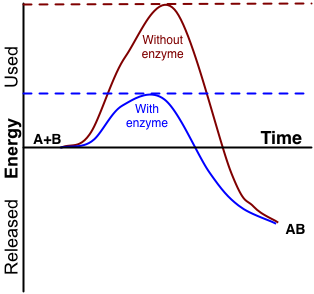
Most of the biochemical reactions that happen inside of living organisms require help. Why is this the case? For one thing, temperatures inside living things are usually too low for biochemical reactions to occur quickly enough to maintain life. The concentrations of reactants may also be too low for them to come together and react. Where do the biochemical reactions get the help they need to proceed? From the enzymes.
An enzyme is a protein that speeds up a biochemical reaction. It is a biological catalyst. An enzyme generally works by reducing the amount of activation energy needed to start the reaction. The graph in Figure 3.10.2 shows the activation energy needed for glucose to combine with oxygen. Less activation energy is needed when the correct enzyme is present than when it is not present.
An enzyme speeds up the reaction by lowering the required activation energy. Compare the activation energy needed with and without the enzyme.
How Well Enzymes Work
Enzymes are involved in most biochemical reactions, and they do their jobs extremely well. A typical biochemical reaction that would take several days or even several centuries to happen without an enzyme is likely to occur in just a split second with the proper enzyme! Without enzymes to speed up biochemical reactions, most organisms could not survive.
Enzymes are substrate-specific. The substrate of an enzyme is the specific substance it affects. Each enzyme works only with a particular substrate, which explains why there are so many different enzymes. In addition, for an enzyme to work, it requires specific conditions, such as the right temperature and pH. Some enzymes work best under acidic conditions, for example, while others work best in neutral environments.
Enzyme-Deficiency Disorders
There are hundreds of known inherited metabolic disorders in humans. In most of them, a single enzyme is either not produced by the body at all, or is otherwise produced in a form that doesn't work. The missing or defective enzyme is like an absentee worker on the cell's assembly line. Imagine the auto assembly line from the image at the start of this section. What if the worker who installed the steering wheel was absent? How would this impact the overall functioning of the vehicle? When an enzyme is missing, toxic chemicals build up, or an essential product isn't made. Generally, the normal enzyme is missing because the individual with the disorder inherited two copies of a gene mutation, which may have originated many generations previously.
Any given inherited metabolic disorder is generally quite rare in the general population. However, there are so many different metabolic disorders that a total of one in 1,000 to 2,500 newborns can be expected to have one.
3.10 Summary
- Biochemical reactions are chemical reactions that take place inside of living things. The sum of all of the biochemical reactions in an organism is called metabolism.
- Metabolism includes catabolic reactions, which are energy-releasing (exothermic) reactions, as well as anabolic reactions, which are energy-absorbing (endothermic) reactions.
- Most biochemical reactions need a biological catalyst called an enzyme to speed up the reaction. Enzymes reduce the amount of activation energy needed for the reaction to begin. Most enzymes are proteins that affect just one specific substance, which is called the enzyme's substrate.
- There are many inherited metabolic disorders in humans. Most of them are caused by a single defective or missing enzyme.
3.10 Review Questions
- What are biochemical reactions?
- Define metabolism.
- Compare and contrast catabolic and anabolic reactions.
- Explain the role of enzymes in biochemical reactions.
- What are enzyme-deficiency disorders?
- Explain why the relatively low temperature of living things, along with the low concentration of reactants, would cause biochemical reactions to occur very slowly in the body without enzymes.
- Answer the following questions about what happens after you eat a sandwich.
- Pieces of the sandwich go into your stomach, where there are digestive enzymes that break down the food. Which type of metabolic reaction is this? Explain your answer.
- During the process of digestion, some of the sandwich is broken down into glucose, which is then further broken down to release energy that your cells can use. Is this an exothermic endothermic reaction? Explain your answer.
- The proteins in the cheese, meat, and bread in the sandwich are broken down into their component amino acids. Then your body uses those amino acids to build new proteins. Which kind of metabolic reaction is represented by the building of these new proteins? Explain your answer.
- Explain why your body doesn’t just use one or two enzymes for all of its biochemical reactions.
- A ________ is the specific substance that an enzyme affects in a biochemical reaction.
- An enzyme is a biological _____________ .
- catabolism
- form of activation energy
- catalyst
- reactant
3.10 Explore More
https://www.youtube.com/watch?v=qgVFkRn8f10&feature=youtu.be
Enzymes (Updated), by The Amoeba Sisters, 2016.
https://www.youtube.com/watch?v=8m6RtOpqvtU&feature=youtu.be
What triggers a chemical reaction? - Kareem Jarrah, TED-Ed, 2015.
Figure 3.10.1
Auto Assembly line by Brian Snelson on Wikimedia Commons is used under a CC BY 2.0 (https://creativecommons.org/licenses/by/2.0) license.
Figure 3.10.2
Enzyme_activation_energy by G. Andruk [IMeowbot at the English language Wikipedia], is used under a CC BY-SA 3.0 (http://creativecommons.org/licenses/by-sa/3.0/) license.
References
Amoeba Sisters. (2016, August 28). Enzymes (updated). YouTube. https://www.youtube.com/watch?v=qgVFkRn8f10&feature=youtu.be
TED-Ed. (2015, January 15). What triggers a chemical reaction? - Kareem Jarrah. YouTube. https://www.youtube.com/watch?v=8m6RtOpqvtU&feature=youtu.be
A type of disease in which cells of the central nervous system stop working or die. Neurodegenerative disorders usually get worse over time and have no cure. They may be genetic or be caused by a tumor or stroke.
Figure 7.4.1 Construction — It's important to have the right materials for the job.
The Right Material for the Job
Building a house is a big job and one that requires a lot of different materials for specific purposes. As you can see in Figure 7.4.1, many different types of materials are used to build a complete house, but each type of material fulfills certain functions. You wouldn't use insulation to cover your roof, and you wouldn't use lumber to wire your home. Just as a builder chooses the appropriate materials to build each aspect of a home (wires for electrical, lumber for framing, shingles for roofing), your body uses the right cells for each type of role. When many cells work together to perform a specific function, this is termed a tissue.
Tissues
Groups of connected cells form tissues. The cells in a tissue may all be the same type, or they may be of multiple types. In either case, the cells in the tissue work together to carry out a specific function, and they are always specialized to be able to carry out that function better than any other type of tissue. There are four main types of human tissues: connective, epithelial, muscle, and nervous tissues. We use tissues to build organs and organ systems. The 200 types of cells that the body can produce based on our single set of DNA can create all the types of tissue in the body.
Epithelial Tissue
Epithelial tissue is made up of cells that line inner and outer body surfaces, such as the skin and the inner surface of the digestive tract. Epithelial tissue that lines inner body surfaces and body openings is called mucous membrane. This type of epithelial tissue produces mucus, a slimy substance that coats mucous membranes and traps pathogens, particles, and debris. Epithelial tissue protects the body and its internal organs, secretes substances (such as hormones) in addition to mucus, and absorbs substances (such as nutrients).
The key identifying feature of epithelial tissue is that it contains a free surface and a basement membrane. The free surface is not attached to any other cells and is either open to the outside of the body, or is open to the inside of a hollow organ or body tube. The basement membrane anchors the epithelial tissue to underlying cells.
Epithelial tissue is identified and named by shape and layering. Epithelial cells exist in three main shapes: squamous, cuboidal, and columnar. These specifically shaped cells can, depending on function, be layered several different ways: simple, stratified, pseudostratified, and transitional.
Epithelial tissue forms coverings and linings and is responsible for a range of functions including diffusion, absorption, secretion and protection. The shape of an epithelial cell can maximize its ability to perform a certain function. The thinner an epithelial cell is, the easier it is for substances to move through it to carry out diffusion and/or absorption. The larger an epithelial cell is, the more room it has in its cytoplasm to be able to make products for secretion, and the more protection it can provide for underlying tissues. Their are three main shapes of epithelial cells: squamous (which is shaped like a pancake- flat and oval), cuboidal (cube shaped), and columnar (tall and rectangular).
Figure 7.4.2 The shape of epithelial tissues is important.
Epithelial tissue will also organize into different layerings depending on their function. For example, multiple layers of cells provide excellent protection, but would no longer be efficient for diffusion, whereas a single layer would work very well for diffusion, but no longer be as protective; a special type of layering called transitional is needed for organs that stretch, like your bladder. Your tissues exhibit the layering that makes them most efficient for the function they are supposed to perform. There are four main layerings found in epithelial tissue: simple (one layer of cells), stratified (many layers of cells), pseudostratified (appears stratified, but upon closer inspection is actually simple), and transitional (can stretch, going from many layers to fewer layers).
Figure 7.4.3 The layerings found in epithelial tissues is important.
See Table 7.4.1 for a summary of the different layering types and shapes epithelial cells can form and their related functions and locations.
Table 7.4.1
Summary of Epithelial Tissue Cells
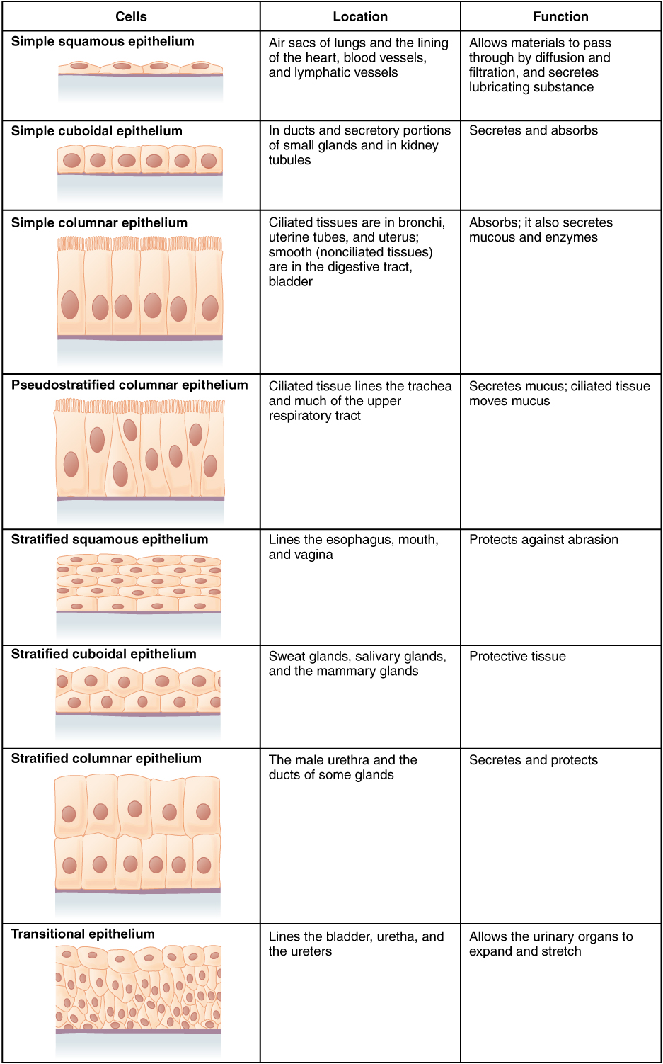
So far, we have identified epithelial tissue based on shape and layering. The representative diagrams we have seen so far are helpful for visualizing the tissue structures, but it is important to look at real examples of these cells. Since cells are too tiny to see with the naked eye, we rely on microscopes to help us study them. Histology is the study of the microscopic anatomy and cells and tissues. See Table 7.4.2 to see some examples of slides of epithelial tissues prepared for the purpose of histology.
Table 7.4.2
Epithelial Tissues and Histological Samples
| Epithelial Tissue Type | Tissue Diagram | Histological Sample |
| Stratified squamous
(from skin) |
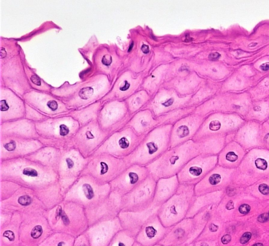 |
|
| Simple cuboidal
(from kidney tubules) |
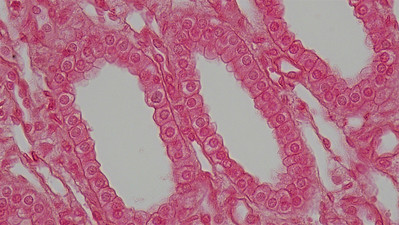 |
|
| Pseudostratified ciliated columnar
(from trachea) |
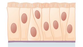 |
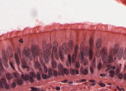 |
Connective Tissue
Bone and blood are examples of connective tissue. Connective tissue is very diverse. In general, it forms a framework and support structure for body tissues and organs. It's made up of living cells separated by non-living material, called extracellular matrix, which can be solid or liquid. The extracellular matrix of bone, for example, is a rigid mineral framework. The extracellular matrix of blood is liquid plasma.
The key identifying feature of connective tissue is that is is composed of a scattering of cells in a non-cellular matrix. There are three main categories of connective tissue, based on the nature of the matrix. They look very different from one another, which is a reflection of their different functions:
- Fibrous connective tissue: is characterized by a matrix which is flexible and is made of protein fibres including collagen, elastin and possibly reticular fibres. These tissues are found making up tendons, ligaments, and body membranes.
- Supportive connective tissue: is characterized by a solid matrix and is what is used to make bone and cartilage. These tissues are used for support and protection.
- Fluid connective tissue: is characterized by a fluid matrix and includes both blood and lymph.
Fibrous Connective Tissue
Fibrous connective tissue contains cells called fibroblasts. These cells produce fibres of collagen, elastin, or reticular fibre which makes up the matrix of this type of connective tissue. Based on how tightly packed these fibres are and how they are oriented changes the properties, and therefore the function of the fibrous connective tissue.
- Loose fibrous connective tissue: composed of a loose and disorganized weave of collagen and elastin fibres, creating a tissue that is thin and flexible, yet still tough. This tissue, which is also sometimes referred to as "areolar tissue", is found in membranes and surrounding blood vessels and most body organs. As you can see from the diagram in Figure 7.4.4, loose fibrous connective tissue fulfills the definition of connectives tissue since it is a scattering of cells (fibroblasts) in a non-cellular matrix (a mesh of collagen and elastin fibres). There are two types of specialized loose fibrous connective tissue: reticular and adipose. Adipose tissue stores fat and reticular tissue forms the spleen and lymph nodes.
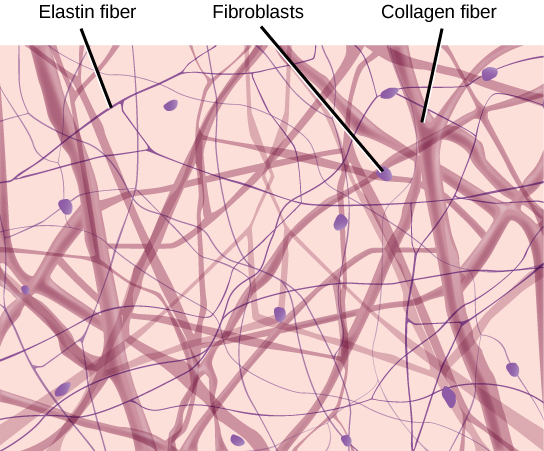
Figure 7.4.4 Diagram of loose fibrous connective tissue consists of a scattering of fibroblasts in a non-cellular matrix of loosely woven collagen and elastin fibres. 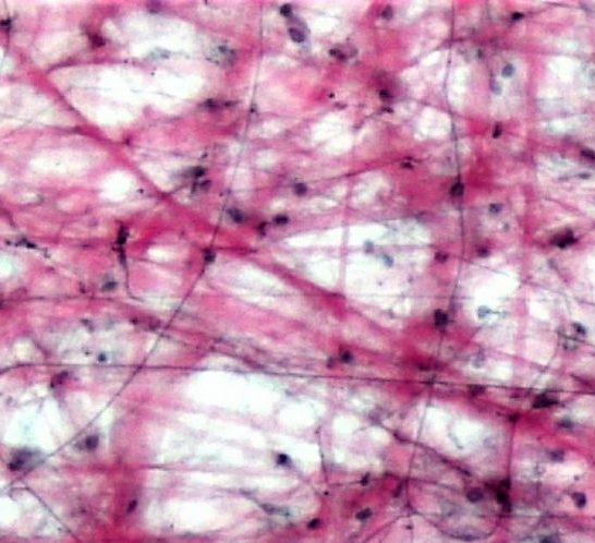
Figure 7.4.5 Microscopic view of loose fibrous connective tissue. - Dense Fibrous Connective Tissue: composed of a dense mat of parallel collagen fibres and a scattering of fibroblasts, creating a tissue that is very strong. Dense fibrous connective tissue forms tendons and ligaments, which connect bones to muscles and/or bones to neighbouring bones.
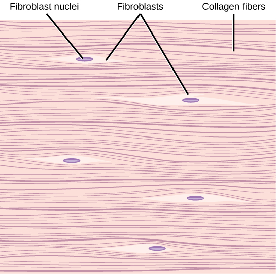
Figure 7.4.6 Dense fibrous connective tissue is composed of fibroblasts and a dense parallel packing of collagen fibres. 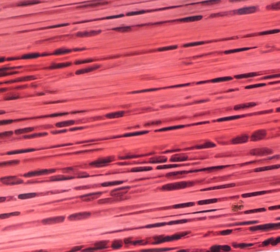
Figure 7.4.7 Microscopic view of dense fibrous connective tissue.
Supportive Connective Tissue
Supportive connective tissue exhibits the defining feature of connective tissue in that it is a scattering of cells in a non-cellular matrix; what sets it apart from other connective tissues is its solid matrix. In this tissue group, the matrix is solid- either bone or cartilage. While fibrous connective tissue contained cells called fibroblasts which produced fibres, supportive connective tissue contains cells that either create bone (osteocytes) or cells that create cartilage (chondrocytes).
Cartilage
Chondrocytes produce the cartilage matrix in which they reside. Cartilage is made up of protein fibres and chondrocytes in lacunae. This is tissue is strong yet flexible and is used many places in the body for protection and support. Cartilage is one of the few tissues that is not vascular (doesn't have a direct blood supply) meaning it relies on diffusion to obtain nutrients and gases; this is the cause of slow healing rates in injuries involving cartilage. There are three main types of cartilage:
- Hyaline cartilage: a smooth, strong and flexible tissue. Found at the ends of ribs and long bones, in the nose, and comprising the entire fetal skeleton.
- Fibrocartilage: a very strong tissue containing thick bundles of collagen. Found in joints that need cushioning from high impact (knees, jaw).
- Elastic cartilage: contains elastic fibres in addition to collagen, giving support with the benefit of elasticity. Found in earlobes and the epiglottis.
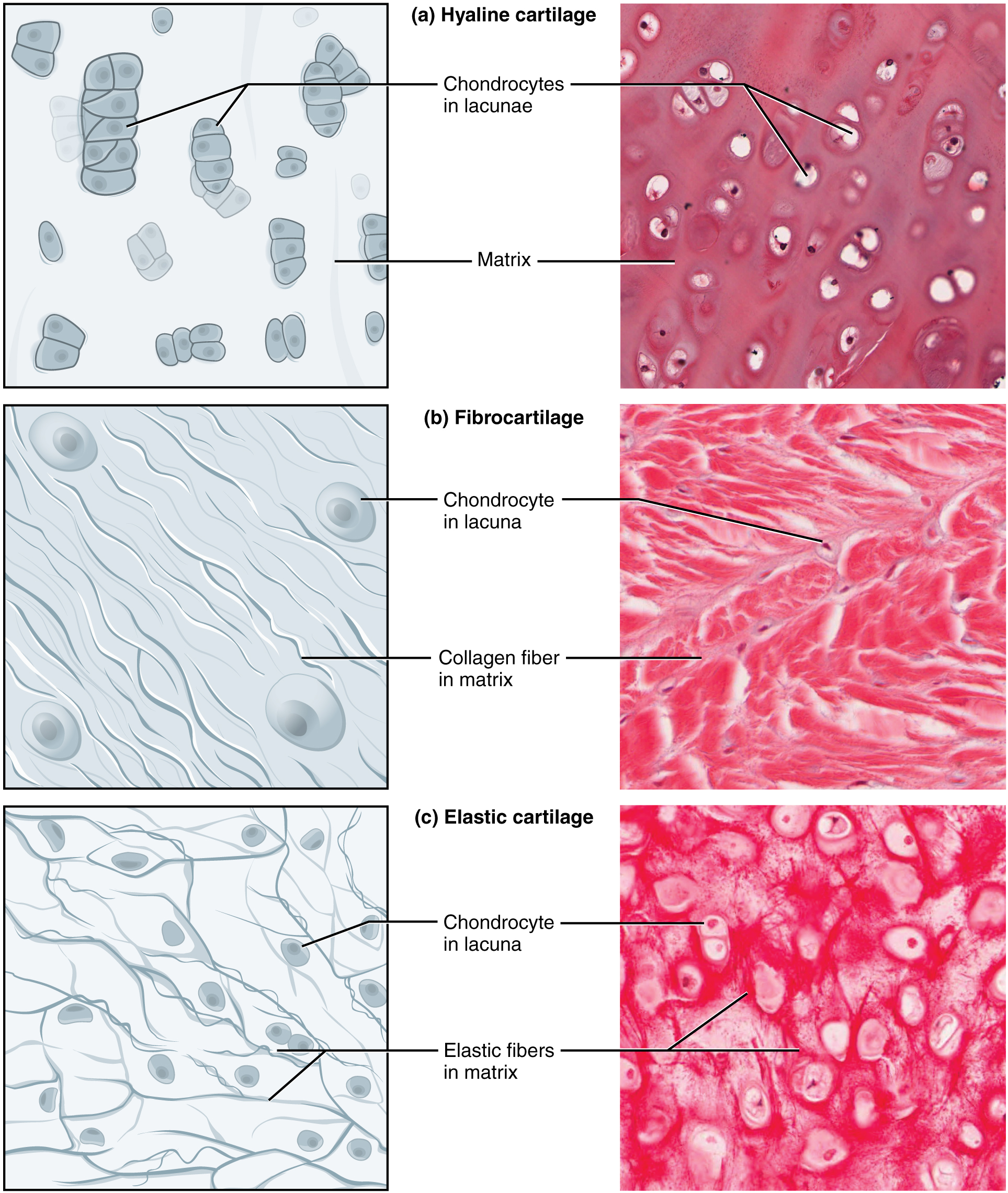
Figure 7.4.8 Three types of cartilage, each with distinct characteristics based on the nature of the matrix.
Bone
Osteocytes produce the bone matrix in which they reside. Since bone is very solid, these cells reside in small spaces called lacunae. This bone tissue is composed of collagen fibres embedded in calcium phosphate giving it strength without brittleness. There are two types of bone: compact and spongy.
- Compact bone: has a dense matrix organized into cylindrical units called osteons. Each osteon contains a central canal (sometimes called a Harversian Canal) which allows for space for blood vessels and nerves, as well as concentric rings of bone matrix and osteocytes in lacunae, as per the diagram here. Compact bone is found in long bones and forms a shell around spongy bone.
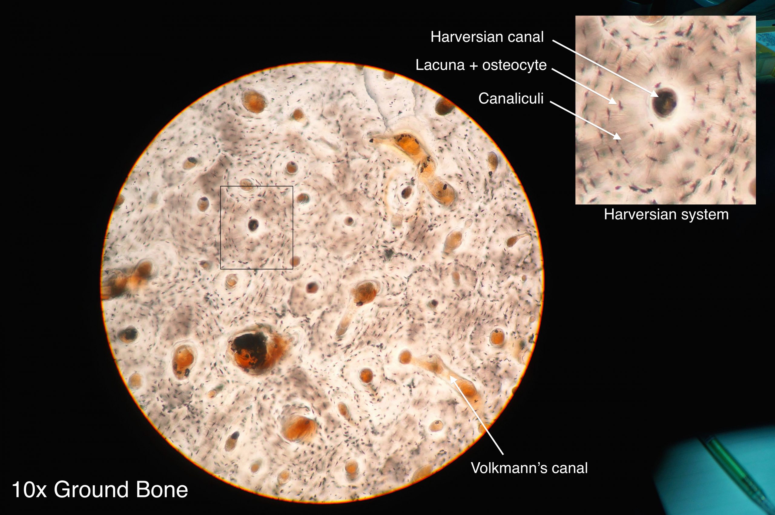
- Spongy bone: a very porous type of bone which most often contains bone marrow. It is found at the end of long bones, and makes up the majority of the ribs, shoulder blades and flat bones of the cranium.
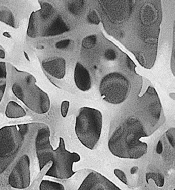
Fluid Connective Tissue
Fluid connective tissue has a matrix that is fluid; unlike the other two categories of connective tissue, the cells that reside in the matrix do not actually produce the matrix. Fibroblasts make the fibrous matrix, chondrocytes make the cartilaginous matrix, osteocytes make the bony matrix, yet blood cells do not make the fluid matrix of either lymph or plasma. This tissue still fits the definition of connective tissue in that it is still a scattering of cells in a non-cellular matrix.
There are two types of fluid connective tissue:
- Blood: blood contains three types of cells suspended in plasma, and is contained in the cardiovascular system.
- Eryththrocytes, more commonly called red blood cells, are present in high numbers (roughly 5 million cells per mL) and are responsible for delivering oxygen from to the lungs to all the other areas of the body. These cells are relatively small in size with a diameter of around 7 micrometres and live no longer than 120 days.
- Leukocytes, often referred to as white blood cells, are present in lower numbers (approximately 5 thousand cells per mL) are responsible for various immune functions. They are typically larger than erythrocytes, but can live much longer, particularly white blood cells responsible for long term immunity. The number of leukocytes in your blood can go up or down based on whether or not you are fighting an infection.
- Thrombocytes, also known as platelets, are very small cells responsible for blood clotting. Thrombocytes are not actually true cells, they are fragments of a much larger cell called a megakaryocyte.
- Lymph: contains a liquid matrix and white blood cells and is contained in the lymphatic system, which ultimately drains into the cardiovascular system.
Figure 7.4.11 A stained lymphocyte surrounded by red blood cells viewed using a light microscope.
Muscular Tissue
Muscular tissue is made up of cells that have the unique ability to contract- which is the defining feature of muscular tissue. There are three major types of muscle tissue, as pictured in Figure 7.4.12 skeletal, smooth, and cardiac muscle tissues.
Skeletal Muscle
Skeletal muscles are voluntary muscles, meaning that you exercise conscious control over them. Skeletal muscles are attached to bones by tendons, a type of connective tissue. When these muscles shorten to pull on the bones to which they are attached, they enable the body to move. When you are exercising, reading a book, or making dinner, you are using skeletal muscles to move your body to carry out these tasks.
Under the microscope, skeletal muscles are striated (or striped) in appearance, because of their internal structure which contains alternating protein fibres of actin and myosin. Skeletal muscle is described as multinucleated, meaning one "cell" has many nuclei. This is because in utero, individual cells destined to become skeletal muscle fused, forming muscle fibres in a process known as myogenesis. You will learn more about skeletal muscle and how it contracts in the Muscular System.
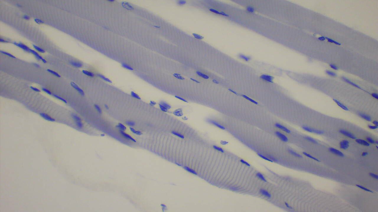
Smooth Muscle
Smooth muscles are nonstriated muscles- they still contain the muscle fibres actin and myosin, but not in the same alternating arrangement seen in skeletal muscle. Smooth muscle is found in the tubes of the body - in the walls of blood vessels and in the reproductive, gastrointestinal, and respiratory tracts. Smooth muscles are not under voluntary control meaning that they operate unconsciously, via the autonomic nervous system. Smooth muscles move substances through a wave of contraction which cascades down the length of a tube, a process termed peristalsis.
Watch the YouTube video "What is Peristalsis" by Mister Science to see peristalsis in action.
https://www.youtube.com/watch?v=kVjeNZA5pi4
What is Peristalsis, Mister Science, 2018.
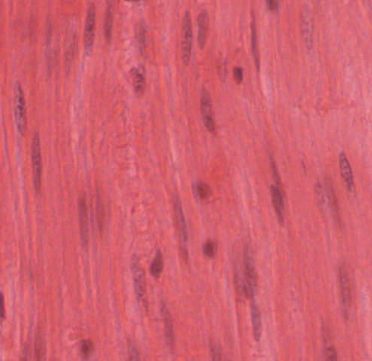
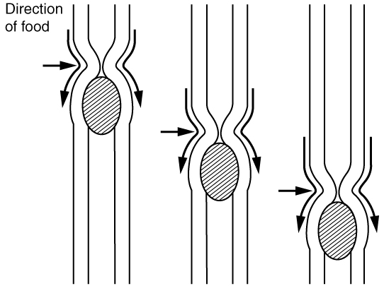
Cardiac Muscle
Cardiac muscles work involuntarily, meaning they are regulated by the autonomic nervous system. This is probably a good thing, since you wouldn't want to have to consciously concentrate on keeping your heart beating all the time! Cardiac muscle, which is found only in the heart, is mononucleated and striated (due to alternating bands of myosin and actin). Their contractions cause the heart to pump blood. In order to make sure entire sections of the heart contract in unison, cardiac muscle tissue contains special cell junctions called intercalated discs, which conduct the electrical signals used to "tell" the chambers of the heart when to contract.
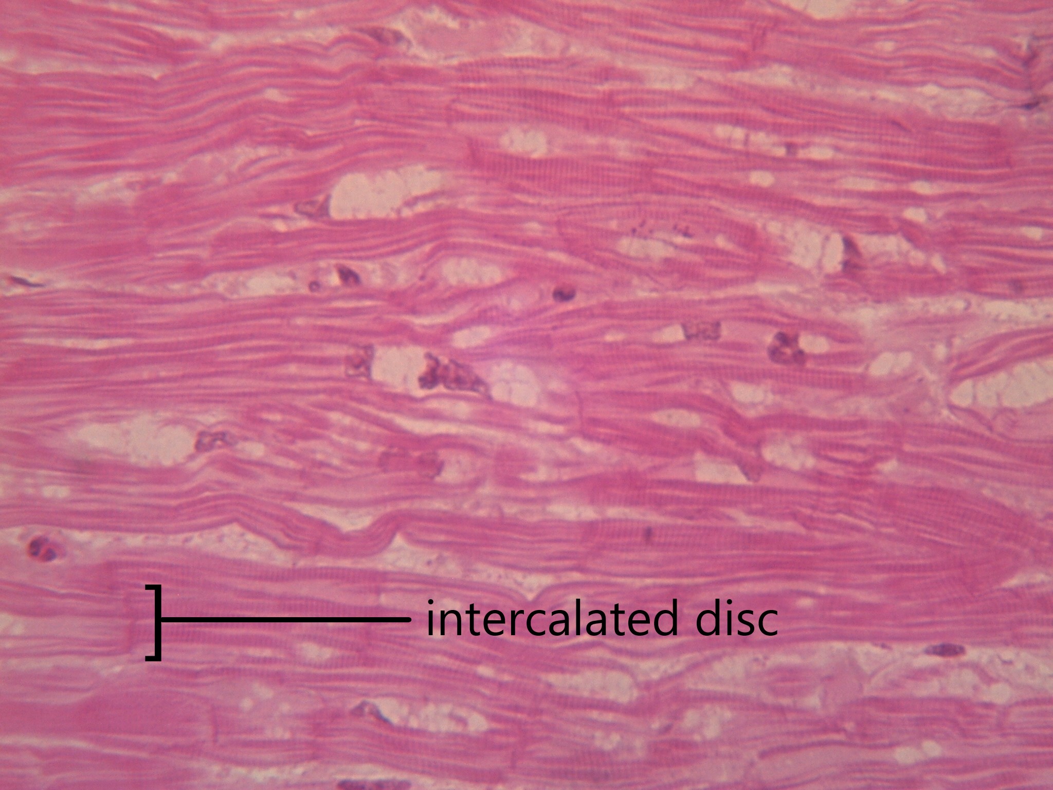
Nervous Tissue
Nervous tissue is made up of neurons and a group of cells called neuroglia (also known as glial cells). Nervous tissue makes up the central nervous system (mainly the brain and spinal cord) and peripheral nervous system (the network of nerves that runs throughout the rest of the body). The defining feature of nervous tissue is that it is specialized to be able to generate and conduct nerve impulses. This function is carried out by neurons, and the purpose of neuroglia is to support neurons.
A neuron has several parts to its structure:
- Dendrites which collect incoming nerve impulses
- A cell body, or soma, which contains the majority of the neuron's organelles, including the nucleus
- An axon, which carries nerve impulses away from the soma, to the next neuron in the chain
- A myelin sheath, which encases the axon and increases that rate at which nerve impulses can be conducted
- Axon terminals, which maintain physical contact with the dendrites of neighbouring neurons
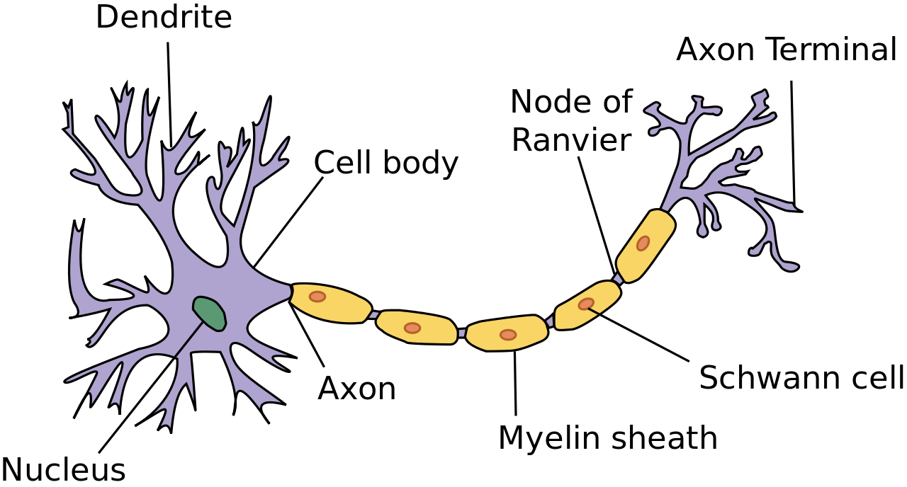
Neuroglia can be understood as support staff for the neuron. The neurons have such an important job, they need cells to bring them nutrients, take away cell waste, and build their mylein sheath. There are many types of neuroglia, which are categorized based on their function and/or their location in the nervous system. Neuroglia outnumber neurons by as much as 50 to 1, and are much smaller. See the diagram in 7.4.17 to compare the size and number of neurons and neuroglia.
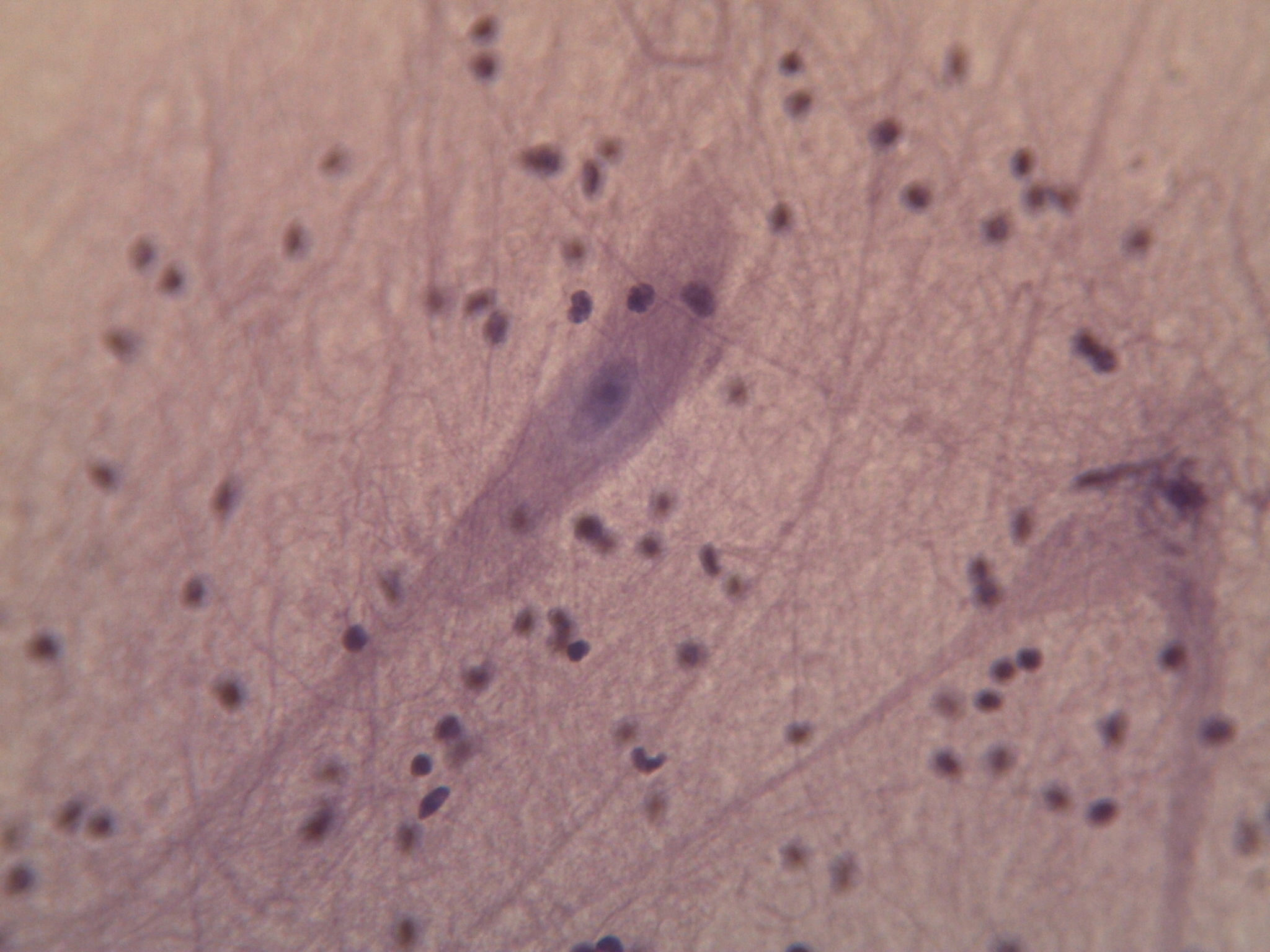
Try out this memory game to test your tissues knowledge:
7.4 Summary
- Tissues are made up of cells working together.
- There are four main types of tissues: epithelial, connective, muscular and nervous.
- Epithelial tissue makes up the linings and coverings of the body and is characterized by having a free surface and a basement membrane. Types of epithelial tissue are distinguished by shape of cell (squamous, cuboidal or columnar) and layering (simple, stratified, pseudostratified and transitional). Different epithelial tissues can carry out diffusion, secretion, absorption, and/or protection depending on their particular cell shape and layering.
- Connective tissue provides structure and support for the body and is characterized as a scattering of cells in a non-cellular matrix. There are three main categories of connective tissue, each characterized by a particular type of matrix:
- Fibrous connective tissue contains protein fibres. Both loose and dense fibrous connective tissue belong in this category.
- Supportive connective tissue contains a very solid matrix, and includes both bone and cartilage.
- Fluid connective tissue contains cells in a fluid matrix with the two types of blood and lymph.
- Muscular tissue's defining feature is that it is contractile. There are three types of muscular tissue: skeletal muscle which is found attached to the skeleton for voluntary movement, smooth muscle which moves substances through body tubes, and cardiac muscle which moves blood through the heart.
- Nervous tissue contains specialized cells called neurons which can conduct electrical impulses. Also found in nervous tissue are neuroglia, which support neurons by providing nutrients, removing wastes, and creating myelin sheath.
7.4 Review Questions
- Define the term tissue.
-
- If a part of the body needed a lining that was both protective, but still able to absorb nutrients, what would be the best type of epithelial tissue to use?
-
- Where do you find skeletal muscle? Smooth muscle? Cardiac muscle?
-
- What are some of the functions of neuroglia?
-
7.4 Explore More
https://www.youtube.com/watch?v=O0ZvbPak4ck
Types of Human Body Tissue, MoomooMath and Science, 2017.
https://www.youtube.com/watch?v=uHbn7wLN_3k
How to 3D print human tissue - Taneka Jones, TED-Ed, 2019.
https://www.youtube.com/watch?v=1Qfmkd6C8u8
How bones make blood - Melody Smith, TED-Ed, 2020.
Attributions
Figure 7.4.1
- Construction man kneeling in front of wall by Charles Deluvio on Unsplash is used under the Unsplash License (https://unsplash.com/license).
- Beige wooden frame by Charles Deluvio on Unsplash is used under the Unsplash License (https://unsplash.com/license).
- Tambour on green by Pierre Châtel-Innocention Unsplash is used under the Unsplash License (https://unsplash.com/license).
- Tags: Construction Studs Plumbing Wiring by JWahl on Pixabay is used under the Pixabay License (https://pixabay.com/es/service/license/).
Figure 7.4.2 and Figure 7.4.3
- Simple columnar epithelium tissue by Kamil Danak on Wikimedia Commons is used under a CC BY-SA 3.0 (https://creativecommons.org/licenses/by-sa/3.0/deed.en) license.
- Simple cuboidal epithelium by Kamil Danak on Wikimedia Commons is used under a CC BY-SA 3.0 (https://creativecommons.org/licenses/by-sa/3.0/deed.en) license.
- Simple squamous epithelium by Kamil Danak on Wikimedia Commons is used under a CC BY-SA 3.0 (https://creativecommons.org/licenses/by-sa/3.0/deed.en) license.
Figure 7.4.4
Loose fibrous connective tissue by CNX OpenStax. Biology. on Wikimedial Commons is used under a CC BY 4.0. (https://creativecommons.org/licenses/by/4.0) license.
Figure 7.4.5
Connective Tissue: Loose Aerolar by Berkshire Community College Bioscience Image Library on Flickr is used under a CC0 1.0 Universal public domain dedication (https://creativecommons.org/publicdomain/zero/1.0/) license.
Figure 7.4.6
Dense Fibrous Connective Tissue by by CNX OpenStax. Biology. on Wikimedial Commons is used under a CC BY 4.0. (https://creativecommons.org/licenses/by/4.0) license.
Figure 7.4.7
Dense_connective_tissue-400x by J Jana on Wikimedia Commons is used under a CC BY-SA 4.0 (https://creativecommons.org/licenses/by-sa/4.0/deed.en) license.
Figure 7.4.8
Types_of_Cartilage-new by OpenStax College on Wikipedia Commons is used under a CC BY 3.0 (https://creativecommons.org/licenses/by/3.0) license.
Figure 7.4.9
Compact_bone_histology_2014 by Athikhun.suw on Wikimedia Commons is used under a CC BY-SA 4.0 (https://creativecommons.org/licenses/by-sa/4.0/deed.en) license.
Figure 7.4.10
Bone_normal_and_degraded_micro_structure by Gtirouflet on Wikimedia Commons is used under a CC BY-SA 3.0 (https://creativecommons.org/licenses/by-sa/3.0/deed.en) license.
Figure 7.4.11
Lymphocyte2 by NicolasGrandjean on Wikimedia Commons is used under a CC BY-SA 3.0 (https://creativecommons.org/licenses/by-sa/3.0/deed.en) license. [No machine-readable author provided. NicolasGrandjean is assumed, based on copyright claims.]
Figure 7.4.12
Skeletal_muscle_横纹肌1 by 乌拉跨氪 on Wikimedia Commons is used under a CC BY-SA 4.0 (https://creativecommons.org/licenses/by-sa/4.0/deed.en) license.
Figure 7.4.13
Smooth_Muscle_new by OpenStax on Wikimedia Commons is used under a CC BY 4.0 (https://creativecommons.org/licenses/by/4.0/deed.en) license.
Figure 7.4.14
Peristalsis by OpenStax on Wikimedia Commons is used under a CC BY 3.0 (https://creativecommons.org/licenses/by/3.0/deed.en) license.
Figure 7.4.15
400x Cardiac Muscle by Jessy731 on Flickr is used and adapted by Christine Miller under a CC BY-NC 2.0 (https://creativecommons.org/licenses/by-nc/2.0/) license.
Figure 7.4.16
Neuron.svg by User:Dhp1080 on Wikimedia Commons is used under a CC BY-SA 3.0 (https://creativecommons.org/licenses/by-sa/3.0/deed.en) license.
Figure 7.4.17
400x Nervous Tissue by Jessy731 on Flickr is used under a CC BY-NC 2.0 (https://creativecommons.org/licenses/by-nc/2.0/) license.
Table 7.4.1
Summary of Epithelial Tissue Cells, by OpenStax College on Wikipedia Commons is used under a CC BY 3.0 (https://creativecommons.org/licenses/by/3.0) license.
Table 7.4.2
- Epithelial_Tissues_Stratified_Squamous_Epithelium_(40230842160) by
Berkshire Community College Bioscience Image Library on Wikimedia Commons is used under a CC0 1.0 Universal Public Domain Dedication (https://creativecommons.org/publicdomain/zero/1.0/) license. - Simple cuboidal epithelial tissue histology by Berkshire Community College on Flickr is used under a CC0 1.0 Universal Public Domain Dedication (https://creativecommons.org/publicdomain/zero/1.0/) license.
- Pseudostratified_Epithelium by OpenStax College on Wikimedia Commons is used under a CC BY 3.0 (https://creativecommons.org/licenses/by/3.0) license.
References
Betts, J. G., Young, K.A., Wise, J.A., Johnson, E., Poe, B., Kruse, D.H., Korol, O., Johnson, J.E., Womble, M., DeSaix, P. (2013, April 25). Figure 4.8 Summary of epithelial tissue cells [digital image]. In Anatomy and Physiology (Section 4.2). OpenStax. https://openstax.org/books/anatomy-and-physiology/pages/4-2-epithelial-tissue
Betts, J. G., Young, K.A., Wise, J.A., Johnson, E., Poe, B., Kruse, D.H., Korol, O., Johnson, J.E., Womble, M., DeSaix, P. (2013, April 25). Figure 4.16 Types of cartilage [digital image]. In Anatomy and Physiology (Section 4.3). OpenStax. https://openstax.org/books/anatomy-and-physiology/pages/4-3-connective-tissue-supports-and-protects
Betts, J. G., Young, K.A., Wise, J.A., Johnson, E., Poe, B., Kruse, D.H., Korol, O., Johnson, J.E., Womble, M., DeSaix, P. (2013, April 25). Figure 10.23 Smooth muscle [digital micrograph]. In Anatomy and Physiology (Section 10.8). OpenStax. https://openstax.org/books/anatomy-and-physiology/pages/10-8-smooth-muscle (Micrograph provided by the Regents of University of Michigan Medical School © 2012)
Betts, J. G., Young, K.A., Wise, J.A., Johnson, E., Poe, B., Kruse, D.H., Korol, O., Johnson, J.E., Womble, M., DeSaix, P. (2013, April 25). Figure 22.5 Pseudostratified ciliated columnar epithelium [digital micrograph]. In Anatomy and Physiology (Section 22.1). OpenStax. https://openstax.org/books/anatomy-and-physiology/pages/22-1-organs-and-structures-of-the-respiratory-system
Betts, J. G., Young, K.A., Wise, J.A., Johnson, E., Poe, B., Kruse, D.H., Korol, O., Johnson, J.E., Womble, M., DeSaix, P. (2013, April 25). Figure 23.5 Peristalsis [diagram]. In Anatomy and Physiology (Section 23.2). OpenStax. https://openstax.org/books/anatomy-and-physiology/pages/23-2-digestive-system-processes-and-regulation
Mister Science. (2018). What is peristalsis? YouTube. https://www.youtube.com/watch?v=kVjeNZA5pi4
MoomooMath and Science. (2017, May 18). Types of human body tissue. YouTube. https://www.youtube.com/watch?v=O0ZvbPak4ck&feature=youtu.be
Open Stax. (2016, May 27). Figure 6 Loose connective tissue [digital image]. In OpenStax Biology (Section 33.2). OpenStax CNX. https://cnx.org/contents/GFy_h8cu@10.53:-LfhWRES@4/Animal-Primary-Tissues
Open Stax. (2016, May 27). Figure 7 Fibrous connective tissue from the tendon [digital image]. In OpenStax Biology (Section 33.2). OpenStax CNX. https://cnx.org/contents/GFy_h8cu@10.53:-LfhWRES@4/Animal-Primary-Tissues
TED-Ed. (2019, October 17). How to 3D print human tissue - Taneka Jones. YouTube. https://www.youtube.com/watch?v=uHbn7wLN_3k&feature=youtu.be
TED-Ed. (2020, January 27). How bones make blood - Melody Smith. YouTube. https://www.youtube.com/watch?v=1Qfmkd6C8u8&feature=youtu.be
Image shows a diagram illustrating how peristalsis pushes food through the digestive tract by squeezing just behind the food, pushing it forward.


