18.2 Introduction to the Reproductive System
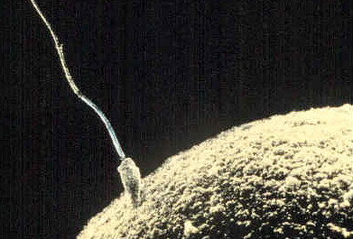
It’s All about Sex
A tiny sperm from dad breaks through the surface of a huge egg from mom. Voilà! In nine months, a new son or daughter will be born. Like most other multicellular organisms, human beings reproduce sexually. In human sexual reproduction, males produce sperm and females produce eggs, and a new offspring forms when a sperm unites with an egg. How do sperm and eggs form? And how do they arrive together at the right place and time so they can unite to form a new offspring? These are functions of the reproductive system.
What Is the Reproductive System?
The reproductive system is the human organ system responsible for the production and fertilization of gametes (sperm or eggs) and, in females, the carrying of a fetus. Both male and female reproductive systems have organs called gonads that produce gametes. A gamete is a haploid cell that combines with another haploid gamete during fertilization, forming a single diploid cell called a zygote. Besides producing gametes, the gonads also produce sex hormones. Sex hormones are endocrine hormones that control the development of sex organs before birth, sexual maturation at puberty, and reproduction once sexual maturation has occurred. Other reproductive system organs have various functions, such as maturing gametes, delivering gametes to the site of fertilization, and providing an environment for the development and growth of an offspring.
Sex Differences in the Reproductive System
The reproductive system is the only human organ system that is significantly different between males and females. Embryonic structures that will develop into the reproductive system start out the same in males and females, but by birth, the reproductive systems have differentiated. How does this happen?
Sex Differentiation
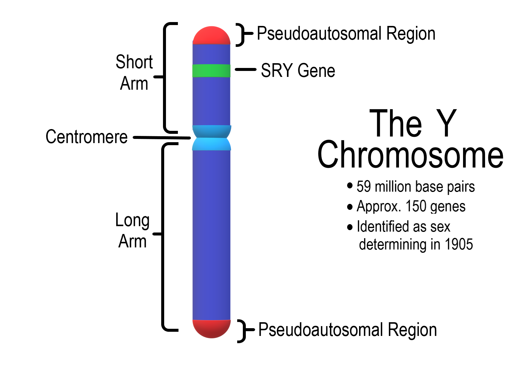
Starting around the seventh week after conception in genetically male (XY) embryos, a gene called SRY on the Y chromosome (shown in Figure 18.2.2) initiates the production of multiple proteins. These proteins cause undifferentiated gonadal tissue to develop into male gonads (testes). The male gonads then secrete hormones — including the male sex hormone testosterone — that trigger other changes in the developing offspring (now called a fetus), causing it to develop a complete male reproductive system. Without a Y chromosome, an embryo will develop female gonads (ovaries) that will produce the female sex hormone estrogen. Estrogen, in turn, will lead to the formation of the other organs of a normal female reproductive system.
Homologous Structures
Undifferentiated embryonic tissues develop into different structures in male and female fetuses. Structures that arise from the same tissues in males and females are called homologous structures. The male testes and female ovaries, for example, are homologous structures that develop from the undifferentiated gonads of the embryo. Likewise, the male penis and female clitoris are homologous structures that develop from the same embryonic tissues.
Sex Hormones and Maturation
Male and female reproductive systems are different at birth, but they are immature and incapable of producing gametes or sex hormones. Maturation of the reproductive system occurs during puberty, when hormones from the hypothalamus and pituitary gland stimulate the testes or ovaries to start producing sex hormones again. The main sex hormones are testosterone in males and estrogen in females. Sex hormones, in turn, lead to the growth and maturation of the reproductive organs, rapid body growth, and the development of secondary sex characteristics. Secondary sex characteristics are traits that are different in mature males and females, but are not directly involved in reproduction. They include facial hair in males and breasts in females.
Male Reproductive System
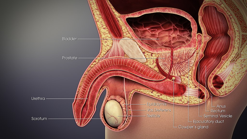
The main structures of the male reproductive system are external to the body and illustrated in Figure 18.2.3. The two testes (singular, testis) hang between the thighs in a sac of skin called the scrotum. The testes produce both sperm and testosterone. Resting atop each testis is a coiled structure called the epididymis (plural, epididymes). The function of the epididymes is to mature and store sperm. The penis is a tubular organ that contains the urethra and has the ability to stiffen during sexual arousal. Sperm passes out of the body through the urethra during a sexual climax (orgasm). This release of sperm is called ejaculation.
In addition to these organs, the male reproductive system consists of several ducts and glands that are internal to the body. The ducts, which include the vas deferens (also called the ductus deferens), transport sperm from the epididymis to the urethra. The glands, which include the prostate gland and seminal vesicles, produce fluids that become part of semen. Semen is the fluid that carries sperm through the urethra and out of the body. It contains substances that control pH and provide sperm with nutrients for energy.
Female Reproductive System
The main structures of the female reproductive system are internal to the body and shown in the following figure. They include the paired ovaries, which are small, ovoid structures that produce ova and secrete estrogen. The two oviducts (sometimes called Fallopian tubes or uterine tubes) start near the ovaries and end at the uterus. Their function is to transport ova from the ovaries to the uterus. If an egg is fertilized, it usually occurs while it is traveling through an oviduct. The uterus is a pear-shaped muscular organ that functions to carry a fetus until birth. It can expand greatly to accommodate a growing fetus, and its muscular walls can contract forcefully during labour to push the baby out of the uterus and into the vagina. The vagina is a tubular tract connecting the uterus to the outside of the body. The vagina is where sperm are usually deposited during sexual intercourse and ejaculation. The vagina is also called the birth canal because a baby travels through the vagina to leave the body during birth.
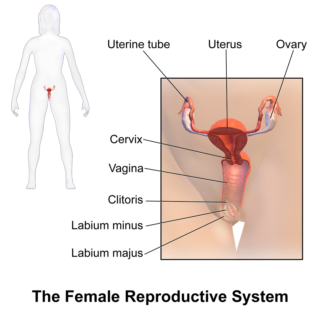
The external structures of the female reproductive system are referred to collectively as the vulva. They include the clitoris, which is homologous to the male penis. They also include two pairs of labia (singular, labium), which surround and protect the openings of the urethra and vagina.
18.2 Summary
- The reproductive system is the human organ system responsible for the production and fertilization of gametes and, in females, the carrying of a fetus.
- Both male and female reproductive systems have organs called gonads (testes in males, ovaries in females) that produce gametes (sperm or ova) and sex hormones (such as testosterone in males and estrogen in females). Sex hormones are endocrine hormones that control the prenatal development of reproductive organs, sexual maturation at puberty, and reproduction after puberty.
- The reproductive system is the only organ system that is significantly different between males and females. A Y-chromosome gene called SRY is responsible for undifferentiated embryonic tissues developing into a male reproductive system. Without a Y chromosome, the undifferentiated embryonic tissues develop into a female reproductive system.
- Structures such as testes and ovaries that arise from the same undifferentiated embryonic tissues in males and females are called homologous structures.
- Male and female reproductive systems are different at birth, but at that point, they are immature and nonfunctioning. Maturation of the reproductive system occurs during puberty, when hormones from the hypothalamus and pituitary gland stimulate the gonads to produce sex hormones again. The sex hormones, in turn, cause the changes of puberty.
- Male reproductive system organs include the testes, epididymis, penis, vas deferens, prostate gland, and seminal vesicles.
- Female reproductive system organs include the ovaries, oviducts, uterus, vagina, clitoris, and labia.
18.2 Review Questions
- What is the reproductive system?
-
- Explain the difference between the vulva and the vagina.
18.2 Explore More
Sex Determination: More Complicated Than You Thought, TED-Ed, 2012.
The evolution of animal genitalia – Menno Schilthuizen, TED-Ed, 2017.
Hormones and Gender Transition, Reactions, 2015.
Attributions
Figure 18.2.1
Sperm-egg by Unknown author on Wikimedia Commons is in the public domain (https://en.wikipedia.org/wiki/public_domain).
Figure 18.2.2
Y Chromosome by Christinelmiller on Wikimedia Commons is used under a CC BY-SA 4.0 (https://creativecommons.org/licenses/by-sa/4.0) license.
Figure 18.2.3
3D_Medical_Animation_Vas_Deferens by https://www.scientificanimations.com on Wikimedia Commons is used under a CC BY-SA 4.0 (https://creativecommons.org/licenses/by-sa/4.0) license.
Figure 18.2.4
Blausen_0399_FemaleReproSystem_01 by BruceBlaus on Wikimedia Commons is used under a CC BY 3.0 (https://creativecommons.org/licenses/by/3.0) license.
References
Blausen.com Staff. (2014). Medical gallery of Blausen Medical 2014. WikiJournal of Medicine 1 (2). DOI:10.15347/wjm/2014.010. ISSN 2002-4436.
Reactions. (2015, June 8). Hormones and gender transition. YouTube. https://www.youtube.com/watch?v=l5knvmy1Z3s&feature=youtu.be
TED-Ed. (2012, April 23). Sex determination: More complicated than you thought. YouTube. https://www.youtube.com/watch?v=kMWxuF9YW38&feature=youtu.be
TED-Ed. (2017, April 24). The evolution of animal genitalia – Menno Schilthuizen. YouTube. https://www.youtube.com/watch?v=vcPJkz-D5II&feature=youtu.be
A biological process which converts sugars such as glucose, fructose, and sucrose into cellular energy, producing ethanol and carbon dioxide as by-products.
Created by: CK-12/Adapted by Christine Miller
Divide and Split
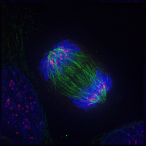
Can you guess what the colourful image in Figure 4.13.1 represents? It shows a eukaryotic cell during the process of cell division. In particular, the image shows the cell in a part of cell division called anaphase, where the DNA is being pulled to opposite ends of the cell. Normally, DNA is located in the nucleus of most human cells. The nucleus divides before the cell itself splits in two, and before the nucleus divides, the cell’s DNA is replicated (or copied). There must be two copies of the DNA so that each daughter cell will have a complete copy of the genetic material from the parent cell. How is the replicated DNA sorted and separated so that each daughter cell gets a complete set of the genetic material? To answer that question, you first need to know more about DNA and the forms it takes.
The Forms of DNA
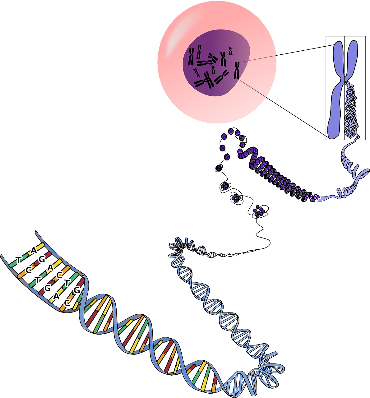
Except when a eukaryotic cell divides, its nuclear DNA exists as a grainy material called chromatin. Only once a cell is about to divide and its DNA has replicated does DNA condense and coil into the familiar X-shaped form of a chromosome, like the one shown below.
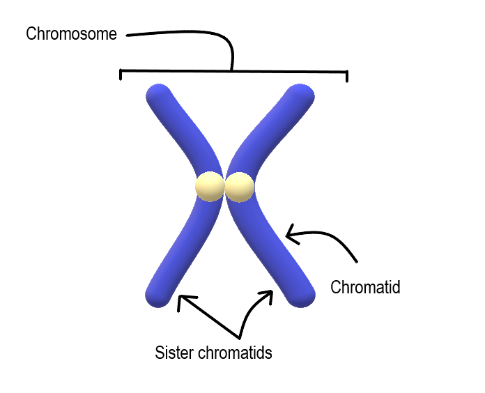
Most cells in the human body have two pairs of 23 different chromosomes, for a total of 46 chromosomes. Cells that have two pairs of chromosomes are called diploid. Because DNA has already replicated when it coils into a chromosome, each chromosome actually consists of two identical structures called sister chromatids. Sister chromatids are joined together at a region called a centromere.
Mitosis
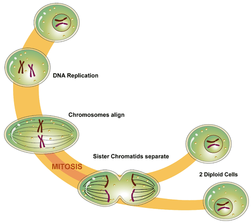
The process in which the nucleus of a eukaryotic cell divides is called mitosis. During mitosis, the two sister chromatids that make up each chromosome separate from each other and move to opposite poles of the cell. This is shown in the figure below.
Mitosis actually occurs in four phases. The phases are called prophase, metaphase, anaphase, and telophase.
Prophase
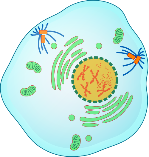
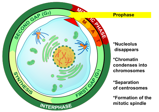
The first and longest phase of mitosis is prophase. During prophase, chromatin condenses into chromosomes, and the nuclear envelope (the membrane surrounding the nucleus) breaks down. In animal cells, the centrioles near the nucleus begin to separate and move to opposite poles of the cell. Centrioles are small organelles found only in eukaryotic cells. They help ensure that the new cells that form after cell division each contain a complete set of chromosomes. As the centrioles move apart, a spindle starts to form between them. The spindle consists of fibres made of microtubules.
Metaphase
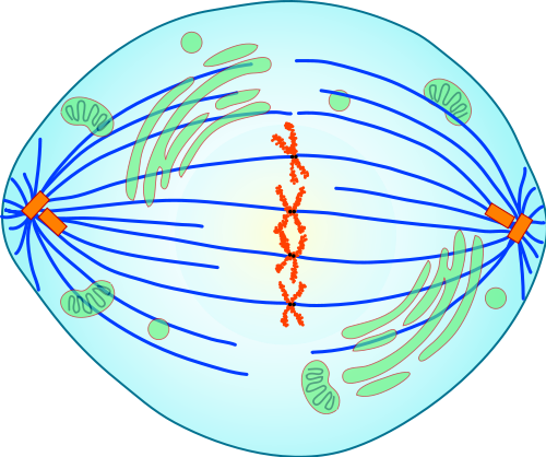
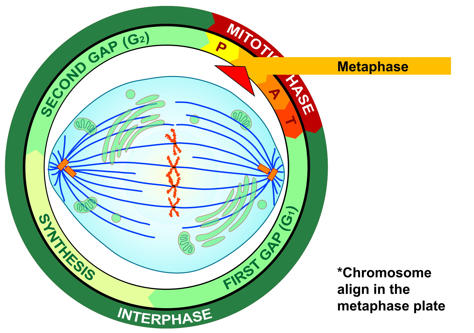
During metaphase, spindle fibres attach to the centromere of each pair of sister chromatids. As you can see in Figure 4.13.7, the sister chromatids line up at the equator (or center) of the cell. The spindle fibres ensure that sister chromatids will separate and go to different daughter cells when the cell divides.
Anaphase
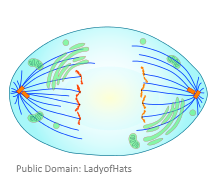
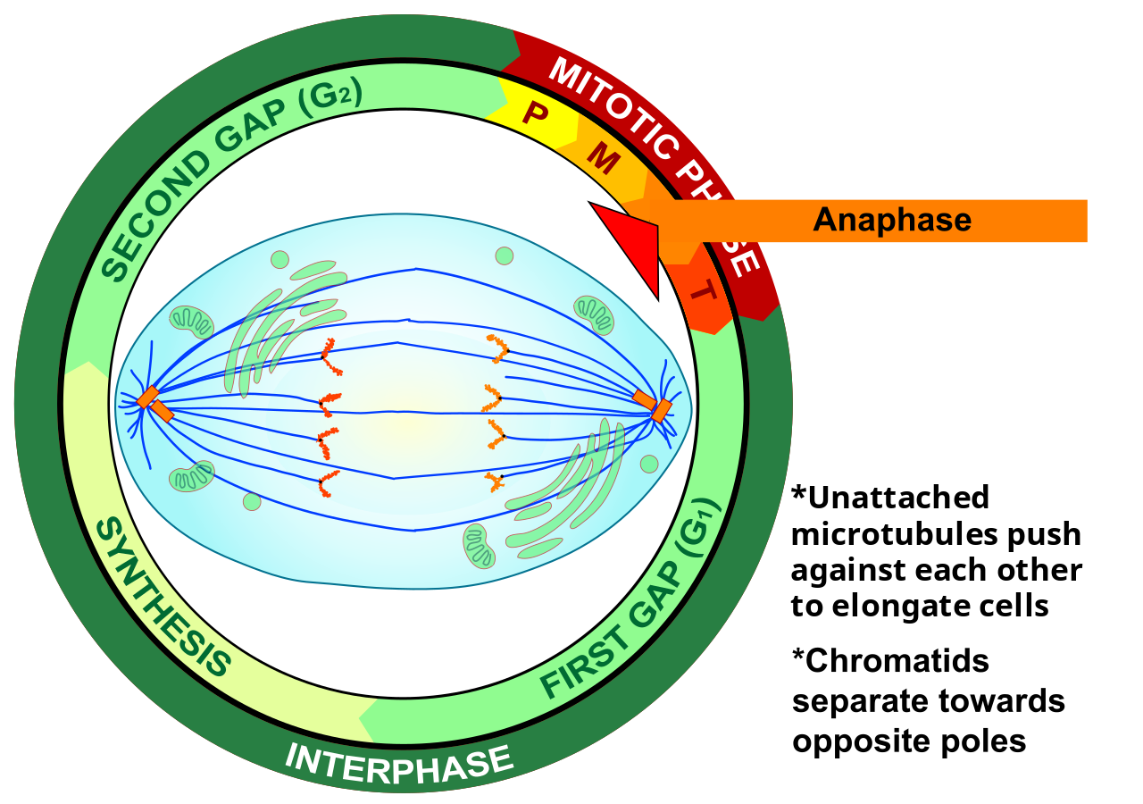
During anaphase, sister chromatids separate and the centromeres divide. The sister chromatids are pulled apart by the shortening of the spindle fibres. This is a little like reeling in a fish by shortening the fishing line. One sister chromatid moves to one pole of the cell, and the other sister chromatid moves to the opposite pole. At the end of anaphase, each pole of the cell has a complete set of chromosomes.
Telophase
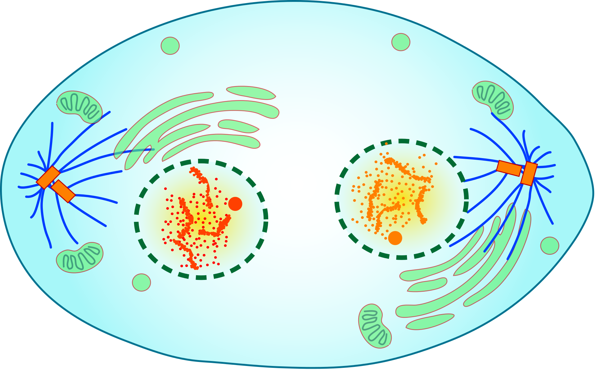
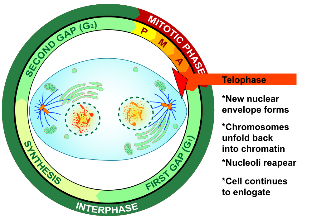
During telophase, the chromosomes begin to uncoil and form chromatin. This prepares the genetic material for directing the metabolic activities of the new cells. The spindle also breaks down, and new nuclear envelopes form.
Cytokinesis

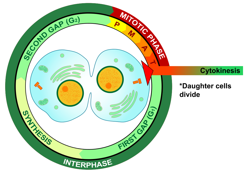
Cytokinesis is the final stage of cell division. During cytokinesis, the cytoplasm splits in two and the cell divides, as shown below. In animal cells, the plasma membrane of the parent cell pinches inward along the cell’s equator until two daughter cells form. Thus, the goal of mitosis and cytokinesis is now complete, because one parent cell has given rise to two daughter cells. The daughter cells have the same chromosomes as the parent cell.
4.13 Summary
- Until a eukaryotic cell divides, its nuclear DNA exists as a grainy material called chromatin. After DNA replicates and the cell is about to divide, the DNA condenses and coils into the X-shaped form of a chromosome. Each chromosome actually consists of two sister chromatids, which are joined together at a centromere.
- Mitosis is the process during which the nucleus of a eukaryotic cell divides. During this process, sister chromatids separate from each other and move to opposite poles of the cell. This happens in four phases: prophase, metaphase, anaphase, and telophase.
- Cytokinesis is the final stage of cell division, during which the cytoplasm splits in two and two daughter cells form.
4.13 Review Questions
- Describe the different forms that DNA takes before and during cell division in a eukaryotic cell.
-
- Identify the four phases of mitosis in an animal cell, and summarize what happens during each phase.
- Order the diagrams of the stages of mitosis:
- Explain what happens during cytokinesis in an animal cell.
- What do you think would happen if the sister chromatids of one of the chromosomes did not separate during mitosis?
- True or False:
4.13 Explore More
https://www.youtube.com/watch?time_continue=3&v=C6hn3sA0ip0&feature=emb_logo
Mitosis, NDSU Virtual Cell Animations project (ndsuvirtualcell), 2012.
https://www.youtube.com/watch?time_continue=19&v=EA0qxhR2oOk&feature=emb_logo
Nondisjunction (Trisomy 21) - An Animated Tutorial, Kristen Koprowski, 2012.
Attributions
Figure 4.13.1
Anaphase_IF by Roy van Heesbeen on Wikimedia Commons is released into the public domain (https://en.wikipedia.org/wiki/Public_domain).
Figure 4.13.2
Chromosomes by OpenClipArt-Vectors on Pixabay is used under the Pixabay License (https://pixabay.com/service/license/).
Figure 4.13.3
Chromosome/ Chromatid/ Sister Chromatid by Christine Miller is released into the public domain (https://en.wikipedia.org/wiki/Public_domain).
Figure 4.13.4
Simple Mitosis by Mariana Ruiz Villarreal [LadyofHats] via CK-12 Foundation is used under a CC BY-NC 3.0 (https://creativecommons.org/licenses/by-nc/3.0/) license.
 ©CK-12 Foundation Licensed under
 ![]() • Terms of Use • Attribution
• Terms of Use • Attribution
Figure 4.13.5
Mitotic Prophase [tiny] by Mariana Ruiz Villarreal [LadyofHats] on Wikimedia Commons is released into the public domain (https://en.wikipedia.org/wiki/Public_domain).
Figure 4.13.6
Prophase Eukaryotic Mitosis by Mariana Ruiz Villarreal [LadyofHats] on Wikimedia Commons is released into the public domain (https://en.wikipedia.org/wiki/Public_domain).
Figure 4.13.7
Mitotic_Metaphase by Mariana Ruiz Villarreal [LadyofHats] on Wikimedia Commons is released into the public domain (https://en.wikipedia.org/wiki/Public_domain).
Figure 4.13.8
Metaphase Eukaryotic Mitosis by Mariana Ruiz Villarreal [LadyofHats] on Wikimedia Commons is released into the public domain (https://en.wikipedia.org/wiki/Public_domain).
Figure 4.13.9
Anaphase [adapted] by Mariana Ruiz Villarreal [LadyofHats] on Wikimedia Commons is released into the public domain (https://en.wikipedia.org/wiki/Public_domain).
Figure 4.13.10
Anaphase_eukaryotic_mitosis.svg by Mariana Ruiz Villarreal [LadyofHats] on Wikimedia Commons is released into the public domain (https://en.wikipedia.org/wiki/Public_domain).
Figure 4.13.11
Mitotic Telophase by Mariana Ruiz Villarreal [LadyofHats] on Wikimedia Commons is released into the public domain (https://en.wikipedia.org/wiki/Public_domain).
Figure 4.13.12
Telophase Eukaryotic Mitosis by Mariana Ruiz Villarreal [LadyofHats] on Wikimedia Commons is released into the public domain (https://en.wikipedia.org/wiki/Public_domain).
Figure 4.13.13
Mitotic Cytokinesis by Mariana Ruiz Villarreal [LadyofHats] on Wikimedia Commons is released into the public domain (https://en.wikipedia.org/wiki/Public_domain).
Figure 4.13.14
Cytokinesis Eukaryotic Mitosis by Mariana Ruiz Villarreal [LadyofHats] on Wikimedia Commons is released into the public domain (https://en.wikipedia.org/wiki/Public_domain).
References
Koprowski, K., Cabey, R. [Kristen Koprowski]. (2012). Nondisjunction (Trisomy 21) - An Animated Tutorial. YouTube. https://www.youtube.com/watch?v=EA0qxhR2oOk&feature=youtu.be
NDSU Virtual Cell Animations project [ndsuvirtualcell]. (2012). Mitosis. YouTube. https://www.youtube.com/watch?v=C6hn3sA0ip0&t=21s
A type of disease in which cells of the central nervous system stop working or die. Neurodegenerative disorders usually get worse over time and have no cure. They may be genetic or be caused by a tumor or stroke.
A mature haploid male or female germ cell which is able to unite with another of the opposite sex in sexual reproduction to form a zygote.
Image shows a man at an oxygen bar. There are several erlenmeyer flasks of differently scented oxygen with tubes coming out of the top of each. The man is wearing a nasal cannula (tube that blows air into your nostrils).
The fusion of haploid gametes, egg and sperm, to form the diploid zygote.
Image shows to small children in a backyard wading pool. One adult is standing by the pool resting their foot on the edge and another adult is sitting nearby in a lawn chair.
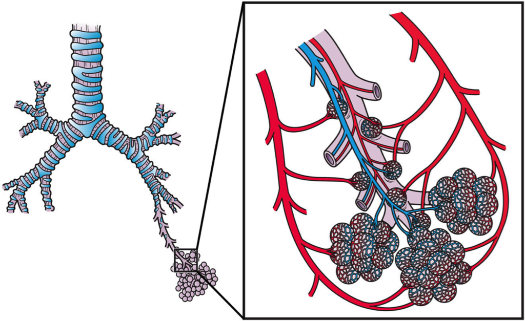
Image shows a diagram of a cluster of alveoli located at the end of a bronchiole. Labelled structures include the alveolar duct (connects bronchiole to alveoli), the pulmonary artery and vein (brings de-oxygenated blood into the lungs and oxygenated blood back to the heart), mucous glands for lubrication and protection, and the alveolar sac (location of gas exchange).
As per caption.
Ant Hill or Plumbing System?
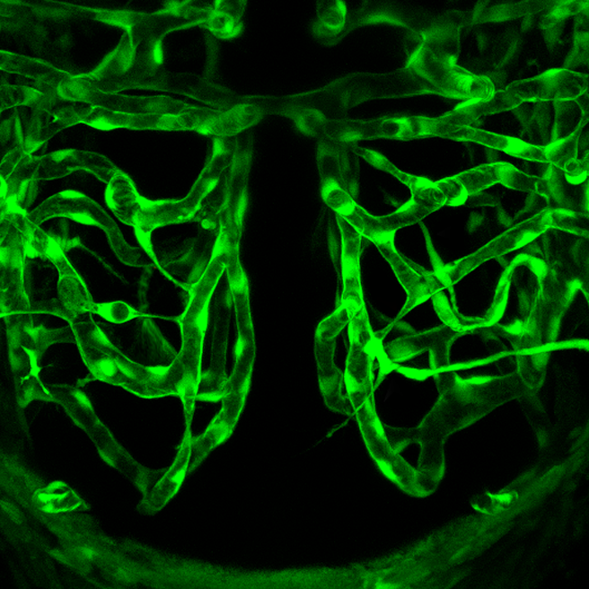
What do you think the picture in Figure 14.2.1 shows? Is it a maze of underground passageways in an ant hill? A network of interconnected pipes in a complex plumbing system? The picture actually shows something that, like ant tunnels and plumbing pipes, functions as a transportation system. It shows a network of blood vessels, which are part of the cardiovascular system.
What is the Cardiovascular System?
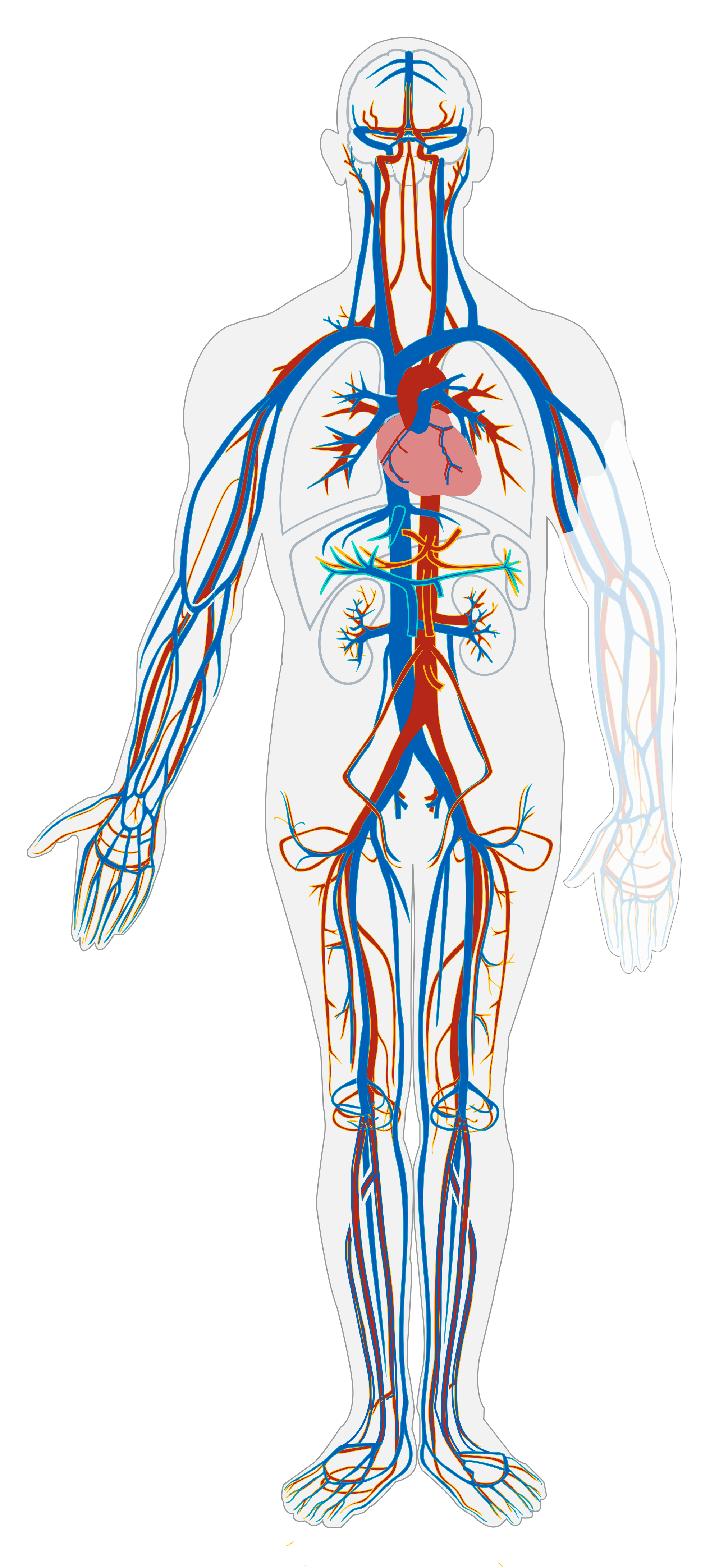
The cardiovascular system, also called the circulatory system, is the organ system that transports materials to and from all the cells of the body. The materials carried by the cardiovascular system include oxygen from the lungs, nutrients from the digestive system, hormones from glands of the endocrine system, and waste materials from cells throughout the body. Transport of these and many other materials is necessary to maintain homeostasis of the body. The main components of the cardiovascular system are the heart, blood vessels, and blood. Each of these components is shown in Figure 14.2.2 and introduced below.
Heart
The heart is a muscular organ in the chest. It consists mainly of cardiac muscle tissue, and it pumps blood through blood vessels by repeated, rhythmic contractions. As shown in Figure 14.2.3, the heart has four inner chambers: a right atrium and ventricle, and a left atrium and ventricle. On each side of the heart, blood is pumped from the atrium to the ventricle below it, and from the ventricle out of the heart. The heart also contains several valves that allow blood to flow only in the proper direction through the heart.
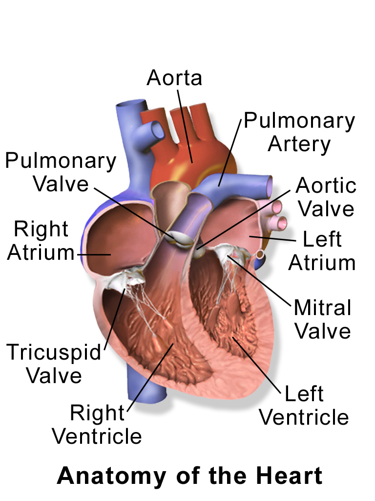
As you may have noticed, the Figure 14.2.3 diagram labels the right side of the heart on the left side of the diagram, and vice versa. This is because it is assumed that in this diagram, the heart appears as if the patient was facing us - the patient's left side is on our right side!
Unlike skeletal muscle, cardiac muscle routinely contracts without stimulation by the nervous system. Specialized cardiac muscle cells send out electrical impulses that stimulate the contractions. As a result, the atria and ventricles normally contract with just the right timing to keep blood pumping efficiently through the heart.
Blood Vessels
The blood vessels of the cardiovascular system are like a network of interconnected, one-way roads that range from superhighways to back alleys. Like a network of roads, the blood vessels are tasked with allowing the transport of materials from one place to another. There are three major types of blood vessels: arteries, veins, and capillaries. They are illustrated in Figure 14.2.4 and described below.
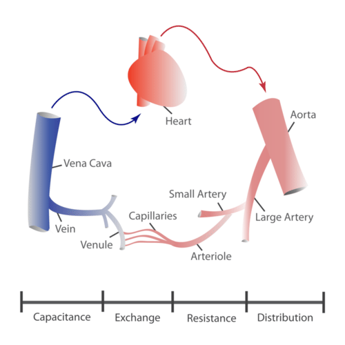
- Arteries are blood vessels that carry blood away from the heart (except for the arteries that actually supply blood to the heart muscle). Most arteries carry oxygen-rich blood, and one of their main functions is distributing oxygen to tissues throughout the body. The smallest arteries are called arterioles.
- Veins are blood vessels that carry blood toward the heart. Most veins carry deoxygenated blood. The smallest veins are called venules.
- Capillaries are the smallest blood vessels, and they connect arterioles and venules. As they pass through tissues, they exchange substances (including oxygen) with cells.
Two Circulations
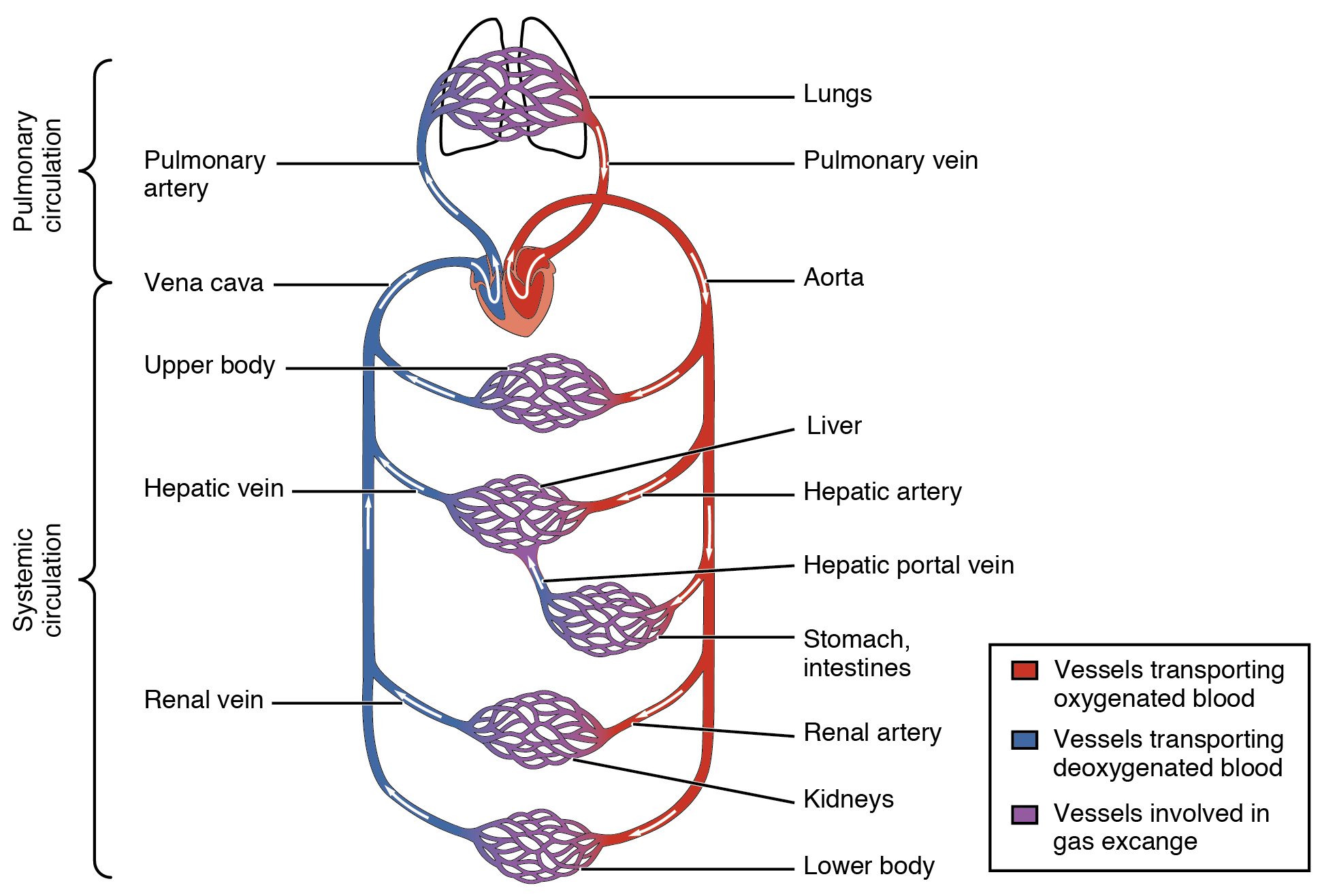
Cells throughout the body need a constant supply of oxygen. They get oxygen from capillaries in the systemic circulation. The systemic circulation is just one of two interconnected circulations that make up the human cardiovascular system. The other circulation is the pulmonary system, which is where blood picks up oxygen to carry to cells. It takes blood about 20 seconds to make one complete transit through both circulations (see Figure 14.2.5).
Pulmonary Circuit
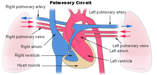
The pulmonary circuit involves only the heart, the lungs, and the major blood vessels that connect them (illustrated in Figure 14.2.6). Blood moves through the pulmonary circuit from the heart, to the lungs, and then back to the heart again, becoming oxygenated in the process. Specifically, the right ventricle of the heart pumps deoxygenated blood into the right and left pulmonary arteries. These arteries carry the blood to the right and left lungs, respectively. Oxygenated blood then returns from the right and left lungs through the two right and two left pulmonary veins. All four pulmonary veins enter the left atrium of the heart.
What happens to the blood while it is in the lungs? It passes through increasingly smaller arteries, and finally through capillary networks surrounding the alveoli (see Figure 14.2.7). This is where gas exchange takes place. The deoxygenated blood in the capillaries picks up oxygen from the alveoli, and gives up carbon dioxide to the alveoli. As a result, the blood returning to the heart in the pulmonary veins is almost completely saturated with oxygen.
Systemic Circulation
The oxygenated blood that enters the left atrium of the heart in the pulmonary circulation then passes into the systemic circuit. This is the part of the cardiovascular system that transports blood to and from all of the tissues of the body to provide oxygen and nutrients, and to pick up wastes. It consists of the heart and blood vessels that supply the metabolic needs of all the cells in the body, including those of the heart and lungs.
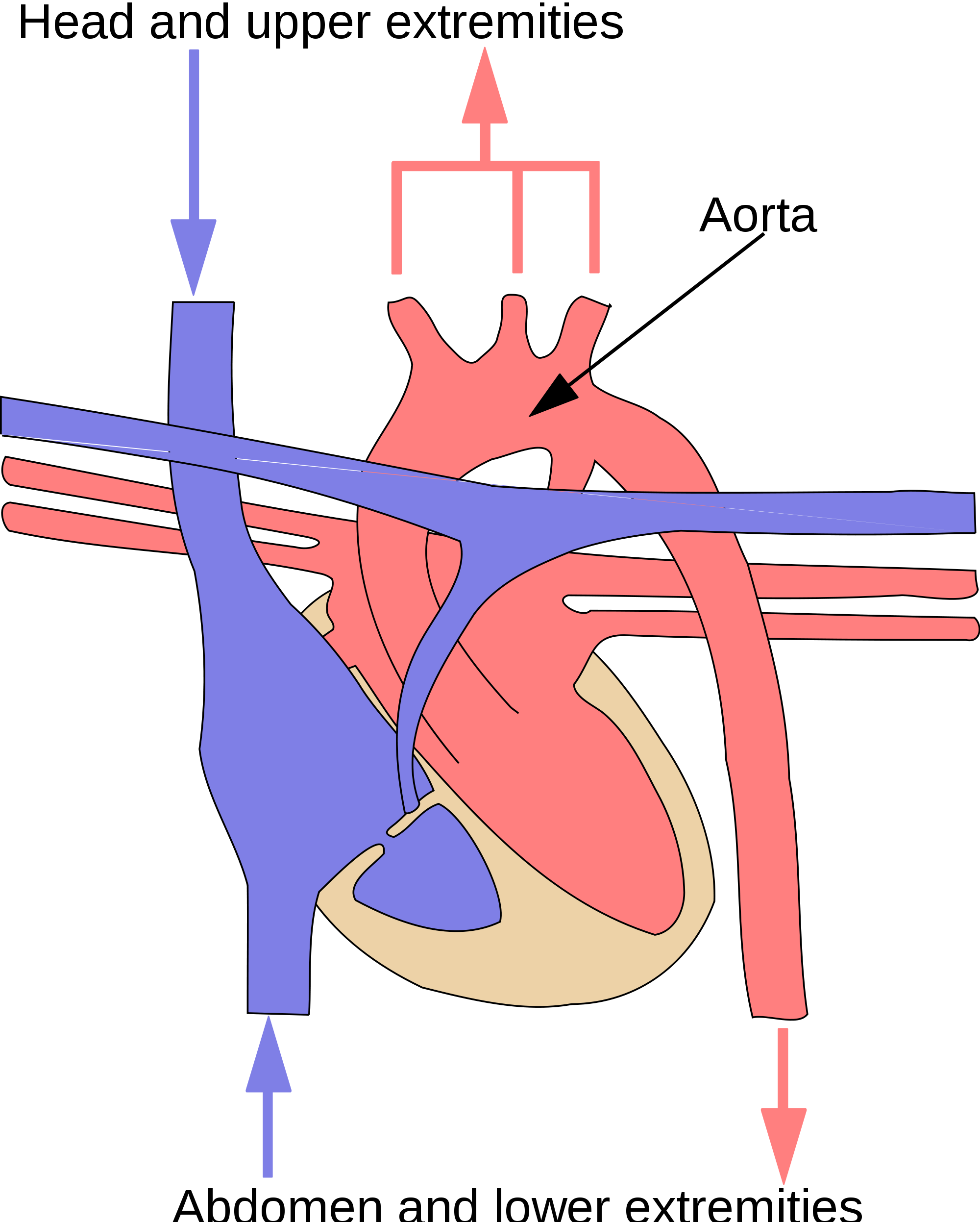
As shown in Figure 14.2.8, in the systemic circulation, the left atrium pumps oxygenated blood to the left ventricle, which pumps the blood directly into the aorta, the body’s largest artery. Major arteries branching off the aorta carry the blood to the head and upper extremities. The aorta continues down through the abdomen and carries blood to the abdomen and lower extremities. The blood then returns to the heart through the network of increasingly larger veins of the systemic circulation. All of the returning blood eventually collects in the superior vena cava (upper body) and inferior vena cava (lower body), which empty directly into the right atrium of the heart.
Blood
Blood is a fluid connective tissue that circulates throughout the body in blood vessels by the pumping action of the heart. Blood carries oxygen and nutrients to all the body’s cells, and it carries carbon dioxide and other wastes away from the cells to be excreted. Blood also transports many other substances, defends the body against infection, repairs body tissues, and controls the body’s pH, among other functions.
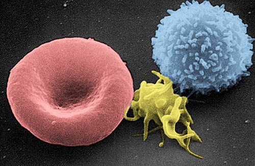
The fluid part of blood is called plasma. It is a yellowish, watery liquid that contains many dissolved substances and blood cells. Types of blood cells in plasma include red blood cells, white blood cells, and platelets, all of which are illustrated in the photomicrograph (Figure 14.2.9) and described below.
- Erythrocytes (red blood cells) have the main function of carrying oxygen in the blood. Red blood cells consist mostly of hemoglobin, a protein containing iron that binds with oxygen.
- Leukocytes (white blood cells) are far fewer in number than red blood cells. They defend the body in various ways. White blood cells called phagocytes, for example, swallow and destroy pathogens, dead cells, and other debris in the blood.
- Thrombocytes (platelets) are cell fragments involved in blood clotting. They stick to tears in blood vessels and to each other, forming a plug at the site of injury. They also release chemicals that are needed for clotting to occur.
14.2 Summary
- The cardiovascular system is the organ system that transports materials to and from all the cells of the body. The main components of the cardiovascular system are the heart, blood vessels, and blood.
- The heart is a muscular organ in the chest that consists mainly of cardiac muscle and pumps blood through blood vessels by repeated, rhythmic contractions. The heart has four chambers through which blood flows, and valves that keep blood flowing in just one direction.
- Blood vessels carry blood throughout the body. Major types of blood vessels are arteries (which mainly carry blood away from the heart), veins (which carry blood toward the heart), and capillaries (which exchange substances between the blood and cells of the body).
- The cardiovascular system has two interconnected circulations. The pulmonary circuit carries blood between the heart and lungs, where blood is oxygenated. The systemic circuit carries blood between the heart and the rest of the body, where it delivers oxygen.
- Blood is a fluid connective tissue that circulates throughout the body in blood vessels. It consists of a liquid part — called plasma — which contains many dissolved substances, and cells, including erythrocytes, leukocytes and thrombocytes.
14.2 Review Questions
- Describe the heart and how it functions.
- Compare and contrast the pulmonary and systemic circulations.
-
- What is blood? What are its chief constituents?
- Name three different types of substances transported by the cardiovascular system.
- Explain why the heart and lungs need blood from the systemic circulation.
- Do blood vessels carrying deoxygenated blood from the body back to the heart get increasingly larger or smaller?
14.2 Explore More
https://www.youtube.com/watch?v=ruM4Xxhx32U&feature=emb_logo
How the heart actually pumps blood - Edmond Hui, TED-Ed, 2014.
https://www.youtube.com/watch?v=9fxm85Fy4sQ&feature=emb_logo
Circulatory & Respiratory Systems - CrashCourse Biology #27, CrashCourse, 2012.
https://www.youtube.com/watch?v=CWFyxn0qDEU
The Heart and Circulatory System - How They Work, Mayo Clinic, 2013.
Attributions
Figure 14.2.1
Brain vascular formation [photo] by Liulin Du/ Chen (The National Cancer Institute at Frederick) on PLOS Biology is used under a CC BY 4.0 license.
Figure 14.2.2
Circulatory_System_no_tags.svg by Mariana Ruiz Villarreal [LadyofHats] on Wikimedia Commons is released into the public domain (https://en.wikipedia.org/wiki/Public_domain).
Figure 14.2.3
Blausen_0462_HeartAnatomy by BruceBlaus on Wikimedia Commons is used under a CC BY 3.0 (https://creativecommons.org/licenses/by/3.0)
Figure 14.2.4
Structure and functions of the different types of blood vessels by CK-12 Foundation is used under a CC BY-NC 3.0 (https://creativecommons.org/licenses/by-nc/3.0/) license.
Figure 14.2.5
2101_Blood_Flow_Through_the_Heart by OpenStax College on Wikimedia Commons is used under a CC BY 3.0 (https://creativecommons.org/licenses/by/3.0) license.
Figure 14.2.6
Illu_pulmonary_circuit by Arcadian from National Cancer Institute/ SEER Training on Wikimedia Commons is in the the public domain (https://en.wikipedia.org/wiki/Public_domain).
Figure 14.2.7
Pulmonary_Blood_Circulation by Artwork by Holly Fischer from Open Michigan (Respiratory Tact Slide 20) on Wikimedia Commons is used under a CC BY 3.0 (https://creativecommons.org/licenses/by/3.0) license.
Figure 14.2.8
systemic_circuit.svg by Surachit on Wikimedia Commons is used under a CC BY-SA 3.0 (http://creativecommons.org/licenses/by-sa/3.0/) license. (Derivative work based on SEER Training by NCI/ U.S. Government).
Figure 14.2.9
Red_White_Blood_cells by Electron Microscopy Facility at The National Cancer Institute at Frederick (NCI-Frederick) on Wikimedia Commons is in the public domain (https://en.wikipedia.org/wiki/Public_domain).
References
Blausen.com Staff. (2014). Medical gallery of Blausen Medical 2014. WikiJournal of Medicine 1 (2). DOI:10.15347/wjm/2014.010. ISSN 2002-4436
Betts, J. G., Young, K.A., Wise, J.A., Johnson, E., Poe, B., Kruse, D.H., Korol, O., Johnson, J.E., Womble, M., DeSaix, P. (2013, June 19). Figure 20.2 Cardiovascular circulation [digital image]. In Anatomy and Physiology (Section 7.3). OpenStax. https://openstax.org/books/anatomy-and-physiology/pages/20-1-structure-and-function-of-blood-vessels
Brainard, J/ CK-12 Foundation. (2016). Figure 4 Diagram represents the structure and functions of the different types of blood vessels in the cardiovascular system [digital image]. In CK-12 College Human Biology (Section 16.2) [online Flexbook]. CK12.org. https://www.ck12.org/book/ck-12-college-human-biology/section/16.2/
CrashCourse. (2012, July 30). Circulatory & respiratory systems - CrashCourse Biology #27. YouTube. https://www.youtube.com/watch?v=9fxm85Fy4sQ&feature=youtu.be
Du, J. (2012, August). Brain vasculature formation [digital image]. PLoS Biology, 10(8): ev10.i08. https://doi.org/10.1371/image.pbio.v10.i08 © Chen.
Mayo Clinic. (2013). The heart and circulatory system - How they work. YouTube. https://www.youtube.com/watch?v=CWFyxn0qDEU&t=1s
TED-Ed. (2014, May 20). How the heart actually pumps blood - Edmond Hui. YouTube. https://www.youtube.com/watch?v=ruM4Xxhx32U&feature=youtu.be
Created by: CK-12/Adapted by Christine Miller
Oh, the Agony!
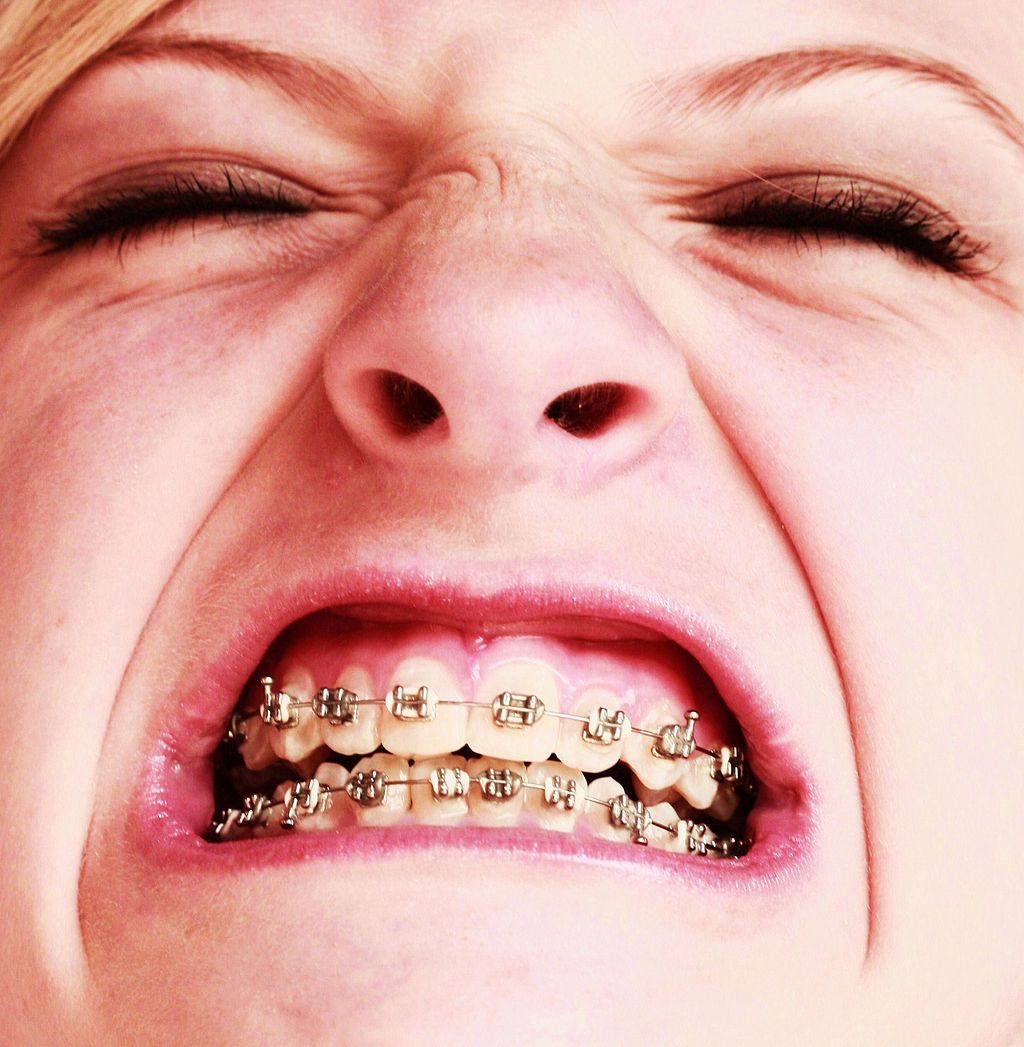
Wearing braces can be very uncomfortable, but it is usually worth it. Braces and other orthodontic treatments can re-align the teeth and jaws to improve bite and appearance. Braces can change the position of the teeth and the shape of the jaws because the human body is malleable. Many phenotypic traits — even those that have a strong genetic basis — can be molded by the environment. Changing the phenotype in response to the environment is just one of several ways we respond to environmental stress.
Types of Responses to Environmental Stress
There are four different types of responses that humans may make to cope with environmental stress:
- Adaptation
- Developmental adjustment
- Acclimatization
- Cultural responses
The first three types of responses are biological in nature, and the fourth type is cultural. Only adaptation involves genetic change and occurs at the level of the population or species. The other three responses do not require genetic change, and they occur at the individual level.
Adaptation
An adaptation is a genetically-based trait that has evolved because it helps living things survive and reproduce in a given environment. Adaptations generally evolve in a population over many generations in response to stresses that last for a long period of time. Adaptations come about through natural selection. Those individuals who inherit a trait that confers an advantage in coping with an environmental stress are likely to live longer and reproduce more. As a result, more of their genes pass on to the next generation. Over many generations, the genes and the trait they control become more frequent in the population.
A Classic Example: Hemoglobin S and Malaria
Probably the most frequently-cited example of a genetic adaptation to an environmental stress is sickle cell trait. As you read in the previous section, people with sickle cell trait have one abnormal allele (S) and one normal allele (A) for hemoglobin, the red blood cell protein that carries oxygen in the blood. Sickle cell trait is an adaptation to the environmental stress of malaria, because people with the trait have resistance to this parasitic disease. In areas where malaria is endemic (present year-round), the sickle cell trait and its allele have evolved to relatively high frequencies. It is a classic example of natural selection favoring heterozygotes for a gene with two alleles. This type of selection keeps both alleles at relatively high frequencies in a population.
To Taste or Not to Taste
Another example of an adaptation in humans is the ability to taste bitter compounds. Plants produce a variety of toxic compounds in order to protect themselves from being eaten, and these toxic compounds often have a bitter taste. The ability to taste bitter compounds is thought to have evolved as an adaptation, because it prevented people from eating poisonous plants. Humans have many different genes that code for bitter taste receptors, allowing us to taste a wide variety of bitter compounds.
A harmless bitter compound called phenylthiocarbamide (PTC) is not found naturally in plants, but it is similar to toxic bitter compounds that are found in plants. Humans' ability to taste this harmless substance has been tested in many different populations. In virtually every population studied, there are some people who can taste PTC (called tasters), and some people who cannot taste PTC, (called nontasters). The ratio of tasters to non-tasters varies among populations, but on average, 75 per cent of people can taste PTC and 25 per cent cannot.
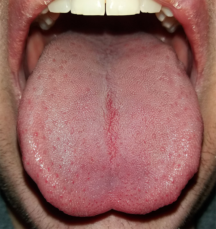
Like many scientific discoveries, human variation in PTC-taster status was discovered by chance. Around 1930, a chemist named Arthur Fox was working with powdered PTC in his lab. Some of the powder accidentally blew into the air. Another lab worker noticed that the powdered PTC tasted bitter, but Fox couldn't detect any taste at all. Fox wondered how to explain this difference in PTC-tasting ability. Geneticists soon determined that PTC-taster status is controlled by a single gene with two common alleles, usually represented by the letters T and t. The T allele encodes a chemical receptor protein (found in taste buds on the tongue, as illustrated in Figure 6.4.2) that can strongly bind to PTC. The other allele, t, encodes a version of the receptor protein that cannot bind as strongly to PTC. The particular combination of these two alleles that a person inherits determines whether the person finds PTC to taste very bitter (TT), somewhat bitter (Tt), or not bitter at all (tt).
If the ability to taste bitter compounds is advantageous, why does every human population studied contain a significant percentage of people who are nontasters? Why has the nontasting allele been preserved in human populations at all? Some scientists hypothesize that the nontaster allele actually confers the ability to taste some other, yet-to-be identified, bitter compound in plants. People who inherit both alleles would presumably be able to taste a wider range of bitter compounds, so they would have the greatest ability to avoid plant toxins. In other words, the heterozygote genotype for the taster gene would be the most fit and favored by natural selection.
Most people no longer have to worry whether the plants they eat contain toxins. The produce you grow in your garden or buy at the supermarket consists of known varieties that are safe to eat. However, natural selection may still be at work in human populations for the PTC-taster gene, because PTC tasters may be more sensitive than nontasters to bitter compounds in tobacco and vegetables in the cabbage family (that is, cruciferous vegetables, such as the broccoli, cauliflower, and cabbage pictured in Figure 6.4.3).
- People who find PTC to taste very bitter are less likely to smoke tobacco, presumably because tobacco smoke has a stronger bitter taste to these individuals. In this case, selection would favor taster genotypes, because tasters would be more likely to avoid smoking and its serious health risks.
- Strong tasters find cruciferous vegetables to taste bitter. As a result, they may avoid eating these vegetables (and perhaps other foods, as well), presumably resulting in a diet that is less varied and nutritious. In this scenario, natural selection might work against taster genotypes.
Figure 6.4.3 Cruciferous vegetables.
Developmental Adjustment
It takes a relatively long time for genetic change in response to environmental stress to produce a population with adaptations. Fortunately, we can adjust to some environmental stresses more quickly by changing in nongenetic ways. One type of nongenetic response to stress is developmental adjustment. This refers to phenotypic change that occurs during development in infancy or childhood, and that may persist into adulthood. This type of change may be irreversible by adulthood.
Phenotypic Plasticity
Developmental adjustment is possible because humans have a high degree of phenotypic plasticity, which is the ability to alter the phenotype in response to changes in the environment. Phenotypic plasticity allows us to respond to changes that occur within our lifetime, and it is particularly important for species (like our own) that have a long generation time. With long generations, evolution of genetic adaptations may occur too slowly to keep up with changing environmental stresses.
Developmental Adjustment and Cultural Practices
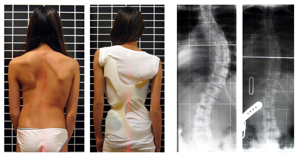
Developmental adjustment may be the result of naturally occurring environmental stresses or cultural practices, including medical or dental treatments. Like our example at the beginning of this section, using braces to change the shape of the jaw and the position of the teeth is an example of a dental practice that brings about a developmental adjustment. Another example of developmental adjustment is the use of a back brace to treat scoliosis (see images in Figure 6.4.4). Scoliosis is an abnormal curvature from side to side in the spine. If the problem is not too severe, a brace, if worn correctly, should prevent the curvature from worsening as a child grows, although it cannot straighten a curve that is already present. Surgery may be required to do that.
Developmental Adjustment and Nutritional Stress
An important example of developmental adjustment that results from a naturally occurring environmental stress is the cessation of physical growth that occurs in children who are under nutritional stress. Children who lack adequate food to fuel both growth and basic metabolic processes are likely to slow down in their growth rate — or even to stop growing entirely. Shunting all available calories and nutrients into essential life functions may keep the child alive at the expense of increasing body size.
Table 6.4.1 shows the effects of inadequate diet on children's' growth in several countries worldwide. For each country, the table gives the prevalence of stunting in children under the age of five. Children are considered stunted if their height is at least two standard deviations below the median height for their age in an international reference population.
Table 6.4.1
Percentage of Stunting in Young Children in Selected Countries (2011-2015)
| Percentage of Stunting in Young Children in Selected Countries (2011-2015) | |
| Country | Per cent of Children Under Age 5 with Stunting |
| United States | 2.1 |
| Turkey | 9.5 |
| Mexico | 13.6 |
| Thailand | 16.3 |
| Iraq | 22.6 |
| Philippines | 33.6 |
| Pakistan | 45.0 |
| Papua New Guinea | 49.5 |
After a growth slow-down occurs and if adequate food becomes available, a child may be able to make up the loss of growth. If food is plentiful, the child may grow more rapidly than normal until the original, genetically-determined growth trajectory is reached. If the inadequate diet persists, however, the failure of growth may become chronic, and the child may never reach his or her full potential adult size.
Phenotypic plasticity of body size in response to dietary change has been observed in successive generations within populations. For example, children in Japan were taller, on average, in each successive generation after the end of World War II. Boys aged 14-15 years old in 1986 were an average of about 18 cm (7 in.) taller than boys of the same age in 1959, a generation earlier. This is a highly significant difference, and it occurred too quickly to be accounted for by genetic change. Instead, the increase in height is a developmental adjustment, thought to be largely attributable to changes in the Japanese diet since World War II. During this period, there was an increase in the amount of animal protein and fat, as well as in the total calories consumed.
Acclimatization
Other responses to environmental stress are reversible and not permanent, whether they occur in childhood or adulthood. The development of reversible changes to environmental stress is called acclimatization. Acclimatization generally develops over a relatively short period of time. It may take just a few days or weeks to attain a maximum response to a stress. When the stress is no longer present, the acclimatized state declines, and the body returns to its normal baseline state. Generally, the shorter the time for acclimatization to occur, the more quickly the condition is reversed when the environmental stress is removed.
Acclimatization to UV Light
A common example of acclimatization is tanning of the skin (see Figure 6.4.5). This occurs in many people in response to exposure to ultraviolet radiation from the sun. Special pigment cells in the skin, called melanocytes, produce more of the brown pigment melanin when exposed to sunlight. The melanin collects near the surface of the skin where it absorbs UV radiation so it cannot penetrate and potentially damage deeper skin structures. Tanning is a reversible change in the phenotype that helps the body deal temporarily with the environmental stress of high levels of UV radiation. When the skin is no longer exposed to the sun’s rays, the tan fades, generally over a period of a few weeks or months.
Figure 6.4.5 Tanning of the skin occurs in many people in response to exposure to ultraviolet radiation from the sun.
Acclimatization to Heat
Another common example of acclimatization occurs in response to heat. Changes that occur with heat acclimatization include increased sweat output and earlier onset of sweat production, which helps the body stay cool because evaporation of sweat takes heat from the body’s surface in a process called evaporative cooling. It generally takes a couple of weeks for maximum heat acclimatization to come about by gradually working out harder and longer at high air temperatures. The changes that occur with acclimatization just as quickly subside when the body is no longer exposed to excessive heat.
Acclimatization to High Altitude
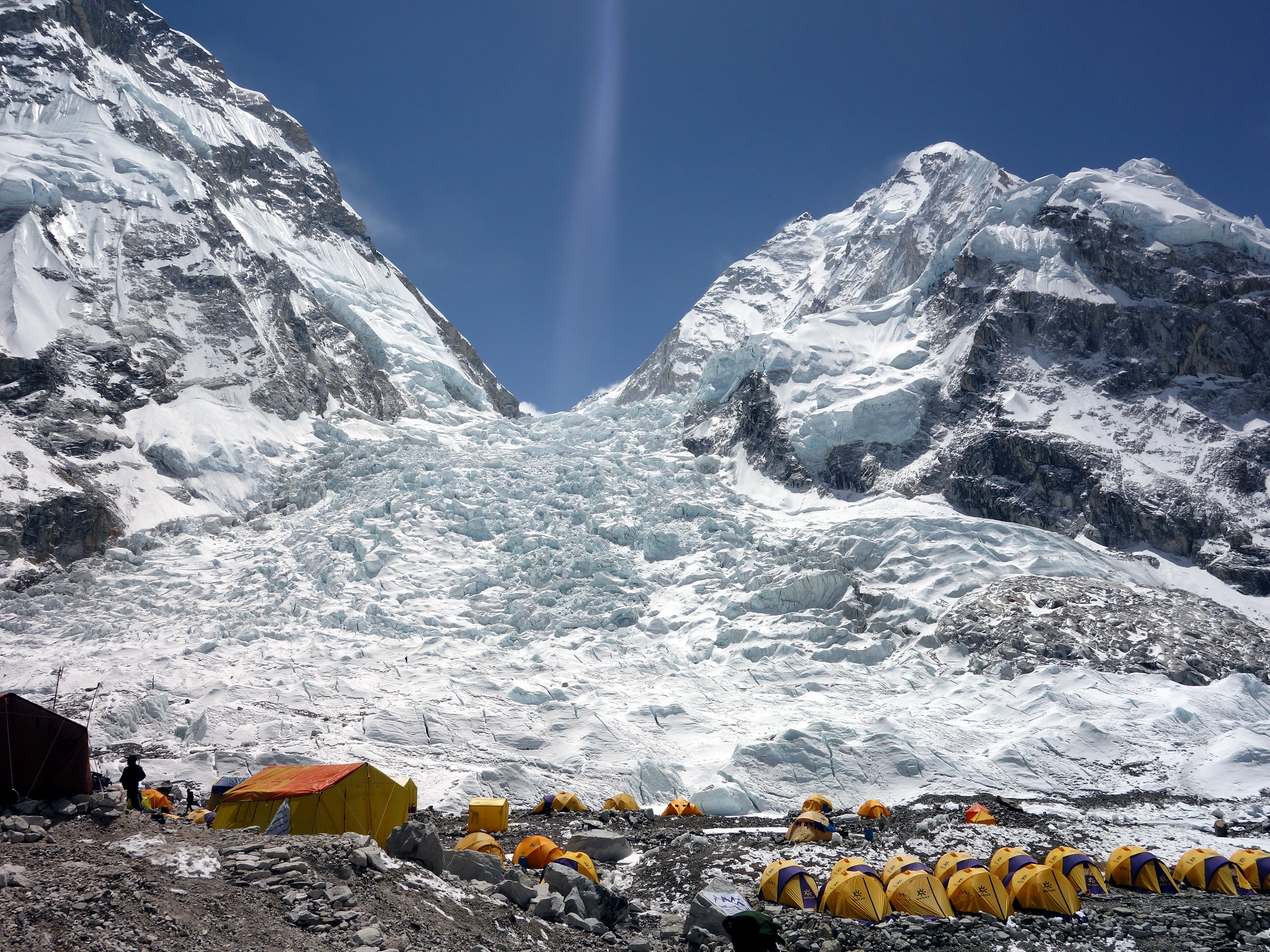
Short term acclimatization to high altitude occurs as a response to low levels of oxygen in the blood. This reduced level of oxygen is detected by carotid bodies, which will trigger in increase in breathing and heart rate. Over a period of weeks the body will compensate by increasing red blood cell production, thereby improving the oxygen-carrying capacity of the blood. This is why mountaineers wishing to climb to the peak of Mount Everest must complete the full climb in portions; it is recommended that climbers spend 2-3 days acclimatizing for every 600 metres of elevation increase. In addition, the higher to altitude, the longer it make take to acclimatize; climbers are advised to spend 4-5 days acclimatizing at base camp (whether the base camp in Nepal or China) before completing the final leg of the climb to the peak. The concentration of red blood cells gradually decreases to normal levels once a climber returns to their normal elevation.
Cultural Responses
More than any other species, humans respond to environmental stresses with learned behaviors and technology. These cultural responses allow us to change our environments to control stresses, rather than changing our bodies genetically or physiologically to cope with the stresses. Even archaic humans responded to some environmental stresses in this way. For example, Neanderthals used shelters, fires, and animal hides as clothing to stay warm in the cold climate in Europe during the last ice age. Today, we use more sophisticated technologies to stay warm in cold climates while retaining our essentially tropical-animal anatomy and physiology. We also use technology (such as furnaces and air conditioners) to avoid temperature stress and stay comfortable in hot or cold climates.
6.4 Summary
- Humans may respond to environmental stress in four different ways: adaptation, developmental adjustment, acclimatization, and cultural responses.
- An adaptation is a genetically based trait that has evolved because it helps living things survive and reproduce in a given environment. Adaptations evolve by natural selection in populations over a relatively long period to time. Examples of adaptations include sickle cell trait as an adaptation to the stress of endemic malaria and the ability to taste bitter compounds as an adaptation to the stress of bitter-tasting toxins in plants.
- A developmental adjustment is a non-genetic response to stress that occurs during infancy or childhood, and that may persist into adulthood. This type of change may be irreversible. Developmental adjustment is possible because humans have a high degree of phenotypic plasticity. It may be the result of environmental stresses (such as inadequate food), which may stunt growth, or cultural practices (such as orthodontic treatments), which re-align the teeth and jaws.
- Acclimatization is the development of reversible changes to environmental stress that develop over a relatively short period of time. The changes revert to the normal baseline state after the stress is removed. Examples of acclimatization include tanning of the skin and physiological changes (such as increased sweating) that occur with heat acclimatization.
- More than any other species, humans respond to environmental stress with learned behaviors and technology, which are cultural responses. These responses allow us to change our environment to control stress, rather than changing our bodies genetically or physiologically to cope with stress. Examples include using shelter, fire, and clothing to cope with a cold climate.
6.4 Review Questions
- List four different types of responses that humans may make to cope with environmental stress.
- Define adaptation.
-
- Explain how natural selection may have resulted in most human populations having people who can and people who cannot taste PTC.
- What is a developmental adjustment?
- Define phenotypic plasticity.
- Explain why phenotypic plasticity may be particularly important in a species with a long generation time.
- Why may stunting of growth occur in children who have an inadequate diet? Why is stunting preferable to the alternative?
- What is acclimatization?
- How does acclimatization to heat come about, and what are two physiological changes that occur in heat acclimatization?
- Give an example of a cultural response to heat stress.
- Which is more likely to be reversible — a change due to acclimatization, or a change due to developmental adjustment? Explain your answer.
6.4 Explore More
https://www.youtube.com/watch?v=upp9-w6GPhU
Could we survive prolonged space travel? - Lisa Nip, TED-Ed, 2016.
https://www.youtube.com/watch?v=hRnrIpUMyZQ&t=182s
How this disease changes the shape of your cells - Amber M. Yates, TED-Ed, 2019.
Attributions
Figure 6.4.1
Free_Awesome_Girl_With_Braces_Close_Up by D. Sharon Pruitt from Hill Air Force Base, Utah, USA on Wikimedia Commons is used under a CC BY 2.0 (https://creativecommons.org/licenses/by/2.0/deed.en) license.
Figure 6.4.2
Tongue by Mahdiabbasinv on Wikimedia Commons is used under a CC BY-SA 4.0 (https://creativecommons.org/licenses/by-sa/4.0/deed.en) license.
Figure 6.4.3
- White cauliflower on brown wooden chopping board by Louis Hansel @shotsoflouis on Unsplash is used under the Unsplash License (https://unsplash.com/license).
- Broccoli on wooden chopping board by Louis Hansel @shotsoflouis on Unsplash is used under the Unsplash License (https://unsplash.com/license).
- Green cabbage close up by Craig Dimmick on Unsplash is used under the Unsplash License (https://unsplash.com/license).
- Cabbage hybrid/ brussel sprouts by Solstice Hannan on Unsplash is used under the Unsplash License (https://unsplash.com/license).
- Kale by Laura Johnston on Unsplash is used under the Unsplash License (https://unsplash.com/license).
- Tiny bok choy at the Asian market by Jodie Morgan on Unsplash is used under the Unsplash License (https://unsplash.com/license).
Figure 6.4.4
Scoliosis_patient_in_cheneau_brace_correcting_from_56_to_27_deg by Weiss H.R. from Scoliosis Journal/BioMed Central Ltd. on Wikimedia Commons is used under a CC BY 2.0 (https://creativecommons.org/licenses/by/2.0) license.
Figure 6.4.5
- Tan Lines by k.steudel on Flickr is used under a CC BY 2.0 (https://creativecommons.org/licenses/by/2.0/) license.
- Twin tan lines (all sizes) by Quinn Dombrowski on Flickr is used under a CC BY-SA 2.0 (https://creativecommons.org/licenses/by-sa/2.0/) license.
- Wedding ring tan line by Quinn Dombrowski on Flickr is used under a CC BY-SA 2.0 (https://creativecommons.org/licenses/by-sa/2.0/) license.
- Tan by Evil Erin on Flickr is used under a CC BY 2.0 (https://creativecommons.org/licenses/by/2.0/) license.
Figure 6.4.6
Nepalese base camp by Mark Horrell on Flickr is used under a CC BY-NC-SA 2.0 (https://creativecommons.org/licenses/by-nc-sa/2.0/) license.
References
TED-Ed. (2016, October 4). Could we survive prolonged space travel? - Lisa Nip. YouTube. https://www.youtube.com/watch?v=upp9-w6GPhU&feature=youtu.be
TED-Ed. (2019, May 6). How this disease changes the shape of your cells - Amber M. Yates. YouTube. https://www.youtube.com/watch?v=hRnrIpUMyZQ&feature=youtu.be
Weiss, H. (2007). Is there a body of evidence for the treatment of patients with Adolescent Idiopathic Scoliosis (AIS)? [Figure 2 - digital photograph], Scoliosis, 2(19). https://doi.org/10.1186/1748-7161-2-19
Created by: CK-12/Adapted by Christine Miller
Giving the Gift of Life
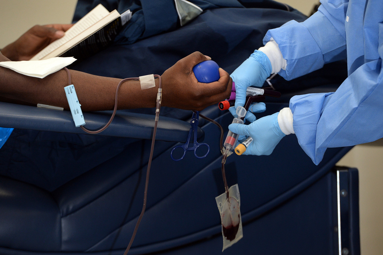
Did you ever donate blood? If you did, then you probably know that your blood type is an important factor in blood transfusions. People vary in the type of blood they inherit, and this determines which type(s) of blood they can safely receive in a transfusion. Do you know your blood type?
What Are Blood Types?
Blood is composed of cells suspended in a liquid called plasma. There are three types of cells in blood: red blood cells, which carry oxygen; white blood cells, which fight infections and other threats; and platelets, which are cell fragments that help blood clot. Blood type (or blood group) is a genetic characteristic associated with the presence or absence of certain molecules, called antigens, on the surface of red blood cells. These molecules may help maintain the integrity of the cell membrane, act as receptors, or have other biological functions. A blood group system refers to all of the gene(s), alleles, and possible genotypes and phenotypes that exist for a particular set of blood type antigens. Human blood group systems include the well-known ABO and Rhesus (Rh) systems, as well as at least 33 others that are less well known.
Antigens and Antibodies
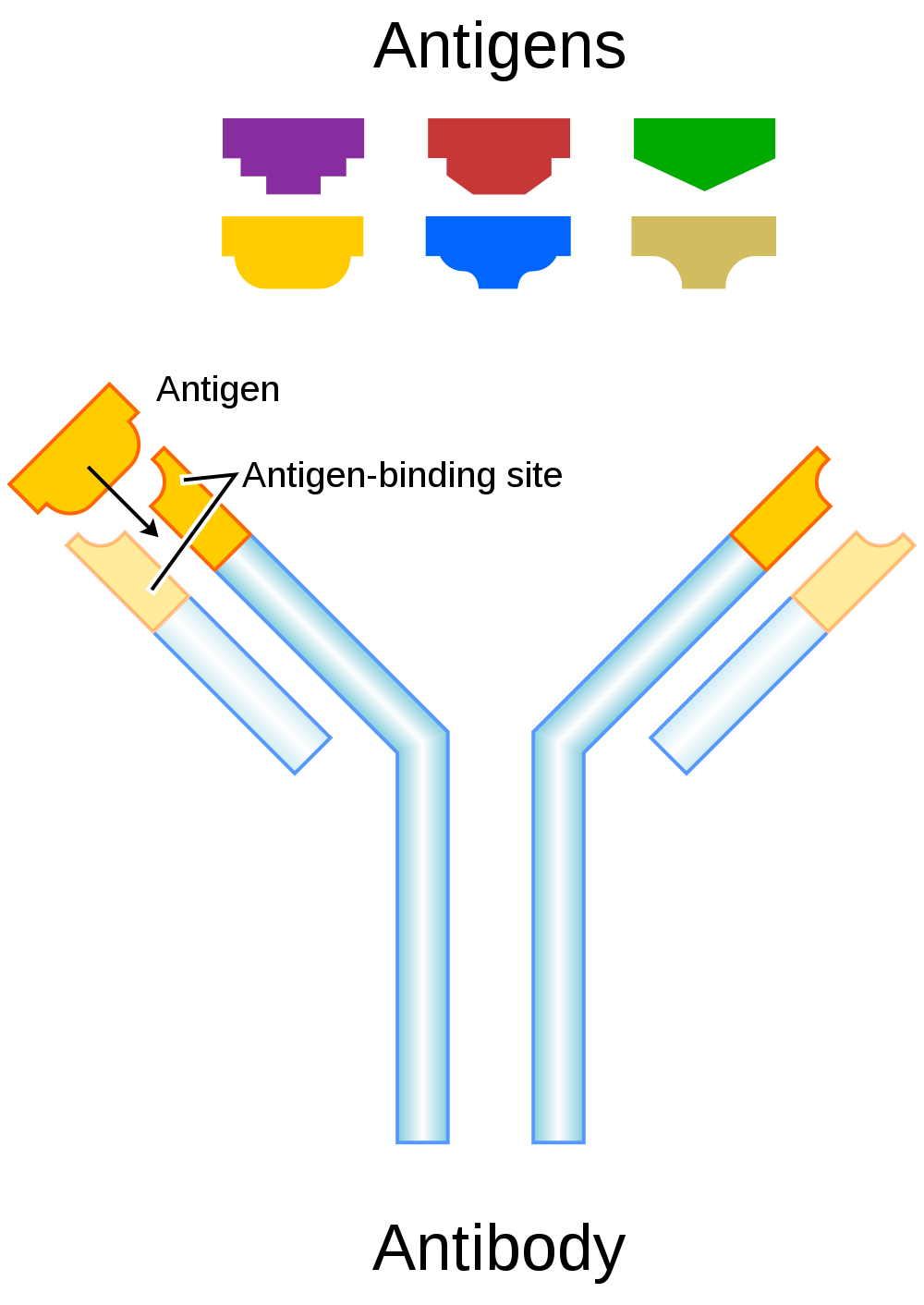
Antigens — such as those on the red blood cells — are molecules that the immune system identifies as either self (produced by your own body) or non-self (not produced by your own body). Blood group antigens may be proteins, carbohydrates, glycoproteins (proteins attached to chains of sugars), or glycolipids (lipids attached to chains of sugars), depending on the particular blood group system. If antigens are identified as non-self, the immune system responds by forming antibodies that are specific to the non-self antigens. Antibodies are large, Y-shaped proteins produced by the immune system that recognize and bind to non-self antigens. The analogy of a lock and key is often used to represent how an antibody and antigen fit together, as shown in the illustration below (Figure 6.5.2). When antibodies bind to antigens, it marks them for destruction by other immune system cells. Non-self antigens may enter your body on pathogens (such as bacteria or viruses), on foods, or on red blood cells in a blood transfusion from someone with a different blood type than your own. The last way is virtually impossible nowadays because of effective blood typing and screening protocols.
Genetics of Blood Type
An individual’s blood type depends on which alleles for a blood group system were inherited from their parents. Generally, blood type is controlled by alleles for a single gene, or for two or more very closely linked genes. Closely linked genes are almost always inherited together, because there is little or no recombination between them. Like other genetic traits, a person’s blood type is generally fixed for life, but there are rare instances in which blood type can change. This could happen, for example, if an individual receives a bone marrow transplant to treat a disease, such as leukemia. If the bone marrow comes from a donor who has a different blood type, the patient’s blood type may eventually convert to the donor’s blood type, because red blood cells are produced in bone marrow.
ABO Blood Group System
The ABO blood group system is the best known human blood group system. Antigens in this system are glycoproteins. These antigens are shown in the list below. There are four common blood types for the ABO system:
- Type A, in which only the A antigen is present.
- Type B, in which only the B antigen is present.
- Type AB, in which both the A and B antigens are present.
- Type O, in which neither the A nor the B antigen is present.
Genetics of the ABO System
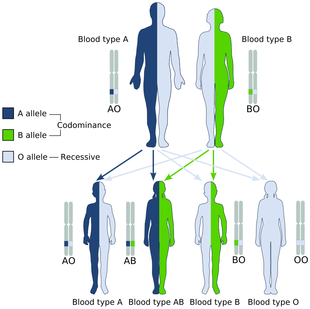
The ABO blood group system is controlled by a single gene on chromosome 9. There are three common alleles for the gene, often represented by the letters A , B , and O. With three alleles, there are six possible genotypes for ABO blood group. Alleles A and B, however, are both dominant to allele O and codominant to each other. This results in just four possible phenotypes (blood types) for the ABO system. These genotypes and phenotypes are shown in Table 6.5.1.
Table 6.5.1
ABO Blood Group System: Genotypes and Phenotypes
| ABO Blood Group System | |
| Genotype | Phenotype (Blood Type, or Group) |
| AA | A |
| AO | A |
| BB | B |
| BO | B |
| OO | O |
| AB | AB |
The diagram below (Figure 6.5.3) shows an example of how ABO blood type is inherited. In this particular example, the father has blood type A (genotype AO) and the mother has blood type B (genotype BO). This mating type can produce children with each of the four possible ABO phenotypes, although in any given family, not all phenotypes may be present in the children.
Medical Significance of ABO Blood Type
The ABO system is the most important blood group system in blood transfusions. If red blood cells containing a particular ABO antigen are transfused into a person who lacks that antigen, the person’s immune system will recognize the antigen on the red blood cells as non-self. Antibodies specific to that antigen will attack the red blood cells, causing them to agglutinate (or clump) and break apart. If a unit of incompatible blood were to be accidentally transfused into a patient, a severe reaction (called acute hemolytic transfusion reaction) is likely to occur, in which many red blood cells are destroyed. This may result in kidney failure, shock, and even death. Fortunately, such medical accidents virtually never occur today.
These antibodies are often spontaneously produced in the first years of life, after exposure to common microorganisms in the environment that have antigens similar to blood antigens. Specifically, a person with type A blood will produce anti-B antibodies, while a person with type B blood will produce anti-A antibodies. A person with type AB blood does not produce either antibody, while a person with type O blood produces both anti-A and anti-B antibodies. Once the antibodies have been produced, they circulate in the plasma. The relationship between ABO red blood cell antigens and plasma antibodies is shown in Figure 6.5.4.
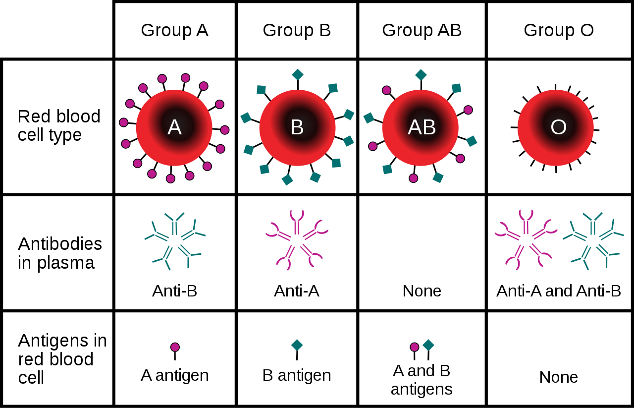
The antibodies that circulate in the plasma are for different antigens than those on red blood cells, which are recognized as self antigens.
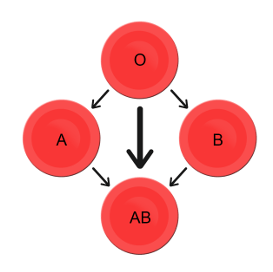
Which blood types are compatible and which are not? Type O blood contains both anti-A and anti-B antibodies, so people with type O blood can only receive type O blood. However, they can donate blood to people of any ABO blood type, which is why individuals with type O blood are called universal donors. Type AB blood contains neither anti-A nor anti-B antibodies, so people with type AB blood can receive blood from people of any ABO blood type. That’s why individuals with type AB blood are called universal recipients. They can donate blood, however, only to people who also have type AB blood. These and other relationships between blood types of donors and recipients are summarized in the simple diagram to the right.
Geographic Distribution of ABO Blood Groups
The frequencies of blood groups for the ABO system vary around the world. You can see how the A and B alleles and the blood group O are distributed geographically on the maps in Figure 6.5.6.
- Worldwide, B is the rarest ABO allele, so type B blood is the least common ABO blood type. Only about 16 per cent of all people have the B allele. Its highest frequency is in Asia. Its lowest frequency is among the indigenous people of Australia and the Americas.
- The A allele is somewhat more common around the world than the B allele, so type A blood is also more common than type B blood. The highest frequencies of the A allele are in Australian Aborigines, the Lapps (Sami) of Northern Scandinavia, and Blackfoot Native Americans in North America. The allele is nearly absent among Native Americans in Central and South America.
- The O allele is the most common ABO allele around the world, and type O blood is the most common ABO blood type. Almost two-thirds of people have at least one copy of the O allele. It is especially common in Native Americans in Central and South America, where it reaches frequencies close to 100 per cent. It also has relatively high frequencies in Australian Aborigines and Western Europeans. Its frequencies are lowest in Eastern Europe and Central Asia.
Figure 6.5.6 Maps of populations that have the A, B and O alleles.
Evolution of the ABO Blood Group System
The geographic distribution of ABO blood type alleles provides indirect evidence for the evolutionary history of these alleles. Evolutionary biologists hypothesize that the allele for blood type A evolved first, followed by the allele for blood type O, and then by the allele for blood type B. This chronology accounts for the percentages of people worldwide with each blood group, and is also consistent with known patterns of early population movements.
The evolutionary forces of founder effect and genetic drift have no doubt played a significant role in the current distribution of ABO blood types worldwide. Geographic variation in ABO blood groups is also likely to be influenced by natural selection, because different blood types are thought to vary in their susceptibility to certain diseases. For example:
- People with type O blood may be more susceptible to cholera and plague. They are also more likely to develop gastrointestinal ulcers.
- People with type A blood may be more susceptible to smallpox and more likely to develop certain cancers.
- People with types A, B, and AB blood appear to be less likely to form blood clots that can cause strokes. However, early in our history, the ability of blood to form clots — which appears greater in people with type O blood — may have been a survival advantage.
- Perhaps the greatest natural selective force associated with ABO blood types is malaria. There is considerable evidence to suggest that people with type O blood are somewhat resistant to malaria, giving them a selective advantage where malaria is endemic.
Rhesus Blood Group System
Another well-known blood group system is the Rhesus (Rh) blood group system. The Rhesus system has dozens of different antigens, but only five main antigens (called D, C, c, E, and e). The major Rhesus antigen is the D antigen. People with the D antigen are called Rh positive (Rh+), and people who lack the D antigen are called Rh negative (Rh-). Rhesus antigens are thought to play a role in transporting ions across cell membranes by acting as channel proteins.
The Rhesus blood group system is controlled by two linked genes on chromosome 1. One gene, called RHD, produces a single antigen, antigen D. The other gene, called RHCE, produces the other four relatively common Rhesus antigens (C, c, E, and e), depending on which alleles for this gene are inherited.
Rhesus Blood Group and Transfusions
After the ABO system, the Rhesus system is the second most important blood group system in blood transfusions. The D antigen is the one most likely to provoke an immune response in people who lack the antigen. People who have the D antigen (Rh+) can be safely transfused with either Rh+ or Rh- blood, whereas people who lack the D antigen (Rh-) can be safely transfused only with Rh- blood.
Unlike anti-A and anti-B antibodies to ABO antigens, anti-D antibodies for the Rhesus system are not usually produced by sensitization to environmental substances. People who lack the D antigen (Rh-), however, may produce anti-D antibodies if exposed to Rh+ blood. This may happen accidentally in a blood transfusion, although this is extremely unlikely today. It may also happen during pregnancy with an Rh+ fetus if some of the fetal blood cells pass into the mother’s blood circulation.
Hemolytic Disease of the Newborn
If a woman who is Rh- is carrying an Rh+ fetus, the fetus may be at risk. This is especially likely if the mother has formed anti-D antibodies during a prior pregnancy because of a mixing of maternal and fetal blood during childbirth. Unlike antibodies against ABO antigens, antibodies against the Rhesus D antigen can cross the placenta and enter the blood of the fetus. This may cause hemolytic disease of the newborn (HDN), also called erythroblastosis fetalis, an illness in which fetal red blood cells are destroyed by maternal antibodies, causing anemia. This illness may range from mild to severe. If it is severe, it may cause brain damage and is sometimes fatal for the fetus or newborn. Fortunately, HDN can be prevented by preventing the formation of anti-D antibodies in the Rh- mother. This is achieved by injecting the mother with a medication called Rho(D) immune globulin.
Geographic Distribution of Rhesus Blood Types
The majority of people worldwide are Rh+, but there is regional variation in this blood group system, as there is with the ABO system. The aboriginal inhabitants of the Americas and Australia originally had very close to 100 per cent Rh+ blood. The frequency of the Rh+ blood type is also very high in African populations, at about 97 to 99 per cent. In East Asia, the frequency of Rh+ is slightly lower, at about 93 to 99 per cent. Europeans have the lowest frequency of the Rh+ blood type at about 83 to 85 per cent.
What explains the population variation in Rhesus blood types? Prior to the advent of modern medicine, Rh+ positive children conceived by Rh- women were at risk of fetal or newborn death or impairment from HDN. This was an enigma, because presumably, natural selection would work to remove the rarer phenotype (Rh-) from populations. However, the frequency of this phenotype is relatively high in many populations.
Recent studies have found evidence that natural selection may actually favor heterozygotes for the Rhesus D antigen. The selective agent in this case is thought to be toxoplasmosis, a parasitic disease caused by the protozoan Toxoplasma gondii, which is very common worldwide. You can see a life cycle diagram of the parasite in Figure 6.5.7. Infection by this parasite often causes no symptoms at all, or it may cause flu-like symptoms for a few days or weeks. Exposure to the parasite has been linked, however, to increased risk of mental disorders (such as schizophrenia), neurological disorders (such as Alzheimer’s), and other neurological problems, including delayed reaction times. One study found that people who tested positive for antibodies to the parasite were more than twice as likely to be involved in traffic accidents.
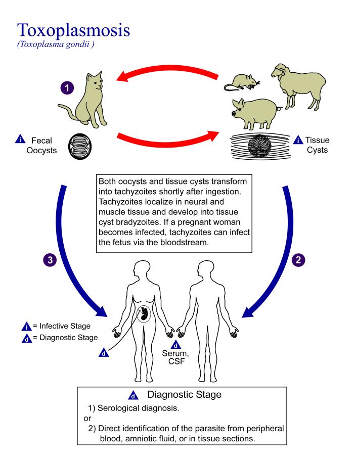
People who are heterozygous for the D antigen appear less likely to develop the negative neurological and mental effects of Toxoplasma gondii infection. This could help explain why both phenotypes (Rh+ and Rh-) are maintained in most populations. There are also striking geographic differences in the prevalence of toxoplasmosis worldwide, ranging from zero to 95 per cent in different regions. This could explain geographic variation in the D antigen worldwide, because its strength as a selective agent would vary with its prevalence.
Feature: Myth vs. Reality
Myth |
Reality |
| "Your nutritional needs can be determined by your ABO blood type. Knowing your blood type allows you to choose the appropriate foods that will help you lose weight, increase your energy, and live a longer, healthier life." | This idea was proposed in 1996 in a New York Times bestseller Eat Right for Your Type, by Peter D’Adamo, a naturopath. Naturopathy is a method of treating disorders that involves the use of herbs, sunlight, fresh air, and other natural substances. Some medical doctors consider naturopathy a pseudoscience. A major scientific review of the blood type diet could find no evidence to support it. In one study, adults eating the diet designed for blood type A showed improved health — but this occurred in everyone, regardless of their blood type. Because the blood type diet is based solely on blood type, it fails to account for other factors that might require dietary adjustments or restrictions. For example, people with diabetes — but different blood types — would follow different diets, and one or both of the diets might conflict with standard diabetes dietary recommendations and be dangerous. |
| "ABO blood type is associated with certain personality traits. People with blood type A, for example, are patient and responsible, but may also be stubborn and tense, whereas people with blood type B are energetic and creative, but may also be irresponsible and unforgiving. In selecting a spouse, both your own and your potential mate’s blood type should be taken into account to ensure compatibility of your personalities." | The belief that blood type is correlated with personality is widely held in Japan and other East Asian countries. The idea was originally introduced in the 1920s in a study commissioned by the Japanese government, but it was later shown to have no scientific support. The idea was revived in the 1970s by a Japanese broadcaster, who wrote popular books about it. There is no scientific basis for the idea, and it is generally dismissed as pseudoscience by the scientific community. Nonetheless, it remains popular in East Asian countries, just as astrology is popular in many other countries. |
6.5 Summary
- Blood type (or blood group) is a genetic characteristic associated with the presence or absence of antigens on the surface of red blood cells. A blood group system refers to all of the gene(s), alleles, and possible genotypes and phenotypes that exist for a particular set of blood type antigens.
- Antigens are molecules that the immune system identifies as either self or non-self. If antigens are identified as non-self, the immune system responds by forming antibodies that are specific to the non-self antigens, leading to the destruction of cells bearing the antigens.
- The ABO blood group system is a system of red blood cell antigens controlled by a single gene with three common alleles on chromosome 9. There are four possible ABO blood types: A, B, AB, and O. The ABO system is the most important blood group system in blood transfusions. People with type O blood are universal donors, and people with type AB blood are universal recipients.
- The frequencies of ABO blood type alleles and blood groups vary around the world. The allele for the B antigen is least common, and blood type O is the most common. The evolutionary forces of founder effect, genetic drift, and natural selection are responsible for the geographic distribution of ABO alleles and blood types. People with type O blood, for example, may be somewhat resistant to malaria, possibly giving them a selective advantage where malaria is endemic.
- The Rhesus blood group system is a system of red blood cell antigens controlled by two genes with many alleles on chromosome 1. There are five common Rhesus antigens, of which antigen D is most significant. Individuals who have antigen D are called Rh+, and individuals who lack antigen D are called Rh-. Rh- mothers of Rh+ fetuses may produce antibodies against the D antigen in the fetal blood, causing hemolytic disease of the newborn (HDN).
- The majority of people worldwide are Rh+, but there is regional variation in this blood group system. This variation may be explained by natural selection that favors heterozygotes for the D antigen, because this genotype seems to be protected against some of the neurological consequences of the common parasitic infection toxoplasmosis.
6.5 Review Questions
- Define blood type and blood group system.
- Explain the relationship between antigens and antibodies.
- Identify the alleles, genotypes, and phenotypes in the ABO blood group system.
- Discuss the medical significance of the ABO blood group system.
- Compare the relative worldwide frequencies of the three ABO alleles.
- Give examples of how different ABO blood types vary in their susceptibility to diseases.
- Describe the Rhesus blood group system.
- Relate Rhesus blood groups to blood transfusions.
- What causes hemolytic disease of the newborn?
- Describe how toxoplasmosis may explain the persistence of the Rh- blood type in human populations.
- A woman is blood type O and Rh-, and her husband is blood type AB and Rh+. Answer the following questions about this couple and their offspring.
- What are the possible genotypes of their offspring in terms of ABO blood group?
- What are the possible phenotypes of their offspring in terms of ABO blood group?
- Can the woman donate blood to her husband? Explain your answer.
- Can the man donate blood to his wife? Explain your answer.
- Type O blood is characterized by the presence of O antigens — explain why this statement is false.
- Explain why newborn hemolytic disease may be more likely to occur in a second pregnancy than in a first.
6.5 Explore More
https://www.youtube.com/watch?v=xfZhb6lmxjk
Why do blood types matter? - Natalie S. Hodge, TED-Ed, 2015.
https://www.youtube.com/watch?v=qcZKbjYyOfE
How do blood transfusions work? - Bill Schutt, TED-Ed, 2020.
Attributes
Figure 6.5.1
Following the Blood Donation Trail by EJ Hersom/ USA Department of Defense is in the public domain. [Disclaimer: The appearance of U.S. Department of Defense (DoD) visual information does not imply or constitute DoD endorsement.]
Figure 6.5.2
Antibody by Fvasconcellos on Wikimedia Commons is released into the public domain (https://en.wikipedia.org/wiki/Public_domain).
Figure 6.5.3
ABO system codominance.svg, adapted by YassineMrabet (original "Codominant" image from US National Library of Medicine) on Wikimedia Commons is in the public domain (https://en.wikipedia.org/wiki/Public_domain).
Figure 6.5.4
ABO_blood_type.svg by InvictaHOG on Wikimedia Commons is released into the public domain (https://en.wikipedia.org/wiki/Public_domain).
Figure 6.5.5
Blood Donor and recipient ABO by CK-12 Foundation is used under a CC BY-NC 3.0 (https://creativecommons.org/licenses/by-nc/3.0/) license.
Figure 6.5.6
- Map of Blood Group A by Muntuwandi at en.wikipedia on Wikimedia Commons is used under a CC BY-SA 3.0 (https://creativecommons.org/licenses/by-sa/3.0/) license.
- Map of Blood Group B by Muntuwandi at en.wikipedia on Wikimedia Commons is used under a CC BY-SA 3.0 (https://creativecommons.org/licenses/by-sa/3.0/) license.
- Map of Blood Group O by anthro palomar at en.wikipedia on Wikimedia Commons is used under a CC BY-SA 3.0 (https://creativecommons.org/licenses/by-sa/3.0/) license.
Figure 6.5.7
Toxoplasma_gondii_Life_cycle_PHIL_3421_lores by Alexander J. da Silva, PhD/Melanie Moser, Centers for Disease Control and Prevention's Public Health Image Library (PHIL#3421) on Wikimedia Commons is in the public domain (https://en.wikipedia.org/wiki/Public_domain).
Table 6.5.1
ABO Blood Group System: Genotypes and Phenotypes was created by Christine Miller.
References
Dean, L. (2005). Chapter 4 Hemolytic disease of the newborn. In Blood Groups and Red Cell Antigens [Internet]. National Center for Biotechnology Information (US). https://www.ncbi.nlm.nih.gov/books/NBK2266/
Mayo Clinic Staff. (n.d.). Toxoplasmosis [online article]. MayoClinic.org. https://www.mayoclinic.org/diseases-conditions/toxoplasmosis/symptoms-causes/syc-20356249
MedlinePlus. (2019, January 29). Hemolytic transfusion reaction [online article]. U.S. National Library of Medicine. https://en.wikipedia.org/w/index.php?title=Chromosome_9&oldid=946440619
TED-Ed. (2015, June 29). Why do blood types matter? - Natalie S. Hodge. YouTube. https://www.youtube.com/watch?v=xfZhb6lmxjk&feature=youtu.be
TED-Ed. (2020, February 18). How do blood transfusions work? - Bill Schutt. YouTube. https://www.youtube.com/watch?v=qcZKbjYyOfE&feature=youtu.be
Wikipedia contributors. (2020, May 10). Chromosome 1. In Wikipedia. https://en.wikipedia.org/w/index.php?title=Chromosome_1&oldid=955942444
Wikipedia contributors. (2020, March 20). Chromosome 9. In Wikipedia. https://en.wikipedia.org/w/index.php?title=Chromosome_9&oldid=946440619
A substance that takes part in and undergoes change during a chemical reaction.
The female sex hormone secreted mainly by the ovaries.
Created by CK-12 Foundation/Adapted by Christine Miller

Ant Hill or Plumbing System?
What do you think the picture in Figure 14.2.1 shows? Is it a maze of underground passageways in an ant hill? A network of interconnected pipes in a complex plumbing system? The picture actually shows something that, like ant tunnels and plumbing pipes, functions as a transportation system. It shows a network of blood vessels, which are part of the cardiovascular system.
What is the Cardiovascular System?
The cardiovascular system, also called the circulatory system, is the organ system that transports materials to and from all the cells of the body. The materials carried by the cardiovascular system include oxygen from the lungs, nutrients from the digestive system, hormones from glands of the endocrine system, and waste materials from cells throughout the body. Transport of these and many other materials is necessary to maintain homeostasis of the body. The main components of the cardiovascular system are the heart, blood vessels, and blood. Each of these components is shown in Figure 14.2.2 and introduced below.

Heart
The heart is a muscular organ in the chest. It consists mainly of cardiac muscle tissue, and it pumps blood through blood vessels by repeated, rhythmic contractions. As shown in Figure 14.2.3, the heart has four inner chambers: a right atrium and ventricle, and a left atrium and ventricle. On each side of the heart, blood is pumped from the atrium to the ventricle below it, and from the ventricle out of the heart. The heart also contains several valves that allow blood to flow only in the proper direction through the heart.

As you may have noticed, the Figure 14.2.3 diagram labels the right side of the heart on the left side of the diagram, and vice versa. This is because it is assumed that in this diagram, the heart appears as if the patient was facing us - the patient's left side is on our right side!
Unlike skeletal muscle, cardiac muscle routinely contracts without stimulation by the nervous system. Specialized cardiac muscle cells send out electrical impulses that stimulate the contractions. As a result, the atria and ventricles normally contract with just the right timing to keep blood pumping efficiently through the heart.
Blood Vessels
The blood vessels of the cardiovascular system are like a network of interconnected, one-way roads that range from superhighways to back alleys. Like a network of roads, the blood vessels are tasked with allowing the transport of materials from one place to another. There are three major types of blood vessels: arteries, veins, and capillaries. They are illustrated in Figure 14.2.4 and described below.

- Arteries are blood vessels that carry blood away from the heart (except for the arteries that actually supply blood to the heart muscle). Most arteries carry oxygen-rich blood, and one of their main functions is distributing oxygen to tissues throughout the body. The smallest arteries are called arterioles.
- Veins are blood vessels that carry blood toward the heart. Most veins carry deoxygenated blood. The smallest veins are called venules.
- Capillaries are the smallest blood vessels, and they connect arterioles and venules. As they pass through tissues, they exchange substances (including oxygen) with cells.
Two Circulations
Cells throughout the body need a constant supply of oxygen. They get oxygen from capillaries in the systemic circulation. The systemic circulation is just one of two interconnected circulations that make up the human cardiovascular system. The other circulation is the pulmonary system, which is where blood picks up oxygen to carry to cells. It takes blood about 20 seconds to make one complete transit through both circulations (see Figure 14.2.5).

Pulmonary Circuit
The pulmonary circuit involves only the heart, the lungs, and the major blood vessels that connect them (illustrated in Figure 14.2.6). Blood moves through the pulmonary circuit from the heart, to the lungs, and then back to the heart again, becoming oxygenated in the process. Specifically, the right ventricle of the heart pumps deoxygenated blood into the right and left pulmonary arteries. These arteries carry the blood to the right and left lungs, respectively. Oxygenated blood then returns from the right and left lungs through the two right and two left pulmonary veins. All four pulmonary veins enter the left atrium of the heart.

What happens to the blood while it is in the lungs? It passes through increasingly smaller arteries, and finally through capillary networks surrounding the alveoli (see Figure 14.2.7). This is where gas exchange takes place. The deoxygenated blood in the capillaries picks up oxygen from the alveoli, and gives up carbon dioxide to the alveoli. As a result, the blood returning to the heart in the pulmonary veins is almost completely saturated with oxygen.

Systemic Circulation
The oxygenated blood that enters the left atrium of the heart in the pulmonary circulation then passes into the systemic circuit. This is the part of the cardiovascular system that transports blood to and from all of the tissues of the body to provide oxygen and nutrients, and to pick up wastes. It consists of the heart and blood vessels that supply the metabolic needs of all the cells in the body, including those of the heart and lungs.
As shown in Figure 14.2.8, in the systemic circulation, the left atrium pumps oxygenated blood to the left ventricle, which pumps the blood directly into the aorta, the body’s largest artery. Major arteries branching off the aorta carry the blood to the head and upper extremities. The aorta continues down through the abdomen and carries blood to the abdomen and lower extremities. The blood then returns to the heart through the network of increasingly larger veins of the systemic circulation. All of the returning blood eventually collects in the superior vena cava (upper body) and inferior vena cava (lower body), which empty directly into the right atrium of the heart.

Blood

Blood is a fluid connective tissue that circulates throughout the body in blood vessels by the pumping action of the heart. Blood carries oxygen and nutrients to all the body’s cells, and it carries carbon dioxide and other wastes away from the cells to be excreted. Blood also transports many other substances, defends the body against infection, repairs body tissues, and controls the body’s pH, among other functions.
The fluid part of blood is called plasma. It is a yellowish, watery liquid that contains many dissolved substances and blood cells. Types of blood cells in plasma include red blood cells, white blood cells, and platelets, all of which are illustrated in the photomicrograph (Figure 14.2.9) and described below.
- Erythrocytes (red blood cells) have the main function of carrying oxygen in the blood. Red blood cells consist mostly of hemoglobin, a protein containing iron that binds with oxygen.
- Leukocytes (white blood cells) are far fewer in number than red blood cells. They defend the body in various ways. White blood cells called phagocytes, for example, swallow and destroy pathogens, dead cells, and other debris in the blood.
- Thrombocytes (platelets) are cell fragments involved in blood clotting. They stick to tears in blood vessels and to each other, forming a plug at the site of injury. They also release chemicals that are needed for clotting to occur.
14.2 Summary
- The cardiovascular system is the organ system that transports materials to and from all the cells of the body. The main components of the cardiovascular system are the heart, blood vessels, and blood.
- The heart is a muscular organ in the chest that consists mainly of cardiac muscle and pumps blood through blood vessels by repeated, rhythmic contractions. The heart has four chambers through which blood flows, and valves that keep blood flowing in just one direction.
- Blood vessels carry blood throughout the body. Major types of blood vessels are arteries (which mainly carry blood away from the heart), veins (which carry blood toward the heart), and capillaries (which exchange substances between the blood and cells of the body).
- The cardiovascular system has two interconnected circulations. The pulmonary circuit carries blood between the heart and lungs, where blood is oxygenated. The systemic circuit carries blood between the heart and the rest of the body, where it delivers oxygen.
- Blood is a fluid connective tissue that circulates throughout the body in blood vessels. It consists of a liquid part — called plasma — which contains many dissolved substances, and cells, including erythrocytes, leukocytes and thrombocytes.
14.2 Review Questions
- Describe the heart and how it functions.
- Compare and contrast the pulmonary and systemic circulations.
-
- What is blood? What are its chief constituents?
- Name three different types of substances transported by the cardiovascular system.
- Explain why the heart and lungs need blood from the systemic circulation.
- Do blood vessels carrying deoxygenated blood from the body back to the heart get increasingly larger or smaller?
14.2 Explore More
https://www.youtube.com/watch?v=ruM4Xxhx32U&feature=emb_logo
How the heart actually pumps blood - Edmond Hui, TED-Ed, 2014.
https://www.youtube.com/watch?v=9fxm85Fy4sQ&feature=emb_logo
Circulatory & Respiratory Systems - CrashCourse Biology #27, CrashCourse, 2012.
https://www.youtube.com/watch?v=CWFyxn0qDEU
The Heart and Circulatory System - How They Work, Mayo Clinic, 2013.
Attributions
Figure 14.2.1
Brain vascular formation [photo] by Liulin Du/ Chen (The National Cancer Institute at Frederick) on PLOS Biology is used under a CC BY 4.0 license.
Figure 14.2.2
Circulatory_System_no_tags.svg by Mariana Ruiz Villarreal [LadyofHats] on Wikimedia Commons is released into the public domain (https://en.wikipedia.org/wiki/Public_domain).
Figure 14.2.3
Blausen_0462_HeartAnatomy by BruceBlaus on Wikimedia Commons is used under a CC BY 3.0 (https://creativecommons.org/licenses/by/3.0)
Figure 14.2.4
Structure and functions of the different types of blood vessels by CK-12 Foundation is used under a CC BY-NC 3.0 (https://creativecommons.org/licenses/by-nc/3.0/) license.
Figure 14.2.5
2101_Blood_Flow_Through_the_Heart by OpenStax College on Wikimedia Commons is used under a CC BY 3.0 (https://creativecommons.org/licenses/by/3.0) license.
Figure 14.2.6
Illu_pulmonary_circuit by Arcadian from National Cancer Institute/ SEER Training on Wikimedia Commons is in the the public domain (https://en.wikipedia.org/wiki/Public_domain).
Figure 14.2.7
Pulmonary_Blood_Circulation by Artwork by Holly Fischer from Open Michigan (Respiratory Tact Slide 20) on Wikimedia Commons is used under a CC BY 3.0 (https://creativecommons.org/licenses/by/3.0) license.
Figure 14.2.8
systemic_circuit.svg by Surachit on Wikimedia Commons is used under a CC BY-SA 3.0 (http://creativecommons.org/licenses/by-sa/3.0/) license. (Derivative work based on SEER Training by NCI/ U.S. Government).
Figure 14.2.9
Red_White_Blood_cells by Electron Microscopy Facility at The National Cancer Institute at Frederick (NCI-Frederick) on Wikimedia Commons is in the public domain (https://en.wikipedia.org/wiki/Public_domain).
References
Blausen.com Staff. (2014). Medical gallery of Blausen Medical 2014. WikiJournal of Medicine 1 (2). DOI:10.15347/wjm/2014.010. ISSN 2002-4436
Betts, J. G., Young, K.A., Wise, J.A., Johnson, E., Poe, B., Kruse, D.H., Korol, O., Johnson, J.E., Womble, M., DeSaix, P. (2013, June 19). Figure 20.2 Cardiovascular circulation [digital image]. In Anatomy and Physiology (Section 7.3). OpenStax. https://openstax.org/books/anatomy-and-physiology/pages/20-1-structure-and-function-of-blood-vessels
Brainard, J/ CK-12 Foundation. (2016). Figure 4 Diagram represents the structure and functions of the different types of blood vessels in the cardiovascular system [digital image]. In CK-12 College Human Biology (Section 16.2) [online Flexbook]. CK12.org. https://www.ck12.org/book/ck-12-college-human-biology/section/16.2/
CrashCourse. (2012, July 30). Circulatory & respiratory systems - CrashCourse Biology #27. YouTube. https://www.youtube.com/watch?v=9fxm85Fy4sQ&feature=youtu.be
Du, J. (2012, August). Brain vasculature formation [digital image]. PLoS Biology, 10(8): ev10.i08. https://doi.org/10.1371/image.pbio.v10.i08 © Chen.
Mayo Clinic. (2013). The heart and circulatory system - How they work. YouTube. https://www.youtube.com/watch?v=CWFyxn0qDEU&t=1s
TED-Ed. (2014, May 20). How the heart actually pumps blood - Edmond Hui. YouTube. https://www.youtube.com/watch?v=ruM4Xxhx32U&feature=youtu.be
a hormonal disorder common among women of reproductive age. Women with PCOS may have infrequent or prolonged menstrual periods or excess male hormone (androgen) levels. The ovaries may develop numerous small collections of fluid (follicles) and fail to regularly release eggs.
Image shows a female clinician dressed in scrubs listening to the heartbeat of a patient.
Image shows a diagram of the cross sectional layering of heart tissues. As described in the preceding paragraph, the endocardium is a thin layer on the inside of the heart chambers, and myocardium is an extremely thick layer of cardiac muscle, and the pericardium consists of two membranes separated by fluid. The visceral pericardium envelopes the myocardium and the parietal pericardium envelopes the entire heart structure.
Image shows a diagram of the heart with all chambers and major vessels labelled. Arrows indicate blood flow. Deoxygenated blood is brought the the right atrium by the superior and inferior vena cava. The right atrium moves blood into the right ventricle, which then sends blood to the lungs via the pulmonary arteries. Oxygenated blood returns to the heart from the lungs via the pulmonary veins and enters the left atrium. From there, blood is pumped into the left ventricle and then into the aorta for distribution to the body.
Image shows a flowchart of the path of blood through the heart. Beginning at the lungs, oxygenated blood enters the pulmonary veins, then enters the left atrium. Blood then passes through the bicuspid AV valve to enter the left ventricle, which contracts to send blood through the aortic semilunar valve into the aorta. The aorta connects to arteries of the systemic circuit and ultimately deliver oxygen to body tissues. From here, blood is deoxygenated and returns to the heart via veins which merge to form the two vena cava. The vena cava delivers blood to the right atrium, which contracts to send blood through the tricuspid AV valve into the right ventricle, which contracts to send blood through the pulmonary semilunar valve into the pulmonary arteries and to the lungs for re-oxygenation.
As described in the caption.
Image shows systole and diastole of the heart. In diastole, the ventricles of the heart are relaxed and are able to receive blood from the atria. In systole, both ventricles contract, squeezing the blood out and into the pulmonary trunk and aorta.
Image shows a diagram of the location of the sinoatrial and atrioventricular nodes. The SA node is located at the top right corner of the right atrium, and the AV node is in left wall of the right atrium, very close to the tricuspid AV valve.
Image shows a photograph of an Amish man. His hairstyle and beard with no mustache is evidence that he is married.
A testable proposed explanation for a phenomenon.
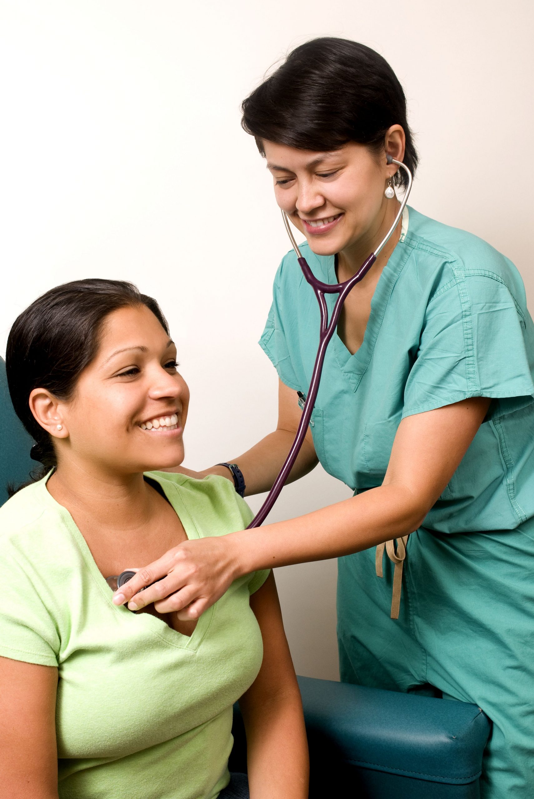
http://humanbiology.pressbooks.tru.ca/wp-content/uploads/sites/6/2019/06/human-heartbeat-daniel_simon.mp3
Lub, Dub
Lub dub, lub dub, lub dub... That’s how the sound of a beating heart is typically described. Those are also the only two sounds that should be audible when listening to a normal, healthy heart through a stethoscope, as in Figure 14.3.1. If a doctor hears something different from the normal lub dub sounds, it’s a sign of a possible heart abnormality. What causes the heart to produce the characteristic lub dub sounds? Read on to find out.
Introduction to the Heart
The heart is a muscular organ behind the sternum (breastbone), slightly to the left of the center of the chest. A normal adult heart is about the size of a fist. The function of the heart is to pump blood through blood vessels of the cardiovascular system. The continuous flow of blood through the system is necessary to provide all the cells of the body with oxygen and nutrients, and to remove their metabolic wastes.
Structure of the Heart
The heart has a thick muscular wall that consists of several layers of tissue. Internally, the heart is divided into four chambers through which blood flows. Because of heart valves, blood flows in just one direction through the chambers.
Heart Wall
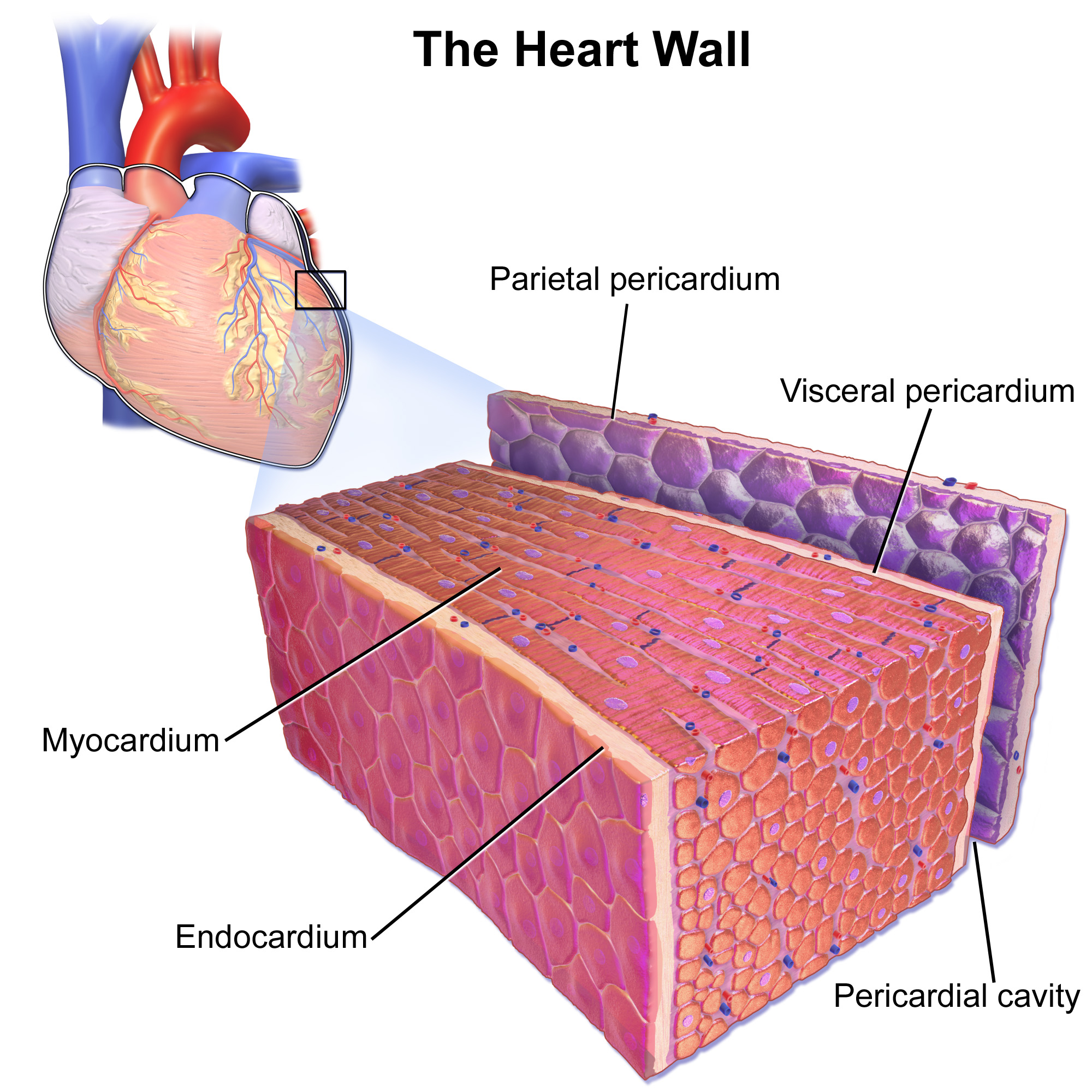
As shown in Figure 14.3.2, the wall of the heart is made up of three layers, called the endocardium, myocardium, and pericardium.
- The endocardium is the innermost layer of the heart wall. It is made up primarily of simple epithelial cells. It covers the heart chambers and valves. A thin layer of connective tissue joins the endocardium to the myocardium.
- The myocardium is the middle and thickest layer of the heart wall. It consists of cardiac muscle surrounded by a framework of collagen. There are two types of cardiac muscle cells in the myocardium: cardiomyocytes — which have the ability to contract easily — and pacemaker cells, which conduct electrical impulses that cause the cardiomyocytes to contract. About 99 per cent of cardiac muscle cells are cardiomyocytes, and the remaining one per cent is pacemaker cells. The myocardium is supplied with blood vessels and nerve fibres via the pericardium.
- The pericardium is a protective sac that encloses and protects the heart. The pericardium consists of two membranes (visceral pericardium and parietal pericardium), between which there is a fluid-filled cavity. The fluid helps to cushion the heart, and also lubricates its outer surface.
Heart Chambers
As shown in Figure 14.3.3 the four chambers of the heart include two upper chambers called atria (singular, atrium), and two lower chambers called ventricles. The atria are also referred to as receiving chambers, because blood coming into the heart first enters these two chambers. The right atrium receives deoxygenated blood from the upper and lower body through the superior vena cava and inferior vena cava, respectively. The left atrium receives oxygenated blood from the lungs through the pulmonary veins. The ventricles are also referred to as discharging chambers, because blood leaving the heart passes out through these two chambers. The right ventricle discharges blood to the lungs through the pulmonary artery, and the left ventricle discharges blood to the rest of the body through the aorta. The four chambers are separated from each other by dense connective tissue consisting mainly of collagen.
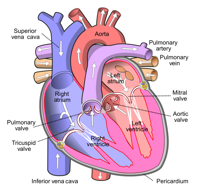
Heart Valves
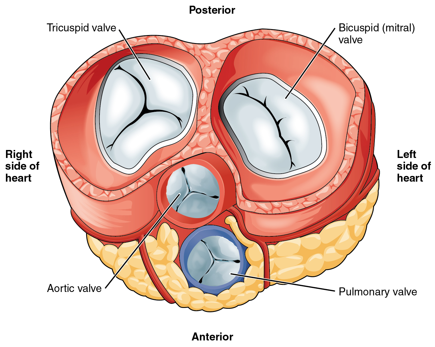
Figure 14.3.4 shows the location of the heart's four valves in a top-down view, looking down at the heart as if the arteries and veins feeding into and out of the heart were removed. The heart valves allow blood to flow from the atria to the ventricles, and from the ventricles to the pulmonary artery and aorta. The valves are constructed in such a way that blood can flow through them in only one direction, thus preventing the backflow of blood. Figure 14.3.5 shows how valves open to let blood into the appropriate chamber, and then close to prevent blood from moving in the wrong direction and the next chamber contracts. The four valves are the:
- Tricuspid atrioventricular valve, (can be shortened to tricuspid AV valve) which allows blood to flow from the right atrium to the right ventricle.
- Bicuspid atrioventricular valve (also know as the mitral valve), which allows blood to flow from the left atrium to the left ventricle.
- Pulmonary semilunar valve, which allows blood to flow from the right ventricle to the pulmonary artery.
- Aortic semilunar valve, which allows blood to flow from the left ventricle to the aorta.
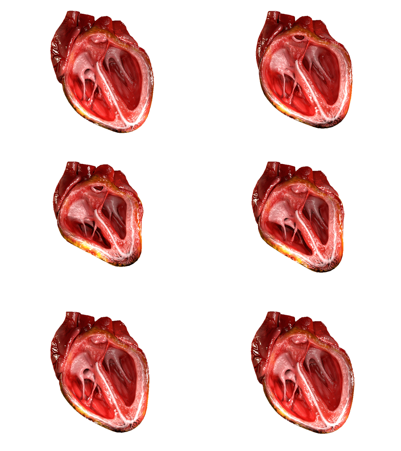
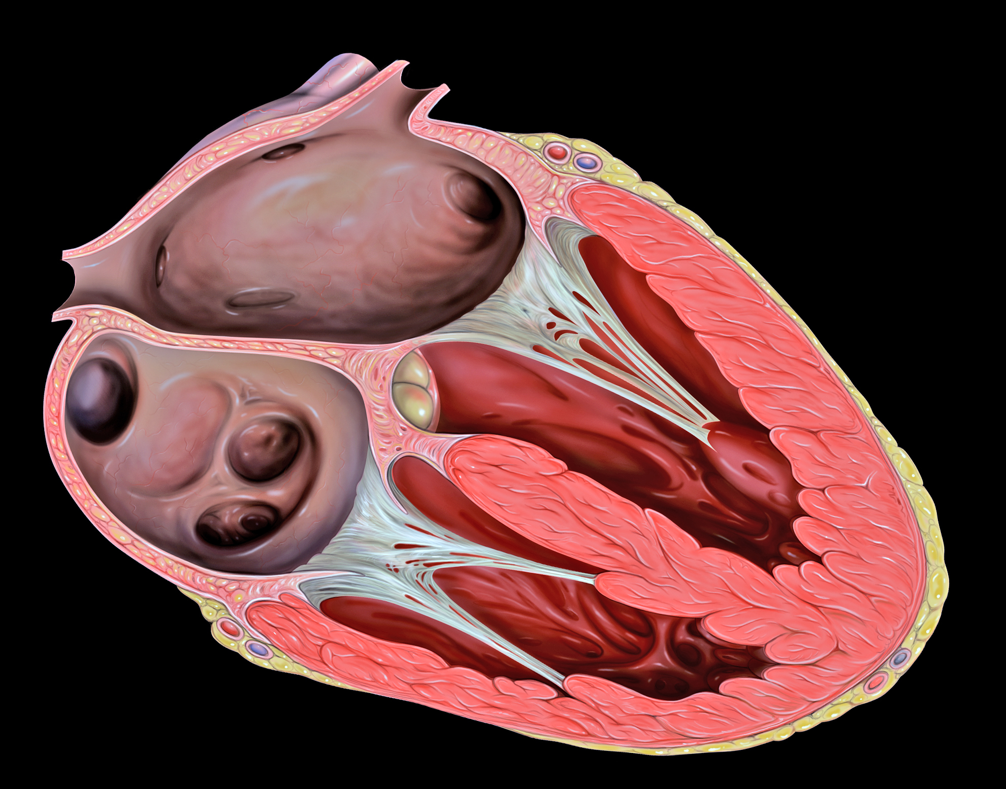
The two atrioventricular (AV) valves prevent backflow when the ventricles are contracting, while the semilunar valves prevent backflow from vessels. This means that the AV valves must withstand much more pressure than do the semilunar valves. In order to withstand the force of the ventricles contracting (to prevent blood from backflowing into the atria), the AV valves are reinforced with structures called chordae tendineae — tendon-like cords of connective tissue which anchor the valve and prevent it from prolapse. Figure 14.3.6 shows the structure and location of the chordae tendoneae.
The chordae tendoneae are under such force that they need special attachments to the interior of the ventricles where they anchor. Papillary muscles are specialized muscles in the interior of the ventricle that provide a strong anchor point for the chordae tendineae.
Coronary Circulation
The cardiomyocytes of the muscular walls of the heart are very active cells, because they are responsible for the constant beating of the heart. These cells need a continuous supply of oxygen and nutrients. The carbon dioxide and waste products they produce also must be continuously removed. The blood vessels that carry blood to and from the heart muscle cells make up the coronary circulation. Note that the blood vessels of the coronary circulation supply heart tissues with blood, and are different from the blood vessels that carry blood to and from the chambers of the heart as part of the general circulation. Coronary arteries supply oxygen-rich blood to the heart muscle cells. Coronary veins remove deoxygenated blood from the heart muscles cells.
- There are two coronary arteries — a right coronary artery that supplies the right side of the heart, and a left coronary artery that supplies the left side of the heart. These arteries branch repeatedly into smaller and smaller arteries and finally into capillaries, which exchange gases, nutrients, and waste products with cardiomyocytes.
- At the back of the heart, small cardiac veins drain into larger veins, and finally into the great cardiac vein, which empties into the right atrium. At the front of the heart, small cardiac veins drain directly into the right atrium.
Blood Circulation Through the Heart
Figure 14.3.7 shows how blood circulates through the chambers of the heart. The right atrium collects blood from two large veins, the superior vena cava (from the upper body) and the inferior vena cava (from the lower body). The blood that collects in the right atrium is pumped through the tricuspid valve into the right ventricle. From the right ventricle, the blood is pumped through the pulmonary valve into the pulmonary artery. The pulmonary artery carries the blood to the lungs, where it enters the pulmonary circulation, gives up carbon dioxide, and picks up oxygen. The oxygenated blood travels back from the lungs through the pulmonary veins (of which there are four), and enters the left atrium of the heart. From the left atrium, the blood is pumped through the mitral valve into the left ventricle. From the left ventricle, the blood is pumped through the aortic valve into the aorta, which subsequently branches into smaller arteries that carry the blood throughout the rest of the body. After passing through capillaries and exchanging substances with cells, the blood returns to the right atrium via the superior vena cava and inferior vena cava, and the process begins anew.
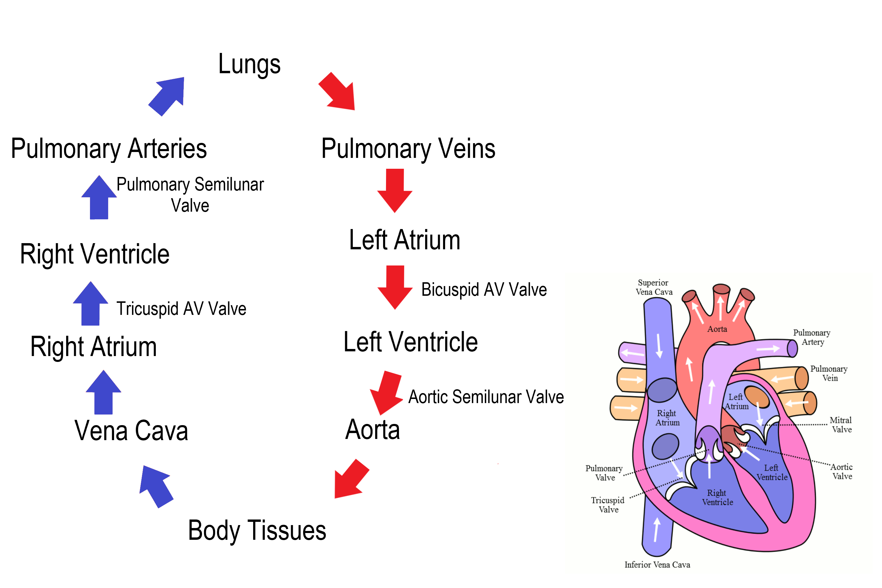
Cardiac Cycle
The cardiac cycle refers to a single complete heartbeat, which includes one iteration of the lub and dub sounds heard through a stethoscope. During the cardiac cycle, the atria and ventricles work in a coordinated fashion so that blood is pumped efficiently through and out of the heart. The cardiac cycle includes two parts, called diastole and systole, which are illustrated in the diagrams in Figure 14.3.8.
- During diastole, the atria contract and pump blood into the ventricles, while the ventricles relax and fill with blood from the atria.
- During systole, the atria relax and collect blood from the lungs and body, while the ventricles contract and pump blood out of the heart.
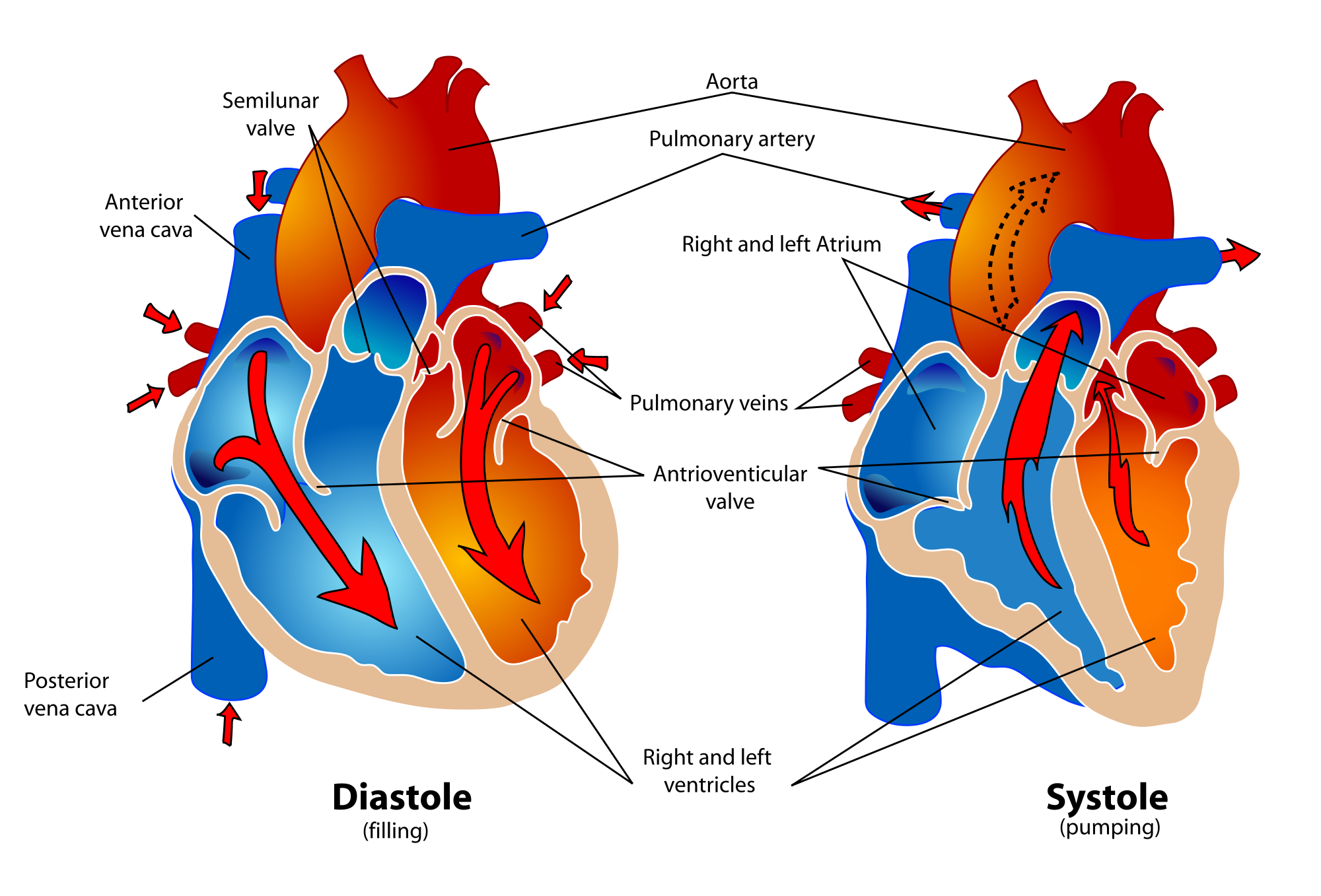
Electrical Stimulation of the Heart
The normal, rhythmical beating of the heart is called sinus rhythm. It is established by the heart’s pacemaker cells, which are located in an area of the heart called the sinoatrial node (shown in Figure 14.3.9). The pacemaker cells create electrical signals with the movement of electrolytes (sodium, potassium, and calcium ions) into and out of the cells. For each cardiac cycle, an electrical signal rapidly travels first from the sinoatrial node, to the right and left atria so they contract together. Then, the signal travels to another node, called the atrioventricular node (Figure 14.3.9), and from there to the right and left ventricles (which also contract together), just a split second after the atria contract.
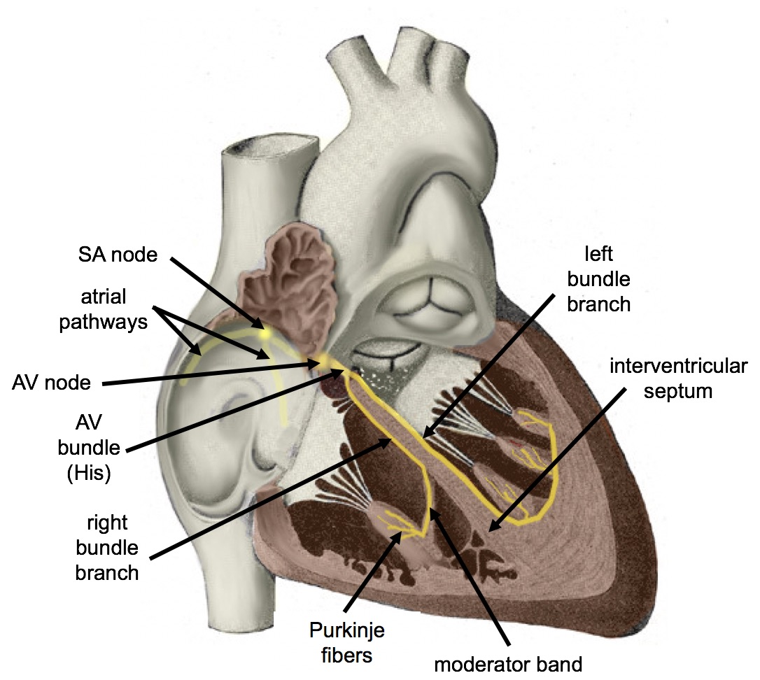
The normal sinus rhythm of the heart is influenced by the autonomic nervous system through sympathetic and parasympathetic nerves. These nerves arise from two paired cardiovascular centers in the medulla of the brainstem. The parasympathetic nerves act to decrease the heart rate, and the sympathetic nerves act to increase the heart rate. Parasympathetic input normally predominates. Without it, the pacemaker cells of the heart would generate a resting heart rate of about 100 beats per minute, instead of a normal resting heart rate of about 72 beats per minute. The cardiovascular centers receive input from receptors throughout the body, and act through the sympathetic nerves to increase the heart rate, as needed. Increased physical activity, for example, is detected by receptors in muscles, joints, and tendons. These receptors send nerve impulses to the cardiovascular centers, causing sympathetic nerves to increase the heart rate, and allowing more blood to flow to the muscles.
Besides the autonomic nervous system, other factors can also affect the heart rate. For example, thyroid hormones and adrenal hormones (such as epinephrine) can stimulate the heart to beat faster. The heart rate also increases when blood pressure drops or the body is dehydrated or overheated. On the other hand, cooling of the body and relaxation — among other factors — can contribute to a decrease in the heart rate.
Feature: Human Biology in the News
When a patient’s heart is too diseased or damaged to sustain life, a heart transplant is likely to be the only long-term solution. The first successful heart transplant was undertaken in South Africa in 1967. There are over 2,200 Canadians walking around today because of life-saving heart transplant surgery. Approximately 180 heart transplant surgeries are performed each year, but there are still so many Canadians on the transplant list that some die while waiting for a heart. The problem is that far too few hearts are available for transplant — there is more demand (people waiting for a heart transplant) than supply (organ donors). Sometimes, recipient hopefuls will receive a device called a Total Artificial Heart (see Figure 14.3.10), which can buy them some time until a donor heart becomes available.
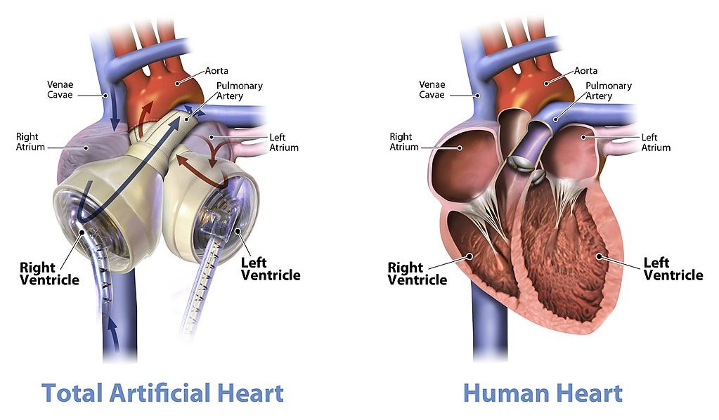
Watch the video below "Total artificial heart option..." from Stanford Health Care to see how it works:
https://youtu.be/1PtxaxcPnGc
Total artificial heart option at Stanford (Includes surgical graphic footage), Stanford Health Care, 2014.
14.3 Summary
- The heart is a muscular organ behind the sternum and slightly to the left of the center of the chest. Its function is to pump blood through the blood vessels of the cardiovascular system.
- The wall of the heart consists of three layers. The middle layer, the myocardium, is the thickest layer and consists mainly of cardiac muscle. The interior of the heart consists of four chambers, with an upper atrium and lower ventricle on each side of the heart. Blood enters the heart through the atria, which pump it to the ventricles. Then the ventricles pump blood out of the heart. Four valves in the heart keep blood flowing in the correct direction and prevent backflow.
- The coronary circulation consists of blood vessels that carry blood to and from the heart muscle cells, and is different from the general circulation of blood through the heart chambers. There are two coronary arteries that supply the two sides of the heart with oxygenated blood. Cardiac veins drain deoxygenated blood back into the heart.
- Deoxygenated blood flows into the right atrium through veins from the upper and lower body (superior and inferior vena cava, respectively), and oxygenated blood flows into the left atrium through four pulmonary veins from the lungs. Each atrium pumps the blood to the ventricle below it. From the right ventricle, deoxygenated blood is pumped to the lungs through the two pulmonary arteries. From the left ventricle, oxygenated blood is pumped to the rest of the body through the aorta.
- The cardiac cycle refers to a single complete heartbeat. It includes diastole — when the atria contract — and systole, when the ventricles contract.
- The normal, rhythmic beating of the heart is called sinus rhythm. It is established by the heart’s pacemaker cells in the sinoatrial node. Electrical signals from the pacemaker cells travel to the atria, and cause them to contract. Then, the signals travel to the atrioventricular node and from there to the ventricles, causing them to contract. Electrical stimulation from the autonomic nervous system and hormones from the endocrine system can also influence heartbeat.
14.3 Review Questions
- What is the heart, where is located, and what is its function?
-
- Describe the coronary circulation.
- Summarize how blood flows into, through, and out of the heart.
- Explain what controls the beating of the heart.
- What are the two types of cardiac muscle cells in the myocardium? What are the differences between these two types of cells?
- Explain why the blood from the cardiac veins empties into the right atrium of the heart. Focus on function (rather than anatomy) in your answer.
14.3 Explore More
https://www.youtube.com/watch?v=1bnzVjOJ6NM
Noel Bairey Merz: The single biggest health threat women face, TED, 2012.
https://www.youtube.com/watch?v=jJm7zBcN6-M
Watch a Transcatheter Aortic Valve Replacement (TAVR) Procedure at St. Luke's in Cedar Rapids, Iowa, UnityPoint Health - Cedar Rapids, 2018.
https://www.youtube.com/watch?v=zU6mmix04PI
A Change of Heart: My Transplant Experience | Thomas Volk | TEDxUWLaCrosse, TEDx Talks, 2018.
https://www.youtube.com/watch?v=biGuwQhuAsk
Heart Transplant Recipient Meets Donor Family For The First Time, WMC Health, 2018.
Attributions
Figure 14.3.1
- Female clinician dressed in scrubs using a stethoscope by Amanda Mills, USCDCP, on Pixnio is used under a CC0 public domain certification license (https://creativecommons.org/licenses/publicdomain/).
- Human heart beating loud and strong (audio) by Daniel Simion on Soundbible.com is used under a CC BY 3.0 (https://creativecommons.org/licenses/by/3.0) license.
Figure 14.3.2
Blausen_0470_HeartWall by BruceBlaus on Wikimedia Commons is used under a CC BY 3.0 (https://creativecommons.org/licenses/by/3.0) license.
Figure 14.3.3
Diagram_of_the_human_heart_(cropped).svg by Wapcaplet on Wikimedia Commons is used under a CC BY-SA 3.0 (http://creativecommons.org/licenses/by-sa/3.0/) license.
Figure 14.3.4
Heart_Valves by OpenStax College on Wikimedia Commons is used under a CC BY 3.0 (https://creativecommons.org/licenses/by/3.0) license.
Figure 14.3.5
CG_Heart Valve Animation by DrJanaOfficial on Wikimedia Commons is used under a CC BY-SA 4.0 (https://creativecommons.org/licenses/by-sa/4.0) license.
Figure 14.3.6
Heart_tee_four_chamber_view by Patrick J. Lynch, medical illustrator from Yale University School of Medicine, on Wikimedia Commons is used under a CC BY 2.5 (https://creativecommons.org/licenses/by/2.5) license.
Figure 14.3.7
Circulation of blood through the heart by Christinelmiller on Wikimedia Commons is used under a CC BY-SA 4.0 (https://creativecommons.org/licenses/by-sa/4.0) license. [Original image in the bottom right is by Wapcaplet / CC BY-SA 3.0 (https://creativecommons.org/licenses/by-sa/3.0/)]
Figure 14.3.8
Human_healthy_pumping_heart_en.svg by Mariana Ruiz Villarreal [LadyofHats] on Wikimedia Common is released into the public domain (https://en.wikipedia.org/wiki/Public_domain).
Figure 14.3.9
Cardiac_Conduction_System by Cypressvine on Wikimedia Commons is used under a CC BY-SA 4.0 (https://creativecommons.org/licenses/by-sa/4.0) license.
References
Betts, J. G., Young, K.A., Wise, J.A., Johnson, E., Poe, B., Kruse, D.H., Korol, O., Johnson, J.E., Womble, M., DeSaix, P. (2013, June 19). Figure 19.12 Heart valves with the atria and major vessels removed [digital image]. In Anatomy and Physiology (Section 19.1). OpenStax. https://openstax.org/books/anatomy-and-physiology/pages/19-1-heart-anatomy#fig-ch20_01_04
Blausen.com Staff. (2014). Medical gallery of Blausen Medical 2014. WikiJournal of Medicine 1 (2). DOI:10.15347/wjm/2014.010. ISSN 2002-4436.
Heart and Stroke Foundation of Canada. (n.d.). https://www.heartandstroke.ca/
Sliwa, K., Zilla, P. (2017, December 7). 50th anniversary of the first human heart transplant—How is it seen today? European Heart Journal, 38(46):3402–3404. https://doi.org/10.1093/eurheartj/ehx695
Stanford Health Care. (2014, December 3). Total artificial heart option at Stanford (Includes surgical graphic footage). YouTube. https://www.youtube.com/watch?v=1PtxaxcPnGc&feature=youtu.be
TED. (2012, March 21). Noel Bairey Merz: The single biggest health threat women face. YouTube. https://www.youtube.com/watch?v=1bnzVjOJ6NM&feature=youtu.be
TEDx Talks. (2018, April 18). A change of heart: My transplant experience | Thomas Volk | TEDxUWLaCrosse. YouTube. https://www.youtube.com/watch?v=zU6mmix04PI&feature=youtu.be
UMagazine. (2015, Fall). The cutting edge: Patient first to bridge from experimental total artificial heart to transplant. UCLA Health. https://www.uclahealth.org/u-magazine/patient-first-to-bridge-from-experimental-total-artificial-heart-to-transplant
UnityPoint Health - Cedar Rapids. (2018, February 7). Watch a transcatheter aortic valve replacement (TAVR) Procedure at St. Luke's in Cedar Rapids, Iowa. YouTube. https://www.youtube.com/watch?v=jJm7zBcN6-M&feature=youtu.be
WMC Health. (2018, September 13). Heart transplant recipient meets donor family for the first time. YouTube. https://www.youtube.com/watch?v=biGuwQhuAsk&feature=youtu.be
Image shows a diagram labeling the major arteries of the body. Some of these include the carotid artery which provides blood to the neck and head, the brachiocephalic artery which supplies blood to the arms and head, the renal artery supplying blood to the kidneys, the mesenteric arteries supplying blood to the intestines, the femoral arteries supplying blood to the legs.
Case Study: Please Don’t Pass the Bread
Angela and Saloni are college students who met in physics class. They decide to study together for their upcoming midterm, but first, they want to grab some lunch. Angela says there is a particular restaurant she would like to go to, because they are able to accommodate her dietary restrictions. Saloni agrees and they head to the restaurant.
At lunch, Saloni asks Angela what is special about her diet. Angela tells her that she can’t eat gluten. Saloni says, “My cousin did that for a while because she heard that gluten is bad for you. But it was too hard for her to not eat bread and pasta, so she gave it up.” Angela tells Saloni that avoiding gluten isn’t optional for her — she has celiac disease. Eating even very small amounts of gluten could damage her digestive system. It can be difficult for people living with celiac disease to find foods when eating out.
You have probably heard of gluten, but what is it, and why is it harmful to people with celiac disease? Gluten is a protein present in wheat and some other grains (such as barley, rye, and oats), so it is commonly found in foods like bread, pasta, baked goods, and many packaged foods, like the ones pictured in Figure 15.1.2.
Figure 15.1.2 Gluten is a protein present in foods like bread, pasta, and baked goods.
For people with celiac disease, eating gluten causes an autoimmune reaction that results in damage to the small, finger-like villi lining the small intestine, causing them to become inflamed and flattened (see Figure 15.1.3). This damage interferes with the digestive process, which can result in a wide variety of symptoms including diarrhea, anemia, skin rash, bone pain, depression, and anxiety, among others. The degree of damage to the villi can vary from mild to severe, with more severe damage generally resulting in more significant symptoms and complications. Celiac disease can have serious long-term consequences, such as osteoporosis, problems in the nervous and reproductive systems, and the development of certain types of cancers.
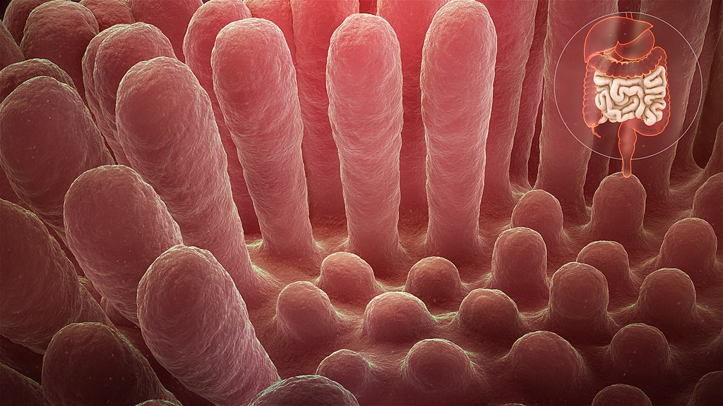
Why does celiac disease cause so many different types of symptoms and have such significant negative health consequences? As you read this chapter and learn about how the digestive system works, you will see just how important the villi of the small intestine are to the body as a whole. At the end of the chapter, you will learn more about celiac disease, why it can be so serious, and whether it is worth avoiding gluten for people who do not have a diagnosed medical issue with it.
Chapter Overview: Digestive System
In this chapter, you will learn about the digestive system, which processes food so that our bodies can obtain nutrients. Specifically, you will learn about:
- The structures and organs of the gastrointestinal (GI) tract through which food directly passes. This includes the mouth, pharynx, esophagus, stomach, small intestine, and large intestine.
- The functions of the GI tract, including mechanical and chemical digestion, absorption of nutrients, and the elimination of solid waste.
- The accessory organs of digestion — the liver, gallbladder, and pancreas — which secrete substances needed for digestion into the GI tract, in addition to performing other important functions.
- Specializations of the tissues of the digestive system that allow it to carry out its functions.
- How different types of nutrients (such as carbohydrates, proteins, and fats) are digested and absorbed by the body.
- Beneficial bacteria that live in the GI tract and help us digest food, produce vitamins, and protect us from harmful pathogens and toxic substances.
- Disorders of the digestive system, including inflammatory bowel diseases, ulcers, diverticulitis, and gastroenteritis (commonly known as “stomach flu”).
As you read this chapter, think about the following questions related to celiac disease:
- What are the general functions of the small intestine? What do the villi in the small intestine do?
- Why do you think celiac disease causes so many different types of symptoms and potentially serious complications?
- What are some other autoimmune diseases that involve the body attacking its own digestive system?
Attributions
Figure 15.1.1
Bread [photo] by Sergio Arze on Unsplash is used under the Unsplash License (https://unsplash.com/license).
Figure 15.1.2
- Paste cu sos de roșii by Sestrjevitovschii Ina on Unsplash is used under the Unsplash License (https://unsplash.com/license).
- Cookies and More by Sarah Shaffer on Unsplash is used under the Unsplash License (https://unsplash.com/license).
- Raspberry waffles by Izabelle Acheson on Unsplash is used under the Unsplash License (https://unsplash.com/license).
- Homemade croissant & pain au chocolat by Cristiano Pinto on Unsplash is used under the Unsplash License (https://unsplash.com/license).
Figure 15.1.3
Inflammed_mucous_layer_of_the_intestinal_villi_depicting_Celiac_disease by www.scientificanimations.com (image 140/191) on Wikimedia Commons is used under a CC BY-SA 4.0 (https://creativecommons.org/licenses/by-sa/4.0) license.
Image shows a diagram illustrating how peristalsis pushes food through the digestive tract by squeezing just behind the food, pushing it forward.
As per caption

We All Scream for Ice Cream
If you’re an ice cream lover, then just the sight of this yummy ice cream cone may make your mouth water. The “water” in your mouth is actually saliva, a fluid released by glands that are part of the digestive system. Saliva contains digestive enzymes, among other substances important for digestion. When your mouth waters at the sight of a tasty treat, it’s a sign that your digestive system is preparing to digest food.
What Is the Digestive System?
The digestive system consists of organs that break down food, absorb its nutrients, and expel any remaining waste. Organs of the digestive system are shown in Figure 15.2.2. Most of these organs make up the gastrointestinal (GI) tract, through which food actually passes. The rest of the organs of the digestive system are called accessory organs. These organs secrete enzymes and other substances into the GI tract, but food does not actually pass through them.
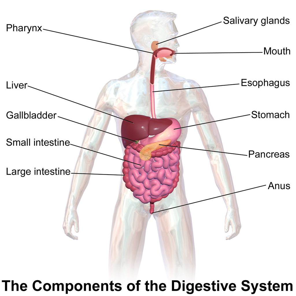
Functions of the Digestive System
The digestive system has three main functions relating to food: digestion of food, absorption of nutrients from food, and elimination of solid food waste. Digestion is the process of breaking down food into components the body can absorb. It consists of two types of processes: mechanical digestion and chemical digestion. Mechanical digestion is the physical breakdown of chunks of food into smaller pieces, and it takes place mainly in the mouth and stomach. Chemical digestion is the chemical breakdown of large, complex food molecules into smaller, simpler nutrient molecules that can be absorbed by body fluids (blood or lymph). This type of digestion begins in the mouth and continues in the stomach, but occurs mainly in the small intestine.
After food is digested, the resulting nutrients are absorbed. Absorption is the process in which substances pass into the bloodstream or lymph system to circulate throughout the body. Absorption of nutrients occurs mainly in the small intestine. Any remaining matter from food that is not digested and absorbed passes out of the body through the anus in the process of elimination.
Gastrointestinal Tract
The gastrointestinal (GI) tract is basically a long, continuous tube that connects the mouth with the anus. If it were fully extended, it would be about nine metres long in adults. It includes the mouth, pharynx, esophagus, stomach, and small and large intestines. Food enters the mouth, and then passes through the other organs of the GI tract, where it is digested and/or absorbed. Finally, any remaining food waste leaves the body through the anus at the end of the large intestine. It takes up to 50 hours for food or food waste to make the complete trip through the GI tract.
Tissues of the GI Tract
The walls of the organs of the GI tract consist of four different tissue layers, which are illustrated in Figure 15.2.3: mucosa, submucosa, muscularis externa, and serosa.
- The mucosa is the innermost layer surrounding the lumen (open space within the organs of the GI tract). This layer consists mainly of epithelium with the capacity to secrete and absorb substances. The epithelium can secret digestive enzymes and mucus, and it can absorb nutrients and water.
- The submucosa layer consists of connective tissue that contains blood and lymph vessels, as well as nerves. The vessels are needed to absorb and carry away nutrients after food is digested, and nerves help control the muscles of the GI tract organs.
- The muscularis externa layer contains two types of smooth muscle: longitudinal muscle and circular muscle. Longitudinal muscle runs the length of the GI tract organs, and circular muscle encircles the organs. Both types of muscles contract to keep food moving through the tract by the process of peristalsis, which is described below.
- The serosa layer is the outermost layer of the walls of GI tract organs. This is a thin layer that consists of connective tissue and separates the organs from surrounding cavities and tissues.
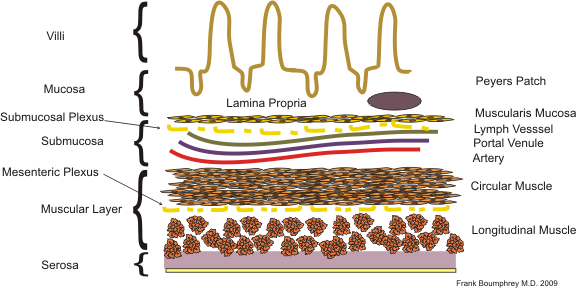 |
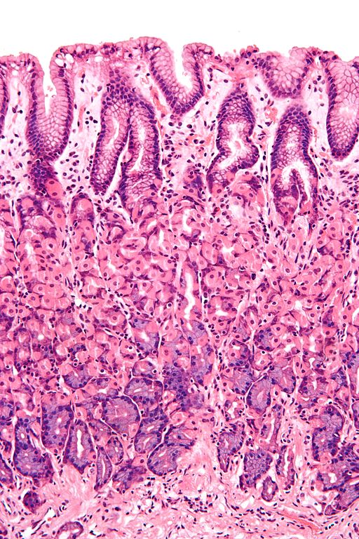 |
Peristalisis in the GI Tract
The muscles in the walls of GI tract organs enable peristalsis, which is illustrated in Figure 15.2.5. Peristalsis is a continuous sequence of involuntary muscle contraction and relaxation that moves rapidly along an organ like a wave, similar to the way a wave moves through a spring toy. Peristalsis in organs of the GI tract propels food through the tract.
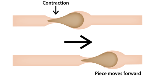
Watch the video "What is peristalsis?" by Mister Science to see peristalsis in action:
https://youtu.be/kVjeNZA5pi4
What is peristalsis?, Mister Science, 2018.
Immune Function of the GI Tract
The GI tract plays an important role in protecting the body from pathogens. The surface area of the GI tract is estimated to be about 32 square metres (105 square feet), or about half the area of a badminton court. This is more than three times the area of the exposed skin of the body, and it provides a lot of area for pathogens to invade the tissues of the body. The innermost mucosal layer of the walls of the GI tract provides a barrier to pathogens so they are less likely to enter the blood or lymph circulations. The mucus produced by the mucosal layer, for example, contains antibodies that mark many pathogenic microorganisms for destruction. Enzymes in some of the secretions of the GI tract also destroy pathogens. In addition, stomach acids have a very low pH that is fatal for many microorganisms that enter the stomach.
Divisions of the GI Tract
The GI tract is often divided into an upper GI tract and a lower GI tract. For medical purposes, the upper GI tract is typically considered to include all the organs from the mouth through the first part of the small intestine, called the duodenum. For our instructional purposes, it makes more sense to include the mouth through the stomach in the upper GI tract, and all of the small intestine — as well as the large intestine — in the lower GI tract.
Upper GI Tract
The mouth is the first digestive organ that food enters. The sight, smell, or taste of food stimulates the release of digestive enzymes and other secretions by salivary glands inside the mouth. The major salivary gland enzyme is amylase. It begins the chemical digestion of carbohydrates by breaking down starches into sugar. The mouth also begins the mechanical digestion of food. When you chew, your teeth break, crush, and grind food into increasingly smaller pieces. Your tongue helps mix the food with saliva and also helps you swallow.
A lump of swallowed food is called a bolus. The bolus passes from the mouth into the pharynx, and from the pharynx into the esophagus. The esophagus is a long, narrow tube that carries food from the pharynx to the stomach. It has no other digestive functions. Peristalsis starts at the top of the esophagus when food is swallowed and continues down the esophagus in a single wave, pushing the bolus of food ahead of it.
From the esophagus, food passes into the stomach, where both mechanical and chemical digestion continue. The muscular walls of the stomach churn and mix the food, thus completing mechanical digestion, as well as mixing the food with digestive fluids secreted by the stomach. One of these fluids is hydrochloric acid (HCl). In addition to killing pathogens in food, it gives the stomach the low pH needed by digestive enzymes that work in the stomach. One of these enzymes is pepsin, which chemically digests proteins. The stomach stores the partially digested food until the small intestine is ready to receive it. Food that enters the small intestine from the stomach is in the form of a thick slurry (semi-liquid) called chyme.
Lower GI Tract
The small intestine is a narrow, but very long tubular organ. It may be almost seven metres long in adults. It is the site of most chemical digestion and virtually all absorption of nutrients. Many digestive enzymes are active in the small intestine, some of which are produced by the small intestine itself, and some of which are produced by the pancreas, an accessory organ of the digestive system. Much of the inner lining of the small intestine is covered by tiny finger-like projections called villi, each of which is covered by even tinier projections called microvilli. These projections, shown in the drawing below (Figure 15.2.6), greatly increase the surface area through which nutrients can be absorbed from the small intestine.
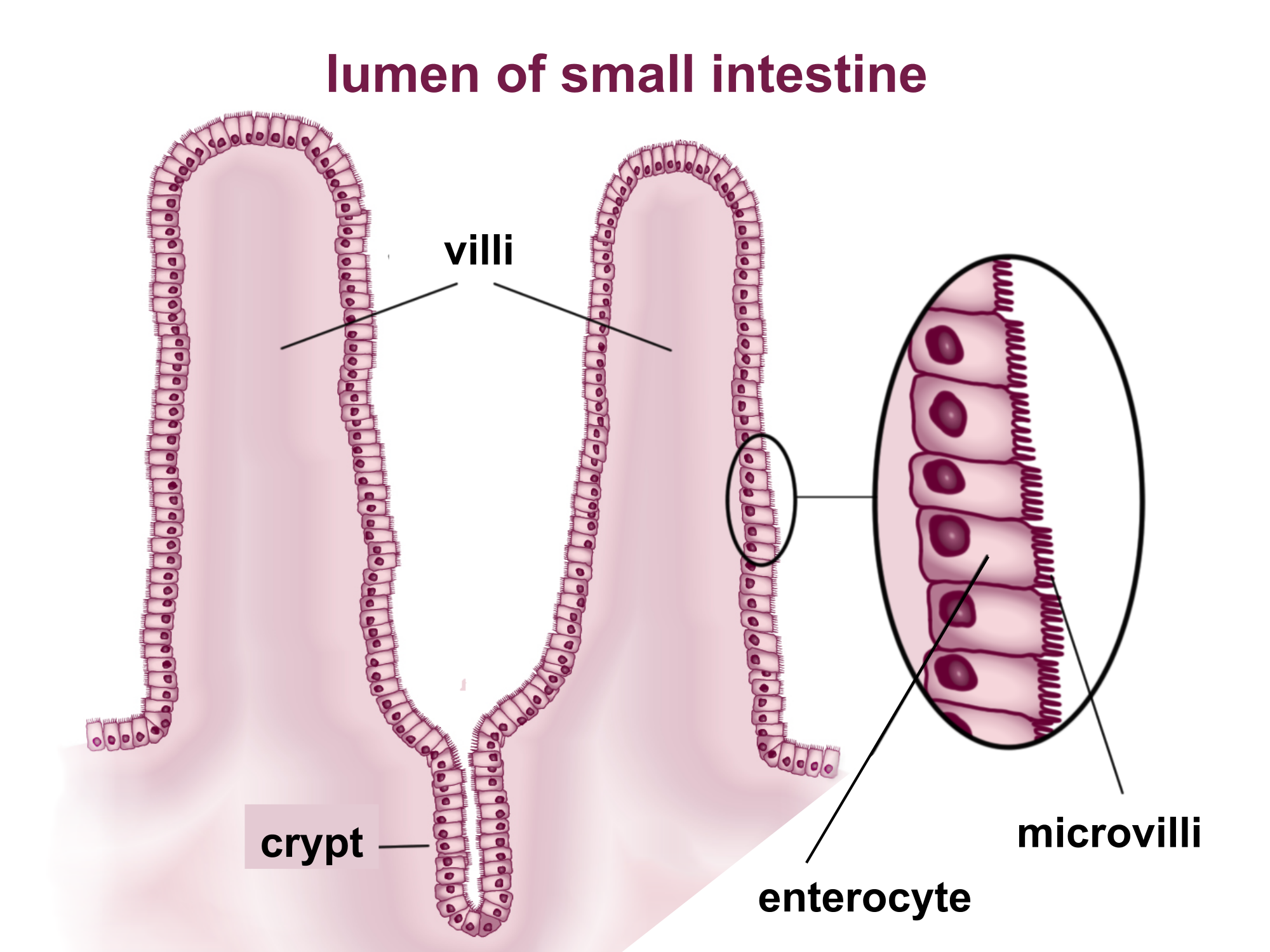
From the small intestine, any remaining nutrients and food waste pass into the large intestine. The large intestine is another tubular organ, but it is wider and shorter than the small intestine. It connects the small intestine and the anus. Waste that enters the large intestine is in a liquid state. As it passes through the large intestine, excess water is absorbed from it. The remaining solid waste — called feces — is eventually eliminated from the body through the anus.
Accessory Organs of the Digestive System
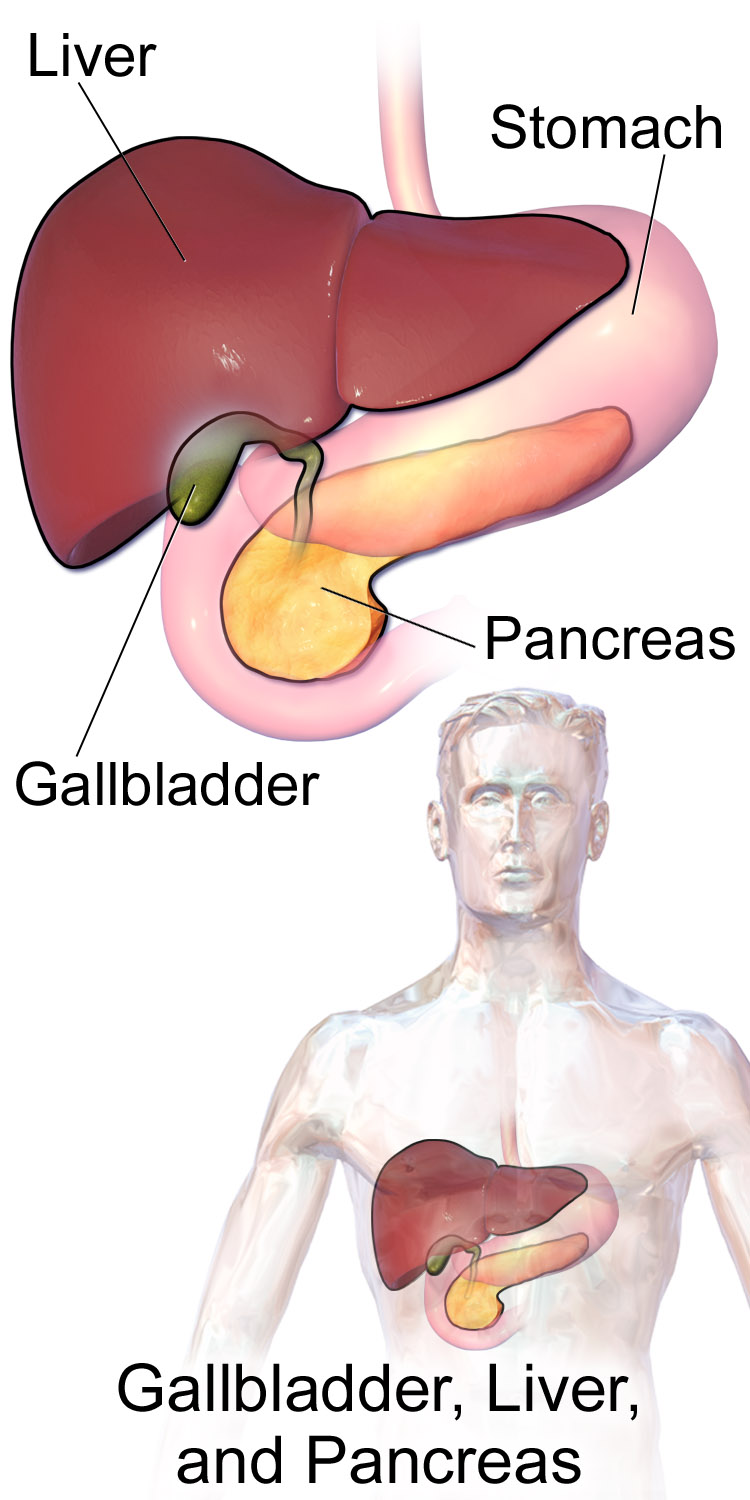
Accessory organs of the digestive system are not part of the GI tract, so they are not sites where digestion or absorption take place. Instead, these organs secrete or store substances needed for the chemical digestion of food. The accessory organs include the liver, gallbladder, and pancreas. They are shown in Figure 15.2.7 and described in the text that follows.
- The liver is an organ with multitude of functions. Its main digestive function is producing and secreting a fluid called bile, which reaches the small intestine through a duct. Bile breaks down large globules of lipids into smaller ones that are easier for enzymes to chemically digest. Bile is also needed to reduce the acidity of food entering the small intestine from the highly acidic stomach, because enzymes in the small intestine require a less acidic environment in order to work.
- The gallbladder is a small sac below the liver that stores some of the bile from the liver. The gallbladder also concentrates the bile by removing some of the water from it. It then secretes the concentrated bile into the small intestine as needed for fat digestion following a meal.
- The pancreas secretes many digestive enzymes, and releases them into the small intestine for the chemical digestion of carbohydrates, proteins, and lipids. The pancreas also helps lessen the acidity of the small intestine by secreting bicarbonate, a basic substance that neutralizes acid.
15.2 Summary
- The digestive system consists of organs that break down food, absorb its nutrients, and expel any remaining food waste.
- Digestion is the process of breaking down food into components that the body can absorb. It includes mechanical digestion and chemical digestion. Absorption is the process of taking up nutrients from food by body fluids for circulation to the rest of the body. Elimination is the process of excreting any remaining food waste after digestion and absorption are finished.
- Most digestive organs form a long, continuous tube called the gastrointestinal (GI) tract. It starts at the mouth, which is followed by the pharynx, esophagus, stomach, small intestine, and large intestine. The upper GI tract consists of the mouth through the stomach, while the lower GI tract consists of the small and large intestines.
- Digestion and/or absorption take place in most of the organs of the GI tract. Organs of the GI tract have walls that consist of several tissue layers that enable them to carry out these functions. The inner mucosa has cells that secrete digestive enzymes and other digestive substances, as well as cells that absorb nutrients. The muscle layer of the organs enables them to contract and relax in waves of peristalsis to move food through the GI tract.
- Three digestive organs — the liver, gallbladder, and pancreas — are accessory organs of digestion. They secrete substances needed for chemical digestion into the small intestine.
15.2 Review Questions
- What is the digestive system?
- What are the three main functions of the digestive system? Define each function.
-
- Relate the tissues in the walls of GI tract organs to the functions the organs perform.
15.2 Explore More
https://youtu.be/Og5xAdC8EUI
How your digestive system works - Emma Bryce, TED-Ed, 2017.
https://youtu.be/YVfyYrEmzgM
How does your body know you're full? - Hilary Coller, TED-Ed, 2017.
Attributions
Figure 15.2.1
Ice Cream [photo] by Mark Cruz on Unsplash is used under the Unsplash License (https://unsplash.com/license).
Figure 15.2.2
Blausen_0316_DigestiveSystem by BruceBlaus on Wikimedia Commons is used under a CC BY 3.0 (https://creativecommons.org/licenses/by/3.0) license.
Figure 15.2.3
Intestinal_layers by Boumphreyfr on Wikimedia Commons is used under a CC BY-SA 3.0 (https://creativecommons.org/licenses/by-sa/3.0) license.
Figure 15.2.4
512px-Normal_gastric_mucosa_intermed_mag by Nephron on Wikimedia Commons is used under a CC BY-SA 3.0 (https://creativecommons.org/licenses/by-sa/3.0) license.
Figure 15.2.5
Peristalsis pushes food through the GI tract by CK-12 Foundation is used under a CC BY NC 3.0 (https://creativecommons.org/licenses/by-nc/3.0/) license.
Figure 15.2.6
Villi_&_microvilli_of_small_intestine.svg by BallenaBlanca on Wikimedia Commons is used under a CC BY-SA 4.0 (https://creativecommons.org/licenses/by-sa/4.0) license.
Figure 15.2.7
Blausen_0428_Gallbladder-Liver-Pancreas_Location by BruceBlaus on Wikimedia Commons is used under a CC BY 3.0 (https://creativecommons.org/licenses/by/3.0) license.
References
Blausen.com Staff. (2014). Medical gallery of Blausen Medical 2014. WikiJournal of Medicine 1 (2). DOI:10.15347/wjm/2014.010. ISSN 2002-4436.
Brainard, J/ CK-12 Foundation. (2016). Figure 4 Peristalsis pushes food through the GI tract. [digital image]. In CK-12 College Human Biology (Section 17.2) [online Flexbook]. CK12.org. https://www.ck12.org/book/ck-12-college-human-biology/section/17.2/
Mister Science. (2018). What is peristalsis? YouTube. https://www.youtube.com/channel/UCxTlkZfjArUobBAeVwzJjYg/videos
TED-Ed. (2017, November 13). How does your body know you're full? - Hilary Coller. YouTube. https://www.youtube.com/watch?v=YVfyYrEmzgM&feature=youtu.be
TED-Ed. (2017, December 14). How your digestive system works - Emma Bryce. YouTube. https://www.youtube.com/watch?v=Og5xAdC8EUI&feature=youtu.be
Created by CK-12 Foundation/Adapted by Christine Miller

We All Scream for Ice Cream
If you’re an ice cream lover, then just the sight of this yummy ice cream cone may make your mouth water. The “water” in your mouth is actually saliva, a fluid released by glands that are part of the digestive system. Saliva contains digestive enzymes, among other substances important for digestion. When your mouth waters at the sight of a tasty treat, it’s a sign that your digestive system is preparing to digest food.
What Is the Digestive System?
The digestive system consists of organs that break down food, absorb its nutrients, and expel any remaining waste. Organs of the digestive system are shown in Figure 15.2.2. Most of these organs make up the gastrointestinal (GI) tract, through which food actually passes. The rest of the organs of the digestive system are called accessory organs. These organs secrete enzymes and other substances into the GI tract, but food does not actually pass through them.

Functions of the Digestive System
The digestive system has three main functions relating to food: digestion of food, absorption of nutrients from food, and elimination of solid food waste. Digestion is the process of breaking down food into components the body can absorb. It consists of two types of processes: mechanical digestion and chemical digestion. Mechanical digestion is the physical breakdown of chunks of food into smaller pieces, and it takes place mainly in the mouth and stomach. Chemical digestion is the chemical breakdown of large, complex food molecules into smaller, simpler nutrient molecules that can be absorbed by body fluids (blood or lymph). This type of digestion begins in the mouth and continues in the stomach, but occurs mainly in the small intestine.
After food is digested, the resulting nutrients are absorbed. Absorption is the process in which substances pass into the bloodstream or lymph system to circulate throughout the body. Absorption of nutrients occurs mainly in the small intestine. Any remaining matter from food that is not digested and absorbed passes out of the body through the anus in the process of elimination.
Gastrointestinal Tract
The gastrointestinal (GI) tract is basically a long, continuous tube that connects the mouth with the anus. If it were fully extended, it would be about nine metres long in adults. It includes the mouth, pharynx, esophagus, stomach, and small and large intestines. Food enters the mouth, and then passes through the other organs of the GI tract, where it is digested and/or absorbed. Finally, any remaining food waste leaves the body through the anus at the end of the large intestine. It takes up to 50 hours for food or food waste to make the complete trip through the GI tract.
Tissues of the GI Tract
The walls of the organs of the GI tract consist of four different tissue layers, which are illustrated in Figure 15.2.3: mucosa, submucosa, muscularis externa, and serosa.
- The mucosa is the innermost layer surrounding the lumen (open space within the organs of the GI tract). This layer consists mainly of epithelium with the capacity to secrete and absorb substances. The epithelium can secret digestive enzymes and mucus, and it can absorb nutrients and water.
- The submucosa layer consists of connective tissue that contains blood and lymph vessels, as well as nerves. The vessels are needed to absorb and carry away nutrients after food is digested, and nerves help control the muscles of the GI tract organs.
- The muscularis externa layer contains two types of smooth muscle: longitudinal muscle and circular muscle. Longitudinal muscle runs the length of the GI tract organs, and circular muscle encircles the organs. Both types of muscles contract to keep food moving through the tract by the process of peristalsis, which is described below.
- The serosa layer is the outermost layer of the walls of GI tract organs. This is a thin layer that consists of connective tissue and separates the organs from surrounding cavities and tissues.
 |
 |
Peristalisis in the GI Tract
The muscles in the walls of GI tract organs enable peristalsis, which is illustrated in Figure 15.2.5. Peristalsis is a continuous sequence of involuntary muscle contraction and relaxation that moves rapidly along an organ like a wave, similar to the way a wave moves through a spring toy. Peristalsis in organs of the GI tract propels food through the tract.

Watch the video "What is peristalsis?" by Mister Science to see peristalsis in action:
https://youtu.be/kVjeNZA5pi4
What is peristalsis?, Mister Science, 2018.
Immune Function of the GI Tract
The GI tract plays an important role in protecting the body from pathogens. The surface area of the GI tract is estimated to be about 32 square metres (105 square feet), or about half the area of a badminton court. This is more than three times the area of the exposed skin of the body, and it provides a lot of area for pathogens to invade the tissues of the body. The innermost mucosal layer of the walls of the GI tract provides a barrier to pathogens so they are less likely to enter the blood or lymph circulations. The mucus produced by the mucosal layer, for example, contains antibodies that mark many pathogenic microorganisms for destruction. Enzymes in some of the secretions of the GI tract also destroy pathogens. In addition, stomach acids have a very low pH that is fatal for many microorganisms that enter the stomach.
Divisions of the GI Tract
The GI tract is often divided into an upper GI tract and a lower GI tract. For medical purposes, the upper GI tract is typically considered to include all the organs from the mouth through the first part of the small intestine, called the duodenum. For our instructional purposes, it makes more sense to include the mouth through the stomach in the upper GI tract, and all of the small intestine — as well as the large intestine — in the lower GI tract.
Upper GI Tract
The mouth is the first digestive organ that food enters. The sight, smell, or taste of food stimulates the release of digestive enzymes and other secretions by salivary glands inside the mouth. The major salivary gland enzyme is amylase. It begins the chemical digestion of carbohydrates by breaking down starches into sugar. The mouth also begins the mechanical digestion of food. When you chew, your teeth break, crush, and grind food into increasingly smaller pieces. Your tongue helps mix the food with saliva and also helps you swallow.
A lump of swallowed food is called a bolus. The bolus passes from the mouth into the pharynx, and from the pharynx into the esophagus. The esophagus is a long, narrow tube that carries food from the pharynx to the stomach. It has no other digestive functions. Peristalsis starts at the top of the esophagus when food is swallowed and continues down the esophagus in a single wave, pushing the bolus of food ahead of it.
From the esophagus, food passes into the stomach, where both mechanical and chemical digestion continue. The muscular walls of the stomach churn and mix the food, thus completing mechanical digestion, as well as mixing the food with digestive fluids secreted by the stomach. One of these fluids is hydrochloric acid (HCl). In addition to killing pathogens in food, it gives the stomach the low pH needed by digestive enzymes that work in the stomach. One of these enzymes is pepsin, which chemically digests proteins. The stomach stores the partially digested food until the small intestine is ready to receive it. Food that enters the small intestine from the stomach is in the form of a thick slurry (semi-liquid) called chyme.
Lower GI Tract
The small intestine is a narrow, but very long tubular organ. It may be almost seven metres long in adults. It is the site of most chemical digestion and virtually all absorption of nutrients. Many digestive enzymes are active in the small intestine, some of which are produced by the small intestine itself, and some of which are produced by the pancreas, an accessory organ of the digestive system. Much of the inner lining of the small intestine is covered by tiny finger-like projections called villi, each of which is covered by even tinier projections called microvilli. These projections, shown in the drawing below (Figure 15.2.6), greatly increase the surface area through which nutrients can be absorbed from the small intestine.

From the small intestine, any remaining nutrients and food waste pass into the large intestine. The large intestine is another tubular organ, but it is wider and shorter than the small intestine. It connects the small intestine and the anus. Waste that enters the large intestine is in a liquid state. As it passes through the large intestine, excess water is absorbed from it. The remaining solid waste — called feces — is eventually eliminated from the body through the anus.
Accessory Organs of the Digestive System

Accessory organs of the digestive system are not part of the GI tract, so they are not sites where digestion or absorption take place. Instead, these organs secrete or store substances needed for the chemical digestion of food. The accessory organs include the liver, gallbladder, and pancreas. They are shown in Figure 15.2.7 and described in the text that follows.
- The liver is an organ with multitude of functions. Its main digestive function is producing and secreting a fluid called bile, which reaches the small intestine through a duct. Bile breaks down large globules of lipids into smaller ones that are easier for enzymes to chemically digest. Bile is also needed to reduce the acidity of food entering the small intestine from the highly acidic stomach, because enzymes in the small intestine require a less acidic environment in order to work.
- The gallbladder is a small sac below the liver that stores some of the bile from the liver. The gallbladder also concentrates the bile by removing some of the water from it. It then secretes the concentrated bile into the small intestine as needed for fat digestion following a meal.
- The pancreas secretes many digestive enzymes, and releases them into the small intestine for the chemical digestion of carbohydrates, proteins, and lipids. The pancreas also helps lessen the acidity of the small intestine by secreting bicarbonate, a basic substance that neutralizes acid.
15.2 Summary
- The digestive system consists of organs that break down food, absorb its nutrients, and expel any remaining food waste.
- Digestion is the process of breaking down food into components that the body can absorb. It includes mechanical digestion and chemical digestion. Absorption is the process of taking up nutrients from food by body fluids for circulation to the rest of the body. Elimination is the process of excreting any remaining food waste after digestion and absorption are finished.
- Most digestive organs form a long, continuous tube called the gastrointestinal (GI) tract. It starts at the mouth, which is followed by the pharynx, esophagus, stomach, small intestine, and large intestine. The upper GI tract consists of the mouth through the stomach, while the lower GI tract consists of the small and large intestines.
- Digestion and/or absorption take place in most of the organs of the GI tract. Organs of the GI tract have walls that consist of several tissue layers that enable them to carry out these functions. The inner mucosa has cells that secrete digestive enzymes and other digestive substances, as well as cells that absorb nutrients. The muscle layer of the organs enables them to contract and relax in waves of peristalsis to move food through the GI tract.
- Three digestive organs — the liver, gallbladder, and pancreas — are accessory organs of digestion. They secrete substances needed for chemical digestion into the small intestine.
15.2 Review Questions
- What is the digestive system?
- What are the three main functions of the digestive system? Define each function.
-
- Relate the tissues in the walls of GI tract organs to the functions the organs perform.
15.2 Explore More
https://youtu.be/Og5xAdC8EUI
How your digestive system works - Emma Bryce, TED-Ed, 2017.
https://youtu.be/YVfyYrEmzgM
How does your body know you're full? - Hilary Coller, TED-Ed, 2017.
Attributions
Figure 15.2.1
Ice Cream [photo] by Mark Cruz on Unsplash is used under the Unsplash License (https://unsplash.com/license).
Figure 15.2.2
Blausen_0316_DigestiveSystem by BruceBlaus on Wikimedia Commons is used under a CC BY 3.0 (https://creativecommons.org/licenses/by/3.0) license.
Figure 15.2.3
Intestinal_layers by Boumphreyfr on Wikimedia Commons is used under a CC BY-SA 3.0 (https://creativecommons.org/licenses/by-sa/3.0) license.
Figure 15.2.4
512px-Normal_gastric_mucosa_intermed_mag by Nephron on Wikimedia Commons is used under a CC BY-SA 3.0 (https://creativecommons.org/licenses/by-sa/3.0) license.
Figure 15.2.5
Peristalsis pushes food through the GI tract by CK-12 Foundation is used under a CC BY NC 3.0 (https://creativecommons.org/licenses/by-nc/3.0/) license.
Figure 15.2.6
Villi_&_microvilli_of_small_intestine.svg by BallenaBlanca on Wikimedia Commons is used under a CC BY-SA 4.0 (https://creativecommons.org/licenses/by-sa/4.0) license.
Figure 15.2.7
Blausen_0428_Gallbladder-Liver-Pancreas_Location by BruceBlaus on Wikimedia Commons is used under a CC BY 3.0 (https://creativecommons.org/licenses/by/3.0) license.
References
Blausen.com Staff. (2014). Medical gallery of Blausen Medical 2014. WikiJournal of Medicine 1 (2). DOI:10.15347/wjm/2014.010. ISSN 2002-4436.
Brainard, J/ CK-12 Foundation. (2016). Figure 4 Peristalsis pushes food through the GI tract. [digital image]. In CK-12 College Human Biology (Section 17.2) [online Flexbook]. CK12.org. https://www.ck12.org/book/ck-12-college-human-biology/section/17.2/
Mister Science. (2018). What is peristalsis? YouTube. https://www.youtube.com/channel/UCxTlkZfjArUobBAeVwzJjYg/videos
TED-Ed. (2017, November 13). How does your body know you're full? - Hilary Coller. YouTube. https://www.youtube.com/watch?v=YVfyYrEmzgM&feature=youtu.be
TED-Ed. (2017, December 14). How your digestive system works - Emma Bryce. YouTube. https://www.youtube.com/watch?v=Og5xAdC8EUI&feature=youtu.be
Image shows a man participating in a hot-dog eating contest. His mouth is so full of hot dog that he can't close his lips.
A hormone is a signaling molecule produced by glands in multicellular organisms that target distant organs to regulate physiology and behavior.
Image shows a diagram of all the locations that chemical and mechanical digestion take place along the GI tract. In the mouth and pharynx, mechanical digestion includes chewing and swallowing and chemical digestion of carbohydrates and fats occurs. In the stomach, mechanical digestion includes peristaltic mixing and propulsion, and the chemical digestion of proteins and fats occurs. In the small intestine, mechanical digestion includes mixing and propulsion, and chemical digestion of carbohydrates, fats, polypeptides and nucleic acids takes place.

