17.6 Disorders of the Immune System
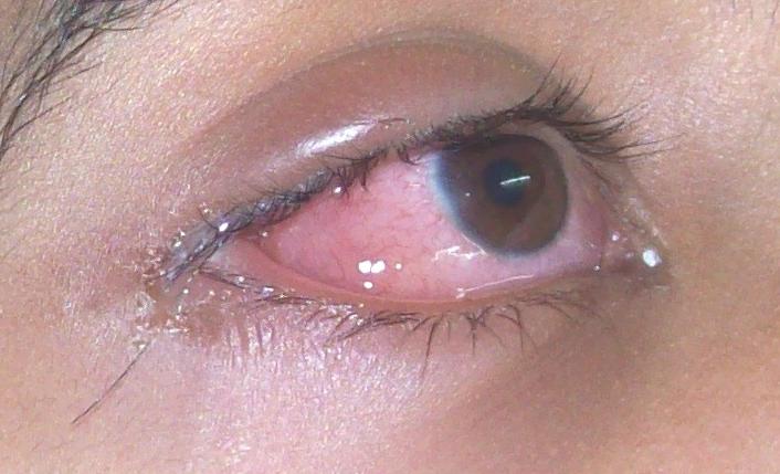
Allergy Eyes
Eyes that are red, watery, and itchy are typical of an allergic reaction known as allergic rhinitis. Commonly called hay fever, allergic rhinitis is an immune system reaction, typically to the pollen of certain plants. Your immune system usually protects you from pathogens and keeps you well. However, like any other body system, the immune system itself can develop problems. Sometimes, it responds to harmless foreign substances as though they were pathogens. This is the basis of allergies like hay fever.
Allergies
An allergy is a disorder in which the immune system makes an inflammatory response to a harmless antigen. It occurs when the immune system is hypersensitive to an antigen in the environment that causes little or no response in most people. Allergies are strongly familial. Allergic parents are more likely to have allergic children, and those children’s allergies are likely to be more severe, which is evidence that there is a heritable tendency to develop allergies. Allergies are more common in children than adults, because many children outgrow their allergies by adulthood.
Allergens
Any antigen that causes an allergy is called an allergen. Common allergens are plant pollens, dust mites, mold, specific foods (such as peanuts or shellfish), insect stings, and certain common medications (such as aspirin and penicillin). Allergens may be inhaled or ingested, or they may come into contact with the skin or eyes. Symptoms vary depending on the type of exposure, and the severity of the immune system response. Some of the most common causes of allergies are shown in Figure 17.6.2: latex, pollen, dust mites, pet dander, insect stings and various foods. Inhaling pollen may cause symptoms of allergic rhinitis, such as sneezing and red itchy eyes. Insect stings may cause an itchy rash. This type of allergy is called contact dermatitis.
Figure 17.6.2 Common allergens include latex, pollen, dust mites, pet dander, insect stings, and foods.
Prevalence of Allergies
There has been a significant increase in the prevalence of allergies over the past several decades, especially in the rich nations of the world, where allergies are now very common disorders. In the developed countries, about 20% of people have or have had hay fever, another 20% have had contact dermatitis, and about 6% have food allergies. In the poorer nations of the world, on the other hand, allergies of all types are much less common.
One explanation for the rise in allergies in the developed world is the hygiene hypothesis. According to this hypothesis, people in developed countries live in relatively sterile environments because of hygienic practices and sanitation systems. As a result, people in these countries are exposed to fewer pathogens than their immune system evolved to cope with. To compensate, their immune system “keeps busy” by attacking harmless antigens in allergic responses.
How Allergies Occur
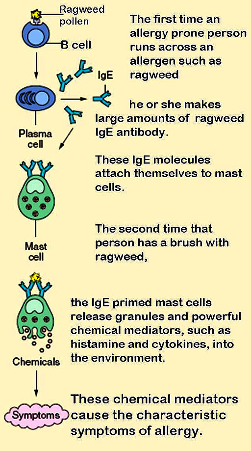
The diagram in Figure 17.6.3 shows how an allergic reaction occurs. At the first exposure to an allergen, B cells are activated to form plasma cells that produce large amounts of antibodies to the allergen. These antibodies attach to leukocytes called mast cells. Subsequently, every time the person encounters the allergen again, the mast cells are already primed and ready to deal with it. The primed mast cells immediately release cytokines and histamines, which in turn cause inflammation and recruitment of leukocytes, among other responses. These responses are responsible for the signs and symptoms of allergies.
Treating Allergies
The symptoms of allergies can range from mild to life-threatening. Mild allergy symptoms are often treated with antihistamines. These are drugs that reduce or eliminate the effects of the histamines that produce allergy symptoms.
Treating Anaphylaxis
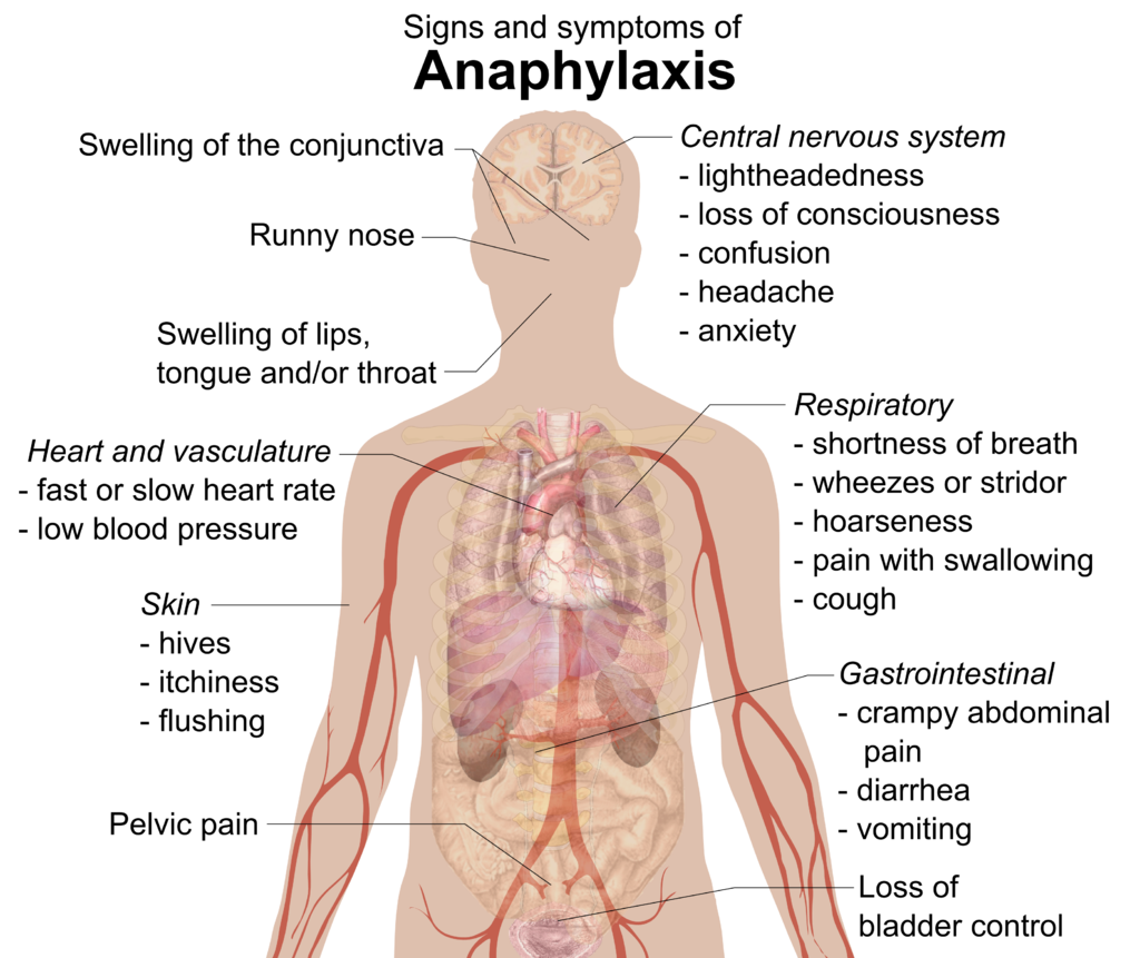
The most severe allergic reaction is a systemic reaction called anaphylaxis. This is a life-threatening response caused by a massive release of histamines. Many of the signs and symptoms of anaphylaxis are shown in Figure 17.6.4. Some of them include a drop in blood pressure, changes in heart rate, shortness of breath, and swelling of the tongue and throat, which may threaten the patient with suffocation unless emergency treatment is given. People who have had anaphylactic reactions may carry an epinephrine autoinjector (widely known by its brand name EpiPen®) so they can inject themselves with epinephrine if they start to experience an anaphylactic response. The epinephrine helps control the immune reaction until medical care can be provided. Epinephrine constricts blood vessels to increase blood pressure, relaxes smooth muscles in the lungs to reduce wheezing and improve breathing, modulates heart rate, and works to reduce swelling that may otherwise block the airways.
Immunotherapy for Allergies
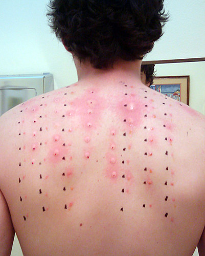
Another way to treat allergies is called immunotherapy, commonly called “allergy shots.” This approach may actually cure specific allergies, at least for several years if not permanently. It may be particularly beneficial for allergens that are difficult or impossible to avoid (such as pollen). First, however, patients must be tested to identify the specific allergens that are causing their allergies. As shown in Figure 17.6.5, this may involve scratching tiny amounts of common allergens into the skin, and then observing whether there is a localized reaction to any of them. Each allergen is applied in a different numbered location on the skin, so if there is a reaction — such as redness or swelling — the responsible allergens can be identified. Then, through periodic injections (usually weekly or monthly), patients are gradually exposed to larger and larger amounts of the allergens. Over time, generally from months to years, the immune system becomes desensitized to the allergens. This method of treating allergies is often effective for allergies to pollen or insect stings, but its usefulness for allergies to food is unclear.
Autoimmune Diseases
Autoimmune diseases occur when the immune system fails to recognize the body’s own molecules as self. As a result, instead of ignoring the body’s healthy cells, it attacks them, causing damage to tissues and altering organ growth and function. Most often, B cells are at fault for autoimmune responses. They are generally the cells that lose tolerance for self. Why does this occur? Some autoimmune diseases are thought to be caused by exposure to pathogens that have antigens similar to the body’s own molecules. After this exposure, the immune system responds to body cells as though they were pathogens, as well.
Certain individuals are genetically susceptible to developing autoimmune diseases. These individuals are also more likely to develop more than one such disease. Gender is a risk factor for autoimmunity — females are much more likely than males to develop autoimmune diseases. This is likely due, in part, to gender differences in sex hormones.
At a population level, autoimmune diseases are less common where infectious diseases are more common. The hygiene hypothesis has been proposed to explain the inverse relationship between infectious and autoimmune diseases, as well as the prevalence of allergies. According to the hypothesis, without infectious diseases to “keep it busy,” the immune system may attack the body’s own cells instead.
Common Autoimmune Diseases
An estimated 15 million or more people worldwide have one or more autoimmune diseases. Two of the most common autoimmune diseases are type I diabetes and multiple sclerosis. In terms of the specific body cells that are attacked by the immune system, both are localized diseases. In the case of type I diabetes, the immune system attacks and destroys insulin-secreting islet cells in the pancreas. In the case of multiple sclerosis, the immune system attacks and destroys the myelin sheaths that normally insulate the axons of neurons and allow rapid transmission of nerve impulses.
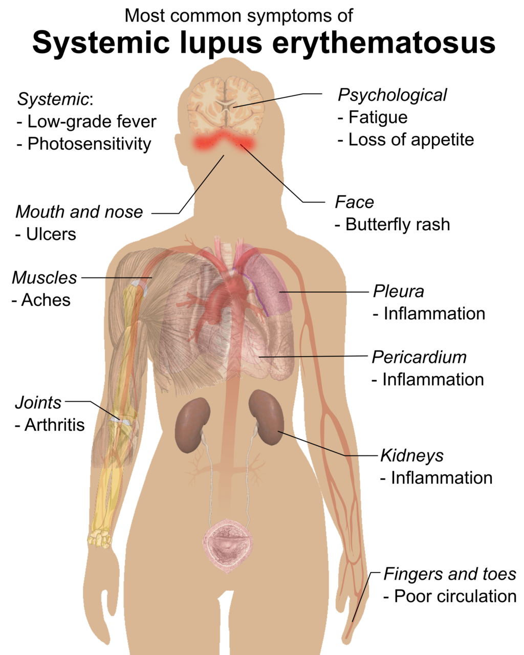
Some relatively common autoimmune diseases are systemic — or body-wide — diseases. They include rheumatoid arthritis and systemic lupus erythematosus (SLE). In these diseases, the immune system may attack and injure many tissues and organs. For example, as you can see in Figure 17.6.6, symptoms of SLE may involve the muscular, skeletal, integumentary, respiratory, and cardiovascular systems.
Treatment for Autoimmune Diseases
None of these common autoimmune diseases can be cured, although all of them have treatments that may help relieve symptoms and prevent some of the long-term damage they may cause. Traditional treatments for autoimmune diseases include immunosuppressive drugs to block the immune response, as well as anti-inflammatory drugs to quell inflammation. Hormone replacement may be another option. Type I diabetes, for example, is treated with injections of the hormone insulin, because islet cells in the pancreas can no longer secrete it.
Immunodeficiency
Immunodeficiency occurs when the immune system is not working properly, generally because one or more components of the immune system are inactive. As a result, the immune system may be unable to fight off pathogens or cancers that a normal immune system would be able to resist. Immunodeficiency may occur for a variety of reasons.
Causes of Immunodeficiency
Dozens of rare genetic diseases can result in a defective immune system. This type of immunodeficiency is called primary immunodeficiency. One is born with one of these diseases, rather than acquiring it after birth. Probably the best known of these primary immunodeficiency diseases is severe combined immunodeficiency (SCID). It is also known as “bubble boy disease,” because people with this disorder are extremely vulnerable to infectious diseases, and some of them have become well known for living inside a bubble that provides a sterile environment. SCID is most often caused by an X-linked recessive mutation that interferes with normal B cell and T cell production.
Other types of immunodeficiency are not present at birth, but are acquired due to experiences or exposures that occur after birth. Acquired immunodeficiency is called secondary immunodeficiency because it is secondary to some other event or exposure. Secondary immunodeficiency may occur for a number of different reasons:
- Some pathogens attack and destroy immune system cells. An example is the virus known as HIV, which attacks and destroys T cells.
- The immune system naturally becomes less effective as people get older. This age-related decline — called immunosenescence — generally begins around the age of 50 and worsens with increasing age. Immunosenescence is the reason older people are generally more susceptible to disease than younger people.
- The immune system may be damaged by another disorder, such as obesity, alcoholism, or the abuse of other drugs.
- In developing countries, malnutrition is the most common cause of immune system damage and immunodeficiency. Inadequate protein intake is especially damaging to the immune system. It can lead to impaired complement system activity, phagocyte malfunction, and lower-than-normal production of antibodies and cytokines.
- Certain medications can suppress the immune system. This is the intended effect of immunosuppressant drugs given to people with transplanted organs so they do not reject them. In many cases, however, immunosuppression is an unwanted side effect of drugs used to treat other disorders.
Focus on HIV
Human immunodeficiency virus (HIV) is the most common cause of immunodeficiency in the world today. HIV infections of human hosts are a relatively recent phenomenon. Scientists think that the virus originally infected monkeys, but then jumped to human populations. most likely from a bite, probably sometime during the early to mid-1900s. This most likely occurred in West Africa, but the virus soon spread around the world. HIV was first identified by medical researchers in 1981. Since then, HIV has killed almost 40 million people worldwide, and its economic toll has also been enormous. The hardest hit countries are in Africa, where the virus has infected human populations the longest, and medications to control the virus are least available. In 2016, over 63,000 Canadians were living with HIV.
HIV Transmission
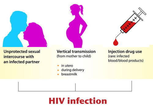
HIV is transmitted through direct contact of mucous membranes or body fluids such as blood, semen, or breast milk. As shown in Figure 17.6.7, transmission of the virus can occur through sexual contact or the use of contaminated hypodermic needles. It can also be transmitted from an infected mother’s blood during late pregnancy or childbirth, or through breast milk after birth. In the past, HIV was also transmitted occasionally through blood transfusions. Because donated blood is now screened for HIV, the virus is no longer transmitted this way.
HIV and the Immune System
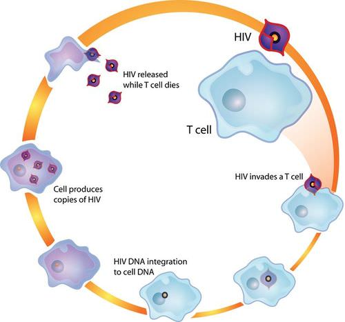
HIV infects and destroys helper T cells, the type of lymphocytes that regulate the immune response. This process is illustrated in the diagram in Figure 17.6.8. The virus injects its own DNA into a helper T cell and uses the T cell’s “machinery” to make copies of itself. In the process, the helper T cell is destroyed, and the virus copies go on to infect other helper T cells. HIV is able to evade the immune system and keep destroying helper T cells by mutating frequently so its surface antigens keep changing, and by using the host cell’s membrane to hide its own antigens.
Acquired immunodeficiency syndrome (AIDS) may result from years of damage to the immune system by HIV. It occurs when helper T cells fall to a very low level and opportunistic diseases occur. Opportunistic diseases are infections and tumors that are rare, except in people with a damaged immune system. The diseases take advantage of the “opportunity” presented by people whose immune system cannot fight back. Opportunistic diseases are usually the direct cause of death for people with AIDS.
Treating HIV/AIDS
For patients who have access to HIV medications, infection with the virus is no longer the death sentence that it once was. By 1995, combinations of drugs called “highly active antiretroviral therapy” were developed. For some patients, these drugs can reduce the amount of virus they are carrying to undetectable levels. However, some level of virus always hides in the body’s immune cells, and it will multiply again if a patient stops taking the medications. Researchers are trying to develop drugs to kill these hidden viruses, as well. If their efforts are successful, it could end AIDS.
Feature: Human Biology in the News
EpiPens® and their sole manufacturer (pharmaceutical company Mylan) were featured in headlines in 2016, but not for a good reason. The media outburst was triggered by a drastic price hike in EpiPens® — and Mylan’s apparent greed.

EpiPens® are auto-injectable syringes preloaded with a measured dose of epinephrine, a drug that can rapidly stop a life-threatening anaphylactic response to an allergen. Using the device is easy and does not require any special training. The injector just needs to be jammed against the thigh, which can be done through clothing or on bare skin. Each year, doctors write millions of prescriptions for EpiPens®. Many people with severe allergies always carry two of the devices with them, just in case they experience anaphylaxis, although most of them never need to use them. Other people with severe allergies have literally had their lives saved multiple times by EpiPens® when they had anaphylactic reactions. Even when the devices haven’t been used, they must be replaced each year due to expiration of the epinephrine.
You might think that EpiPens® would be relatively inexpensive, given their life-saving potential. As recently as 2009, a two-pack of EpiPens® cost about $100. However, in just seven years, the cost of the same two-pack of EpiPens® skyrocketed by an incredible 400%! By 2016, the cost was $600 or more. Mylan apparently raised the price for the sole purpose of increasing profits. The company also raised prices significantly on many other drugs. The price hike in EpiPens® alone was certainly profitable. In 2015, the sale of EpiPens® earned Mylan $1 billion. Mylan’s CEO took home almost $19 million the same year, which was an increase of more than 600% over her prior salary.
News coverage of the price hike in EpiPens® began in the summer of 2016 after a price increase in May of that year. Both private citizens and elected officials expressed outrage over the price increase, especially when coupled with the gluttonous profits of the company and its CEO. By late August, Mylan responded to the backlash by offering discount coupons for EpiPens®. A few days later, the company promised to introduce a cheaper, generic version of the device. Analysts quickly determined that selling a generic version would allow Mylan to make more money on the product than reducing the price of the name-brand device, which they still declined to do. By September of 2016, Mylan was being investigated for antitrust violations related to sales of EpiPens® to public schools in New York City.
The Mylan/EpiPen® story may still be making the news. But whatever its outcome, the story has already added fuel to public and private debates about important ethical issues — issues such as the excessive costs of life-saving drugs and the huge profits of big pharma. What is the most recent news on EpiPens® and Mylan? If you are interested, you can check the headlines online to find out. What are your views on the ethical issues they raise?
17.6 Summary
- An allergy is a disorder in which the immune system makes an inflammatory response to a harmless antigen. Any antigen that causes allergies is called an allergen. Common allergens include pollen, dust mites, mold, specific foods (such as peanuts), insect stings, and certain medications (such as aspirin).
- The prevalence of allergies has been increasing for decades, especially in developed countries, where they are much more common than in developing countries. The hygiene hypothesis posits that this has occurred because humans evolved to cope with more pathogens than we now typically face in our relatively sterile environments in developed countries. As a result, the immune system “keeps busy” by attacking harmless antigens.
- Allergies occur when B cells are first activated to produce large amounts of antibodies to an otherwise harmless allergen, and the antibodies attach to mast cells. On subsequent exposures to the allergen, the mast cells immediately release cytokines and histamines that cause inflammation.
- Mild allergy symptoms are frequently treated with antihistamines that counter histamines and reduce allergy symptoms. A severe systemic allergic reaction, called anaphylaxis, is a medical emergency that is usually treated with injections of epinephrine. Immunotherapy for allergies involves injecting increasing amounts of allergens to desensitize the immune system to them.
- Autoimmune diseases occur when the immune system fails to recognize the body’s own molecules as self and attacks them, causing damage to tissues and organs. A family history of autoimmunity and female gender are risk factors for autoimmune diseases.
- In some autoimmune diseases, such as type I diabetes, the immune system attacks and damages specific body cells. In other autoimmune diseases, such as systemic lupus erythematosus, many different tissues and organs may be attacked and injured. Autoimmune diseases generally cannot be cured, but their symptoms can often be managed with drugs or other treatments.
- Immunodeficiency occurs when the immune system is not working properly, generally because one or more of its components are inactive. As a result, the immune system is unable to fight off pathogens or cancers that a normal immune system would be able to resist.
- Primary immunodeficiency is present at birth and caused by rare genetic diseases. An example is severe combined immunodeficiency. Secondary immunodeficiency occurs because of some event or exposure experienced after birth. Possible causes include aging, certain medications, infections with pathogens, and other disorders, such as obesity or malnutrition.
- The most common cause of immunodeficiency in the world today is human immunodeficiency virus (HIV), which infects and destroys helper T cells. HIV is transmitted through mucous membranes or body fluids. The virus may eventually lead to such low levels of helper T cells that opportunistic infections occur. When this happens, the patient is diagnosed with acquired immunodeficiency syndrome (AIDS). Medications can control the multiplication of HIV in the human body — but they don’t eliminate the virus completely.
17.6 Review Questions
-
- How does immunotherapy for allergies work?
- What are autoimmune diseases?
- Identify two risk factors for autoimmune diseases.
- Autoimmune diseases may be specific to particular tissues, or they may be systemic. Give an example of each type of autoimmune disease.
- What is immunodeficiency? Compare and contrast primary and secondary immunodeficiency. Give an example of each.
- What is the most common cause of immunodeficiency in the world today? How does this affect the immune system?
- Distinguish between HIV and AIDS.
17.6 Explore More
Why do people have seasonal allergies? – Eleanor Nelsen, TED-Ed, 2016.
Why it’s so hard to cure HIV/AIDS – Janet Iwasa, TED-Ed, 2015.
The Boy in the Bubble | Retro Report | The New York Times, 2015.
Why Are Peanut Allergies Becoming So Common? Seeker, 2014.
What Are Tonsil Stones? | Gross Science, 2015.
Attributions
Figure 17.6.1
Oedema by Championswimmer on Wikimedia Commons is in the public domain (https://en.wikipedia.org/wiki/en:public_domain).
Figure 17.6.2
- Medical (latex) gloves from pngimg.com is used under a CC BY-NC 4.0 (https://creativecommons.org/licenses/by-nc/4.0/) license.
- House dust mites (5247996458) by Gilles San Martin from Namur, Belgium on Wikimedia Commons is used under a CC BY-SA 2.0 (https://creativecommons.org/licenses/by-sa/2.0/deed.en) license.
- Honey bee macro by Karunakar Rayker on Flickr is used under a CC BY 2.0 (https://creativecommons.org/licenses/by/2.0/) license.
- Peanuts by Karolina Grabowska on Pexels is used under a CC0 1.0 Universal Public Domain Dedication license (https://creativecommons.org/publicdomain/zero/1.0/).
- Photo of dog and grey cat by ERC4N51 on pxhere is used under a CC0 1.0 Universal Public Domain Dedication license (https://creativecommons.org/publicdomain/zero/1.0/).
- Tags: Pollen Allergy Spring by Castagnari53 on Pixabay, is used under the Pixabay License (https://pixabay.com/service/license/).
Figure 17.6.3
512px-Mast_cells by National Institute of Allergy and Infectious Diseases (U.S.) & National Cancer Institute (p.29) is in the public domain (https://en.wikipedia.org/wiki/en:public_domain).
Figure 17.6.4
Signs_and_symptoms_of_anaphylaxis by Mikael Häggström on Wikimedia Commons is used under a CC0 1.0 Universal Public Domain Dedication license (https://creativecommons.org/publicdomain/zero/1.0/).
Figure 17.6.5
Allergy Tests by Dan Pupius on Flickr is used under a CC BY-NC-SA 2.0 (https://creativecommons.org/licenses/by-nc-sa/2.0/) license.
Figure 17.6.6
1024px-Symptoms_of_SLE by Mikael Häggström on Wikimedia Commons is used under a CC0 1.0 Universal Public Domain Dedication license (https://creativecommons.org/publicdomain/zero/1.0/).
Figure 17.6.7
HIV transmission by CK-12 Foundation is used under a CC BY NC 3.0 (https://creativecommons.org/licenses/by-nc/3.0/) license.
Figure 17.6.8
HIV life cycle by CK-12 Foundation is used under a CC BY NC 3.0 (https://creativecommons.org/licenses/by-nc/3.0/) license.
 ©CK-12 Foundation Licensed under
 ![]() • Terms of Use • Attribution
• Terms of Use • Attribution
Figure 17.6.9
Epipen by Stock Catalog on flickr by Stock Catalog on Flickr is used under a CC BY 2.0 (https://creativecommons.org/licenses/by/2.0/) license.
References
Brainard, J/ CK-12 Foundation. (2016). Figure 8 HIV may be transmitted in all of the ways shown here [digital image]. In CK-12 College Human Biology (Section 19.6) [online Flexbook]. CK12.org. https://www.ck12.org/book/ck-12-human-biology/section/19.6/
Brainard, J/ CK-12 Foundation. (2016). Figure 9 This diagram shows how HIV infects and destroys helper T cells [digital image]. In CK-12 College Human Biology (Section 19.6) [online Flexbook]. CK12.org. https://www.ck12.org/book/ck-12-human-biology/section/19.6/
CBS News. (2016, August 16). Rising cost of potentially life-saving EpiPen puts pinch on families [online article]. CBS Interactive Inc. https://www.cbsnews.com/news/allergy-medication-epipen-epinephrine-rising-costs-impact-on-families/
Gross Science. (2015, June 29). What are tonsil stones? | Gross Science. YouTube. https://www.youtube.com/watch?v=RiMSmDBvgto&feature=youtu.be
Häggström, M. (2014). Medical gallery of Mikael Häggström 2014. WikiJournal of Medicine 1 (2). DOI:10.15347/wjm/2014.008. ISSN 2002-4436
National Institute of Allergy and Infectious Diseases (NIAID). (n.d.). Severe combined immunodeficiency (SCID) [online article]. National Institute of Health (NIH). https://www.niaid.nih.gov/diseases-conditions/severe-combined-immunodeficiency-scid
National Institute of Allergy and Infectious Diseases (U.S.) & National Cancer Institute (U.S.). (2003, September). Understanding the immune system and how it works [NIH Publication No. 03-5423]. Scholar Works – Indiana University. https://scholarworks.iupui.edu/handle/1805/748
[The] New York Times. (2015, December 15). The boy in the bubble | Retro Report | The New York Times. YouTube. https://www.youtube.com/watch?v=pJa6KVLwl9U&feature=youtu.be
Seeker. (2014, October 3). Why are peanut allergies becoming so common? YouTube. https://www.youtube.com/watch?v=Mjr9h_QmdeM&feature=youtu.be
Summary: Estimates of HIV incidence, prevalence, and Canada’s progress on meeting the 90-90-90 HIV targets 2016. (2018, July). Public Health Agency of Canada. https://www.canada.ca/content/dam/phac-aspc/documents/services/publications/diseases-conditions/summary-estimates-hiv-incidence-prevalence-canadas-progress-90-90-90/pub-eng.pdf
Swetlitz, I., Silverman, E. (2016, August 25). Mylan may have violated antitrust law in its EpiPen sales to schools, legal experts say [online article]. STATNews.com. https://www.statnews.com/2016/08/25/mylan-antitrust-epipen-schools/
TED-Ed. (2016, May 26). Why do people have seasonal allergies? – Eleanor Nelsen. YouTube. https://www.youtube.com/watch?v=-q7Fz7NIMWM&feature=youtu.be
TED-Ed. (2015, March 16). Why it’s so hard to cure HIV/AIDS – Janet Iwasa, https://www.youtube.com/watch?v=0TipTogQT3E&feature=youtu.be
Goose Bumps
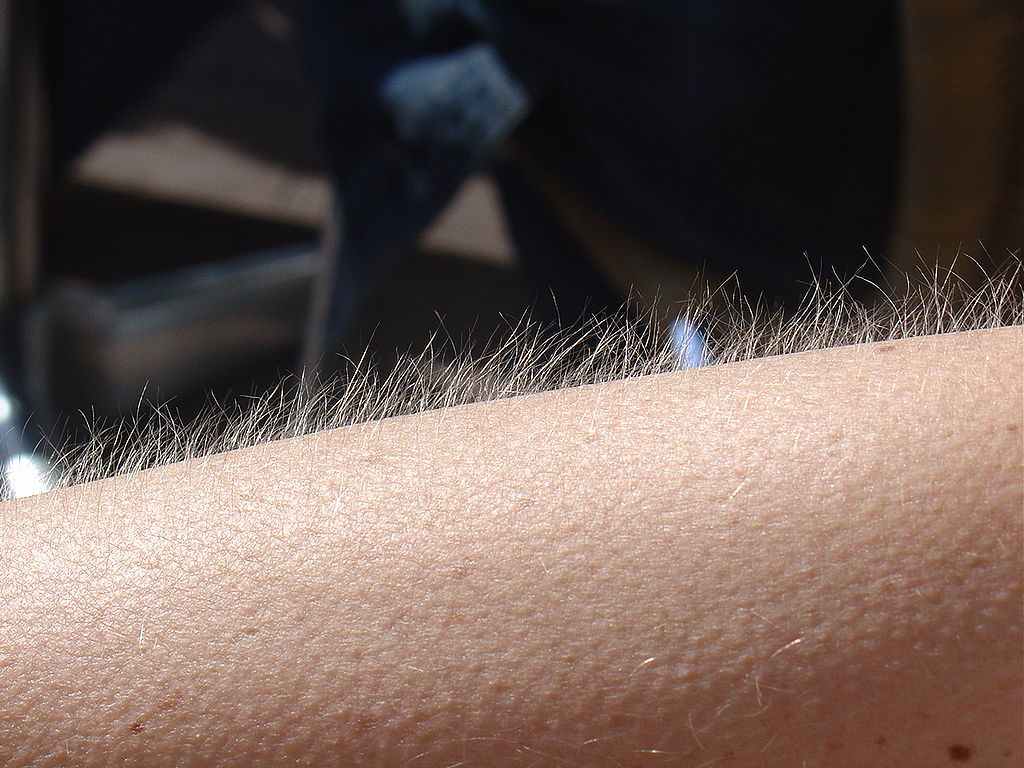
No doubt you’ve experienced the tiny, hair-raising skin bumps called goose bumps, like those you see in Figure 10.4.1. They happen when you feel chilly. Do you know what causes goose bumps, or why they pop up when you are cold? The answers to these questions involve the layer of skin known as the dermis.
What is the Dermis?
The dermis is the inner of the two major layers that make up the skin, the outer layer being the epidermis. The dermis consists mainly of connective tissues. It also contains most skin structures, such as glands and blood vessels. The dermis is anchored to the tissues below it by flexible collagen bundles that permit most areas of the skin to move freely over subcutaneous (“below the skin”) tissues. Functions of the dermis include cushioning subcutaneous tissues, regulating body temperature, sensing the environment, and excreting wastes.
Anatomy of the Dermis
The basic anatomy of the dermis is a matrix, or sort of scaffolding, composed of connective tissues. These tissues include collagen fibres — which provide toughness — and elastin fibres, which provide elasticity. Surrounding these fibres, the matrix also includes a gel-like substance made of proteins. The tissues of the matrix give the dermis both strength and flexibility.
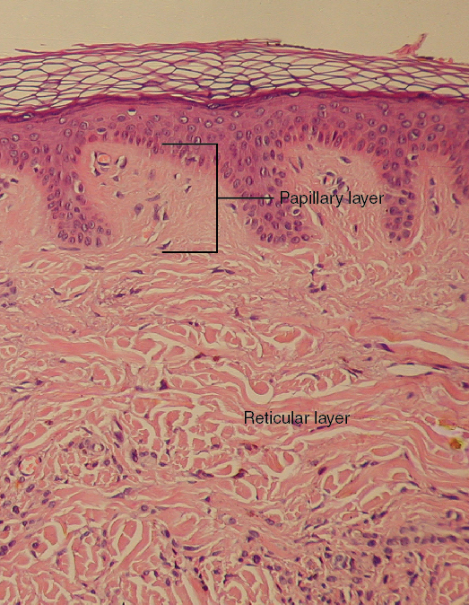
The dermis is divided into two layers: the papillary layer and the reticular layer. Both layers are shown in Figure 10.4.2 below and described in the text that follows.
Papillary Layer
The papillary layer is the upper layer of the dermis, just below the basement membrane that connects the dermis to the epidermis above it. The papillary layer is the thinner of the two dermal layers. It is composed mainly of loosely arranged collagen fibres. The papillary layer is named for its fingerlike projections — or papillae — that extend upward into the epidermis. The papillae contain capillaries and sensory touch receptors.
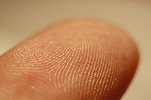
The papillae give the dermis a bumpy surface that interlocks with the epidermis above it, strengthening the connection between the two layers of skin. On the palms and soles, the papillae create epidermal ridges. Epidermal ridges on the fingers are commonly called fingerprints (see Figure 10.4.3). Fingerprints are genetically determined, so no two people (other than identical twins) have exactly the same fingerprint pattern. Therefore, fingerprints can be used as a means of identification, for example, at crime scenes. Fingerprints were much more commonly used forensically before DNA analysis was introduced for this purpose.
Reticular Layer
The reticular layer is the lower layer of the dermis, located below the papillary layer. It is the thicker of the two dermal layers. It is composed of densely woven collagen and elastin fibres. These protein fibres give the dermis its properties of strength and elasticity. This layer of the dermis cushions subcutaneous tissues of the body from stress and strain. The reticular layer of the dermis also contains most of the structures in the dermis, such as glands and hair follicles.
Structures in the Dermis
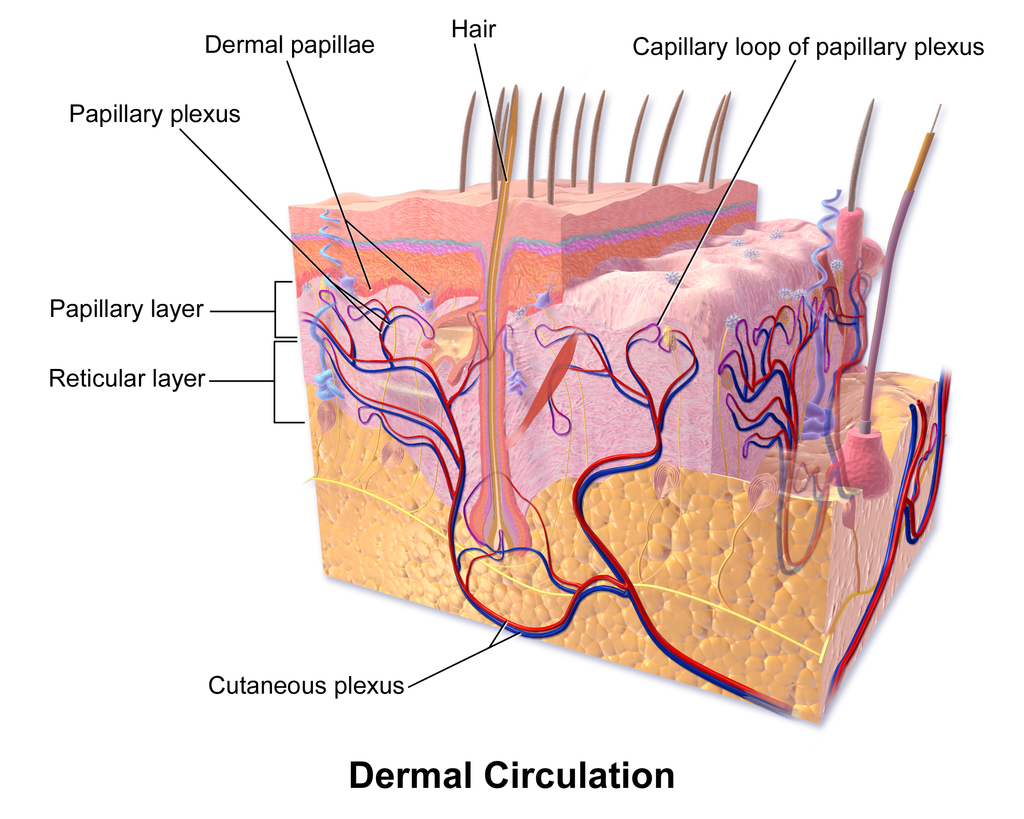
Both papillary and reticular layers of the dermis contain numerous sensory receptors, which make the skin the body’s primary sensory organ for the sense of touch. Both dermal layers also contain blood vessels. They provide nutrients to remove wastes from dermal cells, as well as cells in the lowest layer of the epidermis, the stratum basale. The circulatory components of the dermis are shown in Figure 10.4.4 below.
Glands
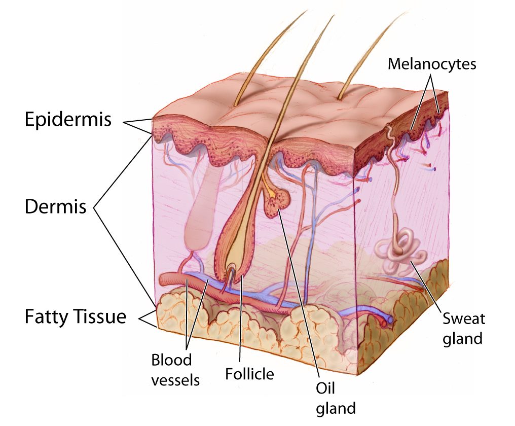
Glands in the reticular layer of the dermis include sweat glands and sebaceous (oil) glands. Both are exocrine glands, which are glands that release their secretions through ducts to nearby body surfaces. The diagram in Figure 10.4.5 shows these glands, as well as several other structures in the dermis.
Sweat Glands
Sweat glands produce the fluid called sweat, which contains mainly water and salts. The glands have ducts that carry the sweat to hair follicles, or to the surface of the skin. There are two different types of sweat glands: eccrine glands and apocrine glands.
- Eccrine sweat glands occur in skin all over the body. Their ducts empty through tiny openings called pores onto the skin surface. These sweat glands are involved in temperature regulation.
- Apocrine sweat glands are larger than eccrine glands, and occur only in the skin of the armpits and groin. The ducts of apocrine glands empty into hair follicles, and then the sweat travels along hairs to reach the surface. Apocrine glands are inactive until puberty, at which point they start producing an oily sweat that is consumed by bacteria living on the skin. The digestion of apocrine sweat by bacteria causes body odor.
Sebaceous Glands
Sebaceous glands are exocrine glands that produce a thick, fatty substance called sebum. Sebum is secreted into hair follicles and makes its way to the skin surface along hairs. It waterproofs the hair and skin, and helps prevent them from drying out. Sebum also has antibacterial properties, so it inhibits the growth of microorganisms on the skin. Sebaceous glands are found in every part of the skin — except for the palms of the hands and soles of the feet, where hair does not grow.
Hair Follicles
Hair follicles are the structures where hairs originate (see the diagram above). Hairs grow out of follicles, pass through the epidermis, and exit at the surface of the skin. Associated with each hair follicle is a sebaceous gland, which secretes sebum that coats and waterproofs the hair. Each follicle also has a bed of capillaries, a nerve ending, and a tiny muscle called an arrector pili.
Functions of the Dermis
The main functions of the dermis are regulating body temperature, enabling the sense of touch, and eliminating wastes from the body.
Temperature Regulation
Several structures in the reticular layer of the dermis are involved in regulating body temperature. For example, when body temperature rises, the hypothalamus of the brain sends nerve signals to sweat glands, causing them to release sweat. An adult can sweat up to four litres an hour. As the sweat evaporates from the surface of the body, it uses energy in the form of body heat, thus cooling the body. The hypothalamus also causes dilation of blood vessels in the dermis when body temperature rises. This allows more blood to flow through the skin, bringing body heat to the surface, where it can radiate into the environment.
When the body is too cool, sweat glands stop producing sweat, and blood vessels in the skin constrict, thus conserving body heat. The arrector pili muscles also contract, moving hair follicles and lifting hair shafts. This results in more air being trapped under the hairs to insulate the surface of the skin. These contractions of arrector pili muscles are the cause of goose bumps.
Sensing the Environment
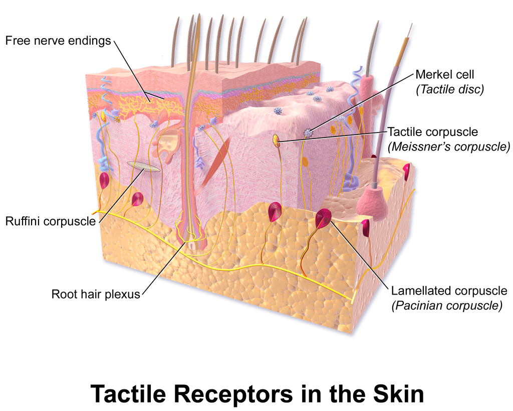
Sensory receptors in the dermis are mainly responsible for the body’s tactile senses. The receptors detect such tactile stimuli as warm or cold temperature, shape, texture, pressure, vibration, and pain. They send nerve impulses to the brain, which interprets and responds to the sensory information. Sensory receptors in the dermis can be classified on the basis of the type of touch stimulus they sense. Mechanoreceptors sense mechanical forces such as pressure, roughness, vibration, and stretching. Thermoreceptors sense variations in temperature that are above or below body temperature. Nociceptors sense painful stimuli. Figure 10.4.6 shows several specific kinds of tactile receptors in the dermis. Each kind of receptor senses one or more types of touch stimuli.
- Free nerve endings sense pain and temperature variations.
- Merkel cells sense light touch, shapes, and textures.
- Meissner’s corpuscles sense light touch.
- Pacinian corpuscles sense pressure and vibration.
- Ruffini corpuscles sense stretching and sustained pressure.
Excreting Wastes
The sweat released by eccrine sweat glands is one way the body excretes waste products. Sweat contains excess water, salts (electrolytes), and other waste products that the body must get rid of to maintain homeostasis. The most common electrolytes in sweat are sodium and chloride. Potassium, calcium, and magnesium electrolytes may be excreted in sweat, as well. When these electrolytes reach high levels in the blood, more are excreted in sweat. This helps to bring their blood levels back into balance. Besides electrolytes, sweat contains small amounts of waste products from metabolism, including ammonia and urea. Sweat may also contain alcohol in someone who has been drinking alcoholic beverages.
Feature: My Human Body
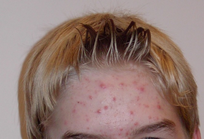
Acne is the most common skin disorder in the Canada. At least 20% of Canadians have acne at any given time and it affects approximately 90% of adolescents (as in Figure 10.4.7). Although acne occurs most commonly in teens and young adults, but it can occur at any age. Even newborn babies can get acne.
The main sign of acne is the appearance of pimples (pustules) on the skin, like those in the photo above. Other signs of acne may include whiteheads, blackheads, nodules, and other lesions. Besides the face, acne can appear on the back, chest, neck, shoulders, upper arms, and buttocks. Acne can permanently scar the skin, especially if it isn’t treated appropriately. Besides its physical effects on the skin, acne can also lead to low self-esteem and depression.
Acne is caused by clogged, sebum-filled pores that provide a perfect environment for the growth of bacteria. The bacteria cause infection, and the immune system responds with inflammation. Inflammation, in turn, causes swelling and redness, and may be associated with the formation of pus. If the inflammation goes deep into the skin, it may form an acne nodule.
Mild acne often responds well to treatment with over-the-counter (OTC) products containing benzoyl peroxide or salicylic acid. Treatment with these products may take a month or two to clear up the acne. Once the skin clears, treatment generally needs to continue for some time to prevent future breakouts.
If acne fails to respond to OTC products, nodules develop, or acne is affecting self-esteem, a visit to a dermatologist is in order. A dermatologist can determine which treatment is best for a given patient. A dermatologist can also prescribe prescription medications (which are likely to be more effective than OTC products) and provide other medical treatments, such as laser light therapies or chemical peels.
What can you do to maintain healthy skin and prevent or reduce acne? Dermatologists recommend the following tips:
- Wash affected or acne-prone skin (such as the face) twice a day, and after sweating.
- Use your fingertips to apply a gentle, non-abrasive cleanser. Avoid scrubbing, which can make acne worse.
- Use only alcohol-free products and avoid any products that irritate the skin, such as harsh astringents or exfoliants.
- Rinse with lukewarm water, and avoid using very hot or cold water.
- Shampoo your hair regularly.
- Do not pick, pop, or squeeze acne. If you do, it will take longer to heal and is more likely to scar.
- Keep your hands off your face. Avoid touching your skin throughout the day.
- Stay out of the sun and tanning beds. Some acne medications make your skin very sensitive to UV light.
10.4 Summary
- The dermis is the inner and thicker of the two major layers that make up the skin. It consists mainly of a matrix of connective tissues that provide strength and stretch. It also contains almost all skin structures, including sensory receptors and blood vessels.
- The dermis has two layers. The upper papillary layer has papillae extending upward into the epidermis and loose connective tissues. The lower reticular layer has denser connective tissues and structures, such as glands and hair follicles. Glands in the dermis include eccrine and apocrine sweat glands and sebaceous glands. Hair follicles are structures where hairs originate.
- Functions of the dermis include cushioning subcutaneous tissues, regulating body temperature, sensing the environment, and excreting wastes. The dense connective tissues of the dermis provide cushioning. The dermis regulates body temperature mainly by sweating and by vasodilation or vasoconstriction. The many tactile sensory receptors in the dermis make it the main organ for the sense of touch. Wastes excreted in sweat include excess water, electrolytes, and certain metabolic wastes.
10.4 Review Questions
- What is the dermis?
- Describe the basic anatomy of the dermis.
- Compare and contrast the papillary and reticular layers of the dermis.
- What causes epidermal ridges, and why can they be used to identify individuals?
- Name the two types of sweat glands in the dermis, and explain how they differ.
- What is the function of sebaceous glands?
- Describe the structures associated with hair follicles.
- Explain how the dermis helps regulate body temperature.
- Identify three specific kinds of tactile receptors in the dermis, along with the type of stimuli they sense.
- How does the dermis excrete wastes? What waste products does it excrete?
- What are subcutaneous tissues? Which layer of the dermis provides cushioning for subcutaneous tissues? Why does this layer provide most of the cushioning, instead of the other layer?
- For each of the functions listed below, describe which structure within the dermis carries it out.
- Brings nutrients to and removes wastes from dermal and lower epidermal cells
- Causes hairs to move
- Detects painful stimuli on the skin
10.4 Explore More
https://www.youtube.com/watch?v=FX-FwK0IIrE
How do you get rid of acne? SciShow, 2016.
https://www.youtube.com/watch?v=VcHQWMAClhQ&feature=emb_logo
When You Can't Scratch Away An Itch, Seeker, 2013.
Attributions
Figure 10.4.1
Goose_bumps by EverJean on Wikimedia Commons is used under a CC BY 2.0 (https://creativecommons.org/licenses/by/2.0) license.
Figure 10.4.2
Layers_of_the_Dermis by OpenStax College on Wikimedia Commons is used under a CC BY 3.0 (https://creativecommons.org/licenses/by/3.0) license.
Figure 10.4.3
Fingerprint_detail_on_male_finger_in_Třebíč,_Třebíč_District by Frettie on Wikimedia Commons is used under a CC BY 3.0 (https://creativecommons.org/licenses/by/3.0) license.
Figure 10.4.4
Blausen_0802_Skin_Dermal Circulation by BruceBlaus on Wikimedia commons is used under a CC BY 3.0 (https://creativecommons.org/licenses/by/3.0) license.
Figure 10.4.5
Anatomy_The_Skin_-_NCI_Visuals_Online by Don Bliss (artist) / National Cancer Institute (National Institutes of Health, with the ID 4604) is in the public domain (https://en.wikipedia.org/wiki/public_domain).
Figure 10.4.6
Blausen_0809_Skin_TactileReceptors by BruceBlaus on Wikimedia commons is used under a CC BY 3.0 (https://creativecommons.org/licenses/by/3.0) license.
Figure 10.4.7
Akne-jugend by Ellywa on Wikimedia Commons is released into the public domain (https://en.wikipedia.org/wiki/public_domain). (No machine-readable author provided. Ellywa assumed, based on copyright claims).
References
Betts, J. G., Young, K.A., Wise, J.A., Johnson, E., Poe, B., Kruse, D.H., Korol, O., Johnson, J.E., Womble, M., DeSaix, P. (2013, June 19). Figure 5.7 Layers of the dermis [digital image]. In Anatomy and Physiology (Section 5.1 Layers of the skin). OpenStax. https://openstax.org/books/anatomy-and-physiology/pages/5-1-layers-of-the-skin
Blausen.com staff. (2014). Medical gallery of Blausen Medical 2014. WikiJournal of Medicine 1 (2). DOI:10.15347/wjm/2014.010. ISSN 2002-4436.
SciShow. (2016, October 26). How do you get rid of acne? YouTube. https://www.youtube.com/watch?v=FX-FwK0IIrE
Seeker. (2013, October 26). When you can't scratch away an itch. YouTube. https://www.youtube.com/watch?v=VcHQWMAClhQ&feature=emb_logo
Image shows a man jogging in the forest. His shirt is wet with sweat.
The process by which information from a gene is used in the synthesis of a functional protein.
As per caption.
Drugs that combat the histamine released during an allergic reaction by blocking the action of the histamine on the tissue.
Created by CK-12 Foundation/Adapted by Christine Miller

Arm Wrestling
It’s obvious that a sport like arm wrestling (Figure 12.4.1) depends on muscle contractions. Arm wrestlers must contract muscles in their hands and arms, and keep them contracted in order to resist the opposing force exerted by their opponent. The wrestler whose muscles can contract with greater force wins the match.
What Is a Muscle Contraction?
A muscle contraction is an increase in the tension or a decrease in the length of a muscle. Muscle tension is the force exerted by the muscle on a bone or other object. A muscle contraction is isometric if muscle tension changes, but muscle length remains the same. An example of isometric muscle contraction is holding a book in the same position. A muscle contraction is isotonic if muscle length changes, but muscle tension remains the same. An example of isotonic muscle contraction is raising a book by bending the arm at the elbow. The termination of a muscle contraction of either type occurs when the muscle relaxes and returns to its non-contracted tension or length.
To use our arm wrestling example, if both arm wrestlers have equal strength and they are pulling with all their might, but there is no movement, that is isometric muscle contraction. However, as soon as one arm wrestler starts to win and is able to start pulling the opponents arm down, that is isotonic muscle contraction.
How a Skeletal Muscle Contraction Begins
Excluding reflexes, all skeletal muscle contractions occur as a result of conscious effort originating in the brain. The brain sends electrochemical signals through the somatic nervous system to motor neurons that innervate muscle fibres (to review how the brain and neurons function, see the chapter Nervous System). A single motor neuron with multiple axon terminals is able to innervate multiple muscle fibres, thereby causing all of them to contract at the same time. The connection between a motor neuron axon terminal and a muscle fibre occurs at a site called a neuromuscular junction. This is a chemical synapse where a motor neuron transmits a signal to a muscle fibre to initiate a muscle contraction. The process by which a signal is transmitted at a neuromuscular junction is illustrated in Figure 12.4.2 below.
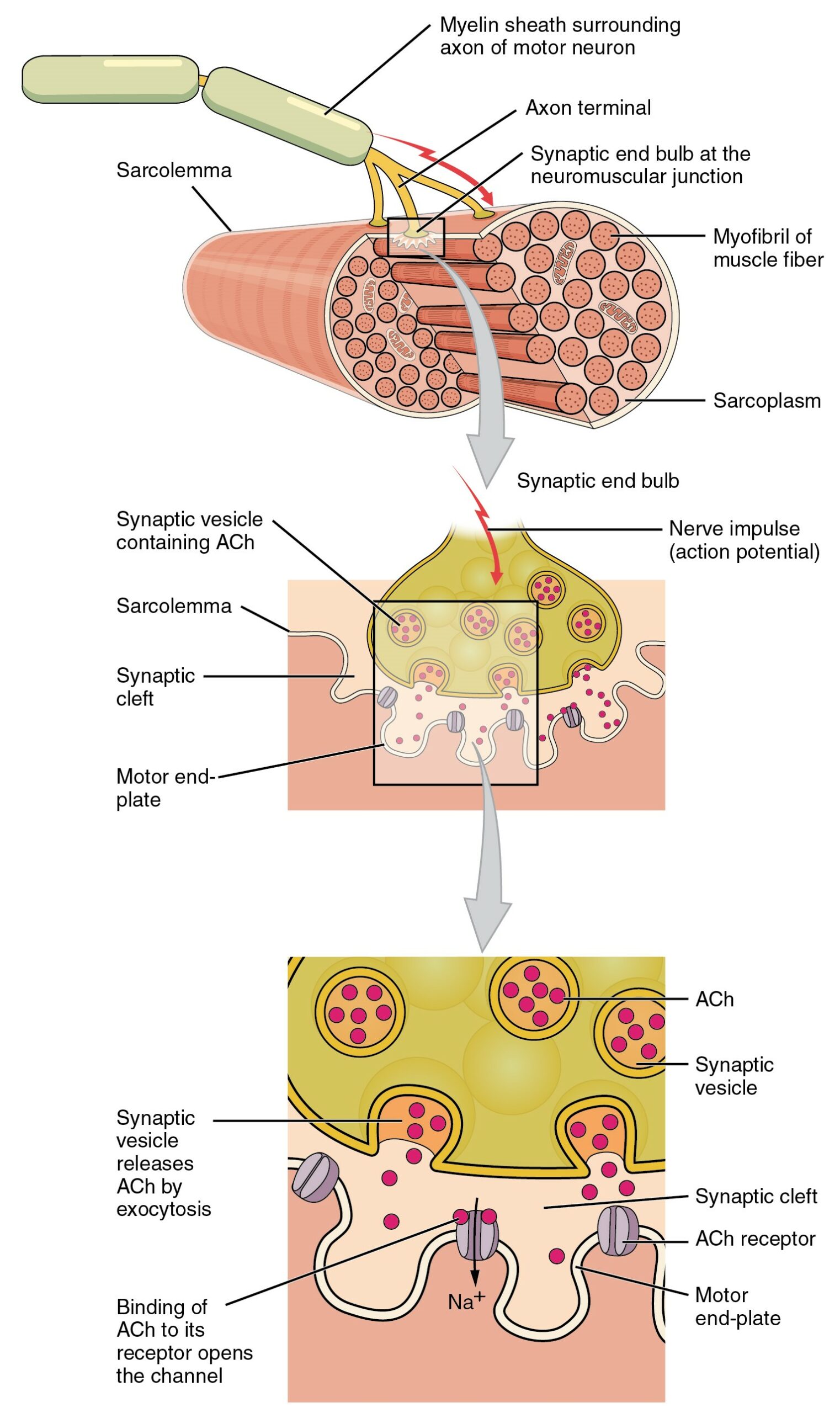
The sequence of events begins when an action potential is initiated in the cell body of a motor neuron, and the action potential is propagated along the neuron’s axon to the neuromuscular junction. Once the action potential reaches the end of the axon terminal, it causes the release of the neurotransmitter acetylcholine (ACh) from synaptic vesicles in the axon terminal. The ACh molecules diffuse across the synaptic cleft and bind to receptors on the muscle fibre, thereby initiating a muscle contraction.
Sliding Filament Theory of Muscle Contraction
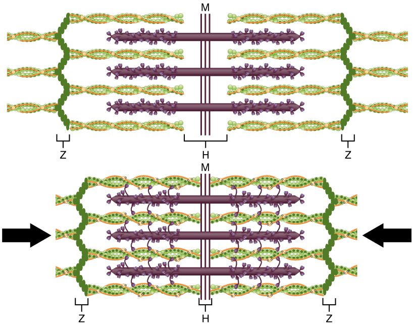
Once the muscle fibre is stimulated by the motor neuron, actin and myosin protein filaments within the skeletal muscle fibre slide past each other to produce a contraction. The sliding filament theory is the most widely accepted explanation for how this occurs. According to this theory, muscle contraction is a cycle of molecular events in which thick myosin filaments repeatedly attach to and pull on thin actin filaments, so the filaments slide over one another, as illustrated in Figure 12.4.3. The actin filaments are attached to Z discs, each of which marks the end of a sarcomere. The sliding of the filaments pulls the Z discs of a sarcomere closer together, thus shortening the sarcomere. As this occurs, the muscle contracts.
Crossbridge Cycling
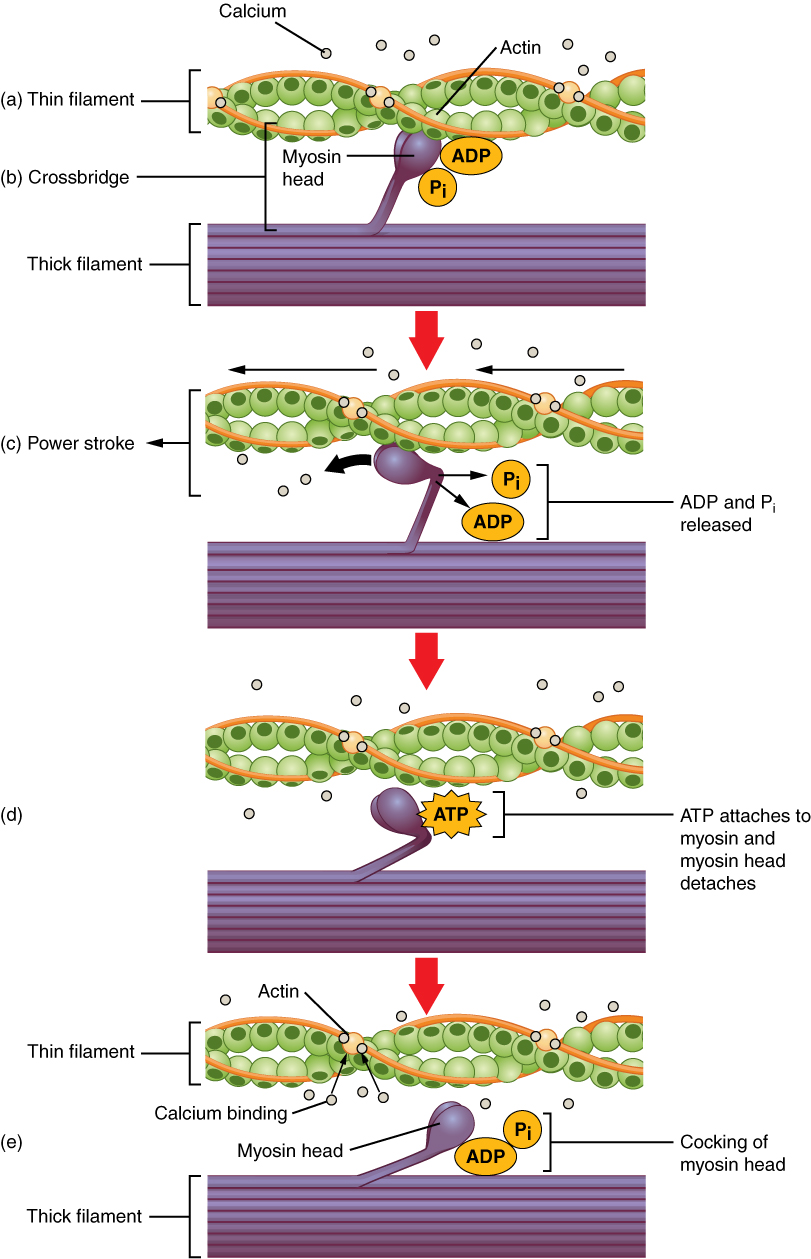
Crossbridge cycling is a sequence of molecular events that underlies the sliding filament theory. There are many projections from the thick myosin filaments, each of which consists of two myosin heads (you can see the projections and heads in Figures 12.4.3 and 12.4.4). Each myosin head has binding sites for ATP (or the products of ATP hydrolysis: ADP and Pi) and for actin. The thin actin filaments also have binding sites for the myosin heads. A crossbridge forms when a myosin head binds with an actin filament.
The process of crossbridge cycling is shown in the video "Muscle Contraction 3D" by 3DBiology (below), and in Figure 12.4.4. A crossbridge cycle begins when the myosin head binds to an actin filament. ADP and Pi are also bound to the myosin head at this stage. Next, a power stroke moves the actin filament inward toward the center of sarcomere, thereby shortening the sarcomere. At the end of the power stroke, ADP and Pi are released from the myosin head, leaving the myosin head attached just to the thin filament until another ATP binds to the myosin head. When ATP binds to the myosin head, it causes the myosin head to detach from the actin. ATP is once again split into ADP and Pi and the energy released is used to move the myosin head into a "cocked" position. Once in this position, the myosin head can bind to the actin filament again, and another crossbridge cycle begins.
https://www.youtube.com/watch?v=GrHsiHazpsw
Muscle Contraction 3D, 3DBiology, 2017.
Energy for Muscle Contraction
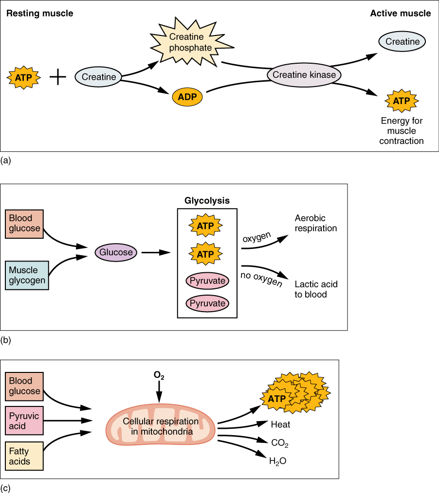
According to the sliding filament theory, ATP is needed to provide the energy for a muscle contraction. Where does this ATP come from? Actually, there are multiple potential sources, as illustrated in Figure 12.4.5 below.
- As you can see from the first diagram, some ATP is already available in a resting muscle. As a muscle contraction starts, this ATP is used up in just a few seconds. More ATP is generated from creatine phosphate, but this ATP is used up rapidly as well. It’s gone in another 15 seconds or so.
- Glucose from the blood and glycogen stored in muscle can then be used to make more ATP. Glycogen breaks down to form glucose, and each glucose molecule produces two molecules of ATP and two molecules of pyruvate. Pyruvate (as pyruvic acid) can be used in aerobic respiration if oxygen is available. Alternatively, pyruvate can be used in anaerobic respiration, if oxygen is not available. The latter produces lactic acid, which may contribute to muscle fatigue. Anaerobic respiration typically occurs only during strenuous exercise when so much ATP is needed that sufficient oxygen cannot be delivered to the muscle to keep up.
- Resting or moderately active muscles can get most of the ATP they need for contractions by aerobic respiration. This process takes place in the mitochondria of muscle cells. In the process, glucose and oxygen react to produce carbon dioxide, water, and many molecules of ATP.
Feature: Human Biology in the News
Basic research on muscle contraction, especially if it is interesting and hopeful, is often in the news, because muscle contractions are involved in so many different body processes and disorders, including heart failure and stroke.
- Heart failure is a chronic condition in which cardiac muscle cells cannot contract forcefully enough to keep body cells adequately supplied with oxygen. According to a 2016 report by the Heart and Stroke Foundation of Canada, 600,000 Canadians are living with heart failure and each year, 50,000 new cases are diagnosed. Heart failure costs the Canadian medical system more than $2.8 billion annually. In 2016, researchers at the University of Texas Southwestern Medical Center identified a potential new target for the development of drugs to increase the strength of cardiac muscle contractions in patients with heart failure. The UT researchers found a previously unidentified protein involved in muscle contraction. The protein, which is very small, turns off the “brake” on the heart so it pumps blood more vigorously. At the molecular level, the protein affects the calcium-ion pump that controls muscle contraction. The scientists also found the same protein in slow-twitch skeletal muscle fibres. Interestingly, the protein is encoded by a stretch of mRNA that had been dismissed by scientists as non-coding RNA, commonly referred to as “junk” RNA. According to one of the researchers, “We dipped into the RNA ‘junk’ pile and came up with a hidden treasure.” This result is likely to lead to searches for additional treasures that might be hiding in the RNA junk pile.
- A stroke occurs when a blood clot lodges in an artery in the brain and cuts off blood flow to part of the brain. Approximately 6% of deaths in Canada are due to stroke and while men and women experiences strokes almost equally, women are more likely to die from a stroke. Damage from the clot associated with strokes would be reduced if the smooth muscles lining brain arteries relaxed following a stroke, because the arteries would dilate and allow greater blood flow to the brain. In a recent study undertaken at the Yale University School of Medicine, researchers determined that the muscles lining blood vessels in the brain actually contract after a stroke. This constricts the vessels, reduces blood flow to the brain, and appears to contribute to permanent brain damage. The hopeful takeaway of this finding is that it suggests a new target for stroke therapy.
12.4 Summary
- A muscle contraction is an increase in the tension or a decrease in the length of a muscle. A muscle contraction is isometric if muscle tension changes, but muscle length remains the same. It is isotonic if muscle length changes, but muscle tension remains the same.
- A skeletal muscle contraction begins with electrochemical stimulation of a muscle fibre by a motor neuron. This occurs at a chemical synapse called a neuromuscular junction. The neurotransmitter acetylcholine diffuses across the synaptic cleft and binds to receptors on the muscle fibre. This initiates a muscle contraction.
- Once stimulated, the protein filaments within the skeletal muscle fibre slide past each other to produce a contraction. The sliding filament theory is the most widely accepted explanation for how this occurs. According to this theory, thick myosin filaments repeatedly attach to and pull on thin actin filaments, thus shortening sarcomeres.
- Crossbridge cycling is a cycle of molecular events that underlies the sliding filament theory. Using energy in ATP, myosin heads repeatedly bind with and pull on actin filaments. This moves the actin filaments toward the center of a sarcomere, shortening the sarcomere and causing a muscle contraction.
- The ATP needed for a muscle contraction comes first from ATP already available in the cell, and more is generated from creatine phosphate. These sources are quickly used up. Glucose and glycogen can be broken down to form ATP and pyruvate. Pyruvate can then be used to produce ATP in aerobic respiration if oxygen is available, or it can be used in anaerobic respiration if oxygen is not available.
12.4 Review Questions
- What is a skeletal muscle contraction?
-
- Explain sliding filament theory and describe crossbridge cycling.
- If the acetylcholine receptors on muscle fibres were blocked by a drug, what do you think this would do to muscle contraction? Explain your answer.
- Explain how crossbridge cycling and sliding filament theory are related to each other.
- When does anaerobic respiration typically occur in human muscle cells?
- If there were no ATP available in a muscle, how would this affect crossbridge cycling? What would this do to muscle contraction?
12.4 Explore More
https://www.youtube.com/watch?v=NfEJUPnqxk0
The Mechanism of Muscle Contraction: Sarcomeres, Action Potential, and the Neuromuscular Junction, Professor Dave Explains, 2019.
https://www.youtube.com/watch?v=8Y_FdjI2v4I
Aerobic vs Anaerobic Difference, Dorian Wilson, 2017.
Attributions
Figure 12.4.1
Armwrestling_Championships by Jnadler1 on Wikimedia Commons is used under a CC BY-SA 3.0 (https://creativecommons.org/licenses/by-sa/3.0) license.
Figure 12.4.2
Motor_End_Plate_and_Innervation by OpenStax on Wikimedia Commons is used under a CC BY 4.0 (https://creativecommons.org/licenses/by 4.0) license.
Figure 12.4.3
Sliding_Filament_Model_of_Muscle_Contraction by OpenStax on Wikimedia Commons is used under a CC BY 4.0 (https://creativecommons.org/licenses/by 4.0) license.
Figure 12.4.4
Skeletal_Muscle_Contraction by OpenStax on Wikimedia Commons is used under a CC BY 4.0 (https://creativecommons.org/licenses/by 4.0) license.
Figure 12.4.5
Muscle_Metabolism by OpenStax on Wikimedia Commons is used under a CC BY 4.0 (https://creativecommons.org/licenses/by 4.0) license.
References
3DBiology. (2017). Muscle contraction 3D. YouTube. https://www.youtube.com/watch?v=GrHsiHazpsw
Betts, J. G., Young, K.A., Wise, J.A., Johnson, E., Poe, B., Kruse, D.H., Korol, O., Johnson, J.E., Womble, M., DeSaix, P. (2016, May 27). Figure 10.6 Motor end-plate and innervation [digital image]. In Anatomy and Physiology (Section 10.2). OpenStax. https://openstax.org/books/anatomy-and-physiology/pages/10-2-skeletal-muscle
Betts, J. G., Young, K.A., Wise, J.A., Johnson, E., Poe, B., Kruse, D.H., Korol, O., Johnson, J.E., Womble, M., DeSaix, P. (2016, May 27). Figure 10.10 The sliding filament model of muscle contraction [digital image]. In Anatomy and Physiology (Section 10.3). OpenStax. https://openstax.org/books/anatomy-and-physiology/pages/10-3-muscle-fiber-contraction-and-relaxation
Betts, J. G., Young, K.A., Wise, J.A., Johnson, E., Poe, B., Kruse, D.H., Korol, O., Johnson, J.E., Womble, M., DeSaix, P. (2016, May 27). Figure 10.11 Skeletal muscle contraction [digital image]. In Anatomy and Physiology (Section 10.3). OpenStax. https://openstax.org/books/anatomy-and-physiology/pages/10-3-muscle-fiber-contraction-and-relaxation
Betts, J. G., Young, K.A., Wise, J.A., Johnson, E., Poe, B., Kruse, D.H., Korol, O., Johnson, J.E., Womble, M., DeSaix, P. (2016, May 27). Figure 10.12 Muscle metabolism [digital image]. In Anatomy and Physiology (Section 10.3). OpenStax. https://openstax.org/books/anatomy-and-physiology/pages/10-3-muscle-fiber-contraction-and-relaxation
Dorian Wilson. (2017, March 8). Aerobic vs anaerobic difference. YouTube. https://www.youtube.com/watch?v=8Y_FdjI2v4I&feature=youtu.be
Heart and Stroke Foundation. (2016). 2016 Report on the health of Canadians: The burden of heart failure. https://www.heartandstroke.ca/-/media/pdf-files/canada/2017-heart-month/heartandstroke-reportonhealth-2016.ashx?la=en
Hill, R. A., Tong, L., Yuan, P., Murikinati, S., Gupta, S., & Grutzendler, J. (2015). Regional blood flow in the normal and ischemic brain is controlled by arteriolar smooth muscle cell contractility and not by capillary pericytes. Neuron, 87(1), 95–110. https://doi.org/10.1016/j.neuron.2015.06.001
UTSouthwestern Newsroom. (2016, January 14). Researchers find a small protein that plays a big role in heart muscle contraction [online article]. https://www.utsouthwestern.edu/newsroom/articles/year-2016/dworf-protein-olson.html
What we do. (n.d.). Heart and Stroke Foundation of Canada. https://www.heartandstroke.ca/what-we-do
Image shows a photo of two children in a kayak. The older child is sitting behind the younger child and paddling.
Image shows a photo of a woman doing a yoga pose which involves a backbend.
A type of white blood cell and, specifically, a type of lymphocyte.
Many B cells mature into what are called plasma cells that produce antibodies (proteins) necessary to fight off infections while other B cells mature into memory B cells.
A microorganism which causes disease.
A groups of mammals characterized by flexible hands and feet, each with five digits, including humans, great apes, monkeys, and lemurs.
Image shows three competing rowing teams in the 2016 Rio Summer Olympics. Each rowboat holds two women. In the photo are Canada, France and China.
Created by CK-12 Foundation/Adapted by Christine Miller

Case Study: Flight Risk
Nineteen-year-old Malcolm is about to take his first plane flight. Shortly after he boards the plane and sits down, a man in his late sixties sits next to him in the aisle seat. About half an hour after the plane takes off, the pilot announces that she is turning the seat belt light off, and that it is safe to move around the cabin.
The man in the aisle seat — who has introduced himself to Malcolm as Willie — immediately unbuckles his seat belt and paces up and down the aisle a few times before returning to his seat. After about 45 minutes, Willie gets up again, walks some more, then sits back down and does some foot and leg exercises. After the third time Willie gets up and paces the aisles, Malcolm asks him whether he is walking so much to accumulate steps on a pedometer or fitness tracking device. Willie laughs and says no. He is actually trying to do something even more important for his health — prevent a blood clot from forming in his legs.
Willie explains that he has a chronic condition: heart failure. Although it sounds scary, his condition is currently well-managed, and he is able to lead a relatively normal lifestyle. However, it does put him at risk of developing other serious health conditions, such as deep vein thrombosis (DVT), which is when a blood clot occurs in the deep veins, usually in the legs. Air travel — and other situations where a person has to sit for a long period of time — increases the risk of DVT. Willie’s doctor said that he is healthy enough to fly, but that he should walk frequently and do leg exercises to help avoid a blood clot.
As you read this chapter, you will learn about the heart, blood vessels, and blood that make up the cardiovascular system, as well as disorders of the cardiovascular system, such as heart failure. At the end of the chapter you will learn more about why DVT occurs, why Willie has to take extra precautions when he flies, and what can be done to lower the risk of DVT and its potentially deadly consequences.
Chapter Overview: Cardiovascular System
In this chapter, you will learn about the cardiovascular system, which transports substances throughout the body. Specifically, you will learn about:
- The major components of the cardiovascular system: the heart, blood vessels, and blood.
- The functions of the cardiovascular system, including transporting needed substances (such as oxygen and nutrients) to the cells of the body, and picking up waste products.
- How blood is oxygenated through the pulmonary circulation, which transports blood between the heart and lungs.
- How blood is circulated throughout the body through the systemic circulation.
- The components of blood — including plasma, red blood cells, white blood cells, and platelets — and their specific functions.
- Types of blood vessels — including arteries, veins, and capillaries — and their functions, similarities, and differences.
- The structure of the heart, how it pumps blood, and how contractions of the heart are controlled.
- What blood pressure is and how it is regulated.
- Blood disorders, including anemia, HIV, and leukemia.
- Cardiovascular diseases (including heart attack, stroke, and angina), and the risk factors and precursors — such as high blood pressure and atherosclerosis — that contribute to them.
As you read the chapter, think about the following questions:
- What is heart failure?Why do you think it increases the risk of DVT?
- What is a blood clot? What are possible health consequences of blood clots?
- Why do you think sitting for long periods of time increases the risk of DVT? Why does walking and exercising the legs help reduce this risk?
Attribution
Figure 14.1.1
aircraft-1583871_1920 [photo] by olivier89 from Pixabay is used under the Pixabay License (https://pixabay.com/de/service/license/).
Image shows a diagram of Killer T Cell funtion. An infected cell displays a pathogen antigen on an MHC. The Killer T Cell interacts with the MHC and in response produces perforin ( a protein that pokes holes in cell membranes) and granzymes (proteins that instruct a cell to carry out programmed cell death). The infected cell dies from the combination of these substances, and as it dies, so does the pathogen inside the infected cell. The Killer T Cell is free to move on and find and destroy other infected cells.
Created by CK-12 Foundation/Adapted by Christine Miller
Vaccinating Against Cancer
Can a vaccine prevent cancer? In the case of cervical cancer, it can. Cervical cancer is one of three disorders of the female reproductive system described in detail in this concept. Of the three, only cervical cancer can be prevented with a vaccine.
Cervical Cancer
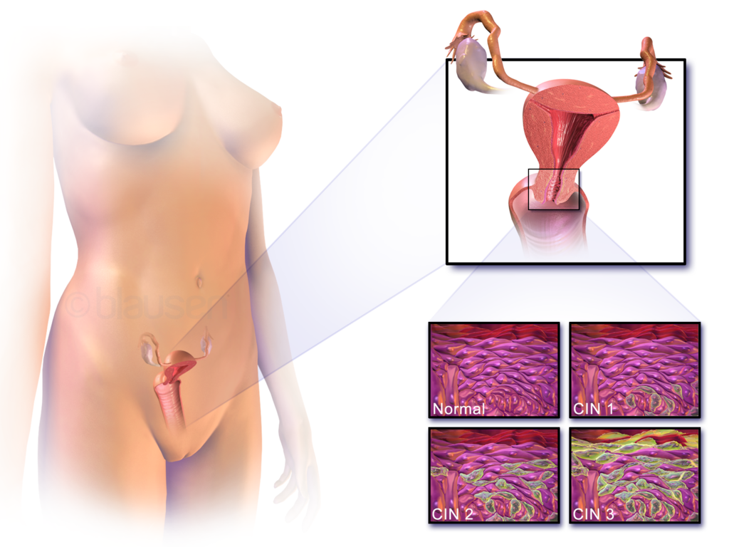
Cervical cancer occurs when cells of the cervix (neck of the uterus) grow abnormally and develop the ability to invade nearby tissues or spread to other parts of the body, such as the abdomen or lungs. Figure 18.9.2 shows the location of the cervix and the appearance of normal and abnormal cervical cells when examined with a microscope.
Cervical Cancer Prevalence and Death Rates
Worldwide, cervical cancer is the second most common type of cancer in females (after breast cancer) and the fourth-most common cause of cancer death in females. In Canada and other high-income nations, the widespread use of cervical cancer screening has detected many cases of precancerous cervical changes and has dramatically reduced rates of cervical cancer deaths. About 75% of cervical cancer cases occur in developing countries, where routine screening is less likely because of cost and other factors. Cervical cancer is also the most common cause of cancer death in low-income countries.
Symptoms of Cervical Cancer
Early in the development of cervical cancer, there are typically no symptoms. As the disease progresses, however, symptoms are likely to occur. The symptoms may include abnormal vaginal bleeding, pelvic pain, or pain during sexual intercourse. Unfortunately, by the time symptoms start to occur, cervical cancer has typically progressed to a stage at which treatment is less likely to be successful.
Cervical Cancer Causes and Risk Factors
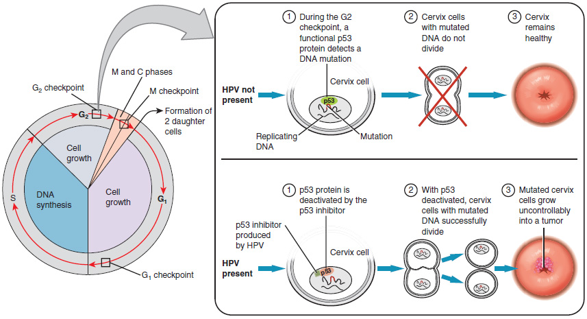
More than 90 per cent of cases of cervical cancer are caused at least in part by human papillomavirus (HPV), which is a sexually transmitted virus that also causes genital warts. Figure 18.9.3 shows how HPV infection can cause cervical cancer by interfering with a normal cell division checkpoint. When HPV is not present, cervical cells containing mutations are not allowed to divide, so the cervix remains healthy. When HPV is present, however, cervical cells with mutations may be allowed to divide, leading to uncontrolled growth of mutated cells and the formation of a tumor.
Other risk factors for cervical cancer include smoking, a weakened immune system (for example, due to HIV infection), use of birth control pills, becoming sexually active at a young age, and having many sexual partners. However, these risk factors are less important than HPV infection. Instead, the risk factors are more likely to increase the risk of cervical cancer in females who are already infected with HPV. For example, among HPV-infected women, current and former smokers have roughly two to three times the incidence of cervical cancer as non-smokers. Passive smoking, or secondhand smoke, is also associated with an increased risk of cervical cancer, but to a lesser extent.
Diagnosis of Cervical Cancer
Diagnosis of cervical cancer is typically made by looking for microscopic abnormal cervical cells in a smear of cells scraped off the cervix. This is called a Pap smear. If cancerous cells are detected or suspected in the smear, this test is usually followed up with a biopsy to confirm the Pap smear results. Medical imaging (by CT scan or MRI, for example) is also likely to be done to provide more information, such as whether the cancer has spread.
Prevention of Cervical Cancer
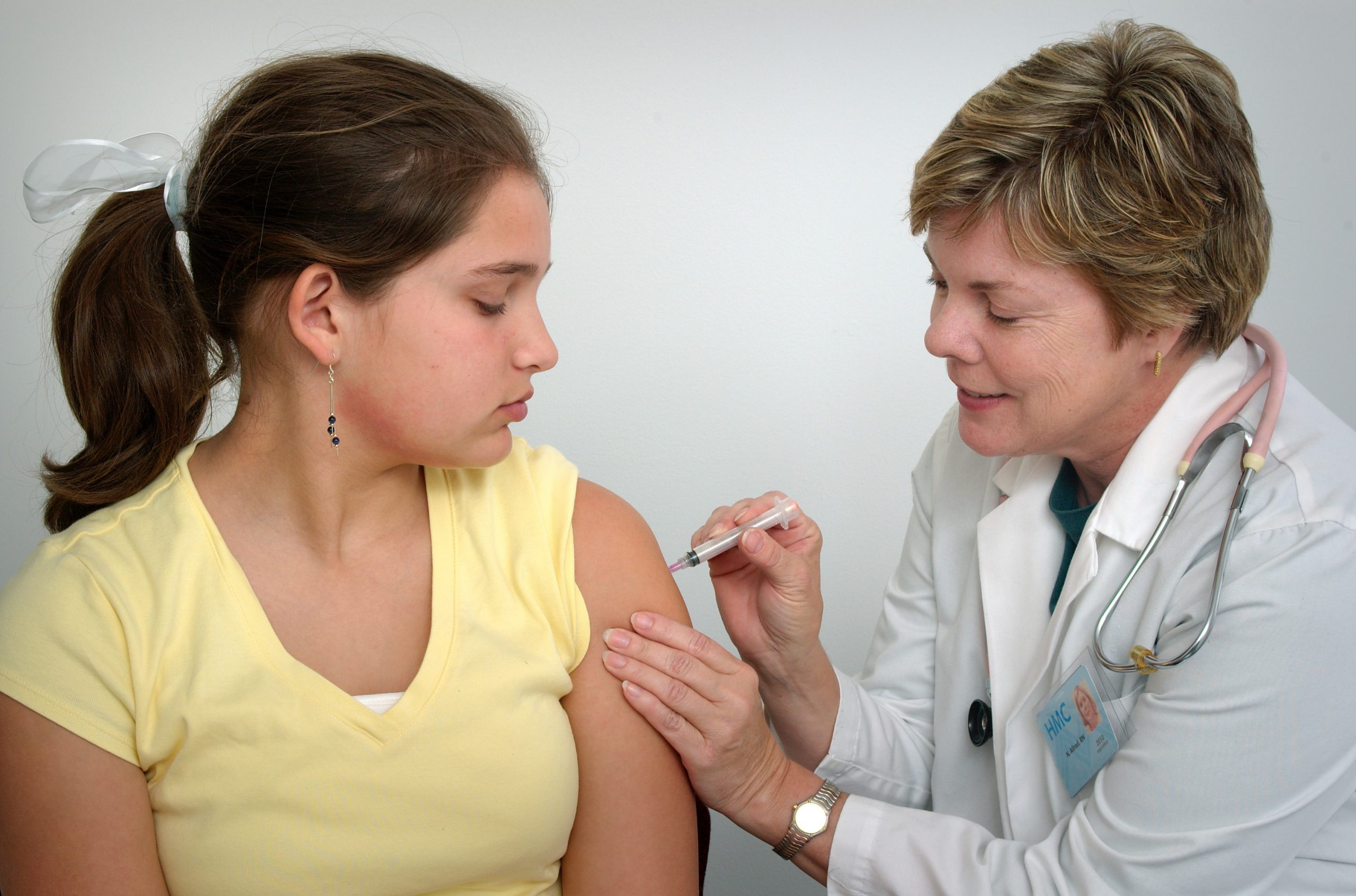
It is now possible to prevent HPV infection with a vaccine. The first HPV vaccine was approved by the U.S. Food and Drug Administration in 2006. The vaccine protects against the strains of HPV that have the greatest risk of causing cervical cancer. It is thought that widespread use of the vaccine will prevent up to 90% of cervical cancer cases. Current recommendations are for females to be given the vaccine between the ages of nine and 26. (Boys should be vaccinated against HPV, as well, because the virus may also cause cancer of the penis and certain other male cancers.) The vaccine is effective only if it is given before HPV infection has occurred. Using condoms during sexual intercourse can also help prevent HPV infection and cervical cancer, in addition to preventing pregnancy and sexually transmitted infections (such as HIV).
Even in women who have received the HPV vaccine, there is still a small risk of developing cervical cancer. Therefore, it is recommended that women continue to be examined with regular Pap smears.
Treatment of Cervical Cancer
Treatment of cervical cancer generally depends on the stage at which the cancer is diagnosed, but it is likely to include some combination of surgery, radiation therapy, and/or chemotherapy. Outcomes of treatment depend largely on how early the cancer is diagnosed and treated. For surgery to cure cervical cancer, the entire tumor must be removed with no cancerous cells found at the margins of the removed tissue on microscopic examination. If cancer is found and treated very early when it is still in the microscopic stage, the five-year survival rate is virtually 100%.
Vaginitis
Vaginitis is inflammation of the vagina — and sometimes the vulva, as well. Symptoms may include a discharge that is yellow, gray, or green; itching; pain; and a burning sensation. There may also be a foul vaginal odor and pain or irritation with sexual intercourse.
Causes of Vaginitis
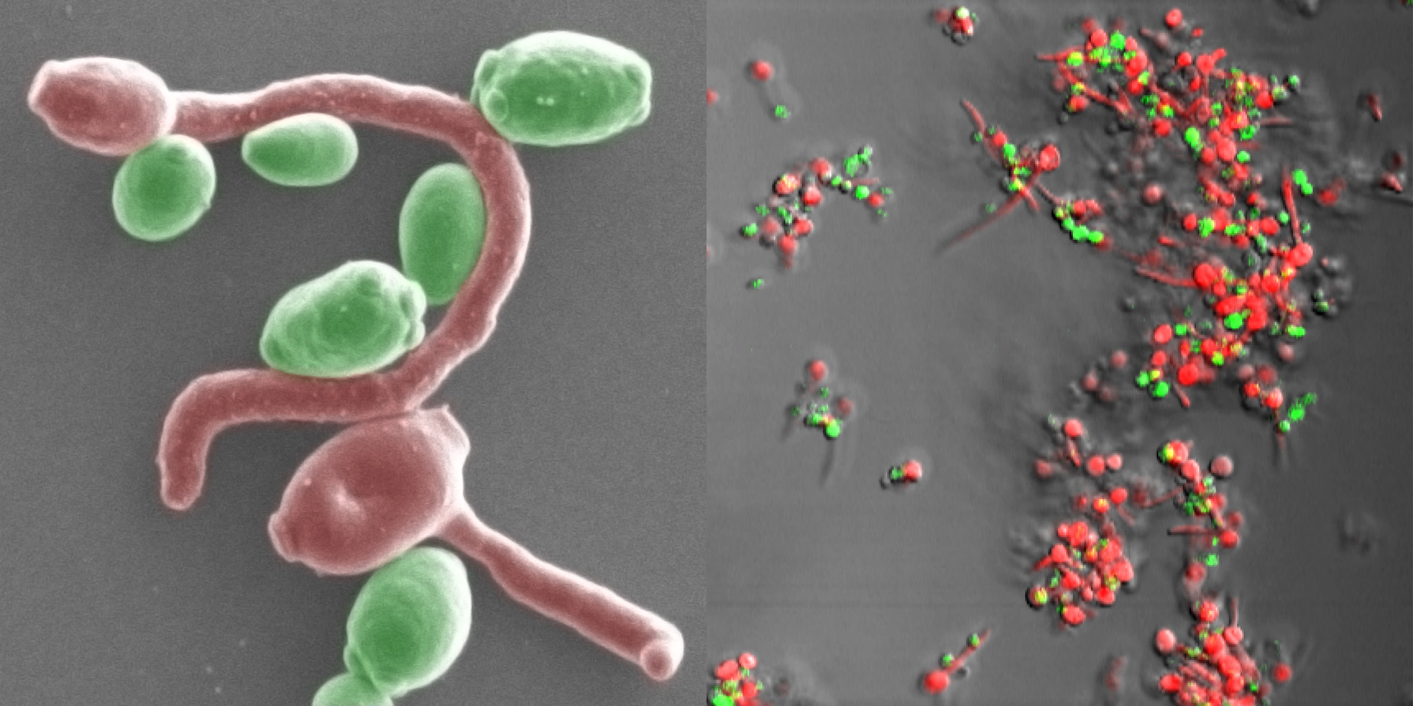
About 90% of cases of vaginitis are caused by infection with microorganisms. Most commonly, vaginal infections are caused by the yeast Candida albicans (see Figure 18.9.4). Such infections are referred to as vaginal candidiasis or more commonly as a yeast infection. Candida albicans is one of the most common opportunistic infections in the world and can affect not only the vagina, but any of the mucus membranes and skin. Other possible causes of vaginal infections include bacteria, especially Gardnerella vaginalis, and some single-celled parasites, notably the protist parasite Trichomonas vaginalis, which is usually transmitted through vaginal intercourse. The risk of vaginal infections may be greater in women who wear tight clothing, are taking antibiotics for another condition, use birth control pills, or have improper hygiene. Poor hygiene allows organisms that are normally present in the stool (such as yeast) to contaminate the vagina.
Most of the remaining cases of vaginitis are due to irritation by — or allergic reactions to — various products. These irritants may include condoms, spermicides, soaps, douches, lubricants, and even semen. Using tampons or soaking in hot tubs may be additional causes of this type of vaginitis.
Diagnosis of Vaginitis
Diagnosis of vaginitis typically begins with symptoms reported by the patient. This may be followed by a microscopic examination or culture of the vaginal discharge in order to identify the specific cause. The colour, consistency, acidity, and other characteristics of the discharge may be predictive of the causative agent. For example, infection with Candida albicans may cause a cottage cheese-like discharge with a low pH, whereas infection with Gardnerella vaginalis may cause a discharge with a fish-like odor and a high pH.
Prevention of Vaginitis
Prevention of vaginitis includes wearing loose cotton underwear that helps keep the vulva dry. Yeasts and bacteria that may cause vaginitis tend to grow best in a moist environment. It is also important to avoid the use of perfumed soaps, personal hygiene sprays, and douches, all of which may upset the normal pH and bacterial balance in the vagina. To help avoid vaginitis caused by infection with Trichomonas vaginalis, the use of condoms during sexual intercourse is advised.
Treatment of Vaginitis
The appropriate treatment of vaginitis depends on the cause. In many cases of vaginitis, there is more than one cause, and all of the causes must be treated to ensure a cure.
- Yeast infections of the vagina are typically treated with topical anti-fungal medications, which are available over the counter. The medications may be in the form of tablets or creams that are inserted into the vagina. Depending on the particular medication used, treatment may involve one, three, or seven days of applications.
- Bacterial infections of the vagina are usually treated with antibiotics. These may be taken orally as pills, or applied topically to the vagina in creams.
- Trichomonas vaginalis infections of the vagina are generally treated with a single dose of an oral antibiotic. Sexual partners should be treated at the same time, and intercourse should be avoided for at least a week until both partners have completed treatment, and have been followed-up by a physician.
Endometriosis
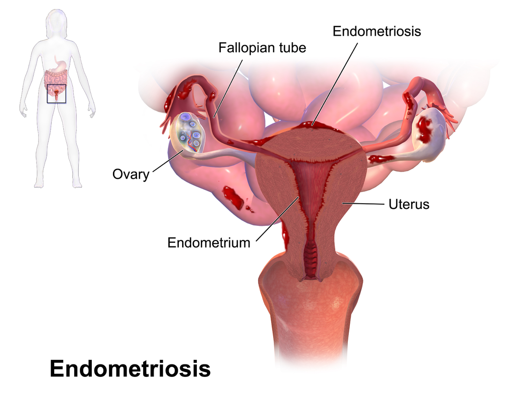
Endometriosis is a disease in which endometrial tissue, which normally grows inside the uterus, grows outside it, as shown in Figure 18.9.5. Most often, the endometrial tissue grows around the ovaries, Fallopian tubes, and uterus. In rare instances, the tissue may grow elsewhere in the body. The areas of endometriosis typically bleed each month during the menstrual period, and this often results in inflammation, pain, and scarring. An estimated six to ten per cent of women are believed to have endometriosis. It is most common in women during their thirties and forties, and only rarely occurs before menarche or after menopause.
Signs and Symptoms of Endometriosis
The main symptom of endometriosis is pelvic pain, which may range from mild to severe. There appears to be little or no relationship between the amount of endometrial tissue growing outside the uterus and the severity of the pain. For many women with the disease, the pain occurs mainly during menstruation. However, nearly half of those affected have chronic pelvic pain. The pain of endometriosis may be caused by bleeding in the pelvis, which triggers inflammation. Pain can also occur from internal scar tissue that binds internal organs to each other.
Another problem often associated with endometriosis is infertility, or the inability to conceive or bear children. Among women with endometriosis, up to half may experience infertility. Infertility can be related to scar formation or to anatomical distortions due to the abnormal endometrial tissue. Other possible symptoms of endometriosis may include diarrhea or constipation, chronic fatigue, nausea and vomiting, headaches, and heavy or irregular menstrual bleeding.
Causes of Endometriosis
The causes of endometriosis are not known for certain, but several risk factors have been identified, including a family history of endometriosis. Daughters or sisters of women with endometriosis have about six times the normal risk of developing the disease themselves. It has been suggested that endometriosis results from mutations in several genes. It is likely that endometriosis is multifactorial, involving the interplay of several factors.
At the physiological level, the predominant idea for how endometriosis comes about is retrograde menstruation. This happens when some of the endometrial debris from a woman’s menstrual flow exits the uterus through the oviducts, rather than through the vagina. The debris then attaches itself to the outside of organs in the abdominal cavity, or to the lining of the abdominal cavity itself. Retrograde menstruation, however, does not explain all cases of endometriosis, so other factors are apparently involved. Suggestions include environmental toxins and autoimmune responses.
Diagnosis of Endometriosis
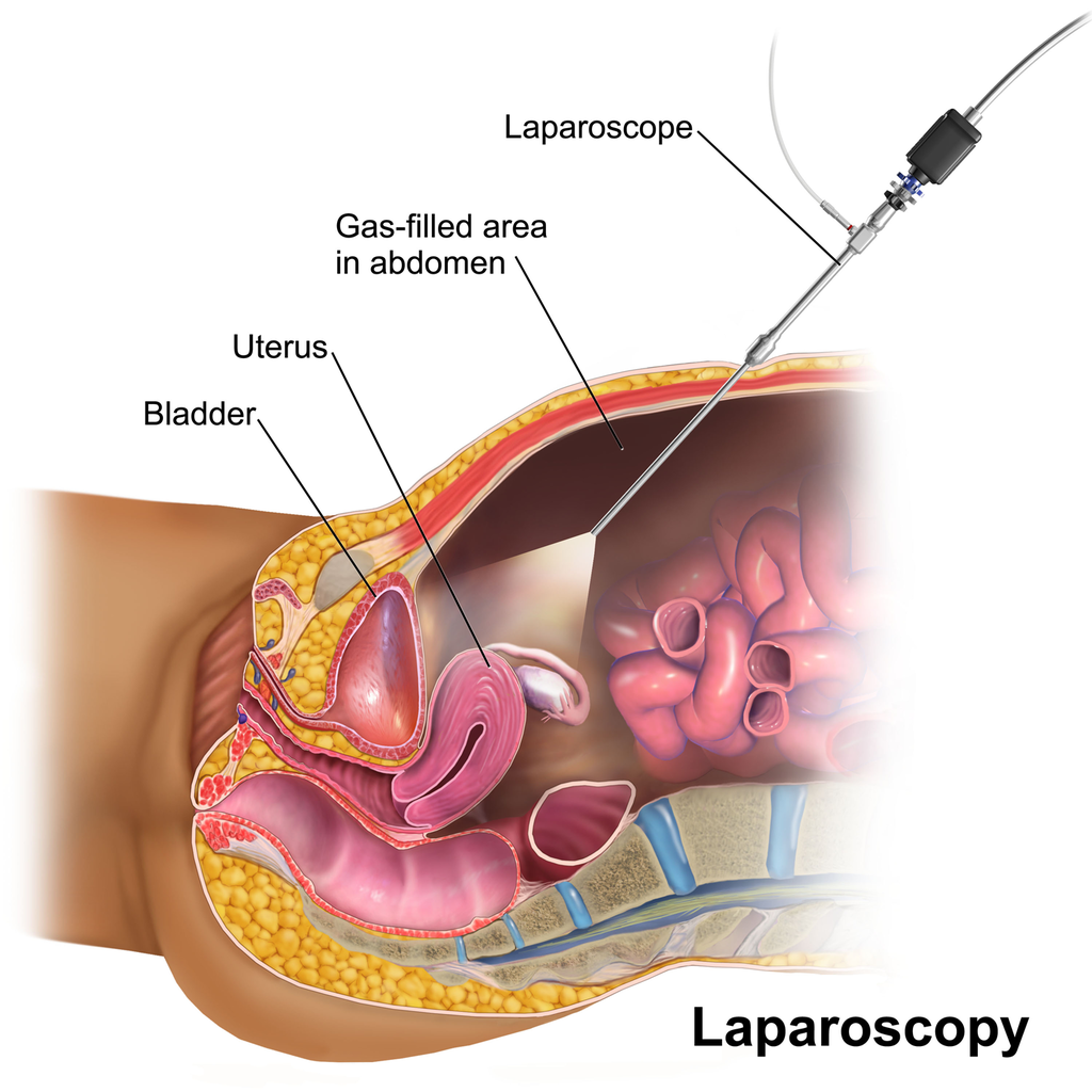
Diagnosis of endometriosis is usually based on self-reported symptoms and a physical examination by a doctor, often combined with medical imaging, such as ultrasonography. The only way to definitively diagnose endometriosis, however, is through visual inspection of the endometrial tissue. This can be done with a surgical procedure called laparoscopy, shown in Figure 18.9.6, in which a tiny camera is inserted into the abdomen through a small incision. The camera allows the physician to visually inspect the area where endometrial tissue is suspected.
Treatment of Endometriosis
The most common treatments for endometriosis are medications to control the pain, and surgery to remove the abnormal tissue. Frequently used pain medications are non-steroidal inflammatory drugs (NSAIDS), such as naproxen. Opiates may be used in cases of severe pain. Laparoscopy can be used to surgically treat endometriosis, as well as to diagnose the condition. In this type of surgery, an additional small incision is made to insert instruments that the surgeon can manipulate externally in order to burn (cauterize) or cut away the endometrial growths. In younger women who want to have children, surgery is conservative to keep the reproductive organs intact and functional. However, with conservative surgery, endometriosis recurs in 20–40% of cases within five years of the surgery. In older women who have completed childbearing, hysterectomy may be undertaken to remove all or part of the internal reproductive organs. This is the only procedure that is likely to cure endometriosis and prevent relapses.
Feature: My Human Body
A Pap smear is a method of cervical cancer screening used to detect potentially pre-cancerous and cancerous cells in the cervix. It is the most widely used screening test for this type of cancer, and it is very effective. The test may also detect vaginal infections and abnormal endometrial cells, but it is not designed for these purposes.
According to HealthLink BC, females should start receiving routine Pap smears by age 25. Because most cases of cervical cancer are caused by infection with human papillomavirus (HPV), which is a sexually transmitted infection, there is little or no benefit to screening people who have not had sexual contact. Starting at age 25, general guidelines are for Pap smears to be repeated every three years until age 69. Screening may be discontinued after 69 years of age, if the last three Pap smears were normal. If a woman has a complete hysterectomy, she no longer has a cervix and there is no need for further Pap smears. On the other hand, if a woman has had a history of abnormal Pap smears or cancer, she will likely be screened more frequently. Pap smears can be done safely during the first several months of pregnancy, and resumed about three months after childbirth. Generally, better results are obtained if Pap smears are not done during menstruation.
If you’ve never had a Pap smear, knowing what to expect may help prepare you for the procedure. The patient lies on the examining table with her feet in “stirrups” to hold the legs up and apart. An instrument called a speculum is inserted into the vagina to hold back the vaginal walls and give access to the cervix. A tiny amount of tissue is brushed off the cervix and smeared onto a microscope slide. The speculum is then removed, and the procedure is over. The slide is later examined under a microscope for abnormal cells. Some women experience light spotting or mild diarrhea after a Pap smear, but most have no lasting effects.
Pap smears are uncomfortable and may be somewhat painful for some women. If you experience pain during a Pap smear, tell your health care provider. Many steps can be taken to minimize the pain, which might include using a smaller speculum, using warm instruments and a lubricant, and applying a topical anesthetic such as lidocaine to the cervix before obtaining the smear. Any pain is generally very brief, and the potential reward is worth it. Pap tests are estimated to reduce up to 80% of cervical cancer deaths. One of the lives saved could be your own.
18.9 Summary
- Cervical cancer occurs when cells of the cervix grow abnormally and develop the ability to invade nearby tissues or spread to other parts of the body. Worldwide, cervical cancer is the second-most common type of cancer in females and the fourth-most common cause of cancer death in females. Early on, cervical cancer often has no symptoms. Later, symptoms (such as abnormal vaginal bleeding and pain) are likely.
- Most cases of cervical cancer occur because of infection with human papillomavirus (HPV), so the HPV vaccine is expected to greatly reduce the incidence of the disease. Other risk factors include smoking and a weakened immune system. A Pap smear can diagnose cervical cancer at an early stage. Where Pap smears are done routinely, cervical cancer death rates have fallen dramatically. Treatment of cervical cancer generally includes surgery, which may be followed by radiation therapy or chemotherapy.
- Vaginitis is inflammation of the vagina. A discharge is likely, and there may be itching and pain. About 90% of cases of vaginitis are caused by infection with microorganisms, typically by the yeast Candida albicans. A minority of cases are caused by irritants or allergens in soaps, spermicides, or douches.
- Diagnosis of vaginitis may be based on characteristics of the discharge, which can be examined microscopically or cultured. Treatment of vaginitis depends on the cause and is usually an oral or topical anti-fungal or antibiotic medication.
- Endometriosis is a disease in which endometrial tissue grows outside the uterus. This tissue may bleed during the menstrual period and cause inflammation, pain, and scarring. The main symptom of endometriosis is pelvic pain, which may be severe. Endometriosis may also lead to infertility.
- Endometriosis is thought to have multiple causes, including genetic mutations. Retrograde menstruation may be the immediate cause of endometrial tissue escaping the uterus and entering the pelvic cavity. Endometriosis is usually treated with surgery to remove the abnormal tissue and medication for pain. If surgery is more conservative than hysterectomy, endometriosis may recur.
18.9 Review Questions
- What is cervical cancer? Worldwide, how prevalent is it, and how does it rank as a cause of cancer deaths?
- Identify symptoms of cervical cancer. What are causes of — and risk factors for — cervical cancer?
- What roles can Pap smears and HPV vaccines play in preventing cervical cancer cases and cervical cancer deaths?
- How is cervical cancer treated?
- Define vaginitis and identify its symptoms.
- What are some of the causes of vaginitis? Which cause is responsible for most of the cases?
- How is vaginitis diagnosed and treated?
- What is endometriosis, and what are its symptoms?
- Discuss possible causes of endometriosis.
- How is endometriosis treated? Which treatment is most likely to prevent recurrence of the disorder?
-
- In the case of infection with Trichomonas vaginalis, why is the woman’s sexual partner usually treated at the same time?
18.9 Explore More
https://youtu.be/KOz-bNhEHhQ
What is HPV and how can you protect yourself from it? - Emma Bryce, TED-Ed, 2019.
https://youtu.be/6HeQ4iEqAUk
Endometriosis - The Mystery Disease of Women | Cécile Real | TEDxBinnenhof, TEDx Talks, 2016.
https://youtu.be/ryNjSP5VVI8
The brain and ovarian hormones | Marwa Azab | TEDxMontrealWomen, TEDxTalks, 2015.
Attributions
Figure 18.9.1
a-nurse-giving-a-young-girl-a-vaccine-shot-or by CDC/ Judy Schmidt from Public Health Image Library (PHIL) #9424 is in the public domain (https://en.wikipedia.org/wiki/Public_domain).
Figure 18.9.2
1024px-Blausen_0221_CervicalDysplasia by Blausen Medical Communications, Inc. on Wikimedia Commons is used under a CC BY 3.0 (https://creativecommons.org/licenses/by/3.0) license.
Figure 18.9.3
HPV and Cervical Cancer by OpenStax by OpenStax College on Wikimedia Commons is used under a CC BY 3.0 (https://creativecommons.org/licenses/by/3.0) license.
Figure 18.9.4
Candida by NIH on Flickr from the NIH Image Gallery on Flickr is used under a CC BY NC 2.0 (https://creativecommons.org/licenses/by-nc/2.0/) license.
Figure 18.9.5
Blausen_0349_Endometriosis by BruceBlaus on Wikimedia Commons is used under a CC BY 3.0 (https://creativecommons.org/licenses/by/3.0) license.
Figure 18.9.6
1024px-Blausen_0602_Laparoscopy_02 by BruceBlaus on Wikimedia Commons is used under a CC BY 3.0 (https://creativecommons.org/licenses/by/3.0) license.
References
Betts, J. G., Young, K.A., Wise, J.A., Johnson, E., Poe, B., Kruse, D.H., Korol, O., Johnson, J.E., Womble, M., DeSaix, P. (2013, June 19). Figure 27.16 Development of cervical cancer [digital image]. In Anatomy and Physiology (Section 27.2). OpenStax. https://openstax.org/books/anatomy-and-physiology/pages/27-2-anatomy-and-physiology-of-the-female-reproductive-system
Blausen.com Staff. (2014). Medical gallery of Blausen Medical 2014. WikiJournal of Medicine 1 (2). DOI:10.15347/wjm/2014.010. ISSN 2002-4436.
HealthLink BC. (n.d.). Pap test: British Columbia specific information. https://www.healthlinkbc.ca/medical-tests/hw5266
TED-Ed. (2019, July 9). What is HPV and how can you protect yourself from it? - Emma Bryce. YouTube. https://www.youtube.com/watch?v=KOz-bNhEHhQ&feature=youtu.be
TEDx Talks. (2016, April 14). Endometriosis - The mystery disease of women | Cécile Real | TEDxBinnenhof. YouTube. https://www.youtube.com/watch?v=6HeQ4iEqAUk&feature=youtu.be
TEDx Talks. (2015, July 27). The brain and ovarian hormones | Marwa Azab | TEDxMontrealWomen. YouTube. https://www.youtube.com/watch?v=ryNjSP5VVI8&feature=youtu.be
A long extension of the cell body of a neuron that transmits nerve impulses to other cells.
Created by CK-12 Foundation/Adapted by Christine Miller
Stroller Moms
These moms (Figure 12.5.1) are setting a great example for their children by engaging in physical exercise. Adopting a habit of regular physical exercise is one of the most important ways to maintain fitness and good health. From higher self-esteem to a healthier heart, physical exercise can have a positive effect on virtually all aspects of health, including physical, mental, and emotional health.
What Is Physical Exercise?
Physical exercise is any bodily activity that enhances or maintains physical fitness and overall health and wellness. We generally think of physical exercise as activities that are undertaken for the main purpose of improving physical fitness and health. However, physical activities that are undertaken for other purposes may also count as physical exercise. Scrubbing a floor, raking a lawn, or playing active games with young children or a pet are all activities that can have fitness and health benefits, even though they generally are not done mainly for this purpose.
How much physical exercise should people get? In the Canada, both the Canadian Food Guide and the Canadian Society for Exercise Physiology recommend that every child and adult who is able should participate in moderate exercise for a minimum of 60 minutes a day. This might include walking, swimming, and/or household or yard work.
Types of Physical Exercise
Physical exercise can be classified into three types, depending on the effects it has on the body: aerobic exercise, anaerobic exercise, and flexibility exercise. Many specific examples of physical exercise (including playing soccer and rock climbing) can be classified as more than one type.
Aerobic Exercise

Aerobic exerciseis any physical activity in which muscles are used at well below their maximum contraction strength, but for long periods of time. Aerobic exercise uses a relatively high percentage of slow-twitch muscle fibres that consume a large amount of oxygen. The main goal of aerobic exercise is to increase cardiovascular endurance, although it can have many other benefits, including muscle toning. Examples of aerobic exercise include cycling, swimming, brisk walking, jumping rope, rowing, hiking, tennis, and kayaking as shown in Figure 12.5.2 .
Anaerobic Exercise
Anaerobic exercise is any physical activity in which muscles are used at close to their maximum contraction strength, but for relatively short periods of time. Anaerobic exercise uses a relatively high percentage of fast-twitch muscle fibres that consume a small amount of oxygen. Goals of anaerobic exercise include building and strengthening muscles, as well as improving bone strength, balance, and coordination. Examples of anaerobic exercise include push-ups, lunges, sprinting, interval training, resistance training, and weight training (such as biceps curls with a dumbbell, as pictured in Figure 12.5.3).

Flexibility Exercise

Flexibility exercise is any physical activity that stretches and lengthens muscles. Goals of flexibility exercise include increasing joint flexibility, keeping muscles limber, and improving the range of motion, all of which can reduce the risk of injury. Examples of flexibility exercise include stretching, yoga (as in Figure 12.5.4), and tai chi.
Health Benefits of Physical Exercise
Many studies have shown that physical exercise is positively correlated with a diversity of health benefits. Some of these benefits include maintaining physical fitness, losing weight and maintaining a healthy weight, regulating digestive health, building and maintaining healthy bone density, increasing muscle strength, improving joint mobility, strengthening the immune system, boosting cognitive ability, and promoting psychological well-being. Some studies have also found a significant positive correlation between exercise and both quality of life and life expectancy. People who participate in moderate to high levels of physical activity have been shown to have lower mortality rates than people of the same ages who are not physically active and daily exercise has been shown to increase life expectancy up to an average of five years.
The underlying physiological mechanisms explaining why exercise has these positive health benefits are not completely understood. However, developing research suggests that many of the benefits of exercise may come about because of the role of skeletal muscles as endocrine organs. Contracting muscles release hormones called myokines, which promote tissue repair and the growth of new tissue. Myokines also have anti-inflammatory effects, which, in turn, reduce the risk of developing inflammatory diseases. Exercise also reduces levels of cortisol, the adrenal cortex stress hormone that may cause many health problems — both physical and mental — at sustained high levels.
Cardiovascular Benefits of Physical Exercise
The beneficial effects of exercise on the cardiovascular system are well documented. Physical inactivity has been identified as a risk factor for the development of coronary artery disease. There is also a direct correlation between physical inactivity and cardiovascular disease mortality. Physical exercise, in contrast, has been demonstrated to reduce several risk factors for cardiovascular disease, including hypertension (high blood pressure), “bad” cholesterol (low-density lipoproteins), high total cholesterol, and excess body weight. Physical exercise has also been shown to increase “good” cholesterol (high-density lipoproteins), insulin sensitivity, the mechanical efficiency of the heart, and exercise tolerance, which is the ability to perform physical activity without undue stress and fatigue.
Cognitive Benefits of Physical Exercise
Physical exercise has been shown to help protect people from developing neurodegenerative disorders, such as dementia. A 30-year study of almost 2,400 men found that those who exercised regularly had a 59 per cent reduction in dementia when compared with those who did not exercise. Similarly, a review of cognitive enrichment therapies for the elderly found that physical activity — in particular, aerobic exercise — can enhance the cognitive function of older adults. Anecdotal evidence suggests that frequent exercise may even help reverse alcohol-induced brain damage. There are several possible reasons why exercise is so beneficial for the brain. Physical exercise:
- Increases blood flow and oxygen availability to the brain.
- Increases growth factors that promote new brain cells and new neuronal pathways in the brain.
- Increases levels of neurotransmitters (such as serotonin), which increase memory retention, information processing, and cognition.
Mental Health Benefits of Physical Exercise
Numerous studies suggest that regular aerobic exercise works as well as pharmaceutical antidepressants in treating mild-to-moderate depression. A possible reason for this effect is that exercise increases the biosynthesis of at least three neurochemicals that may act as euphoriants. The euphoric effect of exercise is well known. Distance runners may refer to it as “runner’s high,” and people who participate in crew (as in Figure 12.5.5) may refer to it as “rower’s high.” Because of these effects, health care providers often promote the use of aerobic exercise as a treatment for depression.

Additional mental health benefits of physical exercise include reducing stress, improving body image, and promoting positive self-esteem. Conversely, there is evidence to suggest that being sedentary is associated with increased risk of anxiety.
Sleep Benefits of Physical Exercise
A recent review of published scientific research suggests that exercise generally improves sleep for most people, and helps sleep disorders, such as insomnia. In fact, exercise is the most recommended alternative to sleeping pills for people with insomnia. For sleep benefits, the optimum time to exercise may be four to eight hours before bedtime, although exercise at any time of day seems to be beneficial. The only possible exception is heavy exercise undertaken shortly before bedtime, which may actually interfere with sleep.
Other Benefits of Physical Exercise
Some studies suggest that physical activity may benefit the immune system. For example, moderate exercise has been found to be associated with a decreased incidence of upper respiratory tract infections. Evidence from many studies has found a correlation between physical exercise and reduced death rates from cancer, specifically breast cancer and colon cancer. Physical exercise has also been shown to reduce the risk of type 2 diabetes and obesity.
Variation in Responses to Physical Exercise

Not everyone benefits equally from physical exercise. When participating in aerobic exercise, most people will have a moderate increase in their endurance, but some people will as much as double their endurance. Some people, on the other hand, will show little or no increase in endurance from aerobic exercise. Genetic differences in slow-twitch and fast-twitch skeletal muscle fibres may play a role in these different results. People with more slow-twitch fibres may be able to develop greater endurance, because these muscle fibres have more capillaries, mitochondria, and myoglobin than fast-twitch fibres. As a result, slow-twitch fibres can carry more oxygen and sustain aerobic activity for a longer period of time than fast-twitch fibres. Studies show that endurance athletes (like the marathoner pictured in Figure 12.5.6) generally do tend to have a higher proportion of slow-twitch fibres than other people.
There is also great variation in individual responses to muscle building as a result of anaerobic exercise. Some people have a much greater capacity to increase muscle size and strength, whereas other people never develop large muscles, no matter how much they exercise them. People who have more fast-twitch than slow-twitch muscle fibres may be able to develop bigger, stronger muscles, because fast-twitch muscle fibres contribute more to muscle strength and have greater potential to increase in mass. Evidence suggests that athletes who excel at power activities (such as throwing and jumping) tend to have a higher proportion of fast-twitch fibres than do endurance athletes.
Can You “Overdose” on Physical Exercise?
Is it possible to exercise too much? Can too much exercise be harmful? Evidence suggests that some adverse effects may occur if exercise is extremely intense and the body is not given proper rest between exercise sessions. Athletes who train for multiple marathons have been shown to develop scarring of the heart and heart rhythm abnormalities. Doing too much exercise without prior conditioning also increases the risk of injuries to muscles and joints. Damage to muscles due to overexertion is often seen in new military recruits (see Figure 12.5.7). Too much exercise in females may cause amenorrhea, which is a cessation of menstrual periods. When this occurs, it generally indicates that a woman is pushing her body too hard.
Many people develop delayed onset muscle soreness (DOMS), which is pain or discomfort in muscles that is felt one to three days after exercising, and generally subsides two or three days later. DOMS was once thought to be caused by the buildup of lactic acid in the muscles. Lactic acid is a product of anaerobic respiration in muscle tissues. However, lactic acid disperses fairly rapidly, so it is unlikely to explain pain experienced several days after exercise. The current theory is that DOMS is caused by tiny tears in muscle fibres, which occur when muscles are used at too high a level of intensity.
Feature: My Human Body
Most people know that exercise is important for good health, and it’s easy to find endless advice about exercise programs and fitness plans. What is not so easy to find is the motivation to start exercising — and to stick with it. This is the main reason why so many people fail to get regular exercise. Practical concerns like a busy schedule and bad weather can certainly make exercising more of a challenge, but the biggest barriers to adopting a regular exercise routine are mental. If you want to exercise but find yourself making excuses or getting discouraged and giving up, here are some tips that may help you get started and stay moving:
- Avoid an all-or-nothing point of view. Don’t think you need to spend hours sweating at the gym or training for a marathon to get healthy. Even a little bit of exercise is better than nothing at all. Start out with ten or 15 minutes of moderate activity each day. Taking a walk around your neighborhood is a great way to begin! From there, gradually increase the amount of time until you are exercising to at least 30 minutes a day, five days a week. Make it a routine.
- Be kind to yourself, and reinforce positive behaviors with rewards. Don’t be down on yourself because you are overweight or out of shape. Don’t beat yourself up because of a supposed lack of willpower. Instead, look at any past failures as opportunities to learn and do better. When you do achieve even small exercise goals, treat yourself to something special. Did you just complete your first workout? Reward yourself with a relaxing bath or other treat.
- Don’t make excuses for not exercising. Common complaints include being too busy or tired or not athletic enough. Such excuses are not valid reasons to avoid exercising, and they will sabotage any plans to improve your fitness. If you can’t find a 30-minute period to work out, try to find ten minutes, three times a day. If you’re feeling tired, know that exercise can actually reduce fatigue and boost your energy level. If you feel clumsy and uncoordinated, remind yourself that you don’t need to be athletic to take a walk or engage in vigorous house or yard work.
- Find an activity that you truly enjoy doing. Don’t think you have to lift weights or run on a treadmill to exercise your muscles. If you find such activities boring or unpleasant, you won’t stick with them. Any activity that increases your heart rate and uses large muscles can provide a workout, especially if you’re not in the habit of exercising, so find something you like to do. Do you like to dance? Put on some music and dance up a sweat! Do you enjoy gardening? Get out in the yard and dig up some dirt! Still not interested? Try an activity-based video game, such as Wii or Kinect. You may find it so much fun that it doesn’t seem like exercise until you realize you’ve worked up a sweat.
- Make yourself accountable. Tell friends and family members that you’re going to start exercising. You’ll be letting them — as well as yourself — down if you don’t follow through. Some people find that keeping an exercise log to track their progress is a good way to be accountable and stick to an exercise program. Perhaps the best way to keep at it is to find an exercise partner. If you’ve got someone waiting to exercise with you, you will be less likely to make excuses for not exercising.
- Add more physical activity to your daily life. You don’t need to follow a structured exercise program to increase your activity level. Do your house or yard work briskly for a workout. Park your car further than necessary from work or the mall, and walk the extra distance. If you live close enough, leave the car at home and walk to and from your destination. Rather than taking elevators or escalators, walk up and down stairs. When you take breaks at work, take a walk instead of sitting. Every time a commercial comes on while you’re watching TV, take a quick exercise break — run in place or do some curls with hand weights.
12.5 Summary
- Physical exercise is any bodily activity that enhances or maintains physical fitness and overall health. Activities such as household chores may count as physical exercise, even if they are not done for their health benefits. Current recommendations for adults are 30 minutes a day of moderate exercise.
- Aerobic exercise is any physical activity that uses muscles at less than their maximum contraction strength, but for long periods of time. This type of exercise uses a relatively high percentage of slow-twitch muscle fibres that consume large amounts of oxygen. Aerobic exercises increase cardiovascular endurance and include cycling and brisk walking.
- Anaerobic exercise is any physical activity that uses muscles at close to their maximum contraction strength, but for short periods of time. This type of exercise uses a relatively high percentage of fast-twitch muscle fibres that consume small amounts of oxygen. Anaerobic exercises increase muscle and bone mass and strength, and they include push-ups and sprinting.
- Flexibility exercise is any physical activity that stretches and lengthens muscles, thereby improving range of motion and reducing risk of injury. Examples include stretching and yoga.
- Many studies have shown that physical exercise is positively correlated with a diversity of physical, mental, and emotional health benefits. Physical exercise also increases quality of life and life expectancy.
- Many of the benefits of exercise may come about because contracting muscles release hormones called myokines, which promote tissue repair and growth and have anti-inflammatory effects.
- Physical exercise can reduce risk factors for cardiovascular disease, including hypertension and excess body weight. Physical exercise can also increase factors associated with cardiovascular health, such as mechanical efficiency of the heart.
- Physical exercise has been shown to offer protection from dementia and other cognitive problems, perhaps because it increases blood flow or neurotransmitters in the brain, among other potential effects.
- Numerous studies suggest that regular aerobic exercise works as well as pharmaceutical antidepressants in treating mild-to-moderate depression, possibly because it increases synthesis of natural euphoriants in the brain.
- Research shows that physical exercise generally improves sleep for most people and helps sleep disorders, such as insomnia. Other health benefits of physical exercise include better immune system function and reduced risk of type 2 diabetes and obesity.
- There is great variation in individual responses to exercise, partly due to genetic differences in proportions of slow-twitch and fast-twitch skeletal muscle fibres. People with more slow-twitch fibres may be able to develop greater endurance from aerobic exercise, whereas people with more fast-twitch fibres may be able to develop greater muscle size and strength from anaerobic exercise.
- Some adverse effects may occur if exercise is extremely intense and the body is not given proper rest between exercise sessions. Many people who overwork their muscles develop delayed onset muscle soreness (DOMS), which may be caused by tiny tears in muscle fibres.
12.5 Review Questions
- How do we define physical exercise?
- What are current recommendations for physical exercise for adults?
-
- Define flexibility exercise, and state its benefits. What are two examples of flexibility exercises?
- In general, how does physical exercise affect health, quality of life, and longevity?
- What mechanism may underlie many of the general health benefits of physical exercise?
- Relate physical exercise to cardiovascular disease risk.
- What may explain the positive benefits of physical exercise on cognition?
- How does physical exercise compare with antidepressant drugs in the treatment of depression?
- Identify several other health benefits of physical exercise.
- Explain how genetics may influence the way individuals respond to physical exercise.
- Can too much physical exercise be harmful?
12.5 Explore More
https://www.youtube.com/watch?v=hmFQqjMF_f0
How playing sports benefits your body ... and your brain - Leah Lagos and Jaspal Ricky Singh, TED-Ed, 2016.
https://www.youtube.com/watch?v=rLsimrBoYXc&t=12s
The surprising reason our muscles get tired - Christian Moro, TED-Ed, 2019.
https://youtu.be/2tM1LFFxeKg
What makes muscles grow? - Jeffrey Siegel, TED-Ed, 2015.
https://www.youtube.com/watch?v=QeIrdqU0o9s
Why some people find exercise harder than others | Emily Balcetis, TED, 2014.
Attributions
Figure 12.5.1
stroller fit by Serge Melki from Indianapolis, USA on Wikimedia Commons is used under a CC BY 2.0 (https://creativecommons.org/licenses/by/2.0) license.
Figure 12.5.2
Children kayaking young sport by Hagerty Ryan, USFWS on Pixnio is used under a public domain (CC0) Certification (https://creativecommons.org/licenses/publicdomain/).
Figure 12.5.3
Bicep curls [photo] by Senior Airman Jarrod Grammel from U.S. Moody Air Force Base is released into the public domain (https://en.wikipedia.org/wiki/Public_domain).
Figure 12.5.4
Flexibility exercise by carl-barcelo-nqUHQkuVj3c [photo] by Carl Barcelo on Unsplash is used under the Unsplash License (https://unsplash.com/license).
Figure 12.5.5
Canadian women’s double scull silver Rio 2016 by Gerhard Pratt on Flickr is used under a CC BY-NC 2.0 (https://creativecommons.org/licenses/by-nc/2.0/) license.
Figure 12.5.6
Toronto Marathon 2012 by Marc Roberts on Flickr is used under a CC BY-NC-SA 2.0 (https://creativecommons.org/licenses/by-nc-sa/2.0/) license.
Figure 12.5.7
Muscle damage in military recruits by Lance Cpl. Bridget M. Keane from the United States Marine Corps Recruit Depot is in the public domain (https://en.wikipedia.org/wiki/Public_domain).
References
Elwood, P., Galante, J., Pickering, J., Palmer, S., Bayer, A., Ben-Shlomo, Y., Longley, M., & Gallacher, J. (2013). Healthy lifestyles reduce the incidence of chronic diseases and dementia: evidence from the Caerphilly cohort study. PloS one, 8(12), e81877. https://doi.org/10.1371/journal.pone.0081877
Mayo Clinic Staff. (n.d.). Amenorrhea [online article]. MayoClinic.org. https://www.mayoclinic.org/diseases-conditions/amenorrhea/symptoms-causes/syc-20369299#
Mayo Clinic Staff. (n.d.). Coronary artery disease [online article]. MayoClinic.org. https://www.mayoclinic.org/diseases-conditions/coronary-artery-disease/symptoms-causes/syc-20350613
TED-Ed. (2016, June 28). How playing sports benefits your body ... and your brain - Leah Lagos and Jaspal Ricky Singh. YouTube. https://www.youtube.com/watch?v=hmFQqjMF_f0&feature=youtu.be
TED-Ed. (2019, April 18). The surprising reason our muscles get tired - Christian Moro. YouTube. https://www.youtube.com/watch?v=rLsimrBoYXc&feature=youtu.be
TED-Ed. (2015, November 3). What makes muscles grow? - Jeffrey Siegel. YouTube https://www.youtube.com/watch?v=2tM1LFFxeKg&feature=youtu.be
TED. (2014, November 14). Why some people find exercise harder than others | Emily Balcetis, YouTube. https://www.youtube.com/watch?v=QeIrdqU0o9s&feature=youtu.be
Wikipedia contributors. (2020, August 1). Delayed onset muscle soreness. In Wikipedia. https://en.wikipedia.org/w/index.php?title=Delayed_onset_muscle_soreness&oldid=970682631
Image shows a female runner participating in the 2012 Toronto Marathon.
Image shows a group of American military recruits jogging while being encouraged/yelled at by their drill instructor.
Created by CK-12 Foundation/Adapted by Christine Miller
Stroller Moms
These moms (Figure 12.5.1) are setting a great example for their children by engaging in physical exercise. Adopting a habit of regular physical exercise is one of the most important ways to maintain fitness and good health. From higher self-esteem to a healthier heart, physical exercise can have a positive effect on virtually all aspects of health, including physical, mental, and emotional health.
What Is Physical Exercise?
Physical exercise is any bodily activity that enhances or maintains physical fitness and overall health and wellness. We generally think of physical exercise as activities that are undertaken for the main purpose of improving physical fitness and health. However, physical activities that are undertaken for other purposes may also count as physical exercise. Scrubbing a floor, raking a lawn, or playing active games with young children or a pet are all activities that can have fitness and health benefits, even though they generally are not done mainly for this purpose.
How much physical exercise should people get? In the Canada, both the Canadian Food Guide and the Canadian Society for Exercise Physiology recommend that every child and adult who is able should participate in moderate exercise for a minimum of 60 minutes a day. This might include walking, swimming, and/or household or yard work.
Types of Physical Exercise
Physical exercise can be classified into three types, depending on the effects it has on the body: aerobic exercise, anaerobic exercise, and flexibility exercise. Many specific examples of physical exercise (including playing soccer and rock climbing) can be classified as more than one type.
Aerobic Exercise

Aerobic exerciseis any physical activity in which muscles are used at well below their maximum contraction strength, but for long periods of time. Aerobic exercise uses a relatively high percentage of slow-twitch muscle fibres that consume a large amount of oxygen. The main goal of aerobic exercise is to increase cardiovascular endurance, although it can have many other benefits, including muscle toning. Examples of aerobic exercise include cycling, swimming, brisk walking, jumping rope, rowing, hiking, tennis, and kayaking as shown in Figure 12.5.2 .
Anaerobic Exercise
Anaerobic exercise is any physical activity in which muscles are used at close to their maximum contraction strength, but for relatively short periods of time. Anaerobic exercise uses a relatively high percentage of fast-twitch muscle fibres that consume a small amount of oxygen. Goals of anaerobic exercise include building and strengthening muscles, as well as improving bone strength, balance, and coordination. Examples of anaerobic exercise include push-ups, lunges, sprinting, interval training, resistance training, and weight training (such as biceps curls with a dumbbell, as pictured in Figure 12.5.3).

Flexibility Exercise

Flexibility exercise is any physical activity that stretches and lengthens muscles. Goals of flexibility exercise include increasing joint flexibility, keeping muscles limber, and improving the range of motion, all of which can reduce the risk of injury. Examples of flexibility exercise include stretching, yoga (as in Figure 12.5.4), and tai chi.
Health Benefits of Physical Exercise
Many studies have shown that physical exercise is positively correlated with a diversity of health benefits. Some of these benefits include maintaining physical fitness, losing weight and maintaining a healthy weight, regulating digestive health, building and maintaining healthy bone density, increasing muscle strength, improving joint mobility, strengthening the immune system, boosting cognitive ability, and promoting psychological well-being. Some studies have also found a significant positive correlation between exercise and both quality of life and life expectancy. People who participate in moderate to high levels of physical activity have been shown to have lower mortality rates than people of the same ages who are not physically active and daily exercise has been shown to increase life expectancy up to an average of five years.
The underlying physiological mechanisms explaining why exercise has these positive health benefits are not completely understood. However, developing research suggests that many of the benefits of exercise may come about because of the role of skeletal muscles as endocrine organs. Contracting muscles release hormones called myokines, which promote tissue repair and the growth of new tissue. Myokines also have anti-inflammatory effects, which, in turn, reduce the risk of developing inflammatory diseases. Exercise also reduces levels of cortisol, the adrenal cortex stress hormone that may cause many health problems — both physical and mental — at sustained high levels.
Cardiovascular Benefits of Physical Exercise
The beneficial effects of exercise on the cardiovascular system are well documented. Physical inactivity has been identified as a risk factor for the development of coronary artery disease. There is also a direct correlation between physical inactivity and cardiovascular disease mortality. Physical exercise, in contrast, has been demonstrated to reduce several risk factors for cardiovascular disease, including hypertension (high blood pressure), “bad” cholesterol (low-density lipoproteins), high total cholesterol, and excess body weight. Physical exercise has also been shown to increase “good” cholesterol (high-density lipoproteins), insulin sensitivity, the mechanical efficiency of the heart, and exercise tolerance, which is the ability to perform physical activity without undue stress and fatigue.
Cognitive Benefits of Physical Exercise
Physical exercise has been shown to help protect people from developing neurodegenerative disorders, such as dementia. A 30-year study of almost 2,400 men found that those who exercised regularly had a 59 per cent reduction in dementia when compared with those who did not exercise. Similarly, a review of cognitive enrichment therapies for the elderly found that physical activity — in particular, aerobic exercise — can enhance the cognitive function of older adults. Anecdotal evidence suggests that frequent exercise may even help reverse alcohol-induced brain damage. There are several possible reasons why exercise is so beneficial for the brain. Physical exercise:
- Increases blood flow and oxygen availability to the brain.
- Increases growth factors that promote new brain cells and new neuronal pathways in the brain.
- Increases levels of neurotransmitters (such as serotonin), which increase memory retention, information processing, and cognition.
Mental Health Benefits of Physical Exercise
Numerous studies suggest that regular aerobic exercise works as well as pharmaceutical antidepressants in treating mild-to-moderate depression. A possible reason for this effect is that exercise increases the biosynthesis of at least three neurochemicals that may act as euphoriants. The euphoric effect of exercise is well known. Distance runners may refer to it as “runner’s high,” and people who participate in crew (as in Figure 12.5.5) may refer to it as “rower’s high.” Because of these effects, health care providers often promote the use of aerobic exercise as a treatment for depression.

Additional mental health benefits of physical exercise include reducing stress, improving body image, and promoting positive self-esteem. Conversely, there is evidence to suggest that being sedentary is associated with increased risk of anxiety.
Sleep Benefits of Physical Exercise
A recent review of published scientific research suggests that exercise generally improves sleep for most people, and helps sleep disorders, such as insomnia. In fact, exercise is the most recommended alternative to sleeping pills for people with insomnia. For sleep benefits, the optimum time to exercise may be four to eight hours before bedtime, although exercise at any time of day seems to be beneficial. The only possible exception is heavy exercise undertaken shortly before bedtime, which may actually interfere with sleep.
Other Benefits of Physical Exercise
Some studies suggest that physical activity may benefit the immune system. For example, moderate exercise has been found to be associated with a decreased incidence of upper respiratory tract infections. Evidence from many studies has found a correlation between physical exercise and reduced death rates from cancer, specifically breast cancer and colon cancer. Physical exercise has also been shown to reduce the risk of type 2 diabetes and obesity.
Variation in Responses to Physical Exercise

Not everyone benefits equally from physical exercise. When participating in aerobic exercise, most people will have a moderate increase in their endurance, but some people will as much as double their endurance. Some people, on the other hand, will show little or no increase in endurance from aerobic exercise. Genetic differences in slow-twitch and fast-twitch skeletal muscle fibres may play a role in these different results. People with more slow-twitch fibres may be able to develop greater endurance, because these muscle fibres have more capillaries, mitochondria, and myoglobin than fast-twitch fibres. As a result, slow-twitch fibres can carry more oxygen and sustain aerobic activity for a longer period of time than fast-twitch fibres. Studies show that endurance athletes (like the marathoner pictured in Figure 12.5.6) generally do tend to have a higher proportion of slow-twitch fibres than other people.
There is also great variation in individual responses to muscle building as a result of anaerobic exercise. Some people have a much greater capacity to increase muscle size and strength, whereas other people never develop large muscles, no matter how much they exercise them. People who have more fast-twitch than slow-twitch muscle fibres may be able to develop bigger, stronger muscles, because fast-twitch muscle fibres contribute more to muscle strength and have greater potential to increase in mass. Evidence suggests that athletes who excel at power activities (such as throwing and jumping) tend to have a higher proportion of fast-twitch fibres than do endurance athletes.
Can You “Overdose” on Physical Exercise?
Is it possible to exercise too much? Can too much exercise be harmful? Evidence suggests that some adverse effects may occur if exercise is extremely intense and the body is not given proper rest between exercise sessions. Athletes who train for multiple marathons have been shown to develop scarring of the heart and heart rhythm abnormalities. Doing too much exercise without prior conditioning also increases the risk of injuries to muscles and joints. Damage to muscles due to overexertion is often seen in new military recruits (see Figure 12.5.7). Too much exercise in females may cause amenorrhea, which is a cessation of menstrual periods. When this occurs, it generally indicates that a woman is pushing her body too hard.
Many people develop delayed onset muscle soreness (DOMS), which is pain or discomfort in muscles that is felt one to three days after exercising, and generally subsides two or three days later. DOMS was once thought to be caused by the buildup of lactic acid in the muscles. Lactic acid is a product of anaerobic respiration in muscle tissues. However, lactic acid disperses fairly rapidly, so it is unlikely to explain pain experienced several days after exercise. The current theory is that DOMS is caused by tiny tears in muscle fibres, which occur when muscles are used at too high a level of intensity.
Feature: My Human Body
Most people know that exercise is important for good health, and it’s easy to find endless advice about exercise programs and fitness plans. What is not so easy to find is the motivation to start exercising — and to stick with it. This is the main reason why so many people fail to get regular exercise. Practical concerns like a busy schedule and bad weather can certainly make exercising more of a challenge, but the biggest barriers to adopting a regular exercise routine are mental. If you want to exercise but find yourself making excuses or getting discouraged and giving up, here are some tips that may help you get started and stay moving:
- Avoid an all-or-nothing point of view. Don’t think you need to spend hours sweating at the gym or training for a marathon to get healthy. Even a little bit of exercise is better than nothing at all. Start out with ten or 15 minutes of moderate activity each day. Taking a walk around your neighborhood is a great way to begin! From there, gradually increase the amount of time until you are exercising to at least 30 minutes a day, five days a week. Make it a routine.
- Be kind to yourself, and reinforce positive behaviors with rewards. Don’t be down on yourself because you are overweight or out of shape. Don’t beat yourself up because of a supposed lack of willpower. Instead, look at any past failures as opportunities to learn and do better. When you do achieve even small exercise goals, treat yourself to something special. Did you just complete your first workout? Reward yourself with a relaxing bath or other treat.
- Don’t make excuses for not exercising. Common complaints include being too busy or tired or not athletic enough. Such excuses are not valid reasons to avoid exercising, and they will sabotage any plans to improve your fitness. If you can’t find a 30-minute period to work out, try to find ten minutes, three times a day. If you’re feeling tired, know that exercise can actually reduce fatigue and boost your energy level. If you feel clumsy and uncoordinated, remind yourself that you don’t need to be athletic to take a walk or engage in vigorous house or yard work.
- Find an activity that you truly enjoy doing. Don’t think you have to lift weights or run on a treadmill to exercise your muscles. If you find such activities boring or unpleasant, you won’t stick with them. Any activity that increases your heart rate and uses large muscles can provide a workout, especially if you’re not in the habit of exercising, so find something you like to do. Do you like to dance? Put on some music and dance up a sweat! Do you enjoy gardening? Get out in the yard and dig up some dirt! Still not interested? Try an activity-based video game, such as Wii or Kinect. You may find it so much fun that it doesn’t seem like exercise until you realize you’ve worked up a sweat.
- Make yourself accountable. Tell friends and family members that you’re going to start exercising. You’ll be letting them — as well as yourself — down if you don’t follow through. Some people find that keeping an exercise log to track their progress is a good way to be accountable and stick to an exercise program. Perhaps the best way to keep at it is to find an exercise partner. If you’ve got someone waiting to exercise with you, you will be less likely to make excuses for not exercising.
- Add more physical activity to your daily life. You don’t need to follow a structured exercise program to increase your activity level. Do your house or yard work briskly for a workout. Park your car further than necessary from work or the mall, and walk the extra distance. If you live close enough, leave the car at home and walk to and from your destination. Rather than taking elevators or escalators, walk up and down stairs. When you take breaks at work, take a walk instead of sitting. Every time a commercial comes on while you’re watching TV, take a quick exercise break — run in place or do some curls with hand weights.
12.5 Summary
- Physical exercise is any bodily activity that enhances or maintains physical fitness and overall health. Activities such as household chores may count as physical exercise, even if they are not done for their health benefits. Current recommendations for adults are 30 minutes a day of moderate exercise.
- Aerobic exercise is any physical activity that uses muscles at less than their maximum contraction strength, but for long periods of time. This type of exercise uses a relatively high percentage of slow-twitch muscle fibres that consume large amounts of oxygen. Aerobic exercises increase cardiovascular endurance and include cycling and brisk walking.
- Anaerobic exercise is any physical activity that uses muscles at close to their maximum contraction strength, but for short periods of time. This type of exercise uses a relatively high percentage of fast-twitch muscle fibres that consume small amounts of oxygen. Anaerobic exercises increase muscle and bone mass and strength, and they include push-ups and sprinting.
- Flexibility exercise is any physical activity that stretches and lengthens muscles, thereby improving range of motion and reducing risk of injury. Examples include stretching and yoga.
- Many studies have shown that physical exercise is positively correlated with a diversity of physical, mental, and emotional health benefits. Physical exercise also increases quality of life and life expectancy.
- Many of the benefits of exercise may come about because contracting muscles release hormones called myokines, which promote tissue repair and growth and have anti-inflammatory effects.
- Physical exercise can reduce risk factors for cardiovascular disease, including hypertension and excess body weight. Physical exercise can also increase factors associated with cardiovascular health, such as mechanical efficiency of the heart.
- Physical exercise has been shown to offer protection from dementia and other cognitive problems, perhaps because it increases blood flow or neurotransmitters in the brain, among other potential effects.
- Numerous studies suggest that regular aerobic exercise works as well as pharmaceutical antidepressants in treating mild-to-moderate depression, possibly because it increases synthesis of natural euphoriants in the brain.
- Research shows that physical exercise generally improves sleep for most people and helps sleep disorders, such as insomnia. Other health benefits of physical exercise include better immune system function and reduced risk of type 2 diabetes and obesity.
- There is great variation in individual responses to exercise, partly due to genetic differences in proportions of slow-twitch and fast-twitch skeletal muscle fibres. People with more slow-twitch fibres may be able to develop greater endurance from aerobic exercise, whereas people with more fast-twitch fibres may be able to develop greater muscle size and strength from anaerobic exercise.
- Some adverse effects may occur if exercise is extremely intense and the body is not given proper rest between exercise sessions. Many people who overwork their muscles develop delayed onset muscle soreness (DOMS), which may be caused by tiny tears in muscle fibres.
12.5 Review Questions
- How do we define physical exercise?
- What are current recommendations for physical exercise for adults?
-
- Define flexibility exercise, and state its benefits. What are two examples of flexibility exercises?
- In general, how does physical exercise affect health, quality of life, and longevity?
- What mechanism may underlie many of the general health benefits of physical exercise?
- Relate physical exercise to cardiovascular disease risk.
- What may explain the positive benefits of physical exercise on cognition?
- How does physical exercise compare with antidepressant drugs in the treatment of depression?
- Identify several other health benefits of physical exercise.
- Explain how genetics may influence the way individuals respond to physical exercise.
- Can too much physical exercise be harmful?
12.5 Explore More
https://www.youtube.com/watch?v=hmFQqjMF_f0
How playing sports benefits your body ... and your brain - Leah Lagos and Jaspal Ricky Singh, TED-Ed, 2016.
https://www.youtube.com/watch?v=rLsimrBoYXc&t=12s
The surprising reason our muscles get tired - Christian Moro, TED-Ed, 2019.
https://youtu.be/2tM1LFFxeKg
What makes muscles grow? - Jeffrey Siegel, TED-Ed, 2015.
https://www.youtube.com/watch?v=QeIrdqU0o9s
Why some people find exercise harder than others | Emily Balcetis, TED, 2014.
Attributions
Figure 12.5.1
stroller fit by Serge Melki from Indianapolis, USA on Wikimedia Commons is used under a CC BY 2.0 (https://creativecommons.org/licenses/by/2.0) license.
Figure 12.5.2
Children kayaking young sport by Hagerty Ryan, USFWS on Pixnio is used under a public domain (CC0) Certification (https://creativecommons.org/licenses/publicdomain/).
Figure 12.5.3
Bicep curls [photo] by Senior Airman Jarrod Grammel from U.S. Moody Air Force Base is released into the public domain (https://en.wikipedia.org/wiki/Public_domain).
Figure 12.5.4
Flexibility exercise by carl-barcelo-nqUHQkuVj3c [photo] by Carl Barcelo on Unsplash is used under the Unsplash License (https://unsplash.com/license).
Figure 12.5.5
Canadian women’s double scull silver Rio 2016 by Gerhard Pratt on Flickr is used under a CC BY-NC 2.0 (https://creativecommons.org/licenses/by-nc/2.0/) license.
Figure 12.5.6
Toronto Marathon 2012 by Marc Roberts on Flickr is used under a CC BY-NC-SA 2.0 (https://creativecommons.org/licenses/by-nc-sa/2.0/) license.
Figure 12.5.7
Muscle damage in military recruits by Lance Cpl. Bridget M. Keane from the United States Marine Corps Recruit Depot is in the public domain (https://en.wikipedia.org/wiki/Public_domain).
References
Elwood, P., Galante, J., Pickering, J., Palmer, S., Bayer, A., Ben-Shlomo, Y., Longley, M., & Gallacher, J. (2013). Healthy lifestyles reduce the incidence of chronic diseases and dementia: evidence from the Caerphilly cohort study. PloS one, 8(12), e81877. https://doi.org/10.1371/journal.pone.0081877
Mayo Clinic Staff. (n.d.). Amenorrhea [online article]. MayoClinic.org. https://www.mayoclinic.org/diseases-conditions/amenorrhea/symptoms-causes/syc-20369299#
Mayo Clinic Staff. (n.d.). Coronary artery disease [online article]. MayoClinic.org. https://www.mayoclinic.org/diseases-conditions/coronary-artery-disease/symptoms-causes/syc-20350613
TED-Ed. (2016, June 28). How playing sports benefits your body ... and your brain - Leah Lagos and Jaspal Ricky Singh. YouTube. https://www.youtube.com/watch?v=hmFQqjMF_f0&feature=youtu.be
TED-Ed. (2019, April 18). The surprising reason our muscles get tired - Christian Moro. YouTube. https://www.youtube.com/watch?v=rLsimrBoYXc&feature=youtu.be
TED-Ed. (2015, November 3). What makes muscles grow? - Jeffrey Siegel. YouTube https://www.youtube.com/watch?v=2tM1LFFxeKg&feature=youtu.be
TED. (2014, November 14). Why some people find exercise harder than others | Emily Balcetis, YouTube. https://www.youtube.com/watch?v=QeIrdqU0o9s&feature=youtu.be
Wikipedia contributors. (2020, August 1). Delayed onset muscle soreness. In Wikipedia. https://en.wikipedia.org/w/index.php?title=Delayed_onset_muscle_soreness&oldid=970682631
Image shows a photo of a young man sitting and staring down at his cell phone
Body cavity that fills the lower half of the trunk and holds the kidneys and the digestive and reproductive organs.
A molecule that can undergo polymerization, creating macromolecules. Large numbers of monomers combine to form polymers in a process called polymerization.
Image shows a photo of the bruise associated with a pulled hamstring. I large, dark purple and black bruise covers parts of lower back thigh, behind the knee and down into the calf.
Divide and Split
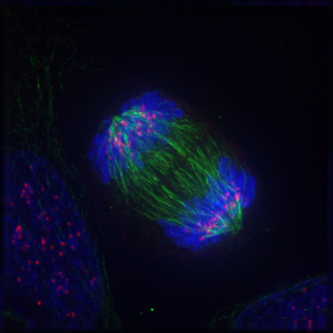
Can you guess what the colourful image in Figure 4.13.1 represents? It shows a eukaryotic cell during the process of cell division. In particular, the image shows the cell in a part of cell division called anaphase, where the DNA is being pulled to opposite ends of the cell. Normally, DNA is located in the nucleus of most human cells. The nucleus divides before the cell itself splits in two, and before the nucleus divides, the cell’s DNA is replicated (or copied). There must be two copies of the DNA so that each daughter cell will have a complete copy of the genetic material from the parent cell. How is the replicated DNA sorted and separated so that each daughter cell gets a complete set of the genetic material? To answer that question, you first need to know more about DNA and the forms it takes.
The Forms of DNA
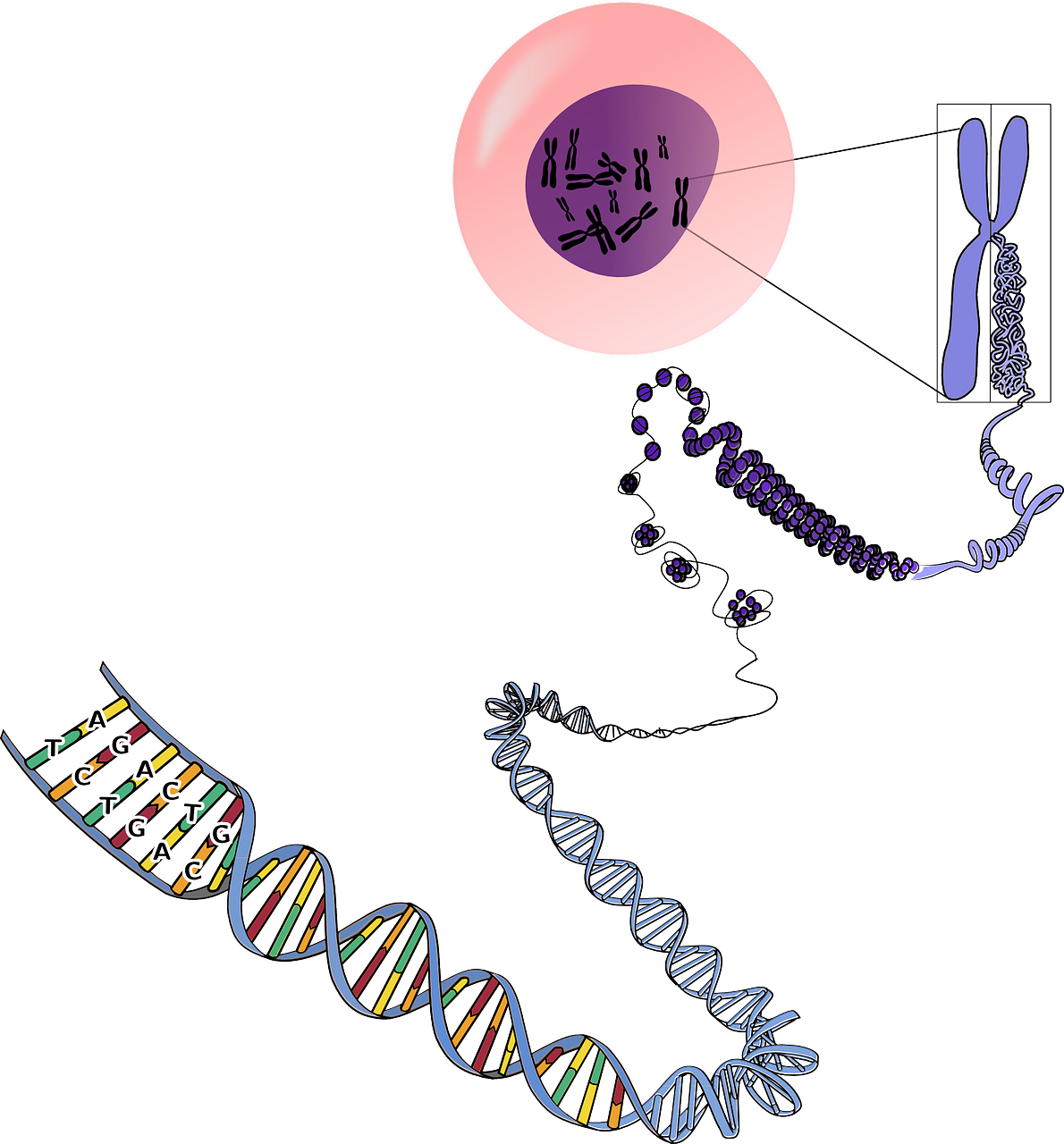
Except when a eukaryotic cell divides, its nuclear DNA exists as a grainy material called chromatin. Only once a cell is about to divide and its DNA has replicated does DNA condense and coil into the familiar X-shaped form of a chromosome, like the one shown below.
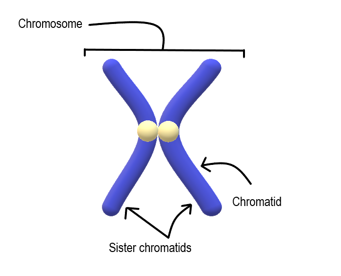
Most cells in the human body have two pairs of 23 different chromosomes, for a total of 46 chromosomes. Cells that have two pairs of chromosomes are called diploid. Because DNA has already replicated when it coils into a chromosome, each chromosome actually consists of two identical structures called sister chromatids. Sister chromatids are joined together at a region called a centromere.
Mitosis
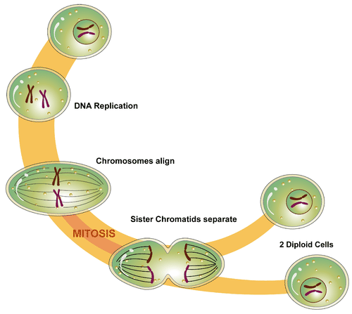
The process in which the nucleus of a eukaryotic cell divides is called mitosis. During mitosis, the two sister chromatids that make up each chromosome separate from each other and move to opposite poles of the cell. This is shown in the figure below.
Mitosis actually occurs in four phases. The phases are called prophase, metaphase, anaphase, and telophase.
Prophase
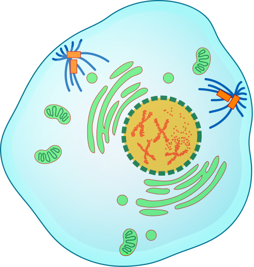
The first and longest phase of mitosis is prophase. During prophase, chromatin condenses into chromosomes, and the nuclear envelope (the membrane surrounding the nucleus) breaks down. In animal cells, the centrioles near the nucleus begin to separate and move to opposite poles of the cell. Centrioles are small organelles found only in eukaryotic cells. They help ensure that the new cells that form after cell division each contain a complete set of chromosomes. As the centrioles move apart, a spindle starts to form between them. The spindle consists of fibres made of microtubules.
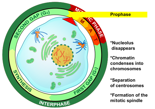
Metaphase
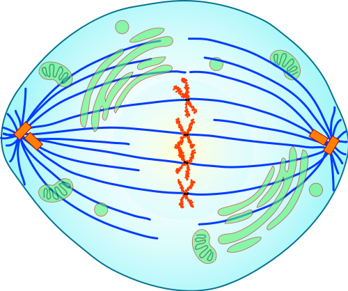
During metaphase, spindle fibres attach to the centromere of each pair of sister chromatids. As you can see in Figure 4.13.7, the sister chromatids line up at the equator (or center) of the cell. The spindle fibres ensure that sister chromatids will separate and go to different daughter cells when the cell divides.
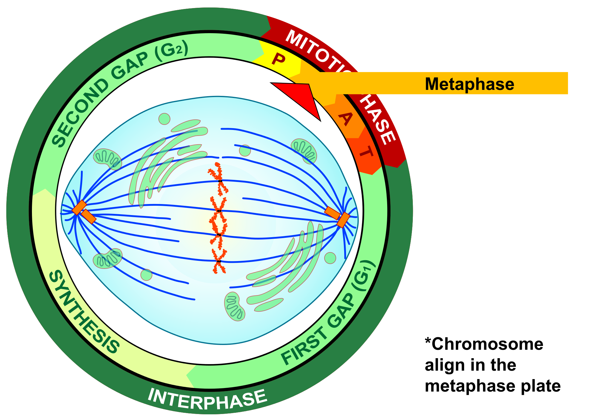
Anaphase
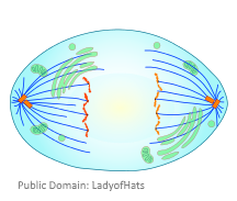
During anaphase, sister chromatids separate and the centromeres divide. The sister chromatids are pulled apart by the shortening of the spindle fibres. This is a little like reeling in a fish by shortening the fishing line. One sister chromatid moves to one pole of the cell, and the other sister chromatid moves to the opposite pole. At the end of anaphase, each pole of the cell has a complete set of chromosomes.
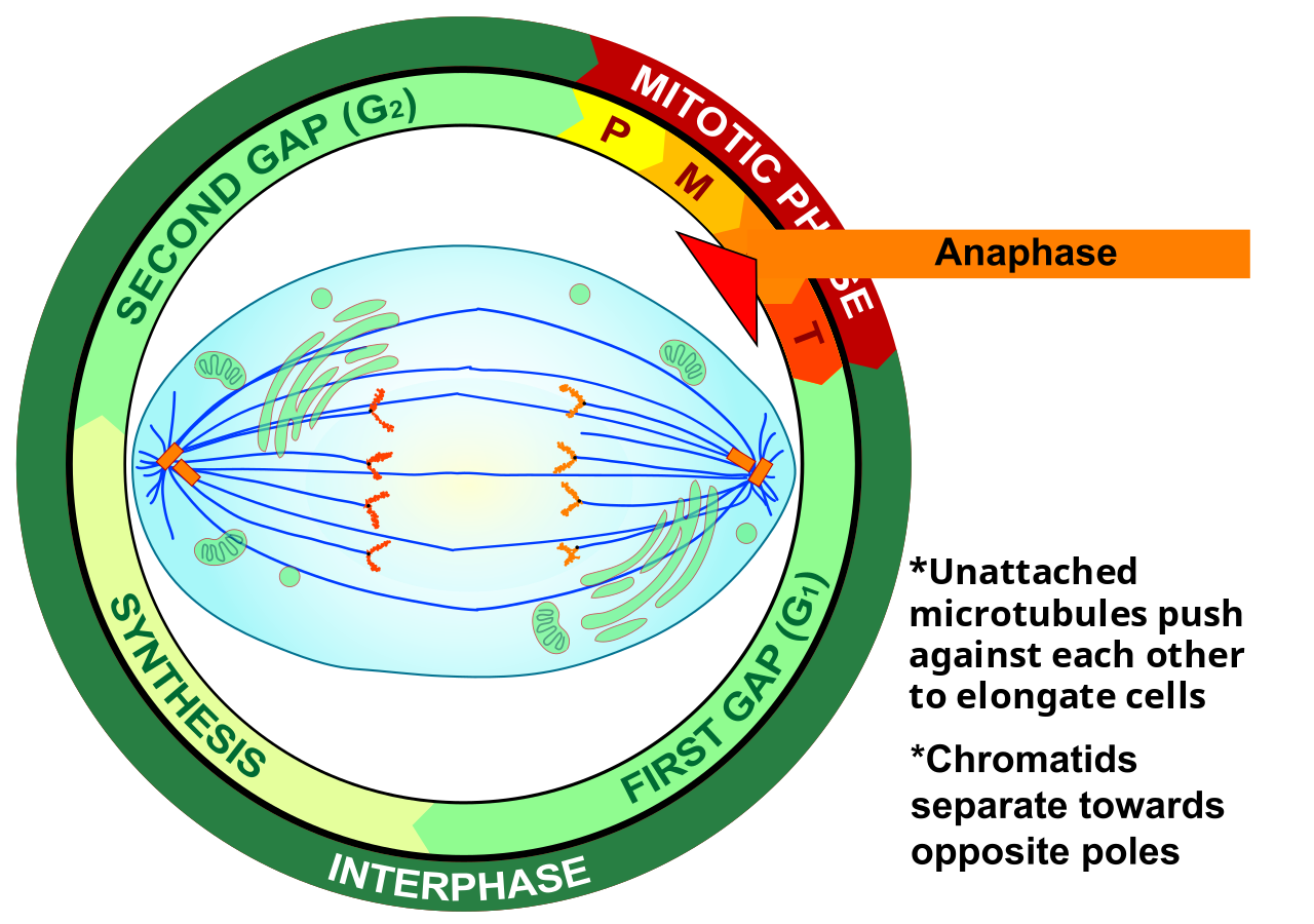
Telophase
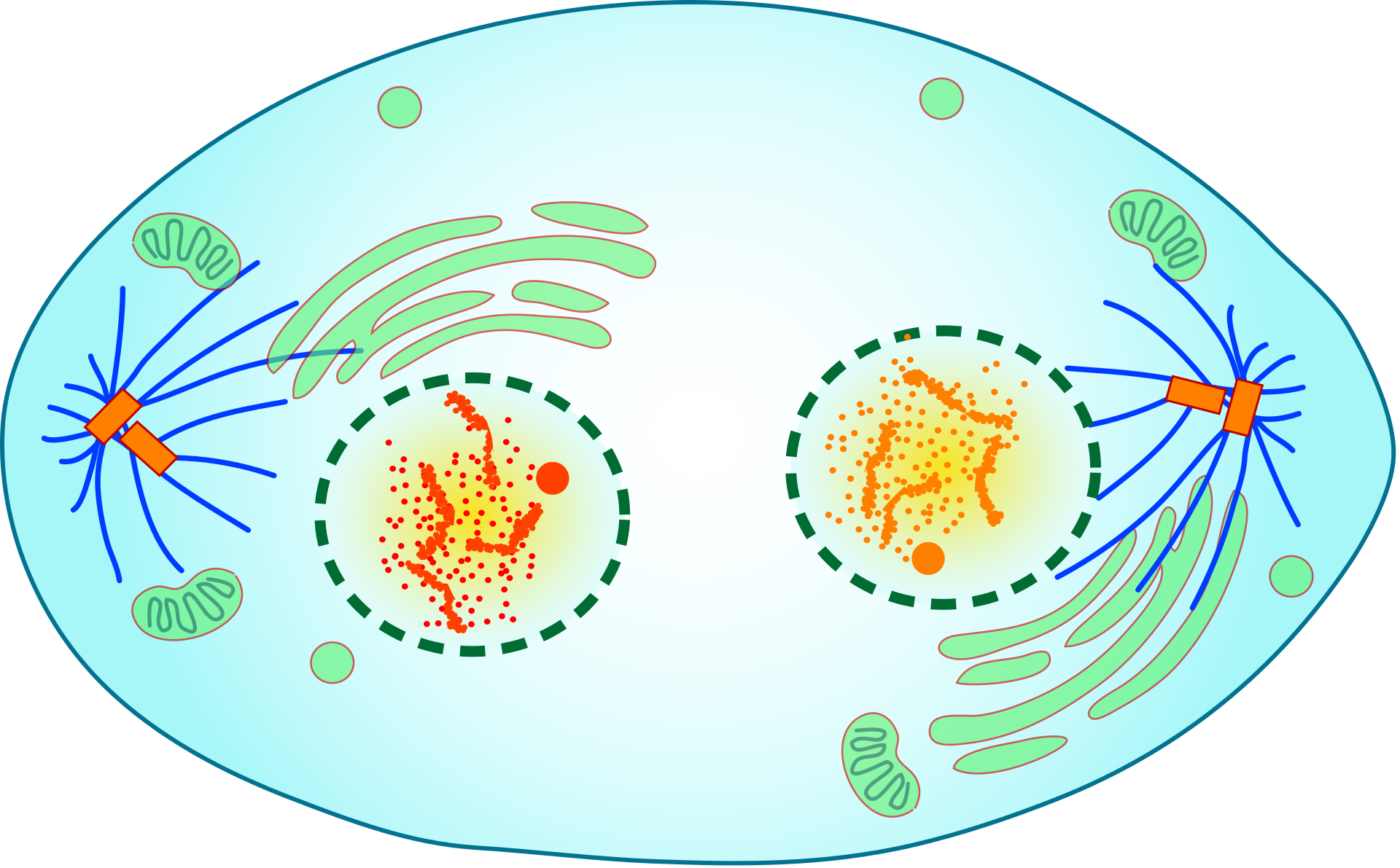
During telophase, the chromosomes begin to uncoil and form chromatin. This prepares the genetic material for directing the metabolic activities of the new cells. The spindle also breaks down, and new nuclear envelopes form.
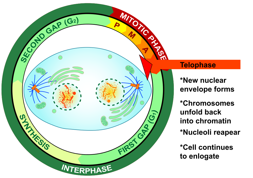
Cytokinesis

Cytokinesis is the final stage of cell division. During cytokinesis, the cytoplasm splits in two and the cell divides, as shown below. In animal cells, the plasma membrane of the parent cell pinches inward along the cell’s equator until two daughter cells form. Thus, the goal of mitosis and cytokinesis is now complete, because one parent cell has given rise to two daughter cells. The daughter cells have the same chromosomes as the parent cell.
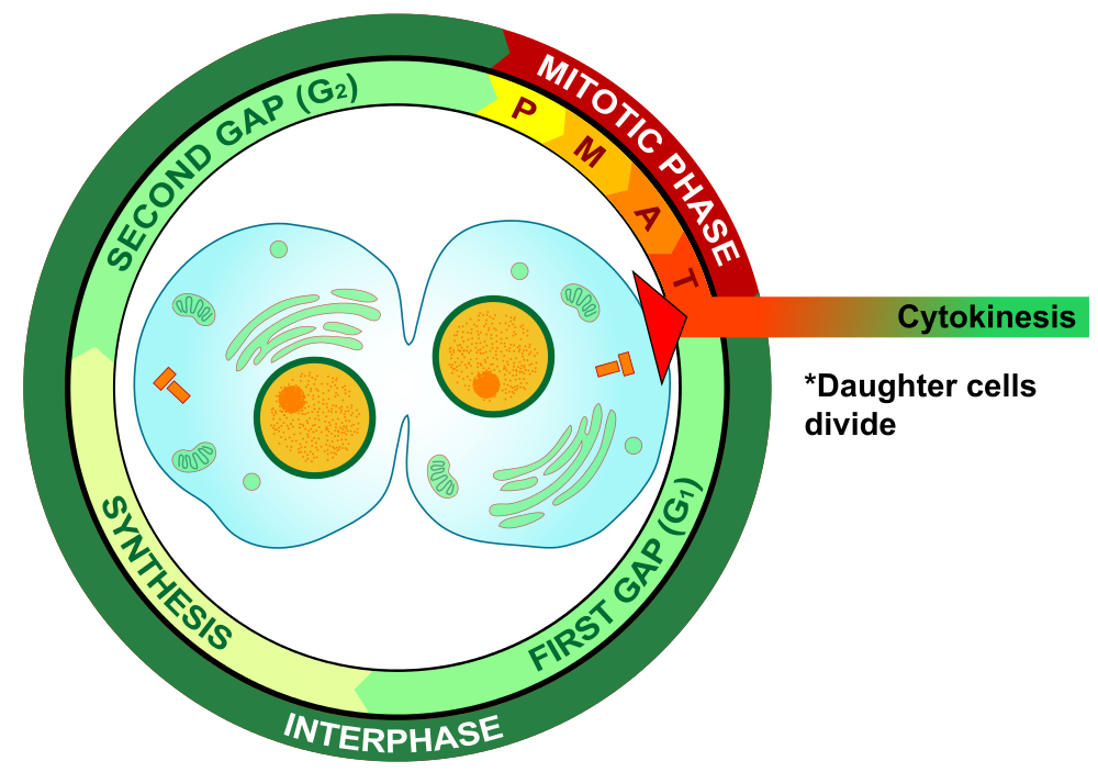
4.13 Summary
- Until a eukaryotic cell divides, its nuclear DNA exists as a grainy material called chromatin. After DNA replicates and the cell is about to divide, the DNA condenses and coils into the X-shaped form of a chromosome. Each chromosome actually consists of two sister chromatids, which are joined together at a centromere.
- Mitosis is the process during which the nucleus of a eukaryotic cell divides. During this process, sister chromatids separate from each other and move to opposite poles of the cell. This happens in four phases: prophase, metaphase, anaphase, and telophase.
- Cytokinesis is the final stage of cell division, during which the cytoplasm splits in two and two daughter cells form.
4.13 Review Questions
- Describe the different forms that DNA takes before and during cell division in a eukaryotic cell.
-
- Identify the four phases of mitosis in an animal cell, and summarize what happens during each phase.
- Order the diagrams of the stages of mitosis:
- Explain what happens during cytokinesis in an animal cell.
- What do you think would happen if the sister chromatids of one of the chromosomes did not separate during mitosis?
- True or False:
4.13 Explore More
https://www.youtube.com/watch?time_continue=3&v=C6hn3sA0ip0&feature=emb_logo
Mitosis, NDSU Virtual Cell Animations project (ndsuvirtualcell), 2012.
https://www.youtube.com/watch?time_continue=19&v=EA0qxhR2oOk&feature=emb_logo
Nondisjunction (Trisomy 21) - An Animated Tutorial, Kristen Koprowski, 2012.
Attributions
Figure 4.13.1
Anaphase_IF by Roy van Heesbeen on Wikimedia Commons is released into the public domain (https://en.wikipedia.org/wiki/Public_domain).
Figure 4.13.2
Chromosomes by OpenClipArt-Vectors on Pixabay is used under the Pixabay License (https://pixabay.com/service/license/).
Figure 4.13.3
Chromosome/ Chromatid/ Sister Chromatid by Christine Miller is released into the public domain (https://en.wikipedia.org/wiki/Public_domain).
Figure 4.13.4
Simple Mitosis by Mariana Ruiz Villarreal [LadyofHats] via CK-12 Foundation is used under a CC BY-NC 3.0 (https://creativecommons.org/licenses/by-nc/3.0/) license.
 ©CK-12 Foundation Licensed under
 ![]() • Terms of Use • Attribution
• Terms of Use • Attribution
Figure 4.13.5
Mitotic Prophase [tiny] by Mariana Ruiz Villarreal [LadyofHats] on Wikimedia Commons is released into the public domain (https://en.wikipedia.org/wiki/Public_domain).
Figure 4.13.6
Prophase Eukaryotic Mitosis by Mariana Ruiz Villarreal [LadyofHats] on Wikimedia Commons is released into the public domain (https://en.wikipedia.org/wiki/Public_domain).
Figure 4.13.7
Mitotic_Metaphase by Mariana Ruiz Villarreal [LadyofHats] on Wikimedia Commons is released into the public domain (https://en.wikipedia.org/wiki/Public_domain).
Figure 4.13.8
Metaphase Eukaryotic Mitosis by Mariana Ruiz Villarreal [LadyofHats] on Wikimedia Commons is released into the public domain (https://en.wikipedia.org/wiki/Public_domain).
Figure 4.13.9
Anaphase [adapted] by Mariana Ruiz Villarreal [LadyofHats] on Wikimedia Commons is released into the public domain (https://en.wikipedia.org/wiki/Public_domain).
Figure 4.13.10
Anaphase_eukaryotic_mitosis.svg by Mariana Ruiz Villarreal [LadyofHats] on Wikimedia Commons is released into the public domain (https://en.wikipedia.org/wiki/Public_domain).
Figure 4.13.11
Mitotic Telophase by Mariana Ruiz Villarreal [LadyofHats] on Wikimedia Commons is released into the public domain (https://en.wikipedia.org/wiki/Public_domain).
Figure 4.13.12
Telophase Eukaryotic Mitosis by Mariana Ruiz Villarreal [LadyofHats] on Wikimedia Commons is released into the public domain (https://en.wikipedia.org/wiki/Public_domain).
Figure 4.13.13
Mitotic Cytokinesis by Mariana Ruiz Villarreal [LadyofHats] on Wikimedia Commons is released into the public domain (https://en.wikipedia.org/wiki/Public_domain).
Figure 4.13.14
Cytokinesis Eukaryotic Mitosis by Mariana Ruiz Villarreal [LadyofHats] on Wikimedia Commons is released into the public domain (https://en.wikipedia.org/wiki/Public_domain).
References
Koprowski, K., Cabey, R. [Kristen Koprowski]. (2012). Nondisjunction (Trisomy 21) - An Animated Tutorial. YouTube. https://www.youtube.com/watch?v=EA0qxhR2oOk&feature=youtu.be
NDSU Virtual Cell Animations project [ndsuvirtualcell]. (2012). Mitosis. YouTube. https://www.youtube.com/watch?v=C6hn3sA0ip0&t=21s
The space occurring between two or more membranes. In cell biology, it's most commonly described as the region between the inner membrane and the outer membrane of a mitochondrion or a chloroplast.
A type of immune cell that stimulates killer T cells, macrophages, and B cells to make immune responses. A helper T cell is a type of white blood cell and a type of lymphocyte.
Image shows a diagram of the hormones secreted by the thyroid gland, and how it is both controlled by and acting upon in a negative feedback the hypothalamus and the anterior pituitary gland.
Image shows two photos of the eyes of the same patient. In the first photo, the patient's right eyelid is drooping substantially. In the second photograph the same eyelid is drooping, but not as drastically.
Image shows a picture of a child with very curly hair.
Image shows a diagram of the layers of the epidermis. The outermost layer is the stratum corneum, below that is the stratum lucidum, below that the stratum granulosum, below that the stratum spinosum, below that the stratum basale, and then a basement membrane which connects the dermis to the epidermis.

