17.3 Lymphatic System
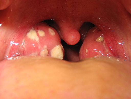
Tonsillitis
The white patches on either side of the throat in Figure 17.3.1 are signs of tonsillitis. The tonsils are small structures in the throat that are very common sites of infection. The white spots on the tonsils pictured here are evidence of infection. The patches consist of large amounts of dead bacteria, cellular debris, and white blood cells — in a word: pus. Children with recurrent tonsillitis may have their tonsils removed surgically to eliminate this type of infection. The tonsils are organs of the lymphatic system.
What Is the Lymphatic System?
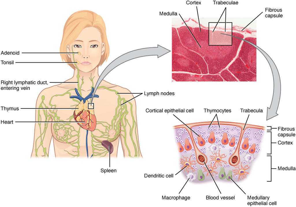
The lymphatic system is a collection of organs involved in the production, maturation, and harboring of white blood cells called lymphocytes. It also includes a network of vessels that transport or filter the fluid known as lymph in which lymphocytes circulate. Figure 17.3.2 shows major lymphatic vessels and other structures that make up the lymphatic system. Besides the tonsils, organs of the lymphatic system include the thymus, the spleen, and hundreds of lymph nodes distributed along the lymphatic vessels.
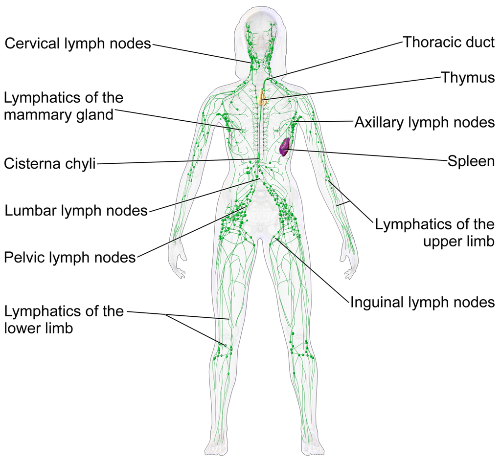
The lymphatic vessels form a transportation network similar in many respects to the blood vessels of the cardiovascular system. However, unlike the cardiovascular system, the lymphatic system is not a closed system. Instead, lymphatic vessels carry lymph in a single direction — always toward the upper chest, where the lymph empties from lymphatic vessels into blood vessels.
Cardiovascular Function of the Lymphatic System
The return of lymph to the bloodstream is one of the major functions of the lymphatic system. When blood travels through capillaries of the cardiovascular system, it is under pressure, which forces some of the components of blood (such as water, oxygen, and nutrients) through the walls of the capillaries and into the tissue spaces between cells, forming tissue fluid, also called interstitial fluid (see Figure 17.3.3). Interstitial fluid bathes and nourishes cells, and also absorbs their waste products. Much of the water from interstitial fluid is reabsorbed into the capillary blood by osmosis. Most of the remaining fluid is absorbed by tiny lymphatic vessels called lymph capillaries. Once interstitial fluid enters the lymphatic vessels, it is called lymph. Lymph is very similar in composition to blood plasma. Besides water, lymph may contain proteins, waste products, cellular debris, and pathogens. It also contains numerous white blood cells, especially the subset of white blood cells known as lymphocytes. In fact, lymphocytes are the main cellular components of lymph.
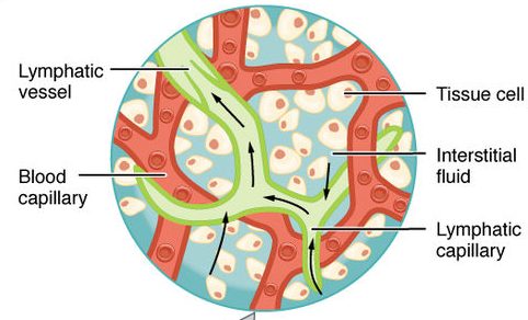
The lymph that enters lymph capillaries in tissues is transported through the lymphatic vessel network to two large lymphatic ducts in the upper chest. From there, the lymph flows into two major veins (called subclavian veins) of the cardiovascular system. Unlike blood, lymph is not pumped through its network of vessels. Instead, lymph moves through lymphatic vessels via a combination of contractions of the vessels themselves and the forces applied to the vessels externally by skeletal muscles, similarly to how blood moves through veins. Lymphatic vessels also contain numerous valves that keep lymph flowing in just one direction, thereby preventing backflow.
Digestive Function of the Lymphatic System
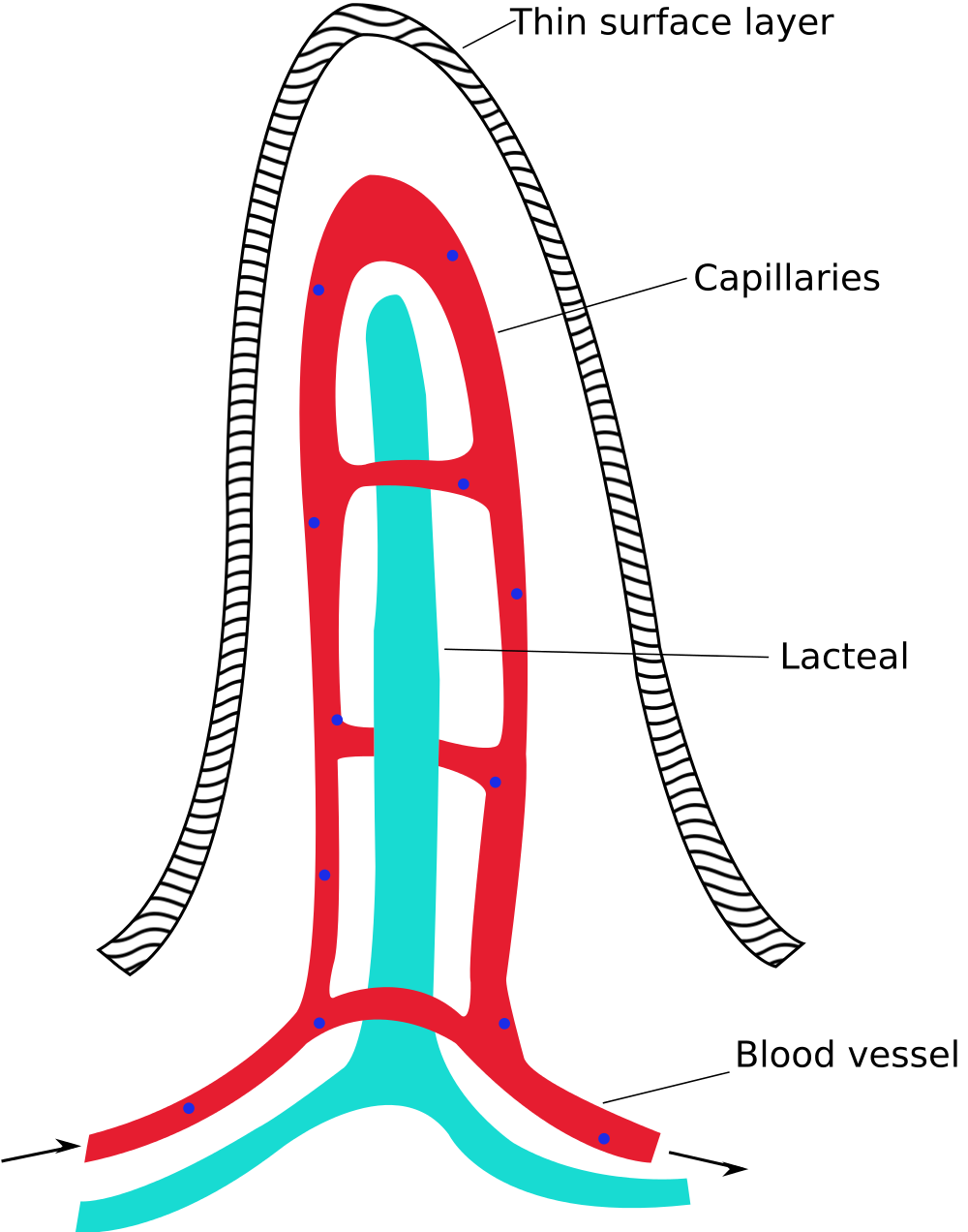
Lymphatic vessels called lacteals (see Figure 17.3.4) are present in the lining of the gastrointestinal tract, mainly in the small intestine. Each tiny villus in the lining of the small intestine has an internal bed of capillaries and lacteals. The capillaries absorb most nutrients from the digestion of food into the blood. The lacteals absorb mainly fatty acids from lipid digestion into the lymph, forming a fatty-acid-enriched fluid called chyle. Vessels of the lymphatic network then transport chyle from the small intestine to the main lymphatic ducts in the chest, from which it drains into the blood circulation. The nutrients in chyle then circulate in the blood to the liver, where they are processed along with the other nutrients that reach the liver directly via the bloodstream.
Immune Function of the Lymphatic System
The primary immune function of the lymphatic system is to protect the body against pathogens and cancerous cells. This function of the lymphatic system is centred on the production, maturation, and circulation of lymphocytes. Lymphocytes are leukocytes that are involved in the adaptive immune system. They are responsible for the recognition of — and tailored defense against — specific pathogens or tumor cells. Lymphocytes may also create a lasting memory of pathogens, so they can be attacked quickly and strongly if they ever invade the body again. In this way, lymphocytes bring about long-lasting immunity to specific pathogens.
There are two major types of lymphocytes, called B cells and T cells. Both B cells and T cells are involved in the adaptive immune response, but they play different roles.
Production and Maturation of Lymphocytes
Like all other types of blood cells (including erythrocytes), both B cells and T cells are produced from stem cells in the red marrow inside bones. After lymphocytes first form, they must go through a complicated maturation process before they are ready to search for pathogens. In this maturation process, they “learn” to distinguish self from non-self. Only those lymphocytes that successfully complete this maturation process go on to actually fight infections by pathogens.
B cells mature in the bone marrow, which is why they are called B cells. After they mature and leave the bone marrow, they travel first to the circulatory system and then enter the lymphatic system to search for pathogens. T cells, on the other hand, mature in the thymus, which is why they are called T cells. The thymus is illustrated in Figure 17.3.5. It is a small lymphatic organ in the chest that consists of an outer cortex and inner medulla, all surrounded by a fibrous capsule. After maturing in the thymus, T cells enter the rest of the lymphatic system to join B cells in the hunt for pathogens. The bone marrow and thymus are called primary lymphoid organs because of their role in the production and/or maturation of lymphocytes.
Lymphocytes in Secondary Lymphoid Organs
The tonsils, spleen, and lymph nodes are referred to as secondary lymphoid organs. These organs do not produce or mature lymphocytes. Instead, they filter lymph and store lymphocytes. It is in these secondary lymphoid organs that pathogens (or their antigens) activate lymphocytes and initiate adaptive immune responses. Activation leads to cloning of pathogen-specific lymphocytes, which then circulate between the lymphatic system and the blood, searching for and destroying their specific pathogens by producing antibodies against them.
Tonsils
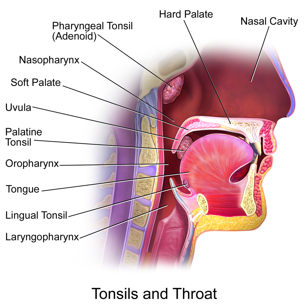
There are four pairs of human tonsils. Three of the four are shown in Figure 17.3.6. The fourth pair, called tubal tonsils, is located at the back of the nasopharynx. The palatine tonsils are the tonsils that are visible on either side of the throat. All four pairs of tonsils encircle a part of the anatomy where the respiratory and gastrointestinal tracts intersect, and where pathogens have ready access to the body. This ring of tonsils is called Waldeyer’s ring.
Spleen
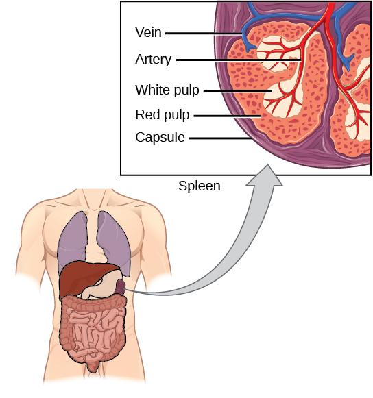
The spleen (Figure 17.3.7) is the largest of the secondary lymphoid organs, and is centrally located in the body. Besides harboring lymphocytes and filtering lymph, the spleen also filters blood. Most dead or aged erythrocytes are removed from the blood in the red pulp of the spleen. Lymph is filtered in the white pulp of the spleen. In the fetus, the spleen has the additional function of producing red blood cells. This function is taken over by bone marrow after birth.
Lymph Nodes
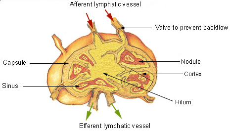
Each lymph node is a small, but organized collection of lymphoid tissue (see Figure 17.3.8) that contains many lymphocytes. Lymph nodes are located at intervals along the lymphatic vessels, and lymph passes through them on its way back to the blood.
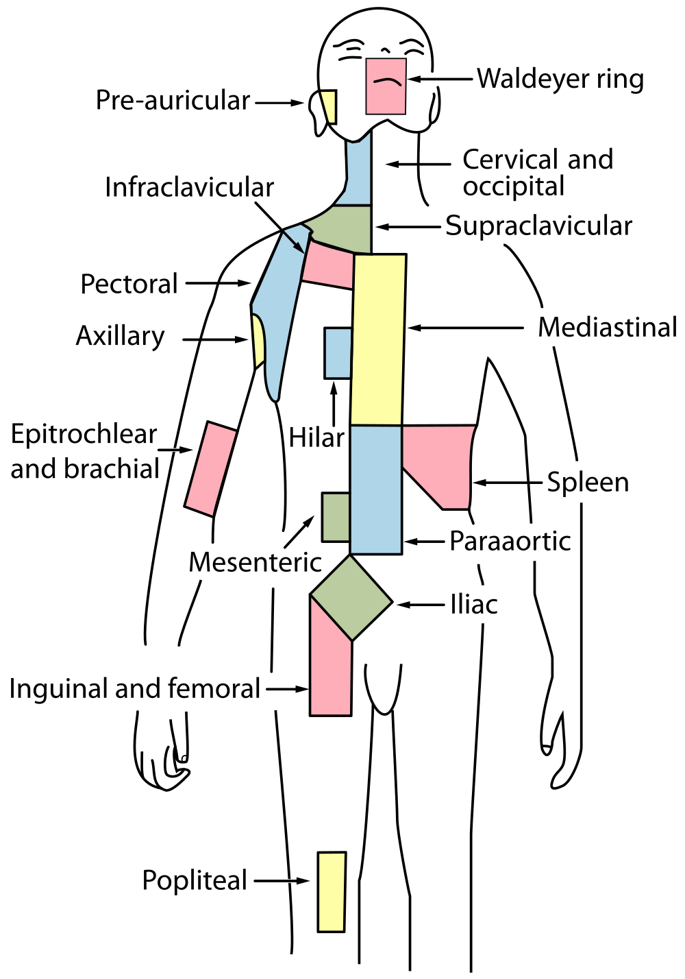
There are at least 500 lymph nodes in the human body. Many of them are clustered at the base of the limbs and in the neck. Figure 17.3.9 shows the major lymph node concentrations, and includes the spleen and the region named Waldeyer’s ring, which consists of the tonsils.
Feature: Myth vs. Reality
When lymph nodes become enlarged and tender to the touch, they are obvious signs of immune system activity. Because it is easy to see and feel swollen lymph nodes, they are one way an individual can monitor his or her own health. To be useful in this way, it is important to know the myths and realities about swollen lymph nodes.
Myth
|
Reality
|
| “You should see a doctor immediately whenever you have swollen lymph nodes.” | Lymph nodes are constantly filtering lymph, so it is expected that they will change in size with varying amounts of debris or pathogens that may be present. A minor, unnoticed infection may cause swollen lymph nodes that may last for a few weeks. Generally, lymph nodes that return to their normal size within two or three weeks are not a cause for concern. |
| “Swollen lymph nodes mean you have a bacterial infection.” | Although an infection is the most common cause of swollen lymph nodes, not all infections are caused by bacteria. Mononucleosis, for example, commonly causes swollen lymph nodes, and it is caused by viruses. There are also other causes of swollen lymph nodes besides infections, such as cancer and certain medications. |
| “A swollen lymph node means you have cancer.” | Cancer is far less likely to be the cause of a swollen lymph node than is an infection. However, if a lymph node remains swollen longer than a few weeks — especially in the absence of an apparent infection — you should have your doctor check it. |
| “Cancer in a lymph node always originates somewhere else. There is no cancer of the lymph nodes.” | Cancers do commonly spread from their site of origin to nearby lymph nodes and then to other organs, but cancer may also originate in the lymph nodes. This type of cancer is called lymphoma. |
17.3 Summary
- The lymphatic system is a collection of organs involved in the production, maturation, and harboring of leukocytes called lymphocytes. It also includes a network of vessels that transport or filter the fluid called lymph in which lymphocytes circulate.
- The return of lymph to the bloodstream is one of the functions of the lymphatic system. Lymph flows from tissue spaces — where it leaks out of blood vessels — to the subclavian veins in the upper chest, where it is returned to the cardiovascular system. Lymph is similar in composition to blood plasma. Its main cellular components are lymphocytes.
- Lymphatic vessels called lacteals are found in villi that line the small intestine. Lacteals absorb fatty acids from the digestion of lipids in the digestive system. The fatty acids are then transported through the network of lymphatic vessels to the bloodstream.
- The primary immune function of the lymphatic system is to protect the body against pathogens and cancerous cells. It is responsible for producing mature lymphocytes and circulating them in lymph. Lymphocytes, which include B cells and T cells, are the subset of white blood cells involved in adaptive immune responses. They may create a lasting memory of and immunity to specific pathogens.
- All lymphocytes are produced in bone marrow and then go through a process of maturation in which they “learn” to distinguish self from non-self. B cells mature in the bone marrow, and T cells mature in the thymus. Both the bone marrow and thymus are considered primary lymphatic organs.
- Secondary lymphatic organs include the tonsils, spleen, and lymph nodes. There are four pairs of tonsils that encircle the throat. The spleen filters blood, as well as lymph. There are hundreds of lymph nodes located in clusters along the lymphatic vessels. All of these secondary organs filter lymph and store lymphocytes, so they are sites where pathogens encounter and activate lymphocytes and initiate adaptive immune responses.
17.3 Review Questions
- What is the lymphatic system?
-
- Summarize the immune function of the lymphatic system.
- Explain the difference between lymphocyte maturation and lymphocyte activation.
17.3 Explore More
What is Lymphoedema or Lymphedema? Compton Care, 2016.
Spleen physiology What does the spleen do in 2 minutes, Simple Nursing, 2015.
How to check your lymph nodes, University Hospitals Bristol and Weston NHS FT, 2020.
Attributions
Figure 17.3.1
512px-Tonsillitis by Michaelbladon at English Wikipedia on Wikimedia Commons is in the public domain (https://en.wikipedia.org/wiki/Public_domain). (Transferred from en.wikipedia to Commons by Kauczuk)
Figure 17.3.2
Blausen_0623_LymphaticSystem_Female by BruceBlaus on Wikimedia Commons is used under a CC BY 3.0 (https://creativecommons.org/licenses/by/3.0) license.
Figure 17.3.3
2201_Anatomy_of_the_Lymphatic_System (cropped) by OpenStax College on Wikimedia Commons is used under a CC BY 3.0 (https://creativecommons.org/licenses/by/3.0) license.
Figure 17.3.4
1000px-Intestinal_villus_simplified.svg by Snow93 on Wikimedia Commons is in the public domain (https://en.wikipedia.org/wiki/Public_domain).
Figure 17.3.5
2206_The_Location_Structure_and_Histology_of_the_Thymus by OpenStax College on Wikimedia Commons is used under a CC BY 3.0 (https://creativecommons.org/licenses/by/3.0) license.
Figure 17.3.6
Blausen_0861_Tonsils&Throat_Anatomy2 by BruceBlaus on Wikimedia Commons is used under a CC BY 3.0 (https://creativecommons.org/licenses/by/3.0) license.
Figure 17.3.7
Figure_42_02_14 by CNX OpenStax on Wikimedia Commons is used under a CC BY 4.0 (https://creativecommons.org/licenses/by/4.0) license.
Figure 17.3.8
Illu_lymph_node_structure by NCI/ SEER Training on Wikimedia Commons is in the public domain (https://en.wikipedia.org/wiki/Public_domain). (Archives: https://web.archive.org/web/20070311015818/http://training.seer.cancer.gov/module_anatomy/unit8_2_lymph_compo1_nodes.html)
Figure 17.3.9
1000px-Lymph_node_regions.svg by Fred the Oyster (derivative work) on Wikimedia Commons is in the public domain (https://en.wikipedia.org/wiki/Public_domain). (Original by NCI/ SEER Training)
References
Betts, J. G., Young, K.A., Wise, J.A., Johnson, E., Poe, B., Kruse, D.H., Korol, O., Johnson, J.E., Womble, M., DeSaix, P. (2013, June 19). Figure 21.2 Anatomy of the lymphatic system [digital image]. In Anatomy and Physiology (Section 21.1). OpenStax. https://openstax.org/books/anatomy-and-physiology/pages/21-1-anatomy-of-the-lymphatic-and-immune-systems
Betts, J. G., Young, K.A., Wise, J.A., Johnson, E., Poe, B., Kruse, D.H., Korol, O., Johnson, J.E., Womble, M., DeSaix, P. (2013, June 19). Figure 21.7 Location, structure, and histology of the thymus [digital image]. In Anatomy and Physiology (Section 21.1). OpenStax. https://openstax.org/books/anatomy-and-physiology/pages/21-1-anatomy-of-the-lymphatic-and-immune-systems
Blausen.com Staff. (2014). Medical gallery of Blausen Medical 2014″. WikiJournal of Medicine 1 (2). DOI:10.15347/wjm/2014.010. ISSN 2002-4436
Compton Care. (2016, March 7). What is lymphoedema or lymphedema? YouTube. https://www.youtube.com/watch?v=RMLPwOiYnII&feature=youtu.be
OpenStax. (2016, May 27) Figure 14. The spleen is similar to a lymph node but is much larger and filters blood instead of lymph [digital image]. In Open Stax, Biology (Section 42.2). OpenStax CNX. https://cnx.org/contents/GFy_h8cu@10.8:etZobsU-@6/Adaptive-Immune-Response
Simple Nursing. (2015, June 28). Spleen physiology What does the spleen do in 2 minutes. YouTube. https://www.youtube.com/watch?v=ah74jT00jBA&feature=youtu.be
University Hospitals Bristol and Weston NHS FT. (2020, May 13). How to check your lymph nodes. YouTube. https://www.youtube.com/watch?v=L4KexZZAdyA&feature=youtu.be
Created by: CK-12/Adapted by Christine Miller

Like Pushing a Humvee Uphill
You can tell by their faces that these airmen (Figure 4.8.1) are expending a lot of energy trying to push this Humvee up a slope. The men are participating in a competition that tests their brute strength against that of other teams. The Humvee weighs about 13 thousand pounds (about 5,897 kilograms), so it takes every ounce of energy they can muster to move it uphill against the force of gravity. Transport of some substances across a plasma membrane is a little like pushing a Humvee uphill — it can't be done without adding energy.
What Is Active Transport?
Some substances can pass into or out of a cell across the plasma membrane without any energy required because they are moving from an area of higher concentration to an area of lower concentration. This type of transport is called passive transport. Other substances require energy to cross a plasma membrane, often because they are moving from an area of lower concentration to an area of higher concentration, against the concentration gradient. This type of transport is called active transport. The energy for active transport comes from the energy-carrying molecule called ATP (adenosine triphosphate). Active transport may also require proteins called pumps, which are embedded in the plasma membrane. Two types of active transport are membrane pumps (such as the sodium-potassium pump) and vesicle transport.
The Sodium-Potassium Pump
The sodium-potassium pump is a mechanism of active transport that moves sodium ions out of the cell and potassium ions into the cells — in all the trillions of cells in the body! Both ions are moved from areas of lower to higher concentration, so energy is needed for this "uphill" process. The energy is provided by ATP. The sodium-potassium pump also requires carrier proteins. Carrier proteins bind with specific ions or molecules, and in doing so, they change shape. As carrier proteins change shape, they carry the ions or molecules across the membrane. Figure 4.8.2 shows in greater detail how the sodium-potassium pump works, as well as the specific roles played by carrier proteins in this process.
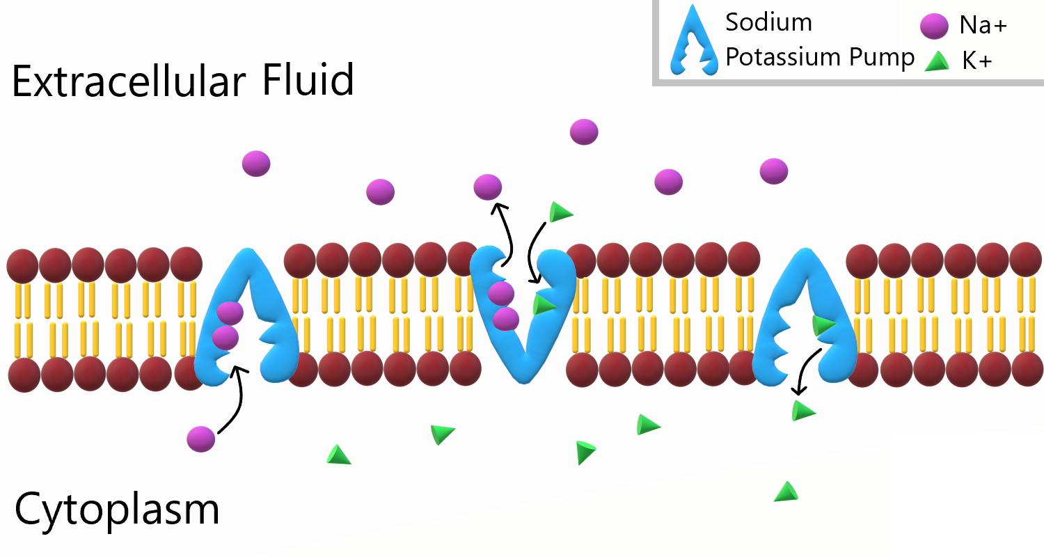
To appreciate the importance of the sodium-potassium pump, you need to know more about the roles of sodium and potassium in the body. Both are essential dietary minerals. You need to get them from the foods you eat. Both sodium and potassium are also electrolytes, which means they dissociate into ions (charged particles) in solution, allowing them to conduct electricity. Normal body functions require a very narrow range of concentrations of sodium and potassium ions in body fluids, both inside and outside of cells.
- Sodium is the principal ion in the fluid outside of cells. Normal sodium concentrations are about ten times higher outside of cells than inside of cells. To move sodium out of the cell is moving it against the concentration gradient
- Potassium is the principal ion in the fluid inside of cells. Normal potassium concentrations are about 30 times higher inside of cells than outside of cells. To move potassium into the cell is moving it against the concentration gradient.
These differences in concentration create an electrical and chemical gradient across the cell membrane, called the membrane potential. Tightly controlling the membrane potential is critical for vital body functions, including the transmission of nerve impulses and contraction of muscles. A large percentage of the body's energy goes to maintaining this potential across the membranes of its trillions of cells with the sodium-potassium pump.
Vesicle Transport
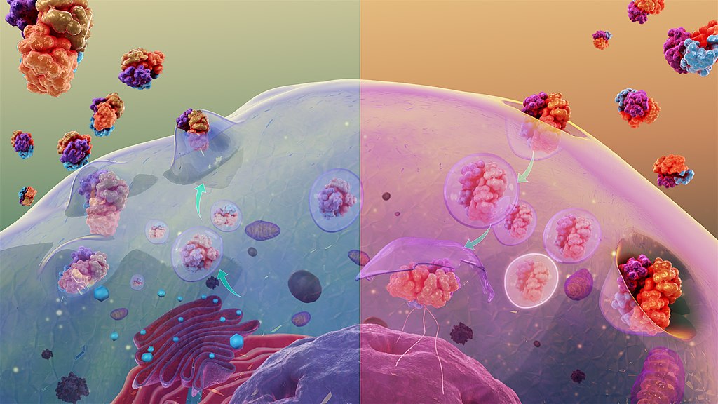
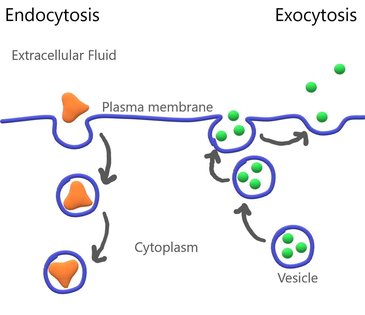
Some molecules, such as proteins, are too large to pass through the plasma membrane, regardless of their concentration inside and outside the cell. Very large molecules cross the plasma membrane with a different sort of help, called vesicle transport. Vesicle transport requires energy input from the cell, so it is also a form of active transport. There are two types of vesicle transport: endocytosis and exocytosis. Both types are shown in Figure 4.8.3.
Endocytosis
Endocytosis is a type of vesicle transport that moves a substance into the cell. The plasma membrane completely engulfs the substance, a vesicle pinches off from the membrane, and the vesicle carries the substance into the cell. When an entire cell or other solid particle is engulfed, the process is called phagocytosis. When fluid is engulfed, the process is called pinocytosis.
Exocytosis
Exocytosis is a type of vesicle transport that moves a substance out of the cell (exo-, like "exit"). A vesicle containing the substance moves through the cytoplasm to the cell membrane. Because the vesicle membrane is a phospholipid bilayer like the plasma membrane, the vesicle membrane fuses with the cell membrane, and the substance is released outside the cell.
Feature: My Human Body
Maintaining the proper balance of sodium and potassium in body fluids by active transport is necessary for life itself, so it's no surprise that getting the right balance of sodium and potassium in the diet is important for good health. Imbalances may increase the risk of high blood pressure, heart disease, diabetes, and other disorders.
If you are like the majority of North Americans, sodium and potassium are out of balance in your diet. You are likely to consume too much sodium and too little potassium. Follow these guidelines to help ensure that these minerals are balanced in the foods you eat:
- Total sodium intake should be less than 2,300 mg/day. Most salt in the diet is found in processed foods, or added with a salt shaker. Stop adding salt and start checking food labels for sodium content. Foods considered low in sodium have less than 140 mg/serving (or 5 per cent daily value).
- Total potassium intake should be 4,700 mg/day. It's easy to add potassium to the diet by choosing the right foods — and there are plenty of choices! Most fruits and vegetables are high in potassium. Potatoes, bananas, oranges, apricots, plums, leafy greens, tomatoes, lima beans, and avocado are especially good sources. Other foods with substantial amounts of potassium are fish, meat, poultry, and whole grains. The collage below shows some of these potassium-rich foods.
Figure 4.8.5 Potassium power!
4.8 Summary
- Active transport requires energy to move substances across a plasma membrane, often because the substances are moving from an area of lower concentration to an area of higher concentration, or because of their large size. Two types of active transport are membrane pumps (such as the sodium-potassium pump) and vesicle transport.
- The sodium-potassium pump is a mechanism of active transport that moves sodium ions out of the cell and potassium ions into the cell against a concentration gradient, in order to maintain the proper concentrations of ions, both inside and outside the cell, and to thereby control membrane potential.
- Vesicle transport is a type of active transport that uses vesicles to move large molecules into or out of cells.
4.8 Review Questions
- Define active transport.
-
- What is the sodium-potassium pump? Why is it so important?
- The drawing below shows the fluid inside and outside of a cell. The dots represent molecules of a substance needed by the cell. Explain which type of transport — active or passive — is needed to move the molecules into the cell.
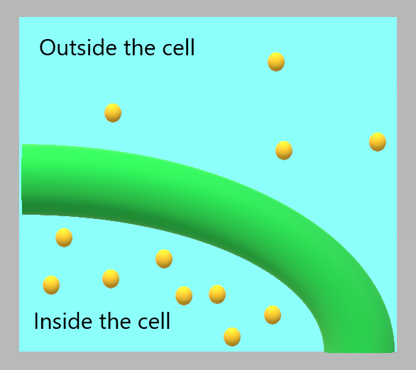
Figure 4.8.6 Use this image to answer question #4 - What are the similarities and differences between phagocytosis and pinocytosis?
- What is the functional significance of the shape change of the carrier protein in the sodium-potassium pump after the sodium ions bind?
- A potentially deadly poison derived from plants called ouabain blocks the sodium-potassium pump and prevents it from working. What do you think this does to the sodium and potassium balance in cells? Explain your answer.
4.8 Explore More
https://www.youtube.com/watch?v=Z_mXDvZQ6dU&feature=emb_logo
Neutrophil Phagocytosis - White Blood Cell Eats Staphylococcus Aureus Bacteria,
ImmiflexImmuneSystem, 2013.
https://www.youtube.com/watch?v=Ptmlvtei8hw
Cell Transport, The Amoeba Sisters, 2016.
Attributions
Figure 4.8.1
Humvee challenge by Airman 1st Class Collin Schmidt on Wikimedia Commons is released into the public domain (https://en.wikipedia.org/wiki/Public_domain).
Figure 4.8.2
Sodium Potassium Pump by Christine Miller is used under a CC BY 4.0 (https://creativecommons.org/licenses/by/4.0/) license.
Figure 4.8.3
Cytosis by Manu5 on Wikimedia Commons is used under a CC BY-SA 4.0 (https://creativecommons.org/licenses/by-sa/4.0) license.
Figure 4.8.4
Endocytosis and Exocytosis by Christine Miller is used under a CC BY 4.0 (https://creativecommons.org/licenses/by/4.0/) license.
Figure 4.8.5
- Canteloupes. Image Number K7355-11 by Scott Bauer/ USDA on Wikimedia Commons is in the public domain (https://en.wikipedia.org/wiki/Public_domain).
- Spinach by chiara conti on Unsplash is used under the Unsplash license (https://unsplash.com/license).
- Eleven long purple eggplants by JVRKPRASAD on Wikimedia commons is used under a CC BY-SA 4.0 (https://creativecommons.org/licenses/by-sa/4.0/deed.en) license.
- Bananas by Marco Antonio Victorino on Pexels is used under the Pexels license (https://www.pexels.com/license/).
- Potato picking by Nic D on Unsplash is used under the Unsplash license (https://unsplash.com/license).
- Maldives by Sebastian Pena Lambarri on Unsplash is used under the Unsplash license (https://unsplash.com/license).
Figure 4.8.6
Active Transport by Christine Miller is released into the public domain (https://en.wikipedia.org/wiki/Public_domain).
References
Amoeba Sisters. (2016, June 24). Cell transport [digital image]. YouTube. https://www.youtube.com/watch?v=Ptmlvtei8hw&feature=youtu.be
ImmiflexImmuneSystem. (2013). Neutrophil phagocytosis - White blood cell eats staphylococcus aureus bacteria. YouTube. https://www.youtube.com/watch?v=Z_mXDvZQ6dU
Mayo Clinic Staff. (n.d.). Diabetes [online]. MayoClinic.org. https://www.mayoclinic.org/diseases-conditions/diabetes/symptoms-causes/syc-20371444
Mayo Clinic Staff. (n.d.). High blood pressure (hypertension) [online]. MayoClinic.org. https://www.mayoclinic.org/diseases-conditions/high-blood-pressure/symptoms-causes/syc-20373410
Mayo Clinic Staff. (n.d.). Heart disease [online]. MayoClinic.org. https://www.mayoclinic.org/diseases-conditions/heart-disease/symptoms-causes/syc-20353118
Wikipedia contributors. (2020, June 19). Ouabain. In Wikipedia. https://en.wikipedia.org/w/index.php?title=Ouabain&oldid=963440756
A hollow, tube-like structure through which blood flows in the cardiovascular system; vein, artery, or capillary.
Refers to the body system consisting of the heart, blood vessels and the blood. Blood contains oxygen and other nutrients which your body needs to survive. The body takes these essential nutrients from the blood.
The smallest type of blood vessel that connects arterioles and venules and that transfers substances between blood and tissues.
Image shows a diagram of polymerase chain reaction, which occurs in three steps: 1) Denaturing, which separates the strands of DNA 2) Annealing, in which primers bind to the template DNA strands 3) Extension, in which Taq polymerase synthesizes new DNA strands from the original, now separated strands.
Image shows a diagram of the three stages of Translation. In Initiation, mRNA, a ribosome and tRNA carry methionine form a complex. In elongation, the ribosome moves along the mRNA, as tRNA with matching anticodons bring and then drop off the corresponding amino acids. In termination, a stop codon is reached, and the entire complex disassembles, and releases the newly synthesize polypeptide.

Eek!
Being bitten on the nose by an eel certainly qualifies as a frightening experience! The fear this man is experiencing produces the same physiological responses in most people — racing heart, rapid breathing, clammy hands. These and other fight-or-flight responses prepare the body to either defend itself or run away from danger. Why does fear elicit these changes in the body? The responses occur in large part because of hormones secreted by the adrenal glands.
Introduction to the Adrenal Glands
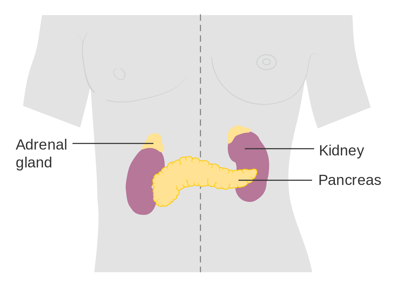
The adrenal glands are endocrine glands that produce a variety of hormones. Adrenal hormones include the fight-or-flight hormone adrenaline and the steroid hormone cortisol. The two adrenal glands are located on both sides of the body, just above the kidneys, as shown in Figure 9.6.2. The right adrenal gland (on the left in the figure) is smaller and has a pyramidal shape. The left adrenal gland (on the right in the figure) is larger and has a half-moon shape.
Each adrenal gland has two distinct parts, and each part has a different function, although both parts produce hormones. There is an outer layer, called the adrenal cortex, which produces steroid hormones including cortisol. There is also an inner layer, called the adrenal medulla, which produces non-steroid hormones including adrenaline.
Adrenal Cortex
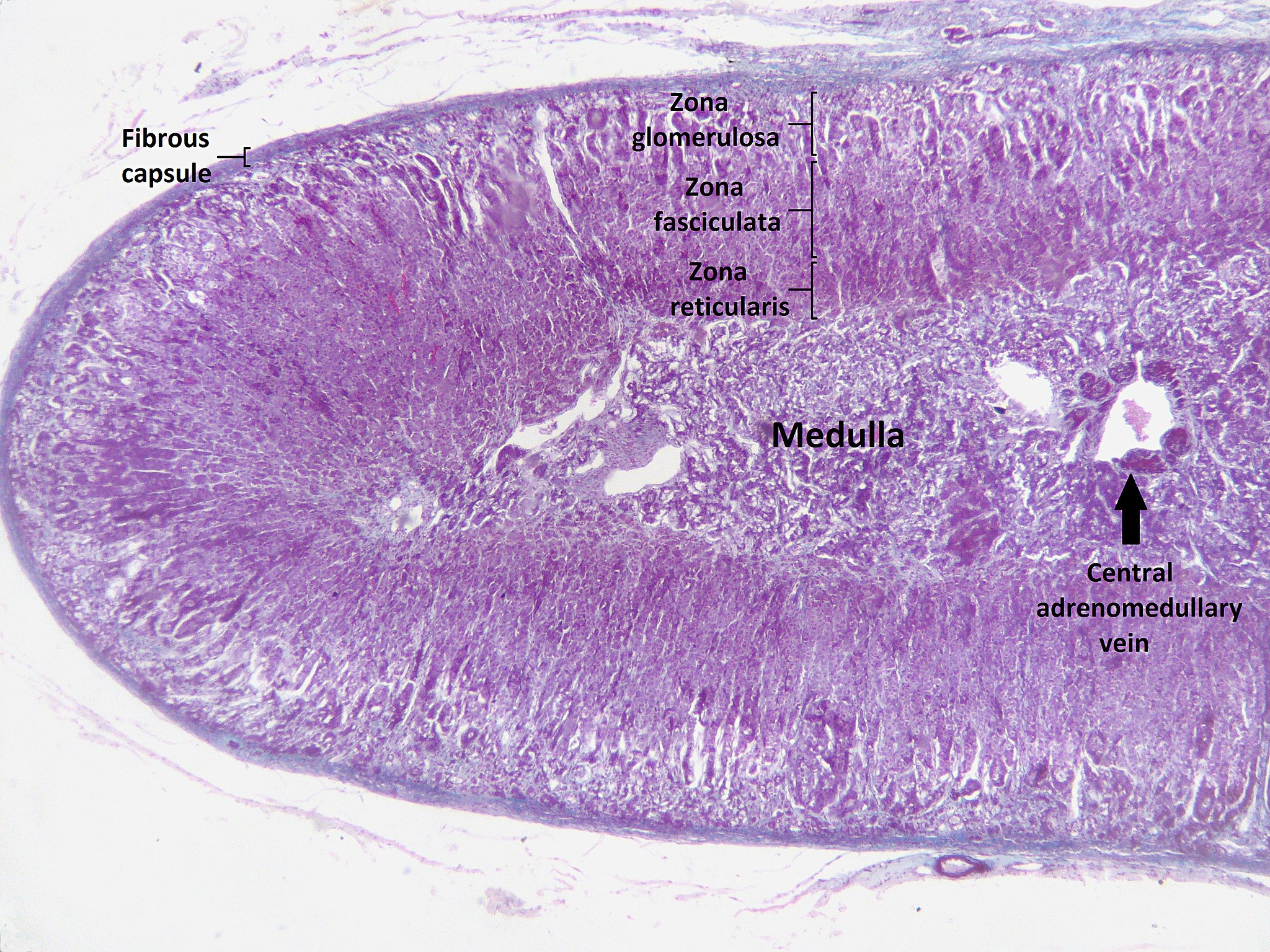
The adrenal cortex, or outer layer of the adrenal gland, is divided into three additional layers, called zones (see Figure 9.6.3). Each zone has distinct enzymes that produce different hormones from the common precursor molecule cholesterol, which is a lipid.
- Zona glomerulosa is the outermost layer of the adrenal cortex. It lies immediately under the outer fibrous capsule that encloses the adrenal gland.
- Zona fasciculata is the middle layer of the adrenal cortex. It is the largest of the three zones, accounting for nearly 80 per cent of the adrenal cortex.
- Zona reticularis is the innermost layer of the adrenal cortex. It is directly adjacent to the medulla of the adrenal gland.
Types of Adrenal Cortex Hormones
Hormones produced by the adrenal cortex are known by the general term corticosteroids. As steroid hormones, corticosteroids are endocrine hormones that are made of lipids and exert their effects on target cells by crossing the plasma membrane and binding with receptors within the cytoplasm. A steroid hormone and its receptor form a complex that enters the cell nucleus and affects gene expression. There are three types of corticosteroids synthesized and secreted by the adrenal cortex. Each type is produced by a different zone of the adrenal cortex, as shown in Figure 9.6.4.

Mineralocorticoids
Mineralocorticoids are produced in the zona glomerulosa and include the hormone aldosterone. These hormones help control the balance of mineral salts (electrolytes) in the body. In the kidneys, aldosterone increases the reabsorption of sodium ions and the excretion of potassium ions. Aldosterone also stimulates the retention of sodium ions by cells in the colon and by the sweat glands. The amount of sodium in the body affects the volume of extracellular fluids (including the blood) and thereby affects blood pressure. In this way, mineralocorticoids help control blood volume and blood pressure.
Glucocorticoids
Glucocorticoids are produced in the zona fasciculata and include the hormone cortisol, which is released in repsonse to stress and is considered the primary stress hormone. Glucocorticoids help control the rate of metabolism of proteins, fats, and sugars. In general, they increase the level of glucose and fatty acids circulating in the blood. Cells rely primarily on glucose for energy, but they can also use fatty acids for energy as an alternative to glucose. Glucocorticoids are also involved in suppression of the immune system, having a potent anti-inflammatory effect. In addition, cortisol reduces the production of new bone and decreases absorption of calcium from the gastrointestinal tract.
Androgens
Androgens are produced in the zona reticularis and include the hormone DHEA (dehydroepiandrosterone). Androgens are a general term for male sex hormones, although this is somewhat misleading, as adrenal cortex androgens are produced by both males and females. In adult males, they are converted to more potent androgens, such as testosterone in the male gonads (testes). In adult females, they are converted to female sex hormones called estrogens in the female gonads (ovaries).
Regulation of Adrenal Cortex Hormones
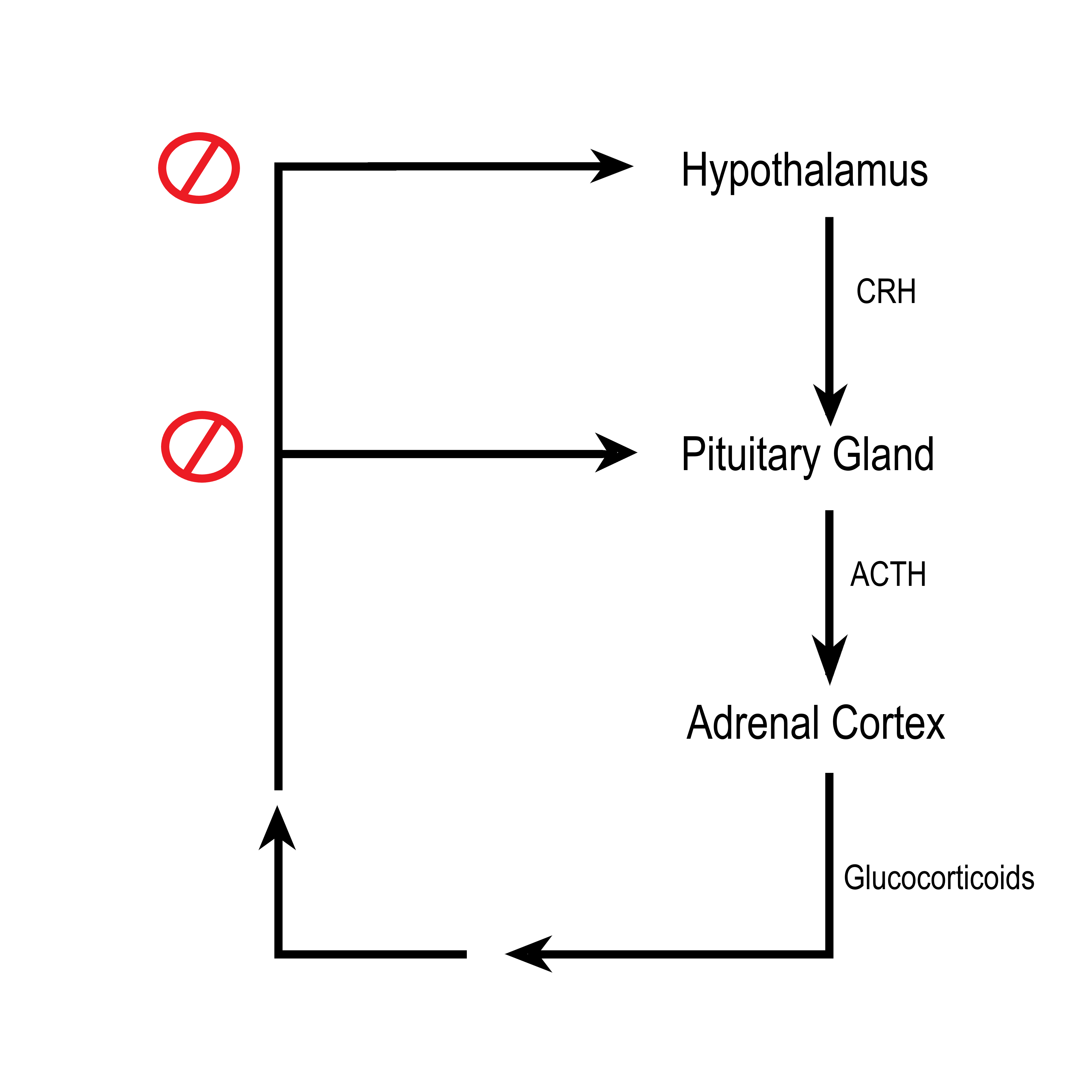
Steroid hormone production by the three zones of the adrenal cortex is regulated by hormones secreted by the anterior lobe of the pituitary gland, as well as by other physiological stimuli. For example, the production of glucocorticoids such as cortisol is stimulated by adrenocorticotropic hormone (ACTH) from the anterior pituitary, which in turn is stimulated by corticotropin releasing hormone (CRH) from the hypothalamus. When levels of glucocorticoids start to rise too high, they provide negative feedback to the hypothalamus and pituitary gland to stop secreting CRH and ACTH, respectively. This negative feedback mechanism is illustrated in Figure 9.6.5. The opposite occurs when levels of glucocorticoids start to fall too low.
Adrenal Medulla
The adrenal medulla is at the center of each adrenal gland and is surrounded by the adrenal cortex. It contains a dense network of blood vessels into which it secretes its hormones. The hormones synthesized and secreted by the adrenal medulla are generally known as catecholamines, and they include adrenaline (also called epinephrine) and noradrenaline (also called norepinephrine). These water-soluble, non-steroid hormones are made of amino acids. As non-steroid hormones, they cannot cross the plasma membrane of target cells. Instead, they exert their effects by binding to receptors on the surface of target cells. The binding of hormone and receptor activates an enzyme in the plasma membrane that controls a second messenger. It is the second messenger that influences processes inside the cell.
Catecholamines function to produce a rapid response throughout the body in stressful situations. They bring about such changes as increased heart rate, more rapid breathing, constriction of blood vessels in certain parts of the body, and an increase in blood pressure. The release of catecholamines by the adrenal medulla is stimulated by activation of the sympathetic division of the autonomic nervous system.
Disorders of the Adrenal Glands
Disorders of the adrenal glands generally include either hypersecretion or hyposecretion of adrenal hormones. The underlying cause of the abnormal secretion may be a problem with the adrenal glands or with the pituitary gland, which controls adrenal cortex hormone production. Both adrenal and pituitary glands are subject to the formation of tumors, which may cause adrenal disorders. The adrenal gland may also be affected by infections or autoimmune diseases.
Adrenal Hypersecretion: Cushing’s Syndrome
Hypersecretion of the glucocorticoid hormone cortisol leads to a disorder called Cushing’s syndrome. The most common cause of Cushing’s syndrome is a pituitary tumor, which causes excessive production of ACTH. The disease produces a wide variety of signs and symptoms, which may include obesity, diabetes, high blood pressure (hypertension), excessive body hair, osteoporosis, and depression. A distinctive sign of Cushing’s syndrome is the appearance of stretch marks in the skin, as the skin becomes progressively thinner. Another distinctive sign is a moon face, in which fat deposits give the face a rounded appearance. Treatment of Cushing’s syndrome depends on its cause and may include surgery to remove a tumor or medications to suppress activity of the adrenal glands.
Adrenal Hyposecretion: Addison’s Disease
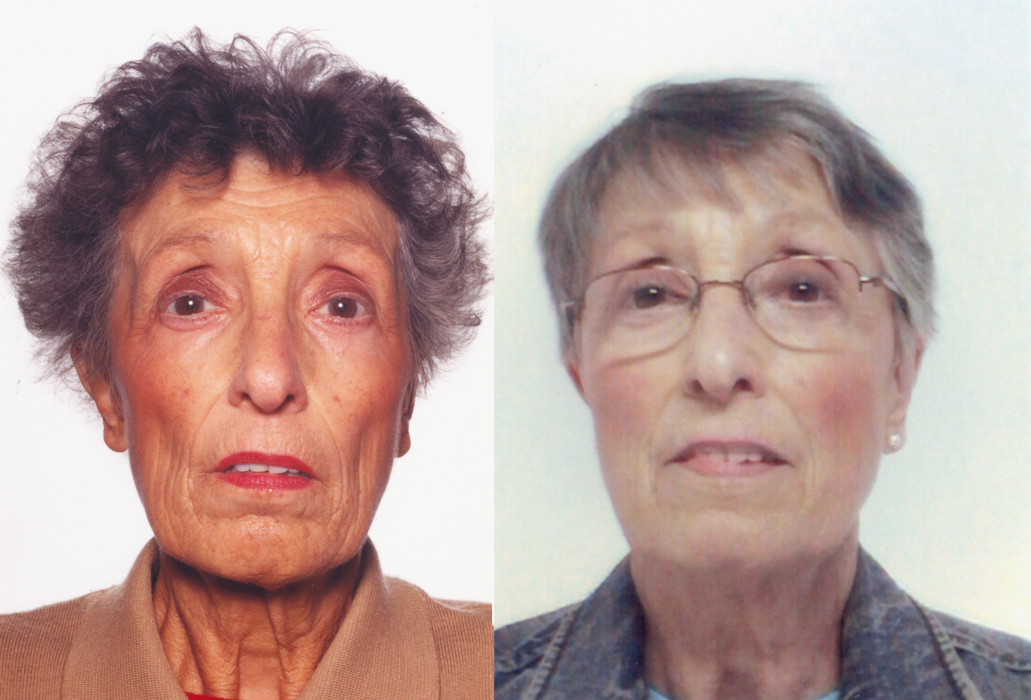
Hyposecretion of the glucocorticoid hormone cortisol leads to a disorder called Addison’s disease. There may also be hyposecretion of mineralocorticoids with this disorder. Addison’s disease is generally an autoimmune disorder, in which the immune system produces abnormal antibodies that attack cells of the adrenal cortex. Untreated infections, especially of tuberculosis, may also damage the adrenal cortex and cause Addison’s disease. A third possible cause is decreased output of ACTH by the pituitary gland, generally due to a pituitary tumor. A distinctive sign of Addison’s disease is hyperpigmentation of the skin (see the photos in Figure 9.6.6). Other symptoms tend to be nonspecific and include excessive fatigue. Addison’s disease is generally treated with replacement hormones in pill form.
Feature: My Human Body

Does just looking at this photo (Figure 9.6.7) cause you to break out in a cold sweat and experience heart palpitations? Imagine how scary it would be to actually fling yourself backward off a tall building like the BASE jumper in the photo! There would be very little time to use a parachute to slow your fall before you hit the ground. BASE jumping is called the most dangerous sport on Earth. In fact, it is so dangerous that it is outlawed in some places.
People who participate in such dangerous activities as BASE jumping are likely to be adrenaline “junkies.” They are addicted to the adrenaline rush and euphoria — or “high” — it causes when their fight-or-flight response is triggered by danger. Why does adrenaline have this effect? Adrenaline is closely related to dopamine, a chemical messenger in the brain that plays a major role in pleasure and addiction.
Adrenaline addicts don’t have to participate in BASE jumping or other dangerous sports to get an adrenaline rush. They might choose a dangerous occupation like firefighting, participate in risky behaviors like reckless driving or bank robbing, or just pick fights with other people. They might even create their own stress by always taking on too much work or delaying projects until close to their deadline.
While some excitement in one’s life is generally a good thing, always putting oneself in danger or constantly being under stress are obviously not good things. If you think you might be an adrenaline addict, note that there are healthier ways to experience a hormonal “high.” Running, biking, or participating in some other form of vigorous aerobic exercise causes the pituitary gland and hypothalamus to produce opiate-like endorphins, leading to a so-called “runner’s high.” Like the euphoric feeling adrenaline causes, a runner’s high may last for hours.
9.6 Summary
- The adrenal glands are endocrine glands that produce a variety of hormones. The two adrenal glands are located on both sides of the body, just above the kidneys. Each gland has two layers: an outer layer called the adrenal cortex and an inner layer called the adrenal medulla.
- The adrenal cortex produces steroid hormones called by the general term corticosteroids, of which there are three types: mineralocorticoids (such as aldosterone), which helps control electrolyte balance; glucocorticoids (such as cortisol), which helps control the rate of metabolism, suppresses the immune system, and is the major stress hormone; and androgens (such as DHEA), which is converted to sex hormones in the gonads.
- The adrenal medulla produces non-steroid catecholamine hormones, including adrenaline and noradrenaline. These hormones stimulate the fight-or-flight response.
- Disorders of the adrenal glands generally include either hypersecretion or hyposecretion of adrenal hormones. The cause may be a problem with the adrenal glands or with the pituitary gland, which controls adrenal cortex hormone production. Examples include Cushing’s syndrome, in which there is hypersecretion of cortisol, and Addison’s disease, in which there is hyposecretion of cortisol and mineralocorticoids.
9.6 Review Questions
- Describe the structure and location of the adrenal glands.
-
- Compare and contrast the adrenal cortex and adrenal medulla.
- Identify the three layers of the adrenal cortex and the type of hormones each layer produces.
- Give an example of each type of corticosteroid and state its function.
- Explain how the production of glucocorticoids is regulated.
- What is a catecholamine? Give an example of a catecholamine and state its function.
- Compare and contrast Cushing’s syndrome and Addison’s disease.
- What are two ways in which the nervous system (which includes the brain, spinal cord, and nerves) controls the adrenal gland?
- Explain why a pituitary tumor can cause either hypersecretion or hyposecretion of cortisol.
9.6 Explore More
https://www.youtube.com/watch?v=v-t1Z5-oPtU
How stress affects your body - Sharon Horesh Bergquist, TED-Ed, 2015.
https://www.youtube.com/watch?time_continue=1&v=WuyPuH9ojCE&feature=emb_logo
How stress affects your brain - Madhumita Murgia, TED-Ed, 2015.
https://www.youtube.com/watch?v=FBnBTkcr6No&feature=emb_logo
Adrenaline: Fight or Flight Response, Henk van 't Klooster, 2013.
Attributions
Figure 9.6.1
Attack from wikimedia commons by Jerry Kirkhart from Los Osos, Calif. on Wikimedia Commons is used under a CC BY 2.0 (https://creativecommons.org/licenses/by/2.0) license.
Figure 9.6.2
Diagram_showing_where_the_adrenal_glands_are_in_the_body_CRUK_415.svg by Cancer Research UK uploader on Wikimedia Commons is used under a CC BY-SA 4.0 (https://creativecommons.org/licenses/by-sa/4.0) license.
Figure 9.6.3
Adrenal_cortex_labelled by Jpogi at English Wikipedia on Wikimedia Commons is used under a CC0 1.0 Universal Public Domain Dedication (https://creativecommons.org/publicdomain/zero/1.0/deed.en) license.
Figure 9.6.4
The_Adrenal_Glands by OpenStax College is used under a CC BY 4.0 (https://creativecommons.org/licenses/by/4.0/) license.
Figure 9.6.5
ACTH negative feedback loop by Christinelmiller is used under a CC BY 4.0 (https://creativecommons.org/licenses/by/4.0/) license.
Figure 9.6.6
A_69-Year-Old_Female_with_Tiredness_and_a_Persistent_Tan_01 by Petros Perros on Wikimedia Commons is used under a CC BY 2.5 (https://creativecommons.org/licenses/by/2.5/deed.en) license.
Figure 9.6.7
BASE_Jumping_from_Sapphire_Tower_in_Istanbul by Kontizas Dimitrios on Wikimedia Commons is used under a CC BY-SA 3.0 (https://creativecommons.org/licenses/by-sa/3.0) license.
References
Betts, J. G., Young, K.A., Wise, J.A., Johnson, E., Poe, B., Kruse, D.H., Korol, O., Johnson, J.E., Womble, M., DeSaix, P. (2013, June 19). Figure 17.17 Adrenal glands [digital image]. In Anatomy and Physiology (Section 17.6). OpenStax College. https://openstax.org/books/anatomy-and-physiology/pages/17-6-the-adrenal-glands
Henk van 't Klooster. (2013). Adrenaline: Fight or flight response. YouTube. https://www.youtube.com/watch?v=FBnBTkcr6No&feature=youtu.be
Perros, P. (2005). A 69-year-old female with tiredness and a persistent tan. PLoS Medicine, 2(8): e229. https://doi.org/10.1371/journal.pmed.0020229
TED-Ed. (2015, October 22). How stress affects your body - Sharon Horesh Bergquist. YouTube. https://www.youtube.com/watch?v=v-t1Z5-oPtU&feature=youtu.be
TED-Ed. (2015, November 6). How stress affects your brain - Madhumita Murgia. YouTube. https://www.youtube.com/watch?v=WuyPuH9ojCE&feature=youtu.be
Image shows a Lego (TM) representation of Gregor Mendel with his plants.
Image shows a diagram of the hormones secreted by the thyroid gland, and how it is both controlled by and acting upon in a negative feedback the hypothalamus and the anterior pituitary gland.
Pills from Pee
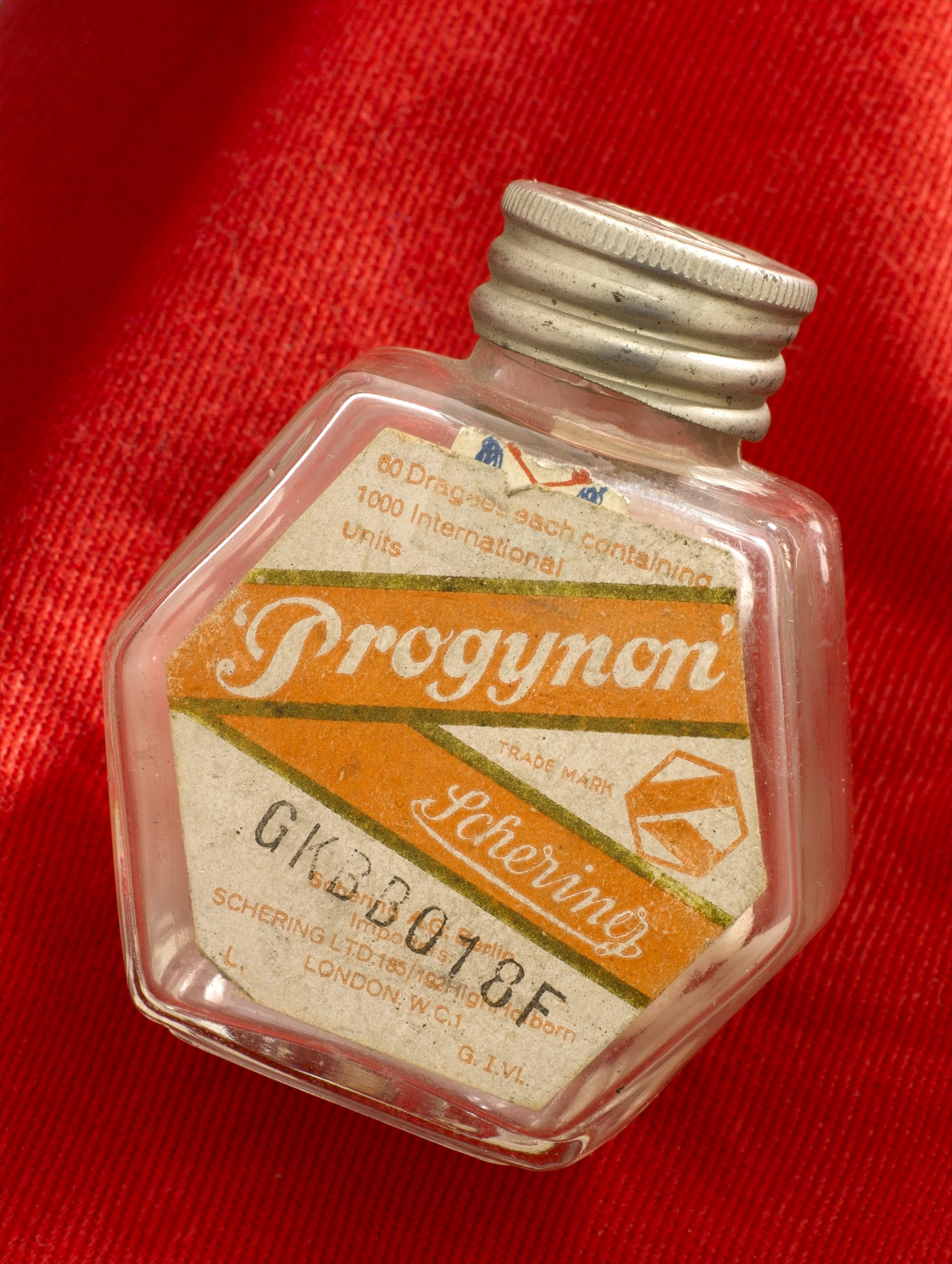
The medication pictured in Figure 9.3.1 with the brand name Progynon was a drug used to control the effects of menopause in women. The pills first appeared in 1928 and contained the human sex hormone estrogen. Estrogen secretion declines in women around the time of menopause and may cause symptoms like mood swings and hot flashes. The pills were supposed to ease the symptoms by supplementing estrogen in the body. The manufacturer of Progynon obtained estrogen for the pills from the urine of pregnant women, because it was a cheap source of the hormone. Progynon is still used today to treat menopausal symptoms. Although the drug has been improved over the years, it still contains estrogen, which is an example of an endocrine hormone.
How Do Endocrine Hormones Work?
Endocrine hormones like estrogen are messenger molecules secreted by endocrine glands into the bloodstream. They travel throughout the body in the circulation. Although they reach virtually every cell in the body in this way, each hormone affects only certain cells, called target cells. A target cell is the type of cell on which a hormone has an effect. A target cell is affected by a particular hormone because it has receptor proteins — either on the cell surface or within the cell — that are specific to that hormone. An endocrine hormone travels through the bloodstream until it finds a target cell with a matching receptor to which it can bind. When the hormone binds to the receptor, it causes changes within the cell. The manner in which it changes the cell depends on whether the hormone is a steroid hormone or a non-steroid hormone.
Steroid Hormones
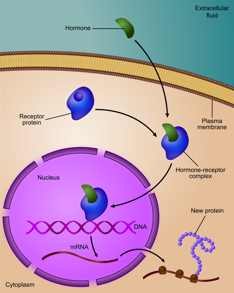
A steroid hormone (such as estrogen) is made of lipids. It is fat soluble, so it can diffuse across a target cell’s plasma membrane, which is also made of lipids. Once inside the cell, a steroid hormone binds with receptor proteins in the cytoplasm. As you can see in Figure 9.3.2, the steroid hormone and its receptor form a complex — called a steroid complex — which moves into the nucleus, where it influences the expression of genes. Examples of steroid hormones include cortisol, which is secreted by the adrenal glands, and sex hormones, which are secreted by the gonads.
Non-Steroid Hormones
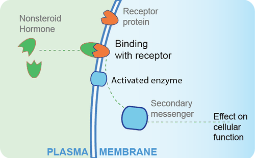
A non-steroid hormoneis made of amino acids. It is not fat soluble, so it cannot diffuse across the plasma membrane of a target cell. Instead, it binds to a receptor protein on the cell membrane. In the Figure 9.3.3 diagram, you can see that the binding of the hormone with the receptor activates an enzyme in the cell membrane. The enzyme then stimulates another molecule, called the second messenger, which influences processes inside the cell. Most endocrine hormones are non-steroid hormones. Examples include glucagon and insulin, both produced by the pancreas.
Regulation of Endocrine Hormones
Endocrine hormones regulate many body processes, but what regulates the secretion of endocrine hormones? Most endocrine hormones are controlled by feedback mechanisms. A feedback mechanism is a loop in which a product feeds back to control its own production. Feedback loops may be either negative or positive.
- Most endocrine hormones are regulated by negative feedback loops. Negative feedback keeps the concentration of a hormone within a relatively narrow range, and maintains homeostasis.
- Very few endocrine hormones are regulated by positive feedback loops. Positive feedback causes the concentration of a hormone to become increasingly higher.
Regulation by Negative Feedback
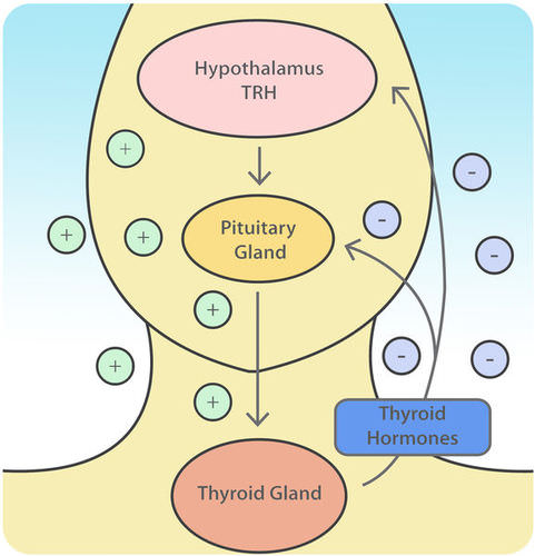
A negative feedback loop controls the synthesis and secretion of hormones by the thyroid gland. This loop includes the hypothalamus and pituitary gland, in addition to the thyroid, as shown in the diagram (Figure 9.3.4). When the levels of thyroid hormones circulating in the blood fall too low, the hypothalamus secretes thyrotropin releasing hormone (TRH). This hormone travels directly to the pituitary gland through the thin stalk connecting the two structures. In the pituitary gland, TRH stimulates the pituitary to secrete thyroid stimulating hormone (TSH). TSH, in turn, travels through the bloodstream to the thyroid gland, and stimulates it to secrete thyroid hormones. This continues until the blood levels of thyroid hormones are high enough. At that point, the thyroid hormones feed back to stop the hypothalamus from secreting TRH and the pituitary from secreting TSH. Without the stimulation of TSH, the thyroid gland stops secreting its hormones. Eventually, the levels of thyroid hormones in the blood start to fall too low again. When that happens, the hypothalamus releases TRH, and the loop repeats.
Regulation by Positive Feedback
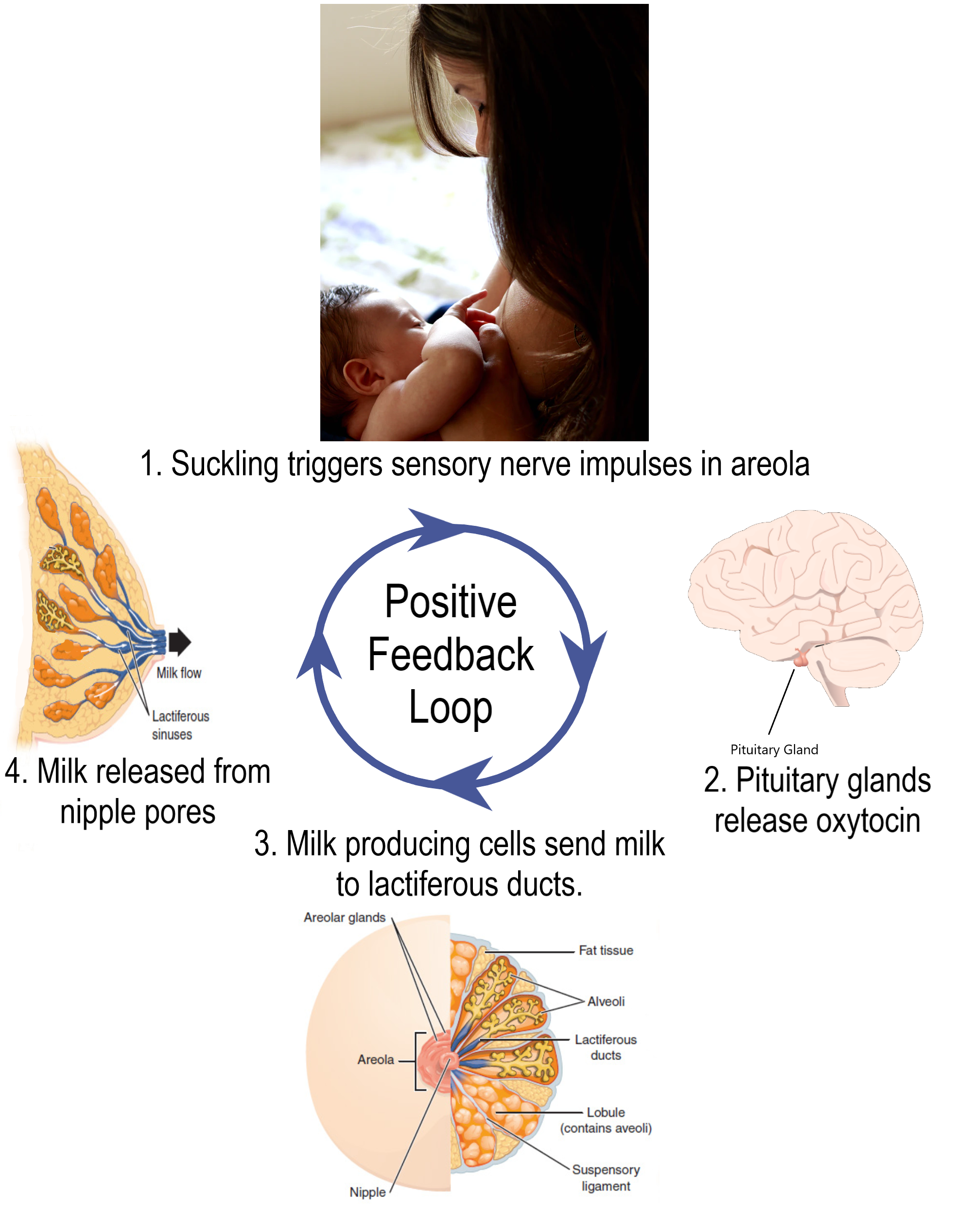
Prolactin is a non-steroid endocrine hormone secreted by the pituitary gland. One of the functions of prolactin is to stimulate a nursing mother’s mammary glands to produce milk. The regulation of prolactin in the mother is controlled by a positive feedback loop that involves the nipples, hypothalamus, pituitary gland, and mammary glands. Positive feedback begins when a baby suckles on the mother’s nipple. Nerve impulses from the nipple reach the hypothalamus, which stimulates the pituitary gland to secrete prolactin. Prolactin travels in the blood to the mammary glands and stimulates them to produce milk. The release of milk causes the baby to continue suckling, which causes more prolactin to be secreted and more milk to be produced. The positive feedback loop continues until the baby stops suckling at the breast.
Feature: Myth vs. Reality
Anabolic steroids are synthetic versions of the naturally occurring male sex hormone testosterone. Male hormones have androgenic (or masculinizing) effects, but they also have anabolic (or muscle-building) effects. The anabolic effects are the reason that synthetic steroids are used by athletes. In addition to building muscles, they also accelerate the development of bones and red blood cells, increase endurance so athletes can train harder and longer, and speed up muscle recovery. Unfortunately, these benefits of steroid use come with costs. If you ever consider taking anabolic steroids to build muscles and improve athletic performance, consider the following myths and corresponding realities.
Myth |
Reality |
| "Steroids are safe." | Steroid use may cause several serious side effects. Prolonged use may increase the risk of liver cancer, heart disease, and high blood pressure. |
| "Steroids will not stunt your growth." | Teens who take steroids before they have finished growing in height may have their growth stunted so they remain shorter throughout life than they would otherwise have been. Such stunting occurs because steroids increase the rate at which skeletal maturity is reached. Once skeletal maturity occurs, additional growth in height is impossible. |
| "Steroids do not cause drug dependency." | Steroid use may cause dependency, as evidenced by the negative effects of stopping steroid use. These negative effects may include insomnia, fatigue, and depressed mood, among others. |
| "There is no such thing as 'roid rage.'" | Steroid use has been shown to increase aggressiveness in some people. It has also been implicated in a number of violent acts committed by people who had not demonstrated violent tendencies until they started using steroids. |
| "Only males use steroids." | Although steroid use is more common in males than females, some females also use steroids. They use them to build muscle and improve physical performance, generally either for athletic competition or for self-defense. |
9.3 Summary
- Endocrine hormones are messenger molecules secreted by endocrine glands into the bloodstream. They travel throughout the body but affect only certain cells, called target cells, which have receptors specific to particular hormones.
- Steroid hormones such as estrogen are endocrine hormones made of lipids that cross plasma membranes and bind to receptors inside target cells. The hormone-receptor complexes then move into the nucleus, where they influence gene expression.
- Non-steroid hormones (such as insulin) are endocrine hormones made of amino acids that bind to receptors on the surface of target cells. This activates an enzyme in the plasma membrane, and the enzyme controls a second messenger molecule, which influences cell processes.
- Most endocrine hormones are controlled by negative feedback loops in which rising levels of a hormone feed back to stop its own production — and vice-versa. For example, a negative feedback loop controls production of thyroid hormones. The loop includes the hypothalamus, pituitary gland, and thyroid gland.
- Only a few endocrine hormones are controlled by positive feedback loops, in which rising levels of a hormone feed back to stimulate continued production of the hormone. Prolactin, the pituitary hormone that stimulates milk production by mammary glands, is controlled by a positive feedback loop. The loop includes the nipples, hypothalamus, pituitary gland, and mammary glands.
9.3 Review Questions
-
-
- Explain how steroid hormones influence target cells.
- How do non-steroid hormones affect target cells?
- Compare and contrast negative and positive feedback loops.
- Outline the way feedback controls the production of thyroid hormones.
- Describe the feedback mechanism that controls milk production by the mammary glands.
- People with a condition called hyperthyroidism produce too much thyroid hormone. What do you think this does to the level of TSH? Explain your answer.
- Which is more likely to maintain homeostasis— negative feedback or positive feedback? Explain your answer.
- Does testosterone bind to receptors on the plasma membrane of target cells or in the cytoplasm of target cells? Explain your answer.
9.3 Explore More
https://www.youtube.com/watch?v=WVrlHH14q3o&feature=emb_logo
Great Glands - Your Endocrine System: CrashCourse Biology #33, CrashCourse, 2012.
https://www.youtube.com/watch?v=qXaDDa3FB5Q&feature=emb_logo
National Geographic | Benefits and Side Effects of Steroids Use 2015, 24 Physic.
Attributions
Figure 9.3.1
L0058274 Glass bottle for ‘Progynon’ pills, United Kingdom, 1928-1948 by Wellcome Collection gallery (2018-03-29)/ Science Museum, London on Wikimedia Commons is used under a CC-BY-4.0 (https://creativecommons.org/licenses/by/4.0/) license.
Figure 9.3.2
Regulation_of_gene_expression_by_steroid_hormone_receptor.svg by Ali Zifan on Wikimedia Commons is used under a CC BY-SA 4.0 (https://creativecommons.org/licenses/by-sa/4.0/deed.en) license.
Figure 9.3.3
Non-steroid hormone pathway by CK-12 Foundation, Biology for High School is used under a CC BY-NC 3.0 (https://creativecommons.org/licenses/by-nc/3.0/) license.
Figure 9.3.4
Thyroid Negative Feedback Loop by CK-12 Foundation, College Human Biology is used under a CC BY-NC 3.0 (https://creativecommons.org/licenses/by-nc/3.0/) license.
 ©CK-12 Foundation Licensed under
 ![]() • Terms of Use • Attribution
• Terms of Use • Attribution
Figure 9.3.5
Lactation Positive Feedback Loop by Christinelmiller on Wikimedia Commons is used under a CC BY-SA 4.0 (https://creativecommons.org/licenses/by-sa/4.0/deed.en) license.
References
24 Physic. (2015,July 19). National Geographic | Benefits and side effects of steroids use 2015. YouTube. https://www.youtube.com/watch?v=qXaDDa3FB5Q&feature=youtu.be
Brainard, J/ CK-12 Foundation. (2016, August 15). Figure 4 Thyroid negative feedback loop [digital image]. In CK-12 College Human Biology (Section 11.3 Endocrine hormones). CK12.org. https://www.ck12.org/book/ck-12-human-biology/section/11.3/
CK-12 Foundation. (2019, March 5). Figure 3 A non-steroid hormone binds with a receptor on the plasma membrane of a target cell [digital image]. In Flexbook 2.0: CK-12 Biology For High School (Section 13.21 Hormone). CK12. https://flexbooks.ck12.org/cbook/ck-12-biology-flexbook-2.0/section/13.21/primary/lesson/hormones-bio
CrashCourse. (2012, September 10). Great glands - Your endocrine system: CrashCourse Biology #33. YouTube. https://www.youtube.com/watch?v=WVrlHH14q3o&feature=youtu.be
TED-Ed. (2018, June 21). How do your hormones work? - Emma Bryce. YouTube. https://www.youtube.com/watch?v=-SPRPkLoKp8&feature=youtu.be
Created by CK-12 Foundation/Adapted by Christine Miller

Steady as She Goes
This device (Figure 7.8.1) looks simple, but it controls a complex system that keeps a home at a steady temperature — it's a thermostat. The device shows the current temperature in the room, and also allows the occupant to set the thermostat to the desired temperature. A thermostat is a commonly cited model of how living systems — including the human body— maintain a steady state called homeostasis.
What Is Homeostasis?
Homeostasis is the condition in which a system (such as the human body) is maintained in a more or less steady state. It is the job of cells, tissues, organs, and organ systems throughout the body to maintain many different variables within narrow ranges compatible with life. Keeping a stable internal environment requires continually monitoring the internal environment and constantly making adjustments to keep things in balance.
Set Point and Normal Range
For any given variable, such as body temperature or blood glucose level, there is a particular set point that is the physiological optimum value. The set point for human body temperature, for example, is about 37 degrees C (98.6 degrees F). As the body works to maintain homeostasis for temperature or any other internal variable, the value typically fluctuates around the set point. Such fluctuations are normal, as long as they do not become too extreme. The spread of values within which such fluctuations are considered insignificant is called the normal range. In the case of body temperature, for example, the normal range for an adult is about 36.5 to 37.5 degrees C (97.7 to 99.5 degrees F).
A good analogy for set point, normal range, and maintenance of homeostasis is driving. When you are driving a vehicle on the road, you are supposed to drive in the centre of your lane — this is analogous to the set point. Sometimes, you are not driving in the exact centre of the lane, but you are still within your lines, so you are in the equivalent of the normal range. However, if you were to get too close to the centre line or the shoulder of the road, you would take action to correct your position. You'd move left if you were too close to the shoulder, or right if too close to the centre line — which is analogous to our next concept, negative feedback to maintain homeostasis.
Maintaining Homeostasis
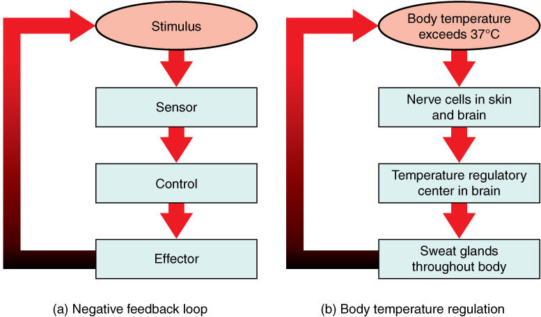
Homeostasis is normally maintained in the human body by an extremely complex balancing act. Regardless of the variable being kept within its normal range, maintaining homeostasis requires at least four interacting components: stimulus, sensor, control centre, and effector.
- The stimulus is provided by the variable being regulated. Generally, the stimulus indicates that the value of the variable has moved away from the set point or has left the normal range.
- The sensor monitors the values of the variable and sends data on it to the control centre.
- The control centre matches the data with normal values. If the value is not at the set point or is outside the normal range, the control centre sends a signal to the effector.
- The effector is an organ, gland, muscle, or other structure that acts on the signal from the control centre to move the variable back toward the set point.
Each of these components is illustrated in Figure 7.8.2. The diagram on the left is a general model showing how the components interact to maintain homeostasis. The diagram on the right shows the example of body temperature. From the diagrams, you can see that maintaining homeostasis involves feedback, which is data that feeds back to control a response. Feedback may be negative (as in the example below) or positive. All the feedback mechanisms that maintain homeostasis use negative feedback. Biological examples of positive feedback are much less common.
Negative Feedback
In a negative feedback loop, feedback serves to reduce an excessive response and keep a variable within the normal range. Two processes controlled by negative feedback are body temperature regulation and control of blood glucose.
Body Temperature
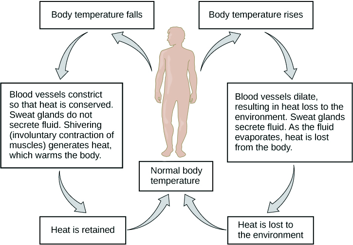
Body temperature regulation involves negative feedback, whether it lowers the temperature or raises it, as shown in Figure 7.8.3 and explained in the text that follows.
Cooling Down
The human body’s temperature regulatory centre is the hypothalamus in the brain. When the hypothalamus receives data from sensors in the skin and brain that body temperature is higher than the set point, it sets into motion the following responses:
- Blood vessels in the skin dilate (vasodilation) to allow more blood from the warm body core to flow close to the surface of the body, so heat can be radiated into the environment.
- As blood flow to the skin increases, sweat glands in the skin are activated to increase their output of sweat (diaphoresis). When the sweat evaporates from the skin surface into the surrounding air, it takes heat with it.
- Breathing becomes deeper, and the person may breathe through the mouth instead of the nasal passages. This increases heat loss from the lungs.
Heating Up
When the brain’s temperature regulatory centre receives data that body temperature is lower than the set point, it sets into motion the following responses:
- Blood vessels in the skin contract (vasoconstriction) to prevent blood from flowing close to the surface of the body, which reduces heat loss from the surface.
- As temperature falls lower, random signals to skeletal muscles are triggered, causing them to contract. This causes shivering, which generates a small amount of heat.
- The thyroid gland may be stimulated by the brain (via the pituitary gland) to secrete more thyroid hormone. This hormone increases metabolic activity and heat production in cells throughout the body.
- The adrenal glands may also be stimulated to secrete the hormone adrenaline. This hormone causes the breakdown of glycogen (the carbohydrate used for energy storage in animals) to glucose, which can be used as an energy source. This catabolic chemical process is exothermic, or heat producing.
Blood Glucose
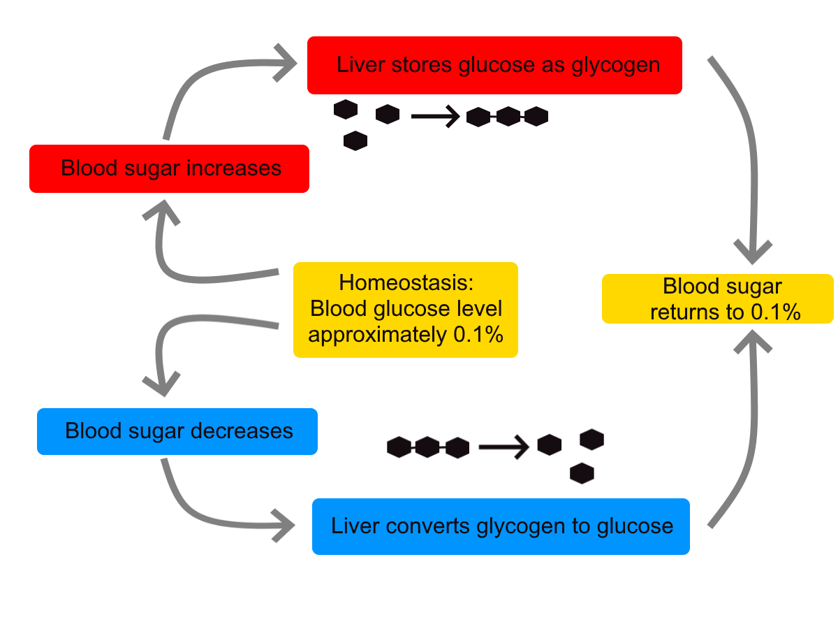
In controlling the blood glucose level, certain endocrine cells in the pancreas (called alpha and beta cells) detect the level of glucose in the blood. They then respond appropriately to keep the level of blood glucose within the normal range.
- If the blood glucose level rises above the normal range, pancreatic beta cells release the hormone insulin into the bloodstream. Insulin signals cells to take up the excess glucose from the blood until the level of blood glucose decreases to the normal range.
- If the blood glucose level falls below the normal range, pancreatic alpha cells release the hormone glucagon into the bloodstream. Glucagon signals cells to break down stored glycogen to glucose and release the glucose into the blood until the level of blood glucose increases to the normal range.
https://www.youtube.com/watch?v=Iz0Q9nTZCw4
Homeostasis and Negative/Positive Feedback, Amoeba Sisters, 2017.
Positive Feedback
In a positive feedback loop, feedback serves to intensify a response until an end point is reached. Examples of processes controlled by positive feedback in the human body include blood clotting and childbirth.
Blood Clotting
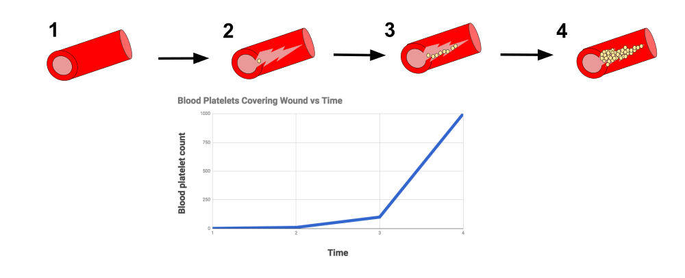
When a wound causes bleeding, the body responds with a positive feedback loop to clot the blood and stop blood loss. Substances released by the injured blood vessel wall begin the process of blood clotting. Platelets in the blood start to cling to the injured site and release chemicals that attract additional platelets. As the platelets continue to amass, more of the chemicals are released and more platelets are attracted to the site of the clot. The positive feedback accelerates the process of clotting until the clot is large enough to stop the bleeding.
Childbirth
Figure 7.8.6 shows the positive feedback loop that controls childbirth. The process normally begins when the head of the infant pushes against the cervix. This stimulates nerve impulses, which travel from the cervix to the hypothalamus in the brain. In response, the hypothalamus sends the hormone oxytocin to the pituitary gland, which secretes it into the bloodstream so it can be carried to the uterus. Oxytocin stimulates uterine contractions, which push the baby harder against the cervix. In response, the cervix starts to dilate in preparation for the passage of the baby. This cycle of positive feedback continues, with increasing levels of oxytocin, stronger uterine contractions, and wider dilation of the cervix until the baby is pushed through the birth canal and out of the body. At that point, the cervix is no longer stimulated to send nerve impulses to the brain, and the entire process stops.
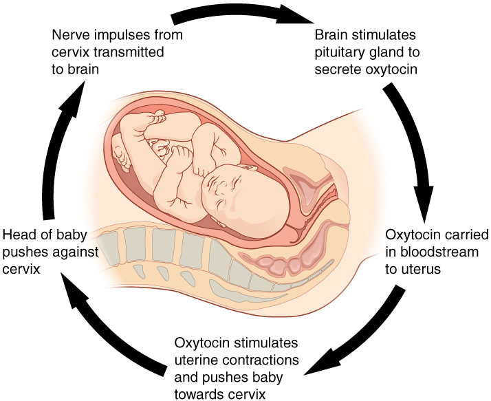
Normal childbirth is driven by a positive feedback loop. Positive feedback causes an increasing deviation from the normal state to a fixed end point, rather than a return to a normal set point as in homeostasis.
When Homeostasis Fails
Homeostatic mechanisms work continuously to maintain stable conditions in the human body. Sometimes, however, the mechanisms fail. When they do, homeostatic imbalance may result, in which cells may not get everything they need or toxic wastes may accumulate in the body. If homeostasis is not restored, the imbalance may lead to disease — or even death. Diabetes is an example of a disease caused by homeostatic imbalance. In the case of diabetes, blood glucose levels are no longer regulated and may be dangerously high. Medical intervention can help restore homeostasis and possibly prevent permanent damage to the organism.
Normal aging may bring about a reduction in the efficiency of the body’s control systems, which makes the body more susceptible to disease. Older people, for example, may have a harder time regulating their body temperature. This is one reason they are more likely than younger people to develop serious heat-induced illnesses, such as heat stroke.
Feature: My Human Body
Diabetes is diagnosed in people who have abnormally high levels of blood glucose after fasting for at least 12 hours. A fasting level of blood glucose below 100 is normal. A level between 100 and 125 places you in the pre-diabetes category, and a level higher than 125 results in a diagnosis of diabetes.
Of the two types of diabetes, type 2 diabetes is the most common, accounting for about 90 per cent of all cases of diabetes in the United States. Type 2 diabetes typically starts after the age of 40. However, because of the dramatic increase in recent decades in obesity in younger people, the age at which type 2 diabetes is diagnosed has fallen. Even children are now being diagnosed with type 2 diabetes. Today, about 3 million Canadians (8.1% of total population) are living with diabetes.
You may at some point have your blood glucose level tested during a routine medical exam. If your blood glucose level indicates that you have diabetes, it may come as a shock to you because you may not have any symptoms of the disease. You are not alone, because as many as one in four diabetics do not know they have the disease. Once the diagnosis of diabetes sinks in, you may be devastated by the news. Diabetes can lead to heart attacks, strokes, blindness, kidney failure, nerve damage, and loss of toes or feet. The risk of death in adults with diabetes is 50 per cent greater than it is in adults without diabetes, and diabetes is the seventh leading cause of death of adults. In addition, controlling diabetes usually requires frequent blood glucose testing, watching what and when you eat, and taking medications or even insulin injections. All of this may seem overwhelming.
The good news is that changing your lifestyle may stop the progression of type 2 diabetes or even reverse it. By adopting healthier habits, you may be able to keep your blood glucose level within the normal range without medications or insulin. Here’s how:
- Lose weight. Any weight loss is beneficial. Losing as little as seven per cent of your weight may be all that is needed to stop diabetes in its tracks. It is especially important to eliminate excess weight around your waist.
- Exercise regularly. You should try to exercise for at least 30 minutes, five days a week. This will not only lower your blood sugar and help your insulin work better, but it will also lower your blood pressure and improve your heart health. Another bonus of exercise is that it will help you lose weight by increasing your basal metabolic rate.
- Adopt a healthy diet. Decrease your consumption of refined carbohydrates, such as sweets and sugary drinks. Increase your intake of fibre-rich foods, such as fruits, vegetables, and whole grains. About one-quarter of each meal should consist of high-protein foods, such as fish, chicken, dairy products, legumes, or nuts.
- Control stress. Stress can increase your blood glucose and also raise your blood pressure and risk of heart disease. When you feel stressed out, do breathing exercises or take a brisk walk or jog. Try to replace stressful thoughts with more calming ones.
- Establish a support system. Enlist the help and support of loved ones, as well as medical professionals, such as a nutritionist and diabetes educator. Having a support system will help ensure that you are on the path to wellness, and that you can stick to your plan.
7.8 Summary
- Homeostasis is the condition in which a system (such as the human body) is maintained in a more or less steady state. It is the job of cells, tissues, organs, and organ systems throughout the body to maintain homeostasis.
- For any given variable, such as body temperature, there is a particular set point that is the physiological optimum value. The spread of values around the set point that is considered insignificant is called the normal range.
- Homeostasis is generally maintained by a negative feedback loop that includes a stimulus, sensor, control centre, and effector. Negative feedback serves to reduce an excessive response and to keep a variable within the normal range. Negative feedback loops control body temperature and the blood glucose level.
- Positive feedback loops are not common in biological systems. Positive feedback serves to intensify a response until an end point is reached. Positive feedback loops control blood clotting and childbirth.
- Sometimes homeostatic mechanisms fail, resulting in homeostatic imbalance. Diabetes is an example of a disease caused by homeostatic imbalance. Aging can bring about a reduction in the efficiency of the body’s control system, which makes the elderly more susceptible to disease.
7.8 Review Questions
-
-
- Compare and contrast negative and positive feedback loops.
- Explain how negative feedback controls body temperature.
- Give two examples of physiological processes controlled by positive feedback loops.
- During breastfeeding, the stimulus of the baby sucking on the nipple increases the amount of milk produced by the mother. The more sucking, the more milk is usually produced. Is this an example of negative or positive feedback? Explain your answer. What do you think might be the evolutionary benefit of the milk production regulation mechanism you described?
- Explain why homeostasis is regulated by negative feedback loops, rather than positive feedback loops.
- The level of a sex hormone, testosterone (T), is controlled by negative feedback. Another hormone, gonadotropin-releasing hormone (GnRH), is released by the hypothalamus of the brain, which triggers the pituitary gland to release luteinizing hormone (LH). LH stimulates the gonads to produce T. When there is too much T in the bloodstream, it feeds back on the hypothalamus, causing it to produce less GnRH. While this does not describe all the feedback loops involved in regulating T, answer the following questions about this particular feedback loop.
- What is the stimulus in this system? Explain your answer.
- What is the control centre in this system? Explain your answer.
- In this system, is the pituitary considered the stimulus, sensor, control centre, or effector? Explain your answer.
7.8 Explore More
https://www.youtube.com/watch?v=LSgEJSlk6W4
Homeostasis - What Is Homeostasis - What Is Set Point For Homeostasis - Homeostasis In The Human Body, Whats Up Dude, 2017.
https://www.youtube.com/watch?v=XMsJ-3qRVJM
Attributions
Figure 7.8.1
Nest_Thermostat by Amanitamano on Wikimedia Commons is used under a CC BY-SA 3.0 (https://creativecommons.org/licenses/by-sa/3.0/deed.en) license.
Figure 7.8.2
Negative_Feedback_Loops by OpenStax on Wikimedia Commons is used under a CC BY 4.0 (https://creativecommons.org/licenses/by/4.0/deed.en) license.
Figure 7.8.3
Body Temperature Homeostasis by OpenStax College, Biology is used under a CC BY 4.0 license.
Figure 7.8.4
Homeostasis_of_blood_sugar by Christinelmiller on Wikimedia Commons is used under a CC0 1.0 Universal Public Domain Dedication (https://creativecommons.org/publicdomain/zero/1.0/deed.en) license.
Figure 7.8.5
Positive_Feedback_Diagram_Blood_Clotting by Elliottuttle on Wikimedia Commons is used under a CC BY-SA 4.0 (https://creativecommons.org/licenses/by-sa/4.0) license.
Figure 7.8.6
Pregnancy-Positive_Feedback by OpenStax on Wikimedia Commons is used under a CC BY 4.0 (https://creativecommons.org/licenses/by/4.0/deed.en) license.
References
Amoeba Sisters. (2017, September 7). Homeostasis and negative/positive feedback. YouTube. https://www.youtube.com/watch?v=Iz0Q9nTZCw4&feature=youtu.be
Betts, J. G., Young, K.A., Wise, J.A., Johnson, E., Poe, B., Kruse, D.H., Korol, O., Johnson, J.E., Womble, M., DeSaix, P. (2013, April 25). Figure 1.10 Negative feedback loop [digital image/ diagram]. In Anatomy and Physiology (Section 1.5). OpenStax. https://openstax.org/books/anatomy-and-physiology/pages/1-5-homeostasis
Betts, J. G., Young, K.A., Wise, J.A., Johnson, E., Poe, B., Kruse, D.H., Korol, O., Johnson, J.E., Womble, M., DeSaix, P. (2013, April 25). Figure 1.11 Positive feedback loop normal childbirth is driven by a positive feedback loop [digital image/ diagram]. In Anatomy and Physiology (Section 1.5). OpenStax. https://openstax.org/books/anatomy-and-physiology/pages/1-5-homeostasis
Cognito. (2018, December 18). GCSE Biology - Homeostasis #38. YouTube. https://www.youtube.com/watch?v=XMsJ-3qRVJM&feature=youtu.be
Mayo Clinic Staff. (n.d.). Type 2 diabetes [online article]. MayoClinic.org. https://www.mayoclinic.org/diseases-conditions/type-2-diabetes/symptoms-causes/syc-20351193
OpenStax CNX. (2016, March 23). Figure 4 The body is able to regulate temperature in response to signals from the nervous system [digital image]. In OpenStax, Biology (Section 33.3). https://cnx.org/contents/GFy_h8cu@10.8:BP24ZReh@7/Homeostasis
Whats Up Dude. (2017, September 20). Homeostasis - What is homeostasis - What is set point for homeostasis - Homeostasis in the human body. YouTube. https://www.youtube.com/watch?v=LSgEJSlk6W4&feature=youtu.be
Image shows a photograph of a women with a goiter. The centre bottom of her throat has a visible enlargement.
Created by CK-12 Foundation/Adapted by Christine Miller

Eek!
Being bitten on the nose by an eel certainly qualifies as a frightening experience! The fear this man is experiencing produces the same physiological responses in most people — racing heart, rapid breathing, clammy hands. These and other fight-or-flight responses prepare the body to either defend itself or run away from danger. Why does fear elicit these changes in the body? The responses occur in large part because of hormones secreted by the adrenal glands.
Introduction to the Adrenal Glands

The adrenal glands are endocrine glands that produce a variety of hormones. Adrenal hormones include the fight-or-flight hormone adrenaline and the steroid hormone cortisol. The two adrenal glands are located on both sides of the body, just above the kidneys, as shown in Figure 9.6.2. The right adrenal gland (on the left in the figure) is smaller and has a pyramidal shape. The left adrenal gland (on the right in the figure) is larger and has a half-moon shape.
Each adrenal gland has two distinct parts, and each part has a different function, although both parts produce hormones. There is an outer layer, called the adrenal cortex, which produces steroid hormones including cortisol. There is also an inner layer, called the adrenal medulla, which produces non-steroid hormones including adrenaline.
Adrenal Cortex
The adrenal cortex, or outer layer of the adrenal gland, is divided into three additional layers, called zones (see Figure 9.6.3). Each zone has distinct enzymes that produce different hormones from the common precursor molecule cholesterol, which is a lipid.
- Zona glomerulosa is the outermost layer of the adrenal cortex. It lies immediately under the outer fibrous capsule that encloses the adrenal gland.
- Zona fasciculata is the middle layer of the adrenal cortex. It is the largest of the three zones, accounting for nearly 80 per cent of the adrenal cortex.
- Zona reticularis is the innermost layer of the adrenal cortex. It is directly adjacent to the medulla of the adrenal gland.

Types of Adrenal Cortex Hormones
Hormones produced by the adrenal cortex are known by the general term corticosteroids. As steroid hormones, corticosteroids are endocrine hormones that are made of lipids and exert their effects on target cells by crossing the plasma membrane and binding with receptors within the cytoplasm. A steroid hormone and its receptor form a complex that enters the cell nucleus and affects gene expression. There are three types of corticosteroids synthesized and secreted by the adrenal cortex. Each type is produced by a different zone of the adrenal cortex, as shown in Figure 9.6.4.

Mineralocorticoids
Mineralocorticoids are produced in the zona glomerulosa and include the hormone aldosterone. These hormones help control the balance of mineral salts (electrolytes) in the body. In the kidneys, aldosterone increases the reabsorption of sodium ions and the excretion of potassium ions. Aldosterone also stimulates the retention of sodium ions by cells in the colon and by the sweat glands. The amount of sodium in the body affects the volume of extracellular fluids (including the blood) and thereby affects blood pressure. In this way, mineralocorticoids help control blood volume and blood pressure.
Glucocorticoids
Glucocorticoids are produced in the zona fasciculata and include the hormone cortisol, which is released in repsonse to stress and is considered the primary stress hormone. Glucocorticoids help control the rate of metabolism of proteins, fats, and sugars. In general, they increase the level of glucose and fatty acids circulating in the blood. Cells rely primarily on glucose for energy, but they can also use fatty acids for energy as an alternative to glucose. Glucocorticoids are also involved in suppression of the immune system, having a potent anti-inflammatory effect. In addition, cortisol reduces the production of new bone and decreases absorption of calcium from the gastrointestinal tract.
Androgens
Androgens are produced in the zona reticularis and include the hormone DHEA (dehydroepiandrosterone). Androgens are a general term for male sex hormones, although this is somewhat misleading, as adrenal cortex androgens are produced by both males and females. In adult males, they are converted to more potent androgens, such as testosterone in the male gonads (testes). In adult females, they are converted to female sex hormones called estrogens in the female gonads (ovaries).
Regulation of Adrenal Cortex Hormones
Steroid hormone production by the three zones of the adrenal cortex is regulated by hormones secreted by the anterior lobe of the pituitary gland, as well as by other physiological stimuli. For example, the production of glucocorticoids such as cortisol is stimulated by adrenocorticotropic hormone (ACTH) from the anterior pituitary, which in turn is stimulated by corticotropin releasing hormone (CRH) from the hypothalamus. When levels of glucocorticoids start to rise too high, they provide negative feedback to the hypothalamus and pituitary gland to stop secreting CRH and ACTH, respectively. This negative feedback mechanism is illustrated in Figure 9.6.5. The opposite occurs when levels of glucocorticoids start to fall too low.

Adrenal Medulla
The adrenal medulla is at the center of each adrenal gland and is surrounded by the adrenal cortex. It contains a dense network of blood vessels into which it secretes its hormones. The hormones synthesized and secreted by the adrenal medulla are generally known as catecholamines, and they include adrenaline (also called epinephrine) and noradrenaline (also called norepinephrine). These water-soluble, non-steroid hormones are made of amino acids. As non-steroid hormones, they cannot cross the plasma membrane of target cells. Instead, they exert their effects by binding to receptors on the surface of target cells. The binding of hormone and receptor activates an enzyme in the plasma membrane that controls a second messenger. It is the second messenger that influences processes inside the cell.
Catecholamines function to produce a rapid response throughout the body in stressful situations. They bring about such changes as increased heart rate, more rapid breathing, constriction of blood vessels in certain parts of the body, and an increase in blood pressure. The release of catecholamines by the adrenal medulla is stimulated by activation of the sympathetic division of the autonomic nervous system.
Disorders of the Adrenal Glands
Disorders of the adrenal glands generally include either hypersecretion or hyposecretion of adrenal hormones. The underlying cause of the abnormal secretion may be a problem with the adrenal glands or with the pituitary gland, which controls adrenal cortex hormone production. Both adrenal and pituitary glands are subject to the formation of tumors, which may cause adrenal disorders. The adrenal gland may also be affected by infections or autoimmune diseases.
Adrenal Hypersecretion: Cushing’s Syndrome
Hypersecretion of the glucocorticoid hormone cortisol leads to a disorder called Cushing’s syndrome. The most common cause of Cushing’s syndrome is a pituitary tumor, which causes excessive production of ACTH. The disease produces a wide variety of signs and symptoms, which may include obesity, diabetes, high blood pressure (hypertension), excessive body hair, osteoporosis, and depression. A distinctive sign of Cushing’s syndrome is the appearance of stretch marks in the skin, as the skin becomes progressively thinner. Another distinctive sign is a moon face, in which fat deposits give the face a rounded appearance. Treatment of Cushing’s syndrome depends on its cause and may include surgery to remove a tumor or medications to suppress activity of the adrenal glands.
Adrenal Hyposecretion: Addison’s Disease

Hyposecretion of the glucocorticoid hormone cortisol leads to a disorder called Addison’s disease. There may also be hyposecretion of mineralocorticoids with this disorder. Addison’s disease is generally an autoimmune disorder, in which the immune system produces abnormal antibodies that attack cells of the adrenal cortex. Untreated infections, especially of tuberculosis, may also damage the adrenal cortex and cause Addison’s disease. A third possible cause is decreased output of ACTH by the pituitary gland, generally due to a pituitary tumor. A distinctive sign of Addison’s disease is hyperpigmentation of the skin (see the photos in Figure 9.6.6). Other symptoms tend to be nonspecific and include excessive fatigue. Addison’s disease is generally treated with replacement hormones in pill form.
Feature: My Human Body

Does just looking at this photo (Figure 9.6.7) cause you to break out in a cold sweat and experience heart palpitations? Imagine how scary it would be to actually fling yourself backward off a tall building like the BASE jumper in the photo! There would be very little time to use a parachute to slow your fall before you hit the ground. BASE jumping is called the most dangerous sport on Earth. In fact, it is so dangerous that it is outlawed in some places.
People who participate in such dangerous activities as BASE jumping are likely to be adrenaline “junkies.” They are addicted to the adrenaline rush and euphoria — or “high” — it causes when their fight-or-flight response is triggered by danger. Why does adrenaline have this effect? Adrenaline is closely related to dopamine, a chemical messenger in the brain that plays a major role in pleasure and addiction.
Adrenaline addicts don’t have to participate in BASE jumping or other dangerous sports to get an adrenaline rush. They might choose a dangerous occupation like firefighting, participate in risky behaviors like reckless driving or bank robbing, or just pick fights with other people. They might even create their own stress by always taking on too much work or delaying projects until close to their deadline.
While some excitement in one’s life is generally a good thing, always putting oneself in danger or constantly being under stress are obviously not good things. If you think you might be an adrenaline addict, note that there are healthier ways to experience a hormonal “high.” Running, biking, or participating in some other form of vigorous aerobic exercise causes the pituitary gland and hypothalamus to produce opiate-like endorphins, leading to a so-called “runner’s high.” Like the euphoric feeling adrenaline causes, a runner’s high may last for hours.
9.6 Summary
- The adrenal glands are endocrine glands that produce a variety of hormones. The two adrenal glands are located on both sides of the body, just above the kidneys. Each gland has two layers: an outer layer called the adrenal cortex and an inner layer called the adrenal medulla.
- The adrenal cortex produces steroid hormones called by the general term corticosteroids, of which there are three types: mineralocorticoids (such as aldosterone), which helps control electrolyte balance; glucocorticoids (such as cortisol), which helps control the rate of metabolism, suppresses the immune system, and is the major stress hormone; and androgens (such as DHEA), which is converted to sex hormones in the gonads.
- The adrenal medulla produces non-steroid catecholamine hormones, including adrenaline and noradrenaline. These hormones stimulate the fight-or-flight response.
- Disorders of the adrenal glands generally include either hypersecretion or hyposecretion of adrenal hormones. The cause may be a problem with the adrenal glands or with the pituitary gland, which controls adrenal cortex hormone production. Examples include Cushing’s syndrome, in which there is hypersecretion of cortisol, and Addison’s disease, in which there is hyposecretion of cortisol and mineralocorticoids.
9.6 Review Questions
- Describe the structure and location of the adrenal glands.
-
- Compare and contrast the adrenal cortex and adrenal medulla.
- Identify the three layers of the adrenal cortex and the type of hormones each layer produces.
- Give an example of each type of corticosteroid and state its function.
- Explain how the production of glucocorticoids is regulated.
- What is a catecholamine? Give an example of a catecholamine and state its function.
- Compare and contrast Cushing’s syndrome and Addison’s disease.
- What are two ways in which the nervous system (which includes the brain, spinal cord, and nerves) controls the adrenal gland?
- Explain why a pituitary tumor can cause either hypersecretion or hyposecretion of cortisol.
9.6 Explore More
https://www.youtube.com/watch?v=v-t1Z5-oPtU
How stress affects your body - Sharon Horesh Bergquist, TED-Ed, 2015.
https://www.youtube.com/watch?time_continue=1&v=WuyPuH9ojCE&feature=emb_logo
How stress affects your brain - Madhumita Murgia, TED-Ed, 2015.
https://www.youtube.com/watch?v=FBnBTkcr6No&feature=emb_logo
Adrenaline: Fight or Flight Response, Henk van 't Klooster, 2013.
Attributions
Figure 9.6.1
Attack from wikimedia commons by Jerry Kirkhart from Los Osos, Calif. on Wikimedia Commons is used under a CC BY 2.0 (https://creativecommons.org/licenses/by/2.0) license.
Figure 9.6.2
Diagram_showing_where_the_adrenal_glands_are_in_the_body_CRUK_415.svg by Cancer Research UK uploader on Wikimedia Commons is used under a CC BY-SA 4.0 (https://creativecommons.org/licenses/by-sa/4.0) license.
Figure 9.6.3
Adrenal_cortex_labelled by Jpogi at English Wikipedia on Wikimedia Commons is used under a CC0 1.0 Universal Public Domain Dedication (https://creativecommons.org/publicdomain/zero/1.0/deed.en) license.
Figure 9.6.4
The_Adrenal_Glands by OpenStax College is used under a CC BY 4.0 (https://creativecommons.org/licenses/by/4.0/) license.
Figure 9.6.5
ACTH negative feedback loop by Christinelmiller is used under a CC BY 4.0 (https://creativecommons.org/licenses/by/4.0/) license.
Figure 9.6.6
A_69-Year-Old_Female_with_Tiredness_and_a_Persistent_Tan_01 by Petros Perros on Wikimedia Commons is used under a CC BY 2.5 (https://creativecommons.org/licenses/by/2.5/deed.en) license.
Figure 9.6.7
BASE_Jumping_from_Sapphire_Tower_in_Istanbul by Kontizas Dimitrios on Wikimedia Commons is used under a CC BY-SA 3.0 (https://creativecommons.org/licenses/by-sa/3.0) license.
References
Betts, J. G., Young, K.A., Wise, J.A., Johnson, E., Poe, B., Kruse, D.H., Korol, O., Johnson, J.E., Womble, M., DeSaix, P. (2013, June 19). Figure 17.17 Adrenal glands [digital image]. In Anatomy and Physiology (Section 17.6). OpenStax College. https://openstax.org/books/anatomy-and-physiology/pages/17-6-the-adrenal-glands
Henk van 't Klooster. (2013). Adrenaline: Fight or flight response. YouTube. https://www.youtube.com/watch?v=FBnBTkcr6No&feature=youtu.be
Perros, P. (2005). A 69-year-old female with tiredness and a persistent tan. PLoS Medicine, 2(8): e229. https://doi.org/10.1371/journal.pmed.0020229
TED-Ed. (2015, October 22). How stress affects your body - Sharon Horesh Bergquist. YouTube. https://www.youtube.com/watch?v=v-t1Z5-oPtU&feature=youtu.be
TED-Ed. (2015, November 6). How stress affects your brain - Madhumita Murgia. YouTube. https://www.youtube.com/watch?v=WuyPuH9ojCE&feature=youtu.be
Image shows an illustration of the thyroid gland. It is located in front of where an Adam's apple would be. It is roughly butterfly shaped. The "wings" are the right and left lobes, and the connecting part is the isthmus.
As per caption.
Image shows a diagram of the situation of the pancreas in relation to the stomach (stomach sits in front), the gallbladder (gallbladder sits above and to the patient's right), and the duodenum, which curls around under the pancreas. The pancreas has major regions including the head, a widened portion at the lowest point, the body, and the tail (the highest point). The pancreas is made up of lobules and the central pancreatic duct.
Structures containing neuronal cell bodies in the peripheral nervous system.
a colorless cell that circulates in the blood and body fluids and is involved in counteracting foreign substances and disease; a white (blood) cell. There are several types, all amoeboid cells with a nucleus, including lymphocytes, granulocytes, monocytes, and macrophages.

