16.3 Introduction to the Urinary System
Figure 16.3.1 The surprising uses of pee.
Surprising Uses
What do gun powder, leather, fabric dyes and laundry service have in common? This may be surprising, but they all historically involved urine. One of the main components in gun powder, potassium nitrate, was difficult to come by pre-1900s, so ingenious gun-owners would evaporate urine to concentrate the nitrates it contains. The ammonium in urine was excellent in breaking down tissues, making it a prime candidate for softening leathers and removing stains in laundry. Ammonia in urine also helps dyes penetrate fabrics, so it was used to make colours stay brighter for longer.
What is the Urinary System?
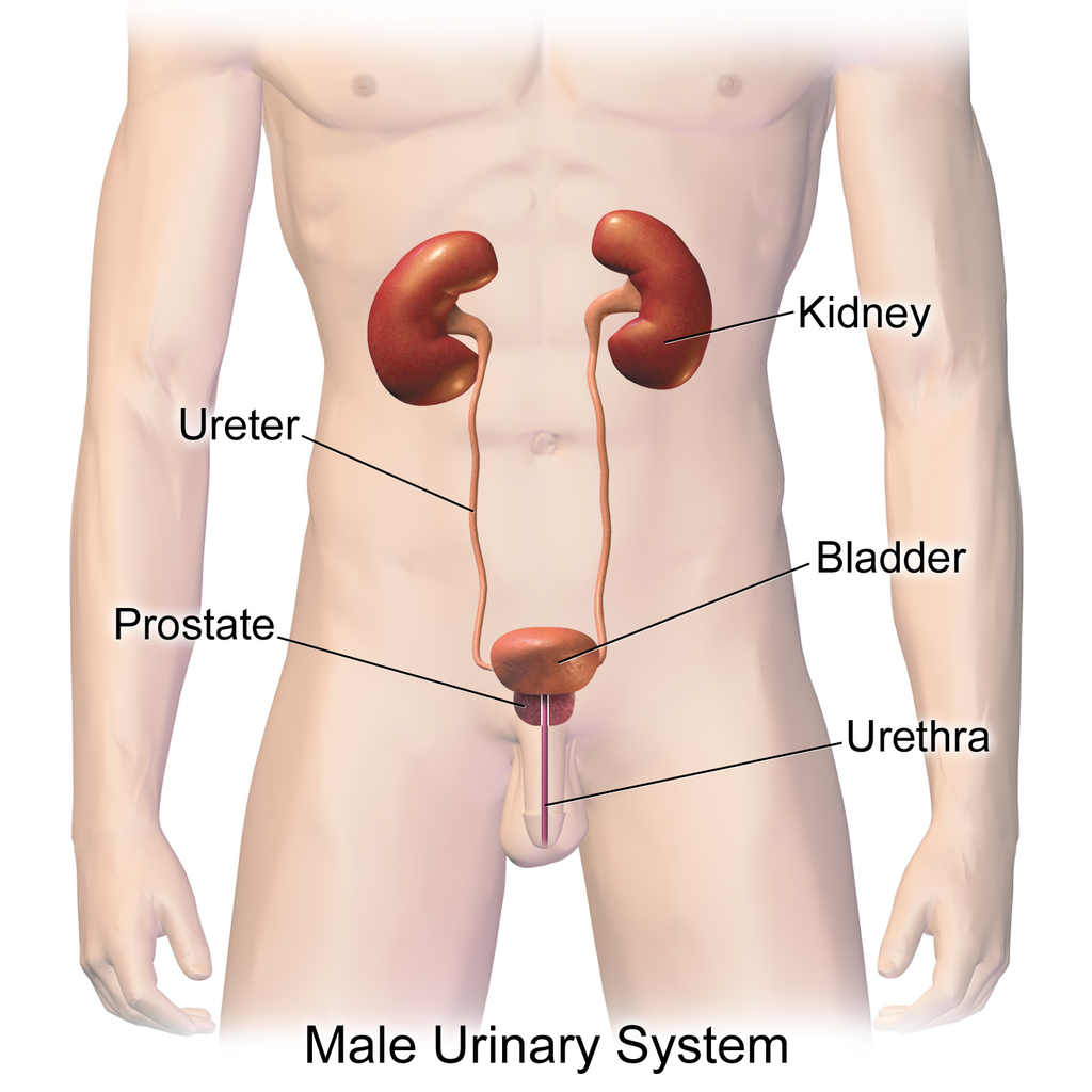
The actual human urinary system, also known as the renal system, is shown in Figure 16.3.2. The system consists of the kidneys, ureters, bladder, and urethra. The main function of the urinary system is to eliminate the waste products of metabolism from the body by forming and excreting urine. Typically, between one and two litres of urine are produced every day in a healthy individual.
Organs of the Urinary System
The urinary system is all about urine. It includes organs that form urine, and also those that transport, store, or excrete urine.
Kidneys
Urine is formed by the kidneys, which filter many substances out of the blood, allow the blood to reabsorb needed materials, and use the remaining materials to form urine. The human body normally has two paired kidneys, although it is possible to get by quite well with just one. As you can see in Figure 16.3.3, each kidney is well supplied with blood vessels by a major artery and vein. Blood to be filtered enters the kidney through the renal artery, and the filtered blood leaves the kidney through the renal vein. The kidney itself is wrapped in a fibrous capsule, and consists of a thin outer layer called the cortex, and a thicker inner layer called the medulla.
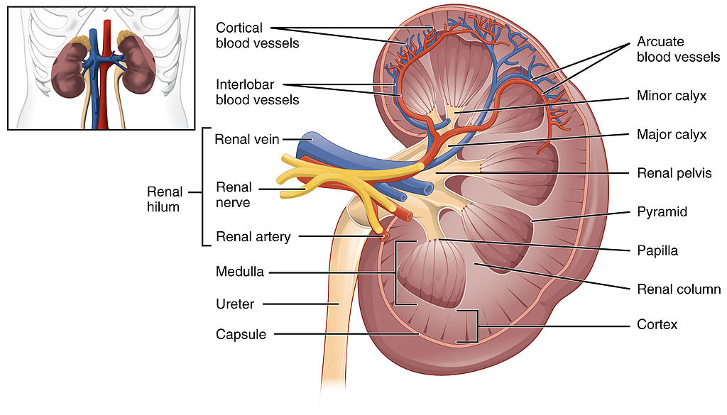
Blood is filtered and urine is formed by tiny filtering units called nephrons. Each kidney contains at least a million nephrons, and each nephron spans the cortex and medulla layers of the kidney. After urine forms in the nephrons, it flows through a system of converging collecting ducts. The collecting ducts join together to form minor calyces (or chambers) that join together to form major calyces (see Figure 16.3.3 above). Ultimately, the major calyces join the renal pelvis, which is the funnel-like end of the ureter where it enters the kidney.
Ureters, Bladder, Urethra
After urine forms in the kidneys, it is transported through the ureters (one per kidney) via peristalsis to the sac-like urinary bladder, which stores the urine until urination. During urination, the urine is released from the bladder and transported by the urethra to be excreted outside the body through the external urethral opening.
Functions of the Urinary System
Waste products removed from the body with the formation and elimination of urine include many water-soluble metabolic products. The main waste products are urea — a by-product of protein catabolism — and uric acid, a by-product of nucleic acid catabolism. Excess water and mineral ions are also eliminated in urine.
Besides the elimination of waste products such as these, the urinary system has several other vital functions. These include:
- Maintaining homeostasis of mineral ions in extracellular fluid: These ions are either excreted in urine or returned to the blood as needed to maintain the proper balance.
- Maintaining homeostasis of blood pH: When pH is too low (blood is too acidic), for example, the kidneys excrete less bicarbonate (which is basic) in urine. When pH is too high (blood is too basic), the opposite occurs, and more bicarbonate is excreted in urine.
- Maintaining homeostasis of extracellular fluids, including the blood volume, which helps maintain blood pressure: The kidneys control fluid volume and blood pressure by excreting more or less salt and water in urine.
Control of the Urinary System
The formation of urine must be closely regulated to maintain body-wide homeostasis. Several endocrine hormones help control this function of the urinary system, including antidiuretic hormone, parathyroid hormone, and aldosterone.
- Antidiuretic hormone (ADH), also called vasopressin, is secreted by the posterior pituitary gland. One of its main roles is conserving body water. It is released when the body is dehydrated, and it causes the kidneys to excrete less water in urine.
- Parathyroid hormone is secreted by the parathyroid glands. It works to regulate the balance of mineral ions in the body via its effects on several organs, including the kidneys. Parathyroid hormone stimulates the kidneys to excrete less calcium and more phosphorus in urine.
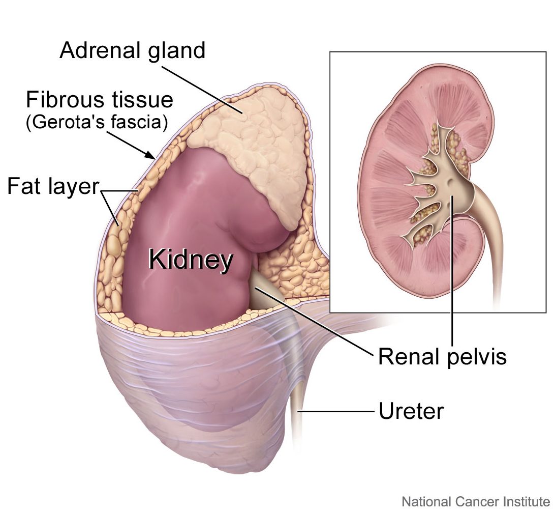
- Aldosterone is secreted by the cortex of the adrenal glands, which rest atop the kidneys, as shown in Figure 16.3.4. Through its effect on the kidneys, it plays a central role in regulating blood pressure. It causes the kidneys to excrete less sodium and water in urine.
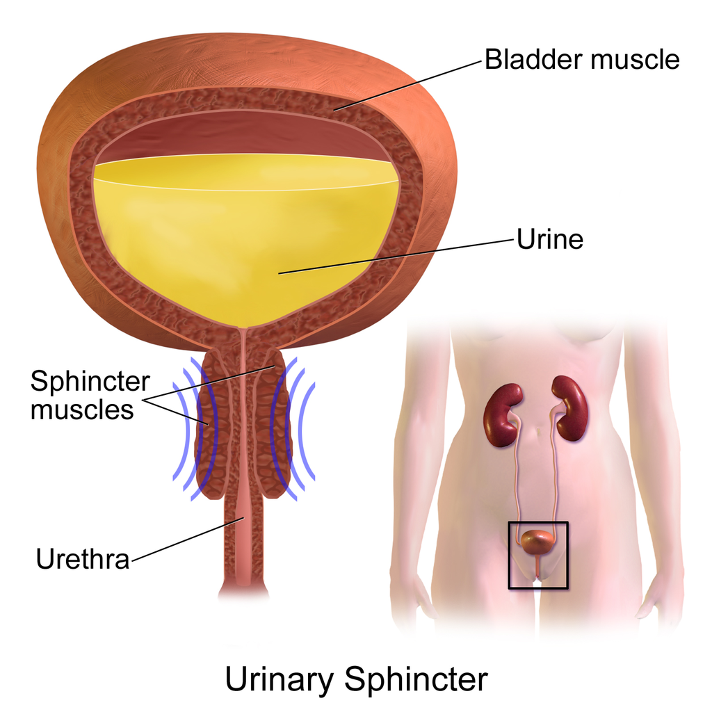
Once urine forms, it is excreted from the body in the process of urination, also sometimes referred to as micturition. This process is controlled by both the autonomic and the somatic nervous systems. As the bladder fills with urine, it causes the autonomic nervous system to signal smooth muscle in the bladder wall to contract (as shown in Figure 16.3.5), and the sphincter between the bladder and urethra to relax and open. This forces urine out of the bladder and through the urethra. Another sphincter at the distal end of the urethra is under voluntary control. When it relaxes under the influence of the somatic nervous system, it allows urine to leave the body through the external urethral opening.
16.3 Summary
- The urinary system consists of the kidneys, ureters, bladder, and urethra. The main function of the urinary system is to eliminate the waste products of metabolism from the body by forming and excreting urine.
- Urine is formed by the kidneys, which filter many substances out of blood, allow the blood to reabsorb needed materials, and use the remaining materials to form urine. Blood to be filtered enters the kidney through the renal artery, and filtered blood leaves the kidney through the renal vein.
- Within each kidney, blood is filtered and urine is formed by tiny filtering units called nephrons, of which there are at least a million in each kidney.
- After urine forms in the kidneys, it is transported through the ureters via peristalsis to the urinary bladder. The bladder stores the urine until urination, when urine is transported by the urethra to be excreted outside the body.
- Besides the elimination of waste products (such as urea, uric acid, excess water, and mineral ions), the urinary system has other vital functions. These include maintaining homeostasis of mineral ions in extracellular fluid, regulating acid-base balance in the blood, regulating the volume of extracellular fluids, and controlling blood pressure.
- The formation of urine must be closely regulated to maintain body-wide homeostasis. Several endocrine hormones help control this function of the urinary system, including antidiuretic hormone from the posterior pituitary gland, parathyroid hormone from the parathyroid glands, and aldosterone from the adrenal glands.
- The process of urination is controlled by both the autonomic and the somatic nervous systems. The autonomic system causes the bladder to empty, but conscious relaxation of the sphincter at the distal end of the urethra allows urine to leave the body.
16.3 Review Questions
-
- State the main function of the urinary system.
- What are nephrons?
- Other than the elimination of waste products, identify functions of the urinary system.
- How is the formation of urine regulated?
- Explain why it is important to have voluntary control over the sphincter at the end of the urethra.
- In terms of how they affect the kidneys, compare aldosterone to antidiuretic hormone.
- If your body needed to retain more calcium, which of the hormones described in this concept is most likely to increase? Explain your reasoning.
16.3 Explore More
The Urinary System – An Introduction | Physiology | Biology | FuseSchool, 2017.
https://youtu.be/pyMcTUQYMQw
Maple Syrup Urine Disease, Alexandria Doody, 2016.
How Accurate Are Drug Tests? Seeker, 2016.
Three Ways Pee Could Change the World, Gross Science, 2015.
Attributions
Figure 16.3.1
- File:Pyrodex powder ffg.jpg by Hustvedt on Wikimedia Commons is used under a CC BY SA 3.0 (https://creativecommons.org/licenses/by-sa/3.0/deed.en).
- Brown leather satchel bag by Álvaro Serrano on Unsplash is used under the Unsplash Licence (https://unsplash.com/license).
- Laundry basket by Andy Fitzsimon on Unsplash is used under the Unsplash Licence (https://unsplash.com/license).
- Tags: Wool Skeins Natural Dyed Colorful Himalayan Weavers by on Pixabay is used under the Pixabay License (https://pixabay.com/service/license/).
Figure 16.3.2
Urinary_System_(Male) by BruceBlaus on Wikimedia Commons is used under a CC BY-SA 4.0 (https://creativecommons.org/licenses/by-sa/4.0) license.
Figure 16.3.3
2610_The_Kidney by OpenStax College on Wikimedia Commons is used under a CC BY 3.0 (https://creativecommons.org/licenses/by/3.0) license.
Figure 16.3.4
Adrenal glands on Kidney by Alan Hoofring (Illustrator)/ NCI Visuals Online is in the public domain (https://en.wikipedia.org/wiki/Public_domain).
Figure 16.3.5
Urinary_Sphincter by BruceBlaus on Wikimedia Commons is used under a CC BY-SA 4.0 (https://creativecommons.org/licenses/by-sa/4.0) license.
References
Alexandria Doody. (2016, March 29). Maple syrup urine disease. YouTube. https://www.youtube.com/watch?v=pyMcTUQYMQw&feature=youtu.be
Betts, J. G., Young, K.A., Wise, J.A., Johnson, E., Poe, B., Kruse, D.H., Korol, O., Johnson, J.E., Womble, M., DeSaix, P. (2013, June 19). Figure 25.8 Left kidney [digital image]. In Anatomy and Physiology (Section 25.3). OpenStax. https://openstax.org/books/anatomy-and-physiology/pages/25-3-gross-anatomy-of-the-kidney
FuseSchool. (2017, June 19). The urinary system – An introduction | Physiology | Biology | FuseSchool. YouTube. https://www.youtube.com/watch?v=dxecGD0m0Xc&feature=youtu.be
Gross Science. (2015, September 15). Three ways pee could change the world. YouTube. https://www.youtube.com/watch?v=xt1Tj5eeS0k&feature=youtu.be
Seeker. (2016, January 16). How accurate are drug tests? YouTube. https://www.youtube.com/watch?v=3z-xjfdJWAI&feature=youtu.be
Created by: CK-12/Adapted by Christine Miller

Fast and Furious
These sprinters' muscles will need a lot of energy to complete this short race, because they will be running at top speed. The action won't last long, but it will be very intense. The energy each sprinter needs can't be provided quickly enough by aerobic cellular respiration. Instead, their muscle cells must use a different process to power their activity.
Making ATP Without Oxygen
Living things' cells power their activities with the energy-carrying molecule ATP (adenosine triphosphate). The cells of most living things make ATP from glucose in the process of cellular respiration. This process occurs in three stages: glycolysis, the Krebs cycle, and electron transport. The latter two stages require oxygen, making cellular respiration an aerobic process. When oxygen is not available in cells, the ETS quickly shuts down. Luckily, there are also ways of making ATP from glucose which are anaerobic, which means that they do not require oxygen. These processes are referred to collectively as anaerobic respiration.
Fermentation
Ome important way of making ATP without oxygen is fermentation. Fermentation starts with glycolysis, which does not require oxygen, but it does not involve the latter two stages of aerobic cellular respiration (the Krebs cycle and electron transport). There are two types of fermentation: alcoholic fermentation and lactic acid fermentation. We make use of both types of fermentation using other organisms, but only lactic acid fermentation actually takes place inside the human body.
Alcoholic Fermentation
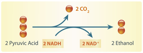
Alcoholic fermentation is carried out by single-celled fungi (called yeasts), as well as some bacteria. We use alcoholic fermentation in these organisms to make biofuels, bread, and wine. The biofuel ethanol (a type of alcohol), for example, is produced by alcoholic fermentation of the glucose in corn or other plants. The process by which this happens is summarized in the diagram below. The two pyruvic acid molecules shown in the diagram come from the splitting of glucose in the first stage of the process (glycolysis). ATP is also made during glycolysis. Two molecules of ATP are produced from each molecule of glucose.

Yeasts in bread dough also use alcoholic fermentation for energy. They produce carbon dioxide gas as a waste product. The carbon dioxide released causes bubbles in the dough and explains why the dough rises. Do you see the small holes in the bread pictured to the right? The holes were formed by bubbles of carbon dioxide gas.
As you have probably guessed, yeast is also used in producing alcoholic beverages. When making beer, brewers will add yeast to a mix of barley and hops. In the absence of oxygen, yeast will carry out alcoholic fermentation in order to convert the glucose in the barley into energy, producing the alcohol content as well as the carbonation present in beer.
Lactic Acid Fermentation
Lactic acid fermentation is carried out by certain bacteria, including the bacteria in yogurt. It is also carried out by your muscle cells when you work them hard and fast. This is how the muscles of the sprinters pictured above get energy for their short-lived — but intense — activity. When this happens, your muscles are using ATP faster than your cardiovascular system can deliver oxygen! The process by which this happens is summarized in the diagram below. Again, the two pyruvic acid molecules shown in the diagram come from the splitting of glucose in the first stage of the process (glycolysis). It is also during this stage that two ATP molecules are produced. The rest of the processes produce lactic acid. Note that, unlike in alcoholic fermentation, there is no carbon dioxide waste product in lactic acid fermentation.
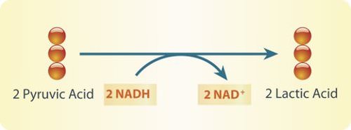
Lactic acid fermentation produces lactic acid and NAD+. The NAD+ cycles back to allow glycolysis to continue so more ATP is made. Each circle represents a carbon atom.
Did you ever run a race, lift heavy weights, or participate in some other intense activity and notice that your muscles start to feel a burning sensation? This may occur when your muscle cells use lactic acid fermentation to provide ATP for energy. The buildup of lactic acid in the muscles causes a burning feeling. This painful sensation is useful if it gets you to stop overworking your muscles and allow them a recovery period, during which cells can eliminate the lactic acid.
Pros and Cons of Anaerobic Respiration
With oxygen, organisms can use aerobic cellular respiration to produce up to 38 molecules of ATP from just one molecule of glucose. Without oxygen, organisms must use anaerobic respiration to produce ATP, and this process produces only two molecules of ATP per molecule of glucose. Although anaerobic respiration produces less ATP, it has the advantage of doing so very quickly. For example, it allows your muscles to get the energy they need for short bursts of intense activity. Aerobic cellular respiration, in contrast, produces ATP more slowly.
Fermentation in Food Production
Anaerobic respiration is also used in the food industry. You read about yeast's role in making bread and beer, but did you know that there are many microbes that are used to create the food we eat, including cheese, sour cream, yogurt, soy sauce, olives, pepperoni, and many more. Watch the video below to learn more about fermentation in the food industry.
https://www.youtube.com/watch?v=eksagPy5tmQ&t=3s
The beneficial bacteria that make delicious food - Erez Garty, TED-Ed, 2016.
4.11 Cultural Connection
Fishing has always been part of the culture and nutrition of Indigenous peoples living on the west coast of Canada. Fish provides delicious important nutrients such as protein, Omega-3 fatty acids, calcium, iron, and Vitamins A, B, C and D. Traditionally, no part of the fish was wasted, including head, eyes, internal organs, and eggs.
Eulachon, also known as candle fish or oolichan, (pictured below) have been prized for their oil for thousands of years. The pathways of these fish dictated "grease trails" and are found from Bristol Bay, Alaska, all the way south to the Klamath River, California. Within BC, the areas of Nass, Knights Inlet, and Bella Coola had large trading centres for this important natural resource.
Photos by Brodie Guy - www.brodieguy.com CC BY-NC-ND 4.0
Euchalon were and are eaten fresh, smoked or dried, and as grease. The grease remains a highly valued food to Indigenous coastal communities. The flavour of the grease varies greatly depending not only on where the fish is from and how it is made, but also how long it is left to ferment. To ferment the eulachon, fish are left in a wood-lined locker dug into the soil for 10 days. Fermentation uses the action of fungi and bacteria to break down the fish making oil extraction much faster and easier.
To learn more, visit the First Nations Health Authority Traditional Foods Fact Sheet and a feature in the Yukon News, "Eulachon, oolicahn, hooligan: A fish by any other name is just as oily."
4.11 Summary
- The cells of most living things produce ATP from glucose by aerobic cellular respiration, which uses oxygen. Some organisms instead produce ATP from glucose by anaerobic respiration, which does not require oxygen.
- An important way of making ATP without oxygen is fermentation. There are two types of fermentation: alcoholic fermentation and lactic acid fermentation. Both start with glycolysis, the first (anaerobic) stage of cellular respiration, in which two molecules of ATP are produced from one molecule of glucose.
- Alcoholic fermentation is carried out by single-celled organisms, including yeasts and some bacteria. We use alcoholic fermentation in these organisms to make biofuels, bread, and wine.
- Lactic acid fermentation is undertaken by certain bacteria, including the bacteria in yogurt, and also by our muscle cells when they are worked hard and fast.
- Anaerobic respiration produces far less ATP than does aerobic cellular respiration, but it has the advantage of being much faster. For example, it allows muscles to get the energy they need for short bursts of intense activity.
4.11 Review Questions
- Explain the primary difference between aerobic cellular respiration and anaerobic respiration.
- What is fermentation?
- Compare and contrast alcoholic and lactic acid fermentation.
- Identify the major pros and the major cons of anaerobic respiration relative to aerobic cellular respiration.
- What process is shared between aerobic cellular respiration and anaerobic respiration? Describe the process briefly. Why can this process happen in anaerobic respiration, as well as aerobic respiration?
-
- What is the reactant (or starting material)common to aerobic respiration and both types of fermentation?
4.11 Explore More
https://www.youtube.com/watch?v=cDC29iBxb3w&t=3s
Anaerobic Respiration, Bozeman Science, 2013.
https://www.youtube.com/watch?v=YbdkbCU20_M
Fermentation, The Amoeba Sisters, 2018.
https://www.youtube.com/watch?time_continue=17&v=TVtqwWGguFk&feature=emb_logo
Science of Beer: Tapping the Power of Brewer's Yeast, KQED Science, 2014.
Attributions
Figure 4.11.1
Sprinters by Jonathan Chng on Unsplash is used under the Unsplash License (https://unsplash.com/license).
Figure 4.11.2
Alcoholic fermentation by Hana Zavadska/ CK-12 Foundation is used under a CC BY-NC 3.0 (https://creativecommons.org/licenses/by-nc/3.0/) license.
Figure 4.11.3
Bread [photo] by Orlova Maria on Unsplash is used under the Unsplash License (https://unsplash.com/license).
Figure 4.11.4
Lactic Acid Fermenation by Hana Zavadska/ CK-12 Foundation is used under a CC BY-NC 3.0 (https://creativecommons.org/licenses/by-nc/3.0/) license.
References
Bozeman Science. (2013, May 2). Anaerobic respiration. YouTube. https://www.youtube.com/watch?v=cDC29iBxb3w&feature=youtu.be
Hana Zavadska/CK-12 Foundation. (2016, August 15). Figure 2: Alcoholic fermentation [digital image]. In Brainard, J., and Henderson, R., CK-12’s College Human Biology FlexBook® (Section 4.11). CK-12 Foundation. https://www.ck12.org/book/ck-12-college-human-biology/section/4.11/
Hana Zavadska/CK-12 Foundation. (2016, August 15). Figure 4: Lactic acid fermentation [digital image]. In Brainard, J., and Henderson, R., CK-12’s College Human Biology FlexBook® (Section 4.11). CK-12 Foundation. https://www.ck12.org/book/ck-12-college-human-biology/section/4.11/
First Nations Health Authority. (2019, September 6). First Nations traditional foods facts Sheet [pdf]. https://www.fnha.ca/Documents/Traditional_Food_Fact_Sheets.pdf
Genest, M. (2017, May 24). Eulachon, oolichan, hooligan: A fish by any other name is just as oily [online article]. YukonNews.com. https://www.yukon-news.com/business/eulachon-oolichan-hooligan-a-fish-by-any-other-name-is-just-as-oily/
KQED Science. (2014, February 11). Science of beer: Tapping the power of brewer's yeast. YouTube. https://www.youtube.com/watch?v=TVtqwWGguFk&feature=youtu.be
TED-Ed. (2016). The beneficial bacteria that make delicious food - Erez Garty. YouTube. https://www.youtube.com/watch?v=eksagPy5tmQ&feature=youtu.be
The Amoeba Sisters. (2018, April 30). Fermentation. YouTube. https://www.youtube.com/watch?v=YbdkbCU20_M&feature=youtu.be
Wikipedia contributors. (2020, June 21). Ethanol fuel. In Wikipedia. https://en.wikipedia.org/w/index.php?title=Ethanol_fuel&oldid=963675942
Figure 7.4.1 Construction — It's important to have the right materials for the job.
The Right Material for the Job
Building a house is a big job and one that requires a lot of different materials for specific purposes. As you can see in Figure 7.4.1, many different types of materials are used to build a complete house, but each type of material fulfills certain functions. You wouldn't use insulation to cover your roof, and you wouldn't use lumber to wire your home. Just as a builder chooses the appropriate materials to build each aspect of a home (wires for electrical, lumber for framing, shingles for roofing), your body uses the right cells for each type of role. When many cells work together to perform a specific function, this is termed a tissue.
Tissues
Groups of connected cells form tissues. The cells in a tissue may all be the same type, or they may be of multiple types. In either case, the cells in the tissue work together to carry out a specific function, and they are always specialized to be able to carry out that function better than any other type of tissue. There are four main types of human tissues: connective, epithelial, muscle, and nervous tissues. We use tissues to build organs and organ systems. The 200 types of cells that the body can produce based on our single set of DNA can create all the types of tissue in the body.
Epithelial Tissue
Epithelial tissue is made up of cells that line inner and outer body surfaces, such as the skin and the inner surface of the digestive tract. Epithelial tissue that lines inner body surfaces and body openings is called mucous membrane. This type of epithelial tissue produces mucus, a slimy substance that coats mucous membranes and traps pathogens, particles, and debris. Epithelial tissue protects the body and its internal organs, secretes substances (such as hormones) in addition to mucus, and absorbs substances (such as nutrients).
The key identifying feature of epithelial tissue is that it contains a free surface and a basement membrane. The free surface is not attached to any other cells and is either open to the outside of the body, or is open to the inside of a hollow organ or body tube. The basement membrane anchors the epithelial tissue to underlying cells.
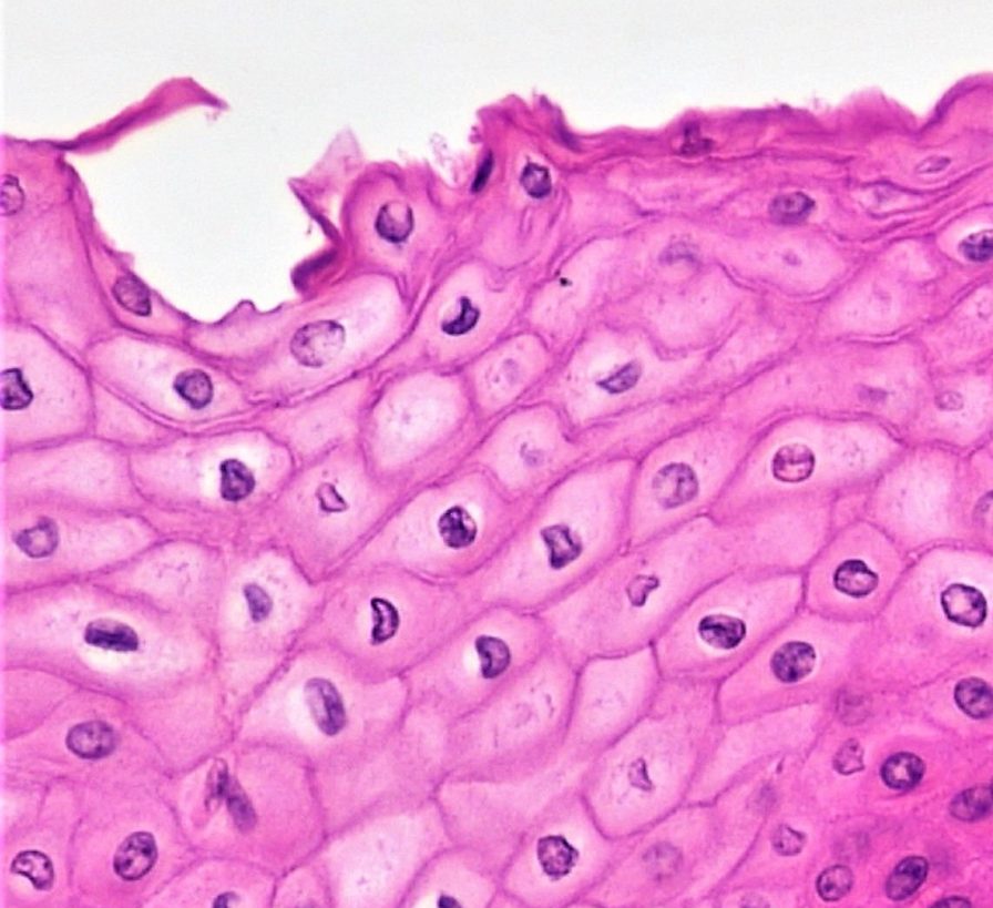
Epithelial tissue is identified and named by shape and layering. Epithelial cells exist in three main shapes: squamous, cuboidal, and columnar. These specifically shaped cells can, depending on function, be layered several different ways: simple, stratified, pseudostratified, and transitional.
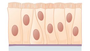
Epithelial tissue forms coverings and linings and is responsible for a range of functions including diffusion, absorption, secretion and protection. The shape of an epithelial cell can maximize its ability to perform a certain function. The thinner an epithelial cell is, the easier it is for substances to move through it to carry out diffusion and/or absorption. The larger an epithelial cell is, the more room it has in its cytoplasm to be able to make products for secretion, and the more protection it can provide for underlying tissues. Their are three main shapes of epithelial cells: squamous (which is shaped like a pancake- flat and oval), cuboidal (cube shaped), and columnar (tall and rectangular).
Figure 7.4.2 The shape of epithelial tissues is important.
Epithelial tissue will also organize into different layerings depending on their function. For example, multiple layers of cells provide excellent protection, but would no longer be efficient for diffusion, whereas a single layer would work very well for diffusion, but no longer be as protective; a special type of layering called transitional is needed for organs that stretch, like your bladder. Your tissues exhibit the layering that makes them most efficient for the function they are supposed to perform. There are four main layerings found in epithelial tissue: simple (one layer of cells), stratified (many layers of cells), pseudostratified (appears stratified, but upon closer inspection is actually simple), and transitional (can stretch, going from many layers to fewer layers).
Figure 7.4.3 The layerings found in epithelial tissues is important.
See Table 7.4.1 for a summary of the different layering types and shapes epithelial cells can form and their related functions and locations.
Table 7.4.1
Summary of Epithelial Tissue Cells
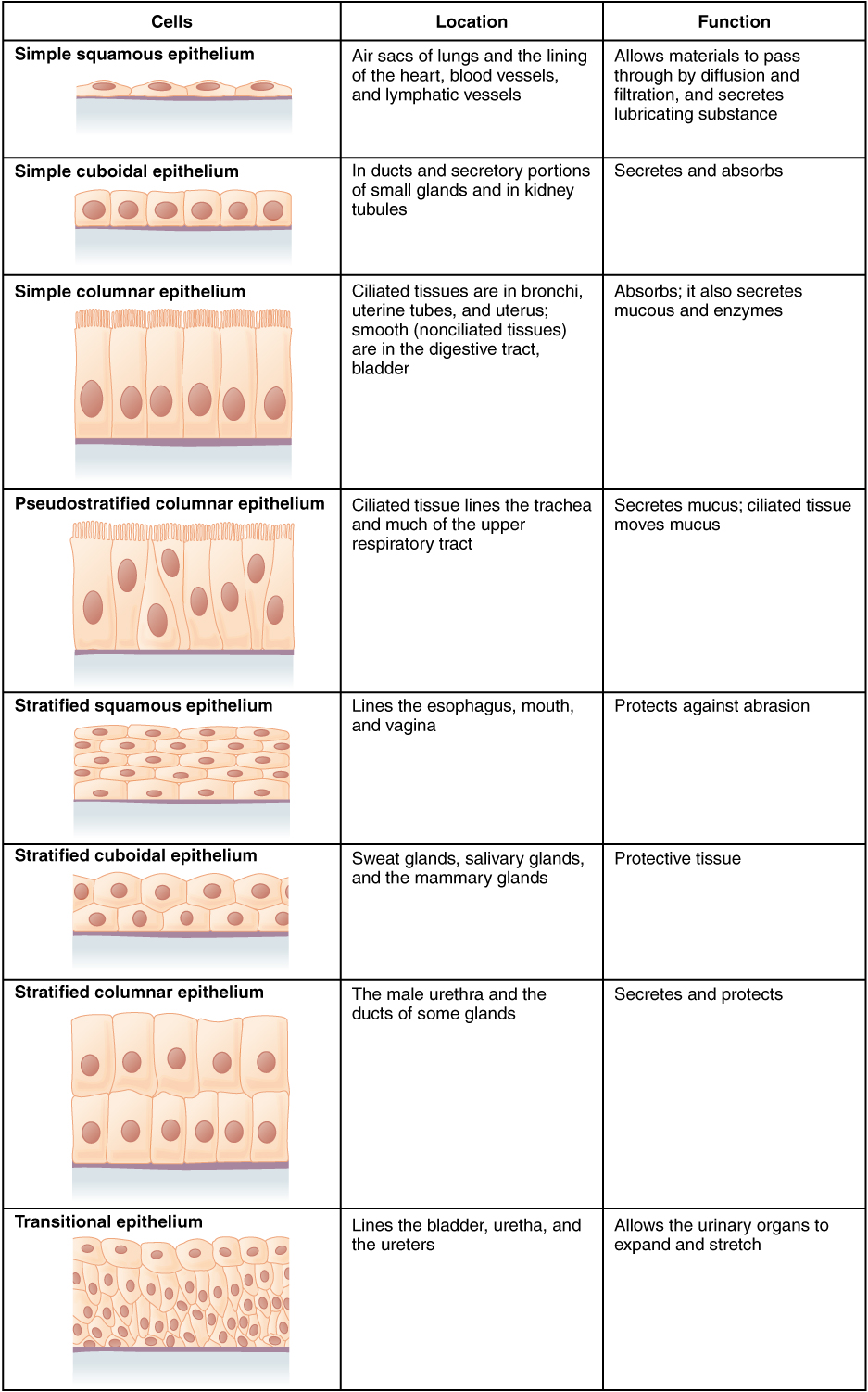
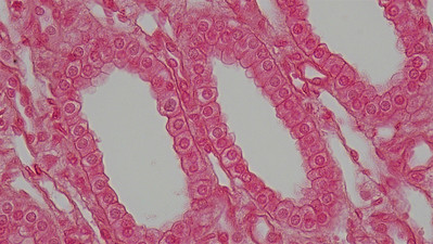
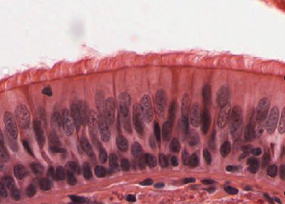
So far, we have identified epithelial tissue based on shape and layering. The representative diagrams we have seen so far are helpful for visualizing the tissue structures, but it is important to look at real examples of these cells. Since cells are too tiny to see with the naked eye, we rely on microscopes to help us study them. Histology is the study of the microscopic anatomy and cells and tissues. See Table 7.4.2 to see some examples of slides of epithelial tissues prepared for the purpose of histology.
Table 7.4.2
Epithelial Tissues and Histological Samples
| Epithelial Tissue Type | Tissue Diagram | Histological Sample |
| Stratified squamous
(from skin) |
 |
|
| Simple cuboidal
(from kidney tubules) |
 |
|
| Pseudostratified ciliated columnar
(from trachea) |
 |
 |
Connective Tissue
Bone and blood are examples of connective tissue. Connective tissue is very diverse. In general, it forms a framework and support structure for body tissues and organs. It's made up of living cells separated by non-living material, called extracellular matrix, which can be solid or liquid. The extracellular matrix of bone, for example, is a rigid mineral framework. The extracellular matrix of blood is liquid plasma.
The key identifying feature of connective tissue is that is is composed of a scattering of cells in a non-cellular matrix. There are three main categories of connective tissue, based on the nature of the matrix. They look very different from one another, which is a reflection of their different functions:
- Fibrous connective tissue: is characterized by a matrix which is flexible and is made of protein fibres including collagen, elastin and possibly reticular fibres. These tissues are found making up tendons, ligaments, and body membranes.
- Supportive connective tissue: is characterized by a solid matrix and is what is used to make bone and cartilage. These tissues are used for support and protection.
- Fluid connective tissue: is characterized by a fluid matrix and includes both blood and lymph.
Fibrous Connective Tissue
Fibrous connective tissue contains cells called fibroblasts. These cells produce fibres of collagen, elastin, or reticular fibre which makes up the matrix of this type of connective tissue. Based on how tightly packed these fibres are and how they are oriented changes the properties, and therefore the function of the fibrous connective tissue.
- Loose fibrous connective tissue: composed of a loose and disorganized weave of collagen and elastin fibres, creating a tissue that is thin and flexible, yet still tough. This tissue, which is also sometimes referred to as "areolar tissue", is found in membranes and surrounding blood vessels and most body organs. As you can see from the diagram in Figure 7.4.4, loose fibrous connective tissue fulfills the definition of connectives tissue since it is a scattering of cells (fibroblasts) in a non-cellular matrix (a mesh of collagen and elastin fibres). There are two types of specialized loose fibrous connective tissue: reticular and adipose. Adipose tissue stores fat and reticular tissue forms the spleen and lymph nodes.
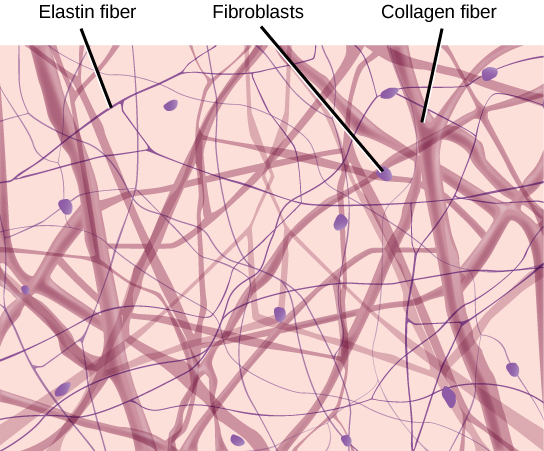
Figure 7.4.4 Diagram of loose fibrous connective tissue consists of a scattering of fibroblasts in a non-cellular matrix of loosely woven collagen and elastin fibres. 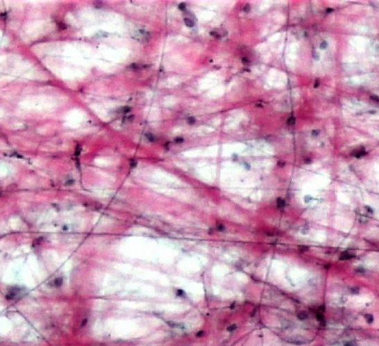
Figure 7.4.5 Microscopic view of loose fibrous connective tissue. - Dense Fibrous Connective Tissue: composed of a dense mat of parallel collagen fibres and a scattering of fibroblasts, creating a tissue that is very strong. Dense fibrous connective tissue forms tendons and ligaments, which connect bones to muscles and/or bones to neighbouring bones.
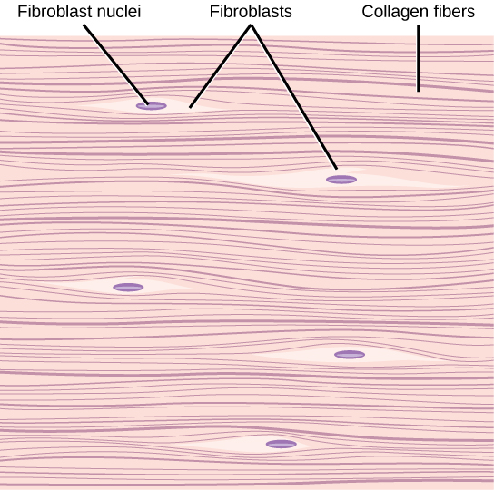
Figure 7.4.6 Dense fibrous connective tissue is composed of fibroblasts and a dense parallel packing of collagen fibres. 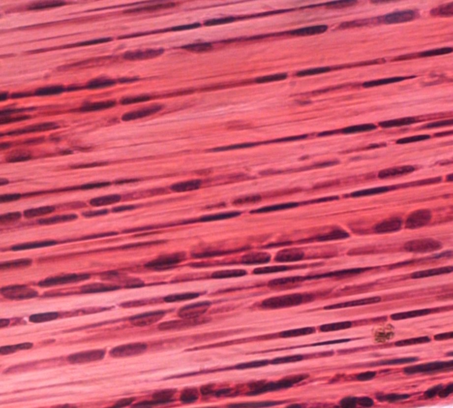
Figure 7.4.7 Microscopic view of dense fibrous connective tissue.
Supportive Connective Tissue
Supportive connective tissue exhibits the defining feature of connective tissue in that it is a scattering of cells in a non-cellular matrix; what sets it apart from other connective tissues is its solid matrix. In this tissue group, the matrix is solid- either bone or cartilage. While fibrous connective tissue contained cells called fibroblasts which produced fibres, supportive connective tissue contains cells that either create bone (osteocytes) or cells that create cartilage (chondrocytes).
Cartilage
Chondrocytes produce the cartilage matrix in which they reside. Cartilage is made up of protein fibres and chondrocytes in lacunae. This is tissue is strong yet flexible and is used many places in the body for protection and support. Cartilage is one of the few tissues that is not vascular (doesn't have a direct blood supply) meaning it relies on diffusion to obtain nutrients and gases; this is the cause of slow healing rates in injuries involving cartilage. There are three main types of cartilage:
- Hyaline cartilage: a smooth, strong and flexible tissue. Found at the ends of ribs and long bones, in the nose, and comprising the entire fetal skeleton.
- Fibrocartilage: a very strong tissue containing thick bundles of collagen. Found in joints that need cushioning from high impact (knees, jaw).
- Elastic cartilage: contains elastic fibres in addition to collagen, giving support with the benefit of elasticity. Found in earlobes and the epiglottis.
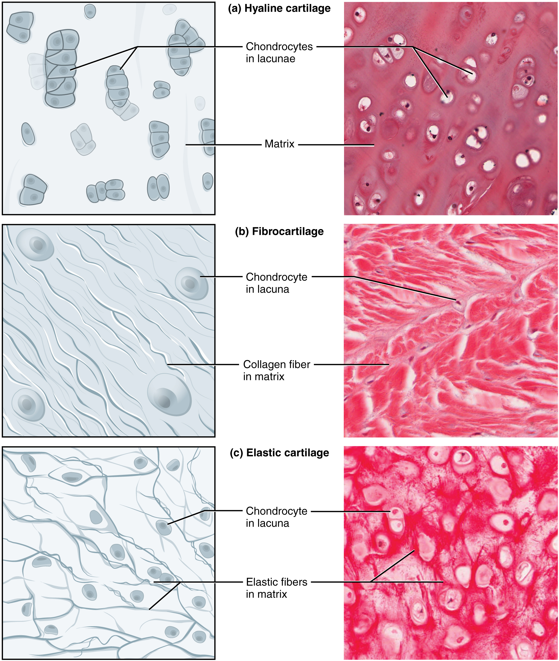
Figure 7.4.8 Three types of cartilage, each with distinct characteristics based on the nature of the matrix.
Bone
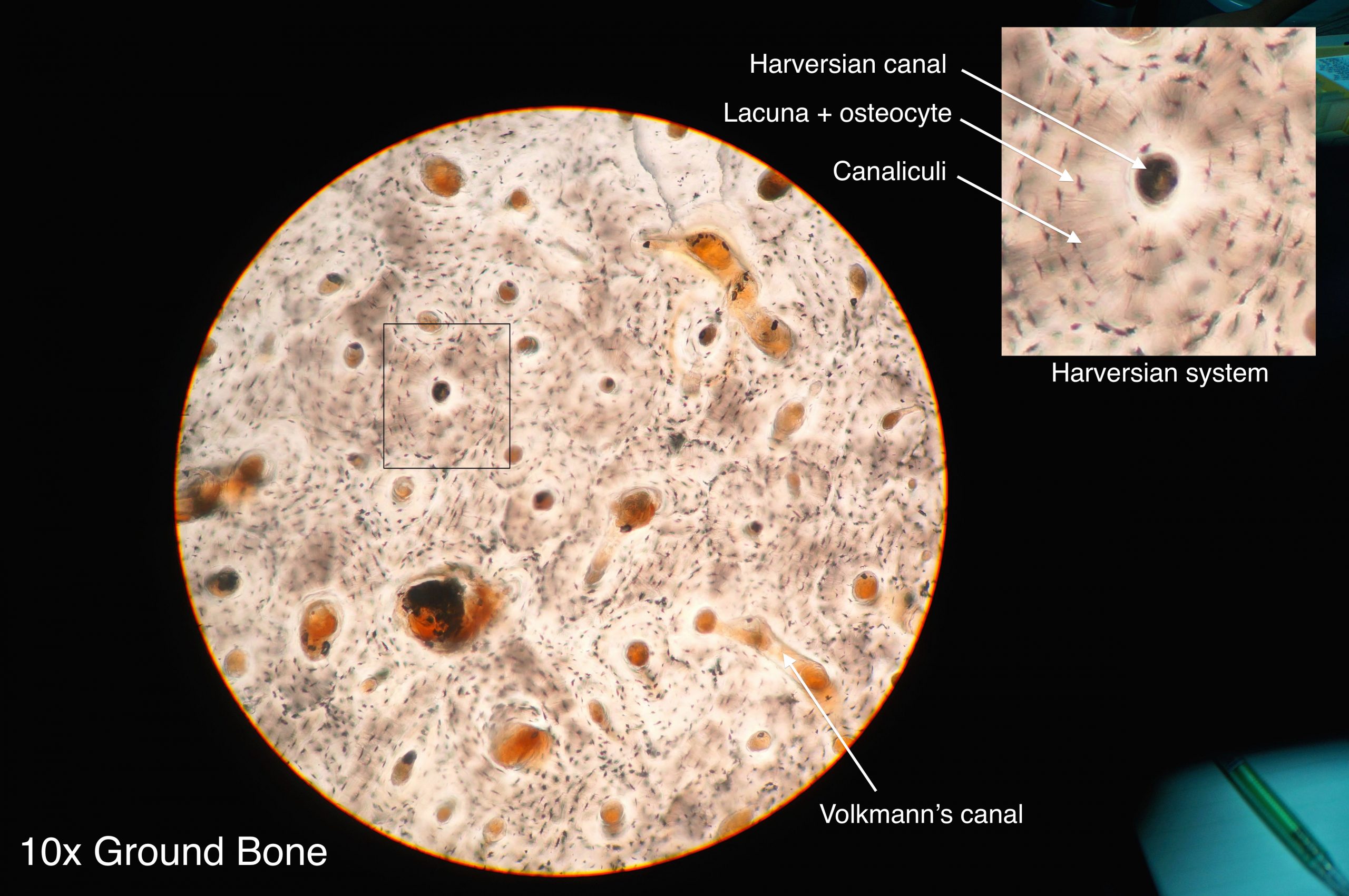
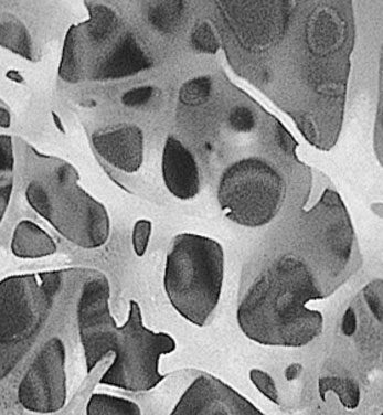
Osteocytes produce the bone matrix in which they reside. Since bone is very solid, these cells reside in small spaces called lacunae. This bone tissue is composed of collagen fibres embedded in calcium phosphate giving it strength without brittleness. There are two types of bone: compact and spongy.
- Compact bone: has a dense matrix organized into cylindrical units called osteons. Each osteon contains a central canal (sometimes called a Harversian Canal) which allows for space for blood vessels and nerves, as well as concentric rings of bone matrix and osteocytes in lacunae, as per the diagram here. Compact bone is found in long bones and forms a shell around spongy bone.
- Spongy bone: a very porous type of bone which most often contains bone marrow. It is found at the end of long bones, and makes up the majority of the ribs, shoulder blades and flat bones of the cranium.
Fluid Connective Tissue
Fluid connective tissue has a matrix that is fluid; unlike the other two categories of connective tissue, the cells that reside in the matrix do not actually produce the matrix. Fibroblasts make the fibrous matrix, chondrocytes make the cartilaginous matrix, osteocytes make the bony matrix, yet blood cells do not make the fluid matrix of either lymph or plasma. This tissue still fits the definition of connective tissue in that it is still a scattering of cells in a non-cellular matrix.
There are two types of fluid connective tissue:
- Blood: blood contains three types of cells suspended in plasma, and is contained in the cardiovascular system.
- Eryththrocytes, more commonly called red blood cells, are present in high numbers (roughly 5 million cells per mL) and are responsible for delivering oxygen from to the lungs to all the other areas of the body. These cells are relatively small in size with a diameter of around 7 micrometres and live no longer than 120 days.
- Leukocytes, often referred to as white blood cells, are present in lower numbers (approximately 5 thousand cells per mL) are responsible for various immune functions. They are typically larger than erythrocytes, but can live much longer, particularly white blood cells responsible for long term immunity. The number of leukocytes in your blood can go up or down based on whether or not you are fighting an infection.
- Thrombocytes, also known as platelets, are very small cells responsible for blood clotting. Thrombocytes are not actually true cells, they are fragments of a much larger cell called a megakaryocyte.
- Lymph: contains a liquid matrix and white blood cells and is contained in the lymphatic system, which ultimately drains into the cardiovascular system.
Figure 7.4.11 A stained lymphocyte surrounded by red blood cells viewed using a light microscope.
Muscular Tissue
Muscular tissue is made up of cells that have the unique ability to contract- which is the defining feature of muscular tissue. There are three major types of muscle tissue, as pictured in Figure 7.4.12 skeletal, smooth, and cardiac muscle tissues.
Skeletal Muscle
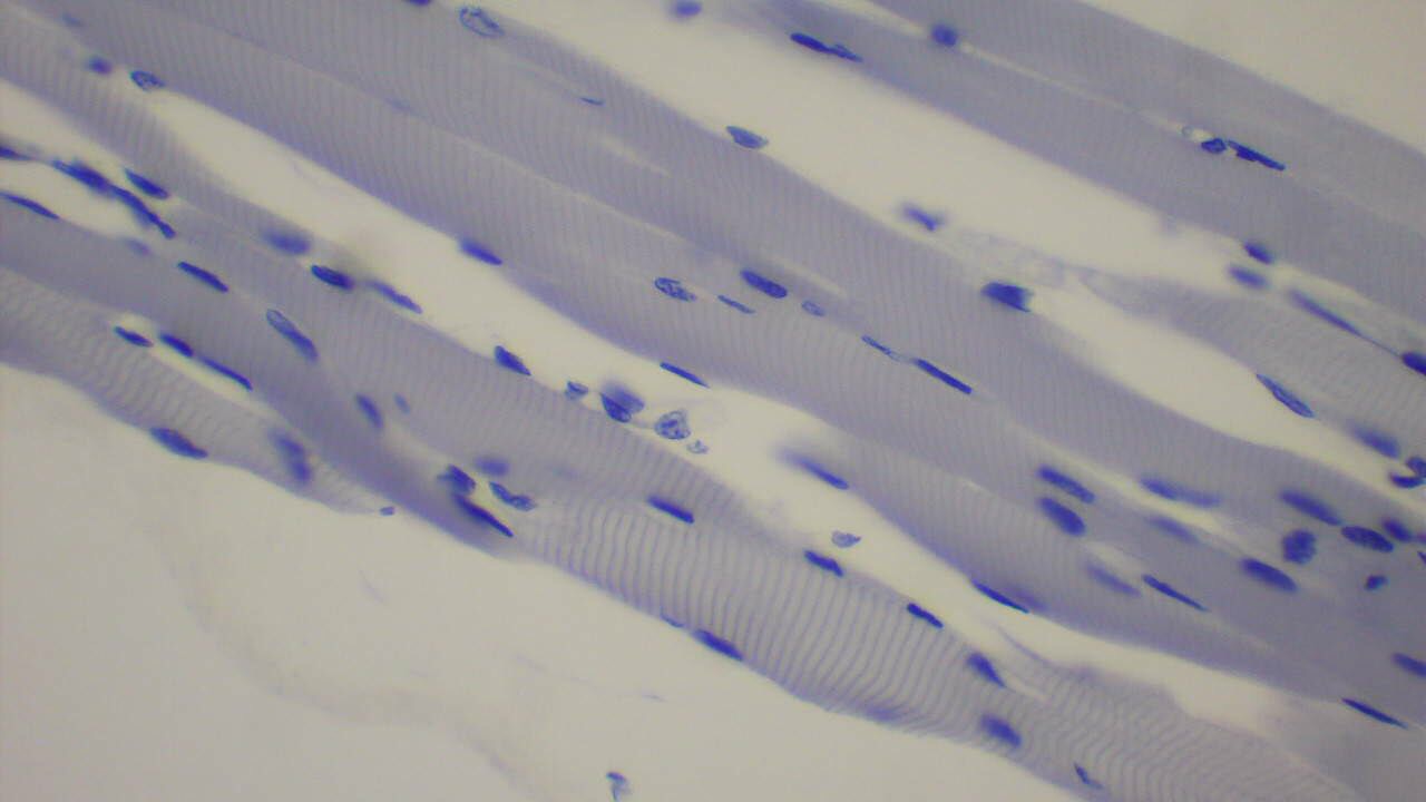
Skeletal muscles are voluntary muscles, meaning that you exercise conscious control over them. Skeletal muscles are attached to bones by tendons, a type of connective tissue. When these muscles shorten to pull on the bones to which they are attached, they enable the body to move. When you are exercising, reading a book, or making dinner, you are using skeletal muscles to move your body to carry out these tasks.
Under the microscope, skeletal muscles are striated (or striped) in appearance, because of their internal structure which contains alternating protein fibres of actin and myosin. Skeletal muscle is described as multinucleated, meaning one "cell" has many nuclei. This is because in utero, individual cells destined to become skeletal muscle fused, forming muscle fibres in a process known as myogenesis. You will learn more about skeletal muscle and how it contracts in the Muscular System.
Smooth Muscle
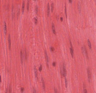
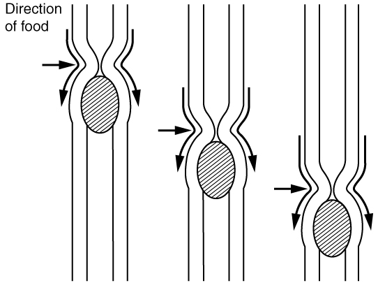
Smooth muscles are nonstriated muscles- they still contain the muscle fibres actin and myosin, but not in the same alternating arrangement seen in skeletal muscle. Smooth muscle is found in the tubes of the body - in the walls of blood vessels and in the reproductive, gastrointestinal, and respiratory tracts. Smooth muscles are not under voluntary control meaning that they operate unconsciously, via the autonomic nervous system. Smooth muscles move substances through a wave of contraction which cascades down the length of a tube, a process termed peristalsis.
Watch the YouTube video "What is Peristalsis" by Mister Science to see peristalsis in action.
https://www.youtube.com/watch?v=kVjeNZA5pi4
What is Peristalsis, Mister Science, 2018.
Cardiac Muscle
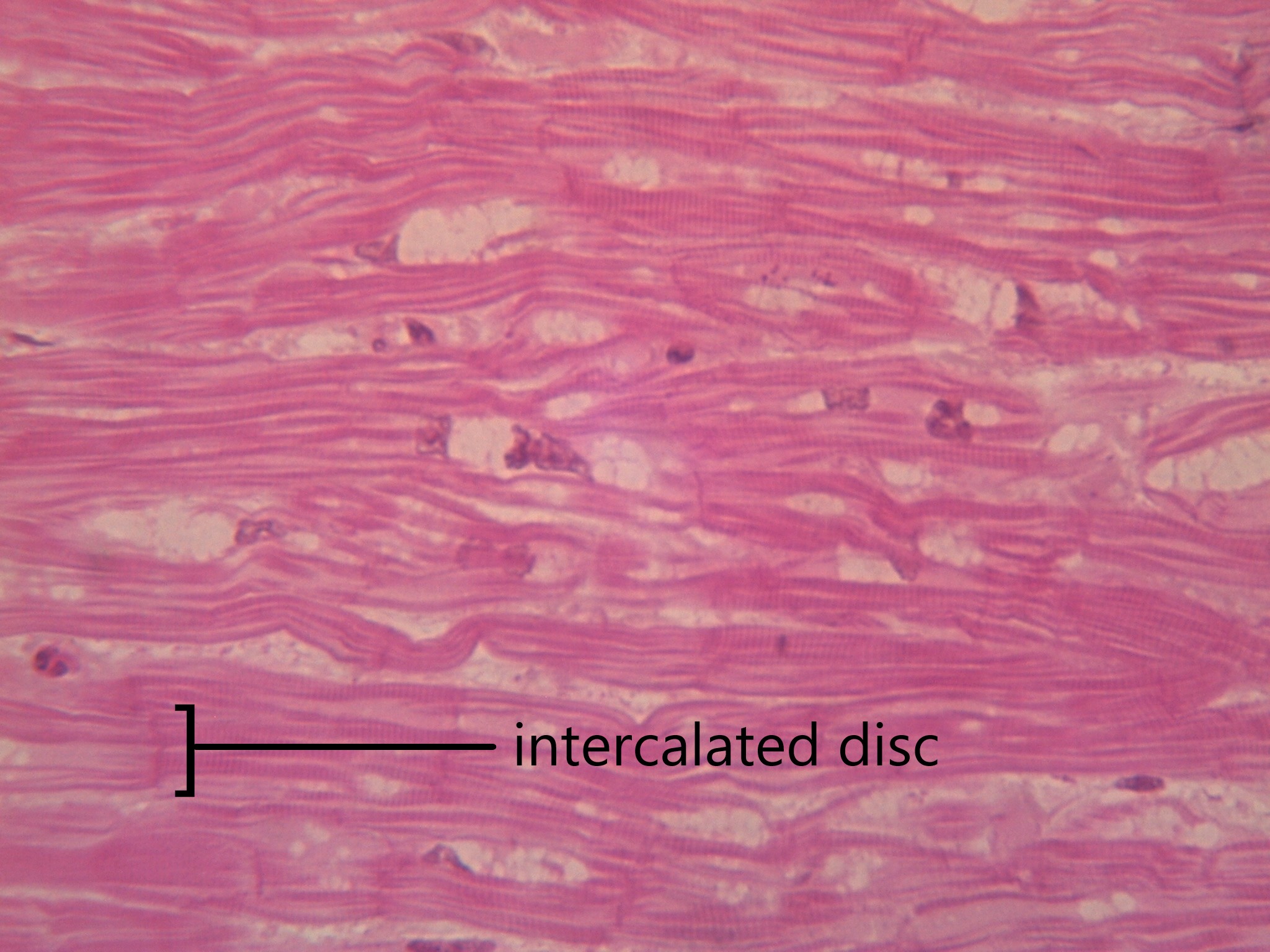
Cardiac muscles work involuntarily, meaning they are regulated by the autonomic nervous system. This is probably a good thing, since you wouldn't want to have to consciously concentrate on keeping your heart beating all the time! Cardiac muscle, which is found only in the heart, is mononucleated and striated (due to alternating bands of myosin and actin). Their contractions cause the heart to pump blood. In order to make sure entire sections of the heart contract in unison, cardiac muscle tissue contains special cell junctions called intercalated discs, which conduct the electrical signals used to "tell" the chambers of the heart when to contract.
Nervous Tissue
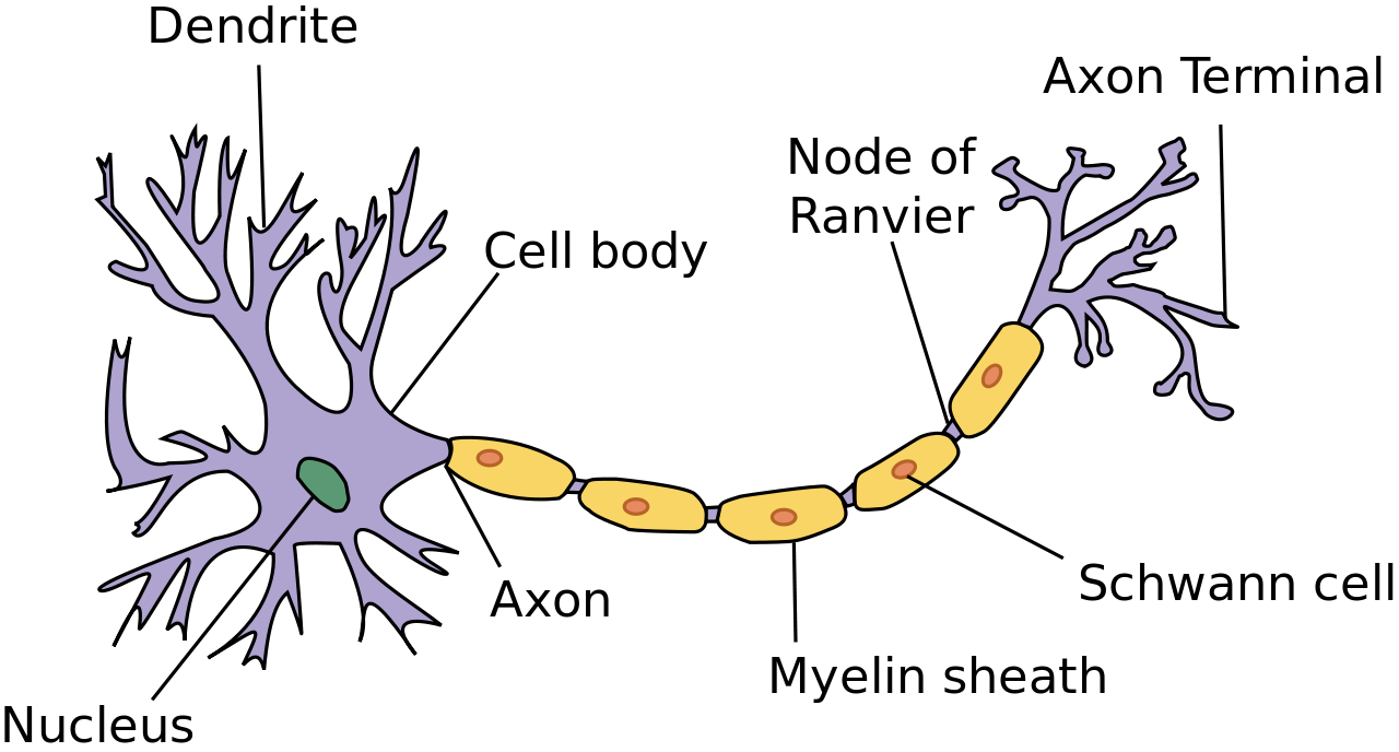
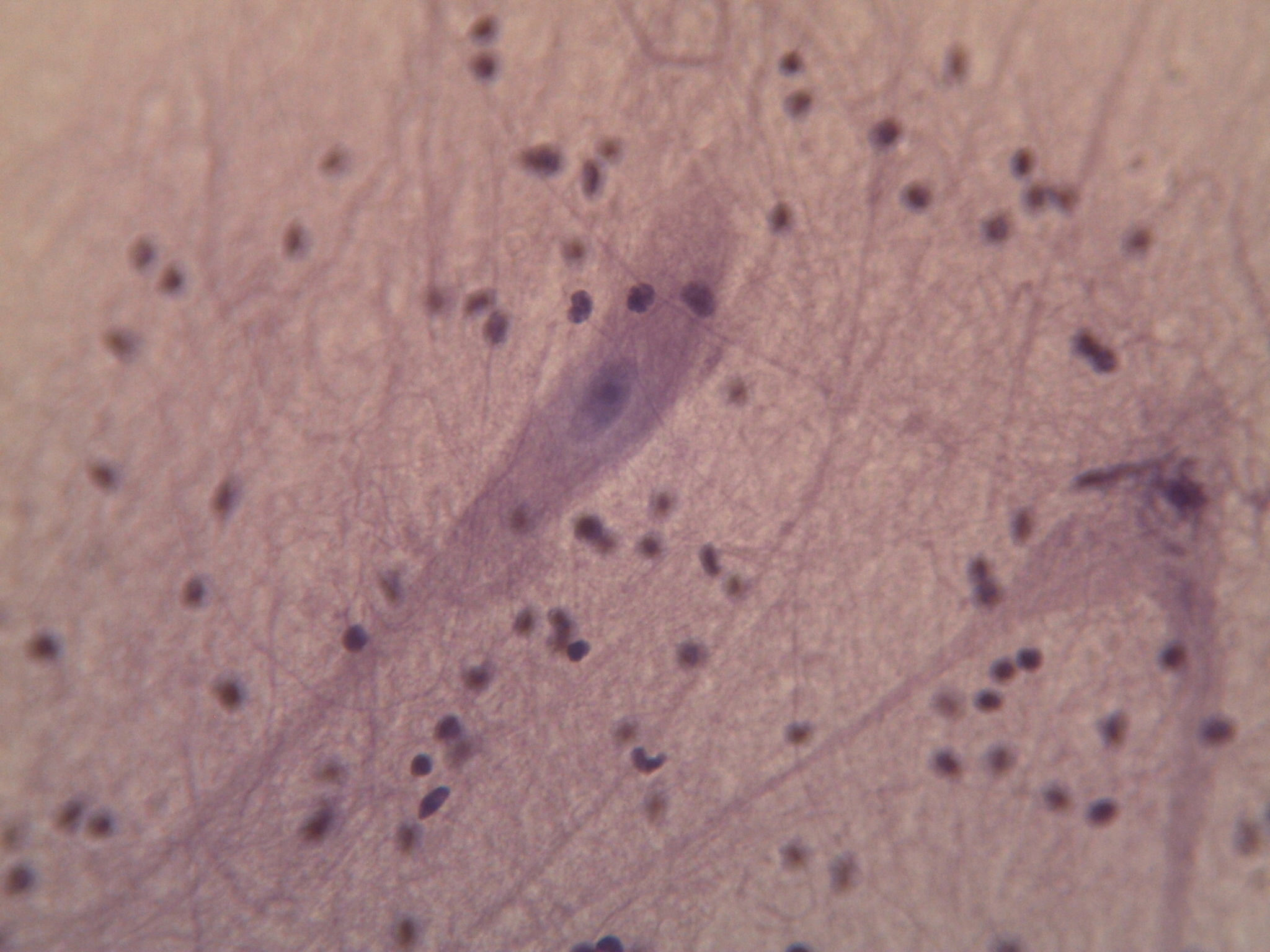
Nervous tissue is made up of neurons and a group of cells called neuroglia (also known as glial cells). Nervous tissue makes up the central nervous system (mainly the brain and spinal cord) and peripheral nervous system (the network of nerves that runs throughout the rest of the body). The defining feature of nervous tissue is that it is specialized to be able to generate and conduct nerve impulses. This function is carried out by neurons, and the purpose of neuroglia is to support neurons.
A neuron has several parts to its structure:
- Dendrites which collect incoming nerve impulses
- A cell body, or soma, which contains the majority of the neuron's organelles, including the nucleus
- An axon, which carries nerve impulses away from the soma, to the next neuron in the chain
- A myelin sheath, which encases the axon and increases that rate at which nerve impulses can be conducted
- Axon terminals, which maintain physical contact with the dendrites of neighbouring neurons
Neuroglia can be understood as support staff for the neuron. The neurons have such an important job, they need cells to bring them nutrients, take away cell waste, and build their mylein sheath. There are many types of neuroglia, which are categorized based on their function and/or their location in the nervous system. Neuroglia outnumber neurons by as much as 50 to 1, and are much smaller. See the diagram in 7.4.17 to compare the size and number of neurons and neuroglia.
Try out this memory game to test your tissues knowledge:
7.4 Summary
- Tissues are made up of cells working together.
- There are four main types of tissues: epithelial, connective, muscular and nervous.
- Epithelial tissue makes up the linings and coverings of the body and is characterized by having a free surface and a basement membrane. Types of epithelial tissue are distinguished by shape of cell (squamous, cuboidal or columnar) and layering (simple, stratified, pseudostratified and transitional). Different epithelial tissues can carry out diffusion, secretion, absorption, and/or protection depending on their particular cell shape and layering.
- Connective tissue provides structure and support for the body and is characterized as a scattering of cells in a non-cellular matrix. There are three main categories of connective tissue, each characterized by a particular type of matrix:
- Fibrous connective tissue contains protein fibres. Both loose and dense fibrous connective tissue belong in this category.
- Supportive connective tissue contains a very solid matrix, and includes both bone and cartilage.
- Fluid connective tissue contains cells in a fluid matrix with the two types of blood and lymph.
- Muscular tissue's defining feature is that it is contractile. There are three types of muscular tissue: skeletal muscle which is found attached to the skeleton for voluntary movement, smooth muscle which moves substances through body tubes, and cardiac muscle which moves blood through the heart.
- Nervous tissue contains specialized cells called neurons which can conduct electrical impulses. Also found in nervous tissue are neuroglia, which support neurons by providing nutrients, removing wastes, and creating myelin sheath.
7.4 Review Questions
- Define the term tissue.
-
- If a part of the body needed a lining that was both protective, but still able to absorb nutrients, what would be the best type of epithelial tissue to use?
-
- Where do you find skeletal muscle? Smooth muscle? Cardiac muscle?
-
- What are some of the functions of neuroglia?
-
7.4 Explore More
https://www.youtube.com/watch?v=O0ZvbPak4ck
Types of Human Body Tissue, MoomooMath and Science, 2017.
https://www.youtube.com/watch?v=uHbn7wLN_3k
How to 3D print human tissue - Taneka Jones, TED-Ed, 2019.
https://www.youtube.com/watch?v=1Qfmkd6C8u8
How bones make blood - Melody Smith, TED-Ed, 2020.
Attributions
Figure 7.4.1
- Construction man kneeling in front of wall by Charles Deluvio on Unsplash is used under the Unsplash License (https://unsplash.com/license).
- Beige wooden frame by Charles Deluvio on Unsplash is used under the Unsplash License (https://unsplash.com/license).
- Tambour on green by Pierre Châtel-Innocention Unsplash is used under the Unsplash License (https://unsplash.com/license).
- Tags: Construction Studs Plumbing Wiring by JWahl on Pixabay is used under the Pixabay License (https://pixabay.com/es/service/license/).
Figure 7.4.2 and Figure 7.4.3
- Simple columnar epithelium tissue by Kamil Danak on Wikimedia Commons is used under a CC BY-SA 3.0 (https://creativecommons.org/licenses/by-sa/3.0/deed.en) license.
- Simple cuboidal epithelium by Kamil Danak on Wikimedia Commons is used under a CC BY-SA 3.0 (https://creativecommons.org/licenses/by-sa/3.0/deed.en) license.
- Simple squamous epithelium by Kamil Danak on Wikimedia Commons is used under a CC BY-SA 3.0 (https://creativecommons.org/licenses/by-sa/3.0/deed.en) license.
Figure 7.4.4
Loose fibrous connective tissue by CNX OpenStax. Biology. on Wikimedial Commons is used under a CC BY 4.0. (https://creativecommons.org/licenses/by/4.0) license.
Figure 7.4.5
Connective Tissue: Loose Aerolar by Berkshire Community College Bioscience Image Library on Flickr is used under a CC0 1.0 Universal public domain dedication (https://creativecommons.org/publicdomain/zero/1.0/) license.
Figure 7.4.6
Dense Fibrous Connective Tissue by by CNX OpenStax. Biology. on Wikimedial Commons is used under a CC BY 4.0. (https://creativecommons.org/licenses/by/4.0) license.
Figure 7.4.7
Dense_connective_tissue-400x by J Jana on Wikimedia Commons is used under a CC BY-SA 4.0 (https://creativecommons.org/licenses/by-sa/4.0/deed.en) license.
Figure 7.4.8
Types_of_Cartilage-new by OpenStax College on Wikipedia Commons is used under a CC BY 3.0 (https://creativecommons.org/licenses/by/3.0) license.
Figure 7.4.9
Compact_bone_histology_2014 by Athikhun.suw on Wikimedia Commons is used under a CC BY-SA 4.0 (https://creativecommons.org/licenses/by-sa/4.0/deed.en) license.
Figure 7.4.10
Bone_normal_and_degraded_micro_structure by Gtirouflet on Wikimedia Commons is used under a CC BY-SA 3.0 (https://creativecommons.org/licenses/by-sa/3.0/deed.en) license.
Figure 7.4.11
Lymphocyte2 by NicolasGrandjean on Wikimedia Commons is used under a CC BY-SA 3.0 (https://creativecommons.org/licenses/by-sa/3.0/deed.en) license. [No machine-readable author provided. NicolasGrandjean is assumed, based on copyright claims.]
Figure 7.4.12
Skeletal_muscle_横纹肌1 by 乌拉跨氪 on Wikimedia Commons is used under a CC BY-SA 4.0 (https://creativecommons.org/licenses/by-sa/4.0/deed.en) license.
Figure 7.4.13
Smooth_Muscle_new by OpenStax on Wikimedia Commons is used under a CC BY 4.0 (https://creativecommons.org/licenses/by/4.0/deed.en) license.
Figure 7.4.14
Peristalsis by OpenStax on Wikimedia Commons is used under a CC BY 3.0 (https://creativecommons.org/licenses/by/3.0/deed.en) license.
Figure 7.4.15
400x Cardiac Muscle by Jessy731 on Flickr is used and adapted by Christine Miller under a CC BY-NC 2.0 (https://creativecommons.org/licenses/by-nc/2.0/) license.
Figure 7.4.16
Neuron.svg by User:Dhp1080 on Wikimedia Commons is used under a CC BY-SA 3.0 (https://creativecommons.org/licenses/by-sa/3.0/deed.en) license.
Figure 7.4.17
400x Nervous Tissue by Jessy731 on Flickr is used under a CC BY-NC 2.0 (https://creativecommons.org/licenses/by-nc/2.0/) license.
Table 7.4.1
Summary of Epithelial Tissue Cells, by OpenStax College on Wikipedia Commons is used under a CC BY 3.0 (https://creativecommons.org/licenses/by/3.0) license.
Table 7.4.2
- Epithelial_Tissues_Stratified_Squamous_Epithelium_(40230842160) by
Berkshire Community College Bioscience Image Library on Wikimedia Commons is used under a CC0 1.0 Universal Public Domain Dedication (https://creativecommons.org/publicdomain/zero/1.0/) license. - Simple cuboidal epithelial tissue histology by Berkshire Community College on Flickr is used under a CC0 1.0 Universal Public Domain Dedication (https://creativecommons.org/publicdomain/zero/1.0/) license.
- Pseudostratified_Epithelium by OpenStax College on Wikimedia Commons is used under a CC BY 3.0 (https://creativecommons.org/licenses/by/3.0) license.
References
Betts, J. G., Young, K.A., Wise, J.A., Johnson, E., Poe, B., Kruse, D.H., Korol, O., Johnson, J.E., Womble, M., DeSaix, P. (2013, April 25). Figure 4.8 Summary of epithelial tissue cells [digital image]. In Anatomy and Physiology (Section 4.2). OpenStax. https://openstax.org/books/anatomy-and-physiology/pages/4-2-epithelial-tissue
Betts, J. G., Young, K.A., Wise, J.A., Johnson, E., Poe, B., Kruse, D.H., Korol, O., Johnson, J.E., Womble, M., DeSaix, P. (2013, April 25). Figure 4.16 Types of cartilage [digital image]. In Anatomy and Physiology (Section 4.3). OpenStax. https://openstax.org/books/anatomy-and-physiology/pages/4-3-connective-tissue-supports-and-protects
Betts, J. G., Young, K.A., Wise, J.A., Johnson, E., Poe, B., Kruse, D.H., Korol, O., Johnson, J.E., Womble, M., DeSaix, P. (2013, April 25). Figure 10.23 Smooth muscle [digital micrograph]. In Anatomy and Physiology (Section 10.8). OpenStax. https://openstax.org/books/anatomy-and-physiology/pages/10-8-smooth-muscle (Micrograph provided by the Regents of University of Michigan Medical School © 2012)
Betts, J. G., Young, K.A., Wise, J.A., Johnson, E., Poe, B., Kruse, D.H., Korol, O., Johnson, J.E., Womble, M., DeSaix, P. (2013, April 25). Figure 22.5 Pseudostratified ciliated columnar epithelium [digital micrograph]. In Anatomy and Physiology (Section 22.1). OpenStax. https://openstax.org/books/anatomy-and-physiology/pages/22-1-organs-and-structures-of-the-respiratory-system
Betts, J. G., Young, K.A., Wise, J.A., Johnson, E., Poe, B., Kruse, D.H., Korol, O., Johnson, J.E., Womble, M., DeSaix, P. (2013, April 25). Figure 23.5 Peristalsis [diagram]. In Anatomy and Physiology (Section 23.2). OpenStax. https://openstax.org/books/anatomy-and-physiology/pages/23-2-digestive-system-processes-and-regulation
Mister Science. (2018). What is peristalsis? YouTube. https://www.youtube.com/watch?v=kVjeNZA5pi4
MoomooMath and Science. (2017, May 18). Types of human body tissue. YouTube. https://www.youtube.com/watch?v=O0ZvbPak4ck&feature=youtu.be
Open Stax. (2016, May 27). Figure 6 Loose connective tissue [digital image]. In OpenStax Biology (Section 33.2). OpenStax CNX. https://cnx.org/contents/GFy_h8cu@10.53:-LfhWRES@4/Animal-Primary-Tissues
Open Stax. (2016, May 27). Figure 7 Fibrous connective tissue from the tendon [digital image]. In OpenStax Biology (Section 33.2). OpenStax CNX. https://cnx.org/contents/GFy_h8cu@10.53:-LfhWRES@4/Animal-Primary-Tissues
TED-Ed. (2019, October 17). How to 3D print human tissue - Taneka Jones. YouTube. https://www.youtube.com/watch?v=uHbn7wLN_3k&feature=youtu.be
TED-Ed. (2020, January 27). How bones make blood - Melody Smith. YouTube. https://www.youtube.com/watch?v=1Qfmkd6C8u8&feature=youtu.be
Created by CK-12 Foundation/Adapted by Christine Miller

Seeing Your Breath
Why can you “see your breath” on a cold day? The air you exhale through your nose and mouth is warm like the inside of your body. Exhaled air also contains a lot of water vapor, because it passes over moist surfaces from the lungs to the nose or mouth. The water vapor in your breath cools suddenly when it reaches the much colder outside air. This causes the water vapor to condense into a fog of tiny droplets of liquid water. You release water vapor and other gases from your body through the process of respiration.
What is Respiration?
Respiration is the life-sustaining process in which gases are exchanged between the body and the outside atmosphere. Specifically, oxygen moves from the outside air into the body; and water vapor, carbon dioxide, and other waste gases move from inside the body to the outside air. Respiration is carried out mainly by the respiratory system. It is important to note that respiration by the respiratory system is not the same process as cellular respiration —which occurs inside cells — although the two processes are closely connected. Cellular respiration is the metabolic process in which cells obtain energy, usually by “burning” glucose in the presence of oxygen. When cellular respiration is aerobic, it uses oxygen and releases carbon dioxide as a waste product. Respiration by the respiratory system supplies the oxygen needed by cells for aerobic cellular respiration, and removes the carbon dioxide produced by cells during cellular respiration.
Respiration by the respiratory system actually involves two subsidiary processes. One process is ventilation, or breathing. Ventilation is the physical process of conducting air to and from the lungs. The other process is gas exchange. This is the biochemical process in which oxygen diffuses out of the air and into the blood, while carbon dioxide and other waste gases diffuse out of the blood and into the air. All of the organs of the respiratory system are involved in breathing, but only the lungs are involved in gas exchange.
Respiratory Organs
The organs of the respiratory system form a continuous system of passages, called the respiratory tract, through which air flows into and out of the body. The respiratory tract has two major divisions: the upper respiratory tract and the lower respiratory tract. The organs in each division are shown in Figure 13.2.2. In addition to these organs, certain muscles of the thorax (body cavity that fills the chest) are also involved in respiration by enabling breathing. Most important is a large muscle called the diaphragm, which lies below the lungs and separates the thorax from the abdomen. Smaller muscles between the ribs also play a role in breathing.
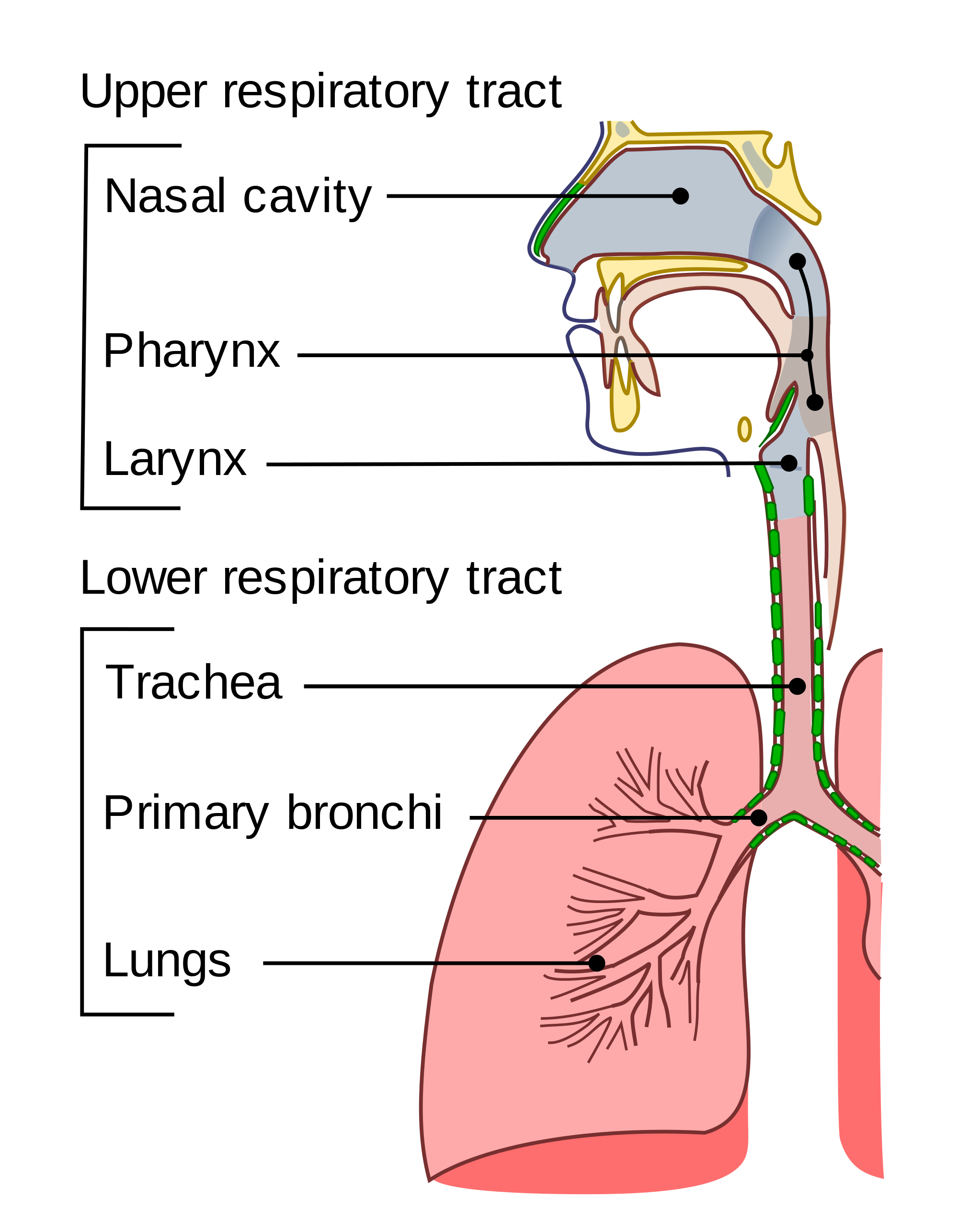
Upper Respiratory Tract
All of the organs and other structures of the upper respiratory tract are involved in conduction, or the movement of air into and out of the body. Upper respiratory tract organs provide a route for air to move between the outside atmosphere and the lungs. They also clean, humidify, and warm the incoming air. No gas exchange occurs in these organs.
Nasal Cavity
The nasal cavity is a large, air-filled space in the skull above and behind the nose in the middle of the face. It is a continuation of the two nostrils. As inhaled air flows through the nasal cavity, it is warmed and humidified by blood vessels very close to the surface of this epithelial tissue . Hairs in the nose and mucous produced by mucous membranes help trap larger foreign particles in the air before they go deeper into the respiratory tract. In addition to its respiratory functions, the nasal cavity also contains chemoreceptors needed for sense of smell, and contribution to the sense of taste.
Pharynx
The pharynx is a tube-like structure that connects the nasal cavity and the back of the mouth to other structures lower in the throat, including the larynx. The pharynx has dual functions — both air and food (or other swallowed substances) pass through it, so it is part of both the respiratory and the digestive systems. Air passes from the nasal cavity through the pharynx to the larynx (as well as in the opposite direction). Food passes from the mouth through the pharynx to the esophagus.
Larynx
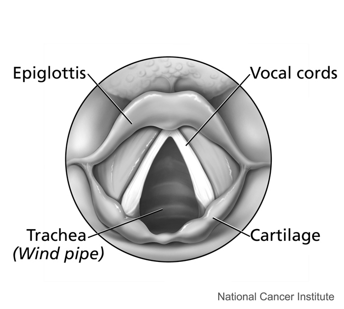
The larynx connects the pharynx and trachea, and helps to conduct air through the respiratory tract. The larynx is also called the voice box, because it contains the vocal cords, which vibrate when air flows over them, thereby producing sound. You can see the vocal cords in the larynx in Figures 13.2.3 and 13.2.4. Certain muscles in the larynx move the vocal cords apart to allow breathing. Other muscles in the larynx move the vocal cords together to allow the production of vocal sounds. The latter muscles also control the pitch of sounds and help control their volume.
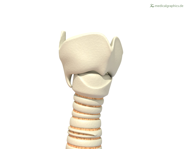 |
Figure 13.2.4 The larynx as viewed from the top. |
A very important function of the larynx is protecting the trachea from aspirated food. When swallowing occurs, the backward motion of the tongue forces a flap called the epiglottis to close over the entrance to the larynx. (You can see the epiglottis in both Figure 13.2.3 and 13.2.4.) This prevents swallowed material from entering the larynx and moving deeper into the respiratory tract. If swallowed material does start to enter the larynx, it irritates the larynx and stimulates a strong cough reflex. This generally expels the material out of the larynx, and into the throat.
https://www.youtube.com/watch?v=BsyB88mq5rQ
Larynx Model - Respiratory System, Dr. Lotz, 2018.
Lower Respiratory Tract
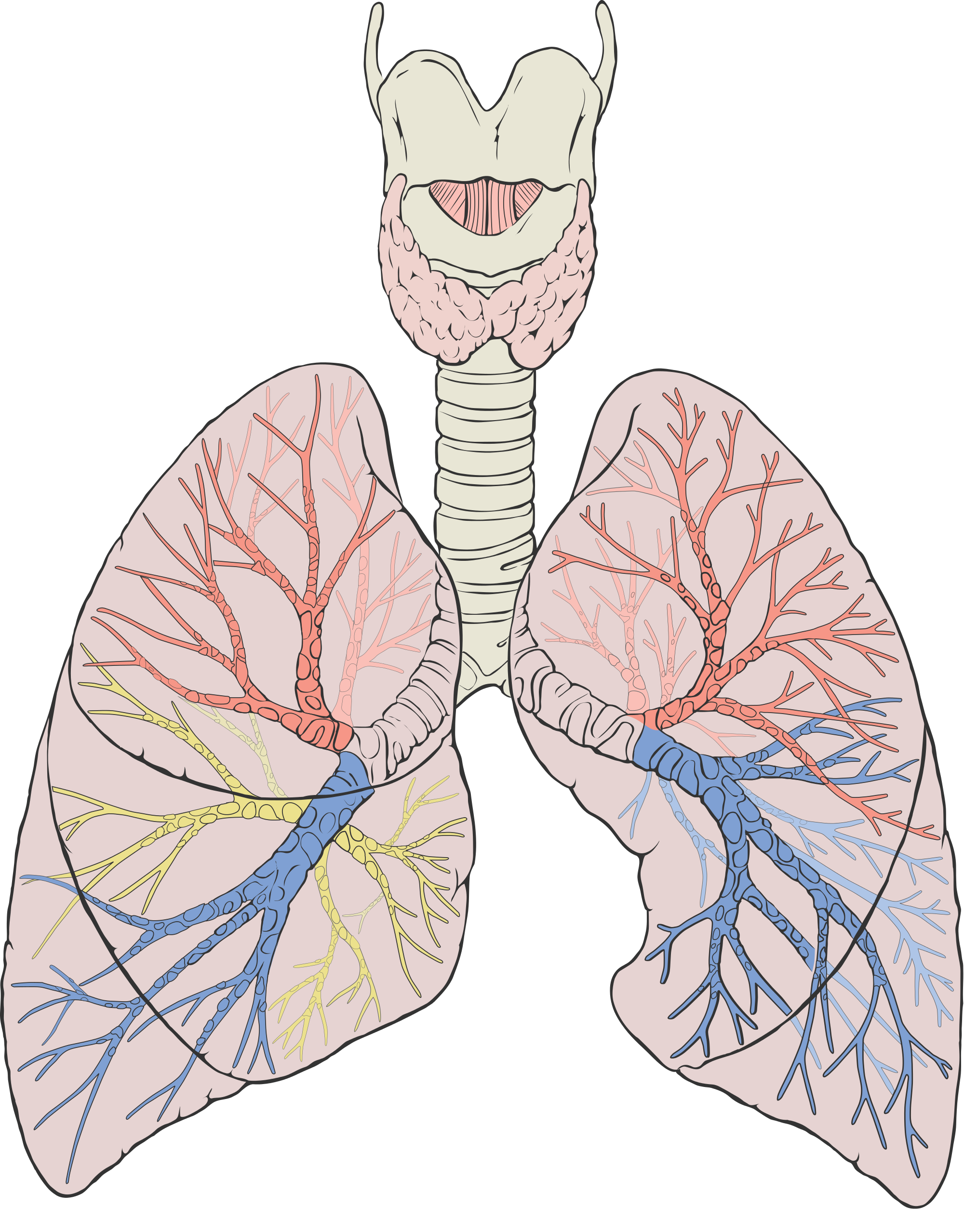
The trachea and other passages of the lower respiratory tract conduct air between the upper respiratory tract and the lungs. These passages form an inverted tree-like shape (Figure 13.2.5), with repeated branching as they move deeper into the lungs. All told, there are an astonishing 2,414 kilometres (1,500 miles) of airways conducting air through the human respiratory tract! It is only in the lungs, however, that gas exchange occurs between the air and the bloodstream.
Trachea
The trachea, or windpipe, is the widest passageway in the respiratory tract. It is about 2.5 cm wide and 10-15 cm long (approximately 1 inch wide and 4–6 inches long). It is formed by rings of cartilage, which make it relatively strong and resilient. The trachea connects the larynx to the lungs for the passage of air through the respiratory tract. The trachea branches at the bottom to form two bronchial tubes.
Bronchi and Bronchioles
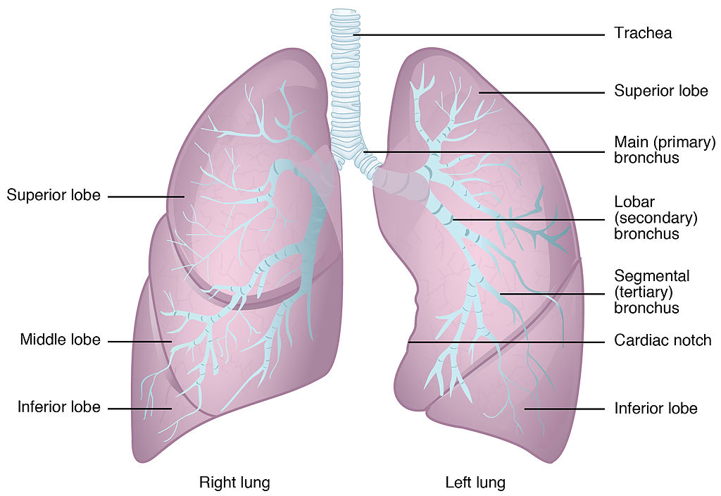
There are two main bronchial tubes, or bronchi (singular, bronchus), called the right and left bronchi. The bronchi carry air between the trachea and lungs. Each bronchus branches into smaller, secondary bronchi; and secondary bronchi branch into still smaller tertiary bronchi. The smallest bronchi branch into very small tubules called bronchioles. The tiniest bronchioles end in alveolar ducts, which terminate in clusters of minuscule air sacs, called alveoli (singular, alveolus), in the lungs.
Lungs
The lungs are the largest organs of the respiratory tract. They are suspended within the pleural cavity of the thorax. The lungs are surrounded by two thin membranes called pleura, which secrete fluid that allows the lungs to move freely within the pleural cavity. This is necessary so the lungs can expand and contract during breathing. In Figure 13.2.6, you can see that each of the two lungs is divided into sections. These are called lobes, and they are separated from each other by connective tissues. The right lung is larger and contains three lobes. The left lung is smaller and contains only two lobes. The smaller left lung allows room for the heart, which is just left of the center of the chest.
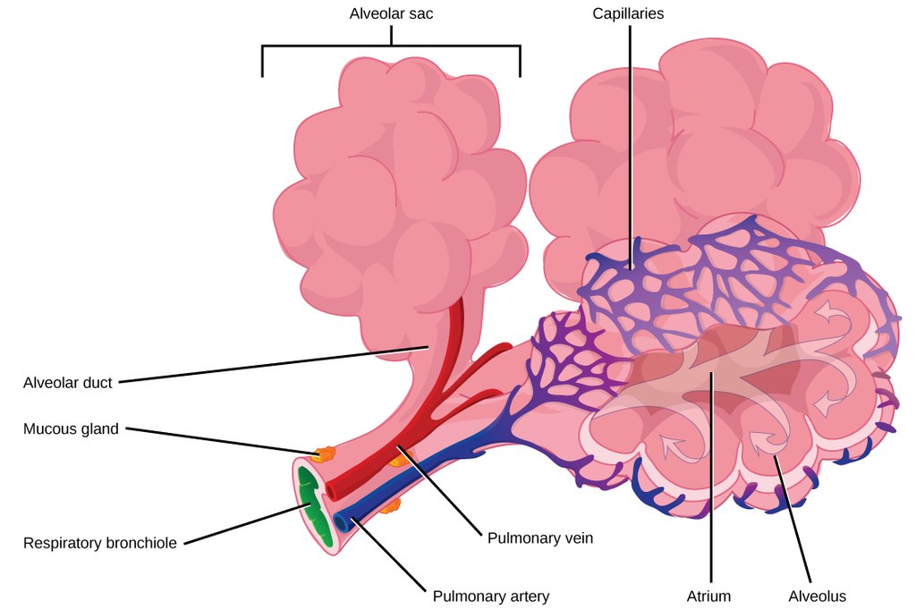
As mentioned previously, the bronchi terminate in bronchioles which feed air into alveoli, tiny sacs of simple squamous epithelial tissue which make up the bulk of the lung. The cross-section of lung tissue in the diagram below (Figure 13.2.7) shows the alveoli, in which gas exchange takes place with the capillary network that surrounds them.
 |
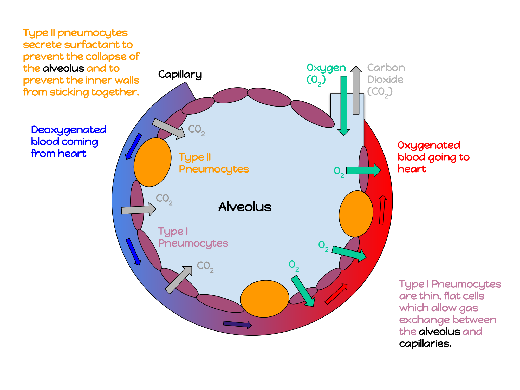 |
Lung tissue consists mainly of alveoli (see Figures 13.2.7 and 13.2.8). These tiny air sacs are the functional units of the lungs where gas exchange takes place. The two lungs may contain as many as 700 million alveoli, providing a huge total surface area for gas exchange to take place. In fact, alveoli in the two lungs provide as much surface area as half a tennis court! Each time you breathe in, the alveoli fill with air, making the lungs expand. Oxygen in the air inside the alveoli is absorbed by the blood via diffusion in the mesh-like network of tiny capillaries that surrounds each alveolus. The blood in these capillaries also releases carbon dioxide (also by diffusion) into the air inside the alveoli. Each time you breathe out, air leaves the alveoli and rushes into the outside atmosphere, carrying waste gases with it.
The lungs receive blood from two major sources. They receive deoxygenated blood from the right side of the heart. This blood absorbs oxygen in the lungs and carries it back to the left side heart to be pumped to cells throughout the body. The lungs also receive oxygenated blood from the heart that provides oxygen to the cells of the lungs for cellular respiration.
Protecting the Respiratory System
You may be able to survive for weeks without food and for days without water, but you can survive without oxygen for only a matter of minutes — except under exceptional circumstances — so protecting the respiratory system is vital. Ensuring that a patient has an open airway is the first step in treating many medical emergencies. Fortunately, the respiratory system is well protected by the ribcage of the skeletal system. The extensive surface area of the respiratory system, however, is directly exposed to the outside world and all its potential dangers in inhaled air. It should come as no surprise that the respiratory system has a variety of ways to protect itself from harmful substances, such as dust and pathogens in the air.
The main way the respiratory system protects itself is called the mucociliary escalator. From the nose through the bronchi, the respiratory tract is covered in epithelium that contains mucus-secreting goblet cells. The mucus traps particles and pathogens in the incoming air. The epithelium of the respiratory tract is also covered with tiny cell projections called cilia (singular, cilium), as shown in the animation. The cilia constantly move in a sweeping motion upward toward the throat, moving the mucus and trapped particles and pathogens away from the lungs and toward the outside of the body. The upward sweeping motion of cilia lining the respiratory tract helps keep it free from dust, pathogens, and other harmful substances.
Watch "Mucociliary clearance" by I-Hsun Wu to learn more:
https://www.youtube.com/watch?v=HMB6flEaZwI
Mucociliary clearance, I-Hsun Wu, 2015.
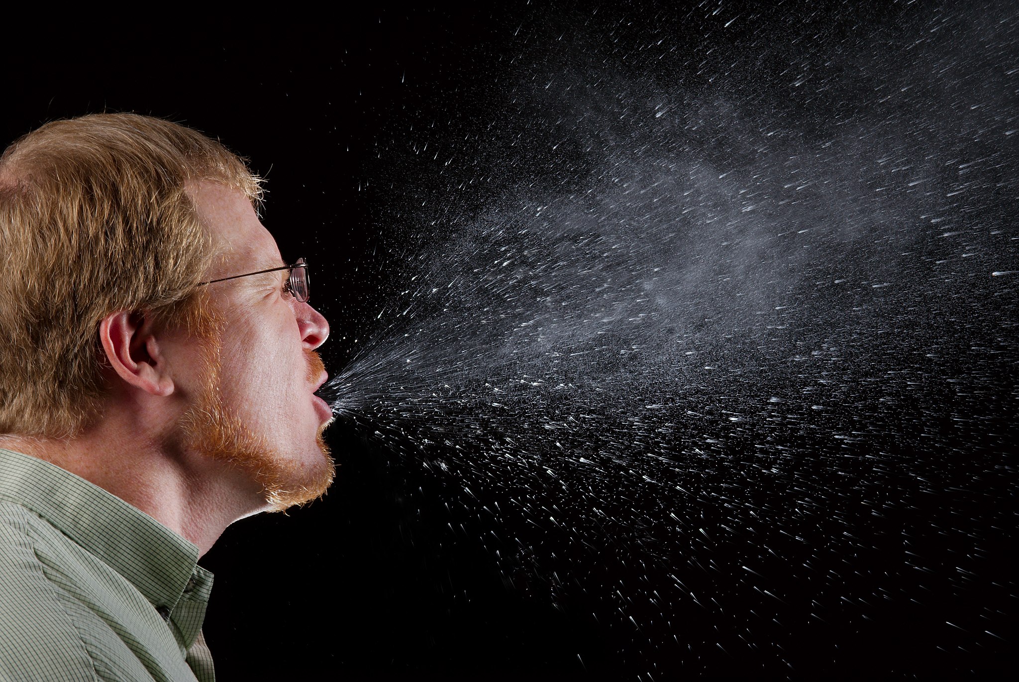
Sneezing is a similar involuntary response that occurs when nerves lining the nasal passage are irritated. It results in forceful expulsion of air from the mouth, which sprays millions of tiny droplets of mucus and other debris out of the mouth and into the air, as shown in Figure 13.2.9. This explains why it is so important to sneeze into a tissue (rather than the air) if we are to prevent the transmission of respiratory pathogens.
How the Respiratory System Works with Other Organ Systems
The amount of oxygen and carbon dioxide in the blood must be maintained within a limited range for survival of the organism. Cells cannot survive for long without oxygen, and if there is too much carbon dioxide in the blood, the blood becomes dangerously acidic (pH is too low). Conversely, if there is too little carbon dioxide in the blood, the blood becomes too basic (pH is too high). The respiratory system works hand-in-hand with the nervous and cardiovascular systems to maintain homeostasis in blood gases and pH.
It is the level of carbon dioxide — rather than the level of oxygen — that is most closely monitored to maintain blood gas and pH homeostasis. The level of carbon dioxide in the blood is detected by cells in the brain, which speed up or slow down the rate of breathing through the autonomic nervous system as needed to bring the carbon dioxide level within the normal range. Faster breathing lowers the carbon dioxide level (and raises the oxygen level and pH), while slower breathing has the opposite effects. In this way, the levels of carbon dioxide, oxygen, and pH are maintained within normal limits.
The respiratory system also works closely with the cardiovascular system to maintain homeostasis. The respiratory system exchanges gases with the outside air, but it needs the cardiovascular system to carry them to and from body cells. Oxygen is absorbed by the blood in the lungs and then transported through a vast network of blood vessels to cells throughout the body, where it is needed for aerobic cellular respiration. The same system absorbs carbon dioxide from cells and carries it to the respiratory system for removal from the body.
Feature: My Human Body
Choking due to a foreign object becoming lodged in the airway results in nearly 5 thousand deaths in Canada each year. In addition, choking accounts for almost 40% of unintentional injuries in infants under the age of one. For the sake of your own human body, as well as those of loved ones, you should be aware of choking risks, signs, and treatments.
Choking is the mechanical obstruction of the flow of air from the atmosphere into the lungs. It prevents breathing, and may be partial or complete. Partial choking allows some — though inadequate — air flow into the lungs. Prolonged or complete choking results in asphyxia, or suffocation, which is potentially fatal.
Obstruction of the airway typically occurs in the pharynx or trachea. Young children are more prone to choking than are older people, in part because they often put small objects in their mouth and do not understand the risk of choking that they pose. Young children may choke on small toys or parts of toys, or on household objects, in addition to food. Foods that are round (hotdogs, carrots, grapes) or can adapt their shape to that of the pharynx (bananas, marshmallows), are especially dangerous, and may cause choking in adults, as well as children.
How can you tell if a loved one is choking? The person cannot speak or cry out, or has great difficulty doing so. Breathing, if possible, is laboured, producing gasping or wheezing. The person may desperately clutch at his or her throat or mouth. If breathing is not soon restored, the person’s face will start to turn blue from lack of oxygen. This will be followed by unconsciousness, brain damage, and possibly death if oxygen deprivation continues beyond a few minutes.
If an infant is choking, turning the baby upside down and slapping him on the back may dislodge the obstructing object. To help an older person who is choking, first encourage the person to cough. Give them a few hard back slaps to help force the lodged object out of the airway. If these steps fail, perform the Heimlich maneuver on the person. See the series of videos below, from ProCPR, which demonstrate several ways to help someone who is choking based on age and consciousness.
https://www.youtube.com/watch?v=XOTbjDGZ7wg&t=46s
Conscious Adult Choking, ProCPR, 2016.
https://www.youtube.com/watch?v=5kmsKNvKAvU
Unconscious Adult Choking, ProCPR, 2011.
https://www.youtube.com/watch?v=ZjmbD7aIaf0
Conscious Child Choking, ProCPR, 2009.
https://www.youtube.com/watch?v=Sba0T2XGIn4
Unconscious Child Choking, ProCPR, 2009.
https://www.youtube.com/watch?v=axqIju9CLKA
Conscious Infant Choking, ProCPR, 2011.
https://www.youtube.com/watch?v=_K7Dwy6b2wQ
Unconscious Infant Choking, ProCPR, 2011.
13.2 Summary
- Respiration is the process in which oxygen moves from the outside air into the body, and in which carbon dioxide and other waste gases move from inside the body into the outside air. It involves two subsidiary processes: ventilation and gas exchange. Respiration is carried out mainly by the respiratory system.
- The organs of the respiratory system form a continuous system of passages, called the respiratory tract. It has two major divisions: the upper respiratory tract and the lower respiratory tract.
- The upper respiratory tract includes the nasal cavity, pharynx, and larynx. All of these organs are involved in conduction, or the movement of air into and out of the body. Incoming air is also cleaned, humidified, and warmed as it passes through the upper respiratory tract. The larynx is called the voice box, because it contains the vocal cords, which are needed to produce vocal sounds.
- The lower respiratory tract includes the trachea, bronchi and bronchioles, and the lungs. The trachea, bronchi, and bronchioles are involved in conduction. Gas exchange takes place only in the lungs, which are the largest organs of the respiratory tract. Lung tissue consists mainly of tiny air sacs called alveoli, which is where gas exchange takes place between air in the alveoli and the blood in capillaries surrounding them.
- The respiratory system protects itself from potentially harmful substances in the air by the mucociliary escalator. This includes mucus-producing cells, which trap particles and pathogens in incoming air. It also includes tiny hair-like cilia that continually move to sweep the mucus and trapped debris away from the lungs and toward the outside of the body.
- The level of carbon dioxide in the blood is monitored by cells in the brain. If the level becomes too high, it triggers a faster rate of breathing, which lowers the level to the normal range. The opposite occurs if the level becomes too low. The respiratory system exchanges gases with the outside air, but it needs the cardiovascular system to carry the gases to and from cells throughout the body.
13.2 Review Questions
-
- What is respiration, as carried out by the respiratory system? Name the two subsidiary processes it involves.
- Describe the respiratory tract.
- Identify the organs of the upper respiratory tract. What are their functions?
- List the organs of the lower respiratory tract. Which organs are involved only in conduction?
- Where does gas exchange take place?
- How does the respiratory system protect itself from potentially harmful substances in the air?
- Explain how the rate of breathing is controlled.
- Why does the respiratory system need the cardiovascular system to help it perform its main function of gas exchange?
- Describe two ways in which the body prevents food from entering the lungs.
- What is the relationship between respiration and cellular respiration?
13.2 Explore More
https://www.youtube.com/watch?v=8NUxvJS-_0k
How do lungs work? - Emma Bryce, TED-Ed, 2014.
https://www.youtube.com/watch?time_continue=1&v=6iFPs6JlSzY&feature=emb_logo
Why Do Men Have Deeper Voices? BrainStuff - HowStuffWorks, 2015.
https://www.youtube.com/watch?v=rjibeBSnpJ0
Why does your voice change as you get older? - Shaylin A. Schundler, TED-Ed, 2018.
Attributions
Figure 13.2.1
Exhale by pavel-lozovikov-HYovA7yPPvI [photo] by Pavel Lozovikov on Unsplash is used under the Unsplash License (https://unsplash.com/license).
Figure 13.2.2
Illu_conducting_passages.svg by Lord Akryl, Jmarchn from SEER Training Modules/ National Cancer Institute on Wikimedia Commons is in the public domain (https://en.wikipedia.org/wiki/Public_domain).
Figure 13.2.3
Larynx by www.medicalgraphics.de is used under a CC BY-ND 4.0 (https://creativecommons.org/licenses/by-nd/4.0/) license.
Figure 13.2.4
Larynx top view by Alan Hoofring (Illustrator) for National Cancer Institute is in the public domain (https://en.wikipedia.org/wiki/Public_domain).
Figure 13.2.5
2000px-Lungs_diagram_detailed.svg by Patrick J. Lynch, medical illustrator on Wikimedia Commons is used under a CC BY 2.5 (https://creativecommons.org/licenses/by/2.5) license. (Derivative work of Fruchtwasserembolie.png.)
Figure 13.2.6
Gross_Anatomy_of_the_Lungs by OpenStax College on Wikimedia Commons is used under a CC BY 3.0 (https://creativecommons.org/licenses/by/3.0) license.
Figure 13.2.7
Alveoli Structure by CNX OpenStax on Wikimedia Commons is used under a CC BY 4.0 (https://creativecommons.org/licenses/by/4.0) license.
Figure 13.2.8
annotated_diagram_of_an_alveolus.svg by Katherinebutler1331 on Wikimedia Commons is used under a CC BY-SA 4.0 (https://creativecommons.org/licenses/by-sa/4.0) license.
Figure 13.2.9
Sneeze by James Gathany at CDC Public Health Imagery Library (PHIL) #11162 on Wikimedia Commons is in the public domain (https://en.wikipedia.org/wiki/Public_domain).
References
Betts, J. G., Young, K.A., Wise, J.A., Johnson, E., Poe, B., Kruse, D.H., Korol, O., Johnson, J.E., Womble, M., DeSaix, P. (2013, June 19). Figure 22.2 Major respiratory structures [digital image]. In Anatomy and Physiology (Section 22.1). OpenStax. https://openstax.org/books/anatomy-and-physiology/pages/22-1-organs-and-structures-of-the-respiratory-system [CC BY 4.0 (https://creativecommons.org/licenses/by/4.0)].
Betts, J. G., Young, K.A., Wise, J.A., Johnson, E., Poe, B., Kruse, D.H., Korol, O., Johnson, J.E., Womble, M., DeSaix, P. (2013, June 19). Figure 22.13 Gross anatomy of the lungs [digital image]. In Anatomy and Physiology (Section 22.2). OpenStax. https://openstax.org/books/anatomy-and-physiology/pages/22-2-the-lungs
BrainStuff - HowStuffWorks. (2015, December 1). Why do men have deeper voices? YouTube. https://www.youtube.com/watch?v=6iFPs6JlSzY&feature=youtu.be
Dr. Lotz. (2018, January 25). Larynx model - Respiratory system. YouTube. https://www.youtube.com/watch?v=BsyB88mq5rQ&feature=youtu.be
I-Hsun Wu. (2015, March 31). Mucociliary clearance. YouTube. https://www.youtube.com/watch?v=HMB6flEaZwI&feature=youtu.be
OpenStax. (2016, May 27). Figure 9 Terminal bronchioles are connected by respiratory bronchioles to alveolar ducts and alveolar sacs [digital image]. In OpenStax, Biology (Section 39.1). OpenStax CNX. https://cnx.org/contents/GFy_h8cu@10.53:35-R0biq@11/Systems-of-Gas-Exchange
ProCPR. (2009, November 24). Conscious child choking. YouTube. https://www.youtube.com/watch?v=ZjmbD7aIaf0&feature=youtu.be
ProCPR. (2009, November 24).Unconscious child choking. YouTube. https://www.youtube.com/watch?v=Sba0T2XGIn4&feature=youtu.be
ProCPR. (2011, February 1). Conscious infant choking. YouTube. https://www.youtube.com/watch?v=axqIju9CLKA&feature=youtu.be
ProCPR. (2011, February 1). Unconscious adult choking. YouTube. https://www.youtube.com/watch?v=5kmsKNvKAvU&feature=youtu.be
ProCPR. (2011, February 1). Unconscious infant choking. YouTube. https://www.youtube.com/watch?v=_K7Dwy6b2wQ&feature=youtu.be
ProCPR. (2016, April 8). Conscious adult choking. YouTube. https://www.youtube.com/watch?v=XOTbjDGZ7wg&feature=youtu.be
TED-Ed. (2014, November 24). How do lungs work? - Emma Bryce. YouTube. https://www.youtube.com/watch?v=8NUxvJS-_0k&feature=youtu.be
TED-Ed. (2018, August 2). Why does your voice change as you get older? - Shaylin A. Schundler. YouTube. https://www.youtube.com/watch?v=rjibeBSnpJ0&feature=youtu.be
Structures containing neuronal cell bodies in the peripheral nervous system.
Created by Christine Miller
Figure 7.4.1 Construction — It's important to have the right materials for the job.
The Right Material for the Job
Building a house is a big job and one that requires a lot of different materials for specific purposes. As you can see in Figure 7.4.1, many different types of materials are used to build a complete house, but each type of material fulfills certain functions. You wouldn't use insulation to cover your roof, and you wouldn't use lumber to wire your home. Just as a builder chooses the appropriate materials to build each aspect of a home (wires for electrical, lumber for framing, shingles for roofing), your body uses the right cells for each type of role. When many cells work together to perform a specific function, this is termed a tissue.
Tissues
Groups of connected cells form tissues. The cells in a tissue may all be the same type, or they may be of multiple types. In either case, the cells in the tissue work together to carry out a specific function, and they are always specialized to be able to carry out that function better than any other type of tissue. There are four main types of human tissues: connective, epithelial, muscle, and nervous tissues. We use tissues to build organs and organ systems. The 200 types of cells that the body can produce based on our single set of DNA can create all the types of tissue in the body.
Epithelial Tissue
Epithelial tissue is made up of cells that line inner and outer body surfaces, such as the skin and the inner surface of the digestive tract. Epithelial tissue that lines inner body surfaces and body openings is called mucous membrane. This type of epithelial tissue produces mucus, a slimy substance that coats mucous membranes and traps pathogens, particles, and debris. Epithelial tissue protects the body and its internal organs, secretes substances (such as hormones) in addition to mucus, and absorbs substances (such as nutrients).
The key identifying feature of epithelial tissue is that it contains a free surface and a basement membrane. The free surface is not attached to any other cells and is either open to the outside of the body, or is open to the inside of a hollow organ or body tube. The basement membrane anchors the epithelial tissue to underlying cells.
Epithelial tissue is identified and named by shape and layering. Epithelial cells exist in three main shapes: squamous, cuboidal, and columnar. These specifically shaped cells can, depending on function, be layered several different ways: simple, stratified, pseudostratified, and transitional.
Epithelial tissue forms coverings and linings and is responsible for a range of functions including diffusion, absorption, secretion and protection. The shape of an epithelial cell can maximize its ability to perform a certain function. The thinner an epithelial cell is, the easier it is for substances to move through it to carry out diffusion and/or absorption. The larger an epithelial cell is, the more room it has in its cytoplasm to be able to make products for secretion, and the more protection it can provide for underlying tissues. Their are three main shapes of epithelial cells: squamous (which is shaped like a pancake- flat and oval), cuboidal (cube shaped), and columnar (tall and rectangular).
Figure 7.4.2 The shape of epithelial tissues is important.
Epithelial tissue will also organize into different layerings depending on their function. For example, multiple layers of cells provide excellent protection, but would no longer be efficient for diffusion, whereas a single layer would work very well for diffusion, but no longer be as protective; a special type of layering called transitional is needed for organs that stretch, like your bladder. Your tissues exhibit the layering that makes them most efficient for the function they are supposed to perform. There are four main layerings found in epithelial tissue: simple (one layer of cells), stratified (many layers of cells), pseudostratified (appears stratified, but upon closer inspection is actually simple), and transitional (can stretch, going from many layers to fewer layers).
Figure 7.4.3 The layerings found in epithelial tissues is important.
See Table 7.4.1 for a summary of the different layering types and shapes epithelial cells can form and their related functions and locations.
Table 7.4.1
Summary of Epithelial Tissue Cells

So far, we have identified epithelial tissue based on shape and layering. The representative diagrams we have seen so far are helpful for visualizing the tissue structures, but it is important to look at real examples of these cells. Since cells are too tiny to see with the naked eye, we rely on microscopes to help us study them. Histology is the study of the microscopic anatomy and cells and tissues. See Table 7.4.2 to see some examples of slides of epithelial tissues prepared for the purpose of histology.
Table 7.4.2
Epithelial Tissues and Histological Samples
| Epithelial Tissue Type | Tissue Diagram | Histological Sample |
| Stratified squamous
(from skin) |
 |
|
| Simple cuboidal
(from kidney tubules) |
 |
|
| Pseudostratified ciliated columnar
(from trachea) |
 |
 |
Connective Tissue
Bone and blood are examples of connective tissue. Connective tissue is very diverse. In general, it forms a framework and support structure for body tissues and organs. It's made up of living cells separated by non-living material, called extracellular matrix, which can be solid or liquid. The extracellular matrix of bone, for example, is a rigid mineral framework. The extracellular matrix of blood is liquid plasma.
The key identifying feature of connective tissue is that is is composed of a scattering of cells in a non-cellular matrix. There are three main categories of connective tissue, based on the nature of the matrix. They look very different from one another, which is a reflection of their different functions:
- Fibrous connective tissue: is characterized by a matrix which is flexible and is made of protein fibres including collagen, elastin and possibly reticular fibres. These tissues are found making up tendons, ligaments, and body membranes.
- Supportive connective tissue: is characterized by a solid matrix and is what is used to make bone and cartilage. These tissues are used for support and protection.
- Fluid connective tissue: is characterized by a fluid matrix and includes both blood and lymph.
Fibrous Connective Tissue
Fibrous connective tissue contains cells called fibroblasts. These cells produce fibres of collagen, elastin, or reticular fibre which makes up the matrix of this type of connective tissue. Based on how tightly packed these fibres are and how they are oriented changes the properties, and therefore the function of the fibrous connective tissue.
- Loose fibrous connective tissue: composed of a loose and disorganized weave of collagen and elastin fibres, creating a tissue that is thin and flexible, yet still tough. This tissue, which is also sometimes referred to as "areolar tissue", is found in membranes and surrounding blood vessels and most body organs. As you can see from the diagram in Figure 7.4.4, loose fibrous connective tissue fulfills the definition of connectives tissue since it is a scattering of cells (fibroblasts) in a non-cellular matrix (a mesh of collagen and elastin fibres). There are two types of specialized loose fibrous connective tissue: reticular and adipose. Adipose tissue stores fat and reticular tissue forms the spleen and lymph nodes.

Figure 7.4.4 Diagram of loose fibrous connective tissue consists of a scattering of fibroblasts in a non-cellular matrix of loosely woven collagen and elastin fibres. 
Figure 7.4.5 Microscopic view of loose fibrous connective tissue. - Dense Fibrous Connective Tissue: composed of a dense mat of parallel collagen fibres and a scattering of fibroblasts, creating a tissue that is very strong. Dense fibrous connective tissue forms tendons and ligaments, which connect bones to muscles and/or bones to neighbouring bones.

Figure 7.4.6 Dense fibrous connective tissue is composed of fibroblasts and a dense parallel packing of collagen fibres. 
Figure 7.4.7 Microscopic view of dense fibrous connective tissue.
Supportive Connective Tissue
Supportive connective tissue exhibits the defining feature of connective tissue in that it is a scattering of cells in a non-cellular matrix; what sets it apart from other connective tissues is its solid matrix. In this tissue group, the matrix is solid- either bone or cartilage. While fibrous connective tissue contained cells called fibroblasts which produced fibres, supportive connective tissue contains cells that either create bone (osteocytes) or cells that create cartilage (chondrocytes).
Cartilage
Chondrocytes produce the cartilage matrix in which they reside. Cartilage is made up of protein fibres and chondrocytes in lacunae. This is tissue is strong yet flexible and is used many places in the body for protection and support. Cartilage is one of the few tissues that is not vascular (doesn't have a direct blood supply) meaning it relies on diffusion to obtain nutrients and gases; this is the cause of slow healing rates in injuries involving cartilage. There are three main types of cartilage:
- Hyaline cartilage: a smooth, strong and flexible tissue. Found at the ends of ribs and long bones, in the nose, and comprising the entire fetal skeleton.
- Fibrocartilage: a very strong tissue containing thick bundles of collagen. Found in joints that need cushioning from high impact (knees, jaw).
- Elastic cartilage: contains elastic fibres in addition to collagen, giving support with the benefit of elasticity. Found in earlobes and the epiglottis.

Figure 7.4.8 Three types of cartilage, each with distinct characteristics based on the nature of the matrix.
Bone
Osteocytes produce the bone matrix in which they reside. Since bone is very solid, these cells reside in small spaces called lacunae. This bone tissue is composed of collagen fibres embedded in calcium phosphate giving it strength without brittleness. There are two types of bone: compact and spongy.
- Compact bone: has a dense matrix organized into cylindrical units called osteons. Each osteon contains a central canal (sometimes called a Harversian Canal) which allows for space for blood vessels and nerves, as well as concentric rings of bone matrix and osteocytes in lacunae, as per the diagram here. Compact bone is found in long bones and forms a shell around spongy bone.

- Spongy bone: a very porous type of bone which most often contains bone marrow. It is found at the end of long bones, and makes up the majority of the ribs, shoulder blades and flat bones of the cranium.

Fluid Connective Tissue
Fluid connective tissue has a matrix that is fluid; unlike the other two categories of connective tissue, the cells that reside in the matrix do not actually produce the matrix. Fibroblasts make the fibrous matrix, chondrocytes make the cartilaginous matrix, osteocytes make the bony matrix, yet blood cells do not make the fluid matrix of either lymph or plasma. This tissue still fits the definition of connective tissue in that it is still a scattering of cells in a non-cellular matrix.
There are two types of fluid connective tissue:
- Blood: blood contains three types of cells suspended in plasma, and is contained in the cardiovascular system.
- Eryththrocytes, more commonly called red blood cells, are present in high numbers (roughly 5 million cells per mL) and are responsible for delivering oxygen from to the lungs to all the other areas of the body. These cells are relatively small in size with a diameter of around 7 micrometres and live no longer than 120 days.
- Leukocytes, often referred to as white blood cells, are present in lower numbers (approximately 5 thousand cells per mL) are responsible for various immune functions. They are typically larger than erythrocytes, but can live much longer, particularly white blood cells responsible for long term immunity. The number of leukocytes in your blood can go up or down based on whether or not you are fighting an infection.
- Thrombocytes, also known as platelets, are very small cells responsible for blood clotting. Thrombocytes are not actually true cells, they are fragments of a much larger cell called a megakaryocyte.
- Lymph: contains a liquid matrix and white blood cells and is contained in the lymphatic system, which ultimately drains into the cardiovascular system.
Figure 7.4.11 A stained lymphocyte surrounded by red blood cells viewed using a light microscope.
Muscular Tissue
Muscular tissue is made up of cells that have the unique ability to contract- which is the defining feature of muscular tissue. There are three major types of muscle tissue, as pictured in Figure 7.4.12 skeletal, smooth, and cardiac muscle tissues.
Skeletal Muscle
Skeletal muscles are voluntary muscles, meaning that you exercise conscious control over them. Skeletal muscles are attached to bones by tendons, a type of connective tissue. When these muscles shorten to pull on the bones to which they are attached, they enable the body to move. When you are exercising, reading a book, or making dinner, you are using skeletal muscles to move your body to carry out these tasks.
Under the microscope, skeletal muscles are striated (or striped) in appearance, because of their internal structure which contains alternating protein fibres of actin and myosin. Skeletal muscle is described as multinucleated, meaning one "cell" has many nuclei. This is because in utero, individual cells destined to become skeletal muscle fused, forming muscle fibres in a process known as myogenesis. You will learn more about skeletal muscle and how it contracts in the Muscular System.

Smooth Muscle
Smooth muscles are nonstriated muscles- they still contain the muscle fibres actin and myosin, but not in the same alternating arrangement seen in skeletal muscle. Smooth muscle is found in the tubes of the body - in the walls of blood vessels and in the reproductive, gastrointestinal, and respiratory tracts. Smooth muscles are not under voluntary control meaning that they operate unconsciously, via the autonomic nervous system. Smooth muscles move substances through a wave of contraction which cascades down the length of a tube, a process termed peristalsis.
Watch the YouTube video "What is Peristalsis" by Mister Science to see peristalsis in action.
https://www.youtube.com/watch?v=kVjeNZA5pi4
What is Peristalsis, Mister Science, 2018.


Cardiac Muscle
Cardiac muscles work involuntarily, meaning they are regulated by the autonomic nervous system. This is probably a good thing, since you wouldn't want to have to consciously concentrate on keeping your heart beating all the time! Cardiac muscle, which is found only in the heart, is mononucleated and striated (due to alternating bands of myosin and actin). Their contractions cause the heart to pump blood. In order to make sure entire sections of the heart contract in unison, cardiac muscle tissue contains special cell junctions called intercalated discs, which conduct the electrical signals used to "tell" the chambers of the heart when to contract.

Nervous Tissue
Nervous tissue is made up of neurons and a group of cells called neuroglia (also known as glial cells). Nervous tissue makes up the central nervous system (mainly the brain and spinal cord) and peripheral nervous system (the network of nerves that runs throughout the rest of the body). The defining feature of nervous tissue is that it is specialized to be able to generate and conduct nerve impulses. This function is carried out by neurons, and the purpose of neuroglia is to support neurons.
A neuron has several parts to its structure:
- Dendrites which collect incoming nerve impulses
- A cell body, or soma, which contains the majority of the neuron's organelles, including the nucleus
- An axon, which carries nerve impulses away from the soma, to the next neuron in the chain
- A myelin sheath, which encases the axon and increases that rate at which nerve impulses can be conducted
- Axon terminals, which maintain physical contact with the dendrites of neighbouring neurons

Neuroglia can be understood as support staff for the neuron. The neurons have such an important job, they need cells to bring them nutrients, take away cell waste, and build their mylein sheath. There are many types of neuroglia, which are categorized based on their function and/or their location in the nervous system. Neuroglia outnumber neurons by as much as 50 to 1, and are much smaller. See the diagram in 7.4.17 to compare the size and number of neurons and neuroglia.

Try out this memory game to test your tissues knowledge:
7.4 Summary
- Tissues are made up of cells working together.
- There are four main types of tissues: epithelial, connective, muscular and nervous.
- Epithelial tissue makes up the linings and coverings of the body and is characterized by having a free surface and a basement membrane. Types of epithelial tissue are distinguished by shape of cell (squamous, cuboidal or columnar) and layering (simple, stratified, pseudostratified and transitional). Different epithelial tissues can carry out diffusion, secretion, absorption, and/or protection depending on their particular cell shape and layering.
- Connective tissue provides structure and support for the body and is characterized as a scattering of cells in a non-cellular matrix. There are three main categories of connective tissue, each characterized by a particular type of matrix:
- Fibrous connective tissue contains protein fibres. Both loose and dense fibrous connective tissue belong in this category.
- Supportive connective tissue contains a very solid matrix, and includes both bone and cartilage.
- Fluid connective tissue contains cells in a fluid matrix with the two types of blood and lymph.
- Muscular tissue's defining feature is that it is contractile. There are three types of muscular tissue: skeletal muscle which is found attached to the skeleton for voluntary movement, smooth muscle which moves substances through body tubes, and cardiac muscle which moves blood through the heart.
- Nervous tissue contains specialized cells called neurons which can conduct electrical impulses. Also found in nervous tissue are neuroglia, which support neurons by providing nutrients, removing wastes, and creating myelin sheath.
7.4 Review Questions
- Define the term tissue.
-
- If a part of the body needed a lining that was both protective, but still able to absorb nutrients, what would be the best type of epithelial tissue to use?
-
- Where do you find skeletal muscle? Smooth muscle? Cardiac muscle?
-
- What are some of the functions of neuroglia?
-
7.4 Explore More
https://www.youtube.com/watch?v=O0ZvbPak4ck
Types of Human Body Tissue, MoomooMath and Science, 2017.
https://www.youtube.com/watch?v=uHbn7wLN_3k
How to 3D print human tissue - Taneka Jones, TED-Ed, 2019.
https://www.youtube.com/watch?v=1Qfmkd6C8u8
How bones make blood - Melody Smith, TED-Ed, 2020.
Attributions
Figure 7.4.1
- Construction man kneeling in front of wall by Charles Deluvio on Unsplash is used under the Unsplash License (https://unsplash.com/license).
- Beige wooden frame by Charles Deluvio on Unsplash is used under the Unsplash License (https://unsplash.com/license).
- Tambour on green by Pierre Châtel-Innocention Unsplash is used under the Unsplash License (https://unsplash.com/license).
- Tags: Construction Studs Plumbing Wiring by JWahl on Pixabay is used under the Pixabay License (https://pixabay.com/es/service/license/).
Figure 7.4.2 and Figure 7.4.3
- Simple columnar epithelium tissue by Kamil Danak on Wikimedia Commons is used under a CC BY-SA 3.0 (https://creativecommons.org/licenses/by-sa/3.0/deed.en) license.
- Simple cuboidal epithelium by Kamil Danak on Wikimedia Commons is used under a CC BY-SA 3.0 (https://creativecommons.org/licenses/by-sa/3.0/deed.en) license.
- Simple squamous epithelium by Kamil Danak on Wikimedia Commons is used under a CC BY-SA 3.0 (https://creativecommons.org/licenses/by-sa/3.0/deed.en) license.
Figure 7.4.4
Loose fibrous connective tissue by CNX OpenStax. Biology. on Wikimedial Commons is used under a CC BY 4.0. (https://creativecommons.org/licenses/by/4.0) license.
Figure 7.4.5
Connective Tissue: Loose Aerolar by Berkshire Community College Bioscience Image Library on Flickr is used under a CC0 1.0 Universal public domain dedication (https://creativecommons.org/publicdomain/zero/1.0/) license.
Figure 7.4.6
Dense Fibrous Connective Tissue by by CNX OpenStax. Biology. on Wikimedial Commons is used under a CC BY 4.0. (https://creativecommons.org/licenses/by/4.0) license.
Figure 7.4.7
Dense_connective_tissue-400x by J Jana on Wikimedia Commons is used under a CC BY-SA 4.0 (https://creativecommons.org/licenses/by-sa/4.0/deed.en) license.
Figure 7.4.8
Types_of_Cartilage-new by OpenStax College on Wikipedia Commons is used under a CC BY 3.0 (https://creativecommons.org/licenses/by/3.0) license.
Figure 7.4.9
Compact_bone_histology_2014 by Athikhun.suw on Wikimedia Commons is used under a CC BY-SA 4.0 (https://creativecommons.org/licenses/by-sa/4.0/deed.en) license.
Figure 7.4.10
Bone_normal_and_degraded_micro_structure by Gtirouflet on Wikimedia Commons is used under a CC BY-SA 3.0 (https://creativecommons.org/licenses/by-sa/3.0/deed.en) license.
Figure 7.4.11
Lymphocyte2 by NicolasGrandjean on Wikimedia Commons is used under a CC BY-SA 3.0 (https://creativecommons.org/licenses/by-sa/3.0/deed.en) license. [No machine-readable author provided. NicolasGrandjean is assumed, based on copyright claims.]
Figure 7.4.12
Skeletal_muscle_横纹肌1 by 乌拉跨氪 on Wikimedia Commons is used under a CC BY-SA 4.0 (https://creativecommons.org/licenses/by-sa/4.0/deed.en) license.
Figure 7.4.13
Smooth_Muscle_new by OpenStax on Wikimedia Commons is used under a CC BY 4.0 (https://creativecommons.org/licenses/by/4.0/deed.en) license.
Figure 7.4.14
Peristalsis by OpenStax on Wikimedia Commons is used under a CC BY 3.0 (https://creativecommons.org/licenses/by/3.0/deed.en) license.
Figure 7.4.15
400x Cardiac Muscle by Jessy731 on Flickr is used and adapted by Christine Miller under a CC BY-NC 2.0 (https://creativecommons.org/licenses/by-nc/2.0/) license.
Figure 7.4.16
Neuron.svg by User:Dhp1080 on Wikimedia Commons is used under a CC BY-SA 3.0 (https://creativecommons.org/licenses/by-sa/3.0/deed.en) license.
Figure 7.4.17
400x Nervous Tissue by Jessy731 on Flickr is used under a CC BY-NC 2.0 (https://creativecommons.org/licenses/by-nc/2.0/) license.
Table 7.4.1
Summary of Epithelial Tissue Cells, by OpenStax College on Wikipedia Commons is used under a CC BY 3.0 (https://creativecommons.org/licenses/by/3.0) license.
Table 7.4.2
- Epithelial_Tissues_Stratified_Squamous_Epithelium_(40230842160) by
Berkshire Community College Bioscience Image Library on Wikimedia Commons is used under a CC0 1.0 Universal Public Domain Dedication (https://creativecommons.org/publicdomain/zero/1.0/) license. - Simple cuboidal epithelial tissue histology by Berkshire Community College on Flickr is used under a CC0 1.0 Universal Public Domain Dedication (https://creativecommons.org/publicdomain/zero/1.0/) license.
- Pseudostratified_Epithelium by OpenStax College on Wikimedia Commons is used under a CC BY 3.0 (https://creativecommons.org/licenses/by/3.0) license.
References
Betts, J. G., Young, K.A., Wise, J.A., Johnson, E., Poe, B., Kruse, D.H., Korol, O., Johnson, J.E., Womble, M., DeSaix, P. (2013, April 25). Figure 4.8 Summary of epithelial tissue cells [digital image]. In Anatomy and Physiology (Section 4.2). OpenStax. https://openstax.org/books/anatomy-and-physiology/pages/4-2-epithelial-tissue
Betts, J. G., Young, K.A., Wise, J.A., Johnson, E., Poe, B., Kruse, D.H., Korol, O., Johnson, J.E., Womble, M., DeSaix, P. (2013, April 25). Figure 4.16 Types of cartilage [digital image]. In Anatomy and Physiology (Section 4.3). OpenStax. https://openstax.org/books/anatomy-and-physiology/pages/4-3-connective-tissue-supports-and-protects
Betts, J. G., Young, K.A., Wise, J.A., Johnson, E., Poe, B., Kruse, D.H., Korol, O., Johnson, J.E., Womble, M., DeSaix, P. (2013, April 25). Figure 10.23 Smooth muscle [digital micrograph]. In Anatomy and Physiology (Section 10.8). OpenStax. https://openstax.org/books/anatomy-and-physiology/pages/10-8-smooth-muscle (Micrograph provided by the Regents of University of Michigan Medical School © 2012)
Betts, J. G., Young, K.A., Wise, J.A., Johnson, E., Poe, B., Kruse, D.H., Korol, O., Johnson, J.E., Womble, M., DeSaix, P. (2013, April 25). Figure 22.5 Pseudostratified ciliated columnar epithelium [digital micrograph]. In Anatomy and Physiology (Section 22.1). OpenStax. https://openstax.org/books/anatomy-and-physiology/pages/22-1-organs-and-structures-of-the-respiratory-system
Betts, J. G., Young, K.A., Wise, J.A., Johnson, E., Poe, B., Kruse, D.H., Korol, O., Johnson, J.E., Womble, M., DeSaix, P. (2013, April 25). Figure 23.5 Peristalsis [diagram]. In Anatomy and Physiology (Section 23.2). OpenStax. https://openstax.org/books/anatomy-and-physiology/pages/23-2-digestive-system-processes-and-regulation
Mister Science. (2018). What is peristalsis? YouTube. https://www.youtube.com/watch?v=kVjeNZA5pi4
MoomooMath and Science. (2017, May 18). Types of human body tissue. YouTube. https://www.youtube.com/watch?v=O0ZvbPak4ck&feature=youtu.be
Open Stax. (2016, May 27). Figure 6 Loose connective tissue [digital image]. In OpenStax Biology (Section 33.2). OpenStax CNX. https://cnx.org/contents/GFy_h8cu@10.53:-LfhWRES@4/Animal-Primary-Tissues
Open Stax. (2016, May 27). Figure 7 Fibrous connective tissue from the tendon [digital image]. In OpenStax Biology (Section 33.2). OpenStax CNX. https://cnx.org/contents/GFy_h8cu@10.53:-LfhWRES@4/Animal-Primary-Tissues
TED-Ed. (2019, October 17). How to 3D print human tissue - Taneka Jones. YouTube. https://www.youtube.com/watch?v=uHbn7wLN_3k&feature=youtu.be
TED-Ed. (2020, January 27). How bones make blood - Melody Smith. YouTube. https://www.youtube.com/watch?v=1Qfmkd6C8u8&feature=youtu.be
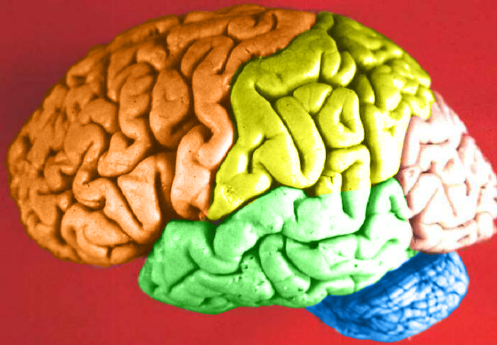
Contain the Brain
You probably recognize the colourful object in this photo (Figure 7.6.1) as a human brain. The brain is arguably the most important organ in the human body. Fortunately for us, the brain has its own special “container,” called the cranial cavity. The cranial cavity enclosing the brain is just one of several cavities in the human body that form “containers” for vital organs.
What Are Body Cavities?
The human body, like that of many other multicellular organisms, is divided into a number of body cavities. A body cavity is a fluid-filled space inside the body that holds and protects internal organs. Human body cavities are separated by membranes and other structures. The two largest human body cavities are the ventral cavity and dorsal cavity. These two body cavities are subdivided into smaller body cavities. Both the dorsal and ventral cavities and their subdivisions are shown in the Figure 7.6.2 diagram.
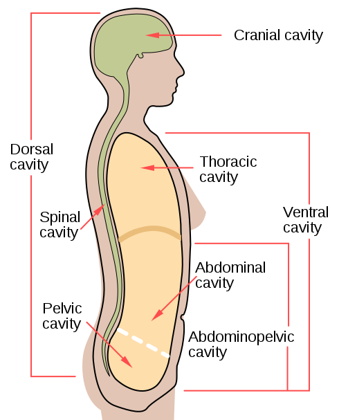
Ventral Cavity
The ventral cavity is at the anterior (or front) of the trunk. Organs contained within this body cavity include the lungs, heart, stomach, intestines, and reproductive organs. The ventral cavity allows for considerable changes in the size and shape of the organs inside as they perform their functions. Organs such as the lungs, stomach, or uterus, for example, can expand or contract without distorting other tissues or disrupting the activities of nearby organs.
The ventral cavity is subdivided into the thoracic and abdominopelvic cavities.
- The thoracic cavity fills the chest and is subdivided into two pleural cavities and the pericardial cavity. The pleural cavities hold the lungs, and the pericardial cavity holds the heart.
- The abdominopelvic cavity fills the lower half of the trunk and is subdivided into the abdominal cavity and the pelvic cavity. The abdominal cavity holds digestive organs and the kidneys, and the pelvic cavity holds reproductive organs and organs of excretion.
Dorsal Cavity
The dorsal cavity is at the posterior (or back) of the body, including both the head and the back of the trunk. The dorsal cavity is subdivided into the cranial and spinal cavities.
- The cranial cavity fills most of the upper part of the skull and contains the brain.
- The spinal cavity is a very long, narrow cavity inside the vertebral column. It runs the length of the trunk and contains the spinal cord.
The brain and spinal cord are protected by the bones of the skull and the vertebrae of the spine. They are further protected by the meninges, a three-layer membrane that encloses the brain and spinal cord. A thin layer of cerebrospinal fluid is maintained between two of the meningeal layers. This clear fluid is produced by the brain, and it provides extra protection and cushioning for the brain and spinal cord.
Feature: My Human Body
The meninges membranes that protect the brain and spinal cord inside their cavities may become inflamed, generally due to a bacterial or viral infection. This condition is called meningitis, and it can lead to serious long-term consequences such as deafness, epilepsy, or cognitive deficits, especially if not treated quickly. Meningitis can also rapidly become life-threatening, so it is classified as a medical emergency.
Learning the symptoms of meningitis may help you or a loved one get prompt medical attention if you ever develop the disease. Common symptoms include fever, headache, and neck stiffness. Other symptoms include confusion or altered consciousness, vomiting, and an inability to tolerate light or loud noises. Young children often exhibit less specific symptoms, such as irritability, drowsiness, or poor feeding.
Meningitis is diagnosed with a lumbar puncture (commonly known as a "spinal tap"), in which a needle is inserted into the spinal canal to collect a sample of cerebrospinal fluid. The fluid is analyzed in a medical lab for the presence of pathogens. If meningitis is diagnosed, treatment consists of antibiotics and sometimes antiviral drugs. Corticosteroids may also be administered to reduce inflammation and the risk of complications (such as brain damage). Supportive measures such as IV fluids may also be provided.
Some types of meningitis can be prevented with a vaccine. Ask your health care professional whether you have had the vaccine or should get it. Giving antibiotics to people who have had significant exposure to certain types of meningitis may reduce their risk of developing the disease. If someone you know is diagnosed with meningitis and you are concerned about contracting the disease yourself, see your doctor for advice.
7.6 Summary
- The human body is divided into a number of body cavities, fluid-filled spaces in the body that hold and protect internal organs. The two largest human body cavities are the ventral cavity and dorsal cavity.
- The ventral cavity is at the anterior (or front) of the trunk. It is subdivided into the thoracic cavity and abdominopelvic cavity.
- The dorsal cavity is at the posterior (or back) of the body, and includes the head and the back of the trunk. It is subdivided into the cranial cavity and spinal cavity.
7.6 Review Questions
-
- What is a body cavity?
- Compare and contrast the ventral and dorsal body cavities.
- Identify the subdivisions of the ventral cavity, and the organs each contains.
- Describe the subdivisions of the dorsal cavity and their contents.
- Identify and describe all the tissues that protect the brain and spinal cord.
- What do you think might happen if fluid were to build up excessively in one of the body cavities?
- Explain why a woman’s body can accommodate a full-term fetus during pregnancy without damaging her internal organs.
- Which body cavity does the needle enter in a lumbar puncture?
- What are the names given to the three body cavity divisions where the heart is located?What are the names given to the three body cavity divisions where the kidneys are located?
-
7.6 Explore More
https://www.youtube.com/watch?v=IaQdv_dBDqM
Why is meningitis so dangerous? - Melvin Sanicas, TED-Ed, 2018.
Attributions
Figure 7.6.1
Brain Lobes by John A Beal, Department of Cellular Biology & Anatomy, Louisiana State University Health Sciences Center Shreveport on Wikimedia Commons is used under a CC BY 2.5 (https://creativecommons.org/licenses/by/2.5/deed.en) license.
Figure 7.6.2
body_cavities-en.svg by Mysid (SVG) on Wikimedia Commons is in the public domain (https://en.wikipedia.org/wiki/Public_domain). (Original by US National Cancer Institute [NCI].)
References
Mayo Clinic Staff. (n.d.). Meningitis. MayoClinic.org. https://www.mayoclinic.org/diseases-conditions/meningitis/symptoms-causes/syc-20350508
TED-Ed. (2018, November 19). Why is meningitis so dangerous? - Melvin Sanicas. YouTube. https://www.youtube.com/watch?v=IaQdv_dBDqM&feature=youtu.be
Created by CK-12 Foundation

Contain the Brain
You probably recognize the colourful object in this photo (Figure 7.6.1) as a human brain. The brain is arguably the most important organ in the human body. Fortunately for us, the brain has its own special “container,” called the cranial cavity. The cranial cavity enclosing the brain is just one of several cavities in the human body that form “containers” for vital organs.
What Are Body Cavities?
The human body, like that of many other multicellular organisms, is divided into a number of body cavities. A body cavity is a fluid-filled space inside the body that holds and protects internal organs. Human body cavities are separated by membranes and other structures. The two largest human body cavities are the ventral cavity and dorsal cavity. These two body cavities are subdivided into smaller body cavities. Both the dorsal and ventral cavities and their subdivisions are shown in the Figure 7.6.2 diagram.

Ventral Cavity
The ventral cavity is at the anterior (or front) of the trunk. Organs contained within this body cavity include the lungs, heart, stomach, intestines, and reproductive organs. The ventral cavity allows for considerable changes in the size and shape of the organs inside as they perform their functions. Organs such as the lungs, stomach, or uterus, for example, can expand or contract without distorting other tissues or disrupting the activities of nearby organs.
The ventral cavity is subdivided into the thoracic and abdominopelvic cavities.
- The thoracic cavity fills the chest and is subdivided into two pleural cavities and the pericardial cavity. The pleural cavities hold the lungs, and the pericardial cavity holds the heart.
- The abdominopelvic cavity fills the lower half of the trunk and is subdivided into the abdominal cavity and the pelvic cavity. The abdominal cavity holds digestive organs and the kidneys, and the pelvic cavity holds reproductive organs and organs of excretion.
Dorsal Cavity
The dorsal cavity is at the posterior (or back) of the body, including both the head and the back of the trunk. The dorsal cavity is subdivided into the cranial and spinal cavities.
- The cranial cavity fills most of the upper part of the skull and contains the brain.
- The spinal cavity is a very long, narrow cavity inside the vertebral column. It runs the length of the trunk and contains the spinal cord.
The brain and spinal cord are protected by the bones of the skull and the vertebrae of the spine. They are further protected by the meninges, a three-layer membrane that encloses the brain and spinal cord. A thin layer of cerebrospinal fluid is maintained between two of the meningeal layers. This clear fluid is produced by the brain, and it provides extra protection and cushioning for the brain and spinal cord.
Feature: My Human Body
The meninges membranes that protect the brain and spinal cord inside their cavities may become inflamed, generally due to a bacterial or viral infection. This condition is called meningitis, and it can lead to serious long-term consequences such as deafness, epilepsy, or cognitive deficits, especially if not treated quickly. Meningitis can also rapidly become life-threatening, so it is classified as a medical emergency.
Learning the symptoms of meningitis may help you or a loved one get prompt medical attention if you ever develop the disease. Common symptoms include fever, headache, and neck stiffness. Other symptoms include confusion or altered consciousness, vomiting, and an inability to tolerate light or loud noises. Young children often exhibit less specific symptoms, such as irritability, drowsiness, or poor feeding.
Meningitis is diagnosed with a lumbar puncture (commonly known as a "spinal tap"), in which a needle is inserted into the spinal canal to collect a sample of cerebrospinal fluid. The fluid is analyzed in a medical lab for the presence of pathogens. If meningitis is diagnosed, treatment consists of antibiotics and sometimes antiviral drugs. Corticosteroids may also be administered to reduce inflammation and the risk of complications (such as brain damage). Supportive measures such as IV fluids may also be provided.
Some types of meningitis can be prevented with a vaccine. Ask your health care professional whether you have had the vaccine or should get it. Giving antibiotics to people who have had significant exposure to certain types of meningitis may reduce their risk of developing the disease. If someone you know is diagnosed with meningitis and you are concerned about contracting the disease yourself, see your doctor for advice.
7.6 Summary
- The human body is divided into a number of body cavities, fluid-filled spaces in the body that hold and protect internal organs. The two largest human body cavities are the ventral cavity and dorsal cavity.
- The ventral cavity is at the anterior (or front) of the trunk. It is subdivided into the thoracic cavity and abdominopelvic cavity.
- The dorsal cavity is at the posterior (or back) of the body, and includes the head and the back of the trunk. It is subdivided into the cranial cavity and spinal cavity.
7.6 Review Questions
-
- What is a body cavity?
- Compare and contrast the ventral and dorsal body cavities.
- Identify the subdivisions of the ventral cavity, and the organs each contains.
- Describe the subdivisions of the dorsal cavity and their contents.
- Identify and describe all the tissues that protect the brain and spinal cord.
- What do you think might happen if fluid were to build up excessively in one of the body cavities?
- Explain why a woman’s body can accommodate a full-term fetus during pregnancy without damaging her internal organs.
- Which body cavity does the needle enter in a lumbar puncture?
- What are the names given to the three body cavity divisions where the heart is located?What are the names given to the three body cavity divisions where the kidneys are located?
-
7.6 Explore More
https://www.youtube.com/watch?v=IaQdv_dBDqM
Why is meningitis so dangerous? - Melvin Sanicas, TED-Ed, 2018.
Attributions
Figure 7.6.1
Brain Lobes by John A Beal, Department of Cellular Biology & Anatomy, Louisiana State University Health Sciences Center Shreveport on Wikimedia Commons is used under a CC BY 2.5 (https://creativecommons.org/licenses/by/2.5/deed.en) license.
Figure 7.6.2
body_cavities-en.svg by Mysid (SVG) on Wikimedia Commons is in the public domain (https://en.wikipedia.org/wiki/Public_domain). (Original by US National Cancer Institute [NCI].)
References
Mayo Clinic Staff. (n.d.). Meningitis. MayoClinic.org. https://www.mayoclinic.org/diseases-conditions/meningitis/symptoms-causes/syc-20350508
TED-Ed. (2018, November 19). Why is meningitis so dangerous? - Melvin Sanicas. YouTube. https://www.youtube.com/watch?v=IaQdv_dBDqM&feature=youtu.be
Created by CK-12/Adapted by Christine Miller
As you read in the beginning of this chapter, new parents Samantha and Aki left their pediatrician’s office still unsure whether or not to vaccinate baby James. Dr. Rodriguez gave them a list of reputable sources where they could look up information about the safety of vaccines, including the Centers for Disease Control and Prevention (CDC). Samantha and Aki read that the consensus within the scientific community is that there is no link between vaccines and autism. They find a long list of studies published in peer-reviewed scientific journals that disprove any link. Additionally, some of the studies are “meta-analyses” that analyzed the findings from many individual studies. The new parents are reassured by the fact that many different researchers, using a large number of subjects in numerous well-controlled and well-reviewed studies, all came to the same conclusion.

Samantha also went back to the web page that originally scared her about the safety of vaccines. She found that the author was not a medical doctor or scientific researcher, but rather a self-proclaimed “child wellness expert.” He sold books and advertising on his site, some of which were related to claims of vaccine injury. She realized that he was both an unqualified and potentially biased source of information.
Samantha also realized that some of his arguments were based on correlations between autism and vaccines, but, as the saying goes, “correlation does not imply causation.” For instance, the recent rise in autism rates may have occurred during the same time period as an increase in the number of vaccines given in childhood, but Samantha could think of many other environmental and social factors that have also changed during this time period. There are just too many variables to come to the conclusion that vaccines, or anything else, are the cause of the rise in autism rates based on that type of argument alone. Also, she learned that the age of onset of autism symptoms happens to typically be around the time that the MMR vaccine is first given, so the apparent association in the timing may just be a coincidence.
Finally, Samantha came across news about a measles outbreak in Vancouver, British Columbia in the winter of 2019. Measles wasn’t just a disease of the past! She learned that measles and whooping cough, which had previously been rare thanks to widespread vaccinations, are now on the rise, and that people choosing not to vaccinate their children seems to be one of the contributing factors. She realized that it is important to vaccinate her baby against these diseases, not only to protect him from their potentially deadly effects, but also to protect others in the population.
In their reading, Samantha and Aki learn that scientists do not yet know the causes of autism, but they feels reassured by the abundance of data that disproves any link with vaccines. Both parents think that the potential benefits of protecting their baby’s health against deadly diseases outweighs any unsubstantiated claims about vaccines. They will be making an appointment to get baby James his shots soon.
Chapter 1 Summary
In this chapter, you learned about some of the same concepts that helped Samantha and Aki make an informed decision. Specifically:
- Science is a distinctive way of gaining knowledge about the natural world that is based on the use of evidence to logically test ideas. As such, science is a process, as well as a body of knowledge.
- A scientific theory, such as the germ theory of disease, is the highest level of explanation in science. A theory is a broad explanation for many phenomena that is widely accepted because it is supported by a great deal of evidence.
- The scientific investigation is the cornerstone of science as a process. A scientific investigation is a systematic approach to answering questions about the physical and natural world. An investigation may be observational or experimental.
- A scientific experiment is a type of scientific investigation in which the researcher manipulates variables under controlled conditions to test expected outcomes. Experiments are the gold standard for scientific investigations and can establish causation between variables.
- Nonexperimental scientific investigations such as observational studies and modeling may be undertaken when experiments are impractical, unethical, or impossible. Observational studies generally can establish correlation — but not causation — between variables.
- A pseudoscience, such as astrology, is a field that is presented as scientific but that does not adhere to scientific standards and methods. Other misuses of science include deliberate hoaxes, frauds, and fallacies made by researchers.
- Strict guidelines must be followed when using human subjects in scientific research. Among the most important protections is the requirement for informed consent.
Now that you know about the nature and process of science, you can apply these concepts in the next chapter to the study of human biology.
Chapter 1 Review
-
- Why does a good hypothesis have to be falsifiable?
- Name one scientific law.
- Name one scientific theory.
- Give an example of a scientific idea that was later discredited.
- A statistical measurement called a P-value is often used in science to determine whether or not a difference between two groups is actually significant or simply due to chance. A P-value of 0.03 means that there is a 3% chance that the difference is due to chance alone. Do you think a P-value of 0.03 would indicate that the difference is likely to be significant? Why or why not?
- Why is it important that scientists communicate their findings to others? How do they usually do this?
- What is a “control group” in science?
- In a scientific experiment, why is it important to only change one variable at a time?
- Which is the dependent variable – the variable that is manipulated or the variable that is being affected by the change?
- You see an ad for a “miracle supplement” called NQP3 that claims the supplement will reduce belly fat. They say it works by reducing the hormone cortisol and by providing your body with missing unspecified “nutrients”, but they do not cite any peer-reviewed clinical studies. They show photographs of three people who appear slimmer after taking the product. A board-certified plastic surgeon endorses the product on television. Answer the following questions about this product.
a. Do you think that because a doctor endorsed the product, it really works? Explain your answer.
b. What are two signs that these claims could actually be pseudoscience instead of true science?
c. Do you think the photographs are good evidence that the product works? Why or why not?
d. If you wanted to do a strong scientific study of whether this supplement does what it claims, what would you do? Be specific about the subjects, data collected, how you would control variables, and how you would analyze the data.
e. What are some ways that you would ensure that the subjects in your experiment in part d are treated ethically and according to human subjects protections regulations?
Attribution
Figure 1.8.1
[Photo of person sitting in front of personal computer] by Avel Chuklanov on Unsplash is used under the Unsplash License (https://unsplash.com/license).
A diagram showing the steps the nervous system takes to initiate a flight or fight response, and then the changes to each body system based on commands from the nervous system. When a threat or harmful event is perceived, the brain processes the signals, beginning in the amygdala and then the hypothalamus. From here, the pituitary gland secretes adrenocorticotropic hormone, which causes the release of cortisol and adrenaline. The physical effects of cortisol and adrenaline are increased heart rate, bladder relaxation, tunnel vision, shaking, dilated pupils, flushed face, dry mouth, slowed digestion, and hearing loss.
Image shows a microscopic view of dense fibrous connective tissue. The parallel strands of collagen show up as pink horizontal fibers. The fibroblasts sit in between fibers and are stained a dark purple.
A class of biological molecule consisting of linked monomers of amino acids and which are the most versatile macromolecules in living systems and serve crucial functions in essentially all biological processes.
The breakdown of larger molecules into smaller ones.
Image shows both a diagram and microscopic view of each of the three types of cartilage: Hyaline cartilage, which contains chondrocytes in paired lacunae and a smooth-looking matrix; Fibrocartilage with looks marbled due to many layers of collagen, and elastic cartilage which irregular, containing both collagen and elastin fibers.
Figure 7.7.1 Everyone on a baseball team has a special job.
Teamwork
Every player on a baseball team has a special job. In the Figure 7.7.1 collage, each player has their part of the infield or outfield covered in case the ball comes their way. Other players on the team cover different parts of the field, or they pitch or catch the ball. Playing baseball clearly requires teamwork. In that regard, the human body is like a baseball team. All of the organ systems of the human body must work together as a team to keep the body alive and well. Teamwork within the body begins with communication.
Communication Among Organ Systems
Communication among organ systems is vital if they are to work together as a team. They must be able to respond to each other and change their responses as needed to keep the body in balance. Communication among organ systems is controlled mainly by the autonomic nervous system and the endocrine system.
The autonomic nervous system is the part of the nervous system that controls involuntary functions. The autonomic nervous system, for example, controls heart rate, blood flow, and digestion. You don’t have to tell your heart to beat faster or to consciously squeeze muscles to push food through the digestive system. You don’t have to even think about these functions at all! The autonomic nervous system orchestrates all the signals needed to control them. It sends messages between parts of the nervous system, as well as between the nervous system and other organ systems via chemical messengers called neurotransmitters.
The endocrine system is the system of glands that secrete hormones directly into the bloodstream. Once in the blood, endocrine hormones circulate to cells everywhere in the body. The endocrine system itself is under control of the nervous system via a part of the brain called the hypothalamus. The hypothalamus secretes hormones that travel directly to cells of the pituitary gland, which is located beneath it. The pituitary gland is the master gland of the endocrine system. Most of its hormones either turn on or turn off other endocrine glands. For example, if the pituitary gland secretes thyroid-stimulating hormone, the hormone travels through the circulation to the thyroid gland, which is stimulated to secrete thyroid hormone. Thyroid hormone then travels to cells throughout the body, where it increases their metabolism.
Examples of Organ System Interactions
An increase in cellular metabolism requires more cellular respiration. Cellular respiration is a good example of organ system interactions, because it is a basic life process that occurs in all living cells.
Cellular Respiration
Cellular respiration is the intracellular process that breaks down glucose with oxygen to produce carbon dioxide and energy in the form of ATP molecules. It is the process by which cells obtain usable energy to power other cellular processes. Which organ systems are involved in cellular respiration? The glucose needed for cellular respiration comes from the digestive system via the cardiovascular system. The oxygen needed for cellular respiration comes from the respiratory system also via the cardiovascular system. The carbon dioxide produced in cellular respiration leaves the body by the opposite route. In short, cellular respiration requires — at a minimum — the digestive, cardiovascular, and respiratory systems.
Fight-or-Flight Response
The well-known fight-or-flight response is a good example of how the nervous and endocrine systems control other organ system responses. The fight-or-flight response begins when the nervous system perceives sudden danger, as shown in the Figure 7.7.2 diagram. The brain sends a message to the endocrine system (via the pituitary gland) for the adrenal glands to secrete the hormones cortisol and adrenaline. These hormones flood the circulation and affect other organ systems throughout the body, including the cardiovascular, urinary, sensory, and digestive systems. Specific responses include increased heart rate, bladder relaxation, tunnel vision, and a shunting of blood away from the digestive system and toward the muscles, brain, and other vital organs needed to fight or flee.
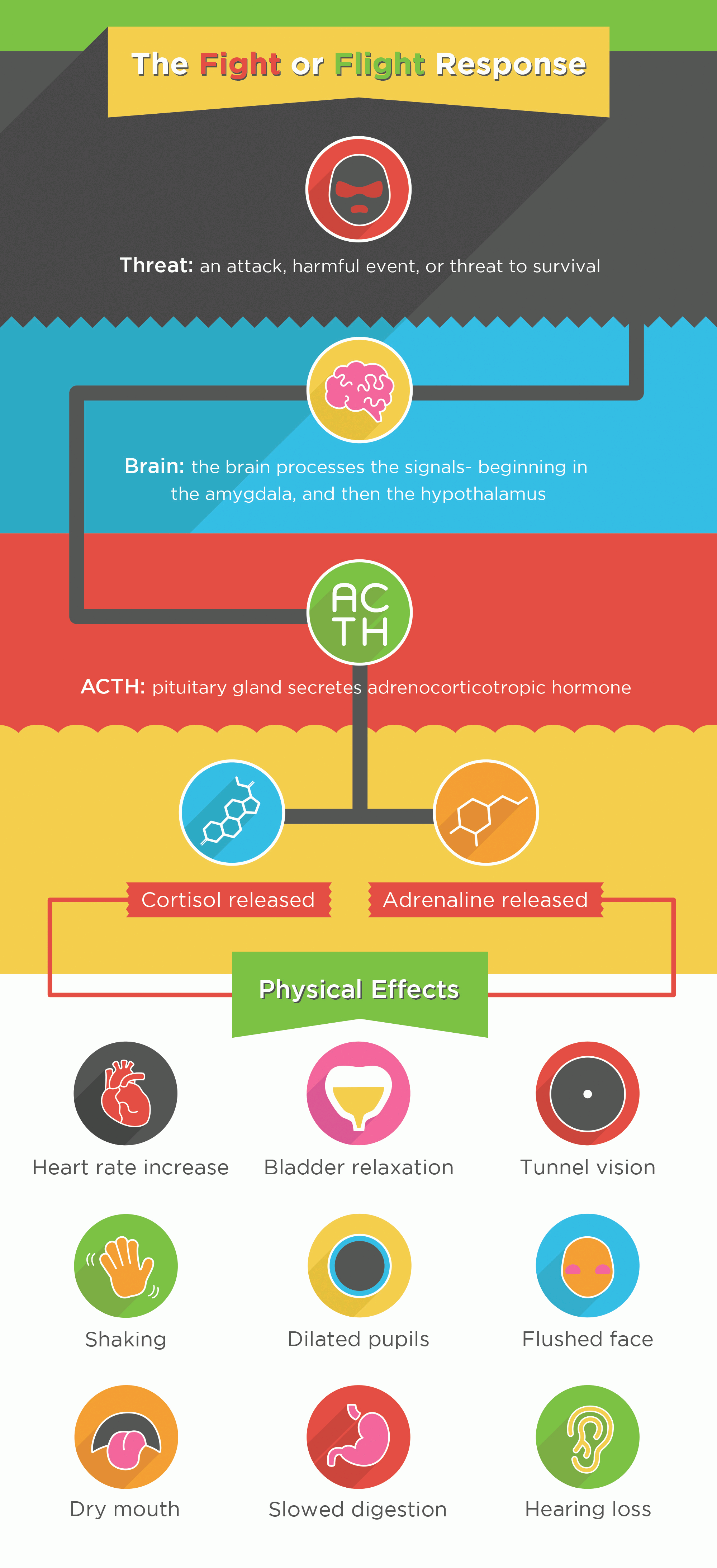
Playing Baseball
The people playing baseball in the opening collage (Figure 7.7.1) are using multiple organ systems in this voluntary activity. Their nervous systems are focused on observing and preparing to respond to the next play. Their other systems are being controlled by the autonomic nervous system. The players are using the muscular, skeletal, respiratory, and cardiovascular systems. Can you explain how each of these organ systems is involved in playing baseball?
Feature: Reliable Sources
Teamwork among organ systems allows the human organism to work like a finely tuned machine — at least, it does until one of the organ systems fails. When that happens, other organ systems interacting in the same overall process will also be affected. This is especially likely if the affected system plays a controlling role in the process. An example is type 1 diabetes. This disorder occurs when the pancreas does not secrete the endocrine hormone insulin. Insulin normally is secreted in response to an increasing level of glucose in the blood, and it brings the level of glucose back to normal by stimulating body cells to take up insulin from the blood.
Learn more about type 1 diabetes. Use several reliable Internet sources to answer the following questions:
- In type 1 diabetes, what causes the endocrine system to fail to produce insulin?
- If type 1 diabetes is not controlled, which organ systems are affected by high blood glucose levels? What are some of the specific effects?
- How can blood glucose levels be controlled in patients with type 1 diabetes?
7.7 Summary
- The human body's organ systems must work together to keep the body alive and functioning normally, which requires communication among systems. This communication is controlled by the autonomic nervous system and endocrine system. The autonomic nervous system controls involuntary body functions, such as heart rate and digestion. The endocrine system secretes hormones into the blood that travel to body cells and influence their activities.
- Cellular respiration is a good example of organ system interactions, because it is a basic life process that happens in all living cells. It is the intracellular process that breaks down glucose with oxygen to produce carbon dioxide and energy. Cellular respiration requires the interaction of the digestive, cardiovascular, and respiratory systems.
- The fight-or-flight response is a good example of how the nervous and endocrine systems control other organ system responses. It is triggered by a message from the brain to the endocrine system and prepares the body for flight or a fight. Many organ systems are stimulated to respond, including the cardiovascular, respiratory, and digestive systems.
- Playing baseball — or doing other voluntary physical activities — may involve the interaction of nervous, muscular, skeletal, respiratory, and cardiovascular systems.
7.7 Review Questions
- What is the autonomic nervous system?
- How do the autonomic nervous system and endocrine system communicate with other organ systems so the systems can interact?
- Explain how the brain communicates with the endocrine system.
- What is the role of the pituitary gland in the endocrine system?
- Identify the organ systems that play a role in cellular respiration.
- How does the hormone adrenaline prepare the body to fight or flee? What specific physiological changes does it bring about?
- Explain the role of the muscular system in digesting food.
- Describe how three different organ systems are involved when a player makes a particular play in baseball, such as catching a fly ball.
-
- What are two types of molecules that the body uses to communicate between organ systems?
- Explain why hormones can have such a wide variety of effects on the body.
7.7 Explore More
https://www.youtube.com/watch?time_continue=19&v=Ujr0UAbyPS4&feature=emb_logo
3D Medical Animation - Peristalsis in Large Intestine/Bowel ||
©Animated Biomedical Productions (ABP), 2013.
https://www.youtube.com/watch?v=FBnBTkcr6No&feature=emb_logo
Adrenaline: Fight or Flight Response, Henk van 't Klooster, 2013.
https://www.youtube.com/watch?v=m2GywoS77qc
Fight or Flight Response, Bozeman Science, 2012.
Attributions
Figure 7.7.1
- Baseball positions by Michael J on Wikimedia Commons is used under a CC BY-SA 4.0 (https://creativecommons.org/licenses/by-sa/4.0/deed.de) license.
- US Navy 040229-N-8629D-070 photo by US Navy's Photographer's Mate 2nd Class Brett A. Dawson on Wikimedia Commons is released into the public domain (https://en.wikipedia.org/wiki/Public_domain).
- David Ortiz batter's box by Albert Yau/ SecondPrint Productions on Flickr is used under a CC BY 2.0 (https://creativecommons.org/licenses/by/2.0/deed.es) license.
- Fenway-from Legend's Box by Jared Vincent on Wikipedia is used under a CC BY 2.0 (https://creativecommons.org/licenses/by/2.0/deed.en) license.
Figure 7.7.2
The_Fight_or_Flight_Response by Jvnkfood (original), converted to PNG and reduced to 8-bit by Pokéfan95 on Wikimedia Commons is used under a CC BY-SA 4.0 (https://creativecommons.org/licenses/by-sa/4.0/deed.en) license.
References
Animated Biomedical Productions. (2013, January 30). 3D Medical animation - Peristalsis in large intestine/bowel || ©ABP. YouTube. https://www.youtube.com/watch?v=Ujr0UAbyPS4&feature=youtu.be
Bozeman Science. (2012, January 9). Fight or flight response. YouTube. https://www.youtube.com/watch?v=m2GywoS77qc&feature=youtu.be
Henk van 't Klooster. (2013). Adrenaline: Fight or flight response. YouTube. https://www.youtube.com/watch?v=FBnBTkcr6No&t=4s
Mayo Clinic Staff. (n.d.). Type 1 diabetes. MayoClinic.org. https://www.mayoclinic.org/diseases-conditions/type-1-diabetes/symptoms-causes/syc-20353011
Wikipedia contributors. (2020, July 22). Thyroid-stimulating hormone. In Wikipedia. https://en.wikipedia.org/w/index.php?title=Thyroid-stimulating_hormone&oldid=968942540
The body system which acts as a chemical messenger system comprising feedback loops of the hormones released by internal glands of an organism directly into the circulatory system, regulating distant target organs. In humans, the major endocrine glands are the thyroid gland and the adrenal glands.
A hormone is a signaling molecule produced by glands in multicellular organisms that target distant organs to regulate physiology and behavior.
also called vasopressin. A hormone made by the hypothalamus in the brain and stored in the posterior pituitary gland. It tells your kidneys how much water to conserve. ADH constantly regulates and balances the amount of water in your blood.
The process by which DNA is copied.
The main mineralocorticoid hormone which is responsible for sodium conservation in the kidney, salivary glands, sweat glands and colon.
division of the peripheral nervous system that controls involuntary activities
Created by CK-12 Foundation/Adapted by Christine Miller
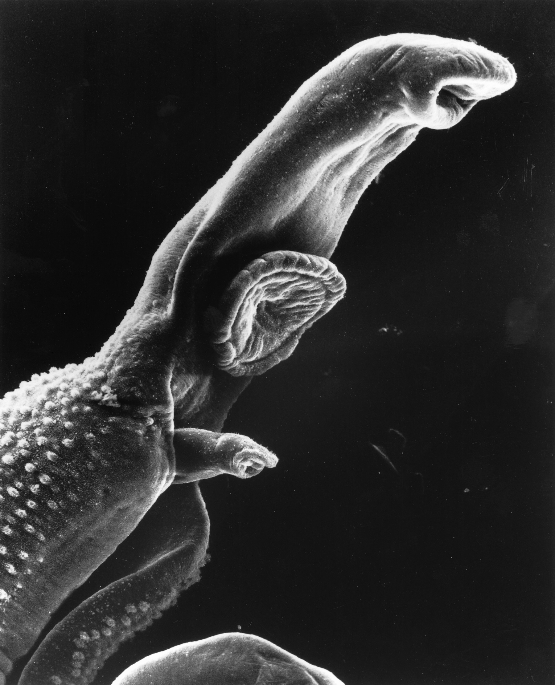
Worm Attack!
Does the organism in Figure 17.2.1 look like a space alien? A scary creature from a nightmare? In fact, it’s a 1-cm long worm in the genus Schistosoma. It may invade and take up residence in the human body, causing a very serious illness known as schistosomiasis. The worm gains access to the human body while it is in a microscopic life stage. It enters through a hair follicle when the skin comes into contact with contaminated water. The worm then grows and matures inside the human organism, causing disease.
Host vs. Pathogen
The Schistosoma worm has a parasitic relationship with humans. In this type of relationship, one organism, called the parasite, lives on or in another organism, called the host. The parasite always benefits from the relationship, and the host is always harmed. The human host of the Schistosoma worm is clearly harmed by the parasite when it invades the host’s tissues. The urinary tract or intestines may be infected, and signs and symptoms may include abdominal pain, diarrhea, bloody stool, or blood in the urine. Those who have been infected for a long time may experience liver damage, kidney failure, infertility, or bladder cancer. In children, Schistosoma infection may cause poor growth and difficulty learning.
Like the Schistosoma worm, many other organisms can make us sick if they manage to enter our body. Any such agent that can cause disease is called a pathogen. Most pathogens are microorganisms, although some — such as the Schistosoma worm — are much larger. In addition to worms, common types of pathogens of human hosts include bacteria, viruses, fungi, and single-celled organisms called protists. You can see examples of each of these types of pathogens in Table 17.1.1. Fortunately for us, our immune system is able to keep most potential pathogens out of the body, or quickly destroy them if they do manage to get in. When you read this chapter, you’ll learn how your immune system usually keeps you safe from harm — including from scary creatures like the Schistosoma worm!
| Type of Pathogen | Description | Disease Caused | |
|---|---|---|---|
| Bacteria:
Example shown: Escherichia coli |
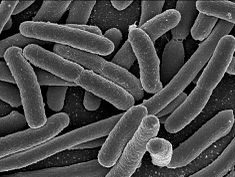 |
Single celled organisms without a nucleus | Strep throat, staph infections, tuberculosis, food poisoning, tetanus, pneumonia, syphillis |
| Viruses:
Example shown: Herpes simplex |
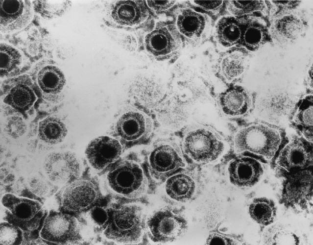 |
Non-living particles that reproduce by taking over living cells | Common cold, flu, genital herpes, cold sores, measles, AIDS, genital warts, chicken pox, small pox |
| Fungi:
Example shown: Death cap mushroom |
 |
Simple organisms, including mushrooms and yeast, that grow as single cells or thread-like filaments | Ringworm, athletes foot, tineas, candidias, histoplasmomis, mushroom poisoning |
| Protozoa:
Example shown: Giardia lamblia |
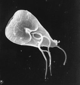 |
Single celled organisms with a nucleus | Malaria, "traveller's diarrhea", giardiasis, typano somiasis ("sleeping sickness") |
What is the Immune System?
The immune system is a host defense system. It comprises many biological structures —ranging from individual leukocytes to entire organs — as well as many complex biological processes. The function of the immune system is to protect the host from pathogens and other causes of disease, such as tumor (cancer) cells. To function properly, the immune system must be able to detect a wide variety of pathogens. It also must be able to distinguish the cells of pathogens from the host’s own cells, and also to distinguish cancerous or damaged host cells from healthy cells. In humans and most other vertebrates, the immune system consists of layered defenses that have increasing specificity for particular pathogens or tumor cells. The layered defenses of the human immune system are usually classified into two subsystems, called the innate immune system and the adaptive immune system.
Innate Immune System
The innate immune system (sometimes referred to as "non-specific defense") provides very quick, but non-specific responses to pathogens. It responds the same way regardless of the type of pathogen that is attacking the host. It includes barriers — such as the skin and mucous membranes — that normally keep pathogens out of the body. It also includes general responses to pathogens that manage to breach these barriers, including chemicals and cells that attack the pathogens inside the human host. Certain leukocytes (white blood cells), for example, engulf and destroy pathogens they encounter in the process called phagocytosis, which is illustrated in Figure 17.2.2. Exposure to pathogens leads to an immediate maximal response from the innate immune system.
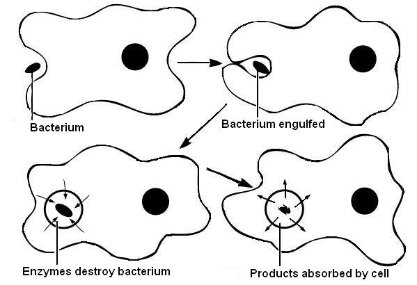
Watch the video below, "Neutrophil Phagocytosis - White Blood Cells Eats Staphylococcus Aureus Bacteria" by ImmiflexImmuneSystem, to see phagocytosis in action.
https://youtu.be/Z_mXDvZQ6dU
Neutrophil Phagocytosis - White Blood Cell Eats Staphylococcus Aureus Bacteria, ImmiflexImmuneSystem, 2013.
Adaptive Immune System
The adaptive immune system is activated if pathogens successfully enter the body and manage to evade the general defenses of the innate immune system. An adaptive response is specific to the particular type of pathogen that has invaded the body, or to cancerous cells. It takes longer to launch a specific attack, but once it is underway, its specificity makes it very effective. An adaptive response also usually leads to immunity. This is a state of resistance to a specific pathogen, due to the adaptive immune system's ability to “remember” the pathogen and immediately mount a strong attack tailored to that particular pathogen if it invades again in the future.
Self vs. Non-Self
Both innate and adaptive immune responses depend on the immune system's ability to distinguish between self- and non-self molecules. Self molecules are those components of an organism’s body that can be distinguished from foreign substances by the immune system. Virtually all body cells have surface proteins that are part of a complex called major histocompatibility complex (MHC). These proteins are one way the immune system recognizes body cells as self. Non-self proteins, in contrast, are recognized as foreign, because they are different from self proteins.
Antigens and Antibodies
Many non-self molecules comprise a class of compounds called antigens. Antigens, which are usually proteins, bind to specific receptors on immune system cells and elicit an adaptive immune response. Some adaptive immune system cells (B cells) respond to foreign antigens by producing antibodies. An antibody is a molecule that precisely matches and binds to a specific antigen. This may target the antigen (and the pathogen displaying it) for destruction by other immune cells.
Antigens on the surface of pathogens are how the adaptive immune system recognizes specific pathogens. Antigen specificity allows for the generation of responses tailored to the specific pathogen. It is also how the adaptive immune system ”remembers” the same pathogen in the future.
Immune Surveillance
Another important role of the immune system is to identify and eliminate tumor cells. This is called immune surveillance. The transformed cells of tumors express antigens that are not found on normal body cells. The main response of the immune system to tumor cells is to destroy them. This is carried out primarily by aptly-named killer T cells of the adaptive immune system.
Lymphatic System
The lymphatic system is a human organ system that is a vital part of the adaptive immune system. It is also part of the cardiovascular system and plays a major role in the digestive system (see section 17.3 Lymphatic System). The major structures of the lymphatic system are shown in Figure 17.2.3 .
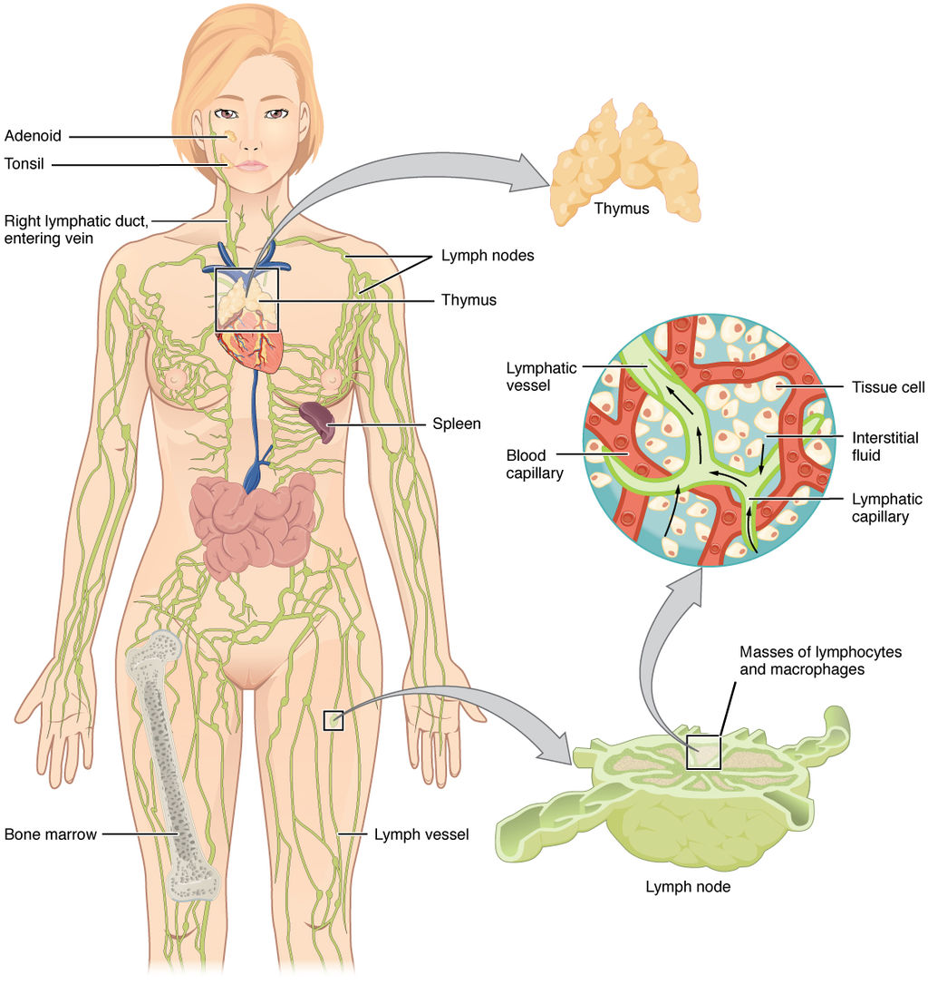
The lymphatic system consists of several lymphatic organs and a body-wide network of lymphatic vessels that transport the fluid called lymph. Lymph is essentially blood plasma that has leaked from capillaries into tissue spaces. It includes many leukocytes, especially lymphocytes, which are the major cells of the lymphatic system. Like other leukocytes, lymphocytes defend the body. There are several different types of lymphocytes that fight pathogens or cancer cells as part of the adaptive immune system.
Major lymphatic organs include the thymus and bone marrow. Their function is to form and/or mature lymphocytes. Other lymphatic organs include the spleen, tonsils, and lymph nodes, which are small clumps of lymphoid tissue clustered along lymphatic vessels. These other lymphatic organs harbor mature lymphocytes and filter lymph. They are sites where pathogens collect, and adaptive immune responses generally begin.
Neuroimmune System vs. Peripheral Immune System
The brain and spinal cord are normally protected from pathogens in the blood by the selectively permeable blood-brain and blood-spinal cord barriers. These barriers are part of the neuroimmune system. The neuroimmune system has traditionally been considered distinct from the rest of the immune system, which is called the peripheral immune system — although that view may be changing. Unlike the peripheral system, in which leukocytes are the main cells, the main cells of the neuroimmune system are thought to be nervous system cells called neuroglia. These cells can recognize and respond to pathogens, debris, and other potential dangers. Types of neuroglia involved in neuroimmune responses include microglial cells and astrocytes.
- Microglial cells are among the most prominent types of neuroglia in the brain. One of their main functions is to phagocytize cellular debris that remains when neurons die. Microglial cells also “prune” obsolete synapses between neurons.
- Astrocytes are neuroglia that have a different immune function. They allow certain immune cells from the peripheral immune system to cross into the brain via the blood-brain barrier to target both pathogens and damaged nervous tissue.
Feature: Human Biology in the News
“They’ll have to rewrite the textbooks!”
That sort of response to a scientific discovery is sure to attract media attention, and it did. It’s what Kevin Lee, a neuroscientist at the University of Virginia, said in 2016 when his colleagues told him they had discovered human anatomical structures that had never before been detected. The structures were tiny lymphatic vessels in the meningeal layers surrounding the brain.
How these lymphatic vessels could have gone unnoticed when all human body systems have been studied so completely is amazing in its own right. The suggested implications of the discovery are equally amazing:
- The presence of these lymphatic vessels means that the brain is directly connected to the peripheral immune system, presumably allowing a close association between the human brain and human pathogens. This suggests an entirely new avenue by which humans and their pathogens may have influenced each other’s evolution. The researchers speculate that our pathogens even may have influenced the evolution of our social behaviors.
- The researchers think there will also be many medical applications of their discovery. For example, the newly discovered lymphatic vessels may play a major role in neurological diseases that have an immune component, such as multiple sclerosis. The discovery might also affect how conditions such as autism spectrum disorders and schizophrenia are treated.
17.2 Summary
- Any agent that can cause disease is called a pathogen. Most human pathogens are microorganisms, such as bacteria and viruses. The immune system is the body system that defends the human host from pathogens and cancerous cells.
- The innate immune system is a subset of the immune system that provides very quick, but non-specific responses to pathogens. It includes multiple types of barriers to pathogens, leukocytes that phagocytize pathogens, and several other general responses.
- The adaptive immune system is a subset of the immune system that provides specific responses tailored to particular pathogens. It takes longer to put into effect, but it may lead to immunity to the pathogens.
- Both innate and adaptive immune responses depend on the immune system's ability to distinguish between self and non-self molecules. Most body cells have major histocompatibility complex (MHC) proteins that identify them as self. Pathogens and tumor cells have non-self antigens that the immune system recognizes as foreign.
- Antigens are proteins that bind to specific receptors on immune system cells and elicit an adaptive immune response. Generally, they are non-self molecules on pathogens or infected cells. Some immune cells (B cells) respond to foreign antigens by producing antibodies that bind with antigens and target pathogens for destruction.
- Tumor surveillance is an important role of the immune system. Killer T cells of the adaptive immune system find and destroy tumor cells, which they can identify from their abnormal antigens.
- The lymphatic system is a human organ system vital to the adaptive immune system. It consists of several organs and a system of vessels that transport lymph. The main immune function of the lymphatic system is to produce, mature, and circulate lymphocytes, which are the main cells in the adaptive immune system.
- The neuroimmune system that protects the central nervous system is thought to be distinct from the peripheral immune system that protects the rest of the human body. The blood-brain and blood-spinal cord barriers are one type of protection for the neuroimmune system. Neuroglia also play role in this system, for example, by carrying out phagocytosis.
17.2 Review Questions
-
- What is a pathogen?
- State the purpose of the immune system.
- Compare and contrast the innate and adaptive immune systems.
- Explain how the immune system distinguishes self molecules from non-self molecules.
- What are antigens?
- Define tumor surveillance.
- Briefly describe the lymphatic system and its role in immune function.
- Identify the neuroimmune system.
- What does it mean that the immune system is not just composed of organs?
- Why is the immune system considered “layered?”
17.2 Explore More
https://youtu.be/xZbcwi7SfZE
The Antibiotic Apocalypse Explained, Kurzgesagt – In a Nutshell, 2016.
https://youtu.be/Nw27_jMWw10
Overview of the Immune System, Handwritten Tutorials, 2011.
https://youtu.be/gVdY9KXF_Sg
The surprising reason you feel awful when you're sick - Marco A. Sotomayor, TED-Ed, 2016.
Attributions
Figure 17.1.1
Schistosome Parasite by Bruce Wetzel and Harry Schaefer (Photographers) from the National Cancer Institute, Visuals online is in the public domain (https://en.wikipedia.org/wiki/Public_domain).
Figure 17.1.2
Phagocytosis by Rlawson at en.wikibooks on Wikimedia Commons is used under a CC BY SA 3.0 (https://creativecommons.org/licenses/by-sa/3.0/deed.en) license. (Transferred from en.wikibooks to Commons by User:Adrignola.)
Figure 17.1.3
2201_Anatomy_of_the_Lymphatic_System by OpenStax College on Wikimedia Commons is used under a CC BY 3.0 (https://creativecommons.org/licenses/by/3.0) license.
Table 17.1.1
- EscherichiaColi NIAID [photo] by Rocky Mountain Laboratories, NIH National Institute of Allergy and Infectious Diseases (NIAID) on Wikimedia Commons is in the public domain (https://en.wikipedia.org/wiki/Public_domain).
- Herpes simplex virus TEM B82-0474 lores by Dr. Erskine Palmer/ CDC Public Health Image Library (PHIL) on Wikimedia Commons is in the public domain (https://en.wikipedia.org/wiki/Public_domain).
- Red death cap mushroom by Rosendahl on Wikimedia Commons is in the public domain (https://en.wikipedia.org/wiki/Public_domain). (Transferred from Pixnio by Fæ.)
- Scanning electron micrograph (SEM) of Giardia lamblia by Janice Haney Carr/ CDC, Public Health Image Library (PHIL) Photo ID# 8698 is in the public domain (https://en.wikipedia.org/wiki/Public_domain).
References
Barney, J. (2016, March 21). They’ll have to rewrite the textbooks [online article]. Illimitable - Discovery. UVA Today/ University of Virginia. https://news.virginia.edu/illimitable/discovery/theyll-have-rewrite-textbooks
Betts, J. G., Young, K.A., Wise, J.A., Johnson, E., Poe, B., Kruse, D.H., Korol, O., Johnson, J.E., Womble, M., DeSaix, P. (2013, June 19). Figure 21.2 Anatomy of the lymphatic system [digital image]. In Anatomy and Physiology (Section 21.1). OpenStax. https://openstax.org/books/anatomy-and-physiology/pages/21-1-anatomy-of-the-lymphatic-and-immune-systems
Handwritten Tutorials. (2011, October 25). Overview of the immune system. YouTube. https://www.youtube.com/watch?v=Nw27_jMWw10&feature=youtu.be
ImmiflexImmuneSystem. (2013). Neutrophil phagocytosis - White blood cell eats staphylococcus aureus bacteria. YouTube. https://www.youtube.com/watch?v=Z_mXDvZQ6dU
Kurzgesagt – In a Nutshell. (2016, March 16). The antibiotic apocalypse explained. YouTube. https://www.youtube.com/watch?v=xZbcwi7SfZE&feature=youtu.be
Louveau, A., Smirnov, I., Keyes, T. J., Eccles, J. D., Rouhani, S. J., Peske, J. D., Derecki, N. C., Castle, D., Mandell, J. W., Lee, K. S., Harris, T. H., & Kipnis, J. (2015). Structural and functional features of central nervous system lymphatic vessels. Nature, 523(7560), 337–341. https://doi.org/10.1038/nature14432
Mayo Clinic Staff. (n.d.). Autism spectrum disorder [online article]. MayoClinic.org. https://www.mayoclinic.org/diseases-conditions/autism-spectrum-disorder/symptoms-causes/syc-20352928
Mayo Clinic Staff. (n.d.). Multiple sclerosis [online article]. MayoClinic.org. https://www.mayoclinic.org/diseases-conditions/multiple-sclerosis/symptoms-causes/syc-20350269
Mayo Clinic Staff. (n.d.). Schizophrenia [online article]. MayoClinic.org. https://www.mayoclinic.org/diseases-conditions/schizophrenia/symptoms-causes/syc-20354443
TED-Ed. (2016, April 19). The surprising reason you feel awful when you're sick - Marco A. Sotomayor. YouTube. https://www.youtube.com/watch?v=gVdY9KXF_Sg&feature=youtu.be
Created by CK-12 Foundation/Adapted by Christine Miller
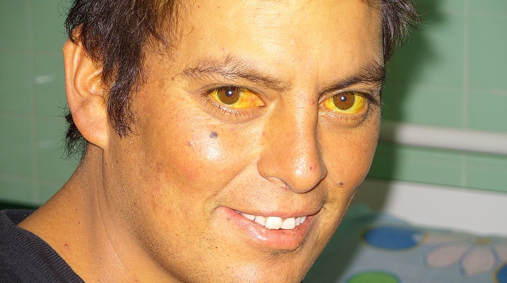
Jaundiced Eyes
Did you ever hear of a person looking at something or someone with a “jaundiced eye”? It means to take a negative view, such as envy, maliciousness, or ill will. The expression may be based on the antiquated idea that liver bile is associated with such negative emotions as these, as well as the fact that excessive liver bile causes jaundice, or yellowing of the eyes and skin. Jaundice is likely a sign of a liver disorder or blockage of the duct that carries bile away from the liver. Bile contains waste products, making the liver an organ of excretion. Bile has an important role in digestion, which makes the liver an accessory organ of digestion, too.
What Are Accessory Organs of Digestion?
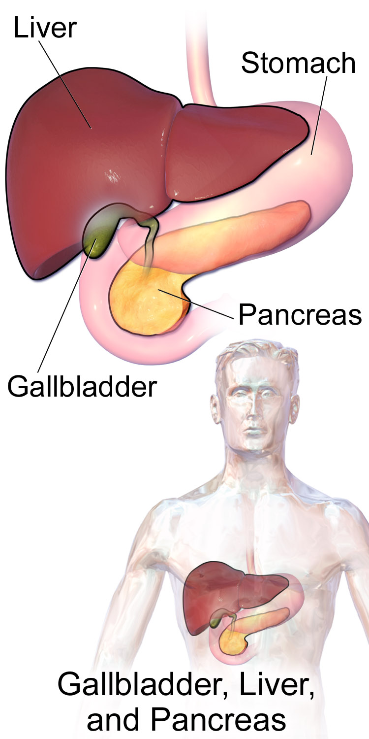
Accessory organs of digestion are organs that secrete substances needed for the chemical digestion of food, but through which food does not actually pass as it is digested. Besides the liver, the major accessory organs of digestion are the gallbladder and pancreas. These organs secrete or store substances that are needed for digestion in the first part of the small intestine — the duodenum — where most chemical digestion takes place. You can see the three organs and their locations in Figure 15.6.2.
Liver
The liver is a vital organ located in the upper right part of the abdomen. It lies just below the diaphragm, to the right of the stomach. The liver plays an important role in digestion by secreting bile, but the liver has a wide range of additional functions unrelated to digestion. In fact, some estimates put the number of functions of the liver at about 500! A few of them are described below.
Structure of the Liver
The liver is a reddish brown, wedge-shaped structure. In adults, the liver normally weighs about 1.5 kg (about 3.3 lb). It is both the heaviest internal organ and the largest gland in the human body. The liver is divided into four lobes of unequal size and shape. Each lobe, in turn, is made up of lobules, which are the functional units of the liver. Each lobule consists of millions of liver cells, called hepatic cells (or hepatocytes). They are the basic metabolic cells that carry out the various functions of the liver.
As shown in Figure 15.6.3, the liver is connected to two large blood vessels: the hepatic artery and the portal vein. The hepatic artery carries oxygen-rich blood from the aorta, whereas the portal vein carries blood that is rich in digested nutrients from the GI tract and wastes filtered from the blood by the spleen. The blood vessels subdivide into smaller arteries and capillaries, which lead into the liver lobules. The nutrients from the GI tract are used to build many vital biochemical compounds, and the wastes from the spleen are degraded and excreted.
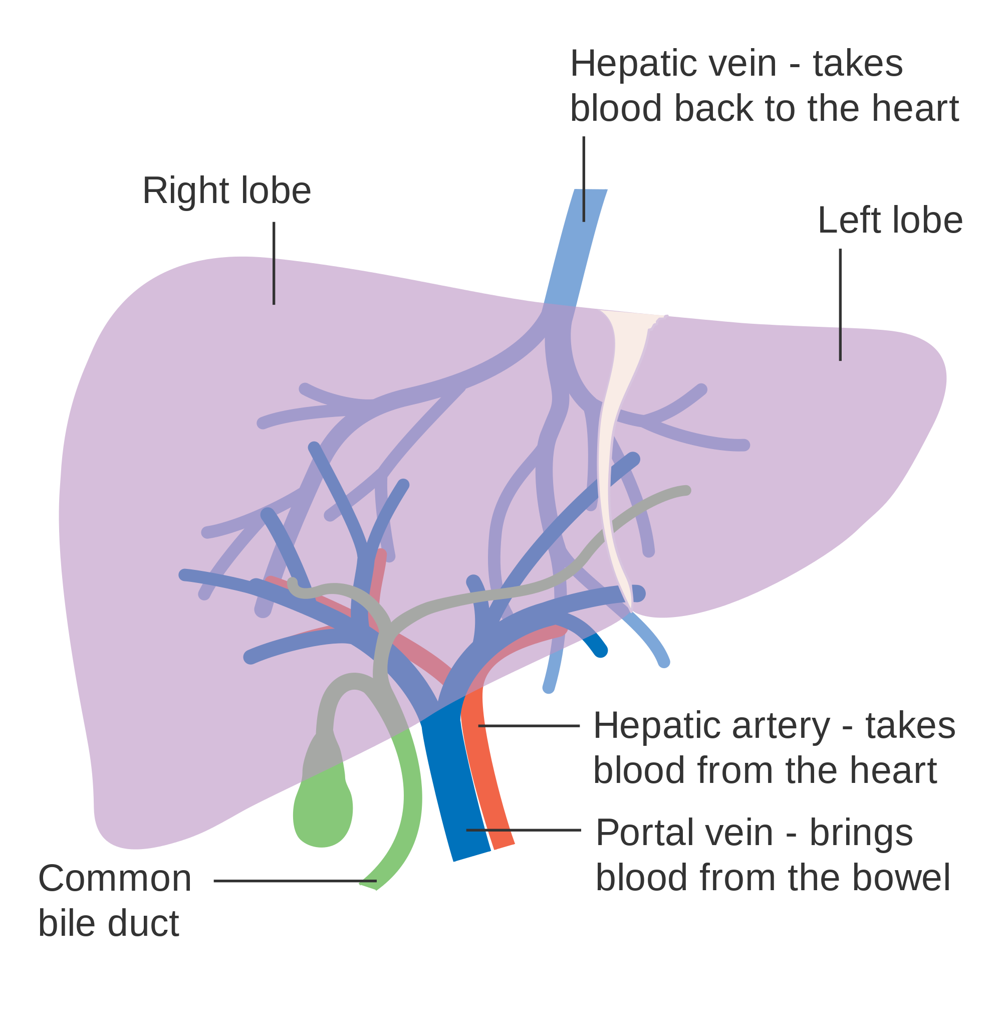
Functions of the Liver
The main digestive function of the liver is the production of bile. Bile is a yellowish alkaline liquid that consists of water, electrolytes, bile salts, and cholesterol, among other substances, many of which are waste products. Some of the components of bile are synthesized by hepatocytes. The rest are extracted from the blood.
As shown in Figure 15.6.4, bile is secreted into small ducts that join together to form larger ducts, with just one large duct carrying bile out of the liver. If bile is needed to digest a meal, it goes directly to the duodenum through the common bile duct. In the duodenum, the bile neutralizes acidic chyme from the stomach and emulsifies fat globules into smaller particles (called micelles) that are easier to digest chemically by the enzyme lipase. Bile also aids with the absorption of vitamin K. Bile that is secreted when digestion is not taking place goes to the gallbladder for storage until the next meal. In either case, the bile enters the duodenum through the common bile duct.
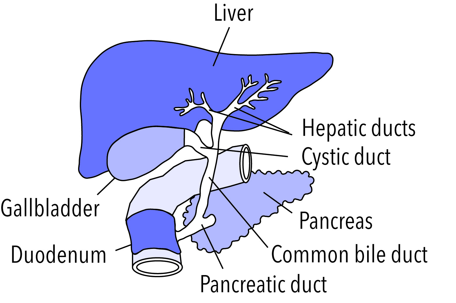
Besides its roles in digestion, the liver has many other vital functions:
- The liver synthesizes glycogen from glucose and stores the glycogen as required to help regulate blood sugar levels. It also breaks down the stored glycogen to glucose and releases it back into the blood as needed.
- The liver stores many substances in addition to glycogen, including vitamins A, D, B12, and K. It also stores the minerals iron and copper.
- The liver synthesizes numerous proteins and many of the amino acids needed to make them. These proteins have a wide range of functions. They include fibrinogen, which is needed for blood clotting; insulin-like growth factor (IGF-1), which is important for childhood growth; and albumen, which is the most abundant protein in blood serum and functions to transport fatty acids and steroid hormones in the blood.
- The liver synthesizes many important lipids, including cholesterol, triglycerides, and lipoproteins.
- The liver is responsible for the breakdown of many waste products and toxic substances. The wastes are excreted in bile or travel to the kidneys, which excrete them in urine.
The liver is clearly a vital organ that supports almost every other organ in the body. Because of its strategic location and diversity of functions, the liver is also prone to many diseases, some of which cause loss of liver function. There is currently no way to compensate for the absence of liver function in the long term, although liver dialysis techniques can be used in the short term. An artificial liver has not yet been developed, so liver transplantation may be the only option for people with liver failure.
Gallbladder
The gallbladder is a small, hollow, pouch-like organ that lies just under the right side of the liver (see Figure 15.6.5). It is about 8 cm (about 3 in) long and shaped like a tapered sac, with the open end continuous with the cystic duct. The gallbladder stores and concentrates bile from the liver until it is needed in the duodenum to help digest lipids. After the bile leaves the liver, it reaches the gallbladder through the cystic duct. At any given time, the gallbladder may store between 30 to 60 mL (1 to 2 oz) of bile. A hormone stimulated by the presence of fat in the duodenum signals the gallbladder to contract and force its contents back through the cystic duct and into the common bile duct to drain into the duodenum.
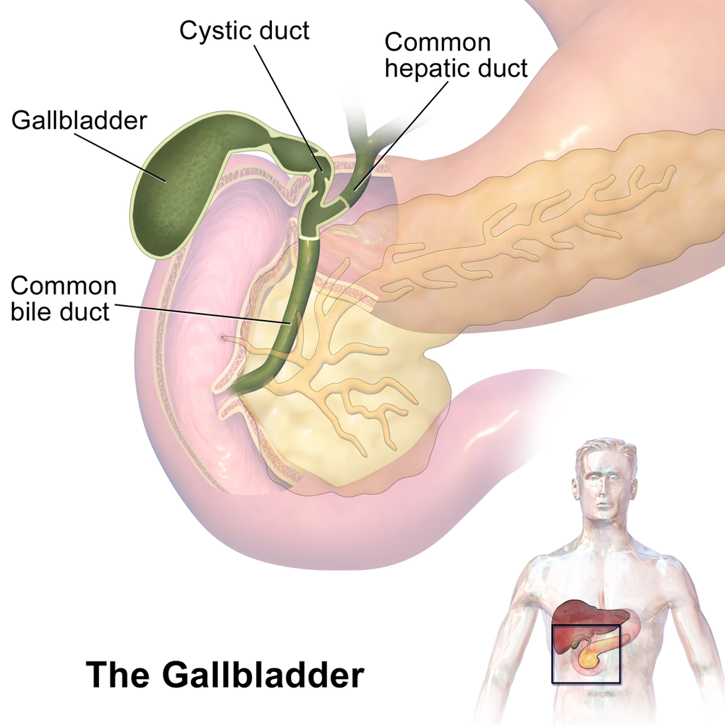
Pancreas
The pancreas is a glandular organ that is part of both the digestive system and the endocrine system. As shown in Figure 15.6.6, it is located in the abdomen behind the stomach, with the head of the pancreas surrounded by the duodenum of the small intestine. The pancreas is about 15 cm (almost 6 in) long, and it has two major ducts: the main pancreatic duct and the accessory pancreatic duct. Both of these ducts drain into the duodenum.
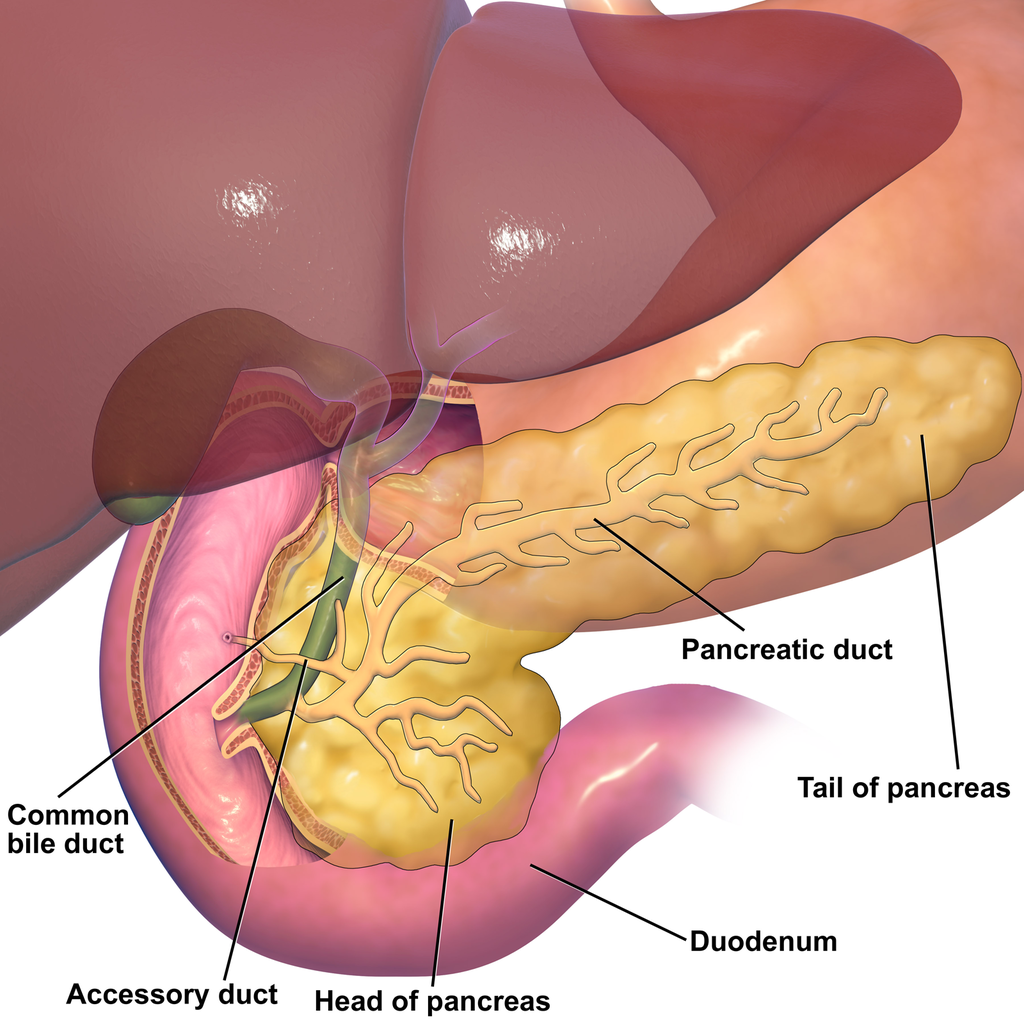
As an endocrine gland, the pancreas secretes several hormones, including insulin and glucagon, which circulate in the blood. The endocrine hormones are secreted by clusters of cells called pancreatic islets (or islets of Langerhans). As a digestive organ, the pancreas secretes many digestive enzymes and also bicarbonate, which helps neutralize acidic chyme after it enters the duodenum. The pancreas is stimulated to secrete its digestive substances when food in the stomach and duodenum triggers the release of endocrine hormones into the blood that reach the pancreas via the bloodstream. The pancreatic digestive enzymes are secreted by clusters of cells called acini, and they travel through the pancreatic ducts to the duodenum. In the duodenum, they help to chemically break down carbohydrates, proteins, lipids, and nucleic acids in chyme. The pancreatic digestive enzymes include:
- Amylase, which helps digest starch and other carbohydrates.
- Trypsin and chymotrypsin, which help digest proteins.
- Lipase, which helps digest lipids.
- Deoxyribonucleases and ribonucleases, which help digest nucleic acids.
15.6 Summary
- Accessory organs of digestion are organs that secrete substances needed for the chemical digestion of food, but through which food does not actually pass as it is digested. The accessory organs include the liver, gallbladder, and pancreas. These organs secrete or store substances that are carried to the duodenum of the small intestine as needed for digestion.
- The liver is a large organ in the abdomen that is divided into lobes and smaller lobules, which consist of metabolic cells called hepatic cells, or hepatocytes. The liver receives oxygen in blood from the aorta through the hepatic artery. It receives nutrients in blood from the GI tract and wastes in blood from the spleen through the portal vein.
- The main digestive function of the liver is the production of the alkaline liquid called bile. Bile is carried directly to the duodenum by the common bile duct or to the gallbladder first for storage. Bile neutralizes acidic chyme that enters the duodenum from the stomach, and also emulsifies fat globules into smaller particles (micelles) that are easier to digest chemically.
- Other vital functions of the liver include regulating blood sugar levels by storing excess sugar as glycogen, storing many vitamins and minerals, synthesizing numerous proteins and lipids, and breaking down waste products and toxic substances.
- The gallbladder is a small pouch-like organ near the liver. It stores and concentrates bile from the liver until it is needed in the duodenum to neutralize chyme and help digest lipids.
- The pancreas is a glandular organ that secretes both endocrine hormones and digestive enzymes. As an endocrine gland, the pancreas secretes insulin and glucagon to regulate blood sugar. As a digestive organ, the pancreas secretes digestive enzymes into the duodenum through ducts. Pancreatic digestive enzymes include amylase (starches) trypsin and chymotrypsin (proteins), lipase (lipids), and ribonucleases and deoxyribonucleases (RNA and DNA).
15.6 Review Questions
- Name three accessory organs of digestion. How do these organs differ from digestive organs that are part of the GI tract?
-
- Describe the liver and its blood supply.
- Explain the main digestive function of the liver and describe the components of bile and it's importance in the digestive process.
- What type of secretions does the pancreas release as part of each body system?
- List pancreatic enzymes that work in the duodenum, along with the substances they help digest.
- What are two substances produced by accessory organs of digestion that help neutralize chyme in the small intestine? Where are they produced?
- People who have their gallbladder removed sometimes have digestive problems after eating high-fat meals. Why do you think this happens?
- Which accessory organ of digestion synthesizes cholesterol?
15.6 Explore More
https://youtu.be/8dgoeYPoE-0
What does the pancreas do? - Emma Bryce, TED-Ed. 2015.
https://youtu.be/wbh3SjzydnQ
What does the liver do? - Emma Bryce, TED-Ed, 2014.
https://youtu.be/a0d1yvGcfzQ
Scar wars: Repairing the liver, nature video, 2018.
Attributions
Figure 15.6.1
Scleral_Icterus by Sheila J. Toro on Wikimedia Commons is used under a CC BY 4.0 (https://creativecommons.org/licenses/by/4.0) license.
Figure 15.6.2
Blausen_0428_Gallbladder-Liver-Pancreas_Location by BruceBlaus on Wikimedia Commons is used under a CC BY 3.0 (https://creativecommons.org/licenses/by/3.0) license.
Figure 15.6.3
Diagram_showing_the_two_lobes_of_the_liver_and_its_blood_supply_CRUK_376.svg by Cancer Research UK on Wikimedia Commons is used under a CC BY-SA 4.0 (https://creativecommons.org/licenses/by-sa/4.0) license.
Figure 15.6.4
Gallbladder by NIH Image Gallery on Flickr is used CC BY-NC 2.0 (https://creativecommons.org/licenses/by-nc/2.0/) license.
Figure 15.6.5
Gallbladder_(organ) (1) by BruceBlaus on Wikimedia Commons is used under a CC BY-SA 4.0 (https://creativecommons.org/licenses/by-sa/4.0) license. (See a full animation of this medical topic at blausen.com.)
Figure 15.6.6
Blausen_0698_PancreasAnatomy by BruceBlaus on Wikimedia Commons is used under a CC BY 3.0 (https://creativecommons.org/licenses/by/3.0) license.
References
Blausen.com Staff. (2014). Medical gallery of Blausen Medical 2014. WikiJournal of Medicine 1 (2). DOI:10.15347/wjm/2014.010. ISSN 2002-4436.
nature video. (2018, December 19). Scar wars: Repairing the liver. YouTube. https://www.youtube.com/watch?v=a0d1yvGcfzQ&feature=youtu.be
TED-Ed. (2014, November 25). What does the liver do? - Emma Bryce. YouTube. https://www.youtube.com/watch?v=wbh3SjzydnQ&feature=youtu.be
TED-Ed. (2015, February 19). What does the pancreas do? - Emma Bryce. YouTube. https://www.youtube.com/watch?v=8dgoeYPoE-0&feature=youtu.be
The chemical processes that occur in a living organism to sustain life.
Image shows a diagram of the negative feedback loops that maintain homeostasis of body temperature. When body temperature falls, blood vessels constrict so that heat is conserved, sweat glands do not secrete fluid, and shivering generates body heat, which warms the body. When body temperature rises, blood vessels dilate, resulting in heat loss to the environment, sweat glands release fluid and as the fluid evaporates, heat is lost from the body.
The ability of an organism to maintain constant internal conditions despite external changes.
A hormone made by the hypothalamus in the brain and stored in the posterior pituitary gland. It tells your kidneys how much water to conserve. ADH constantly regulates and balances the amount of water in your blood. Higher water concentration increases the volume and pressure of your blood.
Created by: CK-12/Adapted by Christine Miller
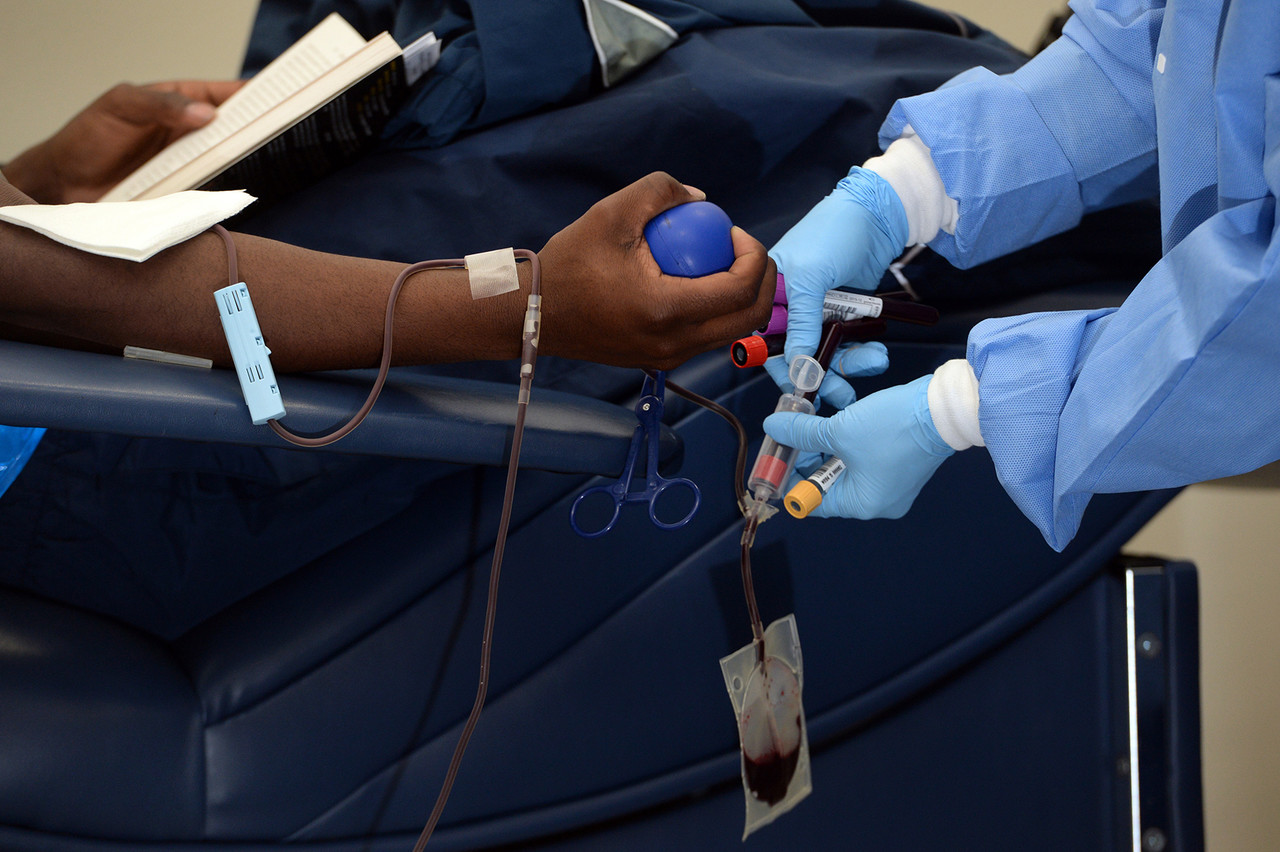
Giving the Gift of Life
Did you ever donate blood? If you did, then you probably know that your blood type is an important factor in blood transfusions. People vary in the type of blood they inherit, and this determines which type(s) of blood they can safely receive in a transfusion. Do you know your blood type?
What Are Blood Types?
Blood is composed of cells suspended in a liquid called plasma. There are three types of cells in blood: red blood cells, which carry oxygen; white blood cells, which fight infections and other threats; and platelets, which are cell fragments that help blood clot. Blood type (or blood group) is a genetic characteristic associated with the presence or absence of certain molecules, called antigens, on the surface of red blood cells. These molecules may help maintain the integrity of the cell membrane, act as receptors, or have other biological functions. A blood group system refers to all of the gene(s), alleles, and possible genotypes and phenotypes that exist for a particular set of blood type antigens. Human blood group systems include the well-known ABO and Rhesus (Rh) systems, as well as at least 33 others that are less well known.
Antigens and Antibodies
Antigens — such as those on the red blood cells — are molecules that the immune system identifies as either self (produced by your own body) or non-self (not produced by your own body). Blood group antigens may be proteins, carbohydrates, glycoproteins (proteins attached to chains of sugars), or glycolipids (lipids attached to chains of sugars), depending on the particular blood group system. If antigens are identified as non-self, the immune system responds by forming antibodies that are specific to the non-self antigens. Antibodies are large, Y-shaped proteins produced by the immune system that recognize and bind to non-self antigens. The analogy of a lock and key is often used to represent how an antibody and antigen fit together, as shown in the illustration below (Figure 6.5.2). When antibodies bind to antigens, it marks them for destruction by other immune system cells. Non-self antigens may enter your body on pathogens (such as bacteria or viruses), on foods, or on red blood cells in a blood transfusion from someone with a different blood type than your own. The last way is virtually impossible nowadays because of effective blood typing and screening protocols.
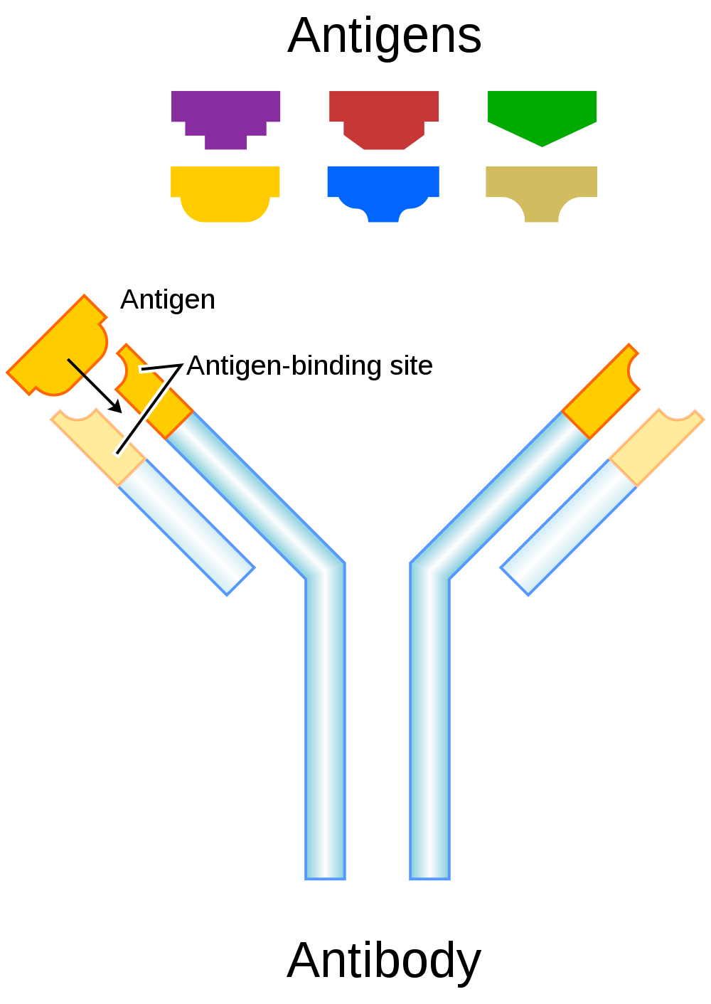
Genetics of Blood Type
An individual’s blood type depends on which alleles for a blood group system were inherited from their parents. Generally, blood type is controlled by alleles for a single gene, or for two or more very closely linked genes. Closely linked genes are almost always inherited together, because there is little or no recombination between them. Like other genetic traits, a person’s blood type is generally fixed for life, but there are rare instances in which blood type can change. This could happen, for example, if an individual receives a bone marrow transplant to treat a disease, such as leukemia. If the bone marrow comes from a donor who has a different blood type, the patient’s blood type may eventually convert to the donor’s blood type, because red blood cells are produced in bone marrow.
ABO Blood Group System
The ABO blood group system is the best known human blood group system. Antigens in this system are glycoproteins. These antigens are shown in the list below. There are four common blood types for the ABO system:
- Type A, in which only the A antigen is present.
- Type B, in which only the B antigen is present.
- Type AB, in which both the A and B antigens are present.
- Type O, in which neither the A nor the B antigen is present.
Genetics of the ABO System
The ABO blood group system is controlled by a single gene on chromosome 9. There are three common alleles for the gene, often represented by the letters A , B , and O. With three alleles, there are six possible genotypes for ABO blood group. Alleles A and B, however, are both dominant to allele O and codominant to each other. This results in just four possible phenotypes (blood types) for the ABO system. These genotypes and phenotypes are shown in Table 6.5.1.
Table 6.5.1
ABO Blood Group System: Genotypes and Phenotypes
| ABO Blood Group System | |
| Genotype | Phenotype (Blood Type, or Group) |
| AA | A |
| AO | A |
| BB | B |
| BO | B |
| OO | O |
| AB | AB |
The diagram below (Figure 6.5.3) shows an example of how ABO blood type is inherited. In this particular example, the father has blood type A (genotype AO) and the mother has blood type B (genotype BO). This mating type can produce children with each of the four possible ABO phenotypes, although in any given family, not all phenotypes may be present in the children.
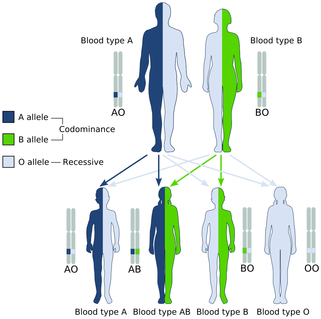
Medical Significance of ABO Blood Type
The ABO system is the most important blood group system in blood transfusions. If red blood cells containing a particular ABO antigen are transfused into a person who lacks that antigen, the person’s immune system will recognize the antigen on the red blood cells as non-self. Antibodies specific to that antigen will attack the red blood cells, causing them to agglutinate (or clump) and break apart. If a unit of incompatible blood were to be accidentally transfused into a patient, a severe reaction (called acute hemolytic transfusion reaction) is likely to occur, in which many red blood cells are destroyed. This may result in kidney failure, shock, and even death. Fortunately, such medical accidents virtually never occur today.
These antibodies are often spontaneously produced in the first years of life, after exposure to common microorganisms in the environment that have antigens similar to blood antigens. Specifically, a person with type A blood will produce anti-B antibodies, while a person with type B blood will produce anti-A antibodies. A person with type AB blood does not produce either antibody, while a person with type O blood produces both anti-A and anti-B antibodies. Once the antibodies have been produced, they circulate in the plasma. The relationship between ABO red blood cell antigens and plasma antibodies is shown in Figure 6.5.4.
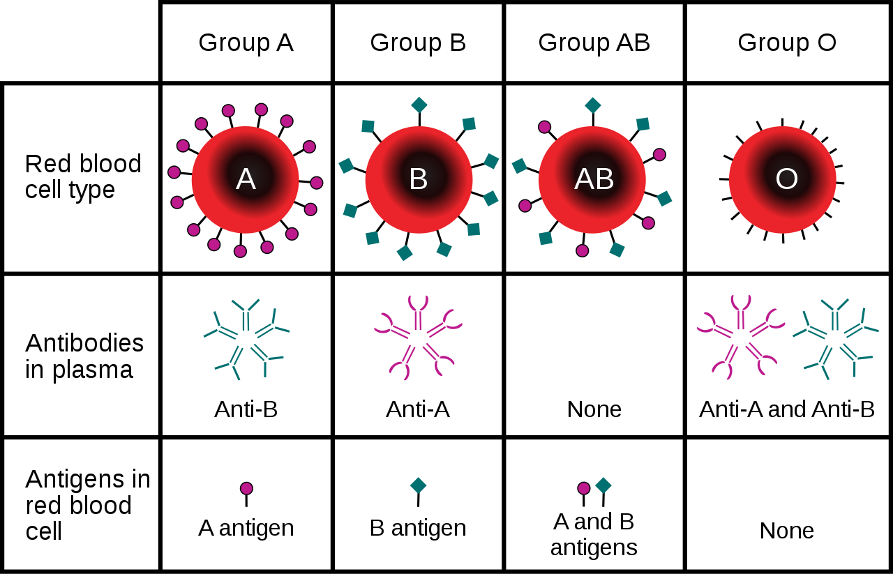
The antibodies that circulate in the plasma are for different antigens than those on red blood cells, which are recognized as self antigens.
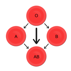
Which blood types are compatible and which are not? Type O blood contains both anti-A and anti-B antibodies, so people with type O blood can only receive type O blood. However, they can donate blood to people of any ABO blood type, which is why individuals with type O blood are called universal donors. Type AB blood contains neither anti-A nor anti-B antibodies, so people with type AB blood can receive blood from people of any ABO blood type. That’s why individuals with type AB blood are called universal recipients. They can donate blood, however, only to people who also have type AB blood. These and other relationships between blood types of donors and recipients are summarized in the simple diagram to the right.
Geographic Distribution of ABO Blood Groups
The frequencies of blood groups for the ABO system vary around the world. You can see how the A and B alleles and the blood group O are distributed geographically on the maps in Figure 6.5.6.
- Worldwide, B is the rarest ABO allele, so type B blood is the least common ABO blood type. Only about 16 per cent of all people have the B allele. Its highest frequency is in Asia. Its lowest frequency is among the indigenous people of Australia and the Americas.
- The A allele is somewhat more common around the world than the B allele, so type A blood is also more common than type B blood. The highest frequencies of the A allele are in Australian Aborigines, the Lapps (Sami) of Northern Scandinavia, and Blackfoot Native Americans in North America. The allele is nearly absent among Native Americans in Central and South America.
- The O allele is the most common ABO allele around the world, and type O blood is the most common ABO blood type. Almost two-thirds of people have at least one copy of the O allele. It is especially common in Native Americans in Central and South America, where it reaches frequencies close to 100 per cent. It also has relatively high frequencies in Australian Aborigines and Western Europeans. Its frequencies are lowest in Eastern Europe and Central Asia.
Figure 6.5.6 Maps of populations that have the A, B and O alleles.
Evolution of the ABO Blood Group System
The geographic distribution of ABO blood type alleles provides indirect evidence for the evolutionary history of these alleles. Evolutionary biologists hypothesize that the allele for blood type A evolved first, followed by the allele for blood type O, and then by the allele for blood type B. This chronology accounts for the percentages of people worldwide with each blood group, and is also consistent with known patterns of early population movements.
The evolutionary forces of founder effect and genetic drift have no doubt played a significant role in the current distribution of ABO blood types worldwide. Geographic variation in ABO blood groups is also likely to be influenced by natural selection, because different blood types are thought to vary in their susceptibility to certain diseases. For example:
- People with type O blood may be more susceptible to cholera and plague. They are also more likely to develop gastrointestinal ulcers.
- People with type A blood may be more susceptible to smallpox and more likely to develop certain cancers.
- People with types A, B, and AB blood appear to be less likely to form blood clots that can cause strokes. However, early in our history, the ability of blood to form clots — which appears greater in people with type O blood — may have been a survival advantage.
- Perhaps the greatest natural selective force associated with ABO blood types is malaria. There is considerable evidence to suggest that people with type O blood are somewhat resistant to malaria, giving them a selective advantage where malaria is endemic.
Rhesus Blood Group System
Another well-known blood group system is the Rhesus (Rh) blood group system. The Rhesus system has dozens of different antigens, but only five main antigens (called D, C, c, E, and e). The major Rhesus antigen is the D antigen. People with the D antigen are called Rh positive (Rh+), and people who lack the D antigen are called Rh negative (Rh-). Rhesus antigens are thought to play a role in transporting ions across cell membranes by acting as channel proteins.
The Rhesus blood group system is controlled by two linked genes on chromosome 1. One gene, called RHD, produces a single antigen, antigen D. The other gene, called RHCE, produces the other four relatively common Rhesus antigens (C, c, E, and e), depending on which alleles for this gene are inherited.
Rhesus Blood Group and Transfusions
After the ABO system, the Rhesus system is the second most important blood group system in blood transfusions. The D antigen is the one most likely to provoke an immune response in people who lack the antigen. People who have the D antigen (Rh+) can be safely transfused with either Rh+ or Rh- blood, whereas people who lack the D antigen (Rh-) can be safely transfused only with Rh- blood.
Unlike anti-A and anti-B antibodies to ABO antigens, anti-D antibodies for the Rhesus system are not usually produced by sensitization to environmental substances. People who lack the D antigen (Rh-), however, may produce anti-D antibodies if exposed to Rh+ blood. This may happen accidentally in a blood transfusion, although this is extremely unlikely today. It may also happen during pregnancy with an Rh+ fetus if some of the fetal blood cells pass into the mother’s blood circulation.
Hemolytic Disease of the Newborn
If a woman who is Rh- is carrying an Rh+ fetus, the fetus may be at risk. This is especially likely if the mother has formed anti-D antibodies during a prior pregnancy because of a mixing of maternal and fetal blood during childbirth. Unlike antibodies against ABO antigens, antibodies against the Rhesus D antigen can cross the placenta and enter the blood of the fetus. This may cause hemolytic disease of the newborn (HDN), also called erythroblastosis fetalis, an illness in which fetal red blood cells are destroyed by maternal antibodies, causing anemia. This illness may range from mild to severe. If it is severe, it may cause brain damage and is sometimes fatal for the fetus or newborn. Fortunately, HDN can be prevented by preventing the formation of anti-D antibodies in the Rh- mother. This is achieved by injecting the mother with a medication called Rho(D) immune globulin.
Geographic Distribution of Rhesus Blood Types
The majority of people worldwide are Rh+, but there is regional variation in this blood group system, as there is with the ABO system. The aboriginal inhabitants of the Americas and Australia originally had very close to 100 per cent Rh+ blood. The frequency of the Rh+ blood type is also very high in African populations, at about 97 to 99 per cent. In East Asia, the frequency of Rh+ is slightly lower, at about 93 to 99 per cent. Europeans have the lowest frequency of the Rh+ blood type at about 83 to 85 per cent.
What explains the population variation in Rhesus blood types? Prior to the advent of modern medicine, Rh+ positive children conceived by Rh- women were at risk of fetal or newborn death or impairment from HDN. This was an enigma, because presumably, natural selection would work to remove the rarer phenotype (Rh-) from populations. However, the frequency of this phenotype is relatively high in many populations.
Recent studies have found evidence that natural selection may actually favor heterozygotes for the Rhesus D antigen. The selective agent in this case is thought to be toxoplasmosis, a parasitic disease caused by the protozoan Toxoplasma gondii, which is very common worldwide. You can see a life cycle diagram of the parasite in Figure 6.5.7. Infection by this parasite often causes no symptoms at all, or it may cause flu-like symptoms for a few days or weeks. Exposure to the parasite has been linked, however, to increased risk of mental disorders (such as schizophrenia), neurological disorders (such as Alzheimer’s), and other neurological problems, including delayed reaction times. One study found that people who tested positive for antibodies to the parasite were more than twice as likely to be involved in traffic accidents.
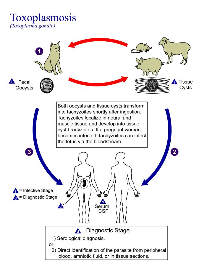
People who are heterozygous for the D antigen appear less likely to develop the negative neurological and mental effects of Toxoplasma gondii infection. This could help explain why both phenotypes (Rh+ and Rh-) are maintained in most populations. There are also striking geographic differences in the prevalence of toxoplasmosis worldwide, ranging from zero to 95 per cent in different regions. This could explain geographic variation in the D antigen worldwide, because its strength as a selective agent would vary with its prevalence.
Feature: Myth vs. Reality
Myth |
Reality |
| "Your nutritional needs can be determined by your ABO blood type. Knowing your blood type allows you to choose the appropriate foods that will help you lose weight, increase your energy, and live a longer, healthier life." | This idea was proposed in 1996 in a New York Times bestseller Eat Right for Your Type, by Peter D’Adamo, a naturopath. Naturopathy is a method of treating disorders that involves the use of herbs, sunlight, fresh air, and other natural substances. Some medical doctors consider naturopathy a pseudoscience. A major scientific review of the blood type diet could find no evidence to support it. In one study, adults eating the diet designed for blood type A showed improved health — but this occurred in everyone, regardless of their blood type. Because the blood type diet is based solely on blood type, it fails to account for other factors that might require dietary adjustments or restrictions. For example, people with diabetes — but different blood types — would follow different diets, and one or both of the diets might conflict with standard diabetes dietary recommendations and be dangerous. |
| "ABO blood type is associated with certain personality traits. People with blood type A, for example, are patient and responsible, but may also be stubborn and tense, whereas people with blood type B are energetic and creative, but may also be irresponsible and unforgiving. In selecting a spouse, both your own and your potential mate’s blood type should be taken into account to ensure compatibility of your personalities." | The belief that blood type is correlated with personality is widely held in Japan and other East Asian countries. The idea was originally introduced in the 1920s in a study commissioned by the Japanese government, but it was later shown to have no scientific support. The idea was revived in the 1970s by a Japanese broadcaster, who wrote popular books about it. There is no scientific basis for the idea, and it is generally dismissed as pseudoscience by the scientific community. Nonetheless, it remains popular in East Asian countries, just as astrology is popular in many other countries. |
6.5 Summary
- Blood type (or blood group) is a genetic characteristic associated with the presence or absence of antigens on the surface of red blood cells. A blood group system refers to all of the gene(s), alleles, and possible genotypes and phenotypes that exist for a particular set of blood type antigens.
- Antigens are molecules that the immune system identifies as either self or non-self. If antigens are identified as non-self, the immune system responds by forming antibodies that are specific to the non-self antigens, leading to the destruction of cells bearing the antigens.
- The ABO blood group system is a system of red blood cell antigens controlled by a single gene with three common alleles on chromosome 9. There are four possible ABO blood types: A, B, AB, and O. The ABO system is the most important blood group system in blood transfusions. People with type O blood are universal donors, and people with type AB blood are universal recipients.
- The frequencies of ABO blood type alleles and blood groups vary around the world. The allele for the B antigen is least common, and blood type O is the most common. The evolutionary forces of founder effect, genetic drift, and natural selection are responsible for the geographic distribution of ABO alleles and blood types. People with type O blood, for example, may be somewhat resistant to malaria, possibly giving them a selective advantage where malaria is endemic.
- The Rhesus blood group system is a system of red blood cell antigens controlled by two genes with many alleles on chromosome 1. There are five common Rhesus antigens, of which antigen D is most significant. Individuals who have antigen D are called Rh+, and individuals who lack antigen D are called Rh-. Rh- mothers of Rh+ fetuses may produce antibodies against the D antigen in the fetal blood, causing hemolytic disease of the newborn (HDN).
- The majority of people worldwide are Rh+, but there is regional variation in this blood group system. This variation may be explained by natural selection that favors heterozygotes for the D antigen, because this genotype seems to be protected against some of the neurological consequences of the common parasitic infection toxoplasmosis.
6.5 Review Questions
- Define blood type and blood group system.
- Explain the relationship between antigens and antibodies.
- Identify the alleles, genotypes, and phenotypes in the ABO blood group system.
- Discuss the medical significance of the ABO blood group system.
- Compare the relative worldwide frequencies of the three ABO alleles.
- Give examples of how different ABO blood types vary in their susceptibility to diseases.
- Describe the Rhesus blood group system.
- Relate Rhesus blood groups to blood transfusions.
- What causes hemolytic disease of the newborn?
- Describe how toxoplasmosis may explain the persistence of the Rh- blood type in human populations.
- A woman is blood type O and Rh-, and her husband is blood type AB and Rh+. Answer the following questions about this couple and their offspring.
- What are the possible genotypes of their offspring in terms of ABO blood group?
- What are the possible phenotypes of their offspring in terms of ABO blood group?
- Can the woman donate blood to her husband? Explain your answer.
- Can the man donate blood to his wife? Explain your answer.
- Type O blood is characterized by the presence of O antigens — explain why this statement is false.
- Explain why newborn hemolytic disease may be more likely to occur in a second pregnancy than in a first.
6.5 Explore More
https://www.youtube.com/watch?v=xfZhb6lmxjk
Why do blood types matter? - Natalie S. Hodge, TED-Ed, 2015.
https://www.youtube.com/watch?v=qcZKbjYyOfE
How do blood transfusions work? - Bill Schutt, TED-Ed, 2020.
Attributes
Figure 6.5.1
Following the Blood Donation Trail by EJ Hersom/ USA Department of Defense is in the public domain. [Disclaimer: The appearance of U.S. Department of Defense (DoD) visual information does not imply or constitute DoD endorsement.]
Figure 6.5.2
Antibody by Fvasconcellos on Wikimedia Commons is released into the public domain (https://en.wikipedia.org/wiki/Public_domain).
Figure 6.5.3
ABO system codominance.svg, adapted by YassineMrabet (original "Codominant" image from US National Library of Medicine) on Wikimedia Commons is in the public domain (https://en.wikipedia.org/wiki/Public_domain).
Figure 6.5.4
ABO_blood_type.svg by InvictaHOG on Wikimedia Commons is released into the public domain (https://en.wikipedia.org/wiki/Public_domain).
Figure 6.5.5
Blood Donor and recipient ABO by CK-12 Foundation is used under a CC BY-NC 3.0 (https://creativecommons.org/licenses/by-nc/3.0/) license.
Figure 6.5.6
- Map of Blood Group A by Muntuwandi at en.wikipedia on Wikimedia Commons is used under a CC BY-SA 3.0 (https://creativecommons.org/licenses/by-sa/3.0/) license.
- Map of Blood Group B by Muntuwandi at en.wikipedia on Wikimedia Commons is used under a CC BY-SA 3.0 (https://creativecommons.org/licenses/by-sa/3.0/) license.
- Map of Blood Group O by anthro palomar at en.wikipedia on Wikimedia Commons is used under a CC BY-SA 3.0 (https://creativecommons.org/licenses/by-sa/3.0/) license.
Figure 6.5.7
Toxoplasma_gondii_Life_cycle_PHIL_3421_lores by Alexander J. da Silva, PhD/Melanie Moser, Centers for Disease Control and Prevention's Public Health Image Library (PHIL#3421) on Wikimedia Commons is in the public domain (https://en.wikipedia.org/wiki/Public_domain).
Table 6.5.1
ABO Blood Group System: Genotypes and Phenotypes was created by Christine Miller.
References
Dean, L. (2005). Chapter 4 Hemolytic disease of the newborn. In Blood Groups and Red Cell Antigens [Internet]. National Center for Biotechnology Information (US). https://www.ncbi.nlm.nih.gov/books/NBK2266/
Mayo Clinic Staff. (n.d.). Toxoplasmosis [online article]. MayoClinic.org. https://www.mayoclinic.org/diseases-conditions/toxoplasmosis/symptoms-causes/syc-20356249
MedlinePlus. (2019, January 29). Hemolytic transfusion reaction [online article]. U.S. National Library of Medicine. https://en.wikipedia.org/w/index.php?title=Chromosome_9&oldid=946440619
TED-Ed. (2015, June 29). Why do blood types matter? - Natalie S. Hodge. YouTube. https://www.youtube.com/watch?v=xfZhb6lmxjk&feature=youtu.be
TED-Ed. (2020, February 18). How do blood transfusions work? - Bill Schutt. YouTube. https://www.youtube.com/watch?v=qcZKbjYyOfE&feature=youtu.be
Wikipedia contributors. (2020, May 10). Chromosome 1. In Wikipedia. https://en.wikipedia.org/w/index.php?title=Chromosome_1&oldid=955942444
Wikipedia contributors. (2020, March 20). Chromosome 9. In Wikipedia. https://en.wikipedia.org/w/index.php?title=Chromosome_9&oldid=946440619
A membrane protein involved in the movement of ions, small molecules, or macromolecules, such as another protein, across a biological membrane.
one of a pair of glands located on top of the kidneys that secretes hormones such as cortisol and adrenaline

Case Study: Your Genes May Help You Save a Life
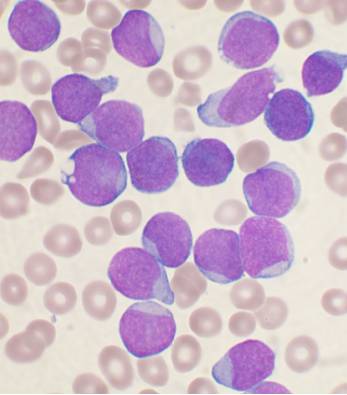
Like the little girl shown in Figure 6.1.1, seven-year-old Mateo is battling leukemia, a type of cancer that affects blood cells. Leukemia usually starts in the bone marrow where blood cells are produced. It causes the production of abnormal blood cells, most commonly white blood cells. Depending on the type of leukemia, it can also affect other types of blood cells. The abnormal blood cells replace the patient’s normal blood cells over time, which can lead to symptoms of fatigue, frequent infections, and easy bruising or bleeding. Leukemia can be fatal, but fortunately, there are some treatment options available that can prolong life — and may even cure the disease.
Mateo has undergone chemotherapy to kill the cancerous cells, but his doctors have told his parents that it is not enough. Mateo needs a bone marrow transplant in order to replace his abnormal bone marrow with healthy bone marrow. His family members are eager to donate bone marrow to him, but first they must be tested to see if they are a compatible match.
For blood transfusions, it is relatively easy to find a compatible blood donor, but bone marrow transplants require much more specific matching between donor and recipient. They must share several of the same type of proteins — called human leukocyte antigens (HLAs) — on the surface of their cells. One type of HLA protein is illustrated in Figure 6.1.3. Different people have different types of HLA proteins (or markers) depending on their specific genes. Typically, eight to ten HLA markers are tested and compared in the potential bone marrow donor and recipient. At least six or seven of these HLA markers must be identical between them in order for a match to be made.
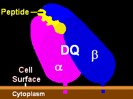
If the match is not good, the patient’s body could reject the bone marrow transplant. Conversely, the transplanted bone marrow could produce immune cells that attack the patient’s body. A good match between donor and recipient is critical for bone marrow donation to be safe and effective.
A full sibling frequently provides the best match for bone marrow donation because they share many of the same genes from their parents. Mateo’s sister is tested, but unfortunately, she is not a match for him. This is not all that surprising since there is only about a 25 per cent chance that a sibling will be an identical HLA match. His parents and other family members are also tested, but none of them are a match, either. Mateo must join the 70 per cent of patients that need to look outside of their families for a bone marrow donor.
How do you find a bone marrow match outside of your family? Fortunately, people from all over the world have signed up to be potential bone marrow donors, usually by providing a simple swab of the inside of their cheek. DNA from the cells collected on the swab is then tested for HLA type. The potential donor’s HLA information is put into a donor registry, and doctors can then search national and international registries for compatible matches for their patients.
Patients are much more likely to be a match with a bone marrow donor of their same race or ethnic background. People with similar ancestry are more likely to share similar HLA genes. In Mateo’s case, his mother is African American, and his father is Japanese and Caucasian. His relatively rare combination of ethnic backgrounds may make it harder for him to find a match in the donor registries, as is the case for many multiethnic patients.
Read the rest of this chapter to learn more about the genetic and phenotypic variations that exist in humans, and how some of these differences came about due to differing natural selection pressures in different areas of the world. At the end of the chapter, learn more about Mateo’s quest for a bone marrow donor, the need for bone marrow donors from diverse ethnic backgrounds, and how you may be able to save someone’s life based on your genetic makeup!
Chapter Overview: Human Variation
In this chapter, you will learn about:
- The extent, types, and patterns of human genetic variation — within and between populations.
- How knowledge about human genetic variation can give insight into human origins and history, and how it may lead to treatments for diseases.
- The ways human variation has been classified, and how some classification methods contribute to racism.
- How gene flow and natural selection can result in a gradual change in the frequency of a trait over a geographic area.
- The ways in which humans can adapt to environmental stresses — genetically, physiologically, and culturally.
- Differences in human blood types (including the ABO and Rh groups), how they may have evolved, and their relationships to diseases.
- How malaria has caused humans to develop a variety of blood cell adaptations over the course of our evolution, including the trait that causes sickle cell anemia.
- Adaptations humans have evolved to deal with the stress of living at high altitudes and in extreme climates, and the ways people can temporarily acclimate to these environmental conditions.
- Human adaptations to our food supply, including lactose tolerance, and weight and blood sugar regulation.
As you read the chapter, think about the following questions:
- How similar are any two people genetically? Based on your answer, why do you think it is not easy to find an HLA match for bone marrow donation between people?
- What is the concept of race? What are its limitations? How does race or ethnicity relate to genetic variation?
- What is an antigen, such as the human leukocyte antigen? On a cellular and molecular level, what happens when there is not a good match between a tissue donor and recipient?
Attributions
Figure 6.1.1
Young_chemotherapy_patient_holds_teddy_bear by Bill Branson (Photographer) at National Cancer Institute/ National Institutes of Health, on Wikimedia Commons is in the public domain (https://en.wikipedia.org/wiki/Public_domain).
Figure 6.1.2
Acute_leukemia-ALL by VashiDonsk at English Wikipedia, now on Wikimedia Commons is used under a CC BY-SA 3.0 (https://creativecommons.org/licenses/by-sa/3.0/deed.en) license.
Figure 6.1.3
HLA_DQ_Illustration by Pdeitiker on Wikimedia Commons is released into the public domain (https://en.wikipedia.org/wiki/Public_domain).
As described in the caption.


