15.6 Accessory Organs of Digestion
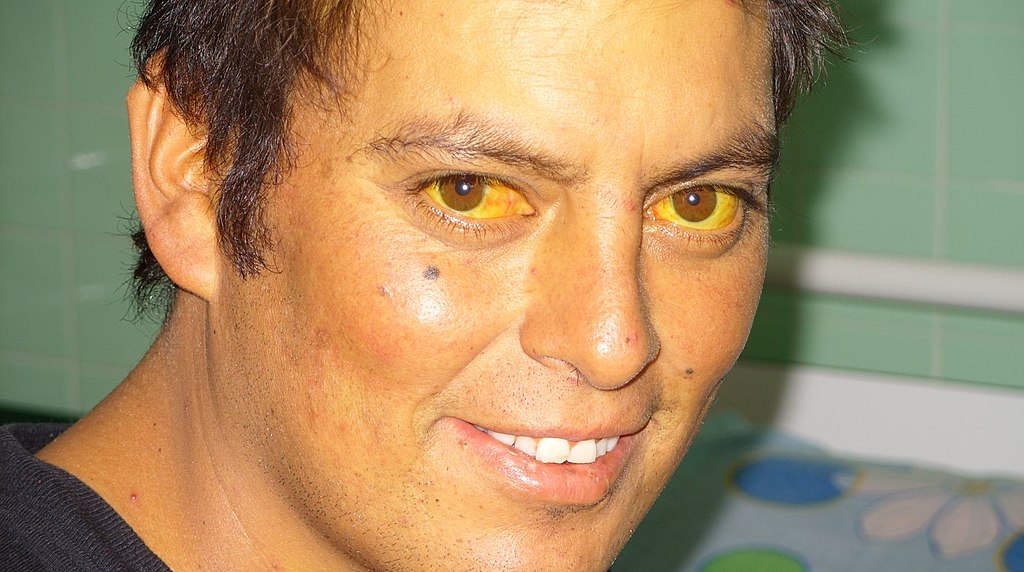
Jaundiced Eyes
Did you ever hear of a person looking at something or someone with a “jaundiced eye”? It means to take a negative view, such as envy, maliciousness, or ill will. The expression may be based on the antiquated idea that liver bile is associated with such negative emotions as these, as well as the fact that excessive liver bile causes jaundice, or yellowing of the eyes and skin. Jaundice is likely a sign of a liver disorder or blockage of the duct that carries bile away from the liver. Bile contains waste products, making the liver an organ of excretion. Bile has an important role in digestion, which makes the liver an accessory organ of digestion, too.
What Are Accessory Organs of Digestion?
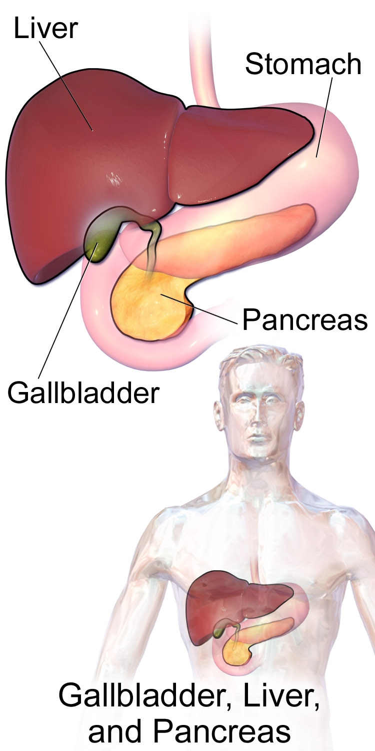
Accessory organs of digestion are organs that secrete substances needed for the chemical digestion of food, but through which food does not actually pass as it is digested. Besides the liver, the major accessory organs of digestion are the gallbladder and pancreas. These organs secrete or store substances that are needed for digestion in the first part of the small intestine — the duodenum — where most chemical digestion takes place. You can see the three organs and their locations in Figure 15.6.2.
Liver
The liver is a vital organ located in the upper right part of the abdomen. It lies just below the diaphragm, to the right of the stomach. The liver plays an important role in digestion by secreting bile, but the liver has a wide range of additional functions unrelated to digestion. In fact, some estimates put the number of functions of the liver at about 500! A few of them are described below.
Structure of the Liver
The liver is a reddish brown, wedge-shaped structure. In adults, the liver normally weighs about 1.5 kg (about 3.3 lb). It is both the heaviest internal organ and the largest gland in the human body. The liver is divided into four lobes of unequal size and shape. Each lobe, in turn, is made up of lobules, which are the functional units of the liver. Each lobule consists of millions of liver cells, called hepatic cells (or hepatocytes). They are the basic metabolic cells that carry out the various functions of the liver.
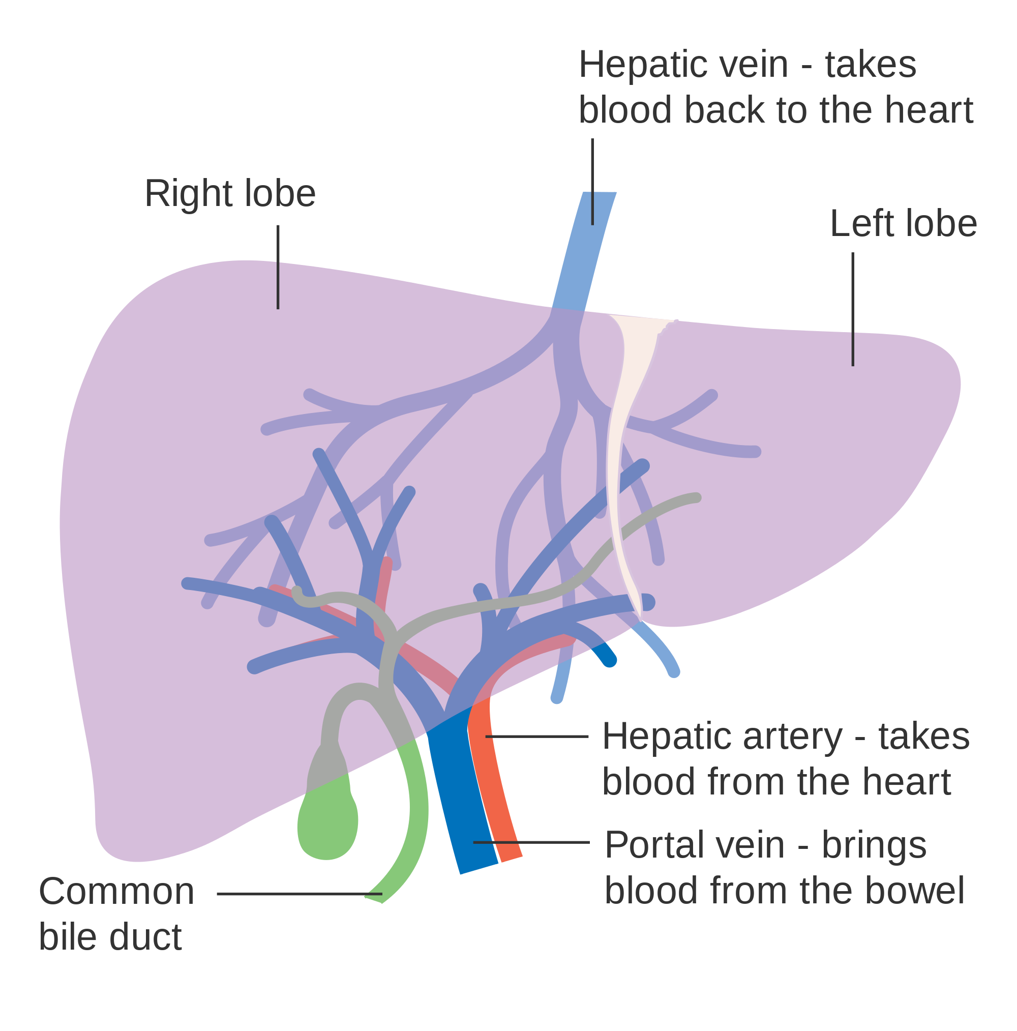
As shown in Figure 15.6.3, the liver is connected to two large blood vessels: the hepatic artery and the portal vein. The hepatic artery carries oxygen-rich blood from the aorta, whereas the portal vein carries blood that is rich in digested nutrients from the GI tract and wastes filtered from the blood by the spleen. The blood vessels subdivide into smaller arteries and capillaries, which lead into the liver lobules. The nutrients from the GI tract are used to build many vital biochemical compounds, and the wastes from the spleen are degraded and excreted.
Functions of the Liver
The main digestive function of the liver is the production of bile. Bile is a yellowish alkaline liquid that consists of water, electrolytes, bile salts, and cholesterol, among other substances, many of which are waste products. Some of the components of bile are synthesized by hepatocytes. The rest are extracted from the blood.
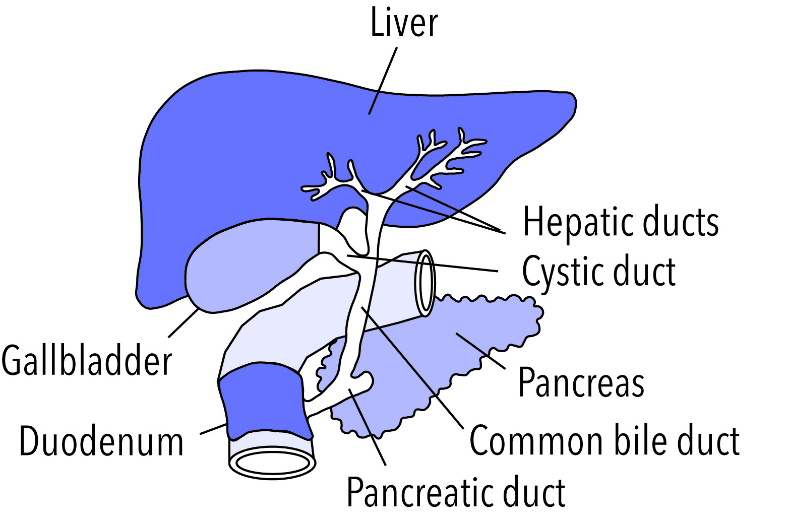
As shown in Figure 15.6.4, bile is secreted into small ducts that join together to form larger ducts, with just one large duct carrying bile out of the liver. If bile is needed to digest a meal, it goes directly to the duodenum through the common bile duct. In the duodenum, the bile neutralizes acidic chyme from the stomach and emulsifies fat globules into smaller particles (called micelles) that are easier to digest chemically by the enzyme lipase. Bile also aids with the absorption of vitamin K. Bile that is secreted when digestion is not taking place goes to the gallbladder for storage until the next meal. In either case, the bile enters the duodenum through the common bile duct.
Besides its roles in digestion, the liver has many other vital functions:
- The liver synthesizes glycogen from glucose and stores the glycogen as required to help regulate blood sugar levels. It also breaks down the stored glycogen to glucose and releases it back into the blood as needed.
- The liver stores many substances in addition to glycogen, including vitamins A, D, B12, and K. It also stores the minerals iron and copper.
- The liver synthesizes numerous proteins and many of the amino acids needed to make them. These proteins have a wide range of functions. They include fibrinogen, which is needed for blood clotting; insulin-like growth factor (IGF-1), which is important for childhood growth; and albumen, which is the most abundant protein in blood serum and functions to transport fatty acids and steroid hormones in the blood.
- The liver synthesizes many important lipids, including cholesterol, triglycerides, and lipoproteins.
- The liver is responsible for the breakdown of many waste products and toxic substances. The wastes are excreted in bile or travel to the kidneys, which excrete them in urine.
The liver is clearly a vital organ that supports almost every other organ in the body. Because of its strategic location and diversity of functions, the liver is also prone to many diseases, some of which cause loss of liver function. There is currently no way to compensate for the absence of liver function in the long term, although liver dialysis techniques can be used in the short term. An artificial liver has not yet been developed, so liver transplantation may be the only option for people with liver failure.
Gallbladder
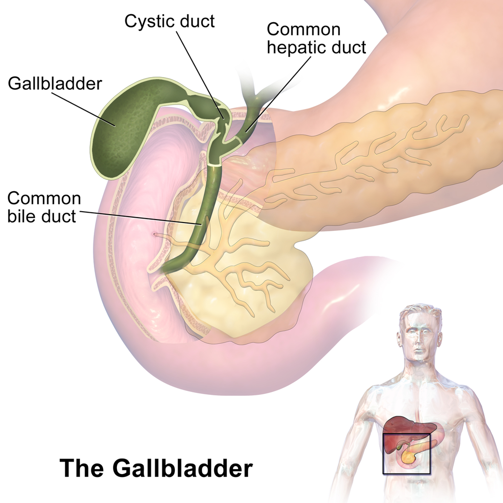
The gallbladder is a small, hollow, pouch-like organ that lies just under the right side of the liver (see Figure 15.6.5). It is about 8 cm (about 3 in) long and shaped like a tapered sac, with the open end continuous with the cystic duct. The gallbladder stores and concentrates bile from the liver until it is needed in the duodenum to help digest lipids. After the bile leaves the liver, it reaches the gallbladder through the cystic duct. At any given time, the gallbladder may store between 30 to 60 mL (1 to 2 oz) of bile. A hormone stimulated by the presence of fat in the duodenum signals the gallbladder to contract and force its contents back through the cystic duct and into the common bile duct to drain into the duodenum.
Pancreas
The pancreas is a glandular organ that is part of both the digestive system and the endocrine system. As shown in Figure 15.6.6, it is located in the abdomen behind the stomach, with the head of the pancreas surrounded by the duodenum of the small intestine. The pancreas is about 15 cm (almost 6 in) long, and it has two major ducts: the main pancreatic duct and the accessory pancreatic duct. Both of these ducts drain into the duodenum.
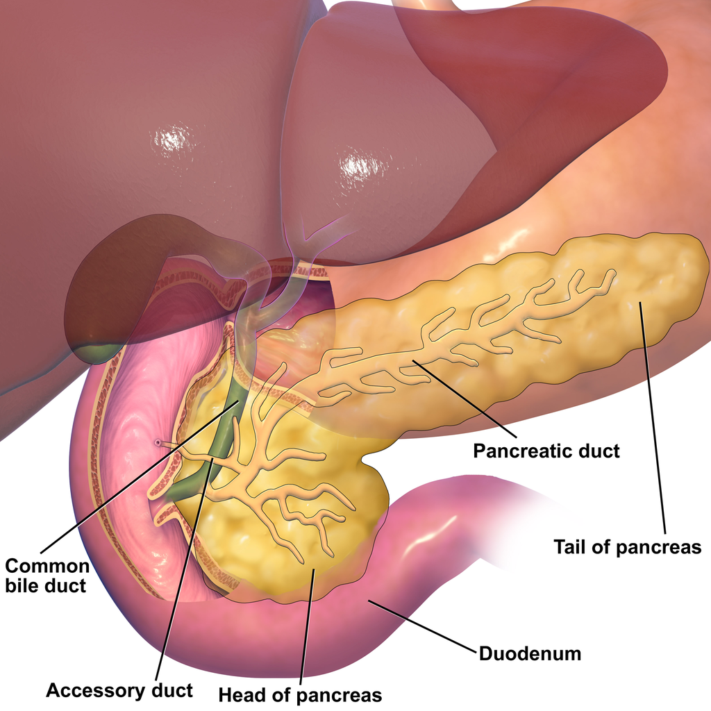
As an endocrine gland, the pancreas secretes several hormones, including insulin and glucagon, which circulate in the blood. The endocrine hormones are secreted by clusters of cells called pancreatic islets (or islets of Langerhans). As a digestive organ, the pancreas secretes many digestive enzymes and also bicarbonate, which helps neutralize acidic chyme after it enters the duodenum. The pancreas is stimulated to secrete its digestive substances when food in the stomach and duodenum triggers the release of endocrine hormones into the blood that reach the pancreas via the bloodstream. The pancreatic digestive enzymes are secreted by clusters of cells called acini, and they travel through the pancreatic ducts to the duodenum. In the duodenum, they help to chemically break down carbohydrates, proteins, lipids, and nucleic acids in chyme. The pancreatic digestive enzymes include:
- Amylase, which helps digest starch and other carbohydrates.
- Trypsin and chymotrypsin, which help digest proteins.
- Lipase, which helps digest lipids.
- Deoxyribonucleases and ribonucleases, which help digest nucleic acids.
15.6 Summary
- Accessory organs of digestion are organs that secrete substances needed for the chemical digestion of food, but through which food does not actually pass as it is digested. The accessory organs include the liver, gallbladder, and pancreas. These organs secrete or store substances that are carried to the duodenum of the small intestine as needed for digestion.
- The liver is a large organ in the abdomen that is divided into lobes and smaller lobules, which consist of metabolic cells called hepatic cells, or hepatocytes. The liver receives oxygen in blood from the aorta through the hepatic artery. It receives nutrients in blood from the GI tract and wastes in blood from the spleen through the portal vein.
- The main digestive function of the liver is the production of the alkaline liquid called bile. Bile is carried directly to the duodenum by the common bile duct or to the gallbladder first for storage. Bile neutralizes acidic chyme that enters the duodenum from the stomach, and also emulsifies fat globules into smaller particles (micelles) that are easier to digest chemically.
- Other vital functions of the liver include regulating blood sugar levels by storing excess sugar as glycogen, storing many vitamins and minerals, synthesizing numerous proteins and lipids, and breaking down waste products and toxic substances.
- The gallbladder is a small pouch-like organ near the liver. It stores and concentrates bile from the liver until it is needed in the duodenum to neutralize chyme and help digest lipids.
- The pancreas is a glandular organ that secretes both endocrine hormones and digestive enzymes. As an endocrine gland, the pancreas secretes insulin and glucagon to regulate blood sugar. As a digestive organ, the pancreas secretes digestive enzymes into the duodenum through ducts. Pancreatic digestive enzymes include amylase (starches) trypsin and chymotrypsin (proteins), lipase (lipids), and ribonucleases and deoxyribonucleases (RNA and DNA).
15.6 Review Questions
- Name three accessory organs of digestion. How do these organs differ from digestive organs that are part of the GI tract?
-
- Describe the liver and its blood supply.
- Explain the main digestive function of the liver and describe the components of bile and it’s importance in the digestive process.
- What type of secretions does the pancreas release as part of each body system?
- List pancreatic enzymes that work in the duodenum, along with the substances they help digest.
- What are two substances produced by accessory organs of digestion that help neutralize chyme in the small intestine? Where are they produced?
- People who have their gallbladder removed sometimes have digestive problems after eating high-fat meals. Why do you think this happens?
- Which accessory organ of digestion synthesizes cholesterol?
15.6 Explore More
What does the pancreas do? – Emma Bryce, TED-Ed. 2015.
What does the liver do? – Emma Bryce, TED-Ed, 2014.
Scar wars: Repairing the liver, nature video, 2018.
Attributions
Figure 15.6.1
Scleral_Icterus by Sheila J. Toro on Wikimedia Commons is used under a CC BY 4.0 (https://creativecommons.org/licenses/by/4.0) license.
Figure 15.6.2
Blausen_0428_Gallbladder-Liver-Pancreas_Location by BruceBlaus on Wikimedia Commons is used under a CC BY 3.0 (https://creativecommons.org/licenses/by/3.0) license.
Figure 15.6.3
Diagram_showing_the_two_lobes_of_the_liver_and_its_blood_supply_CRUK_376.svg by Cancer Research UK on Wikimedia Commons is used under a CC BY-SA 4.0 (https://creativecommons.org/licenses/by-sa/4.0) license.
Figure 15.6.4
Gallbladder by NIH Image Gallery on Flickr is used CC BY-NC 2.0 (https://creativecommons.org/licenses/by-nc/2.0/) license.
Figure 15.6.5
Gallbladder_(organ) (1) by BruceBlaus on Wikimedia Commons is used under a CC BY-SA 4.0 (https://creativecommons.org/licenses/by-sa/4.0) license. (See a full animation of this medical topic at blausen.com.)
Figure 15.6.6
Blausen_0698_PancreasAnatomy by BruceBlaus on Wikimedia Commons is used under a CC BY 3.0 (https://creativecommons.org/licenses/by/3.0) license.
References
Blausen.com Staff. (2014). Medical gallery of Blausen Medical 2014. WikiJournal of Medicine 1 (2). DOI:10.15347/wjm/2014.010. ISSN 2002-4436.
nature video. (2018, December 19). Scar wars: Repairing the liver. YouTube. https://www.youtube.com/watch?v=a0d1yvGcfzQ&feature=youtu.be
TED-Ed. (2014, November 25). What does the liver do? – Emma Bryce. YouTube. https://www.youtube.com/watch?v=wbh3SjzydnQ&feature=youtu.be
TED-Ed. (2015, February 19). What does the pancreas do? – Emma Bryce. YouTube. https://www.youtube.com/watch?v=8dgoeYPoE-0&feature=youtu.be
Created by CK-12 Foundation/Adapted by Christine Miller
Doing the ‘Fly
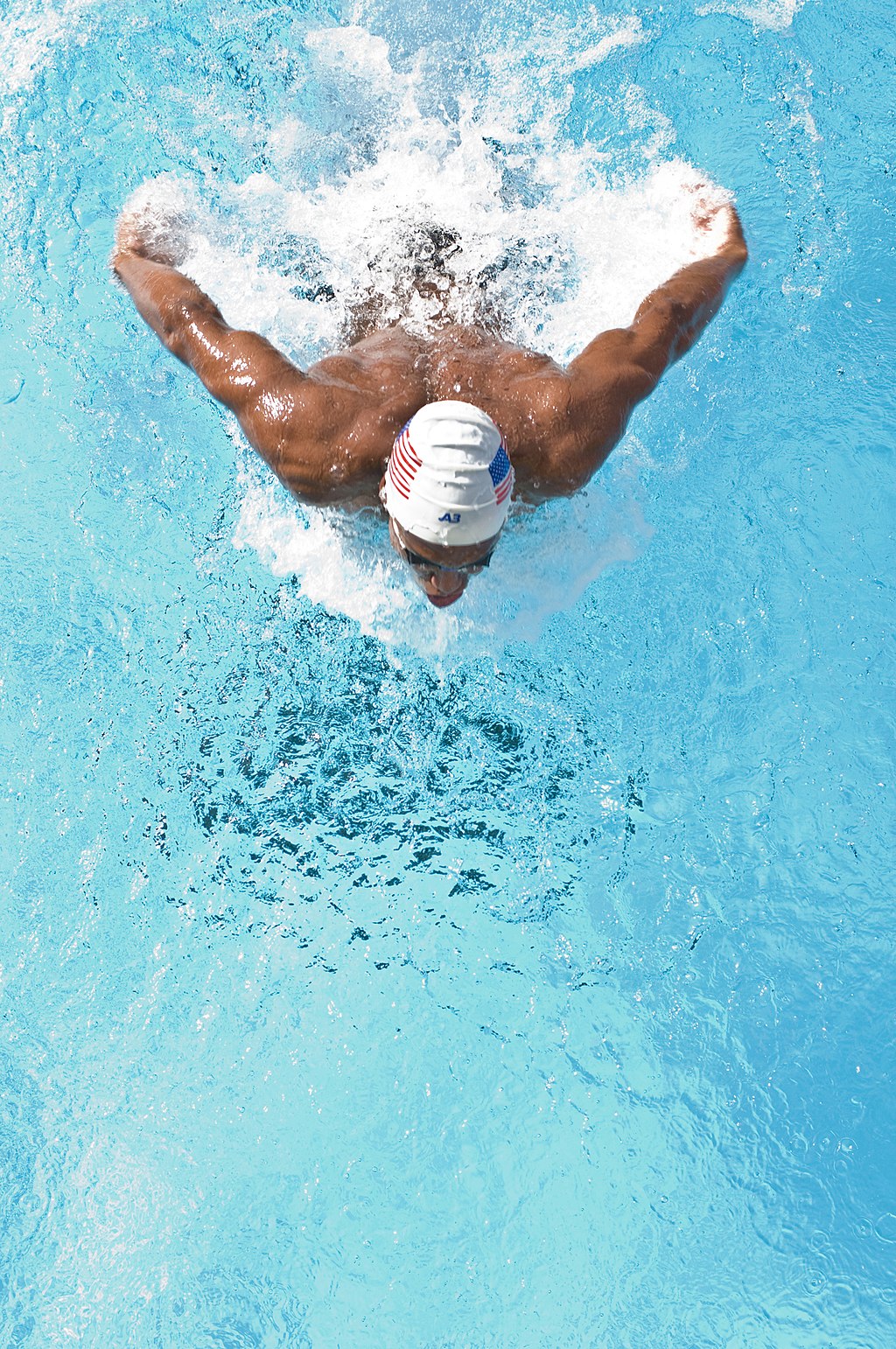
The swimmer in the Figure 13.3.1 photo is doing the butterfly stroke, a swimming style that requires the swimmer to carefully control his breathing so it is coordinated with his swimming movements. Breathing is the process of moving air into and out of the lungs, which are the organs in which gas exchange takes place between the atmosphere and the body. Breathing is also called ventilation, and it is one of two parts of the life-sustaining process of respiration. The other part is gas exchange. Before you can understand how breathing is controlled, you need to know how breathing occurs.
How Breathing Occurs
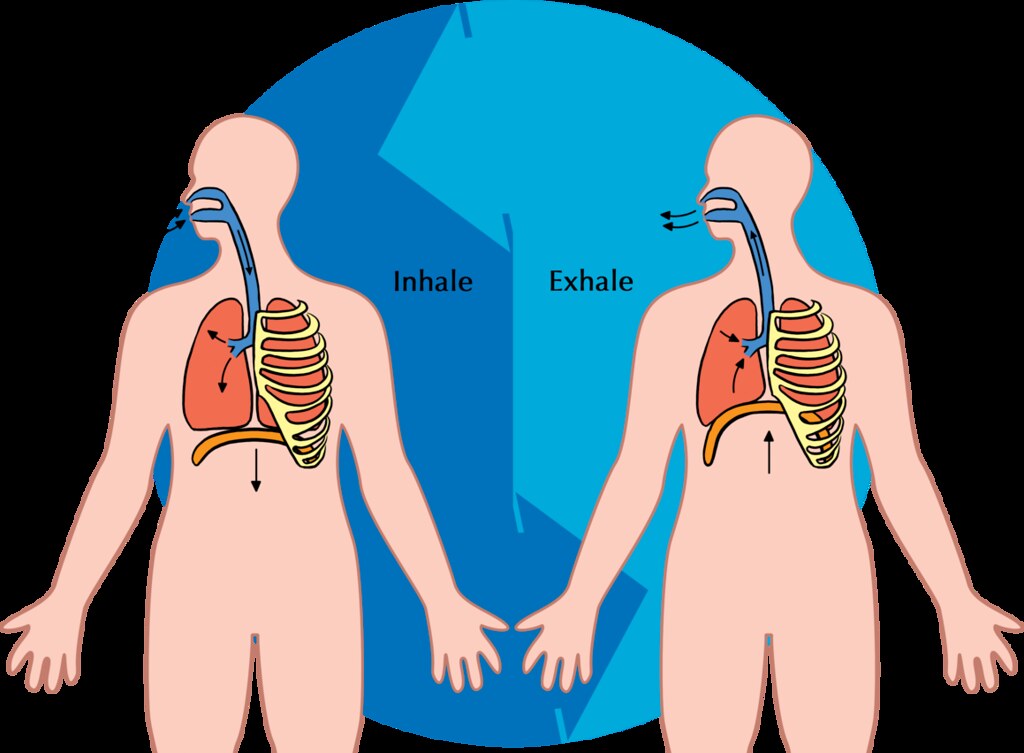
Breathing is a two-step process that includes drawing air into the lungs, or inhaling, and letting air out of the lungs, or exhaling. Both processes are illustrated in Figure 13.3.2.
Inhaling
Inhaling is an active process that results mainly from contraction of a muscle called the diaphragm, shown in Figure 13.3.2. The diaphragm is a large, dome-shaped muscle below the lungs that separates the thoracic (chest) and abdominal cavities. When the diaphragm contracts it moves down causing the thoracic cavity to expand, and the contents of the abdomen to be pushed downward. Other muscles — such as intercostal muscles between the ribs — also contribute to the process of inhalation, especially when inhalation is forced, as when taking a deep breath. These muscles help increase thoracic volume by expanding the ribs outward. The increase in thoracic volume creates a decrease in thoracic air pressure. With the chest expanded, there is lower air pressure inside the lungs than outside the body, so outside air flows into the lungs via the respiratory tract according the the pressure gradient (high pressure flows to lower pressure).
Exhaling
Exhaling involves the opposite series of events. The diaphragm relaxes, so it moves upward and decreases the volume of the thorax. Air pressure inside the lungs increases, so it is higher than the air pressure outside the lungs. Exhalation, unlike inhalation, is typically a passive process that occurs mainly due to the elasticity of the lungs. With the change in air pressure, the lungs contract to their pre-inflated size, forcing out the air they contain in the process. Air flows out of the lungs, similar to the way air rushes out of a balloon when it is released. If exhalation is forced, internal intercostal and abdominal muscles may help move the air out of the lungs.
Control of Breathing
Breathing is one of the few vital bodily functions that can be controlled consciously, as well as unconsciously. Think about using your breath to blow up a balloon. You take a long, deep breath, and then you exhale the air as forcibly as you can into the balloon. Both the inhalation and exhalation are consciously controlled.
Conscious Control of Breathing
You can control your breathing by holding your breath, slowing your breathing, or hyperventilating, which is breathing more quickly and shallowly than necessary. You can also exhale or inhale more forcefully or deeply than usual. Conscious control of breathing is common in many activities besides blowing up balloons, including swimming, speech training, singing, playing many different musical instruments (Figure 13.3.3), and doing yoga, to name just a few.

There are limits on the conscious control of breathing. For example, it is not possible for a healthy person to voluntarily stop breathing indefinitely. Before long, there is an irrepressible urge to breathe. If you were able to stop breathing for a long enough time, you would lose consciousness. The same thing would happen if you were to hyperventilate for too long. Once you lose consciousness so you can no longer exert conscious control over your breathing, involuntary control of breathing takes over.
Unconscious Control of Breathing
Unconscious breathing is controlled by respiratory centers in the medulla and pons of the brainstem (see Figure 13.3.4). The respiratory centers automatically and continuously regulate the rate of breathing based on the body’s needs. These are determined mainly by blood acidity, or pH. When you exercise, for example, carbon dioxide levels increase in the blood, because of increased cellular respiration by muscle cells. The carbon dioxide reacts with water in the blood to produce carbonic acid, making the blood more acidic, so pH falls. The drop in pH is detected by chemoreceptors in the medulla. Blood levels of oxygen and carbon dioxide, in addition to pH, are also detected by chemoreceptors in major arteries, which send the “data” to the respiratory centers. The latter respond by sending nerve impulses to the diaphragm, “telling” it to contract more quickly so the rate of breathing speeds up. With faster breathing, more carbon dioxide is released into the air from the blood, and blood pH returns to the normal range.
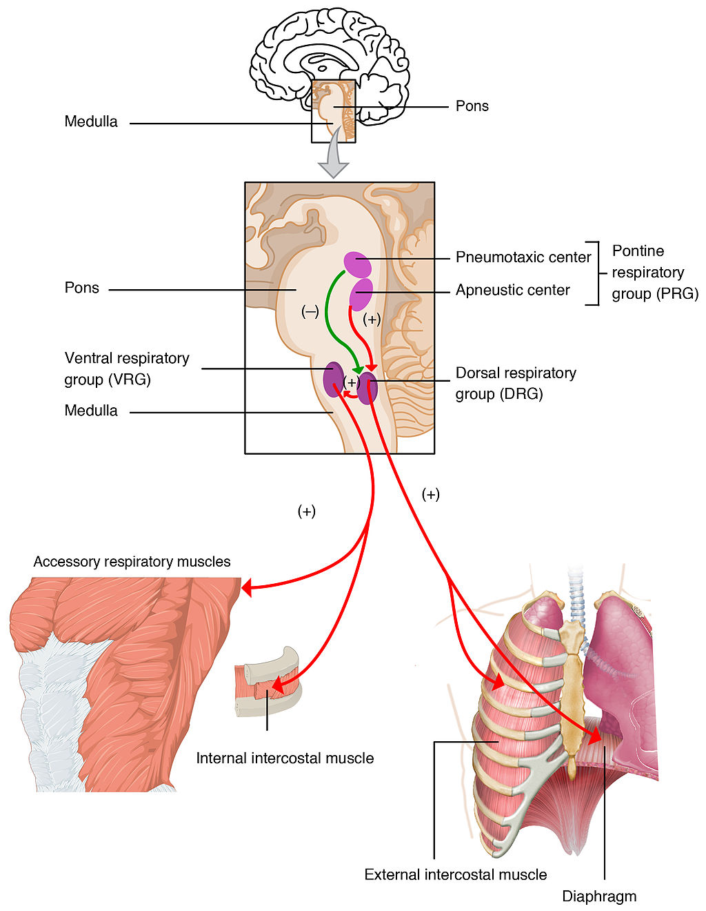
The opposite events occur when the level of carbon dioxide in the blood becomes too low and blood pH rises. This may occur with involuntary hyperventilation, which can happen in panic attacks, episodes of severe pain, asthma attacks, and many other situations. When you hyperventilate, you blow off a lot of carbon dioxide, leading to a drop in blood levels of carbon dioxide. The blood becomes more basic (alkaline), causing its pH to rise.
Nasal vs. Mouth Breathing
Nasal breathing is breathing through the nose rather than the mouth, and it is generally considered to be superior to mouth breathing. The hair-lined nasal passages do a better job of filtering particles out of the air before it moves deeper into the respiratory tract. The nasal passages are also better at warming and moistening the air, so nasal breathing is especially advantageous in the winter when the air is cold and dry. In addition, the smaller diameter of the nasal passages creates greater pressure in the lungs during exhalation. This slows the emptying of the lungs, giving them more time to extract oxygen from the air.
Feature: Myth vs. Reality
Drowning is defined as respiratory impairment from being in or under a liquid. It is further classified according to its outcome into: death, ongoing health problems, or no ongoing health problems (full recovery). Four hundred Canadians die annually from drowning, and drowning is one of the leading causes of death in children under the age of five. There are some potentially dangerous myths about drowning, and knowing what they are might save your life or the life of a loved one, especially a child.
| Myth | Reality |
|---|---|
| "People drown when they aspirate water into their lungs." | Generally, in the early stages of drowning, very little water enters the lungs. A small amount of water entering the trachea causes a muscular spasm in the larynx that seals the airway and prevents the passage of water into the lungs. This spasm is likely to last until unconsciousness occurs. |
| "You can tell when someone is drowning because they will shout for help and wave their arms to attract attention." | The muscular spasm that seals the airway prevents the passage of air, as well as water, so a person who is drowning is unable to shout or call for help. In addition, instinctive reactions that occur in the final minute or so before a drowning person sinks under the water may look similar to calm, safe behavior. The head is likely to be low in the water, tilted back, with the mouth open. The person may have uncontrolled movements of the arms and legs, but they are unlikely to be visible above the water. |
| "It is too late to save a person who is unconscious in the water." | An unconscious person rescued with an airway still sealed from the muscular spasm of the larynx stands a good chance of full recovery if they start receiving CPR within minutes. Without water in the lungs, CPR is much more effective. Even if cardiac arrest has occurred so the heart is no longer beating, there is still a chance of recovery. The longer the brain goes without oxygen, however, the more likely brain cells are to die. Brain death is likely after about six minutes without oxygen, except in exceptional circumstances, such as young people drowning in very cold water. There are examples of children surviving, apparently without lasting ill effects, for as long as an hour in cold water. Rescuers retrieving a child from cold water should attempt resuscitation even after a protracted period of immersion. |
| "If someone is drowning, you should start administering CPR immediately, even before you try to get the person out of the water." | Removing a drowning person from the water is the first priority, because CPR is ineffective in the water. The goal should be to bring the person to stable ground as quickly as possible and then to start CPR. |
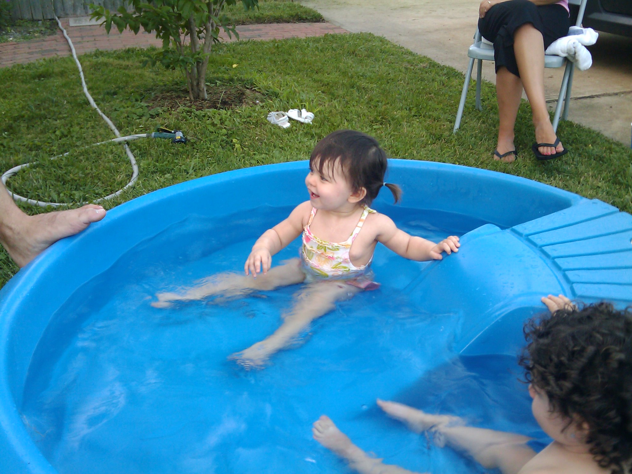
| "You are unlikely to drown unless you are in water over your head." | Depending on circumstances, people have drowned in as little as 30 mm (about 1 ½ in.) of water. Inebriated people or those under the influence of drugs, for example, have been known to have drowned in puddles. Hundreds of children have drowned in the water in toilets, bathtubs, basins, showers, pails, and buckets (see Figure 13.3.5). |
13.3 Summary
- Breathing, or ventilation, is the two-step process of drawing air into the lungs (inhaling) and letting air out of the lungs (exhaling). Inhalation is an active process that results mainly from contraction of a muscle called the diaphragm. Exhalation is typically a passive process that occurs mainly due to the elasticity of the lungs when the diaphragm relaxes.
- Breathing is one of the few vital bodily functions that can be controlled consciously, as well as unconsciously. Conscious control of breathing is common in many activities, including swimming and singing. There are limits on the conscious control of breathing, however. If you try to hold your breath, for example, you will soon have an irrepressible urge to breathe.
- Unconscious breathing is controlled by respiratory centers in the medulla and pons of the brainstem. They respond to variations in blood pH by either increasing or decreasing the rate of breathing as needed to return the pH level to the normal range.
- Nasal breathing is generally considered to be superior to mouth breathing because it does a better job of filtering, warming, and moistening incoming air. It also results in slower emptying of the lungs, which allows more oxygen to be extracted from the air.
- Drowning is a major cause of death in Canada, in particular in children under the age of five. It is important to supervise small children when they are playing in, around, or with water.
13.3 Review Questions
- Define breathing.
-
- Give examples of activities in which breathing is consciously controlled.
- Explain how unconscious breathing is controlled.
- Young children sometimes threaten to hold their breath until they get something they want. Why is this an idle threat?
- Why is nasal breathing generally considered superior to mouth breathing?
- Give one example of a situation that would cause blood pH to rise excessively. Explain why this occurs.
13.3 Explore More
https://www.youtube.com/watch?v=Kl4cU9sG_08
How breathing works - Nirvair Kaur, TED-Ed, 2012.
https://www.youtube.com/watch?v=yDtKBXOEsoM
How do ventilators work? - Alex Gendler, TED-Ed, 2020.
https://www.youtube.com/watch?v=XFnGhrC_3Gs&feature=emb_logo
How I held my breath for 17 minutes | David Blaine, TED, 2010.
https://www.youtube.com/watch?v=Vca6DyFqt4c&feature=emb_logo
The Ultimate Relaxation Technique: How To Practice Diaphragmatic Breathing For Beginners, Kai Simon, 2015.
Attributions
Figure 13.3.1
US_Marines_butterfly_stroke by Cpl. Jasper Schwartz from U.S. Marine Corps on Wikimedia Commons is in the public domain (https://en.wikipedia.org/wiki/Public_domain).
Figure 13.3.2
Inhale Exhale/Breathing cycle by Siyavula Education on Flickr is used under a CC BY 2.0 (https://creativecommons.org/licenses/by/2.0/) license.
Figure 13.3.3
Trumpet/ Frenchmen Street [photo] by Morgan Petroski on Unsplash is used under the Unsplash License (https://unsplash.com/license).
Figure 13.3.4
Respiratory_Centers_of_the_Brain by OpenStax College on Wikimedia Commons is used under a CC BY 3.0 (https://creativecommons.org/licenses/by/3.0) license.
Figure 13.3.5
Lily & Ava in the Kiddie Pool by mob mob on Flickr is used under a CC BY-NC 2.0 (https://creativecommons.org/licenses/by-nc/2.0/) license.
References
Betts, J. G., Young, K.A., Wise, J.A., Johnson, E., Poe, B., Kruse, D.H., Korol, O., Johnson, J.E., Womble, M., DeSaix, P. (2013, June 19). Figure 22.20 Respiratory centers of the brain [digital image]. In Anatomy and Physiology (Section 22.3). OpenStax. https://openstax.org/books/anatomy-and-physiology/pages/22-3-the-process-of-breathing
Kai Simon. (2015, January 11). The ultimate relaxation technique: How to practice diaphragmatic breathing for beginners. YouTube. https://www.youtube.com/watch?v=Vca6DyFqt4c&feature=youtu.be
TED. (2010, January 19). How I held my breath for 17 minutes | David Blaine. YouTube. https://www.youtube.com/watch?v=XFnGhrC_3Gs&feature=youtu.be
TED-Ed. (2012, October 4). How breathing works - Nirvair Kaur. YouTube. https://www.youtube.com/watch?v=Kl4cU9sG_08&feature=youtu.be
TED-Ed. (2020, May 21). How do ventilators work? - Alex Gendler. YouTube. https://www.youtube.com/watch?v=yDtKBXOEsoM&feature=youtu.be
Image shows a diagram of the implications of an x-linked recessive trait when carried by the mother. If a carrier mother has children with an unaffected father, 25% of their children can be expected to be unaffected males, 25% affected males, 25% unaffected females, and 25% carrier females.
Image shows a diagram of Killer T Cell funtion. An infected cell displays a pathogen antigen on an MHC. The Killer T Cell interacts with the MHC and in response produces perforin ( a protein that pokes holes in cell membranes) and granzymes (proteins that instruct a cell to carry out programmed cell death). The infected cell dies from the combination of these substances, and as it dies, so does the pathogen inside the infected cell. The Killer T Cell is free to move on and find and destroy other infected cells.
Image shows the process of crossing over. A pair of homologous chromosomes are side by side. They then overlap the ends of their structures and trades these sections, attaching some maternal DNA onto the paternal chromosome, and some paternal DNA onto the maternal chromosome. The result is that, within the homologous pair of chromosomes, each of the four pieces of DNA, destined to enter 4 separate cells, is now consists of a unique mix of genes.
Image shows a photograph of Gregor Mendel.
Image shows a photo of a young child exhibiting Polydactyly- a condition in which a person is born with extra fingers or toes. In this photo, the child has an extra pinky finger on each hand.
Image shows three Peruvian women standing on a cliff with mountains in the background. There are with 2 baby goats and an alpaca.
Created by: CK-12/Adapted by Christine Miller
Case Study: Cancer in the Family
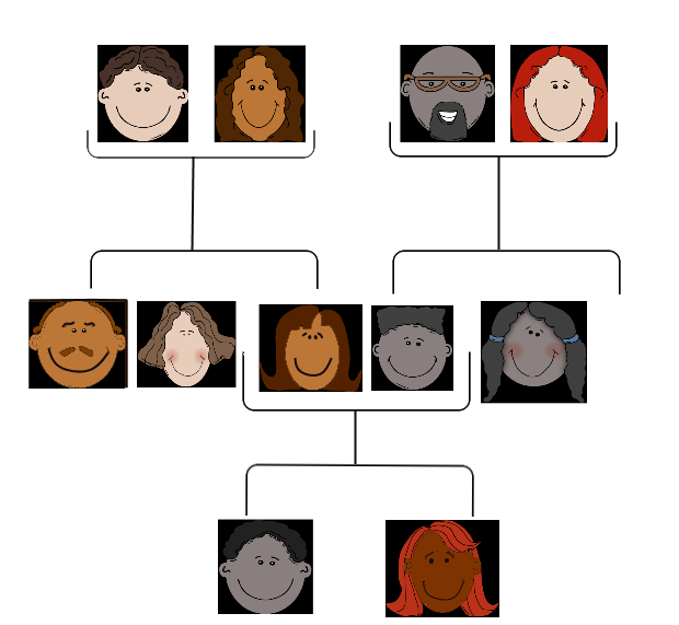
People tend to carry similar traits to their biological parents, as illustrated by the family tree. Beyond just appearance, you can also inherit traits from your parents that you can’t see.
Rebecca becomes very aware of this fact when she visits her new doctor for a physical exam. Her doctor asks several questions about her family medical history, including whether Rebecca has or had relatives with cancer. Rebecca tells her that her grandmother, aunt, and uncle — who have all passed away — had cancer. They all had breast cancer, including her uncle, and her aunt also had ovarian cancer. Her doctor asks how old they were when they were diagnosed with cancer. Rebecca is not sure exactly, but she knows that her grandmother was fairly young at the time, probably in her forties.
Rebecca’s doctor explains that while the vast majority of cancers are not due to inherited factors, a cluster of cancers within a family may indicate that there are mutations in certain genes that increase the risk of getting certain types of cancer, particularly breast and ovarian cancer. Some signs that cancers may be due to these genetic factors are present in Rebecca’s family, such as cancer with an early age of onset (e.g., breast cancer before age 50), breast cancer in men, and breast cancer and ovarian cancer within the same person or family.
Based on her family medical history, Rebecca’s doctor recommends that she see a genetic counselor, because these professionals can help determine whether the high incidence of cancers in her family could be due to inherited mutations in their genes. If so, they can test Rebecca to find out whether she has the particular variations of these genes that would increase her risk of getting cancer.
When Rebecca sees the genetic counselor, he asks how her grandmother, aunt, and uncle with cancer are related to her. She says that these relatives are all on her mother’s side — they are her mother’s mother and siblings. The genetic counselor records this information in the form of a specific type of family tree, called a pedigree, indicating which relatives had which type of cancer, and how they are related to each other and to Rebecca.
He also asks her ethnicity. Rebecca says that her family on both sides are Ashkenazi Jews (Jews whose ancestors came from central and eastern Europe). “But what does that have to do with anything?” she asks. The counselor tells Rebecca that mutations in two tumor-suppressor genes called BRCA1 and BRCA2, located on chromosome 17 and 13, respectively, are particularly prevalent in people of Ashkenazi Jewish descent and greatly increase the risk of getting cancer. About one in 40 Ashkenazi Jewish people have one of these mutations, compared to about one in 800 in the general population. Her ethnicity, along with the types of cancer, age of onset, and the specific relationships between her family members who had cancer, indicate to the counselor that she is a good candidate for genetic testing for the presence of these mutations.

Rebecca says that her 72-year-old mother never had cancer, nor had many other relatives on that side of the family. How could the cancers be genetic? The genetic counselor explains that the mutations in the BRCA1 and BRCA2 genes, while dominant, are not inherited by everyone in a family. Also, even people with mutations in these genes do not necessarily get cancer — the mutations simply increase their risk of getting cancer. For instance, 55 to 65 per cent of women with a harmful mutation in the BRCA1 gene will get breast cancer before age 70, compared to 12 per cent of women in the general population who will get breast cancer sometime over the course of their lives.
Rebecca is not sure she wants to know whether she has a higher risk of cancer. The genetic counselor understands her apprehension, but explains that if she knows that she has harmful mutations in either of these genes, her doctor will screen her for cancer more often and at earlier ages. Therefore, any cancers she may develop are likely to be caught earlier when they are often much more treatable. Rebecca decides to go through with the testing, which involves taking a blood sample, and nervously waits for her results.
Chapter Overview: Genetics
At the end of this chapter, you will find out Rebecca’s test results. By then, you will have learned how traits are inherited from parents to offspring through genes, and how mutations in genes such as BRCA1 and BRCA2 can be passed down and cause disease. Specifically, you will learn about:
- The structure of DNA.
- How DNA replication occurs.
- How DNA was found to be the inherited genetic material.
- How genes and their different alleles are located on chromosomes.
- The 23 pairs of human chromosomes, which include autosomal and sex chromosomes.
- How genes code for proteins using codons made of the sequence of nitrogen bases within RNA and DNA.
- The central dogma of molecular biology, which describes how DNA is transcribed into RNA, and then translated into proteins.
- The structure, functions, and possible evolutionary history of RNA.
- How proteins are synthesized through the transcription of RNA from DNA and the translation of protein from RNA, including how RNA and proteins can be modified, and the roles of the different types of RNA.
- What mutations are, what causes them, different specific types of mutations, and the importance of mutations in evolution and to human health.
- How the expression of genes into proteins is regulated and why problems in this process can cause diseases, such as cancer.
- How Gregor Mendel discovered the laws of inheritance for certain types of traits.
- The science of heredity, known as genetics, and the relationship between genes and traits.
- How gametes, such as eggs and sperm, are produced through meiosis.
- How sexual reproduction works on the cellular level and how it increases genetic variation.
- Simple Mendelian and more complex non-Mendelian inheritance of some human traits.
- Human genetic disorders, such as Down syndrome, hemophilia A, and disorders involving sex chromosomes.
- How biotechnology — which is the use of technology to alter the genetic makeup of organisms — is used in medicine and agriculture, how it works, and some of the ethical issues it may raise.
- The human genome, how it was sequenced, and how it is contributing to discoveries in science and medicine.
As you read this chapter, keep Rebecca’s situation in mind and think about the following questions:
- BCRA1 and BCRA2 are also called Breast cancer type 1 and 2 susceptibility proteins. What do the BRCA1 and BRCA2 genes normally do? How can they cause cancer?
- Are BRCA1 and BRCA2 linked genes? Are they on autosomal or sex chromosomes?
- After learning more about pedigrees, draw the pedigree for cancer in Rebecca’s family. Use the pedigree to help you think about why it is possible that her mother does not have one of the BRCA gene mutations, even if her grandmother, aunt, and uncle did have it.
- Why do you think certain gene mutations are prevalent in certain ethnic groups?
Attributions
Figure 5.1.1
Family Tree [all individual face images] from Clker.com used and adapted by Christine Miller under a CC0 1.0 public domain dedication license (https://creativecommons.org/publicdomain/zero/1.0/).
Figure 5.1.2
Rebecca by Kyle Broad on Unsplash is used under the Unsplash License (https://unsplash.com/license).
References
Wikipedia contributors. (2020, June 27). Ashkenazi Jews. In Wikipedia. https://en.wikipedia.org/w/index.php?title=Ashkenazi_Jews&oldid=964691647
Wikipedia contributors. (2020, June 22). BRCA1. In Wikipedia. https://en.wikipedia.org/w/index.php?title=BRCA1&oldid=963868423
Wikipedia contributors. (2020, May 25). BRCA2. In Wikipedia. https://en.wikipedia.org/w/index.php?title=BRCA2&oldid=958722957
Glucose (also called dextrose) is a simple sugar with the molecular formula C6H12O6. Glucose is the most abundant monosaccharide, a subcategory of carbohydrates. Glucose is mainly made by plants and most algae during photosynthesis from water and carbon dioxide, using energy from sunlight.
A class of biological molecule consisting of linked monomers of amino acids and which are the most versatile macromolecules in living systems and serve crucial functions in essentially all biological processes.
Amino acids are organic compounds that combine to form proteins.
A lipid. Cholesterol and its derivatives are important constituents of cell membranes and precursors of other steroid compounds, but a high proportion in the blood of low-density lipoprotein (which transports cholesterol to the tissues) is associated with an increased risk of coronary heart disease.
A body system including a series of hollow organs joined in a long, twisting tube from the mouth to the anus. The hollow organs that make up the GI tract are the mouth, esophagus, stomach, small intestine, large intestine, and anus. The liver, pancreas, and gallbladder are the solid organs of the digestive system.
The body system which acts as a chemical messenger system comprising feedback loops of the hormones released by internal glands of an organism directly into the circulatory system, regulating distant target organs. In humans, the major endocrine glands are the thyroid gland and the adrenal glands.
A hormone is a signaling molecule produced by glands in multicellular organisms that target distant organs to regulate physiology and behavior.
Created by CK-12 Foundation/Adapted by Christine Miller
Case Study: Flight Risk
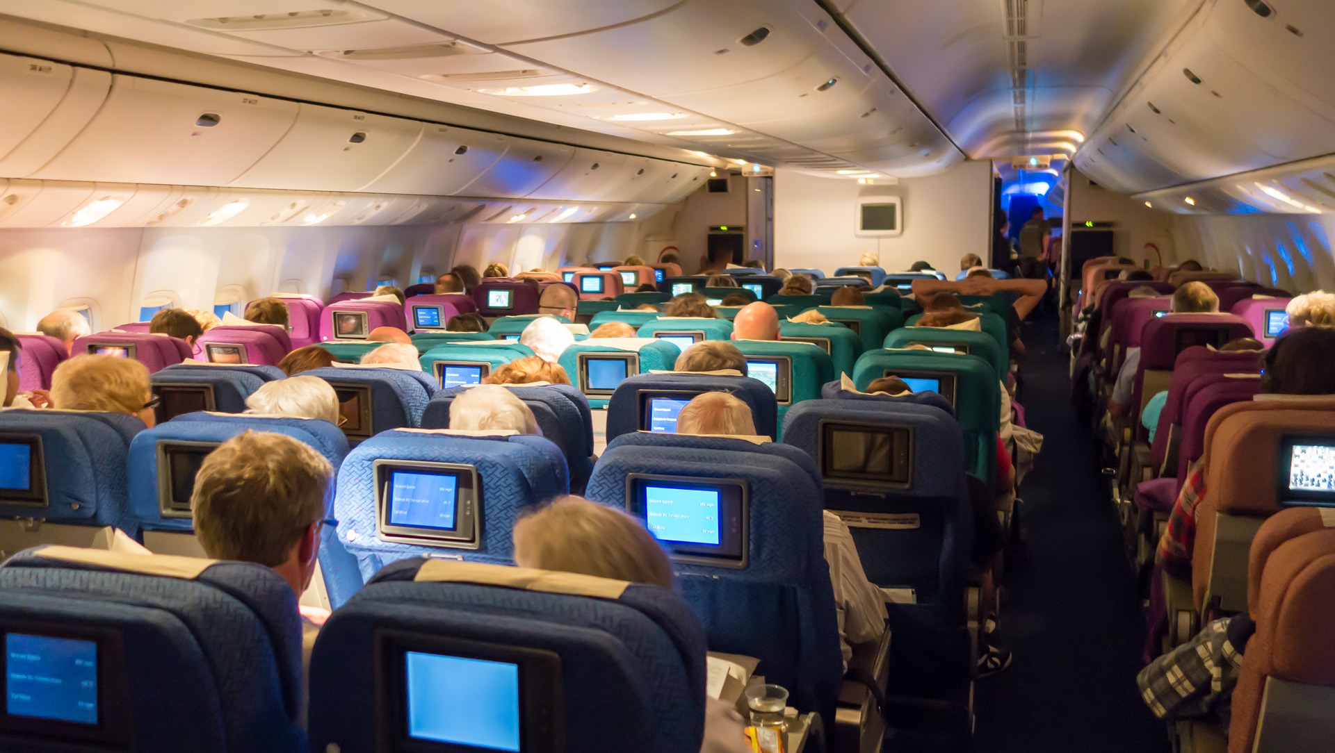
Nineteen-year-old Malcolm is about to take his first plane flight. Shortly after he boards the plane and sits down, a man in his late sixties sits next to him in the aisle seat. About half an hour after the plane takes off, the pilot announces that she is turning the seat belt light off, and that it is safe to move around the cabin.
The man in the aisle seat — who has introduced himself to Malcolm as Willie — immediately unbuckles his seat belt and paces up and down the aisle a few times before returning to his seat. After about 45 minutes, Willie gets up again, walks some more, then sits back down and does some foot and leg exercises. After the third time Willie gets up and paces the aisles, Malcolm asks him whether he is walking so much to accumulate steps on a pedometer or fitness tracking device. Willie laughs and says no. He is actually trying to do something even more important for his health — prevent a blood clot from forming in his legs.
Willie explains that he has a chronic condition: heart failure. Although it sounds scary, his condition is currently well-managed, and he is able to lead a relatively normal lifestyle. However, it does put him at risk of developing other serious health conditions, such as deep vein thrombosis (DVT), which is when a blood clot occurs in the deep veins, usually in the legs. Air travel — and other situations where a person has to sit for a long period of time — increases the risk of DVT. Willie’s doctor said that he is healthy enough to fly, but that he should walk frequently and do leg exercises to help avoid a blood clot.
As you read this chapter, you will learn about the heart, blood vessels, and blood that make up the cardiovascular system, as well as disorders of the cardiovascular system, such as heart failure. At the end of the chapter you will learn more about why DVT occurs, why Willie has to take extra precautions when he flies, and what can be done to lower the risk of DVT and its potentially deadly consequences.
Chapter Overview: Cardiovascular System
In this chapter, you will learn about the cardiovascular system, which transports substances throughout the body. Specifically, you will learn about:
- The major components of the cardiovascular system: the heart, blood vessels, and blood.
- The functions of the cardiovascular system, including transporting needed substances (such as oxygen and nutrients) to the cells of the body, and picking up waste products.
- How blood is oxygenated through the pulmonary circulation, which transports blood between the heart and lungs.
- How blood is circulated throughout the body through the systemic circulation.
- The components of blood — including plasma, red blood cells, white blood cells, and platelets — and their specific functions.
- Types of blood vessels — including arteries, veins, and capillaries — and their functions, similarities, and differences.
- The structure of the heart, how it pumps blood, and how contractions of the heart are controlled.
- What blood pressure is and how it is regulated.
- Blood disorders, including anemia, HIV, and leukemia.
- Cardiovascular diseases (including heart attack, stroke, and angina), and the risk factors and precursors — such as high blood pressure and atherosclerosis — that contribute to them.
As you read the chapter, think about the following questions:
- What is heart failure?Why do you think it increases the risk of DVT?
- What is a blood clot? What are possible health consequences of blood clots?
- Why do you think sitting for long periods of time increases the risk of DVT? Why does walking and exercising the legs help reduce this risk?
Attribution
Figure 14.1.1
aircraft-1583871_1920 [photo] by olivier89 from Pixabay is used under the Pixabay License (https://pixabay.com/de/service/license/).
A peptide hormone, produced by alpha cells of the pancreas. It works to raise the concentration of glucose and fatty acids in the bloodstream, and is considered to be the main catabolic hormone of the body. It is also used as a medication to treat a number of health conditions.
Image shows a sperm fertilizing an egg. The Sperm is much smaller than the egg.
Figure 5.14.1 Collage of Diverse Faces.
This collage shows some of the variation in human skin colour, which can range from very light to very dark, with every possible gradation in between. As you might expect, the skin color trait has a more complex genetic basis than just one gene with two alleles, which is the type of simple trait that Mendel studied in pea plants. Like skin color, many other human traits have more complicated modes of inheritance than Mendelian traits. Such modes of inheritance are called non-Mendelian inheritance, and they include inheritance of multiple allele traits, traits with codominance or incomplete dominance, and polygenic traits, among others. All of these modes are described below.
Multiple Allele Traits
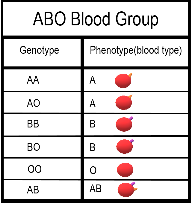
The majority of human genes are thought to have more than two normal versions, or alleles. Traits controlled by a single gene with more than two alleles are called multiple allele traits. An example is ABO blood type. Your blood type refers to which of certain proteins called antigens are found on your red blood cells. There are three common alleles for this trait, which are represented by the letters A, B, and O.
As shown in the table there are six possible ABO genotypes, because the three alleles, taken two at a time, result in six possible combinations. The A and B alleles are dominant to the O allele. As a result, both AA and AO genotypes have the same phenotype, with the A antigen in their blood (type A blood). Similarly, both BB and BO genotypes have the same phenotype, with the B antigen in their blood (type B blood). No antigen is associated with the O allele, so people with the OO genotype have no antigens for ABO blood type in their blood (type O blood).
Codominance
Look at the genotype AB in the ABO blood group table. Alleles A and B for ABO blood type are neither dominant nor recessive to one another. Instead, they are codominant. Codominance occurs when two alleles for a gene are expressed equally in the phenotype of heterozygotes. In the case of ABO blood type, AB heterozygotes have a unique phenotype, with both A and B antigens in their blood (type AB blood).
Incomplete Dominance
Another relationship that may occur between alleles for the same gene is incomplete dominance. This occurs when the dominant allele is not completely dominant. In this case, an intermediate phenotype results in heterozygotes who inherit both alleles. Generally, this happens when the two alleles for a given gene both produce proteins, but one protein is not functional. As a result, the heterozygote individual produces only half the amount of normal protein as is produced by an individual who is homozygous for the normal allele.
An example of incomplete dominance in humans is Tay Sachs disease. The normal allele for the gene in this case produces an enzyme that is responsible for breaking down lipids. A defective allele for the gene results in the production of a nonfunctional enzyme. Heterozygotes who have one normal and one defective allele produce half as much functional enzyme as the normal homozygote, and this is enough for normal development. Homozygotes who have only defective allele, however, produce only nonfunctional enzyme. This leads to the accumulation of lipids in the brain starting in utero, which causes significant brain damage. Most individuals with Tay Sachs disease die at a young age, typically by the age of five years.
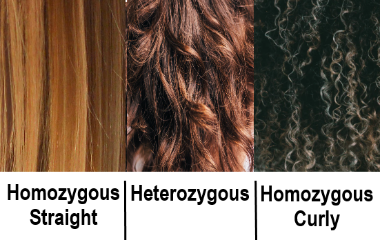
Another good example of incomplete dominance in humans is hair type. There are genes for straight and curly hair, and if an individual is heterozygous, they will typically have the phenotype of wavy hair.
Polygenic Traits
Many human traits are controlled by more than one gene. These traits are called polygenic traits. The alleles of each gene have a minor additive effect on the phenotype. There are many possible combinations of alleles, especially if each gene has multiple alleles. Therefore, a whole continuum of phenotypes is possible.
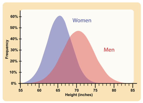
An example of a human polygenic trait is adult height. Several genes, each with more than one allele, contribute to this trait, so there are many possible adult heights. One adult’s height might be 1.655 m (5.430 feet), and another adult’s height might be 1.656 m (5.433 feet). Adult height ranges from less than 5 feet to more than 6 feet, with males, on average, being somewhat taller than females. The majority of people fall near the middle of the range of heights for their sex, as shown in Figure 5.14.4.
Environmental Effects on Phenotype
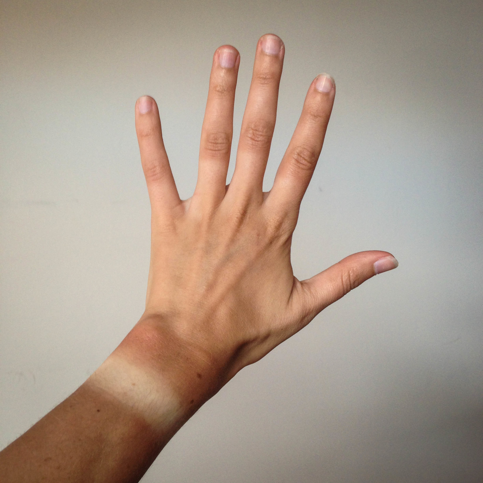
Many traits are affected by the environment, as well as by genes. This may be especially true for polygenic traits. Adult height, for example, might be negatively impacted by poor diet or childhood illness. Skin color is another polygenic trait. There is a wide range of skin colors in people worldwide. In addition to differences in genes, differences in exposure to ultraviolet (UV) light cause some variation. As shown in Figure 5.14.5, exposure to UV light darkens the skin.
Pleiotropy
Some genes affect more than one phenotypic trait. This is called pleiotropy. There are numerous examples of pleiotropy in humans. They generally involve important proteins that are needed for the normal development or functioning of more than one organ system. An example of pleiotropy in humans occurs with the gene that codes for the main protein in collagen, a substance that helps form bones. This protein is also important in the ears and eyes. Mutations in the gene result in problems not only in bones, but also in these sensory organs, which is how the gene's pleiotropic effects were discovered.
Another example of pleiotropy occurs with sickle cell anemia. This recessive genetic disorder occurs when there is a mutation in the gene that normally encodes the red blood cell protein called hemoglobin. People with the disorder have two alleles for sickle cell hemoglobin, so named for the sickle shape (pictured in Figure 5.14.6) that their red blood cells take on under certain conditions (like physical exertion). The sickle-shaped red blood cells clog small blood vessels, causing multiple phenotypic effects, including stunting of physical growth, certain bone deformities, kidney failure, and strokes.
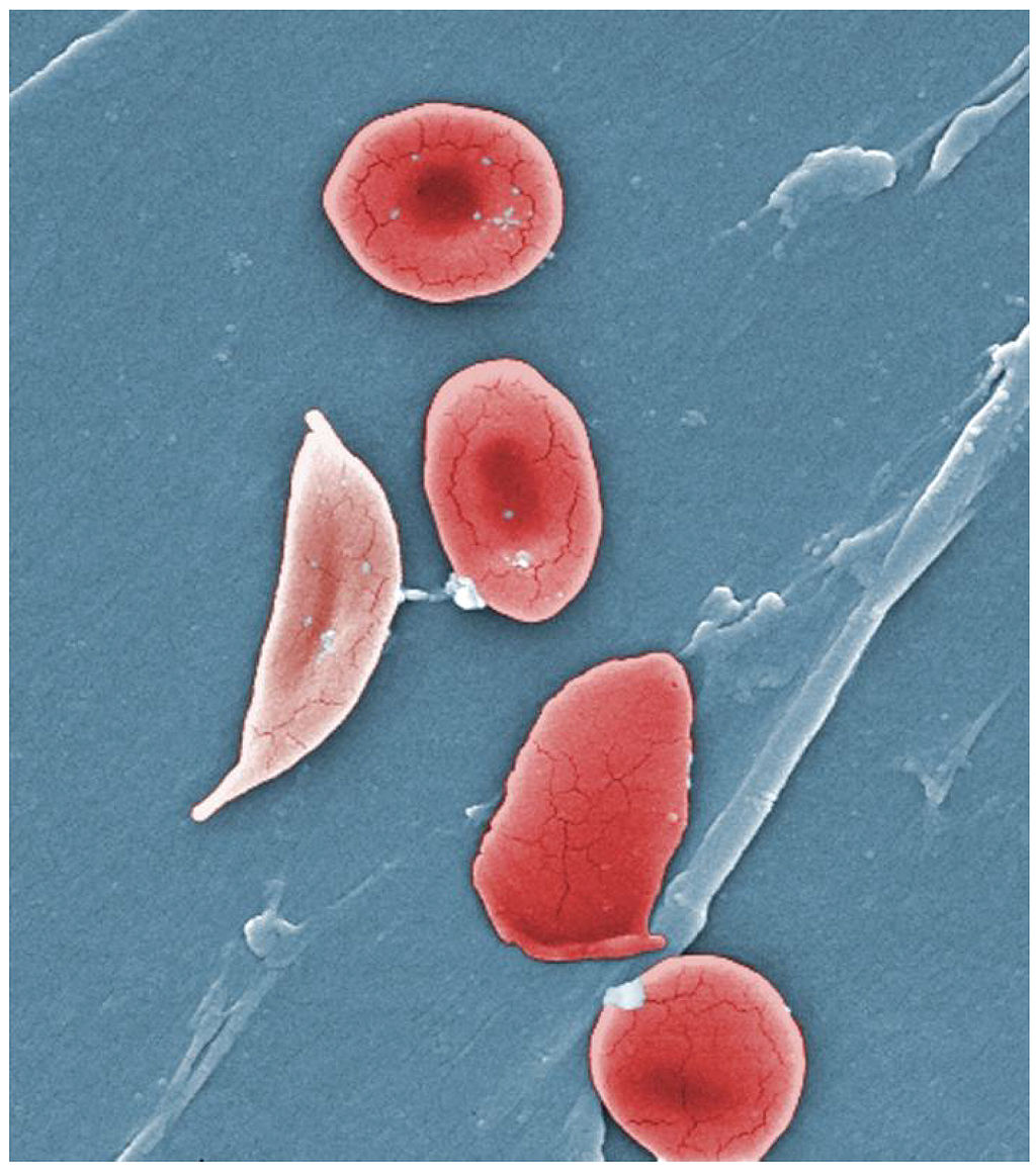
Epistasis
Some genes affect the expression of other genes. This is called epistasis. Epistasis is similar to dominance, except that it occurs between different genes, rather than between different alleles for the same gene.
Albinism is an example of epistasis. A person with albinism has virtually no pigment in the skin. The condition occurs due to an entirely different gene than the genes that encode skin color. Albinism occurs because a protein called tyrosinase, which is needed for the production of normal skin pigment, is not produced, due to a gene mutation. If an individual has the albinism mutation, he or she will not have any skin pigment, regardless of the skin color genes that were inherited.
Feature: My Human Body
Do you know your ABO blood type? In an emergency, knowing this valuable piece of information could possibly save your life. If you ever need a blood transfusion, it is vital that you receive blood that matches your own blood type. Why? If the blood transfused into your body contains an antigen that your own blood does not contain, antibodies in your blood plasma (the liquid part of your blood) will recognize the antigen as foreign to your body and cause a reaction called agglutination. In this reaction, the transfused red blood cells will clump together. The agglutination reaction is serious and potentially fatal.
Knowing the antigens and antibodies present in each of the ABO blood types will help you understand which type(s) of blood you can safely receive if you ever need a transfusion. This information is shown in Figure 5.14.7 for all of the ABO blood types. If you have blood type A, this means that your red blood cells have the A antigen and that your blood plasma contains anti-B antibodies. If you were to receive a transfusion of type B or type AB blood, both of which have the B antigen, your anti-B antibodies would attack the transfused red blood cells, causing agglutination.
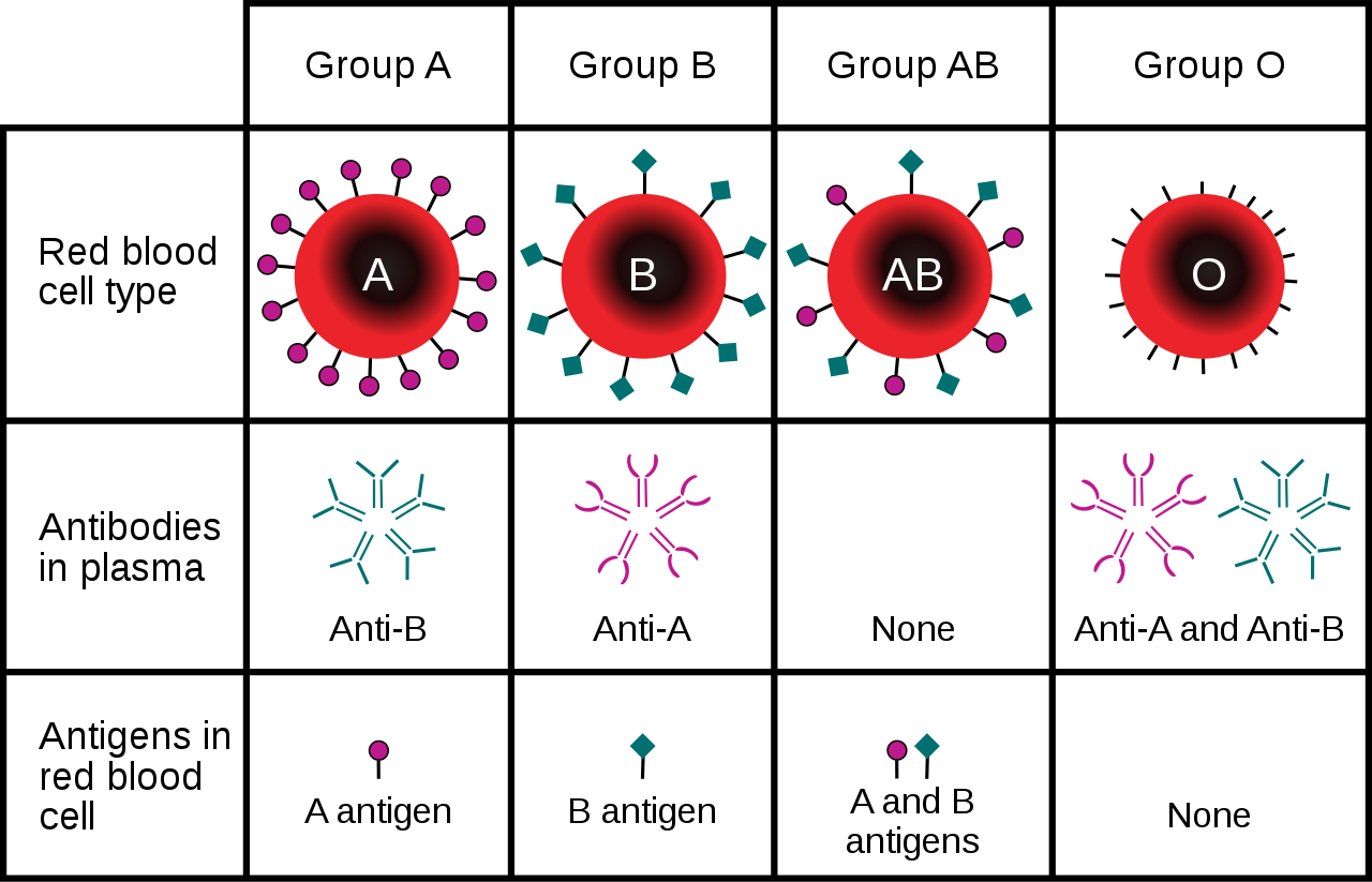
You may have heard that people with blood type O are called "universal donors," and that people with blood type AB are called universal recipients. People with type O blood have neither A nor B antigens in their blood, so if their blood is transfused into someone with a different ABO blood type, it causes no immune reaction, meaning they can donate blood to anyone. On the other hand, people with type AB blood have no anti-A or anti-B antibodies in their blood, so they can receive a transfusion of blood from anyone. Which blood type(s) can safely receive a transfusion of type AB blood, and which blood type(s) can be safely received by those with type O blood?
5.14 Summary
- Non-Mendelian inheritance refers to the inheritance of traits that have a more complex genetic basis than one gene with two alleles and complete dominance.
- Multiple allele traits are controlled by a single gene with more than two alleles. An example of a human multiple allele trait is ABO blood type, for which there are three common alleles: A, B, and O.
- Codominance occurs when two alleles for a gene are expressed equally in the phenotype of heterozygotes. A human example of codominance also occurs in the ABO blood type, in which the A and B alleles are codominant.
- Incomplete dominance is the case in which the dominant allele for a gene is not completely dominant to a recessive allele for the gene, so an intermediate phenotype occurs in heterozygotes who inherit both alleles. A human example of incomplete dominance is Tay Sachs disease, in which heterozygotes produce half as much functional enzyme as normal homozygotes.
- Polygenic traits are controlled by more than one gene, each of which has a minor additive effect on the phenotype. This results in a whole continuum of phenotypes. Examples of human polygenic traits include skin color and adult height.
- Many traits are affected by the environment, as well as by genes. This may be especially true for polygenic traits. Skin color, for example, may be affected by exposure to UV light, and adult stature may be affected by diet or childhood disease.
- Pleiotropy refers to the situation in which a gene affects more than one phenotypic trait. A human example of pleiotropy occurs with sickle cell anemia. People who inherit two recessive alleles for this disorder have abnormal red blood cells and may exhibit multiple other phenotypic effects, such as stunting of physical growth, kidney failure, and strokes.
- Epistasis is the situation in which one gene affects the expression of other genes. An example of epistasis is albinism, in which the albinism mutation negates the expression of skin color genes.
5.14 Review Questions
- What is non-Mendelian inheritance?
-
- Explain why the human ABO blood group is an example of a multiple allele trait with codominance.
- What is incomplete dominance? Give an example of this type of non-Mendelian inheritance in humans.
- Explain the genetic basis of human skin color.
- How can the human trait of adult height be influenced by the environment?
- Define pleiotropy, and give a human example.
- Compare and contrast epistasis and dominance.
- What is the difference between pleiotropy and epistasis?
5.14 Explore More
https://www.youtube.com/watch?time_continue=1&v=YJHGfbW55l0&feature=emb_logo
Incomplete Dominance, Codominance, Polygenic Traits, and Epistasis!,
Amoeba Sisters, 2015.
https://www.youtube.com/watch?v=-4vsio8TZrU&feature=emb_logo
Non-Mendelian Genetics, Teacher's Pet, 2015.
Attributes
Figure 5.14.1
- Woman's Face from Iran by Omid Armin on Unsplash is used under the Unsplash License (https://unsplash.com/license).
- Woman Wearing Black Coat by Anastasiya Pavlova on Unsplash is used under the Unsplash License (https://unsplash.com/license).
- Dark haired man, Queretaro, México by Leonel Hernandez Arteaga on Unsplash is used under the Unsplash License (https://unsplash.com/license). <not found on Unsplash>
- Man in White V-Neck T-Shirt (self) by Joseph Gonzalez on Unsplash is used under the Unsplash License (https://unsplash.com/license).
- Natural Redhead in Brazil by Gabriel Silvério on Unsplash is used under the Unsplash License (https://unsplash.com/license).
- Dark-Skinned Woman with Large White Rose by Oladimeji Oduns on Unsplash is used under the Unsplash License (https://unsplash.com/license).
Figure 5.14.2
ABO Blood Types Per Genotype by Christine Miller is released into the public domain (https://en.wikipedia.org/wiki/Public_domain).
Figure 5.14.3
Three Phenotypes of Hair Based on Inheritance/ Incomplete Dominance Hair by Christine Miller is released into the public domain (https://en.wikipedia.org/wiki/Public_domain).
Figure 5.14.4
Average height /Human Adult Height by CK-12 Foundation is used under a CC BY 3.0 (https://creativecommons.org/licenses/by-nc/3.0/) license.
 ©CK-12 Foundation Licensed under
 ![]() • Terms of Use • Attribution
• Terms of Use • Attribution
Figure 5.14.5
Tan lines by katiebordner on Flickr is used under a CC BY 2.0 (https://creativecommons.org/licenses/by/2.0/) license.
Figure 5.14.6
Sickle cell anemia by OpenStax College on Wikimedia Commons is used under a CC BY 3.0 (https://creativecommons.org/licenses/by/3.0) ©
Figure 5.14.7
ABO_blood_type.svg by InvictaHOG on Wikimedia Commons is in the public domain (https://en.wikipedia.org/wiki/Public_domain).
References
Amoeba Sisters. (2015, May 25). Incomplete dominance, codominance, polygenic traits, and epistasis! YouTube. https://www.youtube.com/watch?v=YJHGfbW55l0
Betts, J. G., Young, K.A., Wise, J.A., Johnson, E., Poe, B., Kruse, D.H., Korol, O., Johnson, J.E., Womble, M., DeSaix, P. (2013, April 25). Figure 18.9 Sickle cells [digital image]. In Anatomy and Physiology. OpenStax. https://openstax.org/books/anatomy-and-physiology/pages/18-3-erythrocytes
Brainard, J/ CK-12 Foundation. (2016). Figure 2 Human adult height [digital image]. In CK-12 College Human Biology (Section 5.13) [online Flexbook]. CK12.org. https://www.ck12.org/book/ck-12-college-human-biology/section/5.13/
Mayo Clinic Staff. (n.d.). Tay-Sachs disease [online article]. MayoClinic.org. https://www.mayoclinic.org/diseases-conditions/tay-sachs-disease/symptoms-causes/syc-20378190
Mayo Clinic Staff. (n.d.). Sickle cell anemia [online article]. MayoClinic.org. https://www.mayoclinic.org/diseases-conditions/sickle-cell-anemia/symptoms-causes/syc-20355876
Teacher's Pet. (2015, January 25). Non-mendelian genetics. YouTube. https://www.youtube.com/watch?v=-4vsio8TZrU
Created by: CK-12/Adapted by Christine Miller
Figure 5.14.1 Collage of Diverse Faces.
This collage shows some of the variation in human skin colour, which can range from very light to very dark, with every possible gradation in between. As you might expect, the skin color trait has a more complex genetic basis than just one gene with two alleles, which is the type of simple trait that Mendel studied in pea plants. Like skin color, many other human traits have more complicated modes of inheritance than Mendelian traits. Such modes of inheritance are called non-Mendelian inheritance, and they include inheritance of multiple allele traits, traits with codominance or incomplete dominance, and polygenic traits, among others. All of these modes are described below.
Multiple Allele Traits

The majority of human genes are thought to have more than two normal versions, or alleles. Traits controlled by a single gene with more than two alleles are called multiple allele traits. An example is ABO blood type. Your blood type refers to which of certain proteins called antigens are found on your red blood cells. There are three common alleles for this trait, which are represented by the letters A, B, and O.
As shown in the table there are six possible ABO genotypes, because the three alleles, taken two at a time, result in six possible combinations. The A and B alleles are dominant to the O allele. As a result, both AA and AO genotypes have the same phenotype, with the A antigen in their blood (type A blood). Similarly, both BB and BO genotypes have the same phenotype, with the B antigen in their blood (type B blood). No antigen is associated with the O allele, so people with the OO genotype have no antigens for ABO blood type in their blood (type O blood).
Codominance
Look at the genotype AB in the ABO blood group table. Alleles A and B for ABO blood type are neither dominant nor recessive to one another. Instead, they are codominant. Codominance occurs when two alleles for a gene are expressed equally in the phenotype of heterozygotes. In the case of ABO blood type, AB heterozygotes have a unique phenotype, with both A and B antigens in their blood (type AB blood).
Incomplete Dominance
Another relationship that may occur between alleles for the same gene is incomplete dominance. This occurs when the dominant allele is not completely dominant. In this case, an intermediate phenotype results in heterozygotes who inherit both alleles. Generally, this happens when the two alleles for a given gene both produce proteins, but one protein is not functional. As a result, the heterozygote individual produces only half the amount of normal protein as is produced by an individual who is homozygous for the normal allele.
An example of incomplete dominance in humans is Tay Sachs disease. The normal allele for the gene in this case produces an enzyme that is responsible for breaking down lipids. A defective allele for the gene results in the production of a nonfunctional enzyme. Heterozygotes who have one normal and one defective allele produce half as much functional enzyme as the normal homozygote, and this is enough for normal development. Homozygotes who have only defective allele, however, produce only nonfunctional enzyme. This leads to the accumulation of lipids in the brain starting in utero, which causes significant brain damage. Most individuals with Tay Sachs disease die at a young age, typically by the age of five years.

Another good example of incomplete dominance in humans is hair type. There are genes for straight and curly hair, and if an individual is heterozygous, they will typically have the phenotype of wavy hair.
Polygenic Traits

Many human traits are controlled by more than one gene. These traits are called polygenic traits. The alleles of each gene have a minor additive effect on the phenotype. There are many possible combinations of alleles, especially if each gene has multiple alleles. Therefore, a whole continuum of phenotypes is possible.
An example of a human polygenic trait is adult height. Several genes, each with more than one allele, contribute to this trait, so there are many possible adult heights. One adult’s height might be 1.655 m (5.430 feet), and another adult’s height might be 1.656 m (5.433 feet). Adult height ranges from less than 5 feet to more than 6 feet, with males, on average, being somewhat taller than females. The majority of people fall near the middle of the range of heights for their sex, as shown in Figure 5.14.4.
Environmental Effects on Phenotype

Many traits are affected by the environment, as well as by genes. This may be especially true for polygenic traits. Adult height, for example, might be negatively impacted by poor diet or childhood illness. Skin color is another polygenic trait. There is a wide range of skin colors in people worldwide. In addition to differences in genes, differences in exposure to ultraviolet (UV) light cause some variation. As shown in Figure 5.14.5, exposure to UV light darkens the skin.
Pleiotropy
Some genes affect more than one phenotypic trait. This is called pleiotropy. There are numerous examples of pleiotropy in humans. They generally involve important proteins that are needed for the normal development or functioning of more than one organ system. An example of pleiotropy in humans occurs with the gene that codes for the main protein in collagen, a substance that helps form bones. This protein is also important in the ears and eyes. Mutations in the gene result in problems not only in bones, but also in these sensory organs, which is how the gene's pleiotropic effects were discovered.
Another example of pleiotropy occurs with sickle cell anemia. This recessive genetic disorder occurs when there is a mutation in the gene that normally encodes the red blood cell protein called hemoglobin. People with the disorder have two alleles for sickle cell hemoglobin, so named for the sickle shape (pictured in Figure 5.14.6) that their red blood cells take on under certain conditions (like physical exertion). The sickle-shaped red blood cells clog small blood vessels, causing multiple phenotypic effects, including stunting of physical growth, certain bone deformities, kidney failure, and strokes.

Epistasis
Some genes affect the expression of other genes. This is called epistasis. Epistasis is similar to dominance, except that it occurs between different genes, rather than between different alleles for the same gene.
Albinism is an example of epistasis. A person with albinism has virtually no pigment in the skin. The condition occurs due to an entirely different gene than the genes that encode skin color. Albinism occurs because a protein called tyrosinase, which is needed for the production of normal skin pigment, is not produced, due to a gene mutation. If an individual has the albinism mutation, he or she will not have any skin pigment, regardless of the skin color genes that were inherited.
Feature: My Human Body
Do you know your ABO blood type? In an emergency, knowing this valuable piece of information could possibly save your life. If you ever need a blood transfusion, it is vital that you receive blood that matches your own blood type. Why? If the blood transfused into your body contains an antigen that your own blood does not contain, antibodies in your blood plasma (the liquid part of your blood) will recognize the antigen as foreign to your body and cause a reaction called agglutination. In this reaction, the transfused red blood cells will clump together. The agglutination reaction is serious and potentially fatal.
Knowing the antigens and antibodies present in each of the ABO blood types will help you understand which type(s) of blood you can safely receive if you ever need a transfusion. This information is shown in Figure 5.14.7 for all of the ABO blood types. If you have blood type A, this means that your red blood cells have the A antigen and that your blood plasma contains anti-B antibodies. If you were to receive a transfusion of type B or type AB blood, both of which have the B antigen, your anti-B antibodies would attack the transfused red blood cells, causing agglutination.

You may have heard that people with blood type O are called "universal donors," and that people with blood type AB are called universal recipients. People with type O blood have neither A nor B antigens in their blood, so if their blood is transfused into someone with a different ABO blood type, it causes no immune reaction, meaning they can donate blood to anyone. On the other hand, people with type AB blood have no anti-A or anti-B antibodies in their blood, so they can receive a transfusion of blood from anyone. Which blood type(s) can safely receive a transfusion of type AB blood, and which blood type(s) can be safely received by those with type O blood?
5.14 Summary
- Non-Mendelian inheritance refers to the inheritance of traits that have a more complex genetic basis than one gene with two alleles and complete dominance.
- Multiple allele traits are controlled by a single gene with more than two alleles. An example of a human multiple allele trait is ABO blood type, for which there are three common alleles: A, B, and O.
- Codominance occurs when two alleles for a gene are expressed equally in the phenotype of heterozygotes. A human example of codominance also occurs in the ABO blood type, in which the A and B alleles are codominant.
- Incomplete dominance is the case in which the dominant allele for a gene is not completely dominant to a recessive allele for the gene, so an intermediate phenotype occurs in heterozygotes who inherit both alleles. A human example of incomplete dominance is Tay Sachs disease, in which heterozygotes produce half as much functional enzyme as normal homozygotes.
- Polygenic traits are controlled by more than one gene, each of which has a minor additive effect on the phenotype. This results in a whole continuum of phenotypes. Examples of human polygenic traits include skin color and adult height.
- Many traits are affected by the environment, as well as by genes. This may be especially true for polygenic traits. Skin color, for example, may be affected by exposure to UV light, and adult stature may be affected by diet or childhood disease.
- Pleiotropy refers to the situation in which a gene affects more than one phenotypic trait. A human example of pleiotropy occurs with sickle cell anemia. People who inherit two recessive alleles for this disorder have abnormal red blood cells and may exhibit multiple other phenotypic effects, such as stunting of physical growth, kidney failure, and strokes.
- Epistasis is the situation in which one gene affects the expression of other genes. An example of epistasis is albinism, in which the albinism mutation negates the expression of skin color genes.
5.14 Review Questions
- What is non-Mendelian inheritance?
-
- Explain why the human ABO blood group is an example of a multiple allele trait with codominance.
- What is incomplete dominance? Give an example of this type of non-Mendelian inheritance in humans.
- Explain the genetic basis of human skin color.
- How can the human trait of adult height be influenced by the environment?
- Define pleiotropy, and give a human example.
- Compare and contrast epistasis and dominance.
- What is the difference between pleiotropy and epistasis?
5.14 Explore More
https://www.youtube.com/watch?time_continue=1&v=YJHGfbW55l0&feature=emb_logo
Incomplete Dominance, Codominance, Polygenic Traits, and Epistasis!,
Amoeba Sisters, 2015.
https://www.youtube.com/watch?v=-4vsio8TZrU&feature=emb_logo
Non-Mendelian Genetics, Teacher's Pet, 2015.
Attributes
Figure 5.14.1
- Woman's Face from Iran by Omid Armin on Unsplash is used under the Unsplash License (https://unsplash.com/license).
- Woman Wearing Black Coat by Anastasiya Pavlova on Unsplash is used under the Unsplash License (https://unsplash.com/license).
- Dark haired man, Queretaro, México by Leonel Hernandez Arteaga on Unsplash is used under the Unsplash License (https://unsplash.com/license). <not found on Unsplash>
- Man in White V-Neck T-Shirt (self) by Joseph Gonzalez on Unsplash is used under the Unsplash License (https://unsplash.com/license).
- Natural Redhead in Brazil by Gabriel Silvério on Unsplash is used under the Unsplash License (https://unsplash.com/license).
- Dark-Skinned Woman with Large White Rose by Oladimeji Oduns on Unsplash is used under the Unsplash License (https://unsplash.com/license).
Figure 5.14.2
ABO Blood Types Per Genotype by Christine Miller is released into the public domain (https://en.wikipedia.org/wiki/Public_domain).
Figure 5.14.3
Three Phenotypes of Hair Based on Inheritance/ Incomplete Dominance Hair by Christine Miller is released into the public domain (https://en.wikipedia.org/wiki/Public_domain).
Figure 5.14.4
Average height /Human Adult Height by CK-12 Foundation is used under a CC BY 3.0 (https://creativecommons.org/licenses/by-nc/3.0/) license.
 ©CK-12 Foundation Licensed under
 ![]() • Terms of Use • Attribution
• Terms of Use • Attribution
Figure 5.14.5
Tan lines by katiebordner on Flickr is used under a CC BY 2.0 (https://creativecommons.org/licenses/by/2.0/) license.
Figure 5.14.6
Sickle cell anemia by OpenStax College on Wikimedia Commons is used under a CC BY 3.0 (https://creativecommons.org/licenses/by/3.0) ©
Figure 5.14.7
ABO_blood_type.svg by InvictaHOG on Wikimedia Commons is in the public domain (https://en.wikipedia.org/wiki/Public_domain).
References
Amoeba Sisters. (2015, May 25). Incomplete dominance, codominance, polygenic traits, and epistasis! YouTube. https://www.youtube.com/watch?v=YJHGfbW55l0
Betts, J. G., Young, K.A., Wise, J.A., Johnson, E., Poe, B., Kruse, D.H., Korol, O., Johnson, J.E., Womble, M., DeSaix, P. (2013, April 25). Figure 18.9 Sickle cells [digital image]. In Anatomy and Physiology. OpenStax. https://openstax.org/books/anatomy-and-physiology/pages/18-3-erythrocytes
Brainard, J/ CK-12 Foundation. (2016). Figure 2 Human adult height [digital image]. In CK-12 College Human Biology (Section 5.13) [online Flexbook]. CK12.org. https://www.ck12.org/book/ck-12-college-human-biology/section/5.13/
Mayo Clinic Staff. (n.d.). Tay-Sachs disease [online article]. MayoClinic.org. https://www.mayoclinic.org/diseases-conditions/tay-sachs-disease/symptoms-causes/syc-20378190
Mayo Clinic Staff. (n.d.). Sickle cell anemia [online article]. MayoClinic.org. https://www.mayoclinic.org/diseases-conditions/sickle-cell-anemia/symptoms-causes/syc-20355876
Teacher's Pet. (2015, January 25). Non-mendelian genetics. YouTube. https://www.youtube.com/watch?v=-4vsio8TZrU
Image shows a medical professional blotting a blood sample from a small prick on an infant's heel onto special filter paper for the purposes of screening for PKU.
Figure 4.2.1 Human cells viewed with a very powerful tool called a scanning electron microscope.
Amazing Cells
What are these incredible objects? Would it surprise you to learn that they are all human cells? Cells are actually too small to see with the unaided eye. It is visible here in such detail because it is being viewed with a very powerful tool called a scanning electron microscope. Cells may be small in size, but they are extremely important to life. Like all other living things, you are made of cells. Cells are the basis of life, and without cells, life as we know it would not exist. You will learn more about these amazing building blocks of life in this section.
What Are Cells?
If you look at living matter with a microscope — even a simple light microscope — you will see that it consists of cells. Cells are the basic units of the structure and function of living things. They are the smallest units that can carry out the processes of life. All organisms are made up of one or more cells, and all cells have many of the same structures and carry out the same basic life processes. Knowing the structure of cells and the processes they carry out is necessary to an understanding of life itself.
Discovery of Cells
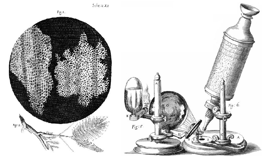
The first time the word cell was used to refer to these tiny units of life was in 1665 by a British scientist named Robert Hooke. Hooke was one of the earliest scientists to study living things under a microscope. The microscopes of his day were not very strong, but Hooke was still able to make an important discovery. When he looked at a thin slice of cork under his microscope, he was surprised to see what looked like a honeycomb. Hooke made the drawing in the figure to the right to show what he saw. As you can see, the cork was made up of many tiny units. Hooke called these units cells because they resembled cells in a monastery.
Soon after Robert Hooke discovered cells in cork, Anton van Leeuwenhoek in Holland made other important discoveries using a microscope. Leeuwenhoek made his own microscope lenses, and he was so good at it that his microscope was more powerful than other microscopes of his day. In fact, Leeuwenhoek’s microscope was almost as strong as modern light microscopes. Using his microscope, Leeuwenhoek was the first person to observe human cells and bacteria.
Cell Theory
By the early 1800s, scientists had observed cells of many different organisms. These observations led two German scientists named Theodor Schwann and Matthias Jakob Schleiden to propose cells as the basic building blocks of all living things. Around 1850, a German doctor named Rudolf Virchow was studying cells under a microscope, when he happened to see them dividing and forming new cells. He realized that living cells produce new cells through division. Based on this realization, Virchow proposed that living cells arise only from other living cells.
The ideas of all three scientists — Schwann, Schleiden, and Virchow — led to cell theory, which is one of the fundamental theories unifying all of biology.
Cell theory states that:
- All organisms are made of one or more cells.
- All the life functions of organisms occur within cells.
- All cells come from existing cells.
Seeing Inside Cells
Starting with Robert Hooke in the 1600s, the microscope opened up an amazing new world — a world of life at the level of the cell. As microscopes continued to improve, more discoveries were made about the cells of living things, but by the late 1800s, light microscopes had reached their limit. Objects much smaller than cells (including the structures inside cells) were too small to be seen with even the strongest light microscope.
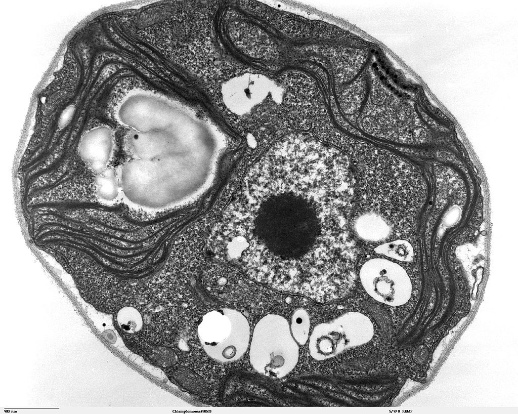
Then, in the 1950s, a new type of microscope was invented. Called the electron microscope, it used a beam of electrons instead of light to observe extremely small objects. With an electron microscope, scientists could finally see the tiny structures inside cells. They could even see individual molecules and atoms. The electron microscope had a huge impact on biology. It allowed scientists to study organisms at the level of their molecules, and it led to the emergence of the molecular biology field. With the electron microscope, many more cell discoveries were made.
Structures Shared By All Cells
Although cells are diverse, all cells have certain parts in common. These parts include a plasma membrane, cytoplasm, ribosomes, and DNA.
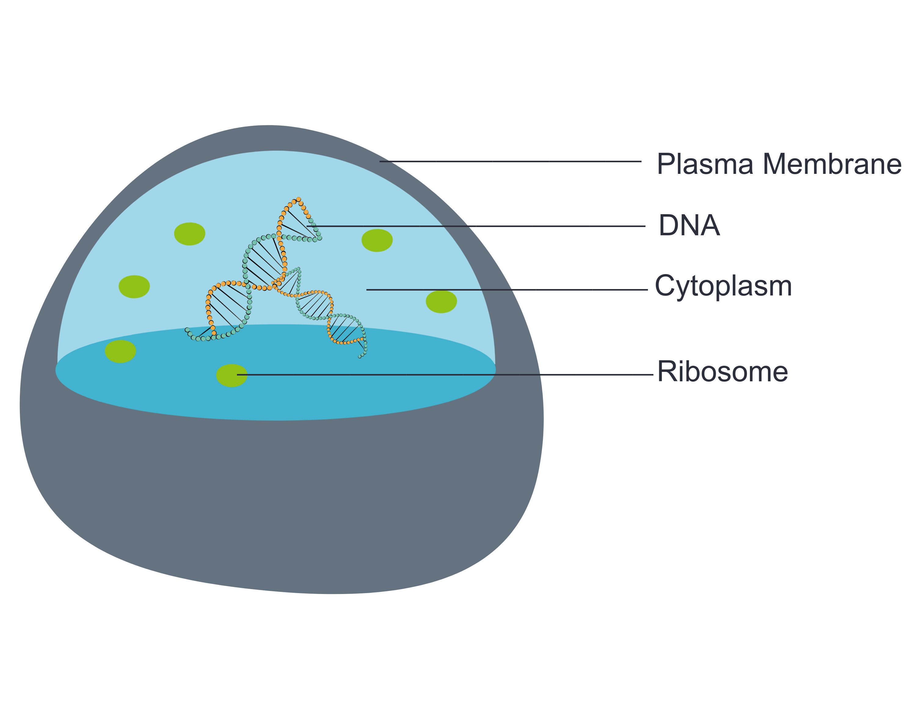
- The plasma membrane (a type of cell membrane) is a thin coat of lipids that surrounds a cell. It forms the physical boundary between the cell and its environment. You can think of it as the “skin” of the cell.
- Cytoplasm refers to all of the cellular material inside of the plasma membrane. Cytoplasm is made up of a watery substance called cytosol, and it contains other cell structures, such as ribosomes.
- Ribosomes are the structures in the cytoplasm in which proteins are made.
- DNA is a nucleic acid found in cells. It contains the genetic instructions that cells need to make proteins.
These four parts are common to all cells, from organisms as different as bacteria and human beings. How did all known organisms come to have such similar cells? The similarities show that all life on Earth has a common evolutionary history.
4.2 Summary
- Cells are the basic units of structure and function in living things. They are the smallest units that can carry out the processes of life.
- In the 1600s, Hooke was the first to observe cells from an organism (cork). Soon after, microscopist van Leeuwenhoek observed many other living cells.
- In the early 1800s, Schwann and Schleiden theorized that cells are the basic building blocks of all living things. Around 1850, Virchow observed cells dividing. To previous learnings, he added that living cells arise only from other living cells. These ideas led to cell theory, which states that all organisms are made of cells, that all life functions occur in cells, and that all cells come from other cells.
- It wasn't until the 1950s that scientists could see what was inside the cell. The invention of the electron microscope allowed them to see organelles and other structures smaller than cells.
- There is variation in cells, but all cells have a plasma membrane, cytoplasm, ribosomes, and DNA. These similarities show that all life on Earth has a common ancestor in the distant past.
4.2 Review Questions
- Describe cells.
- Explain how cells were discovered.
- Outline the development of cell theory.
-
- Identify the structures shared by all cells.
- Proteins are made on _____________ .
-
- Robert Hooke sketched what looked like honeycombs — or repeated circular or square units — when he observed plant cells under a microscope.
- What is each unit?
- Of the shared parts of all cells, what makes up the outer surface of each unit?
- Of the shared parts of all cells, what makes up the inside of each unit?
4.2 Explore More
https://www.youtube.com/watch?v=8IlzKri08kk
Introduction to Cells: The Grand Cell Tour, by The Amoeba Sisters, 2016.
Attributions
Figure 4.2.1
- A white blood cell (WBC) known as a neutrophil by National Institute of Allergy and Infectious Diseases (NIAID) on the CDC/ Public Health Image Library (PHIL) Photo ID# 18129. is in the public domain (https://en.wikipedia.org/wiki/Public_domain).
- Healthy Human T Cell by NIAID on Flickr. is used under a CC BY 2.0 (https://creativecommons.org/licenses/by/2.0/) license.
- Human natural killer cell by NIAID on Flickr. is used under a CC BY 2.0 (https://creativecommons.org/licenses/by/2.0/) license.
- Human blood with red blood cells, T cells (orange) and platelets (green) by ZEISS Microscopy on Flickr. is used under a CC BY-NC-ND 2.0 (https://creativecommons.org/licenses/by-nc-nd/2.0/) license.
- Developing nerve cells by ZEISS Microscopy on Flickr. is used under a CC BY-NC-ND 2.0 (https://creativecommons.org/licenses/by-nc-nd/2.0/) license.
Figure 4.2.2
Hooke-microscope-cork by Robert Hooke (1635-1702) [uploaded by Alejandro Porto] on Wikimedia Commons is released into the public domain (https://en.wikipedia.org/wiki/Public_domain).
Figure 4.2.3
Electron Microscope image of a cell by Dartmouth Electron Microscope Facility, Dartmouth College on Wikimedia Commons is released into the public domain (https://en.wikipedia.org/wiki/Public_domain).
Figure 4.2.4
Basic-Components-of-a-cell by Christine Miller is used under a CC0 1.0 (https://creativecommons.org/publicdomain/zero/1.0/) license.
References
Amoeba Sisters. (2016, November 1). Introduction to cells: The grand cell tour. YouTube. https://www.youtube.com/watch?v=8IlzKri08kk&feature=youtu.be
National Institute of Allergy and Infectious Diseases (NIAID). (2011). A white blood cell (WBC) known as a neutrophil, as it was in the process of ingesting a number of spheroid shaped, methicillin-resistant, Staphylococcus aureus (MRSA) bacteria [digital image]. CDC/ Public Health Image Library (PHIL) Photo ID# 18129. https://phil.cdc.gov/Details.aspx?pid=18129.
Wikipedia contributors. (2020, June 24). Antonie van Leeuwenhoek. In Wikipedia. https://en.wikipedia.org/w/index.php?title=Antonie_van_Leeuwenhoek&oldid=964339564
Wikipedia contributors. (2020, May 25). Matthias Jakob Schleiden. In Wikipedia. https://en.wikipedia.org/w/index.php?title=Matthias_Jakob_Schleiden&oldid=958819219
Wikipedia contributors. (2020, June 4). Rudolf Virchow. In Wikipedia,. https://en.wikipedia.org/w/index.php?title=Rudolf_Virchow&oldid=960708716
Wikipedia contributors. (2020, May 16). Theodor Schwann. In Wikipedia. https://en.wikipedia.org/w/index.php?title=Theodor_Schwann&oldid=956919239

