15.5 Lower Gastrointestinal Tract
What Is It?
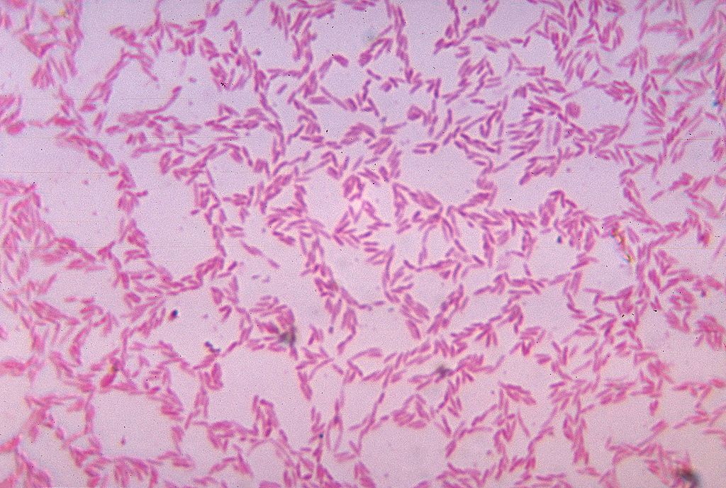
Figure 15.5.1 shows some of the cells of what has been called “the last human organ to be discovered.” This “organ” weighs about 200 grams (about 7 oz) and consists of a hundred trillion cells, yet scientists are only now beginning to learn everything it does and how it varies among individuals. What is it? It’s the mass of bacteria that live in our lower gastrointestinal tract.
Organs of the Lower Gastrointestinal Tract
Most of the bacteria that normally live in the lower gastrointestinal (GI) tract live in the large intestine. They have important and mutually beneficial relationships with the human organism. We provide them with a great place to live, and they provide us with many benefits, some of which you can read about below. Besides the large intestine and its complement of helpful bacteria, the lower GI tract also includes the small intestine. The latter is arguably the most important organ of the digestive system. It is where most chemical digestion and virtually all absorption of nutrients take place.
Small Intestine
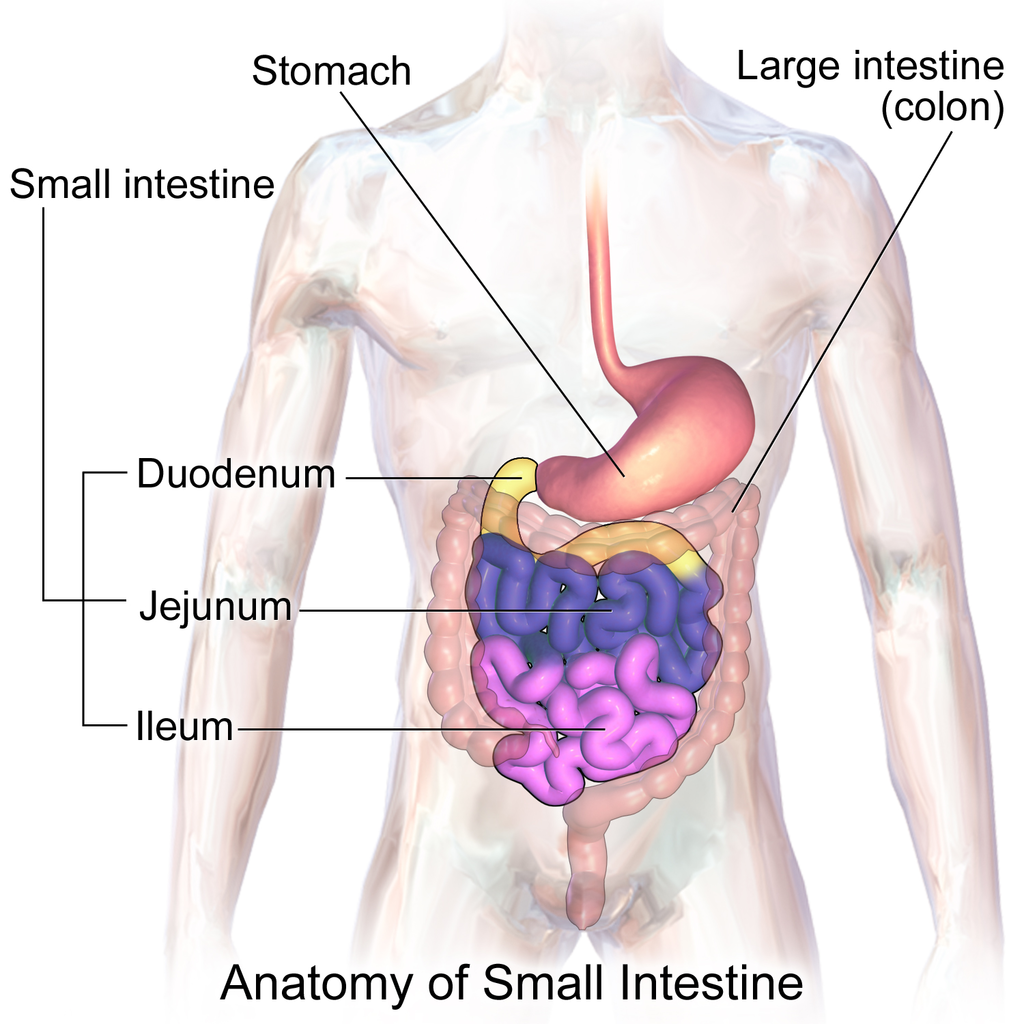
The small intestine (also called the small bowel or gut) is the part of the GI tract between the stomach and large intestine. Its average length in adults is 4.6 m in females and 6.9 m in males. It is approximately 2.5 to 3.0 cm in diameter. It is called “small” because it is much smaller in diameter than the large intestine. The internal surface area of the small intestine totals an average of about 30 m2. Structurally and functionally, the small intestine can be divided into three parts, called the duodenum, jejunum, and ileum, as shown in Figure 15.5.2 and described below.
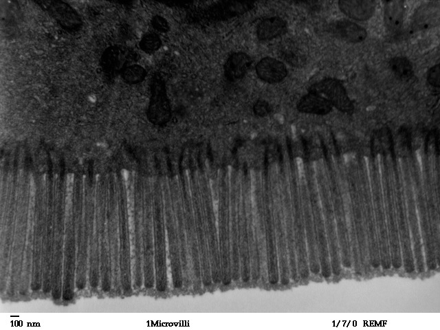
The mucosa lining the small intestine is highly enfolded and covered with the finger-like projections called villi. In fact, each square inch (about 6.5 cm2) of mucosa contains around 20 thousand villi. The individual cells on the surface of the villi also have many finger-like projections — the microvilli, shown in Figure 15.5.3. There are thought to be well over 17 billion microvilli per square centimetre of intestinal mucosa! All of these folds, villi, and microvilli greatly increase the surface area for chyme to come into contact with digestive enzymes, which coat the microvilli, as well as forming a tremendous surface area for the absorption of nutrients. Inside each of the villi is a network of tiny blood and lymph vessels that receive the absorbed nutrients and carry them away in the blood or lymph circulation. The wrinkles and projections in the intestinal mucosa also slow down the passage of chyme so there is more time for digestion and absorption to take place.
Duodenum
The duodenum is the first part of the small intestine, directly connected to the stomach. It is also the shortest part of the small intestine, averaging only about 25 cm (almost 10 in) in length in adults. Its main function is chemical digestion, and it is where most of the chemical digestion in the entire GI tract takes place.
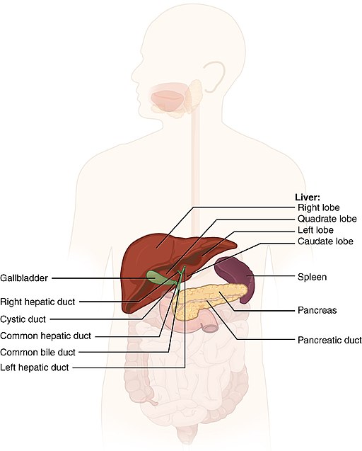
The duodenum receives partially digested, semi-liquid chyme from the stomach. It receives digestive enzymes and alkaline bicarbonate from the pancreas through the pancreatic duct, and it receives bile from the liver via the gallbladder through the common bile duct (see Figure 15.5.4). In addition, the lining of the duodenum secretes digestive enzymes and contains glands — called Brunner’s glands — that secrete mucous and bicarbonate. The bicarbonate from the pancreas and Brunner’s glands — along with bile from the liver — neutralize the highly acidic chyme after it enters the duodenum from the stomach. This is necessary because the digestive enzymes in the duodenum require a nearly neutral environment in order to work. The three major classes of compounds that undergo chemical digestion in the duodenum are carbohydrates, proteins, and lipids.
Digestion of Carbohydrates in the Duodenum
Complex carbohydrates (such as starches) are broken down by the digestive enzyme amylase from the pancreas to short-chain molecules consisting of just a few saccharides (that is, simple sugars). Disaccharides, including sucrose and lactose, are broken down to simple sugars by duodenal enzymes. Sucrase breaks down sucrose, and lactase (if present) breaks down lactose. Some carbohydrates are not digested in the duodenum, and they ultimately pass undigested to the large intestine, where they may be digested by intestinal bacteria.
Digestion of Proteins in the Duodenum
In the duodenum, the pancreatic enzymes trypsin and chymotrypsin cleave proteins into peptides. Then, these smaller molecules are broken down into amino acids. Their digestion is catalyzed by pancreatic enzymes called peptidases.
Digestion of Lipids in the Duodenum
Pancreatic lipase breaks down triglycerides into fatty acids and glycerol. Lipase works with the help of bile secreted by the liver and stored in the gallbladder. Bile salts attach to triglycerides to help them emulsify, or form smaller particles (called micelles) that can disperse through the watery contents of the duodenum. This increases access to the molecules by pancreatic lipase.
Jejunum
The jejunum is the middle part of the small intestine, connecting the duodenum and the ileum. The jejunum is about 2.5 m (about 8 ft) long. Its main function is absorption of the products of digestion, including sugars, amino acids, and fatty acids. Absorption occurs by simple diffusion (water and fatty acids), facilitated diffusion (the simple sugar fructose), or active transport (amino acids, small peptides, water-soluble vitamins, and most glucose). All nutrients are absorbed into the blood, except for fatty acids and fat-soluble vitamins, which are absorbed into the lymph. Although most nutrients are absorbed in the jejunum, there are a few exceptions:
- Iron is absorbed in the duodenum.
- Vitamin B12 and bile salts are absorbed in the ileum.
- Water and lipids are absorbed throughout the small intestine, including the duodenum and ileum in addition to the jejunum.
Ileum
The ileum is the third and final part of the small intestine, directly connected at its distal end to the large intestine. The ileum is about 3 m (almost 10 ft) long. Some cells in the lining of the ileum secrete enzymes that catalyze the final stages of digestion of any undigested protein and carbohydrate molecules. However, the main function of the ileum is to absorb vitamin B12 and bile salts. It also absorbs any other remaining nutrients that were not absorbed in the jejunum. All substances in chyme that remain undigested or unabsorbed by the time they reach the distal end of the ileum pass into the large intestine.
Large Intestine
The large intestine — also called the large bowel — is the last organ of the GI tract. In adults, it averages about 1.5 m (about 5ft) in length. It is shorter than the small intestine, but at least twice as wide, averaging about 6.5 cm (about 2.5 in) in diameter. Water is absorbed from chyme as it passes through the large intestine, turning the chyme into solid feces. Feces is stored in the large intestine until it leaves the body during defecation.
Parts of the Large Intestine
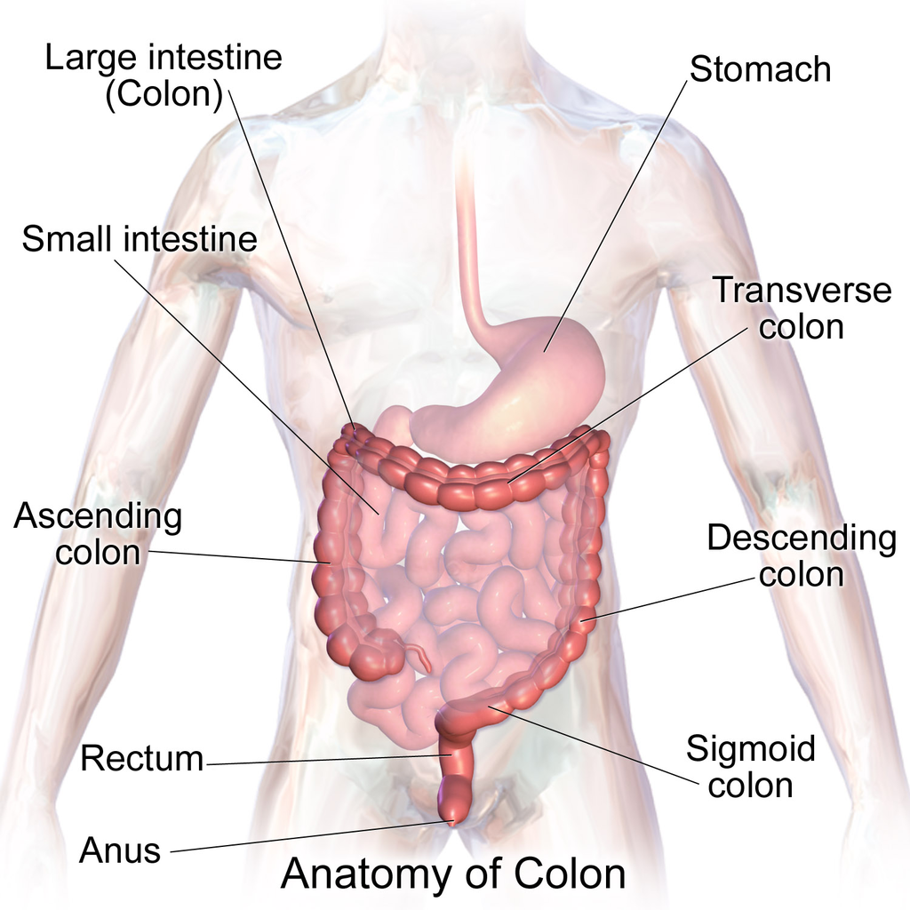
Like the small intestine, the large intestine can be divided into several parts, as shown in Figure 15.5.5. The large intestine begins at the end of the small intestine, where a valve separates the small and large intestines and regulates the movement of chyme into the large intestine. Most of the large intestine is called the colon. The first part of the colon, where chyme enters from the small intestine, is called the cecum. From the cecum, the colon continues upward as the ascending colon, travels across the upper abdomen as the transverse colon, and then continues downward as the descending colon. It then becomes a V-shaped region called the sigmoid colon, which is attached to the rectum. The rectum stores feces until elimination occurs. It transitions to the final part of the large intestine, called the anus, which has an opening to the outside of the body for feces to pass through.
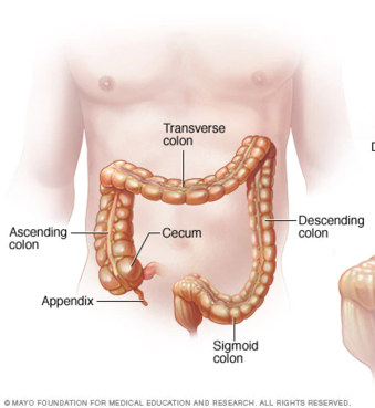
The diagram below (Figure 15.5.6) shows a projection from the cecum of the colon that is known as the appendix. The function of the appendix is somewhat uncertain, but it does not seem to be involved in digestion or absorption. It may play a role in immunity, and in the fetus, it seems to have an endocrine function, releasing hormones needed for homeostasis. Some biologists speculate that the appendix may also store a sample of the colon’s normal bacteria. If so, it may be able to repopulate the colon with the bacteria if illness or antibiotic medications deplete these microorganisms. Appendicitis, or infection and inflammation of the appendix, is a fairly common medical problem, typically resolved by surgical removal of the appendix (appendectomy). People who have had their appendix surgically removed do not seem to suffer any ill effects, so the organ is considered dispensable.
Functions of the Large Intestine
The removal of water from chyme to form feces starts in the ascending colon and continues throughout much of the length of the organ. Salts (such as sodium) are also removed from food wastes in the large intestine before the wastes are eliminated from the body. This allows salts — as well as water — to be recycled in the body.
The large intestine is also the site where huge numbers of beneficial bacteria ferment many unabsorbed materials in food waste. The bacterial breakdown of undigested polysaccharides produces nitrogen, carbon dioxide, methane, and other gases responsible for intestinal gas, or flatulence. These bacteria are particularly prevalent in the descending colon. Some of the bacteria also produce vitamins that are absorbed from the colon. The vitamins include vitamins B1 (thiamine), B2 (riboflavin), B7 (biotin), B12, and K. Another role of bacteria in the colon is an immune function. The bacteria may stimulate the immune system to produce antibodies that are effective against similar, but pathogenic, bacteria, thereby preventing infections. Still other roles played by bacteria in the large intestine include breaking down toxins before they can poison the body, producing substances that help prevent colon cancer, and inhibiting the growth of harmful bacteria.
Feature: My Human Body
Serotonin is a neurotransmitter with a wide variety of functions in the body. Sometimes called the “happy chemical,” it is used in the central nervous system to stabilize mood by contributing to a feeling of well being and happiness. While this “happy chemical” function is important, serotonin is also important for critical brain functions, including support of learning, memory, and reward structures and regulating sleep. Since serotonin’s main target is brain function, you may be surprised to learn that 90% of the human body’s supply of this neurotransmitter is located in the gastrointestinal tract, where it regulates intestinal movements.
The enteric nervous system (ENS), a division of the autonomic nervous system, is a mesh-like system of neurons that control GI function. Interestingly, the ENS can act independently from both the sympathetic/parasympathetic nervous systems and the brain/spinal cord, which is why some scientists refer to the ENS as the “second brain”. The ENS consists of over 500 million neurons (five times as many as in the spinal cord!) and lines the GI tract from the esophagus all the way to the anus. Serotonin is made in and secreted by the gut (small and large intestine) by enterochromaffin cells (also known as Kulchitsky cells) residing in the inner lining of the lower GI tract. While the main function of serotonin in the gut is to regulate digestion, it is also suspected to play a role in brain function — meaning that your gut may actually be partly dictating your mood! Another piece of evidence for the gut-dictating-your-mood theory is the nature vagus nerve, a fibrous visceral nerve, in which 90% of the fibres are dedicated to sending information too the brain, and only 10% of the fibres receiving information from the brain. Some treatments for depression actually involved electrical stimulation of the vagus nerve!
In a 2015 study by researchers at Caltech, it was shown that gut bacteria promote serotonin production by enterochromaffin cells. The study involved measuring the levels of serotonin levels in mice with normal gut bacteria, and then comparing that to the levels of serotonin in a population of mice with no gut bacteria. The germ-free mice were found to produce only 40% of the serotonin that the mice with normal gut flora were producing. Once the germ-free mice were re-colonized with normal gut flora, their production of serotonin returned to normal. This lead researchers to the conclusion that enterochromaffin cells depend on an interaction with gut bacterial flora to be able to produce necessary levels of serotonin.
15.5 Summary
- The lower GI tract includes the small intestine and large intestine. The small intestine is where most chemical digestion and virtually all absorption of nutrients occur. The large intestine contains huge numbers of beneficial bacteria and removes water and salts from food waste before it is eliminated.
- The small intestine consists of three parts: the duodenum, jejunum, and ileum. All three parts of the small intestine are lined with mucosa that is highly enfolded and covered with villi and microvilli, giving the small intestine a huge surface area for digestion and absorption.
- The duodenum secretes digestive enzymes, and also receives bile from the liver via the gallbladder and digestive enzymes and bicarbonate from the pancreas. These digestive substances neutralize acidic chyme and allow for the chemical digestion of carbohydrates, proteins, lipids, and nucleic acids in the duodenum.
- The jejunum carries out most of the absorption of nutrients in the small intestine, including the absorption of simple sugars, amino acids, fatty acids, and many vitamins.
- The ileum carries out any remaining digestion and absorption of nutrients, but its main function is to absorb vitamin B12 and bile salts.
- The large intestine consists of the colon (which, in turn, includes the cecum, ascending colon, transverse colon, descending colon, and sigmoid colon), rectum, and anus. The vestigial organ called the appendix is attached to the cecum of the colon.
- The main function of the large intestine is to remove water and salts from chyme for recycling within the body and eliminating the remaining solid feces from the body through the anus. The large intestine is also the site where trillions of bacteria help digest certain compounds, produce vitamins, stimulate the immune system, and break down toxins, among other important functions.
15.5 Review Questions
-
- How is the mucosa of the small intestine specialized for digestion and absorption?
- What digestive substances are secreted into the duodenum? What compounds in food do they help digest?
- What is the main function of the jejunum?
- What roles does the ileum play?
- How do beneficial bacteria in the large intestine help the human organism?
- When diarrhea occurs, feces leaves the body in a more liquid state than normal. What part of the digestive system do you think is involved in diarrhea? Explain your answer
- What causes intestinal gas, or flatulence?
15.5 Explore More
Why do we pass gas? – Purna Kashyap, TED-Ed, 2014.
What causes constipation? – Heba Shaheed, TED-Ed, 2018.
Attributions
Figure 15.5.1
1024px-Bacteroides_biacutis_01 by CDC/Dr. V.R. Dowell, Jr. Public Health Image Library (PHIL) ID#3087) on Wikimedia Commons is in the public domain (https://en.wikipedia.org/wiki/public_domain).
Figure 15.5.2
Blausen_0817_SmallIntestine_Anatomy by BruceBlaus on Wikimedia Commons is used under a CC BY 3.0 (https://creativecommons.org/licenses/by/3.0) license.
Figure 15.5.3
Human_jejunum_microvilli_1_-_TEM by Louisa Howard, Katherine Connollly / E. M. Facility, Dartmouth, on Wikimedia Commons is in the public domain (https://en.wikipedia.org/wiki/public_domain).
Figure 15.5.4
512px-2422_Accessory_Organs by OpenStax College on Wikimedia Commons is used under a CC BY 3.0 (https://creativecommons.org/licenses/by/3.0) license.
Figure 15.5.5
1024px-Blausen_0603_LargeIntestine_Anatomy by BruceBlaus on Wikimedia Commons is used under a CC BY 3.0 (https://creativecommons.org/licenses/by/3.0) license.
Figure 15.5.6
512px-Ds00070_an01934_im00887_divert_s_gif.webp (1) by Lfreeman04 on Wikimedia Commons is used under a CC BY-SA 4.0 (https://creativecommons.org/licenses/by-sa/4.0) license.
References
Betts, J. G., Young, K.A., Wise, J.A., Johnson, E., Poe, B., Kruse, D.H., Korol, O., Johnson, J.E., Womble, M., DeSaix, P. (2013, June 19). Figure 23.24 Accessory organs [digital image]. In Anatomy and Physiology (Section 23.6). OpenStax. https://openstax.org/books/anatomy-and-physiology/pages/23-6-accessory-organs-in-digestion-the-liver-pancreas-and-gallbladder
Blausen.com Staff. (2014). Medical gallery of Blausen Medical 2014. WikiJournal of Medicine 1 (2). DOI:10.15347/wjm/2014.010. ISSN 2002-4436. /
TED-Ed. (2018, May 7). What causes constipation? – Heba Shaheed. YouTube. https://www.youtube.com/watch?v=0IVO50DuMCs&feature=youtu.be
TED-Ed. (2014, September 8). Why do we pass gas? – Purna Kashyap. YouTube. https://www.youtube.com/watch?v=GTvnjaUU6Xk&feature=youtu.be
Yano, J. M., Yu, K., Donaldson, G. P., Shastri, G. G., Ann, P., Ma, L., Nagler, C. R., Ismagilov, R. F., Mazmanian, S. K., & Hsiao, E. Y. (2015). Indigenous bacteria from the gut microbiota regulate host serotonin biosynthesis. Cell, 161(2), 264–276. https://doi.org/10.1016/j.cell.2015.02.047
Image shows the four stages of Meiosis II: In Prophase II, the dyads condense, the nuclear envelope fragments and spindle fibers form. In Metaphase II, the dyads align at the center of the cell. In Anaphase II, the dyads are split pulled to opposite ends of the cell. In Telophase II the chromatids decondense, nuclear envelope reforms and spindle fibers fragment.
Image shows a Lego (TM) representation of Gregor Mendel with his plants.
A stem cell can become any type of body cell based on gene regulation. Types of cells a stem cell can become include, but are not limited to: Sex cells, muscle cells, fat cells, immune cells, bone cells, epithelial cells, nervous cells, and blood cells.
Image shows the process of crossing over. A pair of homologous chromosomes are side by side. They then overlap the ends of their structures and trades these sections, attaching some maternal DNA onto the paternal chromosome, and some paternal DNA onto the maternal chromosome. The result is that, within the homologous pair of chromosomes, each of the four pieces of DNA, destined to enter 4 separate cells, is now consists of a unique mix of genes.
Polly Who?
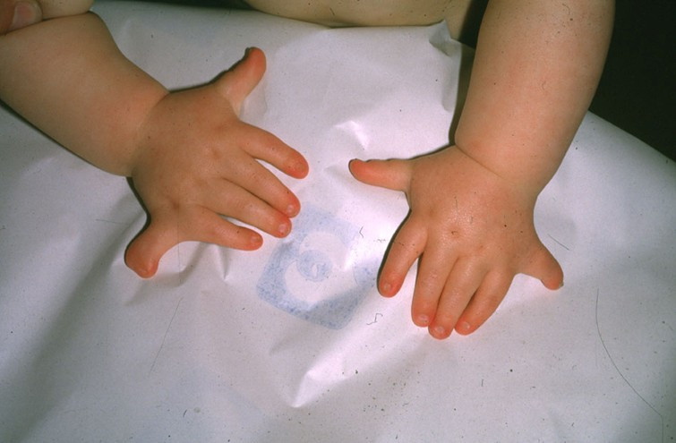
Each hand in the Figure 5.15.1 photo has an extra pinky finger. This is a condition called polydactyly, which literally means "many digits." People with polydactyly may have extra fingers and/or toes, and the condition may affect just one hand or foot, or both hands and feet. Polydactyly is often genetic in origin and may be part of a genetic disorder associated with other abnormalities.
What Are Genetic Disorders?
Genetic disorders are diseases, syndromes, or other abnormal conditions caused by mutations in one or more genes, or by chromosomal alterations. Genetic disorders are typically present at birth, but they should not be confused with congenital disorders, a category that includes any disorder present at birth, regardless of cause. Some congenital disorders are not caused by genetic mutations or chromosomal alterations. Instead, they are caused by problems that arise during embryonic or fetal development, or during the process of birth. An example of a nongenetic congenital disorder is fetal alcohol syndrome. This is a collection of birth defects, including facial anomalies and intellectual disability, caused by maternal alcohol consumption during pregnancy.
Genetic Disorders Caused by Mutations
Table 5.15.1 lists several genetic disorders caused by mutations in just one gene. Some of the disorders are caused by mutations in autosomal genes, others by mutations in X-linked genes. Which disorders would you expect to be more common in males than females?
| Genetic Disorder | Direct Effect of Mutation | Signs and Symptoms of the Disorder | Mode of Inheritance |
|---|---|---|---|
| Marfan syndrome | Defective protein in connective tissue | Heart and bone defects and unusually long, slender limbs and fingers | Autosomal dominant |
| Sickle cell anemia | Abnormal hemoglobin protein in red blood cells | Sickle-shaped red blood cells that clog tiny blood vessels, causing pain and damaging organs and joints | Autosomal recessive |
| Hypophosphatemic (Vitamin D-resistant) rickets | Lack of a substance needed for bones to absorb minerals | Soft bones that easily become deformed, leading to bowed legs and other skeletal deformities | X-linked dominant |
| Hemophilia A | Reduced activity of a protein needed for blood clotting | Internal and external bleeding that occurs easily and is difficult to control | X-linked recessive |
Very few genetic disorders are controlled by dominant mutant [pHypophosphatemicb_glossary id="2119"]alleles[/pb_glossary]. A dominant allele is expressed in every individual who inherits even one copy of it. If it causes a serious disorder, affected people may die young and fail to reproduce. Therefore, the mutant dominant allele is likely to die out of a population.
A recessive mutant allele — such as the allele that causes sickle cell anemia or cystic fibrosis — is not expressed in people who inherit just one copy of it. These people are called carriers. They do not have the disorder themselves, but they carry the mutant allele and their offspring can inherit it. Thus, the allele is likely to pass on to the next generation, rather than die out.
Genetic Disorders Caused by Chromosomal Alterations
Mistakes may occur during meiosis that result in nondisjunction. This is the failure of replicated chromosomes to separate properly during meiosis. Some of the resulting gametes will be missing all or part of a chromosome, while others will have an extra copy of all or part of the chromosome. If such gametes are fertilized and form zygotes, they usually do not survive. If they do survive, the individuals are likely to have serious genetic disorders.
Table 5.15.2 lists several genetic disorders that are caused by abnormal numbers of chromosomes. Most chromosomal disorders involve the X chromosome. The X and Y chromosomes are the only chromosome pair in which the two chromosomes are very different in size. This explains why nondisjunction tends to occur more frequently in sex chromosomes than in autosomes.
| Genetic Disorder | Genotype | Phenotypic Effects |
|---|---|---|
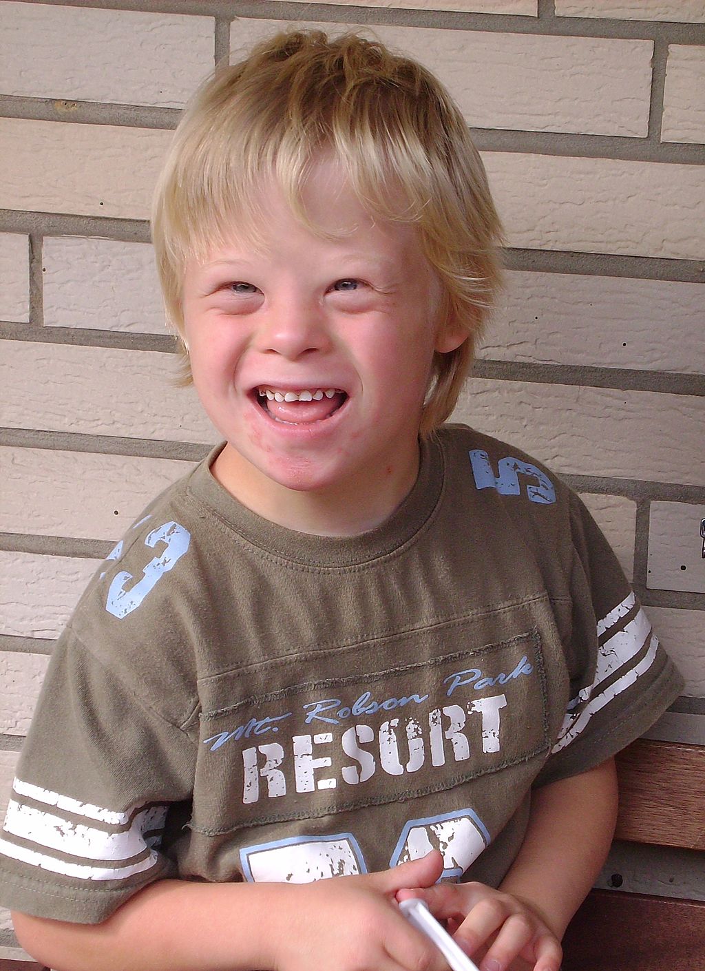
| Down syndrome | Extra copy (complete or partial) of chromosome 21 (see Figure 5.15.3) | Developmental delays, distinctive facial appearance, and other abnormalities (see Figure 5.15.2) |
| Turner syndrome | One X chromosome but no other sex chromosome (XO) | Female with short height and infertility(inability to reproduce) |
| Triple X syndrome | Three X chromosomes (XXX) | Female with mild developmental delays and menstrual irregularities |
| Klinefelter syndrome | One Y chromosome and two or more X chromosomes (XXY, XXXY) | Male with problems in sexual development and reduced levels of the male hormone testosterone |
 |
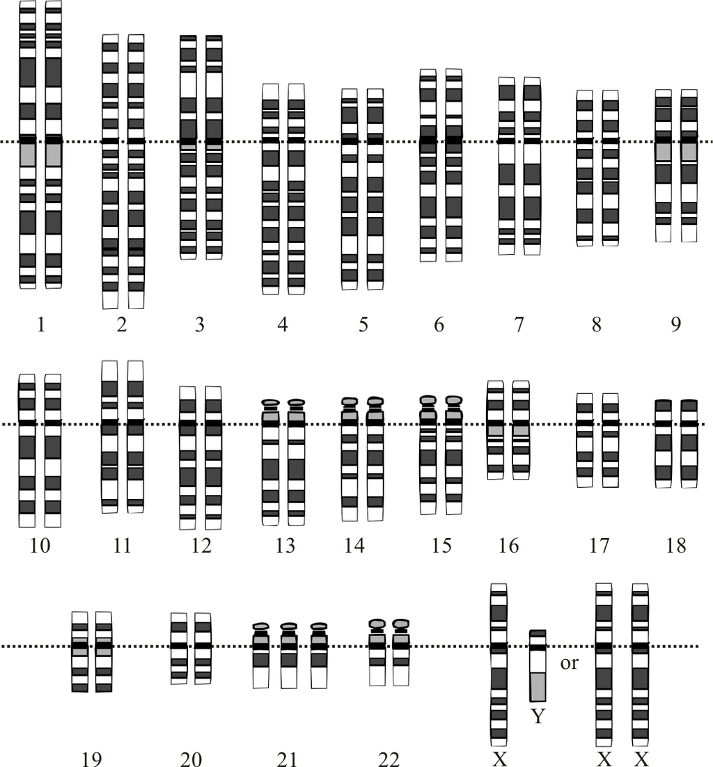 |
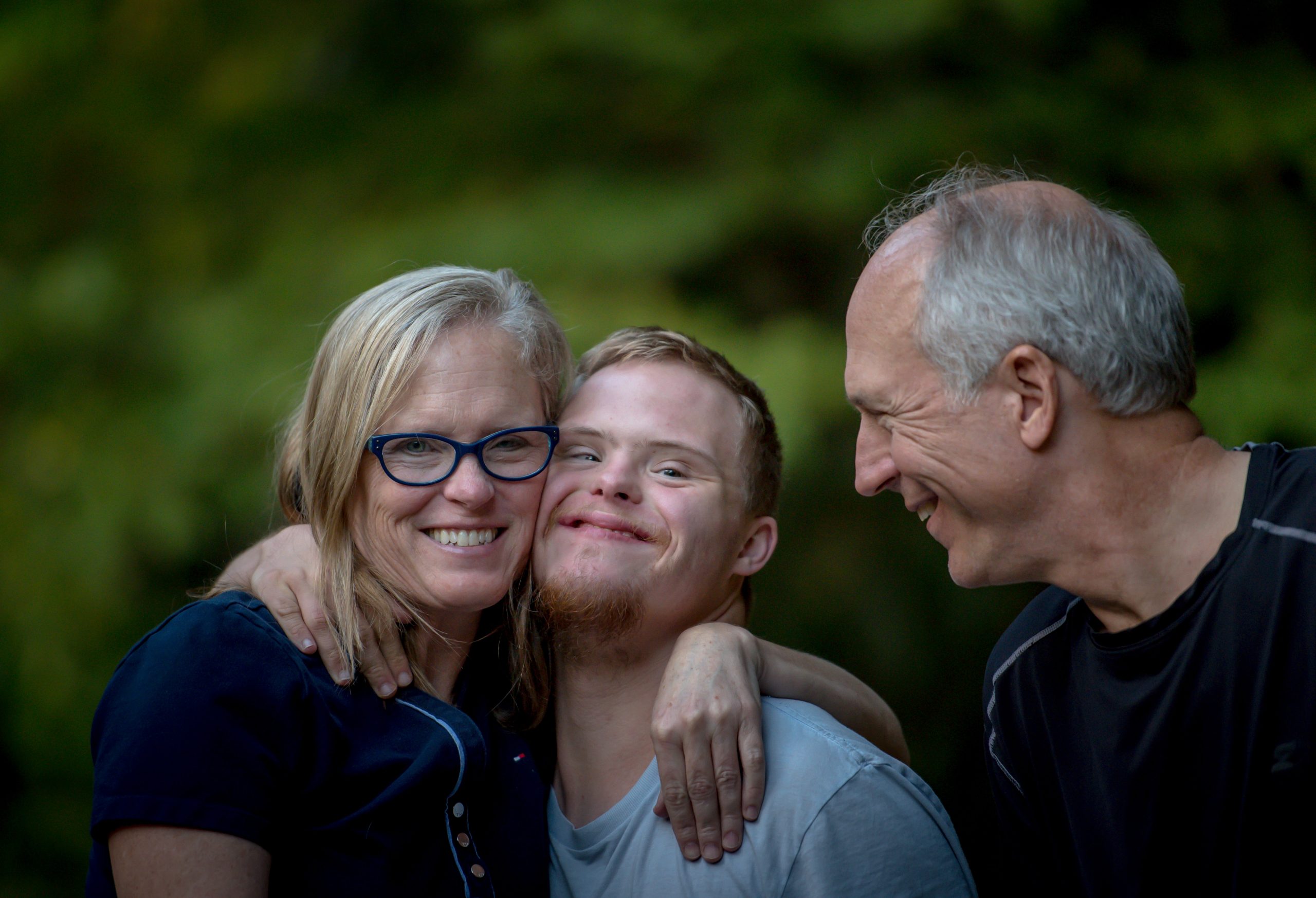
A karyotype is a picture of a cell's chromosomes. In Figure 5.15.3, note the extra chromosome 21. In Figure 5.15.2, a young man with Down syndrome exhibits the characteristic facial appearance.
Diagnosing and Treating Genetic Disorders
A genetic disorder that is caused by a mutation can be inherited. Therefore, people with a genetic disorder in their family may be concerned about having children with the disorder. A genetic counselor can help them understand the risks of their children being affected. If they decide to have children, they may be advised to have prenatal (“before birth”) testing to see if the fetus has any genetic abnormalities. One method of prenatal testing is amniocentesis. In this procedure, a few fetal cells are extracted from the fluid surrounding the fetus in utero, and the fetal chromosomes are examined. Down syndrome and other chromosomal alterations can be detected in this way.
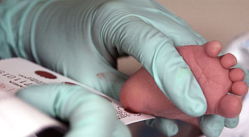
The symptoms of genetic disorders can sometimes be treated or prevented. In the genetic disorder called phenylketonuria (PKU), for example, the amino acid phenylalanine builds up in the body to harmful levels. PKU is caused by a mutation in a gene that normally codes for an enzyme needed to break down phenylalanine. When a person with PKU consumes foods high in phenylalanine (including many high-protein foods), the buildup of PKU can lead to serious health problems. In infants and young children, the build-up of phenylalanine can cause intellectual disability and delayed development, along with other serious problems. All babies in Canada and the United States and many other countries are screened for PKU soon after birth. As shown in Figure 5.15.3, the PKU test involves collecting a small amount of blood from the infant, typically from the heel using a small lancet. The blood is collected on a special type of filter paper and then brought to a laboratory for analysis. If PKU is diagnosed, the infant can be fed a low-phenylalanine diet, which prevents the buildup of phenylalanine and the health problems associated with it, including intellectual disability. As long as a low-phenylalanine diet is followed throughout life, most symptoms of the disorder can be prevented.
Curing Genetic Disorders
Cures for genetic disorders are still in the early stages of development. One potential cure is gene therapy. Gene therapy is an experimental technique that uses genes to treat or prevent disease. In gene therapy, normal genes are introduced into cells to compensate for abnormal genes. If a mutated gene causes a necessary protein to be nonfunctional or missing, gene therapy may be able to introduce a normal copy of the gene to produce the needed functional protein.
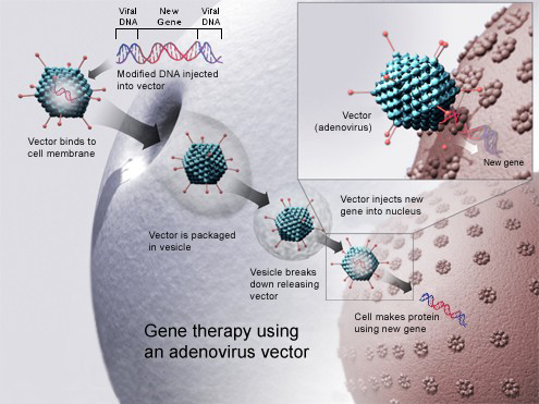
A gene inserted directly into a cell usually does not function, so a carrier called a vector is genetically engineered to deliver the gene (see Figure 5.15.4 illustration). Certain viruses, such as adenoviruses, are often used as vectors. They can deliver the new gene by infecting cells. The viruses are modified so they do not cause disease when used in people. If the treatment is successful, the new gene delivered by the vector will allow the synthesis of a functioning protein. Researchers still must overcome many technical challenges before gene therapy will be a practical approach to curing genetic disorders.
Feature: Human Biology in the News
Down syndrome is the most common genetic cause of intellectual disability. It occurs in about one in every 700 live births, and it currently affects nearly half a million Americans. Until recently, scientists thought that the changes leading to intellectual disability in people with Down syndrome all happen before birth.
Even more recently, researchers discovered a genetic abnormality that affects brain development in people with Down syndrome throughout childhood and into adulthood. The newly discovered genetic abnormality changes communication between nerve cells in the brain, resulting in slower transmission of nerve impulses. This finding may eventually allow the development of strategies to promote brain functioning in Down syndrome patients, and it may also be applicable to other development disabilities, such as autism. The results of this promising study were published in the March 16, 2016 issue of the scientific journal Neuron.
5.15 Summary
- Genetic disorders are diseases, syndromes, or other abnormal conditions that are caused by mutations in one or more genes, or by chromosomal alterations.
- Examples of genetic disorders caused by single-gene mutations include Marfan syndrome (autosomal dominant), sickle cell anemia (autosomal recessive), vitamin D-resistant rickets (X-linked dominant), and hemophilia A (X-linked recessive). Very few genetic disorders are caused by dominant mutations because these alleles are less likely to be passed on to successive generations.
- Nondisjunction is the failure of replicated chromosomes to separate properly during meiosis. This may result in genetic disorders caused by abnormal numbers of chromosomes. An example is Down syndrome, in which the individual inherits an extra copy of chromosome 21. Most chromosomal disorders involve the X chromosome. An example is Klinefelter's syndrome (XXY, XXXY).
- Prenatal genetic testing (by amniocentesis, for example) can detect chromosomal alterations in utero. The symptoms of some genetic disorders can be treated or prevented. For example, symptoms of phenylketonuria (PKU) can be prevented by following a low-phenylalanine diet throughout life.
- Cures for genetic disorders are still in the early stages of development. One potential cure is gene therapy, in which normal genes are introduced into cells by a vector such as a virus to compensate for mutated genes.
5.15 Review Questions
- Define genetic disorder.
- Identify three genetic disorders caused by mutations in a single gene.
- Why are single-gene genetic disorders more commonly controlled by recessive than dominant mutant alleles?
- What is nondisjunction? Why can it cause genetic disorders?
- Explain why genetic disorders caused by abnormal numbers of chromosomes most often involve the X chromosome.
- How is Down syndrome detected in utero?
- Use the example of PKU to illustrate how the symptoms of a genetic disorder can sometimes be prevented.
- Explain how gene therapy works.
- Compare and contrast genetic disorders and congenital disorders.
- Explain why parents that do not have Down syndrome can have a child with Down syndrome.
- Hemophilia A and Turner’s syndrome both involve problems with the X chromosome. In terms of how the X chromosome is affected, what is the major difference between these two types of disorders?
- Can you be a carrier of Marfan syndrome and not have the disorder? Explain your answer.
5.15 Explore More
https://www.youtube.com/watch?v=6tw_JVz_IEc
How CRISPR lets you edit DNA - Andrea M. Henle, TED-Ed, 2019.
https://youtu.be/1BXYSGepx7Q
What you need to know about CRISPR | Ellen Jorgensen, TED, 2016.
https://youtu.be/nOHbn8Q1fBM
The ethical dilemma of designer babies | Paul Knoepfler, TED, 2017.
Attributions
Figure 5.15.1
Polydactyly_ECS by Baujat G, Le Merrer M. on Wikimedia Commons is used under a CC BY 2.0 (https://creativecommons.org/licenses/by/2.0) license.
Figure 5.15.2
Downs/ All the Family [photo] by Nathan Anderson on Unsplash is used under the Unsplash License (https://unsplash.com/license).
Figure 5.15.3
Phenylketonuria_testing by U.S. Air Force photo/Staff Sgt Eric T. Sheler in the US Air Force National Archives on Wikimedia Commons is in the public domain (https://en.wikipedia.org/wiki/Public_domain).
Figure 5.15.4
Gene_therapy by National Institutes of Health (NIH) on Wikimedia Commons is in the public domain (https://en.wikipedia.org/wiki/Public_domain).
Figure 5.15.5
Boy_with_Down_Syndrome by Vanellus Foto on Wikimedia Commons is used under a CC BY-SA 3.0 (https://creativecommons.org/licenses/by-sa/3.0/deed.en) license.
References
Baujat, G., Le Merrer, M. (2007, January 23). Ellis-Van Creveld syndrome. Orphanet Journal of Rare Diseases, 2, 27. https://doi.org/10.1186/1750-1172-2-27
Hecht, M. (2019, June 26). What is polydactyly? [online article]. Healthline. https://www.healthline.com/health/polydactyly
Genetic and Rare Diseases Information Center (GARD). (2016). Hypophosphatemic rickets (previously called vitamin D-resistant rickets) [online article]. NIH. https://rarediseases.info.nih.gov/diseases/6735/hypophosphatemic-rickets [last updated 7/1/2020]
Mayo Clinic Staff. (n.d.). Cystic fibrosis [online article]. MayoClinic.org. https://www.mayoclinic.org/diseases-conditions/cystic-fibrosis/symptoms-causes/syc-20353700
Mayo Clinic Staff. (n.d.). Down syndrome [online article]. MayoClinic.org. https://www.mayoclinic.org/diseases-conditions/down-syndrome/diagnosis-treatment/drc-20355983
Mayo Clinic Staff. (n.d.). Hemophilia [online article]. MayoClinic.org. https://www.mayoclinic.org/diseases-conditions/hemophilia/symptoms-causes/syc-20373327
Mayo Clinic Staff. (n.d.). Klinefelter syndrome [online article]. MayoClinic.org. https://www.mayoclinic.org/diseases-conditions/klinefelter-syndrome/symptoms-causes/syc-20353949
Mayo Clinic Staff. (n.d.). Marfan syndrome [online article]. MayoClinic.org. https://www.mayoclinic.org/diseases-conditions/marfan-syndrome/symptoms-causes/syc-20350782
Mayo Clinic Staff. (n.d.). Phenylketonuria (PKU) [online article]. MayoClinic.org. https://www.mayoclinic.org/diseases-conditions/phenylketonuria/symptoms-causes/syc-20376302
Mayo Clinic Staff. (n.d.). Sickle cell anemia [online article]. MayoClinic.org. https://www.mayoclinic.org/diseases-conditions/sickle-cell-anemia/symptoms-causes/syc-20355876
Mayo Clinic Staff. (n.d.). Turner syndrome [online article]. MayoClinic.org. https://www.mayoclinic.org/diseases-conditions/turner-syndrome/symptoms-causes/syc-20360782
Mayo Clinic Staff. (n.d.). Triple X syndrome [online article]. MayoClinic.org. https://www.mayoclinic.org/diseases-conditions/triple-x-syndrome/symptoms-causes/syc-20350977
National Center on Birth Defects and Developmental Disabilities. (2020). Fetal alcohol spectrum disorders (FASDs): Basics about FASDs [webpage]. Centers for Disease Control and Prevention (CDC). https://www.cdc.gov/ncbddd/fasd/facts.html
TED-Ed. (2019, January 24). How CRISPR lets you edit DNA - Andrea M. Henle. YouTube. https://www.youtube.com/watch?v=6tw_JVz_IEc
TED. (2016, October 24). What you need to know about CRISPR | Ellen Jorgensen. YouTube. https://www.youtube.com/watch?v=1BXYSGepx7Q&feature=youtu.be
TED. (2017, February 10). The ethical dilemma of designer babies | Paul Knoepfler. YouTube. https://www.youtube.com/watch?v=nOHbn8Q1fBM&t=3s
Created by: CK-12/Adapted by Christine Miller

Polly Who?
Each hand in the Figure 5.15.1 photo has an extra pinky finger. This is a condition called polydactyly, which literally means "many digits." People with polydactyly may have extra fingers and/or toes, and the condition may affect just one hand or foot, or both hands and feet. Polydactyly is often genetic in origin and may be part of a genetic disorder associated with other abnormalities.
What Are Genetic Disorders?
Genetic disorders are diseases, syndromes, or other abnormal conditions caused by mutations in one or more genes, or by chromosomal alterations. Genetic disorders are typically present at birth, but they should not be confused with congenital disorders, a category that includes any disorder present at birth, regardless of cause. Some congenital disorders are not caused by genetic mutations or chromosomal alterations. Instead, they are caused by problems that arise during embryonic or fetal development, or during the process of birth. An example of a nongenetic congenital disorder is fetal alcohol syndrome. This is a collection of birth defects, including facial anomalies and intellectual disability, caused by maternal alcohol consumption during pregnancy.
Genetic Disorders Caused by Mutations
Table 5.15.1 lists several genetic disorders caused by mutations in just one gene. Some of the disorders are caused by mutations in autosomal genes, others by mutations in X-linked genes. Which disorders would you expect to be more common in males than females?
| Genetic Disorder | Direct Effect of Mutation | Signs and Symptoms of the Disorder | Mode of Inheritance |
|---|---|---|---|
| Marfan syndrome | Defective protein in connective tissue | Heart and bone defects and unusually long, slender limbs and fingers | Autosomal dominant |
| Sickle cell anemia | Abnormal hemoglobin protein in red blood cells | Sickle-shaped red blood cells that clog tiny blood vessels, causing pain and damaging organs and joints | Autosomal recessive |
| Hypophosphatemic (Vitamin D-resistant) rickets | Lack of a substance needed for bones to absorb minerals | Soft bones that easily become deformed, leading to bowed legs and other skeletal deformities | X-linked dominant |
| Hemophilia A | Reduced activity of a protein needed for blood clotting | Internal and external bleeding that occurs easily and is difficult to control | X-linked recessive |
Very few genetic disorders are controlled by dominant mutant [pHypophosphatemicb_glossary id="2119"]alleles[/pb_glossary]. A dominant allele is expressed in every individual who inherits even one copy of it. If it causes a serious disorder, affected people may die young and fail to reproduce. Therefore, the mutant dominant allele is likely to die out of a population.
A recessive mutant allele — such as the allele that causes sickle cell anemia or cystic fibrosis — is not expressed in people who inherit just one copy of it. These people are called carriers. They do not have the disorder themselves, but they carry the mutant allele and their offspring can inherit it. Thus, the allele is likely to pass on to the next generation, rather than die out.
Genetic Disorders Caused by Chromosomal Alterations
Mistakes may occur during meiosis that result in nondisjunction. This is the failure of replicated chromosomes to separate properly during meiosis. Some of the resulting gametes will be missing all or part of a chromosome, while others will have an extra copy of all or part of the chromosome. If such gametes are fertilized and form zygotes, they usually do not survive. If they do survive, the individuals are likely to have serious genetic disorders.
Table 5.15.2 lists several genetic disorders that are caused by abnormal numbers of chromosomes. Most chromosomal disorders involve the X chromosome. The X and Y chromosomes are the only chromosome pair in which the two chromosomes are very different in size. This explains why nondisjunction tends to occur more frequently in sex chromosomes than in autosomes.
| Genetic Disorder | Genotype | Phenotypic Effects |
|---|---|---|
| Down syndrome | Extra copy (complete or partial) of chromosome 21 (see Figure 5.15.3) | Developmental delays, distinctive facial appearance, and other abnormalities (see Figure 5.15.2) |
| Turner syndrome | One X chromosome but no other sex chromosome (XO) | Female with short height and infertility(inability to reproduce) |
| Triple X syndrome | Three X chromosomes (XXX) | Female with mild developmental delays and menstrual irregularities |
| Klinefelter syndrome | One Y chromosome and two or more X chromosomes (XXY, XXXY) | Male with problems in sexual development and reduced levels of the male hormone testosterone |
 |
 |
A karyotype is a picture of a cell's chromosomes. In Figure 5.15.3, note the extra chromosome 21. In Figure 5.15.2, a young man with Down syndrome exhibits the characteristic facial appearance.
Diagnosing and Treating Genetic Disorders
A genetic disorder that is caused by a mutation can be inherited. Therefore, people with a genetic disorder in their family may be concerned about having children with the disorder. A genetic counselor can help them understand the risks of their children being affected. If they decide to have children, they may be advised to have prenatal (“before birth”) testing to see if the fetus has any genetic abnormalities. One method of prenatal testing is amniocentesis. In this procedure, a few fetal cells are extracted from the fluid surrounding the fetus in utero, and the fetal chromosomes are examined. Down syndrome and other chromosomal alterations can be detected in this way.

The symptoms of genetic disorders can sometimes be treated or prevented. In the genetic disorder called phenylketonuria (PKU), for example, the amino acid phenylalanine builds up in the body to harmful levels. PKU is caused by a mutation in a gene that normally codes for an enzyme needed to break down phenylalanine. When a person with PKU consumes foods high in phenylalanine (including many high-protein foods), the buildup of PKU can lead to serious health problems. In infants and young children, the build-up of phenylalanine can cause intellectual disability and delayed development, along with other serious problems. All babies in Canada and the United States and many other countries are screened for PKU soon after birth. As shown in Figure 5.15.3, the PKU test involves collecting a small amount of blood from the infant, typically from the heel using a small lancet. The blood is collected on a special type of filter paper and then brought to a laboratory for analysis. If PKU is diagnosed, the infant can be fed a low-phenylalanine diet, which prevents the buildup of phenylalanine and the health problems associated with it, including intellectual disability. As long as a low-phenylalanine diet is followed throughout life, most symptoms of the disorder can be prevented.
Curing Genetic Disorders
Cures for genetic disorders are still in the early stages of development. One potential cure is gene therapy. Gene therapy is an experimental technique that uses genes to treat or prevent disease. In gene therapy, normal genes are introduced into cells to compensate for abnormal genes. If a mutated gene causes a necessary protein to be nonfunctional or missing, gene therapy may be able to introduce a normal copy of the gene to produce the needed functional protein.
A gene inserted directly into a cell usually does not function, so a carrier called a vector is genetically engineered to deliver the gene (see Figure 5.15.4 illustration). Certain viruses, such as adenoviruses, are often used as vectors. They can deliver the new gene by infecting cells. The viruses are modified so they do not cause disease when used in people. If the treatment is successful, the new gene delivered by the vector will allow the synthesis of a functioning protein. Researchers still must overcome many technical challenges before gene therapy will be a practical approach to curing genetic disorders.

Feature: Human Biology in the News

Down syndrome is the most common genetic cause of intellectual disability. It occurs in about one in every 700 live births, and it currently affects nearly half a million Americans. Until recently, scientists thought that the changes leading to intellectual disability in people with Down syndrome all happen before birth.
Even more recently, researchers discovered a genetic abnormality that affects brain development in people with Down syndrome throughout childhood and into adulthood. The newly discovered genetic abnormality changes communication between nerve cells in the brain, resulting in slower transmission of nerve impulses. This finding may eventually allow the development of strategies to promote brain functioning in Down syndrome patients, and it may also be applicable to other development disabilities, such as autism. The results of this promising study were published in the March 16, 2016 issue of the scientific journal Neuron.
5.15 Summary
- Genetic disorders are diseases, syndromes, or other abnormal conditions that are caused by mutations in one or more genes, or by chromosomal alterations.
- Examples of genetic disorders caused by single-gene mutations include Marfan syndrome (autosomal dominant), sickle cell anemia (autosomal recessive), vitamin D-resistant rickets (X-linked dominant), and hemophilia A (X-linked recessive). Very few genetic disorders are caused by dominant mutations because these alleles are less likely to be passed on to successive generations.
- Nondisjunction is the failure of replicated chromosomes to separate properly during meiosis. This may result in genetic disorders caused by abnormal numbers of chromosomes. An example is Down syndrome, in which the individual inherits an extra copy of chromosome 21. Most chromosomal disorders involve the X chromosome. An example is Klinefelter's syndrome (XXY, XXXY).
- Prenatal genetic testing (by amniocentesis, for example) can detect chromosomal alterations in utero. The symptoms of some genetic disorders can be treated or prevented. For example, symptoms of phenylketonuria (PKU) can be prevented by following a low-phenylalanine diet throughout life.
- Cures for genetic disorders are still in the early stages of development. One potential cure is gene therapy, in which normal genes are introduced into cells by a vector such as a virus to compensate for mutated genes.
5.15 Review Questions
- Define genetic disorder.
- Identify three genetic disorders caused by mutations in a single gene.
- Why are single-gene genetic disorders more commonly controlled by recessive than dominant mutant alleles?
- What is nondisjunction? Why can it cause genetic disorders?
- Explain why genetic disorders caused by abnormal numbers of chromosomes most often involve the X chromosome.
- How is Down syndrome detected in utero?
- Use the example of PKU to illustrate how the symptoms of a genetic disorder can sometimes be prevented.
- Explain how gene therapy works.
- Compare and contrast genetic disorders and congenital disorders.
- Explain why parents that do not have Down syndrome can have a child with Down syndrome.
- Hemophilia A and Turner’s syndrome both involve problems with the X chromosome. In terms of how the X chromosome is affected, what is the major difference between these two types of disorders?
- Can you be a carrier of Marfan syndrome and not have the disorder? Explain your answer.
5.15 Explore More
https://www.youtube.com/watch?v=6tw_JVz_IEc
How CRISPR lets you edit DNA - Andrea M. Henle, TED-Ed, 2019.
https://youtu.be/1BXYSGepx7Q
What you need to know about CRISPR | Ellen Jorgensen, TED, 2016.
https://youtu.be/nOHbn8Q1fBM
The ethical dilemma of designer babies | Paul Knoepfler, TED, 2017.
Attributions
Figure 5.15.1
Polydactyly_ECS by Baujat G, Le Merrer M. on Wikimedia Commons is used under a CC BY 2.0 (https://creativecommons.org/licenses/by/2.0) license.
Figure 5.15.2
Downs/ All the Family [photo] by Nathan Anderson on Unsplash is used under the Unsplash License (https://unsplash.com/license).
Figure 5.15.3
Phenylketonuria_testing by U.S. Air Force photo/Staff Sgt Eric T. Sheler in the US Air Force National Archives on Wikimedia Commons is in the public domain (https://en.wikipedia.org/wiki/Public_domain).
Figure 5.15.4
Gene_therapy by National Institutes of Health (NIH) on Wikimedia Commons is in the public domain (https://en.wikipedia.org/wiki/Public_domain).
Figure 5.15.5
Boy_with_Down_Syndrome by Vanellus Foto on Wikimedia Commons is used under a CC BY-SA 3.0 (https://creativecommons.org/licenses/by-sa/3.0/deed.en) license.
References
Baujat, G., Le Merrer, M. (2007, January 23). Ellis-Van Creveld syndrome. Orphanet Journal of Rare Diseases, 2, 27. https://doi.org/10.1186/1750-1172-2-27
Hecht, M. (2019, June 26). What is polydactyly? [online article]. Healthline. https://www.healthline.com/health/polydactyly
Genetic and Rare Diseases Information Center (GARD). (2016). Hypophosphatemic rickets (previously called vitamin D-resistant rickets) [online article]. NIH. https://rarediseases.info.nih.gov/diseases/6735/hypophosphatemic-rickets [last updated 7/1/2020]
Mayo Clinic Staff. (n.d.). Cystic fibrosis [online article]. MayoClinic.org. https://www.mayoclinic.org/diseases-conditions/cystic-fibrosis/symptoms-causes/syc-20353700
Mayo Clinic Staff. (n.d.). Down syndrome [online article]. MayoClinic.org. https://www.mayoclinic.org/diseases-conditions/down-syndrome/diagnosis-treatment/drc-20355983
Mayo Clinic Staff. (n.d.). Hemophilia [online article]. MayoClinic.org. https://www.mayoclinic.org/diseases-conditions/hemophilia/symptoms-causes/syc-20373327
Mayo Clinic Staff. (n.d.). Klinefelter syndrome [online article]. MayoClinic.org. https://www.mayoclinic.org/diseases-conditions/klinefelter-syndrome/symptoms-causes/syc-20353949
Mayo Clinic Staff. (n.d.). Marfan syndrome [online article]. MayoClinic.org. https://www.mayoclinic.org/diseases-conditions/marfan-syndrome/symptoms-causes/syc-20350782
Mayo Clinic Staff. (n.d.). Phenylketonuria (PKU) [online article]. MayoClinic.org. https://www.mayoclinic.org/diseases-conditions/phenylketonuria/symptoms-causes/syc-20376302
Mayo Clinic Staff. (n.d.). Sickle cell anemia [online article]. MayoClinic.org. https://www.mayoclinic.org/diseases-conditions/sickle-cell-anemia/symptoms-causes/syc-20355876
Mayo Clinic Staff. (n.d.). Turner syndrome [online article]. MayoClinic.org. https://www.mayoclinic.org/diseases-conditions/turner-syndrome/symptoms-causes/syc-20360782
Mayo Clinic Staff. (n.d.). Triple X syndrome [online article]. MayoClinic.org. https://www.mayoclinic.org/diseases-conditions/triple-x-syndrome/symptoms-causes/syc-20350977
National Center on Birth Defects and Developmental Disabilities. (2020). Fetal alcohol spectrum disorders (FASDs): Basics about FASDs [webpage]. Centers for Disease Control and Prevention (CDC). https://www.cdc.gov/ncbddd/fasd/facts.html
TED-Ed. (2019, January 24). How CRISPR lets you edit DNA - Andrea M. Henle. YouTube. https://www.youtube.com/watch?v=6tw_JVz_IEc
TED. (2016, October 24). What you need to know about CRISPR | Ellen Jorgensen. YouTube. https://www.youtube.com/watch?v=1BXYSGepx7Q&feature=youtu.be
TED. (2017, February 10). The ethical dilemma of designer babies | Paul Knoepfler. YouTube. https://www.youtube.com/watch?v=nOHbn8Q1fBM&t=3s
Image shows the inheritance pattern of blossom colour in pea plants. In the parental generation, a purple blossomed plant was crossed with a white blossomed plant. In the next generation (F1), all blossoms were purple. When the F1 generation self-pollinated, 75% of the resulting F2 generation had purple blossoms while the remaining 25% had white blossoms.
Image shows a diagram of the three stages of Translation. In Initiation, mRNA, a ribosome and tRNA carry methionine form a complex. In elongation, the ribosome moves along the mRNA, as tRNA with matching anticodons bring and then drop off the corresponding amino acids. In termination, a stop codon is reached, and the entire complex disassembles, and releases the newly synthesize polypeptide.
Image shows an example of a Punnett Square. In this example, a mother and father who are both heterozygous for eye colour result in four children of whom: 25% have blue eyes, 75% have brown eyes, 25% are homozygous dominant (with brown eyes being the dominant trait), 25% are homozygous recessive, and 50% are heterozygous
Image shows that Human Adult Height. Like many other polygenic traits, adult height has a bell-shaped distribution. The average height for adult females is 5 feet 5 inches, from ranges from 4 feet 7 inches all the way up to 6 feet 5 inches. In males, the same normal curve is observed, but with slightly higher values.
A hollow, tube-like structure through which blood flows in the cardiovascular system; vein, artery, or capillary.
Image shows a photograph of Gregor Mendel.
Image shows a diagram of Killer T Cell funtion. An infected cell displays a pathogen antigen on an MHC. The Killer T Cell interacts with the MHC and in response produces perforin ( a protein that pokes holes in cell membranes) and granzymes (proteins that instruct a cell to carry out programmed cell death). The infected cell dies from the combination of these substances, and as it dies, so does the pathogen inside the infected cell. The Killer T Cell is free to move on and find and destroy other infected cells.
The excretory duct of the pancreas, extending through the gland from tail to head, where it empties into the duodenum.
Image shows a photo of a young child exhibiting Polydactyly- a condition in which a person is born with extra fingers or toes. In this photo, the child has an extra pinky finger on each hand.
Created by CK-12 Foundation/Adapted by Christine Miller

Doing the ‘Fly
The swimmer in the Figure 13.3.1 photo is doing the butterfly stroke, a swimming style that requires the swimmer to carefully control his breathing so it is coordinated with his swimming movements. Breathing is the process of moving air into and out of the lungs, which are the organs in which gas exchange takes place between the atmosphere and the body. Breathing is also called ventilation, and it is one of two parts of the life-sustaining process of respiration. The other part is gas exchange. Before you can understand how breathing is controlled, you need to know how breathing occurs.
How Breathing Occurs
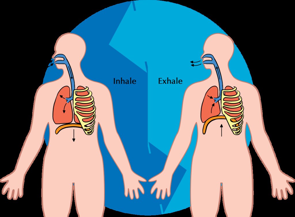
Breathing is a two-step process that includes drawing air into the lungs, or inhaling, and letting air out of the lungs, or exhaling. Both processes are illustrated in Figure 13.3.2.
Inhaling
Inhaling is an active process that results mainly from contraction of a muscle called the diaphragm, shown in Figure 13.3.2. The diaphragm is a large, dome-shaped muscle below the lungs that separates the thoracic (chest) and abdominal cavities. When the diaphragm contracts it moves down causing the thoracic cavity to expand, and the contents of the abdomen to be pushed downward. Other muscles — such as intercostal muscles between the ribs — also contribute to the process of inhalation, especially when inhalation is forced, as when taking a deep breath. These muscles help increase thoracic volume by expanding the ribs outward. The increase in thoracic volume creates a decrease in thoracic air pressure. With the chest expanded, there is lower air pressure inside the lungs than outside the body, so outside air flows into the lungs via the respiratory tract according the the pressure gradient (high pressure flows to lower pressure).
Exhaling
Exhaling involves the opposite series of events. The diaphragm relaxes, so it moves upward and decreases the volume of the thorax. Air pressure inside the lungs increases, so it is higher than the air pressure outside the lungs. Exhalation, unlike inhalation, is typically a passive process that occurs mainly due to the elasticity of the lungs. With the change in air pressure, the lungs contract to their pre-inflated size, forcing out the air they contain in the process. Air flows out of the lungs, similar to the way air rushes out of a balloon when it is released. If exhalation is forced, internal intercostal and abdominal muscles may help move the air out of the lungs.
Control of Breathing
Breathing is one of the few vital bodily functions that can be controlled consciously, as well as unconsciously. Think about using your breath to blow up a balloon. You take a long, deep breath, and then you exhale the air as forcibly as you can into the balloon. Both the inhalation and exhalation are consciously controlled.
Conscious Control of Breathing
You can control your breathing by holding your breath, slowing your breathing, or hyperventilating, which is breathing more quickly and shallowly than necessary. You can also exhale or inhale more forcefully or deeply than usual. Conscious control of breathing is common in many activities besides blowing up balloons, including swimming, speech training, singing, playing many different musical instruments (Figure 13.3.3), and doing yoga, to name just a few.

There are limits on the conscious control of breathing. For example, it is not possible for a healthy person to voluntarily stop breathing indefinitely. Before long, there is an irrepressible urge to breathe. If you were able to stop breathing for a long enough time, you would lose consciousness. The same thing would happen if you were to hyperventilate for too long. Once you lose consciousness so you can no longer exert conscious control over your breathing, involuntary control of breathing takes over.
Unconscious Control of Breathing
Unconscious breathing is controlled by respiratory centers in the medulla and pons of the brainstem (see Figure 13.3.4). The respiratory centers automatically and continuously regulate the rate of breathing based on the body’s needs. These are determined mainly by blood acidity, or pH. When you exercise, for example, carbon dioxide levels increase in the blood, because of increased cellular respiration by muscle cells. The carbon dioxide reacts with water in the blood to produce carbonic acid, making the blood more acidic, so pH falls. The drop in pH is detected by chemoreceptors in the medulla. Blood levels of oxygen and carbon dioxide, in addition to pH, are also detected by chemoreceptors in major arteries, which send the “data” to the respiratory centers. The latter respond by sending nerve impulses to the diaphragm, “telling” it to contract more quickly so the rate of breathing speeds up. With faster breathing, more carbon dioxide is released into the air from the blood, and blood pH returns to the normal range.
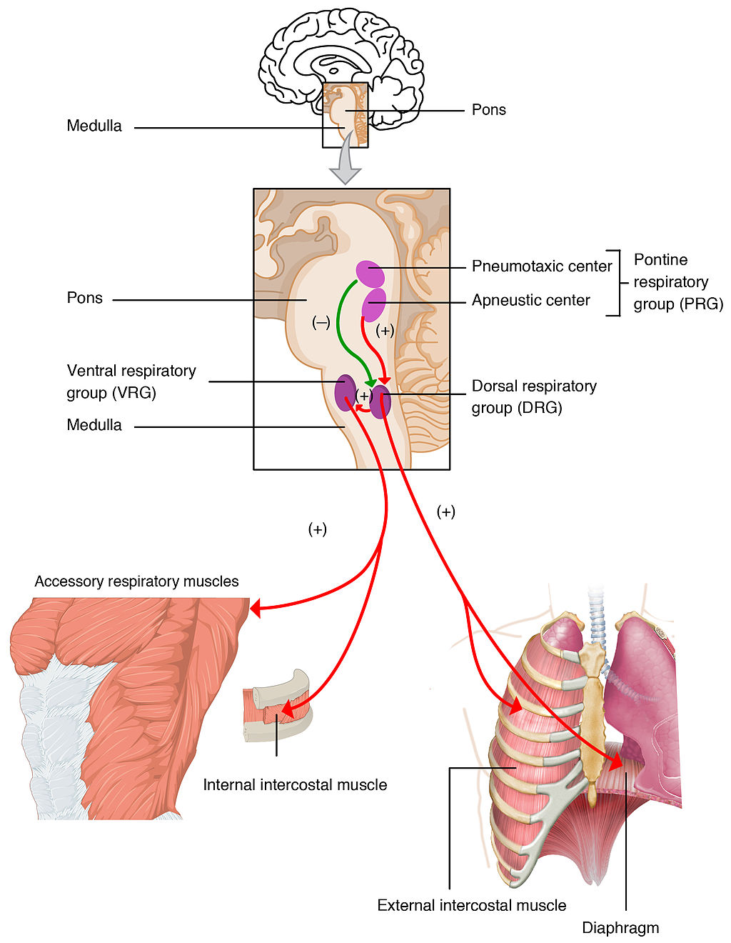
The opposite events occur when the level of carbon dioxide in the blood becomes too low and blood pH rises. This may occur with involuntary hyperventilation, which can happen in panic attacks, episodes of severe pain, asthma attacks, and many other situations. When you hyperventilate, you blow off a lot of carbon dioxide, leading to a drop in blood levels of carbon dioxide. The blood becomes more basic (alkaline), causing its pH to rise.
Nasal vs. Mouth Breathing
Nasal breathing is breathing through the nose rather than the mouth, and it is generally considered to be superior to mouth breathing. The hair-lined nasal passages do a better job of filtering particles out of the air before it moves deeper into the respiratory tract. The nasal passages are also better at warming and moistening the air, so nasal breathing is especially advantageous in the winter when the air is cold and dry. In addition, the smaller diameter of the nasal passages creates greater pressure in the lungs during exhalation. This slows the emptying of the lungs, giving them more time to extract oxygen from the air.
Feature: Myth vs. Reality
Drowning is defined as respiratory impairment from being in or under a liquid. It is further classified according to its outcome into: death, ongoing health problems, or no ongoing health problems (full recovery). Four hundred Canadians die annually from drowning, and drowning is one of the leading causes of death in children under the age of five. There are some potentially dangerous myths about drowning, and knowing what they are might save your life or the life of a loved one, especially a child.
| Myth | Reality |
|---|---|
| "People drown when they aspirate water into their lungs." | Generally, in the early stages of drowning, very little water enters the lungs. A small amount of water entering the trachea causes a muscular spasm in the larynx that seals the airway and prevents the passage of water into the lungs. This spasm is likely to last until unconsciousness occurs. |
| "You can tell when someone is drowning because they will shout for help and wave their arms to attract attention." | The muscular spasm that seals the airway prevents the passage of air, as well as water, so a person who is drowning is unable to shout or call for help. In addition, instinctive reactions that occur in the final minute or so before a drowning person sinks under the water may look similar to calm, safe behavior. The head is likely to be low in the water, tilted back, with the mouth open. The person may have uncontrolled movements of the arms and legs, but they are unlikely to be visible above the water. |
| "It is too late to save a person who is unconscious in the water." | An unconscious person rescued with an airway still sealed from the muscular spasm of the larynx stands a good chance of full recovery if they start receiving CPR within minutes. Without water in the lungs, CPR is much more effective. Even if cardiac arrest has occurred so the heart is no longer beating, there is still a chance of recovery. The longer the brain goes without oxygen, however, the more likely brain cells are to die. Brain death is likely after about six minutes without oxygen, except in exceptional circumstances, such as young people drowning in very cold water. There are examples of children surviving, apparently without lasting ill effects, for as long as an hour in cold water. Rescuers retrieving a child from cold water should attempt resuscitation even after a protracted period of immersion. |
| "If someone is drowning, you should start administering CPR immediately, even before you try to get the person out of the water." | Removing a drowning person from the water is the first priority, because CPR is ineffective in the water. The goal should be to bring the person to stable ground as quickly as possible and then to start CPR. |
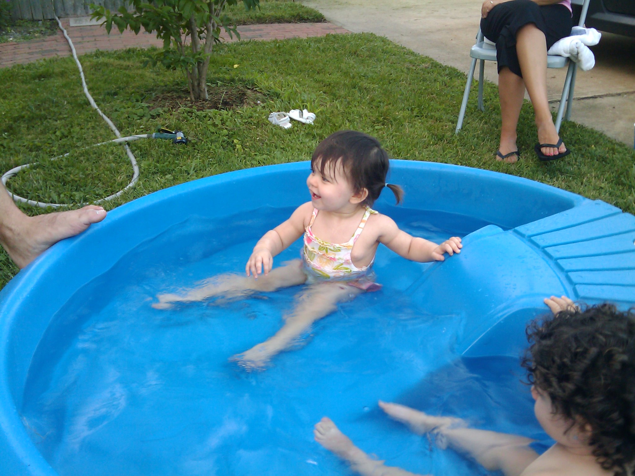
| "You are unlikely to drown unless you are in water over your head." | Depending on circumstances, people have drowned in as little as 30 mm (about 1 ½ in.) of water. Inebriated people or those under the influence of drugs, for example, have been known to have drowned in puddles. Hundreds of children have drowned in the water in toilets, bathtubs, basins, showers, pails, and buckets (see Figure 13.3.5). |
13.3 Summary
- Breathing, or ventilation, is the two-step process of drawing air into the lungs (inhaling) and letting air out of the lungs (exhaling). Inhalation is an active process that results mainly from contraction of a muscle called the diaphragm. Exhalation is typically a passive process that occurs mainly due to the elasticity of the lungs when the diaphragm relaxes.
- Breathing is one of the few vital bodily functions that can be controlled consciously, as well as unconsciously. Conscious control of breathing is common in many activities, including swimming and singing. There are limits on the conscious control of breathing, however. If you try to hold your breath, for example, you will soon have an irrepressible urge to breathe.
- Unconscious breathing is controlled by respiratory centers in the medulla and pons of the brainstem. They respond to variations in blood pH by either increasing or decreasing the rate of breathing as needed to return the pH level to the normal range.
- Nasal breathing is generally considered to be superior to mouth breathing because it does a better job of filtering, warming, and moistening incoming air. It also results in slower emptying of the lungs, which allows more oxygen to be extracted from the air.
- Drowning is a major cause of death in Canada, in particular in children under the age of five. It is important to supervise small children when they are playing in, around, or with water.
13.3 Review Questions
- Define breathing.
-
- Give examples of activities in which breathing is consciously controlled.
- Explain how unconscious breathing is controlled.
- Young children sometimes threaten to hold their breath until they get something they want. Why is this an idle threat?
- Why is nasal breathing generally considered superior to mouth breathing?
- Give one example of a situation that would cause blood pH to rise excessively. Explain why this occurs.
13.3 Explore More
https://www.youtube.com/watch?v=Kl4cU9sG_08
How breathing works - Nirvair Kaur, TED-Ed, 2012.
https://www.youtube.com/watch?v=yDtKBXOEsoM
How do ventilators work? - Alex Gendler, TED-Ed, 2020.
https://www.youtube.com/watch?v=XFnGhrC_3Gs&feature=emb_logo
How I held my breath for 17 minutes | David Blaine, TED, 2010.
https://www.youtube.com/watch?v=Vca6DyFqt4c&feature=emb_logo
The Ultimate Relaxation Technique: How To Practice Diaphragmatic Breathing For Beginners, Kai Simon, 2015.
Attributions
Figure 13.3.1
US_Marines_butterfly_stroke by Cpl. Jasper Schwartz from U.S. Marine Corps on Wikimedia Commons is in the public domain (https://en.wikipedia.org/wiki/Public_domain).
Figure 13.3.2
Inhale Exhale/Breathing cycle by Siyavula Education on Flickr is used under a CC BY 2.0 (https://creativecommons.org/licenses/by/2.0/) license.
Figure 13.3.3
Trumpet/ Frenchmen Street [photo] by Morgan Petroski on Unsplash is used under the Unsplash License (https://unsplash.com/license).
Figure 13.3.4
Respiratory_Centers_of_the_Brain by OpenStax College on Wikimedia Commons is used under a CC BY 3.0 (https://creativecommons.org/licenses/by/3.0) license.
Figure 13.3.5
Lily & Ava in the Kiddie Pool by mob mob on Flickr is used under a CC BY-NC 2.0 (https://creativecommons.org/licenses/by-nc/2.0/) license.
References
Betts, J. G., Young, K.A., Wise, J.A., Johnson, E., Poe, B., Kruse, D.H., Korol, O., Johnson, J.E., Womble, M., DeSaix, P. (2013, June 19). Figure 22.20 Respiratory centers of the brain [digital image]. In Anatomy and Physiology (Section 22.3). OpenStax. https://openstax.org/books/anatomy-and-physiology/pages/22-3-the-process-of-breathing
Kai Simon. (2015, January 11). The ultimate relaxation technique: How to practice diaphragmatic breathing for beginners. YouTube. https://www.youtube.com/watch?v=Vca6DyFqt4c&feature=youtu.be
TED. (2010, January 19). How I held my breath for 17 minutes | David Blaine. YouTube. https://www.youtube.com/watch?v=XFnGhrC_3Gs&feature=youtu.be
TED-Ed. (2012, October 4). How breathing works - Nirvair Kaur. YouTube. https://www.youtube.com/watch?v=Kl4cU9sG_08&feature=youtu.be
TED-Ed. (2020, May 21). How do ventilators work? - Alex Gendler. YouTube. https://www.youtube.com/watch?v=yDtKBXOEsoM&feature=youtu.be
Image shows a diagram of the implications of an x-linked recessive trait when carried by the mother. If a carrier mother has children with an unaffected father, 25% of their children can be expected to be unaffected males, 25% affected males, 25% unaffected females, and 25% carrier females.
A tube that carries bile from the liver and the gallbladder through the pancreas and into the duodenum (the upper part of the small intestine). It is formed where the ducts from the liver and gallbladder are joined. It is part of the biliary duct system.
A biomolecule consisting of carbon (C), hydrogen (H) and oxygen (O) atoms, usually with a hydrogen–oxygen atom ratio of 2:1. Complex carbohydrates are polymers made from monomers of simple carbohydrates, also termed monosaccharides.
A stored form of glucose used by plants.
Image shows a sperm fertilizing an egg. The Sperm is much smaller than the egg.
Figure 5.14.1 Collage of Diverse Faces.
This collage shows some of the variation in human skin colour, which can range from very light to very dark, with every possible gradation in between. As you might expect, the skin color trait has a more complex genetic basis than just one gene with two alleles, which is the type of simple trait that Mendel studied in pea plants. Like skin color, many other human traits have more complicated modes of inheritance than Mendelian traits. Such modes of inheritance are called non-Mendelian inheritance, and they include inheritance of multiple allele traits, traits with codominance or incomplete dominance, and polygenic traits, among others. All of these modes are described below.
Multiple Allele Traits
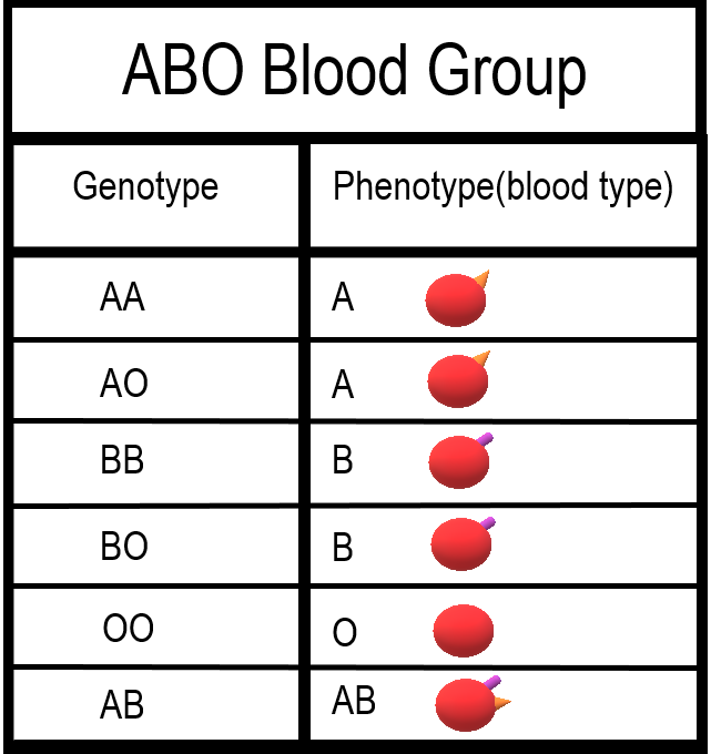
The majority of human genes are thought to have more than two normal versions, or alleles. Traits controlled by a single gene with more than two alleles are called multiple allele traits. An example is ABO blood type. Your blood type refers to which of certain proteins called antigens are found on your red blood cells. There are three common alleles for this trait, which are represented by the letters A, B, and O.
As shown in the table there are six possible ABO genotypes, because the three alleles, taken two at a time, result in six possible combinations. The A and B alleles are dominant to the O allele. As a result, both AA and AO genotypes have the same phenotype, with the A antigen in their blood (type A blood). Similarly, both BB and BO genotypes have the same phenotype, with the B antigen in their blood (type B blood). No antigen is associated with the O allele, so people with the OO genotype have no antigens for ABO blood type in their blood (type O blood).
Codominance
Look at the genotype AB in the ABO blood group table. Alleles A and B for ABO blood type are neither dominant nor recessive to one another. Instead, they are codominant. Codominance occurs when two alleles for a gene are expressed equally in the phenotype of heterozygotes. In the case of ABO blood type, AB heterozygotes have a unique phenotype, with both A and B antigens in their blood (type AB blood).
Incomplete Dominance
Another relationship that may occur between alleles for the same gene is incomplete dominance. This occurs when the dominant allele is not completely dominant. In this case, an intermediate phenotype results in heterozygotes who inherit both alleles. Generally, this happens when the two alleles for a given gene both produce proteins, but one protein is not functional. As a result, the heterozygote individual produces only half the amount of normal protein as is produced by an individual who is homozygous for the normal allele.
An example of incomplete dominance in humans is Tay Sachs disease. The normal allele for the gene in this case produces an enzyme that is responsible for breaking down lipids. A defective allele for the gene results in the production of a nonfunctional enzyme. Heterozygotes who have one normal and one defective allele produce half as much functional enzyme as the normal homozygote, and this is enough for normal development. Homozygotes who have only defective allele, however, produce only nonfunctional enzyme. This leads to the accumulation of lipids in the brain starting in utero, which causes significant brain damage. Most individuals with Tay Sachs disease die at a young age, typically by the age of five years.
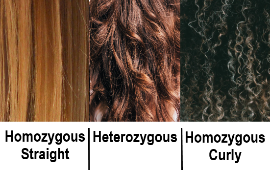
Another good example of incomplete dominance in humans is hair type. There are genes for straight and curly hair, and if an individual is heterozygous, they will typically have the phenotype of wavy hair.
Polygenic Traits
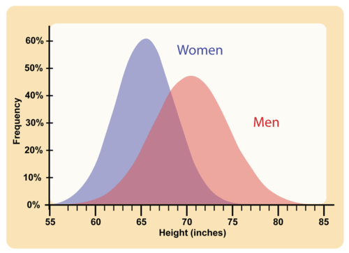
Many human traits are controlled by more than one gene. These traits are called polygenic traits. The alleles of each gene have a minor additive effect on the phenotype. There are many possible combinations of alleles, especially if each gene has multiple alleles. Therefore, a whole continuum of phenotypes is possible.
An example of a human polygenic trait is adult height. Several genes, each with more than one allele, contribute to this trait, so there are many possible adult heights. One adult’s height might be 1.655 m (5.430 feet), and another adult’s height might be 1.656 m (5.433 feet). Adult height ranges from less than 5 feet to more than 6 feet, with males, on average, being somewhat taller than females. The majority of people fall near the middle of the range of heights for their sex, as shown in Figure 5.14.4.
Environmental Effects on Phenotype
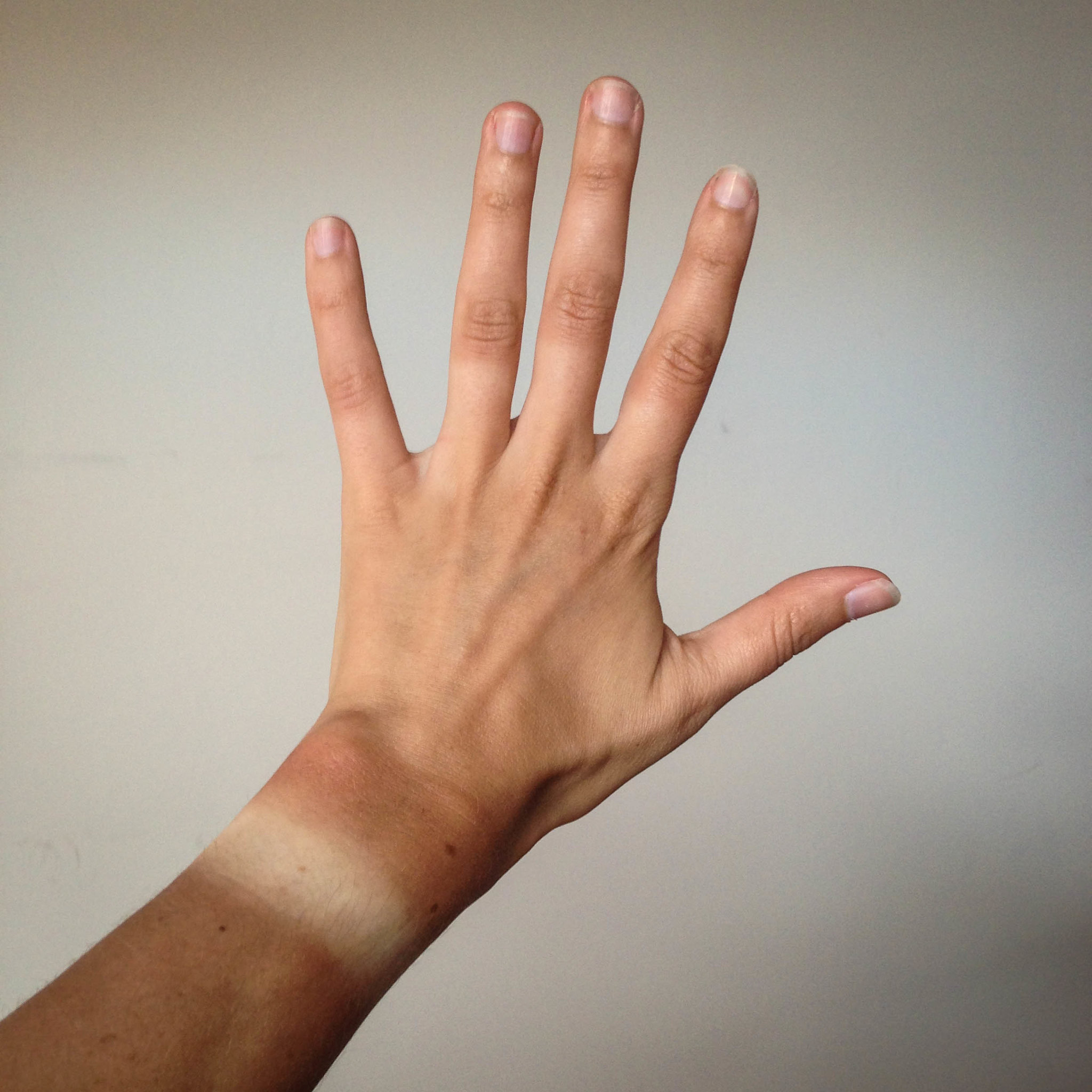
Many traits are affected by the environment, as well as by genes. This may be especially true for polygenic traits. Adult height, for example, might be negatively impacted by poor diet or childhood illness. Skin color is another polygenic trait. There is a wide range of skin colors in people worldwide. In addition to differences in genes, differences in exposure to ultraviolet (UV) light cause some variation. As shown in Figure 5.14.5, exposure to UV light darkens the skin.
Pleiotropy
Some genes affect more than one phenotypic trait. This is called pleiotropy. There are numerous examples of pleiotropy in humans. They generally involve important proteins that are needed for the normal development or functioning of more than one organ system. An example of pleiotropy in humans occurs with the gene that codes for the main protein in collagen, a substance that helps form bones. This protein is also important in the ears and eyes. Mutations in the gene result in problems not only in bones, but also in these sensory organs, which is how the gene's pleiotropic effects were discovered.
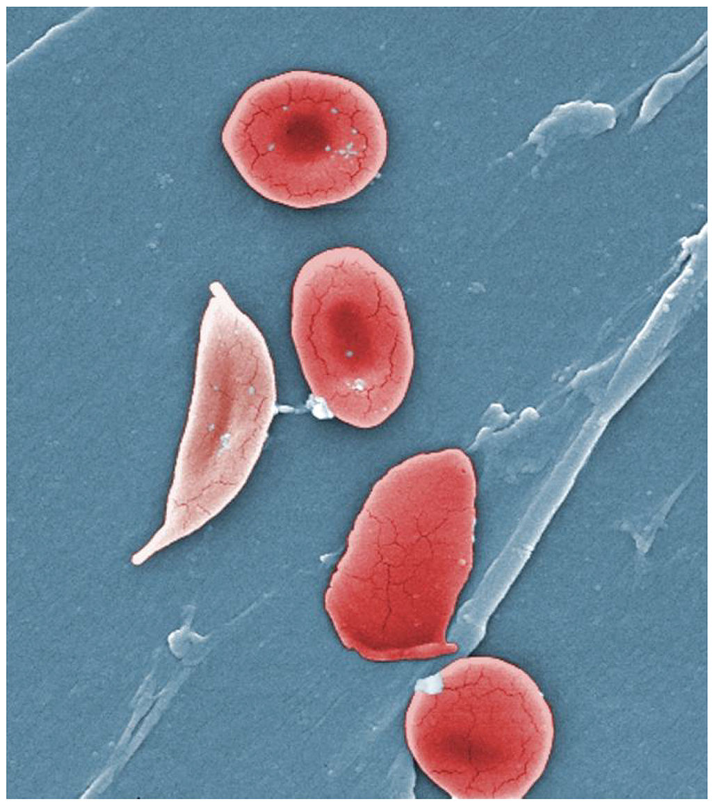
Another example of pleiotropy occurs with sickle cell anemia. This recessive genetic disorder occurs when there is a mutation in the gene that normally encodes the red blood cell protein called hemoglobin. People with the disorder have two alleles for sickle cell hemoglobin, so named for the sickle shape (pictured in Figure 5.14.6) that their red blood cells take on under certain conditions (like physical exertion). The sickle-shaped red blood cells clog small blood vessels, causing multiple phenotypic effects, including stunting of physical growth, certain bone deformities, kidney failure, and strokes.
Epistasis
Some genes affect the expression of other genes. This is called epistasis. Epistasis is similar to dominance, except that it occurs between different genes, rather than between different alleles for the same gene.
Albinism is an example of epistasis. A person with albinism has virtually no pigment in the skin. The condition occurs due to an entirely different gene than the genes that encode skin color. Albinism occurs because a protein called tyrosinase, which is needed for the production of normal skin pigment, is not produced, due to a gene mutation. If an individual has the albinism mutation, he or she will not have any skin pigment, regardless of the skin color genes that were inherited.
Feature: My Human Body
Do you know your ABO blood type? In an emergency, knowing this valuable piece of information could possibly save your life. If you ever need a blood transfusion, it is vital that you receive blood that matches your own blood type. Why? If the blood transfused into your body contains an antigen that your own blood does not contain, antibodies in your blood plasma (the liquid part of your blood) will recognize the antigen as foreign to your body and cause a reaction called agglutination. In this reaction, the transfused red blood cells will clump together. The agglutination reaction is serious and potentially fatal.
Knowing the antigens and antibodies present in each of the ABO blood types will help you understand which type(s) of blood you can safely receive if you ever need a transfusion. This information is shown in Figure 5.14.7 for all of the ABO blood types. If you have blood type A, this means that your red blood cells have the A antigen and that your blood plasma contains anti-B antibodies. If you were to receive a transfusion of type B or type AB blood, both of which have the B antigen, your anti-B antibodies would attack the transfused red blood cells, causing agglutination.
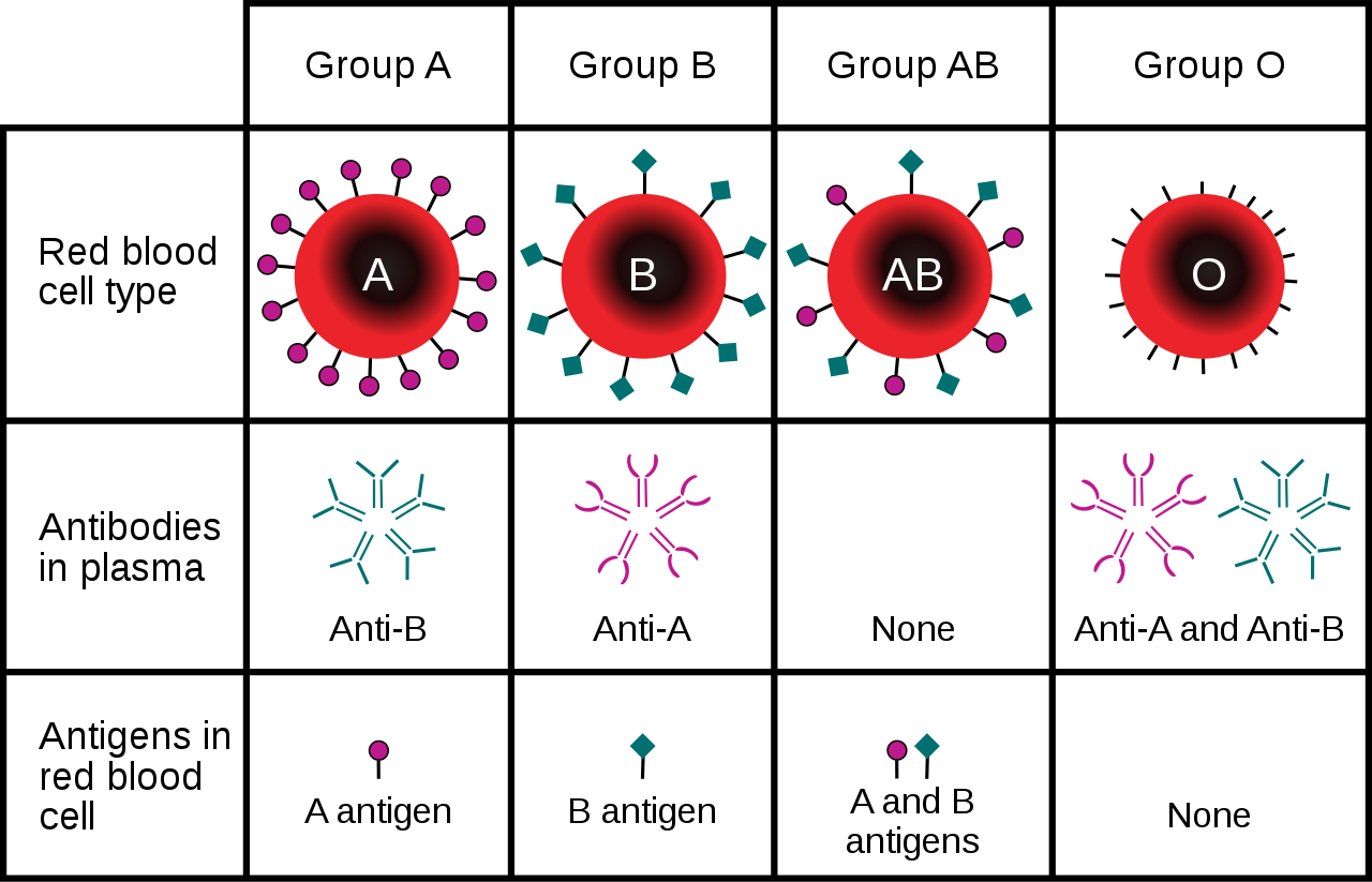
You may have heard that people with blood type O are called "universal donors," and that people with blood type AB are called universal recipients. People with type O blood have neither A nor B antigens in their blood, so if their blood is transfused into someone with a different ABO blood type, it causes no immune reaction, meaning they can donate blood to anyone. On the other hand, people with type AB blood have no anti-A or anti-B antibodies in their blood, so they can receive a transfusion of blood from anyone. Which blood type(s) can safely receive a transfusion of type AB blood, and which blood type(s) can be safely received by those with type O blood?
5.14 Summary
- Non-Mendelian inheritance refers to the inheritance of traits that have a more complex genetic basis than one gene with two alleles and complete dominance.
- Multiple allele traits are controlled by a single gene with more than two alleles. An example of a human multiple allele trait is ABO blood type, for which there are three common alleles: A, B, and O.
- Codominance occurs when two alleles for a gene are expressed equally in the phenotype of heterozygotes. A human example of codominance also occurs in the ABO blood type, in which the A and B alleles are codominant.
- Incomplete dominance is the case in which the dominant allele for a gene is not completely dominant to a recessive allele for the gene, so an intermediate phenotype occurs in heterozygotes who inherit both alleles. A human example of incomplete dominance is Tay Sachs disease, in which heterozygotes produce half as much functional enzyme as normal homozygotes.
- Polygenic traits are controlled by more than one gene, each of which has a minor additive effect on the phenotype. This results in a whole continuum of phenotypes. Examples of human polygenic traits include skin color and adult height.
- Many traits are affected by the environment, as well as by genes. This may be especially true for polygenic traits. Skin color, for example, may be affected by exposure to UV light, and adult stature may be affected by diet or childhood disease.
- Pleiotropy refers to the situation in which a gene affects more than one phenotypic trait. A human example of pleiotropy occurs with sickle cell anemia. People who inherit two recessive alleles for this disorder have abnormal red blood cells and may exhibit multiple other phenotypic effects, such as stunting of physical growth, kidney failure, and strokes.
- Epistasis is the situation in which one gene affects the expression of other genes. An example of epistasis is albinism, in which the albinism mutation negates the expression of skin color genes.
5.14 Review Questions
- What is non-Mendelian inheritance?
-
- Explain why the human ABO blood group is an example of a multiple allele trait with codominance.
- What is incomplete dominance? Give an example of this type of non-Mendelian inheritance in humans.
- Explain the genetic basis of human skin color.
- How can the human trait of adult height be influenced by the environment?
- Define pleiotropy, and give a human example.
- Compare and contrast epistasis and dominance.
- What is the difference between pleiotropy and epistasis?
5.14 Explore More
https://www.youtube.com/watch?time_continue=1&v=YJHGfbW55l0&feature=emb_logo
Incomplete Dominance, Codominance, Polygenic Traits, and Epistasis!,
Amoeba Sisters, 2015.
https://www.youtube.com/watch?v=-4vsio8TZrU&feature=emb_logo
Non-Mendelian Genetics, Teacher's Pet, 2015.
Attributes
Figure 5.14.1
- Woman's Face from Iran by Omid Armin on Unsplash is used under the Unsplash License (https://unsplash.com/license).
- Woman Wearing Black Coat by Anastasiya Pavlova on Unsplash is used under the Unsplash License (https://unsplash.com/license).
- Dark haired man, Queretaro, México by Leonel Hernandez Arteaga on Unsplash is used under the Unsplash License (https://unsplash.com/license). <not found on Unsplash>
- Man in White V-Neck T-Shirt (self) by Joseph Gonzalez on Unsplash is used under the Unsplash License (https://unsplash.com/license).
- Natural Redhead in Brazil by Gabriel Silvério on Unsplash is used under the Unsplash License (https://unsplash.com/license).
- Dark-Skinned Woman with Large White Rose by Oladimeji Oduns on Unsplash is used under the Unsplash License (https://unsplash.com/license).
Figure 5.14.2
ABO Blood Types Per Genotype by Christine Miller is released into the public domain (https://en.wikipedia.org/wiki/Public_domain).
Figure 5.14.3
Three Phenotypes of Hair Based on Inheritance/ Incomplete Dominance Hair by Christine Miller is released into the public domain (https://en.wikipedia.org/wiki/Public_domain).
Figure 5.14.4
Average height /Human Adult Height by CK-12 Foundation is used under a CC BY 3.0 (https://creativecommons.org/licenses/by-nc/3.0/) license.
 ©CK-12 Foundation Licensed under
 ![]() • Terms of Use • Attribution
• Terms of Use • Attribution
Figure 5.14.5
Tan lines by katiebordner on Flickr is used under a CC BY 2.0 (https://creativecommons.org/licenses/by/2.0/) license.
Figure 5.14.6
Sickle cell anemia by OpenStax College on Wikimedia Commons is used under a CC BY 3.0 (https://creativecommons.org/licenses/by/3.0) ©
Figure 5.14.7
ABO_blood_type.svg by InvictaHOG on Wikimedia Commons is in the public domain (https://en.wikipedia.org/wiki/Public_domain).
References
Amoeba Sisters. (2015, May 25). Incomplete dominance, codominance, polygenic traits, and epistasis! YouTube. https://www.youtube.com/watch?v=YJHGfbW55l0
Betts, J. G., Young, K.A., Wise, J.A., Johnson, E., Poe, B., Kruse, D.H., Korol, O., Johnson, J.E., Womble, M., DeSaix, P. (2013, April 25). Figure 18.9 Sickle cells [digital image]. In Anatomy and Physiology. OpenStax. https://openstax.org/books/anatomy-and-physiology/pages/18-3-erythrocytes
Brainard, J/ CK-12 Foundation. (2016). Figure 2 Human adult height [digital image]. In CK-12 College Human Biology (Section 5.13) [online Flexbook]. CK12.org. https://www.ck12.org/book/ck-12-college-human-biology/section/5.13/
Mayo Clinic Staff. (n.d.). Tay-Sachs disease [online article]. MayoClinic.org. https://www.mayoclinic.org/diseases-conditions/tay-sachs-disease/symptoms-causes/syc-20378190
Mayo Clinic Staff. (n.d.). Sickle cell anemia [online article]. MayoClinic.org. https://www.mayoclinic.org/diseases-conditions/sickle-cell-anemia/symptoms-causes/syc-20355876
Teacher's Pet. (2015, January 25). Non-mendelian genetics. YouTube. https://www.youtube.com/watch?v=-4vsio8TZrU
Created by: CK-12/Adapted by Christine Miller
Figure 5.14.1 Collage of Diverse Faces.
This collage shows some of the variation in human skin colour, which can range from very light to very dark, with every possible gradation in between. As you might expect, the skin color trait has a more complex genetic basis than just one gene with two alleles, which is the type of simple trait that Mendel studied in pea plants. Like skin color, many other human traits have more complicated modes of inheritance than Mendelian traits. Such modes of inheritance are called non-Mendelian inheritance, and they include inheritance of multiple allele traits, traits with codominance or incomplete dominance, and polygenic traits, among others. All of these modes are described below.
Multiple Allele Traits

The majority of human genes are thought to have more than two normal versions, or alleles. Traits controlled by a single gene with more than two alleles are called multiple allele traits. An example is ABO blood type. Your blood type refers to which of certain proteins called antigens are found on your red blood cells. There are three common alleles for this trait, which are represented by the letters A, B, and O.
As shown in the table there are six possible ABO genotypes, because the three alleles, taken two at a time, result in six possible combinations. The A and B alleles are dominant to the O allele. As a result, both AA and AO genotypes have the same phenotype, with the A antigen in their blood (type A blood). Similarly, both BB and BO genotypes have the same phenotype, with the B antigen in their blood (type B blood). No antigen is associated with the O allele, so people with the OO genotype have no antigens for ABO blood type in their blood (type O blood).
Codominance
Look at the genotype AB in the ABO blood group table. Alleles A and B for ABO blood type are neither dominant nor recessive to one another. Instead, they are codominant. Codominance occurs when two alleles for a gene are expressed equally in the phenotype of heterozygotes. In the case of ABO blood type, AB heterozygotes have a unique phenotype, with both A and B antigens in their blood (type AB blood).
Incomplete Dominance
Another relationship that may occur between alleles for the same gene is incomplete dominance. This occurs when the dominant allele is not completely dominant. In this case, an intermediate phenotype results in heterozygotes who inherit both alleles. Generally, this happens when the two alleles for a given gene both produce proteins, but one protein is not functional. As a result, the heterozygote individual produces only half the amount of normal protein as is produced by an individual who is homozygous for the normal allele.
An example of incomplete dominance in humans is Tay Sachs disease. The normal allele for the gene in this case produces an enzyme that is responsible for breaking down lipids. A defective allele for the gene results in the production of a nonfunctional enzyme. Heterozygotes who have one normal and one defective allele produce half as much functional enzyme as the normal homozygote, and this is enough for normal development. Homozygotes who have only defective allele, however, produce only nonfunctional enzyme. This leads to the accumulation of lipids in the brain starting in utero, which causes significant brain damage. Most individuals with Tay Sachs disease die at a young age, typically by the age of five years.

Another good example of incomplete dominance in humans is hair type. There are genes for straight and curly hair, and if an individual is heterozygous, they will typically have the phenotype of wavy hair.
Polygenic Traits

Many human traits are controlled by more than one gene. These traits are called polygenic traits. The alleles of each gene have a minor additive effect on the phenotype. There are many possible combinations of alleles, especially if each gene has multiple alleles. Therefore, a whole continuum of phenotypes is possible.
An example of a human polygenic trait is adult height. Several genes, each with more than one allele, contribute to this trait, so there are many possible adult heights. One adult’s height might be 1.655 m (5.430 feet), and another adult’s height might be 1.656 m (5.433 feet). Adult height ranges from less than 5 feet to more than 6 feet, with males, on average, being somewhat taller than females. The majority of people fall near the middle of the range of heights for their sex, as shown in Figure 5.14.4.
Environmental Effects on Phenotype

Many traits are affected by the environment, as well as by genes. This may be especially true for polygenic traits. Adult height, for example, might be negatively impacted by poor diet or childhood illness. Skin color is another polygenic trait. There is a wide range of skin colors in people worldwide. In addition to differences in genes, differences in exposure to ultraviolet (UV) light cause some variation. As shown in Figure 5.14.5, exposure to UV light darkens the skin.
Pleiotropy
Some genes affect more than one phenotypic trait. This is called pleiotropy. There are numerous examples of pleiotropy in humans. They generally involve important proteins that are needed for the normal development or functioning of more than one organ system. An example of pleiotropy in humans occurs with the gene that codes for the main protein in collagen, a substance that helps form bones. This protein is also important in the ears and eyes. Mutations in the gene result in problems not only in bones, but also in these sensory organs, which is how the gene's pleiotropic effects were discovered.
Another example of pleiotropy occurs with sickle cell anemia. This recessive genetic disorder occurs when there is a mutation in the gene that normally encodes the red blood cell protein called hemoglobin. People with the disorder have two alleles for sickle cell hemoglobin, so named for the sickle shape (pictured in Figure 5.14.6) that their red blood cells take on under certain conditions (like physical exertion). The sickle-shaped red blood cells clog small blood vessels, causing multiple phenotypic effects, including stunting of physical growth, certain bone deformities, kidney failure, and strokes.

Epistasis
Some genes affect the expression of other genes. This is called epistasis. Epistasis is similar to dominance, except that it occurs between different genes, rather than between different alleles for the same gene.
Albinism is an example of epistasis. A person with albinism has virtually no pigment in the skin. The condition occurs due to an entirely different gene than the genes that encode skin color. Albinism occurs because a protein called tyrosinase, which is needed for the production of normal skin pigment, is not produced, due to a gene mutation. If an individual has the albinism mutation, he or she will not have any skin pigment, regardless of the skin color genes that were inherited.
Feature: My Human Body
Do you know your ABO blood type? In an emergency, knowing this valuable piece of information could possibly save your life. If you ever need a blood transfusion, it is vital that you receive blood that matches your own blood type. Why? If the blood transfused into your body contains an antigen that your own blood does not contain, antibodies in your blood plasma (the liquid part of your blood) will recognize the antigen as foreign to your body and cause a reaction called agglutination. In this reaction, the transfused red blood cells will clump together. The agglutination reaction is serious and potentially fatal.
Knowing the antigens and antibodies present in each of the ABO blood types will help you understand which type(s) of blood you can safely receive if you ever need a transfusion. This information is shown in Figure 5.14.7 for all of the ABO blood types. If you have blood type A, this means that your red blood cells have the A antigen and that your blood plasma contains anti-B antibodies. If you were to receive a transfusion of type B or type AB blood, both of which have the B antigen, your anti-B antibodies would attack the transfused red blood cells, causing agglutination.

You may have heard that people with blood type O are called "universal donors," and that people with blood type AB are called universal recipients. People with type O blood have neither A nor B antigens in their blood, so if their blood is transfused into someone with a different ABO blood type, it causes no immune reaction, meaning they can donate blood to anyone. On the other hand, people with type AB blood have no anti-A or anti-B antibodies in their blood, so they can receive a transfusion of blood from anyone. Which blood type(s) can safely receive a transfusion of type AB blood, and which blood type(s) can be safely received by those with type O blood?
5.14 Summary
- Non-Mendelian inheritance refers to the inheritance of traits that have a more complex genetic basis than one gene with two alleles and complete dominance.
- Multiple allele traits are controlled by a single gene with more than two alleles. An example of a human multiple allele trait is ABO blood type, for which there are three common alleles: A, B, and O.
- Codominance occurs when two alleles for a gene are expressed equally in the phenotype of heterozygotes. A human example of codominance also occurs in the ABO blood type, in which the A and B alleles are codominant.
- Incomplete dominance is the case in which the dominant allele for a gene is not completely dominant to a recessive allele for the gene, so an intermediate phenotype occurs in heterozygotes who inherit both alleles. A human example of incomplete dominance is Tay Sachs disease, in which heterozygotes produce half as much functional enzyme as normal homozygotes.
- Polygenic traits are controlled by more than one gene, each of which has a minor additive effect on the phenotype. This results in a whole continuum of phenotypes. Examples of human polygenic traits include skin color and adult height.
- Many traits are affected by the environment, as well as by genes. This may be especially true for polygenic traits. Skin color, for example, may be affected by exposure to UV light, and adult stature may be affected by diet or childhood disease.
- Pleiotropy refers to the situation in which a gene affects more than one phenotypic trait. A human example of pleiotropy occurs with sickle cell anemia. People who inherit two recessive alleles for this disorder have abnormal red blood cells and may exhibit multiple other phenotypic effects, such as stunting of physical growth, kidney failure, and strokes.
- Epistasis is the situation in which one gene affects the expression of other genes. An example of epistasis is albinism, in which the albinism mutation negates the expression of skin color genes.
5.14 Review Questions
- What is non-Mendelian inheritance?
-
- Explain why the human ABO blood group is an example of a multiple allele trait with codominance.
- What is incomplete dominance? Give an example of this type of non-Mendelian inheritance in humans.
- Explain the genetic basis of human skin color.
- How can the human trait of adult height be influenced by the environment?
- Define pleiotropy, and give a human example.
- Compare and contrast epistasis and dominance.
- What is the difference between pleiotropy and epistasis?
5.14 Explore More
https://www.youtube.com/watch?time_continue=1&v=YJHGfbW55l0&feature=emb_logo
Incomplete Dominance, Codominance, Polygenic Traits, and Epistasis!,
Amoeba Sisters, 2015.
https://www.youtube.com/watch?v=-4vsio8TZrU&feature=emb_logo
Non-Mendelian Genetics, Teacher's Pet, 2015.
Attributes
Figure 5.14.1
- Woman's Face from Iran by Omid Armin on Unsplash is used under the Unsplash License (https://unsplash.com/license).
- Woman Wearing Black Coat by Anastasiya Pavlova on Unsplash is used under the Unsplash License (https://unsplash.com/license).
- Dark haired man, Queretaro, México by Leonel Hernandez Arteaga on Unsplash is used under the Unsplash License (https://unsplash.com/license). <not found on Unsplash>
- Man in White V-Neck T-Shirt (self) by Joseph Gonzalez on Unsplash is used under the Unsplash License (https://unsplash.com/license).
- Natural Redhead in Brazil by Gabriel Silvério on Unsplash is used under the Unsplash License (https://unsplash.com/license).
- Dark-Skinned Woman with Large White Rose by Oladimeji Oduns on Unsplash is used under the Unsplash License (https://unsplash.com/license).
Figure 5.14.2
ABO Blood Types Per Genotype by Christine Miller is released into the public domain (https://en.wikipedia.org/wiki/Public_domain).
Figure 5.14.3
Three Phenotypes of Hair Based on Inheritance/ Incomplete Dominance Hair by Christine Miller is released into the public domain (https://en.wikipedia.org/wiki/Public_domain).
Figure 5.14.4
Average height /Human Adult Height by CK-12 Foundation is used under a CC BY 3.0 (https://creativecommons.org/licenses/by-nc/3.0/) license.
 ©CK-12 Foundation Licensed under
 ![]() • Terms of Use • Attribution
• Terms of Use • Attribution
Figure 5.14.5
Tan lines by katiebordner on Flickr is used under a CC BY 2.0 (https://creativecommons.org/licenses/by/2.0/) license.
Figure 5.14.6
Sickle cell anemia by OpenStax College on Wikimedia Commons is used under a CC BY 3.0 (https://creativecommons.org/licenses/by/3.0) ©
Figure 5.14.7
ABO_blood_type.svg by InvictaHOG on Wikimedia Commons is in the public domain (https://en.wikipedia.org/wiki/Public_domain).
References
Amoeba Sisters. (2015, May 25). Incomplete dominance, codominance, polygenic traits, and epistasis! YouTube. https://www.youtube.com/watch?v=YJHGfbW55l0
Betts, J. G., Young, K.A., Wise, J.A., Johnson, E., Poe, B., Kruse, D.H., Korol, O., Johnson, J.E., Womble, M., DeSaix, P. (2013, April 25). Figure 18.9 Sickle cells [digital image]. In Anatomy and Physiology. OpenStax. https://openstax.org/books/anatomy-and-physiology/pages/18-3-erythrocytes
Brainard, J/ CK-12 Foundation. (2016). Figure 2 Human adult height [digital image]. In CK-12 College Human Biology (Section 5.13) [online Flexbook]. CK12.org. https://www.ck12.org/book/ck-12-college-human-biology/section/5.13/
Mayo Clinic Staff. (n.d.). Tay-Sachs disease [online article]. MayoClinic.org. https://www.mayoclinic.org/diseases-conditions/tay-sachs-disease/symptoms-causes/syc-20378190
Mayo Clinic Staff. (n.d.). Sickle cell anemia [online article]. MayoClinic.org. https://www.mayoclinic.org/diseases-conditions/sickle-cell-anemia/symptoms-causes/syc-20355876
Teacher's Pet. (2015, January 25). Non-mendelian genetics. YouTube. https://www.youtube.com/watch?v=-4vsio8TZrU
A class of biological molecule consisting of linked monomers of amino acids and which are the most versatile macromolecules in living systems and serve crucial functions in essentially all biological processes.
Amino acids are organic compounds that combine to form proteins.
Image shows a medical professional blotting a blood sample from a small prick on an infant's heel onto special filter paper for the purposes of screening for PKU.
Image shows components of DNA regulating transcription. One a section of DNA regulating a specific gene, upstream, there is an enhancer, promoter sequences, and the TATA box. These preceded the coding strand section of DNA which includes introns and exons.
Image shows the pathway of events in the activation of T Cells. This includes: 1) T Cells are activated when they encounter a foreign antigen on an MHC from an antigen-presenting cell. 2) Cytokines help the T cell to mature. Some T Cells become helper T cells and continue to produce cytokines. 3) Some T Cells become Killer T Cells and search out and destroy infected or cancerous cells.
The passive movement of molecules across the cell membrane with the aid of a membrane protein.
The movement of ions or molecules across a cell membrane into a region of higher concentration, assisted by enzymes and requiring energy.
Structures containing neuronal cell bodies in the peripheral nervous system.
Image shows a table with illustrations showing the variation that exists within pea plans. The peas can either be smooth or wrinkled. The peas can either be green or yellow. The flowers could either be white or purple. The pods could either be smooth or constricted. The pods could either be yellow or green. The plants could either be short or tall. The plants could either end with flowers or end with foliage.
Created by: CK-12/Adapted by Christine Miller
Figure 6.3.1 How would you classify these people?
Why Classify?
What do you see when you look at Figure 6.3.1? Did you sort individuals into categories based in gender, age, body type, facial features, skin colour or other characteristics? As humans, we seem to have a penchant for classifying and labeling people and things. It helps us establish a sense of order in the world around us. The 18th century taxonomist Carl Linnaeus, for example, classified virtually all known living things into different species, genera, families, and other taxonomic categories. His classifications were based on observable phenotypic characteristics, such as skin colour. Modern biological classifications of living things are usually based on phylogenetic relationships. Phylogenies reflect evolutionary history and group together living things that are related by descent from a common ancestor.
Starting with Linnaeus and continuing to the present, scientists and others have attempted to classify human variation. There are three basic approaches to classification: typological, populational, and clinal.
Typological Approach
The typological approach involves creating a typology, which is a system of discrete types, or categories. This approach was widely used by scientists up through the early 20th century. Racial classifications are typological classifications. They place people into a small number of discrete categories, or races, based on a few readily observable traits, such as skin colour, hair texture, facial features, and body build.
Racial Classifications and Racism
Racial classifications of humans probably go back as long as people distinguished “us” from “them.” An early “scientific” classification of humans into races is Linnaeus’ 1735 classification. He divided Homo sapiens into continental races, which he named europaeus, asiaticus, americanus, and afer. Linnaeus described these races in terms of observable physical traits. He also associated, inaccurately, each race with different personality qualities and behaviors. For example, he described Homo sapiens europaeus as active and adventurous and Homo sapiens afer as lazy and careless. In 1795, the German naturalist Johann Friedrich Blumenbach proposed five major races of Homo sapiens, which he named the caucasoid, mongoloid, negroid, American Indian, and Malayan races. Blumenbach thought that the caucasoid race was the original race, and that the other races arose in a process of “degeneration” from the caucasoids.
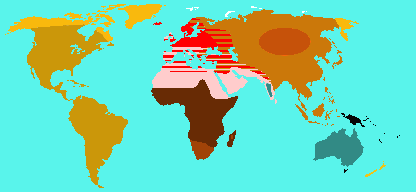
In 1870, the English biologist Thomas Huxley classified Homo sapiens into nine races which were distributed geographically. The map in Figure 6.3.2 shows how Huxley thought the races were distributed worldwide. Each colour represents one of Huxley’s proposed races. These categories included “Australoid,” “Xanthochroi,” “Melanochori,” “Negroes,” and “Mongoloids,” and they are not used today. It should be noted that Huxley did not hold such strong negative stereotypes about non-European (or non-caucasoid) races as did his intellectual forebears. Huxley, however, still attributed different behaviors to racial groups that had nothing to do with the colour of their skin or continent of origin.
By the early 20th century, so-called scientific racism was a popular ideology. This was the idea that race is a biological concept and that human behavior is partly determined by race. At around 1950, in a series of groundbreaking studies of skeletal anatomy, anthropologist Franz Boas showed that cranial (skull) shape and size were highly malleable, depending on environmental factors (such as health and nutrition). He contrasted this with racial anthropologists' claims that head shape is a stable racial trait. In this way, Boas demonstrated that this commonly used racial trait was determined by the environment, and not just genes. Boas also worked to demonstrate that differences in human behavior are not determined primarily by innate biological dispositions, but are largely the result of cultural differences acquired through social learning.
Unfortunately, racism still persists today — in society at large, if not in science. This is the association of racial traits (such as skin colour) with unrelated traits (such as intelligence), often leading to prejudice and discrimination against people based only on how they look. The concept of human race is real, not in a biological sense, but in a social sense. Racial stereotypes and racism are deeply ingrained in our history and culture, and they have real material effects on human lives.
Additional Problems with Typological Classification
Besides the problem of racism, there are other problems with typological approaches to the biological classification of Homo sapiens. One problem is that most human biological traits are not either present or absent, but instead vary on a continuum. This type of distribution cannot be adequately represented by discrete categories, such as races. The typological approach also results in groupings of people that may be similar in terms of some traits, but not others. How people are grouped together depends on which traits are chosen. In addition, the number of groups that are needed to classify people depends on the number of traits that are used. The greater the number of traits, the greater the number of racial categories there must be. If racial categories depend on the traits chosen to define them, it is clear that the racial classifications are arbitrary and do not reflect biological reality.
Another problem with typological classifications is that they lead to the mistaken belief that people within typological categories are more similar to each other than they are to people in other categories. There is actually more variation within than between typological groups. An estimated 90 per cent of human genetic variation occurs between people within races, and only 10 per cent occurs between races. Clearly, races are far from homogenous in terms of their genetic composition. In short, we are all more alike than we are different.
Populational Approach
By the middle of the 20th century, scientists started advocating a populational approach to classifying Homo sapiens. This approach is based on the idea that the breeding population is the only biologically meaningful group. The breeding population is the unit of evolution, and it includes people who have mated and produced offspring together for many generations. As a result, members of the same breeding population should share many genetic traits. You would also expect them to have many of the same phenotypic traits, because of their similar genetic makeup.
While the populational approach makes sense in theory, in reality, it can rarely be applied, because most human populations are not closed breeding populations. Some people have always selected mates from outside their local population (even mating with archaic humans such as Neanderthals). This tendency has increased dramatically in recent centuries with the advent of efficient means of traveling long distances. As a consequence, there are very few remaining distinct breeding populations within the human species.
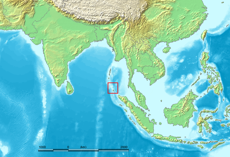
An example of one such population is the Sentinelese, a small population of hunter-gatherers who live alone on a small island in the Andaman Islands (see the map). The Sentinelese are thought to be direct descendants of the first modern humans to leave Africa, and they may have lived in the Andaman Islands for as long as 60 thousand years. The Sentinelese are also one of the most isolated human populations on Earth. The fact that their language is distinctly different from other Andaman Islands languages is evidence that they have had little contact with other people for thousands of years. Although closed breeding populations (such as the Sentinelese) may be useful for investigating questions about evolutionary processes, they are not useful for classifying most of humanity.
Clinal Approach
By the 1960s, scientists began to use a clinal approach to classify human variation. This approach maps variation in traits over geographic regions (such as continents) or even worldwide. Clinal models are a useful way of describing human variation that does not lead to discrete races or other categories of people.
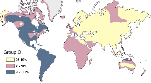
In Figure 6.3.4 you can see a worldwide clinal map for type O blood in the human ABO blood group system. The frequency of this trait is shown for the indigenous populations of various regions. It is lowest throughout Asia and highest in Native American populations in both North and South America. This geographic distribution results from the complex interaction of a variety of factors, including natural selection, genetic drift, and gene flow. You can read more about geographic variation in blood types in the concept Variation in Blood Types.
Clinal maps for many genetic traits show variation that changes gradually from one geographic area to another, which may happen because of the nature of gene flow. Gene flow occurs when mating takes place between people in different populations. The likelihood of mating with others depends on their distance from us. You may not marry the boy or girl next door, but your mate is more likely to be someone in the same state or country than someone on another continent.
Natural selection has a major impact on the clinal distribution of some traits, because variation in the traits tracks variation in selective pressures. For example, the environmental stressor of malaria varies throughout Africa with climate, as you can see in the left-hand map below (Figure 6.3.5). The sickle cell trait that protects from malaria has a similar distribution, as shown in the right-hand map.
Figure 6.3.5
6.3 Summary
- Humans seem to have a need to classify and label people based on their similarities and differences. Three approaches to classifying human variation include typological, populational, and clinal approaches.
- The typological approach involves creating a typology, which is a system of discrete categories, or races. This approach was widely used by scientists until the early 20th century. Racial categories are based on observable phenotypic traits (such as skin colour), but other traits and behaviors are often assumed to apply to racial groups, as well. The use of racial classifications often leads to racism.
- By the mid-20th century, scientists started advocating a population approach. This assumes that the breeding population, which is the unit of evolution, is the only biologically meaningful group. While this approach makes sense in theory, in reality, it can rarely be applied to actual human populations. With few exceptions, most human populations are not closed breeding populations.
- By the 1960s, scientists began to use a clinal approach to classify human variation. This approach maps variation in the frequency of traits or alleles over geographic regions or worldwide. Clinal maps for many genetic traits show variation that changes gradually from one geographic area to another. This type of distribution may result from gene flow and/or natural selection.
6.3 Review Questions
- Name the 18th century taxonomist that classified virtually all known living things.
- Describe the typological approach to classifying human variation.
- Discuss why typological classifications of Homo sapiens are associated with racism.
- Why is the breeding population considered to be the most meaningful biological group?
- Explain why it is generally unrealistic to apply a populational approach to classifying the human species.
- What does a clinal map show?
- Explain how gene flow and natural selection can result in a gradual change in the frequency of a trait over geographic space.
- Most human traits vary on a continuum. Explain why this presents a problem for the typological classification approach.
-
6.3 Explore More
https://www.youtube.com/watch?v=ntimKsWDUpA&feature=emb_logo
The Biology of Race in the Absence of Biological Races,
Centre for Genetic Medicine, 2015.
https://www.youtube.com/watch?v=QOSPNVunyFQ
Nina Jablonski breaks the illusion of skin color, TED, 2009.
https://www.youtube.com/watch?v=_r4c2NT4naQ
The science of skin color - Angela Koine Flynn, TED-Ed, 2016.
Attributions
Figure 6.3.1
- Three women sitting by flowers and laughing by Priscilla Du Preez on Unsplash is used under the Unsplash License (https://unsplash.com/license).
- Two women sitting on sofa by AllGo on Unsplash is used under the Unsplash License (https://unsplash.com/license).
- Young people in conversation by Alexis Brown on Unsplash is used under the Unsplash License (https://unsplash.com/license).
- Men talking in the cold by Anna Vander Stel on Unsplash is used under the Unsplash License (https://unsplash.com/license).
- Laughing by the tracks by Priscilla Du Preez on Unsplash is used under the Unsplash License (https://unsplash.com/license).
Figure 6.3.2
Huxley_races by Wobble on Wikimedia Commons is released into the public domain (https://en.wikipedia.org/wiki/Public_domain).
Figure 6.3.3
Nicobar_Islands is edited by M.Minderhoud on Wikimedia Commons, and was released into the public domain by its original author, www.demis.nl. (See also approval email on de.wp and its clarification.)
Figure 6.3.4
Map_of_Group_O/ (Percent of Native population that has the O blood type) by Ephert on Wikimedia Commons is used under a CC BY 3.0 (https://creativecommons.org/licenses/by/3.0/deed.en) license. (Original Spanish edition by Maulucioni)
Figure 6.3.5
- Malaria distribution by Muntuwandi at English Wikipedia on Wikimedia Commons is used under a CC BY-SA 3.0 (https://creativecommons.org/licenses/by-sa/3.0/deed.en) license.
- Sickle cell distribution by Muntuwandi at English Wikipedia on Wikimedia Commons is used under a CC BY-SA 3.0 (https://creativecommons.org/licenses/by-sa/3.0/deed.en) license.
References
Centre for Genetic Medicine. (2015, July 14). The biology of race in the absence of biological races. YouTube. https://www.youtube.com/watch?v=ntimKsWDUpA&feature=youtu.be
TED. (2009, August 7). Nina Jablonski breaks the illusion of skin color. YouTube. https://www.youtube.com/watch?v=QOSPNVunyFQ&feature=youtu.be
TED-Ed. (2016, February 16). The science of skin color - Angela Koine Flynn. YouTube. https://www.youtube.com/watch?v=_r4c2NT4naQ&feature=youtu.be
Wikipedia contributors. (2020, June 27). Carl Linnaeus. In Wikipedia. https://en.wikipedia.org/w/index.php?title=Carl_Linnaeus&oldid=964690855
Wikipedia contributors. (2020, May 18). Franz Boas. In Wikipedia. https://en.wikipedia.org/w/index.php?title=Franz_Boas&oldid=957282443
Wikipedia contributors. (2020, July 5). Johann Friedrich Blumenbach. In Wikipedia. https://en.wikipedia.org/w/index.php?title=Johann_Friedrich_Blumenbach&oldid=966196943
Wikipedia contributors. (2020, July 11). Sentinelese. In Wikipedia. https://en.wikipedia.org/w/index.php?title=Sentinelese&oldid=967121254
Wikipedia contributors. (2020, July 14). Thomas Henry Huxley. In Wikipedia. https://en.wikipedia.org/w/index.php?title=Thomas_Henry_Huxley&oldid=967701553
Shows a human tongue. There are different areas on the tongue that have varying concentration of taste buds.
Image shows before and after xrays and photos of a patient who has been using a brace to treat their condition. The change in spinal curvature improved from 56 degrees to 27 degrees; possibly preventing the need for surgery.
Created by: CK-12/Adapted by Christine Miller
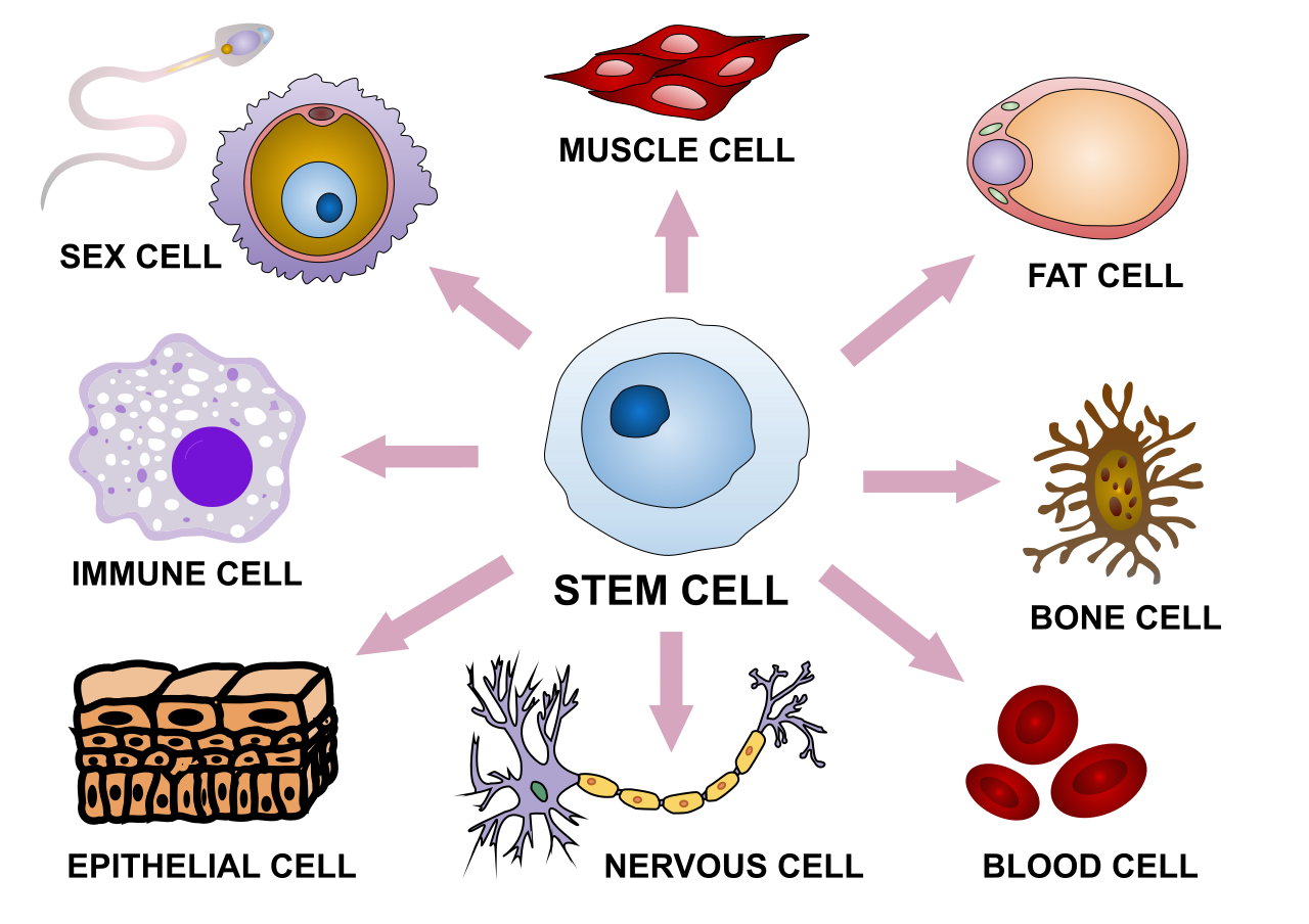
Express Yourself
This sketch illustrates some of the variability in human cells. The shape and other characteristics that make each type of cell unique depend mainly on the specific proteins that particular cell type makes. Proteins are encoded in genes. All the cells in an organism have the same genes, so they all have genetic instructions for the same proteins. Obviously, different types of cells must use (or express) different genes to make different proteins.
What Is Gene Expression?
Using a gene to make a protein is called gene expression. It includes the synthesis of the protein by the processes of transcription of DNA into mRNA, and translation of mRNA into a protein. It may also include further processing of the protein after synthesis.
Gene expression is regulated to ensure that the correct proteins are made when and where they are needed. Regulation may occur at any point in the expression of a gene, from the start of the transcription phase of protein synthesis to the processing of a protein after synthesis occurs. The regulation of transcription is one of the most complicated parts of gene regulation in eukaryotic cells, and it is the focus of this concept.
Regulation of Transcription
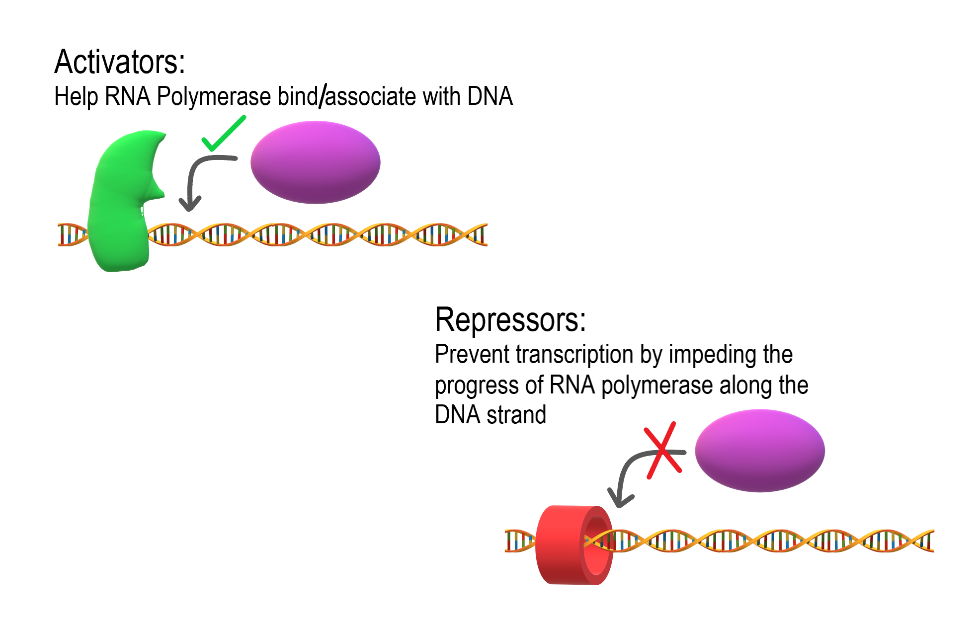
As shown in Figure 5.9.2, transcription is controlled by regulatory proteins. These proteins bind to regions of DNA, called regulatory elements, which are located near promoters. The promoter is the region of a gene where RNA polymerase binds to initiate transcription of the DNA to mRNA. After regulatory proteins bind to regulatory elements, the proteins can interact with RNA polymerase. Regulatory proteins are typically either activators or repressors. Activators are regulatory proteins that promote transcription by enhancing the interaction of RNA polymerase with the promoter. Repressors are regulatory proteins that prevent transcription by impeding the progress of RNA polymerase along the DNA strand, so the DNA cannot be transcribed to mRNA.
Enhancers
Although regulatory proteins and elements are typically the key players in the regulation of transcription, other factors may also be involved. Regulation of transcription may also involve enhancers. Enhancers are distant regions of DNA that can loop back to interact with a gene's promoter. They can also increase the likelihood that transcription of the gene will occur.Enhancers
The TATA Box
Different types of cells have unique patterns of regulatory elements that result in only the necessary genes being transcribed. That’s why a blood cell and nerve cell, for example, are so different from each other. Some regulatory elements, however, are common to virtually all genes, regardless of the cells in which they occur. An example is the TATA box, which is a regulatory element that is part of the promoter of almost every eukaryotic gene. A number of regulatory proteins bind to the TATA box, forming a multi-protein complex. It is only when all of the appropriate proteins are bound to the TATA box that RNA polymerase recognizes the complex and binds to the promoter so transcription can begin.

Regulation During Development
The regulation of gene expression is extremely important in an organism's early development. Regulatory proteins must "turn on" certain genes in particular cells at just the right time, so the individual develops normal organs and organ systems. Homeobox genes are important genes that regulate development.
Homeobox genes are a large group of similar genes that direct the formation of many body structures during the embryonic stage. In humans, there are an estimated 235 functional homeobox genes. They are present on every chromosome and generally grouped in clusters. Homeobox genes contain instructions for making chains of 60 amino acids, called homeodomains. Proteins containing homeodomains are transcription factors that bind to and control the activities of other genes. The homeodomain is the part of the protein that binds to the target gene and controls its expression.
Gene Expression and Cancer
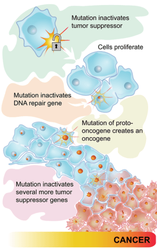
Some types of cancer occur because of mutations in the genes that control the cell cycle. Cancer-causing mutations most often occur in two types of regulatory genes: proto-oncogenes and tumor-suppressor genes. Both are shown in Figure 5.9.4.
- Proto-oncogenes are genes that normally help cells divide. When a proto-oncogene mutates to become an oncogene, it is continuously expressed, even when it is not supposed to be. This is like a car's accelerator pedal being stuck at full throttle. The car keeps racing at top speed. A cell, in this case, keeps dividing out of control, which can lead to cancer.
- Tumor suppressor genes are genes that normally slow down or stop cell division. When a mutation occurs in a tumor suppressor gene, it can no longer control cell division. This is like a car without brakes. The car can't be slowed or stopped. A cell, in this case, keeps dividing out of control, which can lead to cancer.
5.9 Summary
- Using a gene to make a protein is called gene expression. Gene expression is regulated to ensure that the correct proteins are made when and where they are needed. Regulation may occur at any stage of protein synthesis or processing.
- The regulation of transcription is controlled by regulatory proteins that bind to regions of DNA called regulatory elements, which are usually located near promoters. Most regulatory proteins are either activators that promote transcription, or repressors that impede transcription.
- A regulatory element common to almost all eukaryotic genes is the TATA box. A number of regulatory proteins must bind to the TATA box in the promoter before transcription can proceed.
- Regulation of gene expression is extremely important during an organism's early development. Homeobox genes — which encode for chains of amino acids called homeodomains — are important genes that regulate development.
- Some types of cancer occur because of mutations in the genes that control the cell cycle. Cancer-causing mutations most often occur in two types of regulatory genes: tumor-suppressor genes and proto-oncogenes.
5.9 Review Questions
- Define gene expression.
- Why must gene expression be regulated?
- Explain how regulatory proteins may activate or repress transcription.
-
- What is the TATA box, and how does it work?
- Describe homeobox genes and their role in an organism's development.
- Discuss the role of regulatory gene mutations in cancer.
- Explain the relationship between proto-oncogenes and oncogenes.
- If a newly fertilized egg contained a mutation in a homeobox gene, how do you think this would affect the developing embryo? Explain your answer.
- Compare and contrast enhancers and activators.
5.9 Explore More
https://www.youtube.com/watch?time_continue=3&v=vi-zWoobt_Q&feature=emb_logo
Regulated Transcription, ndsuvirtualcell, 2008.
https://www.youtube.com/watch?v=BmFEoCFDi-w
How do cancer cells behave differently from healthy ones? - George Zaidan,
TED-Ed, 2012.
https://www.youtube.com/watch?v=Z3B-AaqjyjE
What is leukemia? - Danilo Allegra and Dania Puggioni, 2015.
Attributions
Figure 5.9.1
Stem_cell_differentiation.svg by Haileyfournier on Wikimedia Commons is used under a CC BY-SA 4.0 (https://creativecommons.org/licenses/by-sa/4.0) license.
Figure 5.9.2
Activators and Repressors by Christine Miller is used under a CC BY-SA 4.0 (https://creativecommons.org/licenses/by-sa/4.0) license.
Figure 5.9.3
TATA_box_description by Luttysar on Wikimedia Commons is used under a CC BY-SA 4.0 (https://creativecommons.org/licenses/by-sa/4.0) license.
Figure 5.9.4
Pathways to cancer by CK-12 Foundation is used under a CC BY-NC 3.0 (http://creativecommons.org/licenses/by-nc/3.0/) license.
 ©CK-12 Foundation Licensed under
 ![]() • Terms of Use • Attribution
• Terms of Use • Attribution
References
Brainard, J/ CK-12 Foundation. (2012). Figure 3 Flow chart (series of mutations leading to cancer) [digital image]. In CK-12 College Human Biology (Section 5.8) [online Flexbook]. CK12.org. https://www.ck12.org/c/physical-science/concentration/?referrer=crossref
ndsuvirtualcell.(2008). Regulated transcription. YouTube. https://www.youtube.com/watch?v=vi-zWoobt_Q&feature=youtu.be
TED-Ed. (2012, December 5). How do cancer cells behave differently from healthy ones? - George Zaidan. YouTube. https://www.youtube.com/watch?v=BmFEoCFDi-w&feature=youtu.be
TED-Ed. (2015, April 15). What is leukemia? - Danilo Allegra and Dania Puggioni. YouTube. https://www.youtube.com/watch?v=Z3B-AaqjyjE&feature=youtu.be
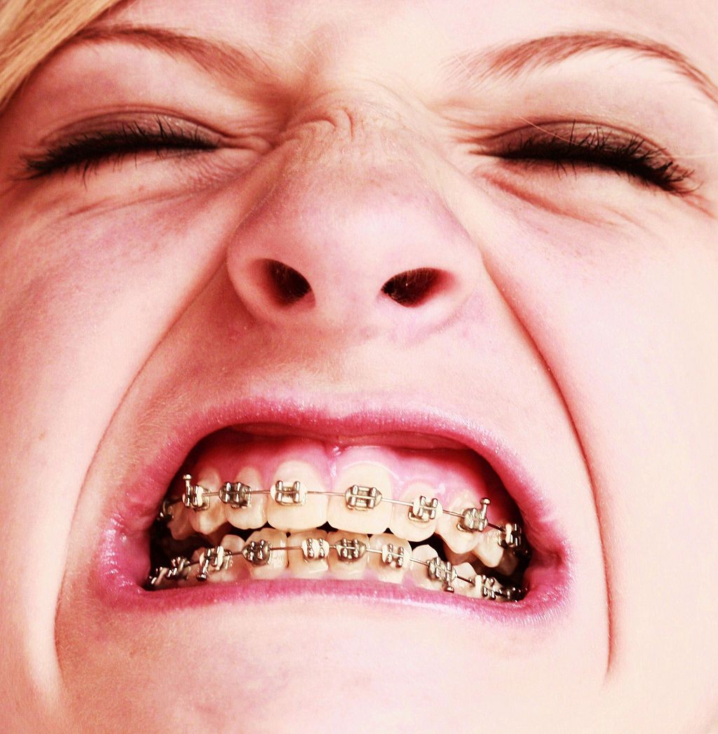
Oh, the Agony!
Wearing braces can be very uncomfortable, but it is usually worth it. Braces and other orthodontic treatments can re-align the teeth and jaws to improve bite and appearance. Braces can change the position of the teeth and the shape of the jaws because the human body is malleable. Many phenotypic traits — even those that have a strong genetic basis — can be molded by the environment. Changing the phenotype in response to the environment is just one of several ways we respond to environmental stress.
Types of Responses to Environmental Stress
There are four different types of responses that humans may make to cope with environmental stress:
- Adaptation
- Developmental adjustment
- Acclimatization
- Cultural responses
The first three types of responses are biological in nature, and the fourth type is cultural. Only adaptation involves genetic change and occurs at the level of the population or species. The other three responses do not require genetic change, and they occur at the individual level.
Adaptation
An adaptation is a genetically-based trait that has evolved because it helps living things survive and reproduce in a given environment. Adaptations generally evolve in a population over many generations in response to stresses that last for a long period of time. Adaptations come about through natural selection. Those individuals who inherit a trait that confers an advantage in coping with an environmental stress are likely to live longer and reproduce more. As a result, more of their genes pass on to the next generation. Over many generations, the genes and the trait they control become more frequent in the population.
A Classic Example: Hemoglobin S and Malaria
Probably the most frequently-cited example of a genetic adaptation to an environmental stress is sickle cell trait. As you read in the previous section, people with sickle cell trait have one abnormal allele (S) and one normal allele (A) for hemoglobin, the red blood cell protein that carries oxygen in the blood. Sickle cell trait is an adaptation to the environmental stress of malaria, because people with the trait have resistance to this parasitic disease. In areas where malaria is endemic (present year-round), the sickle cell trait and its allele have evolved to relatively high frequencies. It is a classic example of natural selection favoring heterozygotes for a gene with two alleles. This type of selection keeps both alleles at relatively high frequencies in a population.
To Taste or Not to Taste
Another example of an adaptation in humans is the ability to taste bitter compounds. Plants produce a variety of toxic compounds in order to protect themselves from being eaten, and these toxic compounds often have a bitter taste. The ability to taste bitter compounds is thought to have evolved as an adaptation, because it prevented people from eating poisonous plants. Humans have many different genes that code for bitter taste receptors, allowing us to taste a wide variety of bitter compounds.
A harmless bitter compound called phenylthiocarbamide (PTC) is not found naturally in plants, but it is similar to toxic bitter compounds that are found in plants. Humans' ability to taste this harmless substance has been tested in many different populations. In virtually every population studied, there are some people who can taste PTC (called tasters), and some people who cannot taste PTC, (called nontasters). The ratio of tasters to non-tasters varies among populations, but on average, 75 per cent of people can taste PTC and 25 per cent cannot.
Like many scientific discoveries, human variation in PTC-taster status was discovered by chance. Around 1930, a chemist named Arthur Fox was working with powdered PTC in his lab. Some of the powder accidentally blew into the air. Another lab worker noticed that the powdered PTC tasted bitter, but Fox couldn't detect any taste at all. Fox wondered how to explain this difference in PTC-tasting ability. Geneticists soon determined that PTC-taster status is controlled by a single gene with two common alleles, usually represented by the letters T and t. The T allele encodes a chemical receptor protein (found in taste buds on the tongue, as illustrated in Figure 6.4.2) that can strongly bind to PTC. The other allele, t, encodes a version of the receptor protein that cannot bind as strongly to PTC. The particular combination of these two alleles that a person inherits determines whether the person finds PTC to taste very bitter (TT), somewhat bitter (Tt), or not bitter at all (tt).
If the ability to taste bitter compounds is advantageous, why does every human population studied contain a significant percentage of people who are nontasters? Why has the nontasting allele been preserved in human populations at all? Some scientists hypothesize that the nontaster allele actually confers the ability to taste some other, yet-to-be identified, bitter compound in plants. People who inherit both alleles would presumably be able to taste a wider range of bitter compounds, so they would have the greatest ability to avoid plant toxins. In other words, the heterozygote genotype for the taster gene would be the most fit and favored by natural selection.
Most people no longer have to worry whether the plants they eat contain toxins. The produce you grow in your garden or buy at the supermarket consists of known varieties that are safe to eat. However, natural selection may still be at work in human populations for the PTC-taster gene, because PTC tasters may be more sensitive than nontasters to bitter compounds in tobacco and vegetables in the cabbage family (that is, cruciferous vegetables, such as the broccoli, cauliflower, and cabbage pictured in Figure 6.4.3).
- People who find PTC to taste very bitter are less likely to smoke tobacco, presumably because tobacco smoke has a stronger bitter taste to these individuals. In this case, selection would favor taster genotypes, because tasters would be more likely to avoid smoking and its serious health risks.
- Strong tasters find cruciferous vegetables to taste bitter. As a result, they may avoid eating these vegetables (and perhaps other foods, as well), presumably resulting in a diet that is less varied and nutritious. In this scenario, natural selection might work against taster genotypes.
Figure 6.4.3 Cruciferous vegetables.
Developmental Adjustment
It takes a relatively long time for genetic change in response to environmental stress to produce a population with adaptations. Fortunately, we can adjust to some environmental stresses more quickly by changing in nongenetic ways. One type of nongenetic response to stress is developmental adjustment. This refers to phenotypic change that occurs during development in infancy or childhood, and that may persist into adulthood. This type of change may be irreversible by adulthood.
Phenotypic Plasticity
Developmental adjustment is possible because humans have a high degree of phenotypic plasticity, which is the ability to alter the phenotype in response to changes in the environment. Phenotypic plasticity allows us to respond to changes that occur within our lifetime, and it is particularly important for species (like our own) that have a long generation time. With long generations, evolution of genetic adaptations may occur too slowly to keep up with changing environmental stresses.
Developmental Adjustment and Cultural Practices
Developmental adjustment may be the result of naturally occurring environmental stresses or cultural practices, including medical or dental treatments. Like our example at the beginning of this section, using braces to change the shape of the jaw and the position of the teeth is an example of a dental practice that brings about a developmental adjustment. Another example of developmental adjustment is the use of a back brace to treat scoliosis (see images in Figure 6.4.4). Scoliosis is an abnormal curvature from side to side in the spine. If the problem is not too severe, a brace, if worn correctly, should prevent the curvature from worsening as a child grows, although it cannot straighten a curve that is already present. Surgery may be required to do that.
Developmental Adjustment and Nutritional Stress
An important example of developmental adjustment that results from a naturally occurring environmental stress is the cessation of physical growth that occurs in children who are under nutritional stress. Children who lack adequate food to fuel both growth and basic metabolic processes are likely to slow down in their growth rate — or even to stop growing entirely. Shunting all available calories and nutrients into essential life functions may keep the child alive at the expense of increasing body size.
Table 6.4.1 shows the effects of inadequate diet on children's' growth in several countries worldwide. For each country, the table gives the prevalence of stunting in children under the age of five. Children are considered stunted if their height is at least two standard deviations below the median height for their age in an international reference population.
Table 6.4.1
Percentage of Stunting in Young Children in Selected Countries (2011-2015)
| Percentage of Stunting in Young Children in Selected Countries (2011-2015) | |
| Country | Per cent of Children Under Age 5 with Stunting |
| United States | 2.1 |
| Turkey | 9.5 |
| Mexico | 13.6 |
| Thailand | 16.3 |
| Iraq | 22.6 |
| Philippines | 33.6 |
| Pakistan | 45.0 |
| Papua New Guinea | 49.5 |
After a growth slow-down occurs and if adequate food becomes available, a child may be able to make up the loss of growth. If food is plentiful, the child may grow more rapidly than normal until the original, genetically-determined growth trajectory is reached. If the inadequate diet persists, however, the failure of growth may become chronic, and the child may never reach his or her full potential adult size.
Phenotypic plasticity of body size in response to dietary change has been observed in successive generations within populations. For example, children in Japan were taller, on average, in each successive generation after the end of World War II. Boys aged 14-15 years old in 1986 were an average of about 18 cm (7 in.) taller than boys of the same age in 1959, a generation earlier. This is a highly significant difference, and it occurred too quickly to be accounted for by genetic change. Instead, the increase in height is a developmental adjustment, thought to be largely attributable to changes in the Japanese diet since World War II. During this period, there was an increase in the amount of animal protein and fat, as well as in the total calories consumed.
Acclimatization
Other responses to environmental stress are reversible and not permanent, whether they occur in childhood or adulthood. The development of reversible changes to environmental stress is called acclimatization. Acclimatization generally develops over a relatively short period of time. It may take just a few days or weeks to attain a maximum response to a stress. When the stress is no longer present, the acclimatized state declines, and the body returns to its normal baseline state. Generally, the shorter the time for acclimatization to occur, the more quickly the condition is reversed when the environmental stress is removed.
Acclimatization to UV Light
A common example of acclimatization is tanning of the skin (see Figure 6.4.5). This occurs in many people in response to exposure to ultraviolet radiation from the sun. Special pigment cells in the skin, called melanocytes, produce more of the brown pigment melanin when exposed to sunlight. The melanin collects near the surface of the skin where it absorbs UV radiation so it cannot penetrate and potentially damage deeper skin structures. Tanning is a reversible change in the phenotype that helps the body deal temporarily with the environmental stress of high levels of UV radiation. When the skin is no longer exposed to the sun’s rays, the tan fades, generally over a period of a few weeks or months.
Figure 6.4.5 Tanning of the skin occurs in many people in response to exposure to ultraviolet radiation from the sun.
Acclimatization to Heat
Another common example of acclimatization occurs in response to heat. Changes that occur with heat acclimatization include increased sweat output and earlier onset of sweat production, which helps the body stay cool because evaporation of sweat takes heat from the body’s surface in a process called evaporative cooling. It generally takes a couple of weeks for maximum heat acclimatization to come about by gradually working out harder and longer at high air temperatures. The changes that occur with acclimatization just as quickly subside when the body is no longer exposed to excessive heat.
Acclimatization to High Altitude
Short term acclimatization to high altitude occurs as a response to low levels of oxygen in the blood. This reduced level of oxygen is detected by carotid bodies, which will trigger in increase in breathing and heart rate. Over a period of weeks the body will compensate by increasing red blood cell production, thereby improving the oxygen-carrying capacity of the blood. This is why mountaineers wishing to climb to the peak of Mount Everest must complete the full climb in portions; it is recommended that climbers spend 2-3 days acclimatizing for every 600 metres of elevation increase. In addition, the higher to altitude, the longer it make take to acclimatize; climbers are advised to spend 4-5 days acclimatizing at base camp (whether the base camp in Nepal or China) before completing the final leg of the climb to the peak. The concentration of red blood cells gradually decreases to normal levels once a climber returns to their normal elevation.
Cultural Responses
More than any other species, humans respond to environmental stresses with learned behaviors and technology. These cultural responses allow us to change our environments to control stresses, rather than changing our bodies genetically or physiologically to cope with the stresses. Even archaic humans responded to some environmental stresses in this way. For example, Neanderthals used shelters, fires, and animal hides as clothing to stay warm in the cold climate in Europe during the last ice age. Today, we use more sophisticated technologies to stay warm in cold climates while retaining our essentially tropical-animal anatomy and physiology. We also use technology (such as furnaces and air conditioners) to avoid temperature stress and stay comfortable in hot or cold climates.
6.4 Summary
- Humans may respond to environmental stress in four different ways: adaptation, developmental adjustment, acclimatization, and cultural responses.
- An adaptation is a genetically based trait that has evolved because it helps living things survive and reproduce in a given environment. Adaptations evolve by natural selection in populations over a relatively long period to time. Examples of adaptations include sickle cell trait as an adaptation to the stress of endemic malaria and the ability to taste bitter compounds as an adaptation to the stress of bitter-tasting toxins in plants.
- A developmental adjustment is a non-genetic response to stress that occurs during infancy or childhood, and that may persist into adulthood. This type of change may be irreversible. Developmental adjustment is possible because humans have a high degree of phenotypic plasticity. It may be the result of environmental stresses (such as inadequate food), which may stunt growth, or cultural practices (such as orthodontic treatments), which re-align the teeth and jaws.
- Acclimatization is the development of reversible changes to environmental stress that develop over a relatively short period of time. The changes revert to the normal baseline state after the stress is removed. Examples of acclimatization include tanning of the skin and physiological changes (such as increased sweating) that occur with heat acclimatization.
- More than any other species, humans respond to environmental stress with learned behaviors and technology, which are cultural responses. These responses allow us to change our environment to control stress, rather than changing our bodies genetically or physiologically to cope with stress. Examples include using shelter, fire, and clothing to cope with a cold climate.
6.4 Review Questions
- List four different types of responses that humans may make to cope with environmental stress.
- Define adaptation.
-
- Explain how natural selection may have resulted in most human populations having people who can and people who cannot taste PTC.
- What is a developmental adjustment?
- Define phenotypic plasticity.
- Explain why phenotypic plasticity may be particularly important in a species with a long generation time.
- Why may stunting of growth occur in children who have an inadequate diet? Why is stunting preferable to the alternative?
- What is acclimatization?
- How does acclimatization to heat come about, and what are two physiological changes that occur in heat acclimatization?
- Give an example of a cultural response to heat stress.
- Which is more likely to be reversible — a change due to acclimatization, or a change due to developmental adjustment? Explain your answer.
6.4 Explore More
https://www.youtube.com/watch?v=upp9-w6GPhU
Could we survive prolonged space travel? - Lisa Nip, TED-Ed, 2016.
https://www.youtube.com/watch?v=hRnrIpUMyZQ&t=182s
How this disease changes the shape of your cells - Amber M. Yates, TED-Ed, 2019.
Attributions
Figure 6.4.1
Free_Awesome_Girl_With_Braces_Close_Up by D. Sharon Pruitt from Hill Air Force Base, Utah, USA on Wikimedia Commons is used under a CC BY 2.0 (https://creativecommons.org/licenses/by/2.0/deed.en) license.
Figure 6.4.2
Tongue by Mahdiabbasinv on Wikimedia Commons is used under a CC BY-SA 4.0 (https://creativecommons.org/licenses/by-sa/4.0/deed.en) license.
Figure 6.4.3
- White cauliflower on brown wooden chopping board by Louis Hansel @shotsoflouis on Unsplash is used under the Unsplash License (https://unsplash.com/license).
- Broccoli on wooden chopping board by Louis Hansel @shotsoflouis on Unsplash is used under the Unsplash License (https://unsplash.com/license).
- Green cabbage close up by Craig Dimmick on Unsplash is used under the Unsplash License (https://unsplash.com/license).
- Cabbage hybrid/ brussel sprouts by Solstice Hannan on Unsplash is used under the Unsplash License (https://unsplash.com/license).
- Kale by Laura Johnston on Unsplash is used under the Unsplash License (https://unsplash.com/license).
- Tiny bok choy at the Asian market by Jodie Morgan on Unsplash is used under the Unsplash License (https://unsplash.com/license).
Figure 6.4.4
Scoliosis_patient_in_cheneau_brace_correcting_from_56_to_27_deg by Weiss H.R. from Scoliosis Journal/BioMed Central Ltd. on Wikimedia Commons is used under a CC BY 2.0 (https://creativecommons.org/licenses/by/2.0) license.
Figure 6.4.5
- Tan Lines by k.steudel on Flickr is used under a CC BY 2.0 (https://creativecommons.org/licenses/by/2.0/) license.
- Twin tan lines (all sizes) by Quinn Dombrowski on Flickr is used under a CC BY-SA 2.0 (https://creativecommons.org/licenses/by-sa/2.0/) license.
- Wedding ring tan line by Quinn Dombrowski on Flickr is used under a CC BY-SA 2.0 (https://creativecommons.org/licenses/by-sa/2.0/) license.
- Tan by Evil Erin on Flickr is used under a CC BY 2.0 (https://creativecommons.org/licenses/by/2.0/) license.
Figure 6.4.6
Nepalese base camp by Mark Horrell on Flickr is used under a CC BY-NC-SA 2.0 (https://creativecommons.org/licenses/by-nc-sa/2.0/) license.
References
TED-Ed. (2016, October 4). Could we survive prolonged space travel? - Lisa Nip. YouTube. https://www.youtube.com/watch?v=upp9-w6GPhU&feature=youtu.be
TED-Ed. (2019, May 6). How this disease changes the shape of your cells - Amber M. Yates. YouTube. https://www.youtube.com/watch?v=hRnrIpUMyZQ&feature=youtu.be
Weiss, H. (2007). Is there a body of evidence for the treatment of patients with Adolescent Idiopathic Scoliosis (AIS)? [Figure 2 - digital photograph], Scoliosis, 2(19). https://doi.org/10.1186/1748-7161-2-19
Created by CK-12 Foundation/Adapted by Christine Miller

Case Study: Flight Risk
Nineteen-year-old Malcolm is about to take his first plane flight. Shortly after he boards the plane and sits down, a man in his late sixties sits next to him in the aisle seat. About half an hour after the plane takes off, the pilot announces that she is turning the seat belt light off, and that it is safe to move around the cabin.
The man in the aisle seat — who has introduced himself to Malcolm as Willie — immediately unbuckles his seat belt and paces up and down the aisle a few times before returning to his seat. After about 45 minutes, Willie gets up again, walks some more, then sits back down and does some foot and leg exercises. After the third time Willie gets up and paces the aisles, Malcolm asks him whether he is walking so much to accumulate steps on a pedometer or fitness tracking device. Willie laughs and says no. He is actually trying to do something even more important for his health — prevent a blood clot from forming in his legs.
Willie explains that he has a chronic condition: heart failure. Although it sounds scary, his condition is currently well-managed, and he is able to lead a relatively normal lifestyle. However, it does put him at risk of developing other serious health conditions, such as deep vein thrombosis (DVT), which is when a blood clot occurs in the deep veins, usually in the legs. Air travel — and other situations where a person has to sit for a long period of time — increases the risk of DVT. Willie’s doctor said that he is healthy enough to fly, but that he should walk frequently and do leg exercises to help avoid a blood clot.
As you read this chapter, you will learn about the heart, blood vessels, and blood that make up the cardiovascular system, as well as disorders of the cardiovascular system, such as heart failure. At the end of the chapter you will learn more about why DVT occurs, why Willie has to take extra precautions when he flies, and what can be done to lower the risk of DVT and its potentially deadly consequences.
Chapter Overview: Cardiovascular System
In this chapter, you will learn about the cardiovascular system, which transports substances throughout the body. Specifically, you will learn about:
- The major components of the cardiovascular system: the heart, blood vessels, and blood.
- The functions of the cardiovascular system, including transporting needed substances (such as oxygen and nutrients) to the cells of the body, and picking up waste products.
- How blood is oxygenated through the pulmonary circulation, which transports blood between the heart and lungs.
- How blood is circulated throughout the body through the systemic circulation.
- The components of blood — including plasma, red blood cells, white blood cells, and platelets — and their specific functions.
- Types of blood vessels — including arteries, veins, and capillaries — and their functions, similarities, and differences.
- The structure of the heart, how it pumps blood, and how contractions of the heart are controlled.
- What blood pressure is and how it is regulated.
- Blood disorders, including anemia, HIV, and leukemia.
- Cardiovascular diseases (including heart attack, stroke, and angina), and the risk factors and precursors — such as high blood pressure and atherosclerosis — that contribute to them.
As you read the chapter, think about the following questions:
- What is heart failure?Why do you think it increases the risk of DVT?
- What is a blood clot? What are possible health consequences of blood clots?
- Why do you think sitting for long periods of time increases the risk of DVT? Why does walking and exercising the legs help reduce this risk?
Attribution
Figure 14.1.1
aircraft-1583871_1920 [photo] by olivier89 from Pixabay is used under the Pixabay License (https://pixabay.com/de/service/license/).

