15.4 Upper Gastrointestinal Tract

Head Stand
Did you ever wonder what would happen if you tried to swallow food while standing on your head like this person in Figure 15.4.1? Many people think that food travels down the gullet from the mouth by the force of gravity. If that were the case, then food you swallowed would stay in your throat while you were standing on your head. In reality, your position doesn’t have much to do with your ability to swallow. Food will travel from your mouth to your stomach whether you are standing upright or upside down. That’s because the tube the food travels through — the esophagus — moves the food along via muscular contractions known as peristalsis. The esophagus is one of several organs that make up the upper gastrointestinal tract.
Organs of the Upper Gastrointestinal Tract
Besides the esophagus, organs of the upper gastrointestinal (GI) tract include the mouth, pharynx, and stomach. These hollow organs are all connected to form a tube through which food passes during digestion. The only role in digestion played by the pharynx and esophagus is to move food through the GI tract. The mouth and stomach, in contrast, are organs where digestion — or the breakdown of food — also occurs. In both of these organs, food is broken into smaller pieces (mechanical digestion), as well as broken down chemically (chemical digestion). It should be noted that the first part of the small intestine (duodenum) is considered in some contexts to be part of the upper GI tract, but that practice is not followed here.
Mouth
The mouth is the first organ of the GI tract. Most of the oral cavity is lined with mucous membrane. This tissue produces mucus, which helps moisten, soften, and lubricate food. Underlying the mucous membrane is a thin layer of smooth muscle to which the mucous membrane is only loosely connected. This gives the mucous membrane considerable ability to stretch as you eat food. The roof of the mouth, called the palate, separates the oral cavity from the nasal cavity. The front part is hard, consisting of mucous membrane covering a plate of bone. The back part of the palate is softer and more pliable, consisting of mucous membrane over muscle and connective tissue. The hard surface of the front of the palate allows for pressure needed in chewing and mixing food. The soft, pliable surface of the back of the palate can move to accommodate the passage of food while swallowing. Muscles at either side of the soft palate contract to create the swallowing action.
Several specific structures in the mouth are specialized for digestion. These include salivary glands, tongue, and teeth.
Salivary Glands
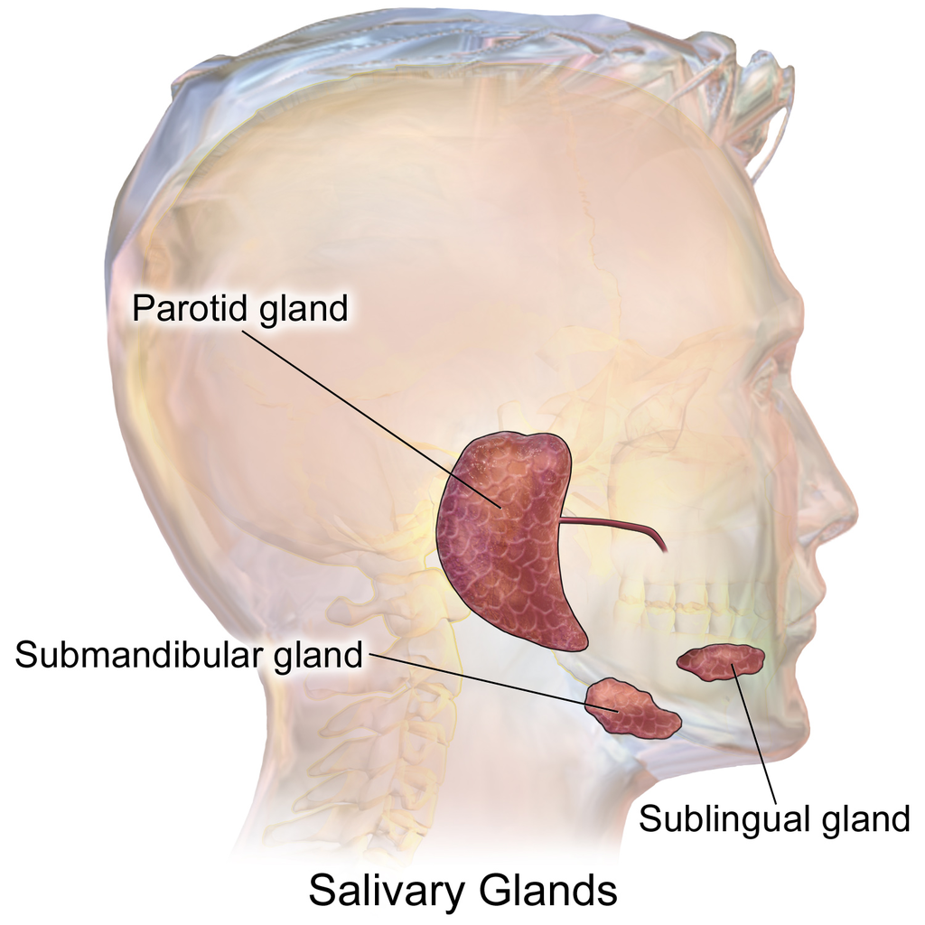
The mouth contains three pairs of major salivary glands, shown in Figure 15.4.2. These three pairs are all exocrine glands that secrete saliva into the mouth through ducts.
- The largest of the three major pairs of salivary glands are the parotid glands, which are located on either side of the mouth in front of the ears.
- The next largest pair is the submandibular glands, located beneath the lower jaw.
- The third pair is the sublingual glands, located underneath the tongue.
In addition to these three pairs of major salivary glands, there are also hundreds of minor salivary glands in the oral mucosa lining the mouth and on the tongue. Along with the major glands, most of the minor glands secrete the digestive enzyme amylase, which begins the chemical digestion of starch and glycogen (polysaccharides). However, the minor salivary glands on the tongue secrete the fat-digesting enzyme lipase, which in the mouth is called lingual lipase (to distinguish it from pancreatic lipase secreted by the pancreas).
Saliva secreted by the salivary glands mainly helps digestion, but it also plays other roles. It helps maintain dental health by cleaning the teeth, and it contains antibodies that help protect against infection. By keeping the mouth lubricated, saliva also allows the mouth movements needed for speech.
Tongue
The tongue is a fleshy, muscular organ that is attached to the floor of the mouth by a band of ligaments that gives it great mobility. This is necessary so the tongue can manipulate food for chewing and swallowing. Movements of the tongue are also necessary for speaking. The upper surface of the tongue is covered with tiny projections called papillae, which contain taste buds. The latter are collections of chemoreceptor cells (shown in Figure 15.4.3). These sensory cells sense chemicals in food and send the information to the brain via cranial nerves, thus enabling the sense of taste.
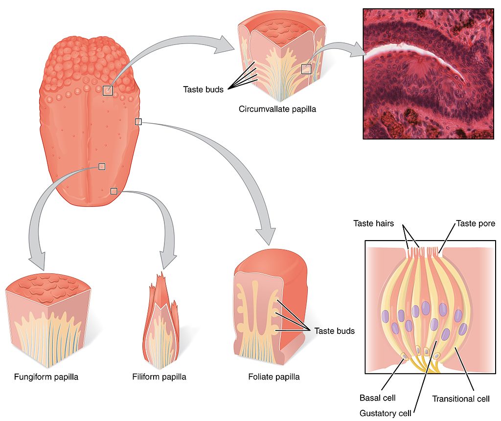
There are five basic tastes detected by the chemoreceptor cells in taste buds: saltiness, sourness, bitterness, sweetness, and umami (often described as a meaty taste). Contrary to popular belief, taste buds for the five basic tastes are not located on different parts of the tongue. Why does taste matter? The taste of food helps to stimulate the secretion of saliva from the salivary glands. It also helps us to eat foods that are good for us, instead of rotten or toxic foods. The detection of saltiness, for example, enables the control of salt intake and salt balance in the body. The detection of sourness may help us avoid spoiled foods, which often taste sour due to fermentation by bacteria. The detection of bitterness warns of poisons, because many plants defend themselves with toxins that taste bitter. The detection of sweetness guides us to foods that supply quick energy. The detection of umami may signal protein-rich foods.
Teeth
The teeth are complex structures made of a bone-like material called dentin and covered with enamel, which is the hardest tissue in the body. Adults normally have a total of 32 teeth, with 16 in each jaw. The right and left sides of each jaw are mirror images in terms of the numbers and types of teeth they contain. Teeth have different shapes to suit them for different aspects of mastication (chewing). The different types of teeth are illustrated in Figure 15.4.4.
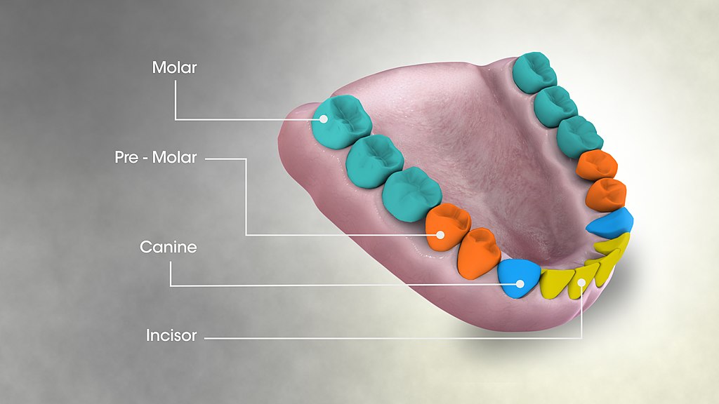
- Incisors are the sharp, blade-like teeth at the front of the mouth. They are used for cutting or biting off pieces of food. In adults, there are normally four incisors in each jaw, or eight in total.
- Canines are the pointed teeth on either side of the incisors. They are used for tearing foods that are tough or stringy. Adults normally have two canines in each jaw, or four altogether.
- Premolars and molars are cuboid teeth with cusps and grooves that are located on the sides and toward the back of the jaws. Premolars are closer to the front of the mouth. Molars are larger and have more cusps than premolars, but both are used for crushing and grinding food. Adults normally have two premolars and three molars on each side of each jaw, for a total of eight premolars and twelve molars.
Pharynx
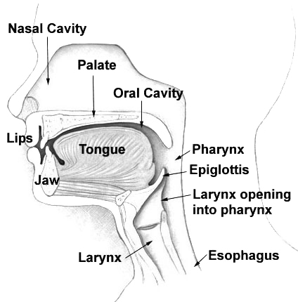
The tube-like pharynx (see Figure 15.4.5 below) plays a dual role as an organ of both respiration and digestion. As part of the respiratory system, it conducts air between the nasal cavity and larynx. As part of the digestive system, it allows swallowed food to pass from the oral cavity to the esophagus. Anything swallowed has priority over inhaled air when passing through the pharynx. During swallowing, the backward motion of the tongue causes a flap of elastic cartilage — called the epiglottis — to close over the opening to the larynx. This prevents food or drink from entering the larynx.
Esophagus
The esophagus (shown in Figure 15.4.6) is a muscular tube through which food is pushed from the pharynx to the stomach. The esophagus passes through an opening in the diaphragm (the large breathing muscle that separates the abdomen from the thorax) before reaching the stomach. In adults, the esophagus averages about 25 cm (about 9.8 inches) in length, depending on a person’s height. The inner lining of the esophagus consists of mucous membrane, which provides a smooth, slippery surface for the passage of food. The cells of this membrane are constantly being replaced as they are worn away from the frequent passage of food over them.
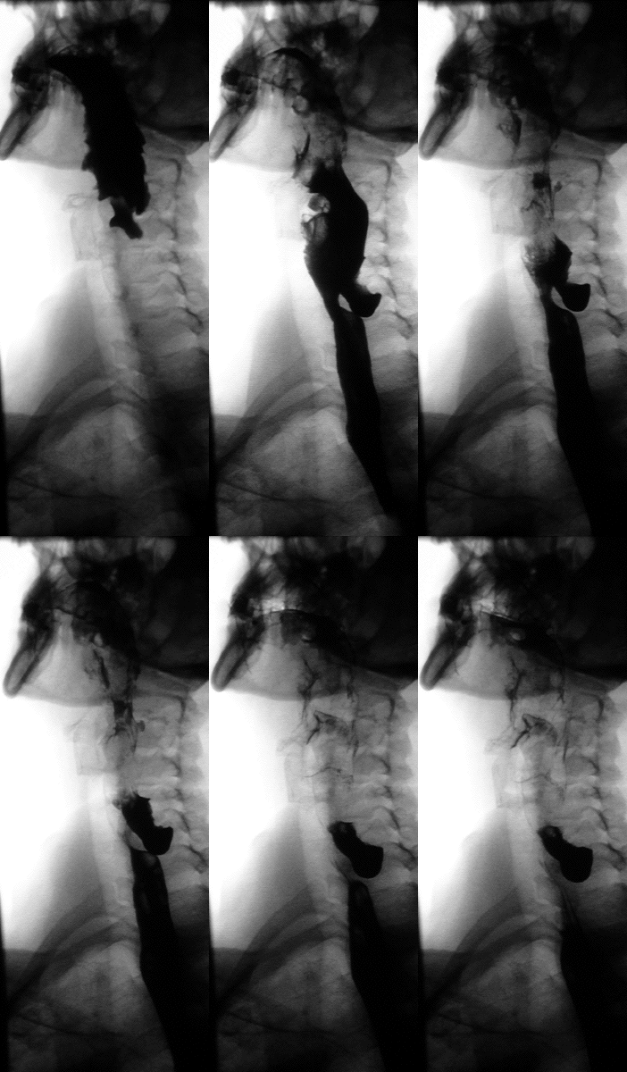
When food is not being swallowed, the esophagus is closed at both ends by upper and lower esophageal sphincters. Sphincters are rings of muscle that can contract to close off openings between structures. The upper esophageal sphincter is triggered to relax and open by the act of swallowing, allowing a bolus of food to enter the esophagus from the pharynx. Then, the esophageal sphincter closes again to prevent food from moving back into the pharynx from the esophagus.
Once in the esophagus, the food bolus travels down to the stomach, pushed along by the rhythmic contraction and relaxation of muscles (peristalsis). The lower esophageal sphincter is located at the junction between the esophagus and the stomach. This sphincter opens when the bolus reaches it, allowing the food to enter the stomach. The sphincter normally remains closed at other times to prevent the contents of the stomach from entering the esophagus. Failure of this sphincter to remain completely closed can lead to heartburn. If it happens chronically, it can lead to gastroesophageal reflux disease (GERD), in which the mucous membrane of the esophagus may become damaged by the highly acidic contents of the stomach.
See the video below to see how the parts of the upper GI tract work together to carry out swallowing:
Swallowing, uploaded by Alejandra Cork, 2012.
Stomach
The stomach is a J-shaped organ (shown in Figure 15.4.7) that is joined to the esophagus at its upper end, and to the first part of the small intestine (duodenum) at its lower end. When the stomach is empty of food, it normally has a volume of about 75 millilitres, but it can expand to hold up to about a litre of food. Waves of muscle contractions (peristalsis) passing through the muscular walls of the stomach cause the food inside to be mixed and churned. The wall of the stomach has an extra layer of muscle tissue not found in other organs of the GI tract that helps it squeeze and mix the food. These movements of the stomach wall contribute greatly to mechanical digestion by breaking the food into much smaller pieces. The churning also helps mix the food with stomach secretions that aid in its chemical digestion.
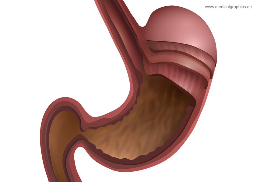
Secretions of the stomach include gastric acid, which consists mainly of hydrochloric acid (HCl). This makes the stomach contents highly acidic, which is necessary so that the enzyme pepsin — also secreted by the stomach — can begin the digestion of protein. Mucus is secreted by the lining of stomach to provide a slimy protective coating against the otherwise damaging effects of gastric acid. The fat-digesting enzyme lipase is secreted in small amounts in the stomach, but very little fat digestion occurs there.
By the time food has been in the stomach for about an hour, it has become the thick, semi-liquid chyme. When the small intestine is ready to receive chyme, a sphincter between the stomach and duodenum — called the pyloric sphincter — opens to allow the chyme to enter the small intestine for further digestion and absorption.
Feature: Reliable Sources
The ongoing epidemic of obesity in the wealthier nations of the world, including Canada, has led to the development of several different bariatric surgeries that modify the stomach to help obese patients reduce their food intake and lose weight. Go online to learn more about bariatric surgery. Find sources you judge to be reliable that answer the following questions:
- Who qualifies for bariatric surgery?
- Describe the bariatric surgeries commonly called stomach stapling, lap band, and gastric sleeve. How does each type of surgery modify the stomach? In terms of weight loss, how effective is each type?
- What are the major potential risks of bariatric surgery?
- Besides weight loss, what other benefits have been shown to result from bariatric surgery?
15.4 Summary
- Organs of the upper gastrointestinal (GI) tract include the mouth, pharynx, esophagus, and stomach.
- The mouth is the first organ of the GI tract. It has several structures that are specialized for digestion, including salivary glands, tongue, and teeth. Both mechanical digestion and chemical digestion of carbohydrates and fats begin in the mouth.
- The pharynx and esophagus move food from the mouth to the stomach, but are not involved in the process of digestion or absorption. Food moves through the esophagus by peristalsis.
- Mechanical and chemical digestion continue in the stomach. Acid and digestive enzymes secreted by the stomach start the chemical digestion of proteins. The stomach turns masticated food into a semi-fluid mixture called chyme.
15.4 Review Questions
-
- Identify structures in the mouth that are specialized for digestion.
- Describe digestion in the mouth.
- What general role do the pharynx and esophagus play in the digestion of food?
- How does food travel through the esophagus?
- Describe digestion in the stomach.
- Describe the differences between how air and food normally move past the pharynx.
- Name two structures in the mouth that contribute to mechanical digestion.
- What structure normally keeps stomach contents from backing up into the esophagus?
- Thirty minutes after you eat a meal, where is most of your food located? Explain your answer.
- What are two roles of mucus in the upper GI tract?
15.4 Explore More
What causes cavities? – Mel Rosenberg, TED-Ed, 2016.
How does alcohol make you drunk? – Judy Grisel, TED-Ed, 2020.
Gastric Bypass Surgery: One Patient’s Journey – Mayo Clinic, 2014.
Here’s What Happens In Your Body When You Swallow Gum | The Human Body, Tech Insider, 2018.
Attributions
Figure 15.4.1
Handstand, Pender Island, B.C. [photo] by Jasper Garratt on Unsplash is used under the Unsplash License (https://unsplash.com/license).
Figure 15.4.2
Blausen_0780_SalivaryGlands by BruceBlaus on Wikimedia Commons is used under a CC BY 3.0 (https://creativecommons.org/licenses/by/3.0) license.
Figure 15.4.3
1402_The_Tongue by OpenStax on Wikimedia Commons is used under a CC BY 4.0 (https://creativecommons.org/licenses/by/4.0) license.
Figure 15.4.4
1024px-3D_Medical_Animation_Still_Showing_Types_of_Teeth by http://www.scientificanimations.com on Wikimedia Commons is used under a CC BY-SA 4.0 (https://creativecommons.org/licenses/by-sa/4.0) license.
Figure 15.4.5
Illu01_head_neck by Arcadian from NCI/ SEER Training Modules on Wikimedia Common is in the public domain (https://en.wikipedia.org/wiki/public_domain).
Figure 15.4.6
ZenkerSchraeg by Bernd Brägelmann Braegel on Wikimedia Commons is used under a CC BY 3.0 (https://creativecommons.org/licenses/by/3.0) license. (Courtesy of Dr. Martin Steinhoff. It is not known whether there is a possibly necessary approval from the patient.)
Figure 15.4.7
Anatomy stomach – white by www.medicalgraphics.de from MedicalGraphics is used under a CC BY-ND 4.0 (https://creativecommons.org/licenses/by-nd/4.0/) license.
References
Alejandra Cork. (2012). Swallowing. YouTube. https://www.youtube.com/watch?v=pNcV6yAfq-g&t=4s
Betts, J. G., Young, K.A., Wise, J.A., Johnson, E., Poe, B., Kruse, D.H., Korol, O., Johnson, J.E., Womble, M., DeSaix, P. (2016, May 27). Figure 14.3 The tongue [digital image]. In Anatomy and Physiology (Section 14.1). OpenStax. https://openstax.org/books/anatomy-and-physiology/pages/14-1-sensory-perception
Blausen.com Staff. (2014). Medical gallery of Blausen Medical 2014. WikiJournal of Medicine 1 (2). DOI:10.15347/wjm/2014.010. ISSN 2002-4436.
Mayo Clinic. (2014, August 26). Gastric bypass surgery: One patient’s journey – Mayo Clinic. https://www.youtube.com/watch?v=twJBEypJDfU&feature=youtu.be
Mayo Clinic Staff. (n.d.). Gastroesophageal reflux disease (GERD) [online article]. MayoClinic.org. https://www.mayoclinic.org/diseases-conditions/gerd/symptoms-causes/syc-20361940
Tech Insider. (2018, March 20). Here’s what happens in your body when you swallow gum | The human body. YouTube. https://www.youtube.com/watch?v=u_1sVri3b2w&feature=youtu.be
TED-Ed. (2020, April 9). How does alcohol make you drunk? – Judy Grisel. YouTube. https://www.youtube.com/watch?v=gCrmFbgT37I&feature=youtu.be
TED-Ed. (2016, October 17). What causes cavities? – Mel Rosenberg. YouTube. https://www.youtube.com/watch?v=zGoBFU1q4g0&feature=youtu.be
Figure 5.10.1
Created by CK-12/Adapted by Christine Miller
As you read in the beginning of this chapter, new parents Samantha and Aki left their pediatrician’s office still unsure whether or not to vaccinate baby James. Dr. Rodriguez gave them a list of reputable sources where they could look up information about the safety of vaccines, including the Centers for Disease Control and Prevention (CDC). Samantha and Aki read that the consensus within the scientific community is that there is no link between vaccines and autism. They find a long list of studies published in peer-reviewed scientific journals that disprove any link. Additionally, some of the studies are “meta-analyses” that analyzed the findings from many individual studies. The new parents are reassured by the fact that many different researchers, using a large number of subjects in numerous well-controlled and well-reviewed studies, all came to the same conclusion.

Samantha also went back to the web page that originally scared her about the safety of vaccines. She found that the author was not a medical doctor or scientific researcher, but rather a self-proclaimed “child wellness expert.” He sold books and advertising on his site, some of which were related to claims of vaccine injury. She realized that he was both an unqualified and potentially biased source of information.
Samantha also realized that some of his arguments were based on correlations between autism and vaccines, but, as the saying goes, “correlation does not imply causation.” For instance, the recent rise in autism rates may have occurred during the same time period as an increase in the number of vaccines given in childhood, but Samantha could think of many other environmental and social factors that have also changed during this time period. There are just too many variables to come to the conclusion that vaccines, or anything else, are the cause of the rise in autism rates based on that type of argument alone. Also, she learned that the age of onset of autism symptoms happens to typically be around the time that the MMR vaccine is first given, so the apparent association in the timing may just be a coincidence.
Finally, Samantha came across news about a measles outbreak in Vancouver, British Columbia in the winter of 2019. Measles wasn’t just a disease of the past! She learned that measles and whooping cough, which had previously been rare thanks to widespread vaccinations, are now on the rise, and that people choosing not to vaccinate their children seems to be one of the contributing factors. She realized that it is important to vaccinate her baby against these diseases, not only to protect him from their potentially deadly effects, but also to protect others in the population.
In their reading, Samantha and Aki learn that scientists do not yet know the causes of autism, but they feels reassured by the abundance of data that disproves any link with vaccines. Both parents think that the potential benefits of protecting their baby’s health against deadly diseases outweighs any unsubstantiated claims about vaccines. They will be making an appointment to get baby James his shots soon.
Chapter 1 Summary
In this chapter, you learned about some of the same concepts that helped Samantha and Aki make an informed decision. Specifically:
- Science is a distinctive way of gaining knowledge about the natural world that is based on the use of evidence to logically test ideas. As such, science is a process, as well as a body of knowledge.
- A scientific theory, such as the germ theory of disease, is the highest level of explanation in science. A theory is a broad explanation for many phenomena that is widely accepted because it is supported by a great deal of evidence.
- The scientific investigation is the cornerstone of science as a process. A scientific investigation is a systematic approach to answering questions about the physical and natural world. An investigation may be observational or experimental.
- A scientific experiment is a type of scientific investigation in which the researcher manipulates variables under controlled conditions to test expected outcomes. Experiments are the gold standard for scientific investigations and can establish causation between variables.
- Nonexperimental scientific investigations such as observational studies and modeling may be undertaken when experiments are impractical, unethical, or impossible. Observational studies generally can establish correlation — but not causation — between variables.
- A pseudoscience, such as astrology, is a field that is presented as scientific but that does not adhere to scientific standards and methods. Other misuses of science include deliberate hoaxes, frauds, and fallacies made by researchers.
- Strict guidelines must be followed when using human subjects in scientific research. Among the most important protections is the requirement for informed consent.
Now that you know about the nature and process of science, you can apply these concepts in the next chapter to the study of human biology.
Chapter 1 Review
-
- Why does a good hypothesis have to be falsifiable?
- Name one scientific law.
- Name one scientific theory.
- Give an example of a scientific idea that was later discredited.
- A statistical measurement called a P-value is often used in science to determine whether or not a difference between two groups is actually significant or simply due to chance. A P-value of 0.03 means that there is a 3% chance that the difference is due to chance alone. Do you think a P-value of 0.03 would indicate that the difference is likely to be significant? Why or why not?
- Why is it important that scientists communicate their findings to others? How do they usually do this?
- What is a “control group” in science?
- In a scientific experiment, why is it important to only change one variable at a time?
- Which is the dependent variable – the variable that is manipulated or the variable that is being affected by the change?
- You see an ad for a “miracle supplement” called NQP3 that claims the supplement will reduce belly fat. They say it works by reducing the hormone cortisol and by providing your body with missing unspecified “nutrients”, but they do not cite any peer-reviewed clinical studies. They show photographs of three people who appear slimmer after taking the product. A board-certified plastic surgeon endorses the product on television. Answer the following questions about this product.
a. Do you think that because a doctor endorsed the product, it really works? Explain your answer.
b. What are two signs that these claims could actually be pseudoscience instead of true science?
c. Do you think the photographs are good evidence that the product works? Why or why not?
d. If you wanted to do a strong scientific study of whether this supplement does what it claims, what would you do? Be specific about the subjects, data collected, how you would control variables, and how you would analyze the data.
e. What are some ways that you would ensure that the subjects in your experiment in part d are treated ethically and according to human subjects protections regulations?
Attribution
Figure 1.8.1
[Photo of person sitting in front of personal computer] by Avel Chuklanov on Unsplash is used under the Unsplash License (https://unsplash.com/license).
Image shows a diagram of the four stages of Meiosis I. In Prophase I, the nuclear envelope is fragmenting, the spindle fibers are forming, and the homologous pairs of chromosomes are undergoing crossing over, in which small segments of DNA are exchanged between homologous pairs. In metaphase I, the tetrads align at the equator. The alignment is termed Independent Alignment, since it is random for each tetrad which "side" the maternal or paternal chromosomes will end up on. In Anaphase I, the tetrads are seperated as dyads are pulled to opposite poles of the cell. In Telophase I the cleavage furrow forms, the dyads decondense and the nuclear envelope reforms as the spindle fibers fragment.
Created by: CK-12/Adapted by Christine Miller

Mutant Cosplay
You probably recognize these costumed comic fans in Figure 5.8.1 as two of the four Teenage Mutant Ninja Turtles. Can a mutation really turn a reptile into an anthropomorphic superhero? Of course not — but mutations can often result in other drastic (but more realistic) changes in living things.
What Are Mutations?
Mutations are random changes in the sequence of bases in DNA or RNA. The word mutation may make you think of the Ninja Turtles, but that's a misrepresentation of how most mutations work. First of all, everyone has mutations. In fact, most people have dozens (or even hundreds!) of mutations in their DNA. Secondly, from an evolutionary perspective, mutations are essential. They are needed for evolution to occur because they are the ultimate source of all new genetic variation in any species.
Causes of Mutations
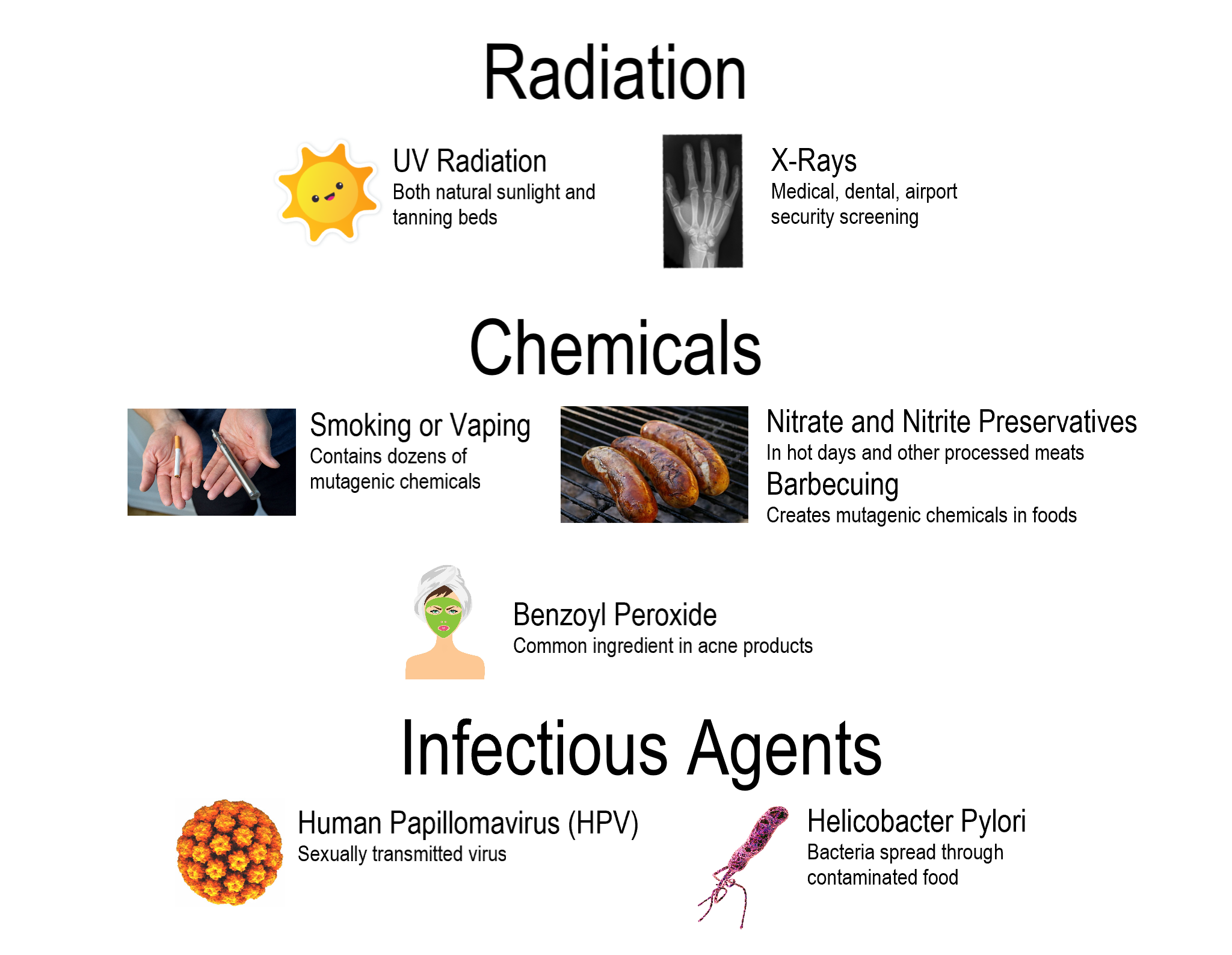
Mutations have many possible causes. Some mutations seem to happen spontaneously, without any outside influence. They occur when errors are made during DNA replication or during the transcription phase of protein synthesis. Other mutations are caused by environmental factors. Anything in the environment that can cause a mutation is known as a mutagen. Examples of mutagens are shown in the figure below.
Types of Mutations
Mutations come in a variety of types. Two major categories of mutations are germline mutations and somatic mutations.
- Germline mutations occur in gametes (the sex cells), such as eggs and sperm. These mutations are especially significant because they can be transmitted to offspring, causing every cell in the offspring to carry those mutations.
- Somatic mutations occur in other cells of the body. These mutations may have little effect on the organism, because they are confined to just one cell and its daughter cells. Somatic mutations cannot be passed on to offspring.
Mutations also differ in the way that the genetic material is changed. Mutations may change an entire chromosome, or they may alter just one or a few nucleotides.
Chromosomal Alterations
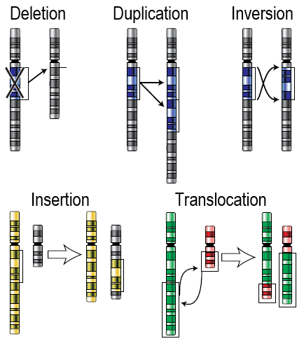
Chromosomal alterations are mutations that change chromosome structure. They occur when a section of a chromosome breaks off and rejoins incorrectly, or otherwise does not rejoin at all. Possible ways in which these mutations can occur are illustrated in the figure below. Chromosomal alterations are very serious. They often result in the death of the organism in which they occur. If the organism survives, it may be affected in multiple ways. An example of a human disease caused by a chromosomal duplication is Charcot-Marie-Tooth disease type 1 (CMT1). It is characterized by muscle weakness, as well as loss of muscle tissue and sensation. The most common cause of CMT1 is a duplication of part of chromosome 17.
Point Mutations
A point mutation is a change in a single nucleotide in DNA. This type of mutation is usually less serious than a chromosomal alteration. An example of a point mutation is a mutation that changes the codon UUU to the codon UCU. Point mutations can be silent, missense, or nonsense mutations, as described in Table 5.8.1. The effects of point mutations depend on how they change the genetic code.
| Type | Description | Example | Effect |
|---|---|---|---|
| Silent | mutated codon codes for the same amino acid | CAA (glutamine) → CAG (glutamine) | none |
| Missense | mutated codon codes for a different amino acid | CAA (glutamine) → CCA (proline) | variable |
| Nonsense | mutated codon is a premature stop codon | CAA (glutamine) → UAA (stop) usually | serious |
Frameshift Mutations
A frameshift mutation is a deletion or insertion of one or more nucleotides, changing the reading frame of the base sequence. Deletions remove nucleotides, and insertions add nucleotides. Consider the following sequence of bases in RNA:
AUG-AAU-ACG-GCU = start-asparagine-threonine-alanine
Now, assume that an insertion occurs in this sequence. Let’s say an A nucleotide is inserted after the start codon AUG. The sequence of bases becomes:
AUG-AAA-UAC-GGC-U = start-lysine-tyrosine-glycine
Even though the rest of the sequence is unchanged, this insertion changes the reading frame and, therefore, all of the codons that follow it. As this example shows, a frameshift mutation can dramatically change how the codons in mRNA are read. This can have a drastic effect on the protein product.
Effects of Mutations
The majority of mutations have neither negative nor positive effects on the organism in which they occur. These mutations are called neutral mutations. Examples include silent point mutations, which are neutral because they do not change the amino acids in the proteins they encode.
Many other mutations have no effects on the organism because they are repaired before protein synthesis occurs. Cells have multiple repair mechanisms to fix mutations in DNA.
Beneficial Mutations
Some mutations — known as beneficial mutations — have a positive effect on the organism in which they occur. They generally code for new versions of proteins that help organisms adapt to their environment. If they increase an organism’s chances of surviving or reproducing, the mutations are likely to become more common over time. There are several well-known examples of beneficial mutations. Here are two such examples:
- Mutations have occurred in bacteria that allow the bacteria to survive in the presence of antibiotic drugs, leading to the evolution of antibiotic-resistant strains of bacteria.
- A unique mutation is found in people in Limone, a small town in Italy. The mutation protects them from developing atherosclerosis, which is the dangerous buildup of fatty materials in blood vessels despite a high-fat diet. The individual in which this mutation first appeared has even been identified and many of his descendants carry this gene.
Harmful Mutations
Imagine making a random change in a complicated machine, such as a car engine. There is a chance that the random change would result in a car that does not run well — or perhaps does not run at all. By the same token, a random change in a gene's DNA may result in the production of a protein that does not function normally... or may not function at all. Such mutations are likely to be harmful. Harmful mutations may cause genetic disorders or cancer.
- A genetic disorder is a disease, syndrome, or other abnormal condition caused by a mutation in one or more genes, or by a chromosomal alteration. An example of a genetic disorder is cystic fibrosis. A mutation in a single gene causes the body to produce thick, sticky mucus that clogs the lungs and blocks ducts in digestive organs.
- Cancer is a disease in which cells grow out of control and form abnormal masses of cells (called tumors). It is generally caused by mutations in genes that regulate the cell cycle. Because of the mutations, cells with damaged DNA are allowed to divide without restriction.
Feature: My Human Body
Inherited mutations are thought to play a role in roughly five to ten per cent of all cancers. Specific mutations that cause many of the known hereditary cancers have been identified. Most of the mutations occur in genes that control the growth of cells or the repair of damaged DNA.
Genetic testing can be done to determine whether individuals have inherited specific cancer-causing mutations. Some of the most common inherited cancers for which genetic testing is available include hereditary breast and ovarian cancer, caused by mutations in genes called BRCA1 and BRCA2. Besides breast and ovarian cancers, mutations in these genes may also cause pancreatic and prostate cancers. Genetic testing is generally done on a small sample of body fluid or tissue, such as blood, saliva, or skin cells. The sample is analyzed by a lab that specializes in genetic testing, and it usually takes at least a few weeks to get the test results.
Should you get genetic testing to find out whether you have inherited a cancer-causing mutation? Such testing is not done routinely just to screen patients for risk of cancer. Instead, the tests are generally done only when the following three criteria are met:
- The test can determine definitively whether a specific gene mutation is present. This is the case with the BRCA1 and BRCA2 gene mutations, for example.
- The test results would be useful to help guide future medical care. For example, if you found out you had a mutation in the BRCA1 or BRCA2 gene, you might get more frequent breast and ovarian cancer screenings than are generally recommended.
- You have a personal or family history that suggests you are at risk of an inherited cancer.
Criterion number 3 is based, in turn, on such factors as:
- Diagnosis of cancer at an unusually young age.
- Several different cancers occurring independently in the same individual.
- Several close genetic relatives having the same type of cancer (such as a maternal grandmother, mother, and sister all having breast cancer).
- Cancer occurring in both organs in a set of paired organs (such as both kidneys or both breasts).
If you meet the criteria for genetic testing and are advised to undergo it, genetic counseling is highly recommended. A genetic counselor can help you understand what the results mean and how to make use of them to reduce your risk of developing cancer. For example, a positive test result that shows the presence of a mutation may not necessarily mean that you will develop cancer. It may depend on whether the gene is located on an autosome or sex chromosome, and whether the mutation is dominant or recessive. Lifestyle factors may also play a role in cancer risk even for hereditary cancers. Early detection can often be life saving if cancer does develop. Genetic counseling can also help you assess the chances that any children you may have will inherit the mutation.
5.8 Summary
- Mutations are random changes in the sequence of bases in DNA or RNA. Most people have multiple mutations in their DNA without ill effects. Mutations are the ultimate source of all new genetic variation in any species.
- Mutations may happen spontaneously during DNA replication or transcription. Other mutations are caused by environmental factors called mutagens. Mutagens include radiation, certain chemicals, and some infectious agents.
- Germline mutations occur in gametes and may be passed onto offspring. Every cell in the offspring will then have the mutation. Somatic mutations occur in cells other than gametes and are confined to just one cell and its daughter cells. These mutations cannot be passed on to offspring.
- Chromosomal alterations are mutations that change chromosome structure and usually affect the organism in multiple ways. Charcot-Marie-Tooth disease type 1 is an example of a chromosomal alteration in humans.
- Point mutations are changes in a single nucleotide. The effects of point mutations depend on how they change the genetic code and may range from no effects to very serious effects.
- Frameshift mutations change the reading frame of the genetic code and are likely to have a drastic effect on the encoded protein.
- Many mutations are neutral and have no effect on the organism in which they occur. Some mutations are beneficial and improve fitness. An example is a mutation that confers antibiotic resistance in bacteria. Other mutations are harmful and decrease fitness, such as the mutations that cause genetic disorders or cancers.
5.8 Review Question
- Define mutation.
- Identify causes of mutation.
- Compare and contrast germline and somatic mutations.
- Describe chromosomal alterations, point mutations, and frameshift mutations. Identify the potential effects of each type of mutation.
- Why do many mutations have neutral effects?
- Give one example of a beneficial mutation and one example of a harmful mutation.
-
- Why do you think that exposure to mutagens (such as cigarette smoke) can cause cancer?
- Explain why the insertion or deletion of a single nucleotide can cause a frameshift mutation.
- Compare and contrast missense and nonsense mutations.
- Explain why mutations are important to evolution.
5.8 Explore More
https://www.youtube.com/watch?time_continue=51&v=PQjL4ZDuq2o&feature=emb_logo
How Radiation Changes Your DNA, Seeker, 2016.
https://www.youtube.com/watch?v=z9HIYjRRaDE&t=93s
Where do genes come from? - Carl Zimmer, TED-Ed, 2014.
https://www.youtube.com/watch?v=a63t8r70QN0&feature=youtu.be
What you should know about vaping and e-cigarettes | Suchitra Krishnan-Sarin,
TED, 2019.
Attributions
Figure 5.8.1
Ninja Turtles by Pat Loika on Flickr is used under a CC BY 2.0 (https://creativecommons.org/licenses/by/2.0/) license.
Figure 5.8.2
Separate images are all in public domain or CC licensed:
- Beauty treatment face mask by no-longer-here on Pixabay is used under the Pixabay License (https://pixabay.com/service/license/).
- HPV by AJC1 on Flickr is used under a CC BY-NC 2.0 (https://creativecommons.org/licenses/by-nc/2.0/) license.
- H Pylori by AJC1 on Flickr is used under a CC BY-NC 2.0 (https://creativecommons.org/licenses/by-nc/2.0/) license.
- Vape and Cigarette by Vaping360 on Flickr is used under a CC BY 2.0 (https://creativecommons.org/licenses/by/2.0/) license.
- Hand X-Ray by Hellerhoff on Wikimedia Commons - CC BY-SA 3.0 (https://creativecommons.org/licenses/by-sa/3.0/deed.en) license.
- Hot dogs by unknown on PxFuel is used under the Pxfuel Terms (https://www.pxfuel.com/terms-of-use).
- Sunshine face is clipart.
Figure 5.8.3
Scheme of possible chromosome mutations/ Chromosomenmutationen by unknown on Wikimedia Commons is adapted from NIH's Talking Glossary of Genetics. [Changes as described by de:user:Dietzel65]. Further use and adapation (text translated to English) by Christine Miller as image is in the public domain (https://en.wikipedia.org/wiki/Public_domain).
References
Seeker. (2016, April 23). How radiation changes your DNA. YouTube. https://www.youtube.com/watch?v=PQjL4ZDuq2o&feature=youtu.be
TED. (2019, June 5). What you should know about vaping and e-cigarettes | Suchitra Krishnan-Sarin. YouTube. https://www.youtube.com/watch?v=a63t8r70QN0&feature=youtu.be
TED-Ed. (2014, September 22). Where do genes come from? - Carl Zimmer. YouTube. https://www.youtube.com/watch?v=z9HIYjRRaDE&feature=youtu.be
Wikipedia contributors. (2020, July 6). Breast cancer. In Wikipedia. https://en.wikipedia.org/w/index.php?title=Breast_cancer&oldid=966366739
Wikipedia contributors. (2020, July 9). Charcot–Marie–Tooth disease. In Wikipedia. https://en.wikipedia.org/w/index.php?title=Charcot%E2%80%93Marie%E2%80%93Tooth_disease&oldid=966912915
Wikipedia contributors. (2020, July 7). Cystic fibrosis. In Wikipedia. https://en.wikipedia.org/w/index.php?title=Cystic_fibrosis&oldid=966566921
Wikipedia contributors. (2020, June 4). Limone sul Garda. In Wikipedia. https://en.wikipedia.org/w/index.php?title=Limone_sul_Garda&oldid=960771991
Wikipedia contributors. (2020, June 23). Ovarian cancer. In Wikipedia. https://en.wikipedia.org/w/index.php?title=Ovarian_cancer&oldid=964157192
Wikipedia contributors. (2020, May 7). BRCA mutation. In Wikipedia. https://en.wikipedia.org/w/index.php?title=BRCA_mutation&oldid=955463902
Wikipedia contributors. (2020, July 10). Teenage Mutant Ninja Turtles. In Wikipedia. https://en.wikipedia.org/w/index.php?title=Teenage_Mutant_Ninja_Turtles&oldid=967030468
A stem cell can become any type of body cell based on gene regulation. Types of cells a stem cell can become include, but are not limited to: Sex cells, muscle cells, fat cells, immune cells, bone cells, epithelial cells, nervous cells, and blood cells.
A chameleon on a branch, surrounded by foliage. The chameleon is camouflaged to blend into its surroundings.
The space occurring between two or more membranes. In cell biology, it's most commonly described as the region between the inner membrane and the outer membrane of a mitochondrion or a chloroplast.
Created by CK-12 Foundation/Adapted by Christine Miller

Work Those Eye Muscles!
Imagine the man in Figure 12.3.1 turns his eyes in your direction. This is a very small movement, considering the conspicuously large and strong external eye muscles that control eyeball movements. These muscles have been called the strongest muscles in the human body relative to the work they do. However, the external eye muscles actually do a surprising amount of work. Eye movements occur almost constantly during waking hours, especially when we are scanning faces or reading. Eye muscles are also exercised nightly during the phase of sleep called rapid eye movement sleep. External eye muscles can move the eyes because they are made mainly of muscle tissue.
What is Muscle Tissue?
Muscle tissue is a soft tissue that makes up most of the tissues in the muscles of the human muscular system. Other tissues in muscles are connective tissues, such as tendons that attach skeletal muscles to bones and sheaths of connective tissues that cover or line muscle tissues. Only muscle tissue per se, has cells with the ability to contract.
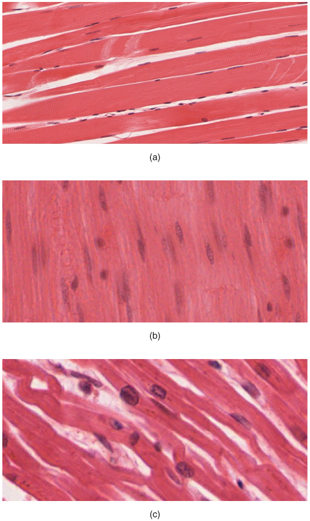
There are three major types of muscle tissues in the human body: skeletal, smooth, and cardiac muscle tissues. Figure 12.3.2 shows how the three types of muscle tissues appear under magnification. When you read about each type below, you will learn why the three types appear as they do.
Skeletal Muscle Tissue
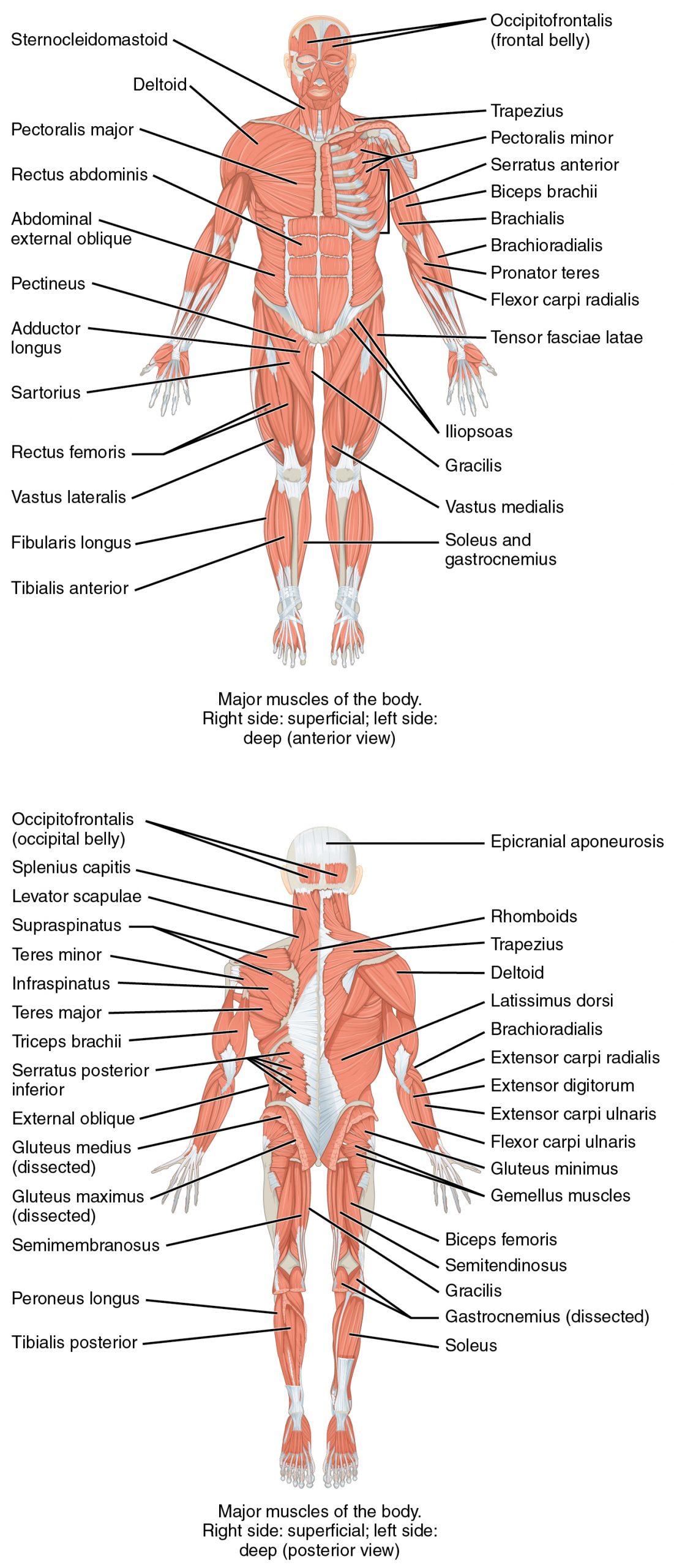
Skeletal muscle is muscle tissue that is attached to bones by tendons, which are bundles of collagen fibres. Whether you are moving your eyes or running a marathon, you are using skeletal muscles. Contractions of skeletal muscles are voluntary, or under conscious control of the central nervous system via the somatic nervous system. Skeletal muscle tissue is the most common type of muscle tissue in the human body. By weight, an average adult male is about 42% skeletal muscles, and the average adult female is about 36% skeletal muscles. Some of the major skeletal muscles in the human body are labeled in Figure 12.3.3 below.
Skeletal Muscle Pairs
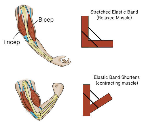
To move bones in opposite directions, skeletal muscles often consist of muscle pairs that work in opposition to one another, also called antagonistic muscle pairs. For example, when the biceps muscle (on the front of the upper arm) contracts, it can cause the elbow joint to flex or bend the arm, as shown in Figure 12.3.4. When the triceps muscle (on the back of the upper arm) contracts, it can cause the elbow to extend or straighten the arm. The biceps and triceps muscles, also shown in Figure 12.3.4, are an example of a muscle pair where the muscles work in opposition to each other.
Skeletal Muscle Structure
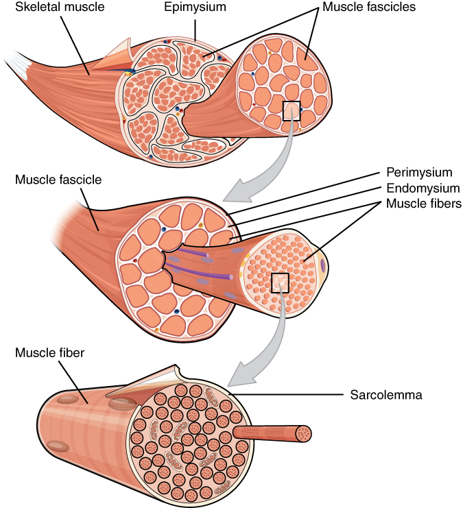
Each skeletal muscle consists of hundreds — or even thousands — of skeletal muscle fibres, which are long, string-like cells. As shown in Figure 12.3.5 below, skeletal muscle fibres are individually wrapped in connective tissue called endomysium. The skeletal muscle fibres are bundled together in units called muscle fascicles, which are surrounded by sheaths of connective tissue called perimysium. Each fascicle contains between ten and 100 (or even more!) skeletal muscle fibres. Fascicles, in turn, are bundled together to form individual skeletal muscles, which are wrapped in connective tissue called epimysium. The connective tissues in skeletal muscles have a variety of functions. They support and protect muscle fibres, allowing them to withstand the forces of contraction by distributing the forces applied to the muscle. They also provide pathways for nerves and blood vessels to reach the muscles. In addition, the epimysium anchors the muscles to tendons.
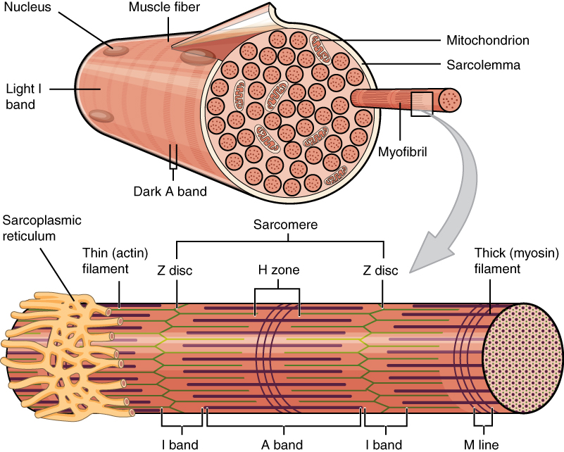
The same bundles-within-bundles structure is replicated within each muscle fibre. As shown in Figure 12.3.6, a muscle fibre consists of a bundle of myofibrils, which are themselves bundles of protein filaments. These protein filaments consist of thin filaments of the protein actin, which are anchored to structures called Z discs, and thick filaments of the protein myosin. The filaments are arranged together within a myofibril in repeating units called sarcomeres, which run from one Z disc to the next. The sarcomere is the basic functional unit of skeletal and cardiac muscles. It contracts as actin and myosin filaments slide over one another. Skeletal muscle tissue is said to be striated, because it appears striped. It has this appearance because of the regular, alternating A (dark) and I (light) bands of filaments arranged in sarcomeres inside the muscle fibres. Other components of a skeletal muscle fibre include multiple nuclei and mitochondria.
Slow- and Fast-Twitch Skeletal Muscle Fibres
Skeletal muscle fibres can be divided into two types, called slow-twitch (or type I) muscle fibres and fast-twitch (or type II) muscle fibres.
- Slow-twitch muscle fibres are dense with capillaries and rich in mitochondria and myoglobin, which is a protein that stores oxygen until needed for muscle activity. Relative to fast-twitch fibres, slow-twitch fibres can carry more oxygen and sustain aerobic (oxygen-using) activity. Slow-twitch fibres can contract for long periods of time, but not with very much force. They are relied upon primarily in endurance events, such as distance running or cycling.
- Fast-twitch muscle fibres contain fewer capillaries and mitochondria and less myoglobin. This type of muscle fibre can contract rapidly and powerfully, but it fatigues very quickly. Fast-twitch fibres can sustain only short, anaerobic (non-oxygen-using) bursts of activity. Relative to slow-twitch fibres, fast-twitch fibres contribute more to muscle strength and have a greater potential for increasing in mass. They are relied upon primarily in short, strenuous events, such as sprinting or weightlifting.
Proportions of fibre types vary considerably from muscle to muscle and from person to person. Individuals may be genetically predisposed to have a larger percentage of one type of muscle fibre than the other. Generally, an individual who has more slow-twitch fibres is better suited for activities requiring endurance, whereas an individual who has more fast-twitch fibres is better suited for activities requiring short bursts of power.
Smooth Muscle
Smooth muscle is muscle tissue in the walls of internal organs and other internal structures such as blood vessels. When smooth muscles contract, they help the organs and vessels carry out their functions. When smooth muscles in the stomach wall contract, for example, they squeeze the food inside the stomach, helping to mix and churn the food and break it into smaller pieces. This is an important part of digestion. Contractions of smooth muscles are involuntary, so they are not under conscious control. Instead, they are controlled by the autonomic nervous system, hormones, neurotransmitters, and other physiological factors.
Structure of Smooth Muscle
The cells that make up smooth muscle are generally called myocytes. Unlike the muscle fibres of striated muscle tissue, the myocytes of smooth muscle tissue do not have their filaments arranged in sarcomeres. Therefore, smooth tissue is not striated. However, the myocytes of smooth muscle do contain myofibrils, which in turn contain bundles of myosin and actin filaments. The filaments cause contractions when they slide over each other, as shown in Figure 12.3.7.

Functions of Smooth Muscle
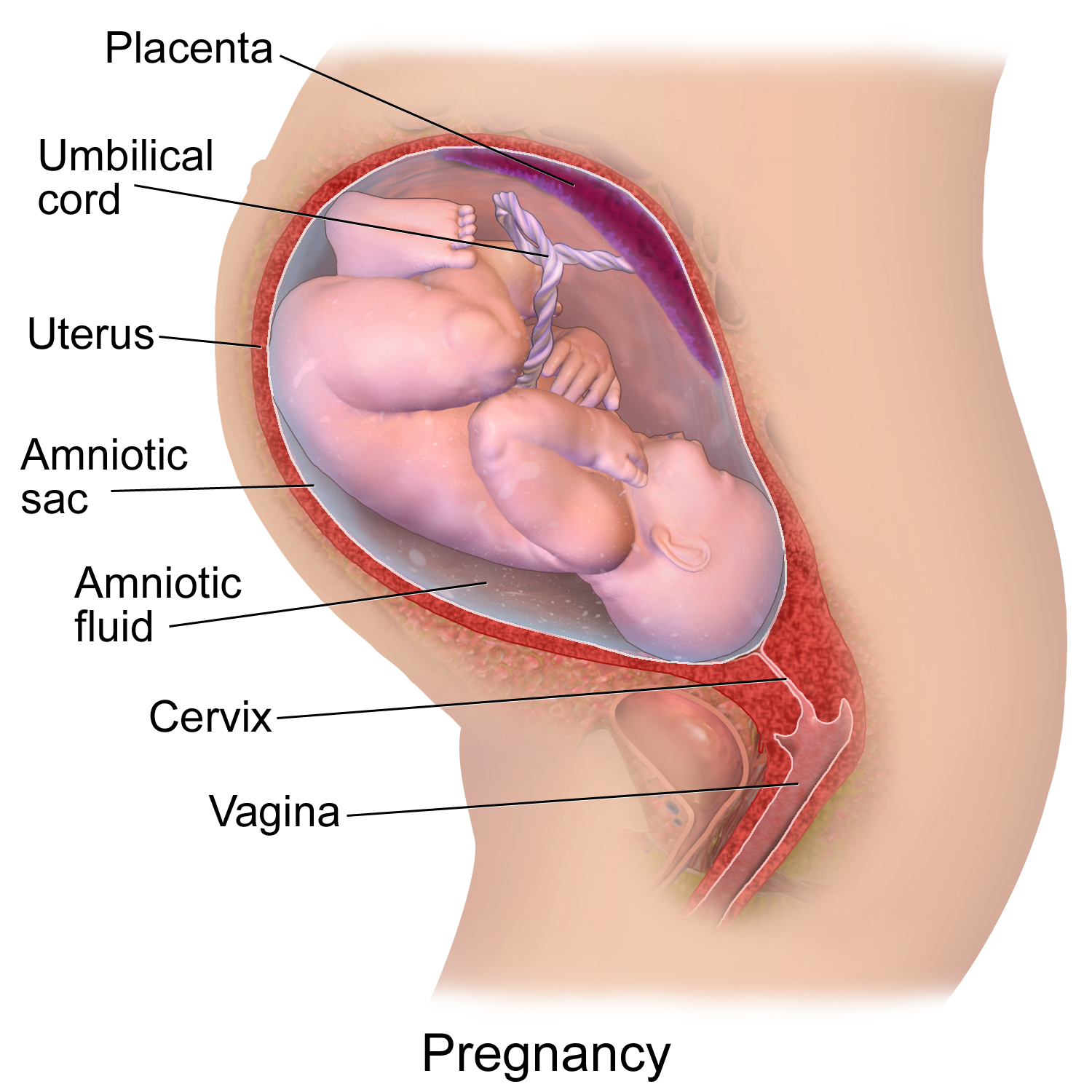
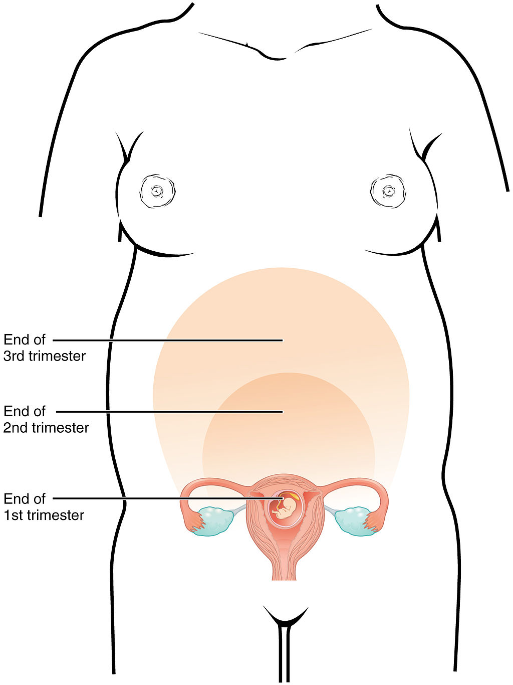
Unlike striated muscle, smooth muscle can sustain very long-term contractions. Smooth muscle can also stretch and still maintain its contractile function, which striated muscle cannot. The elasticity of smooth muscle is enhanced by an extracellular matrix secreted by myocytes. The matrix consists of elastin, collagen, and other stretchy fibres. The ability to stretch and still contract is an important attribute of smooth muscle in organs such as the stomach and uterus (see Figures 12.3.8 and 12.3.9), both of which must stretch considerably as they perform their normal functions.
 |
 |
The following list indicates where many smooth muscles are found, along with some of their specific functions.
- Walls of organs of the gastrointestinal tract (such as the esophagus, stomach, and intestines), moving food through the tract by peristalsis
- Walls of air passages of the respiratory tract (such as the bronchi), controlling the diameter of the passages and the volume of air that can pass through them
- Walls of organs of the male and female reproductive tracts; in the uterus, for example, pushing a baby out of the uterus and into the birth canal
- Walls of structures of the urinary system, including the urinary bladder, allowing the bladder to expand so it can hold more urine, and then contract as urine is released
- Walls of blood vessels, controlling the diameter of the vessels and thereby affecting blood flow and blood pressure
- Walls of lymphatic vessels, squeezing the fluid called lymph through the vessels
- Iris of the eyes, controlling the size of the pupils and thereby the amount of light entering the eyes
- Arrector pili in the skin, raising hairs in hair follicles in the dermis
Cardiac Muscle
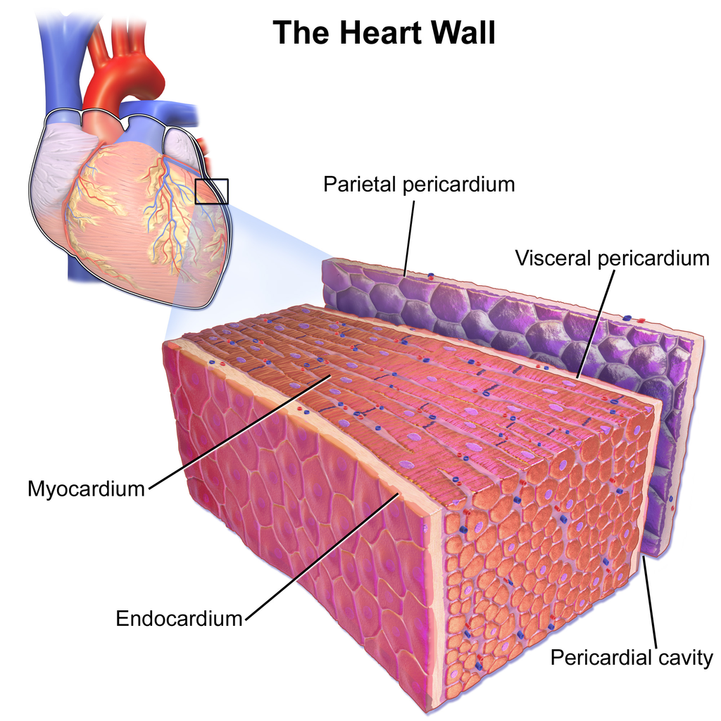
Cardiac muscle is found only in the wall of the heart. It is also called myocardium. As shown in Figure 12.3.10, myocardium is enclosed within connective tissues, including the endocardium on the inside of the heart and pericardium on the outside of the heart. When cardiac muscle contracts, the heart beats and pumps blood. Contractions of cardiac muscle are involuntary, like those of smooth muscles. They are controlled by electrical impulses from specialized cardiac muscle cells in an area of the heart muscle called the sinoatrial node.
Like skeletal muscle, cardiac muscle is striated because its filaments are arranged in sarcomeres inside the muscle fibres. However, in cardiac muscle, the myofibrils are branched at irregular angles rather than arranged in parallel rows (as they are in skeletal muscle). This explains why cardiac and skeletal muscle tissues look different from one another.
The cells of cardiac muscle tissue are arranged in interconnected networks. This arrangement allows rapid transmission of electrical impulses, which stimulate virtually simultaneous contractions of the cells. This enables the cells to coordinate contractions of the heart muscle.
The heart is the muscle that performs the greatest amount of physical work in the course of a lifetime. Although the power output of the heart is much less than the maximum power output of some other muscles in the human body, the heart does its work continuously over an entire lifetime without rest. Cardiac muscle contains a great many mitochondria, which produce ATP for energy and help the heart resist fatigue.
Feature: Human Biology in the News
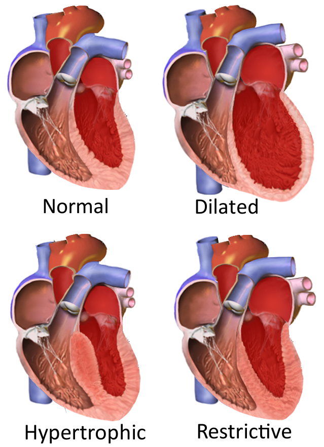
Cardiomyopathy is a disease in which the muscles of the heart are no longer able to effectively pump blood to the body — extreme forms of this disease can lead to heart failure. There are four main types of cardiomyopathy (also illustrated in Figure 12.3.11):
- Dilated (congestive) cardiomyopathy: the left ventricle (the chamber itself) of the heart becomes enlarged and can't pump blood our to the body. This is normally related to coronary artery disease and/or heart attack
- Hypertrophic cardiomyopathy: abnormal thickening of the muscular walls of the left ventricle make the chamber less able to work properly. This condition is more common in patients with a family history of the disease.
- Restrictive cardiomyopathy: the myocardium becomes abnormally rigid and inelastic and is unable to expand in between heartbeats to refill with blood. Restrictive cardiomyopathy typically affects older people.
- Arrhythmogenic right ventricular cardiomyopathy: the right ventricular muscle is replaced by adipose or scar tissue, reducing elasticity and interfering with normal heartbeat and rhythm. This disease is often caused by genetic mutations.
Cardiomyopathy is typically diagnosed with a physical exam supplemented by medical and family history, an angiogram, blood tests, chest x-rays and electrocardiograms. In some cases your doctor would also requisition a CT scan and/or genetic testing.
When treating cardiomyopathy, the goal is to reduce symptoms that affect everyday life. Certain medications can help regularize and slow heart rate, decrease chances of blood clots and cause vasodilation in the coronary arteries. If medication is not sufficient to manage symptoms, a pacemaker or even a heart transplant may be the best option. Lifestyle can also help manage the symptoms of cardiomyopathy — people living with this disease are encouraged to avoid drug and alcohol use, control high blood pressure, eat a healthy diet, get ample rest and exercise, as well as reduce stress levels.
12.3 Summary
- Muscle tissue is a soft tissue that makes up most of the tissues in the muscles of the human muscular system. It is the only type of tissue that has cells with the ability to contract.
- Skeletal muscle tissue is attached to bones by tendons. It allows voluntary body movements.
- Skeletal muscle is the most common type of muscle tissue in the human body. To move bones in opposite directions, skeletal muscles often consist of pairs of muscles that work in opposition to one another to move bones in different directions at joints.
- Skeletal muscle fibres are bundled together in units called muscle fascicles, which are bundled together to form individual skeletal muscles. Skeletal muscles also have connective tissue supporting and protecting the muscle tissue.
- Each skeletal muscle fibre consists of a bundle of myofibrils, which are bundles of protein filaments. The filaments are arranged in repeating units called sarcomeres, which are the basic functional units of skeletal muscles. Skeletal muscle tissue is striated because of the pattern of sarcomeres in its fibres.
- Skeletal muscle fibres can be divided into two types, called slow-twitch and fast-twitch fibres. Slow-twitch fibres are used mainly in aerobic endurance activities, such as long-distance running. Fast-twitch fibres are used mainly for non-aerobic, strenuous activities, such as sprinting. Proportions of the two types of fibres vary from muscle to muscle and person to person.
- Smooth muscle tissue is found in the walls of internal organs and vessels. When smooth muscles contract, they help the organs and vessels carry out their functions. Contractions of smooth muscles are involuntary and controlled by the autonomic nervous system, hormones, and other substances.
- Cells of smooth muscle tissue are not striated because they lack sarcomeres, but the cells contract in the same basic way as striated muscle cells. Unlike striated muscle, smooth muscle can sustain very long-term contractions and maintain its contractile function, even when stretched.
- Cardiac muscle tissue is found only in the wall of the heart. When cardiac muscle contracts, the heart beats and pumps blood. Contractions of cardiac muscle are involuntary, like those of smooth muscles. They are controlled by electrical impulses from specialized cardiac cells.
- Like skeletal muscle, cardiac muscle is striated because its filaments are arranged in sarcomeres inside the muscle fibres. However, the myofibrils are branched instead of arranged in parallel rows, making cardiac and skeletal muscle tissues look different from one another.
- The heart is the muscle that performs the greatest amount of physical work in the course of a lifetime. Its cells contain a great many mitochondria to produce ATP for energy and help the heart resist fatigue.
12.3 Review Questions
- What is muscle tissue?
-
-
- Where is skeletal muscle found, and what is its general function?
- Why do many skeletal muscles work in pairs?
- Describe the structure of a skeletal muscle.
- Relate muscle fibre structure to the functional units of muscles.
- Why is skeletal muscle tissue striated?
- Where is smooth muscle found? What controls the contraction of smooth muscle?
- Where is cardiac muscle found? What controls its contractions?
- The heart muscle is smaller and less powerful than some other muscles in the body. Why is the heart the muscle that performs the greatest amount of physical work in the course of a lifetime? How does the heart resist fatigue?
- Give one example of connective tissue that is found in muscles. Describe one of its functions.
12.3 Explore More
https://www.youtube.com/watch?v=3_PYnWVoUzM
What happens during a heart attack? - Krishna Sudhir, TED-Ed, 2017.
https://www.youtube.com/watch?v=bwOE1MEginA&feature=emb_logo
Three types of muscle | Circulatory system physiology | NCLEX-RN | KhanAcademyMedicine, 2012.
Attributions
Figure 12.3.1
Look by ali-yahya-155huuQwGvA [photo] by Ali Yahya on Unsplash is used under the Unsplash License (https://unsplash.com/license).
Figure 12.3.2
Skeletal_Smooth_Cardiac by OpenStax College on Wikimedia Commons is used under a CC BY 3.0 (https://creativecommons.org/licenses/by/3.0) license.
Figure 12.3.3
Anterior_and_Posterior_Views_of_Muscles by OpenStax on Wikimedia Commons is used under a CC BY 4.0 (https://creativecommons.org/licenses/by/4.0) license.
Figure 12.3.4
Antagonistic Muscle Pair by Laura Guerin at CK-12 Foundation on Wikimedia Commons is used under a CC BY-NC 3.0 (https://creativecommons.org/licenses/by-nc/3.0/) license.
Figure 12.3.5
Muscle_Fibes_(large) by OpenStax on Wikimedia Commons is used under a CC BY 4.0 (https://creativecommons.org/licenses/by/4.0) license.
Figure 12.3.6
Muscle_Fibers_(small) by OpenStax on Wikimedia Commons is used under a CC BY 4.0 (https://creativecommons.org/licenses/by/4.0) license.
Figure 12.3.7
Smooth_Muscle_Contraction by OpenStax on Wikimedia Commons is used under a CC BY 4.0 (https://creativecommons.org/licenses/by/4.0) license.
Figure 12.3.8
Blausen_0747_Pregnancy by BruceBlaus on Wikimedia Commons is used under a CC BY 3.0 (https://creativecommons.org/licenses/by/3.0) license.
Figure 12.3.9
Size_of_Uterus_Throughout_Pregnancy-02 by OpenStax College on Wikimedia Commons is used under a CC BY 3.0 (https://creativecommons.org/licenses/by/3.0) license.
Figure 12.3.10
1024px-Blausen_0470_HeartWall by BruceBlaus on Wikimedia Commons is used under a CC BY 3.0 (https://creativecommons.org/licenses/by/3.0) license.
Figure 12.3.11
Tipet_e_kardiomiopative by Npatchett at English Wikipedia on Wikimedia Commons is used under a CC BY-SA 3.0 (https://creativecommons.org/licenses/by-sa/3.0) license. (Work derived from Blausen 0165 Cardiomyopathy Dilated by BruceBlaus)
References
Betts, J. G., Young, K.A., Wise, J.A., Johnson, E., Poe, B., Kruse, D.H., Korol, O., Johnson, J.E., Womble, M., DeSaix, P. (2013, June 19). Figure 4.18 Muscle tissue [digital image]. In Anatomy and Physiology (Section 4.4). OpenStax. https://openstax.org/books/anatomy-and-physiology/pages/4-4-muscle-tissue-and-motion
Betts, J. G., Young, K.A., Wise, J.A., Johnson, E., Poe, B., Kruse, D.H., Korol, O., Johnson, J.E., Womble, M., DeSaix, P. (2013, June 19). Figure 28.18 Size of uterus throughout pregnancy [digital image]. In Anatomy and Physiology (Section 28.4). OpenStax. https://openstax.org/books/anatomy-and-physiology/pages/28-4-maternal-changes-during-pregnancy-labor-and-birth
Betts, J. G., Young, K.A., Wise, J.A., Johnson, E., Poe, B., Kruse, D.H., Korol, O., Johnson, J.E., Womble, M., DeSaix, P. (2016, May 18). Figure 10.3 The three connective tissue layers [digital image]. In Anatomy and Physiology (Section 10.2). OpenStax. https://openstax.org/books/anatomy-and-physiology/pages/10-2-skeletal-muscle
Betts, J. G., Young, K.A., Wise, J.A., Johnson, E., Poe, B., Kruse, D.H., Korol, O., Johnson, J.E., Womble, M., DeSaix, P. (2016, May 18). Figure 10.4 Muscle fiber [digital image]. In Anatomy and Physiology (Section 10.2). OpenStax. https://openstax.org/books/anatomy-and-physiology/pages/10-2-skeletal-muscle
Betts, J. G., Young, K.A., Wise, J.A., Johnson, E., Poe, B., Kruse, D.H., Korol, O., Johnson, J.E., Womble, M., DeSaix, P. (2016, May 18). Figure 10.24 Muscle contraction [digital image]. In Anatomy and Physiology (Section 10.8). OpenStax. https://openstax.org/books/anatomy-and-physiology/pages/10-8-smooth-muscle
Betts, J. G., Young, K.A., Wise, J.A., Johnson, E., Poe, B., Kruse, D.H., Korol, O., Johnson, J.E., Womble, M., DeSaix, P. (2016, May 18). Figure 11.5 Overview of the muscular system [digital image]. In Anatomy and Physiology (Section 11.2). OpenStax. https://openstax.org/books/anatomy-and-physiology/pages/11-2-naming-skeletal-muscles
Blausen.com staff. (2014). Medical gallery of Blausen Medical 2014. WikiJournal of Medicine 1 (2). DOI:10.15347/wjm/2014.010. ISSN 2002-4436.
Brainard, J/ CK-12 Foundation. (2012). Figure 5 Triceps and biceps muscles in the upper arm are opposing muscles. [digital image]. In CK-12 Biology (Section 21.3) [online Flexbook]. CK12.org. https://www.ck12.org/book/ck-12-biology/section/21.3/ (Last modified August 11, 2017.)
khanacademymedicine. (2012, October 19). Three types of muscle | Circulatory system physiology | NCLEX-RN | Khan Academy. YouTube.
TED-Ed. (2017, February 14). What happens during a heart attack? - Krishna Sudhir. YouTube. https://www.youtube.com/watch?v=3_PYnWVoUzM&feature=youtu.be
Image illustrates how independent alignment greatly increases the genetic diversity among gametes produced. In a single cell undergoing Meiosis, independent alignment means that whether the paternal or maternal chromosome for each homologous pair ends up on the left or right is totally random, and is random for each pair of homologous chromosomes. In a cell which has 3 pairs of homologous chromosomes, there are 8 possible random alignments, which would each result in a different set of gametes being produced.
Gland such as a sweat gland, salivary gland, or mammary gland that secretes a substance into a duct that carries the secretion to the outside of the body.
Image shows the difference in morphology between a sickle cell and a normal red blood cell. The normal red blood cells are shaped like danishes, while the sickle cells are shaped like bananas
Created by: CK-12/Adapted by Christine Miller
Figure 5.16.1 Potato plants: One genetically engineered and healthy (left), and one infected with bacterial ring rot (right).
Please Pass the Potatoes
You might want to pass on the potato plants on the right in Figure 5.16.1. They are infected with a virus, which is quickly killing them. The potato plants on the left are healthy and productive. Why aren't they infected with the same virus? The plants on the left have been genetically engineered to make them resistant to the virus.
What Is Genetic Engineering?
Genetic engineering is the use of technology to change the genetic makeup of living things for human purposes. Generally, the goal of genetic engineering is to modify organisms so they are more useful to humans. Genetic engineering, for example, may be used to create crops that yield more food or resist insect pests or viruses, such as the virus-resistant potatoes pictured (left) in Figure 5.16.1 . Research is also underway to use genetic engineering to cure human genetic disorders with gene therapy.
Genetic Engineering Methods
Genetic engineering uses a variety of techniques to achieve its aims. Two commonly used techniques are gene cloning and the polymerase chain reaction.
Gene Cloning
Gene cloning is the process of isolating and making copies of a gene. This is useful for many purposes. For example, gene cloning might be used to isolate and make copies of a normal gene for gene therapy. Gene cloning involves four steps: isolation, ligation, transformation, and selection.
- In the isolation step, an enzyme is used to break DNA at a specific base sequence. This is done to isolate a gene.
- During ligation, the enzyme DNA ligase combines the isolated gene with plasmid DNA from bacteria. (Plasmid DNA is circular DNA that is not part of a chromosome and can replicate independently). The DNA that results is called recombinant DNA.
- In transformation, the recombinant DNA is inserted into a living cell, usually a bacterial cell.
- Selection involves growing transformed bacteria to make sure they have the recombinant DNA. This is a necessary step because transformation is not always successful. Only bacteria that contain the recombinant DNA are selected for further use.
Polymerase Chain Reaction
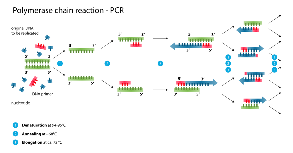
The polymerase chain reaction (PCR) makes many copies of a gene or other DNA segment. This might be done in order to make large quantities of a gene for genetic testing. PCR involves three steps: denaturing, annealing, and extension. The three steps are illustrated in Figure 5.16.2. They are repeated many times in a cycle to make large quantities of the gene.
- Denaturing involves heating DNA to break the bonds holding together the two DNA strands, yielding two single strands of DNA.
- Annealing involves cooling the single strands of DNA and mixing them with short DNA segments called primers. Primers have base sequences that are complementary to segments of the single DNA strands. As a result, bonds form between the DNA strands and primers.
- Extension [or Elongation] occurs when an enzyme (Taq polymerase or Taq DNA polymerase) adds nucleotides to the primers. This produces new DNA molecules, each incorporating one of the original DNA strands.
Uses of Genetic Engineering
Methods of genetic engineering can be used for many practical purposes. They are used widely in both medicine and agriculture.
Applications in Medicine
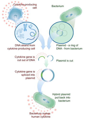
In addition to gene therapy for genetic disorders, genetic engineering can be used to transform bacteria so they are able to make human proteins (see Figure 5.16.3). Proteins made by the bacteria are injected into people who cannot produce them because of mutations.
Insulin was the first human protein to be produced in this way. Insulin helps cells take up glucose from the blood. People with type 1 diabetes have a mutation in the gene that normally codes for insulin. Without insulin, their blood glucose rises to harmfully high levels. At present, the only treatment for type 1 diabetes is the injection of insulin from outside sources. Until recently, there was no known way to make human insulin outside the human body. The problem was solved by gene cloning. The human insulin gene was cloned and used to transform bacterial cells, which could then produce large quantities of human insulin.
Applications in Agriculture
Genetic engineering has been used to create transgenic crops. Transgenic crops are genetically modified with new genes that code for traits useful to humans.
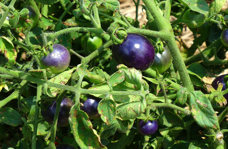
Transgenic crops have been created with a variety of different traits. They can yield more food, taste better, survive drought, tolerate salty soil, and resist insect pests, among other things. Scientists have even created a transgenic purple tomato (Figure 5.16.4) that contains high levels of cancer-fighting compounds called antioxidants.
Ethical, Legal, and Social Issues
The use of genetic engineering has raised a number of ethical, legal, and social issues. Here are just a few:
- Who owns genetically modified organisms (such as bacteria)? Can such organisms be patented like inventions?
- Are genetically modified foods safe to eat? Might they have harmful effects on the people who consume them?
- Are genetically engineered crops safe for the environment? Might they harm other organisms — or even entire ecosystems?
- Who controls a person’s genetic information? What safeguards ensure that the information is kept private?
- How far should we go to ensure that children are free of mutations?
This example shows how complex such issues may be:
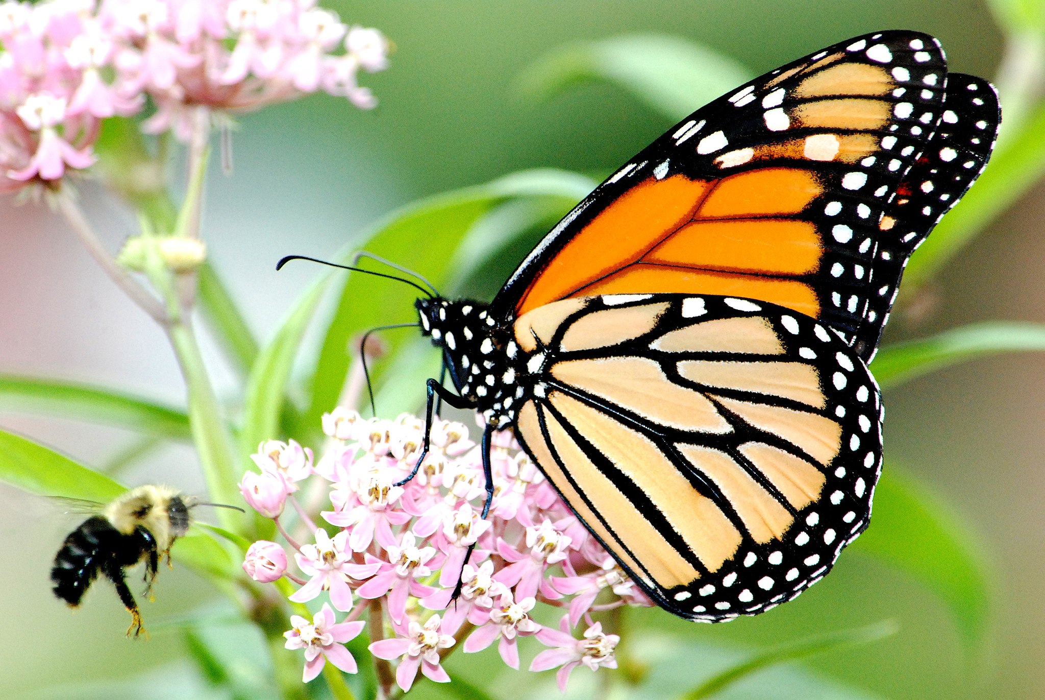
A strain of corn has been created with a gene that encodes a natural pesticide. On the positive side, the transgenic corn is not eaten by insects, so there is more corn for people to eat. The corn also doesn’t need to be sprayed with chemical pesticides, which can harm people and other living things. On the negative side, the transgenic corn has been shown to cross-pollinate nearby milkweed plants. Offspring of the cross-pollinated milkweed plants are now known to be toxic to monarch butterfly caterpillars that depend on them for food. Scientists are concerned that this may threaten the monarch species, as well as other species that normally eat monarchs.
As this example shows, the pros of genetic engineering may be obvious, but the cons may not be known until it is too late, and the damage has already been done. Unforeseen harm may be done to people, other species, and entire ecosystems. No doubt the ethical, legal, and social issues raised by genetic engineering will be debated for decades to come.
Feature: Reliable Sources
Genetically modified foods (or GM foods) are foods produced from genetically modified organisms. These are organisms that have had changes introduced into their DNA using methods of genetic engineering. Commercial sale of GM foods began in 1994, with a tomato that had delayed ripening. By 2015, three major crops grown in the U.S. were raised mainly from GM seeds, including field corn, soybeans, and cotton. Many other crops were also raised from GM seeds, ranging from a variety of vegetables to sugar beets. Other sources of GM foods in our diet include meats, eggs, and dairy products from animals that have eaten GM feed, as well as a plethora of food products that contain some form of soy or corn products, such as soybean oil, soybean flour, corn oil, corn starch, and corn syrup. A quick glance at the ingredients list of most processed foods shows that these products are added to many of the items in a typical American diet.
Most scientists think that GM foods are not necessarily any riskier to human health than conventional foods. Nonetheless, in many countries, including the U.S., GM foods are given more rigorous evaluations than conventional foods. For example, GM foods are assessed for toxicity, ability to cause allergic reactions, and stability of inserted genes. GM crops are also evaluated for possible environmental effects, such as outcrossing, which is the migration of genes from GM plants to conventional crops or wild plant species.
Despite the extra measures used to evaluate GM foods, there is a lot of public concern about them, including whether they are safe for human health, how they are labeled, and their environmental impacts. These concerns are based on a number of factors, such as the worrying belief that scientists are creating entirely new species, and a perceived lack of benefits to the consumer of GM foods. People may also doubt the validity of risk assessments, especially with regard to long-term effects. Lack of labeling of GM foods is also an issue because it denies consumers the choice of buying GM or conventional foods.
Find reliable online sources about GM foods. Look for information to answer the questions below. Make sure you evaluate the nature of the sources when you assess the reliability of the information they provide. Consider whether the sources may have a vested interest in one side of the issue or another. For example, major chemical companies might promote the use of seeds for crops that have been genetically engineered to be herbicide tolerant. Why? Because it boosts the use of the weed-killing chemical herbicides they produce and sell.
- In what ways are crops modified genetically? What traits are introduced, and what methods are used to introduce them?
- What are the main human safety questions about GM foods? How is the human safety of GM foods assessed?
- What are the main environmental concerns about GM crops? How is risk assessment for the environment performed?
- What are the major pros and cons of GM crops and foods? Who is most affected by these pros and cons? For example, for pros, do growers and marketeers receive most of the benefits, or do consumers also reap rewards?
5.16 Summary
- Genetic engineering is the use of technology to change the genetic makeup of living things for human purposes.
- Genetic engineering methods include gene cloning and the polymerase chain reaction. Gene cloning is the process of isolating and making copies of a DNA segment, such as a gene. The polymerase chain reaction makes many copies of a gene or other DNA segment.
- Genetic engineering can be used to transform bacteria so they are able to make human proteins, such as insulin. It can also be used to create transgenic crops, like crops that yield more food or resist insect pests.
- Genetic engineering has raised a number of ethical, legal, and social issues. For example, are genetically modified foods safe to eat? Who controls a person’s genetic information?
5.16 Review Questions
- Define genetic engineering
-
- What is recombinant DNA?
- Identify the steps of gene cloning.
- What is the purpose of the polymerase chain reaction?
- Make a flow chart outlining the steps involved in creating a transgenic crop.
- Explain how bacteria can be genetically engineered to produce a human protein.
- Identify an ethical, legal, or social issue raised bygenetic engineering. State your view on the issue, and develop a logical argument to support your view.
- Explain what primers are and what they do in PCR.
- The enzyme Taq polymerase was originally identified from bacteria that live in very hot environments, such as hotsprings. Why does this fact make Taq polymerase particularly useful in PCR reactions?
5.16 Explore More
https://www.youtube.com/watch?time_continue=1&v=3IsQ92KiBwM&feature=emb_logo
What is Genetic Engineering?, Eco-Wise Videos, 2015.
https://www.youtube.com/watch?time_continue=1&v=g_ZswrLFSdo&feature=emb_logo
Bringing biotechnology into the home: Cathal Garvey at TEDxDublin,
TEDx Talks, 2013.
Attributions
Figure 5.16.1
- Potato Plant by Lehava Maghar (Pikiwikisrael) on Wikimedia Commons via the PikiWiki - Israel free image collection project is used under a CC BY 2.5 (https://creativecommons.org/licenses/by/2.5/) license.
- Potato Plant Infected with Bacterial Ring Rot by William M. Brown Jr. on Wikimedia Commons via William M. Brown Jr., Bugwood.org via forestryimages.org is used under a CC BY 3.0 US (https://creativecommons.org/licenses/by/3.0/us/deed.en) license.
Figure 5.16.2
Polymerase_chain_reaction.svg by Enzoklop on Wikimedia Commons is used under a
CC BY-SA 3.0 (https://creativecommons.org/licenses/by-sa/3.0/deed.en) license.
Figure 5.16.3
Genetic Engineering in Medicine by CK-12 Foundation is used under a CC BY-NC 3.0 (https://creativecommons.org/licenses/by-nc/3.0/) license.
 ©CK-12 Foundation Licensed under
 ![]() • Terms of Use • Attribution
• Terms of Use • Attribution
Figure 5.16.4
Purple Tomato/Indigo Rose by F Delventhal on Flickr is used under a CC BY 2.0 (https://creativecommons.org/licenses/by/2.0/) license.
Figure 5.16.5
Monarch_Butterfly_and_Bumble_Bee_on_Swamp_Milkweed_(28960994212) by U.S. Fish and Wildlife Service on Wikimedia Commons is in the public domain (https://en.wikipedia.org/wiki/Public_domain).
References
Brainard, J/ CK-12 Foundation. (2016). Figure 4 Genetically engineering bacteria to produce a human protein. [digital image]. In CK-12 College Human Biology (Section 5.15) [online Flexbook]. CK12.org. https://www.ck12.org/book/ck-12-college-human-biology/section/5.15/
Eco-Wise Videos. (2015, March 28). What is genetic engineering? YouTube. https://www.youtube.com/watch?v=3IsQ92KiBwM&feature=youtu.be
TEDx Talks. (2013, October 22). Bringing biotechnology into the home: Cathal Garvey at TEDxDublin. YouTube. https://www.youtube.com/watch?v=g_ZswrLFSdo&feature=youtu.be
A salivary gland that is located deep under the mandible (jawbone).
A salivary gland that is located under the floor of the mouth, close to the midline.
The first printout of the human genome to be presented as a series of books, displayed in the 'Medicine Now' room at the Wellcome Collection, London. The 3.4 billion units of DNA code are transcribed into more than a hundred volumes, each a thousand pages long, in type so small as to be barely legible.
Image shows a sperm fertilizing an egg. The Sperm is much smaller than the egg.
Image shows a medical professional blotting a blood sample from a small prick on an infant's heel onto special filter paper for the purposes of screening for PKU.
An antibody, also known as an immunoglobulin, is a large, Y-shaped protein produced mainly by plasma cells that is used by the immune system to neutralize pathogens such as pathogenic bacteria and viruses.
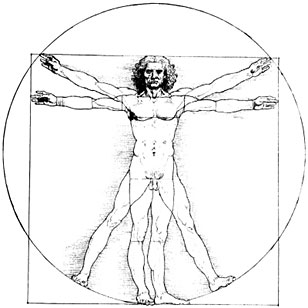
Vitruvian Man
The drawing in Figure 5.17.1, named Vitruvian Man, was created by Leonardo da Vinci in 1490. It was meant to show normal human body proportions. Vitruvian Man is used today to represent a different approach to the human body. It symbolizes a scientific research project that began in 1990, exactly 500 years after da Vinci created the drawing. That project, called the Human Genome Project, is the largest collaborative biological research project ever undertaken.
What Is the Human Genome?
The human genome refers to all the DNA of the human species. Human DNA consists of 3.3 billion base pairs, and it is divided into more than 20 thousand genes on 23 chromosomes. Humans inherit one set of chromosomes from each parent. So there are actually two copies of each of those 20,000 genes. The human genome also includes noncoding sequences of DNA, as shown in Figure 5.17.2.
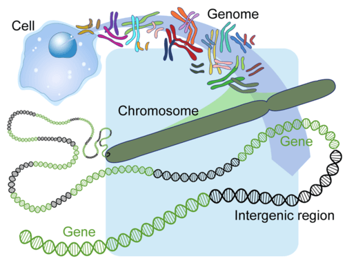
Discovering the Human Genome
Scientists now know the sequence of all the DNA base pairs in the entire human genome. This knowledge was attained by the Human Genome Project (HGP), a $3 billion, international scientific research project that was formally launched in 1990. The project was completed in 2003, two years ahead of its 15-year projected deadline.
Determining the sequence of the billions of base pairs that make up human DNA was the main goal of the HGP. Another goal was to map the location and determine the function of all the genes in the human genome. A somewhat surprising finding of the HGP is the relatively small number of human genes. There are only about 20,500 genes in human beings. This may sound like a lot, but it's about the same number as in mice. Another surprising finding of the HGP is the large number of nearly identical, repeated DNA segments in the human genome. This number was previously believed to be much smaller.
A Collaborative Effort
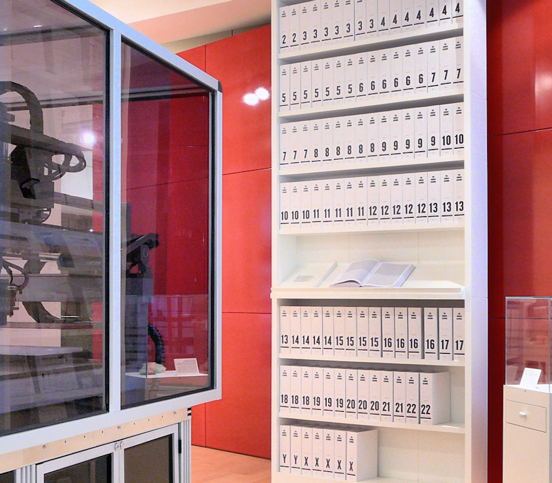
Funding for the HGP came from the U.S. Department of Energy and the National Institutes of Health, as well as from foreign institutions. The actual research was undertaken by scientists in 20 universities in the U.S., United Kingdom, Australia, France, Germany, Japan, and China. A private U.S. company named Celera also contributed to the effort. Although Celera had hoped to patent some of the genes it discovered, this was later denied. The entire DNA sequence of the genome is stored in databases that are available to anyone on the Internet. Additional data and tools for analyzing the human genome are also available online.
Reference Genome of the Human Genome Project
In 2003, the HGP published the results of its sequencing of DNA as a human reference genome. The reference genome sequences a full set of human chromosomes, but it clearly doesn't represent the sequence of every human individual's genome. Instead, it is the combined mosaic of a small number of anonymous donors. The DNA that was sequenced came from blood samples of the female donors and sperm samples of the male donors. All of the donors were of European origin, and more than 70 per cent of the reference DNA came from a single anonymous male donor from Buffalo, New York. Identities of all the donors were protected so neither they nor the researchers could know whose DNA was sequenced.
Subsequent projects have sequenced the genomes of multiple distinct ethnic groups. Ongoing research is searching base by base for variations in the sequence. However, there is still only one reference genome available.
Benefits of the Human Genome Project
The sequencing of the human genome has benefits for many fields, including molecular medicine and human evolution.
- Knowing the human DNA sequence can help us understand many human diseases. For example, it is helping researchers identify mutations linked to different forms of cancer. It is also yielding insights into the genetic basis of cystic fibrosis, liver diseases, blood-clotting disorders, and Alzheimer's disease, among others.
- The human DNA sequence can also help researchers tailor medications to individual genotypes. This is called personalized medicine, and it has led to an entirely new field called pharmacogenomics. Pharmacogenomics, also called pharmacogenetics, is the study of how our genes affect the way we respond to drugs. You can read more about pharmacogenomics in the Feature section below.
- The analysis of similarities between DNA sequences from different organisms is opening new avenues in the study of evolution. For example, analyses are expected to shed light on many questions about the similarities and differences between humans and our closest relatives, the nonhuman primates.
Ethical, Legal, and Social Issues of the Human Genome Project
From its launch in 1990, the HGP proactively established and funded a separate committee to oversee potential ethical, legal, and social issues associated with the project. Some of these possible issues include:
- The possible use of the knowledge generated by the project to discriminate against people. There were worries that that employers and health insurance companies would refuse to hire or insure people based on their genetic makeup, for instance, if they had genes that increased their risk of getting certain diseases.
- The issues surrounding "ownership" of DNA sequences. There have been requests by the both governmental agencies and private companies for patents of certain sequences of human genes called cDNA, although the US Patent Office has rejected all applications.
- Education of healthcare professionals, policy makers and the public about the complex issues involved in the HGP and what is done with the information acquired from it.
Feature: Human Biology in the News
Not everyone responds to medications in the same way. A drug that works well for one person may not be effective for another. The dose of a drug that cures a disease in one individual may be inadequate for someone else. Some people may experience side effects from a given medication, whereas other people do not. This variation in responses to medications can be due to differences in our genes. That's where the field of pharmacogenetics comes in. News media have hailed it as the "new frontier in medicine." It certainly seems to hold promise for improving the treatment of patients with pharmaceutical drugs.
Pharmacogenomics is based on a special kind of genetic testing. It looks for small genetic variations that influence a person's ability to activate and deactivate drugs. Results of the tests can help doctors choose the best drug and most effective dose for a given patient. Some of the greatest successes of pharmacogenomics have been in cancer treatment. Many of the drugs that treat cancer need to be activated by the patient's own enzymes. Inherited variations in enzymes may affect how quickly or efficiently the drugs are activated. For example, if a patient's enzymes break down a particular drug too slowly, then standard doses of the drug may not work very well for that patient. Drugs also must be deactivated to reduce their effects on healthy cells. If a patient's enzymes deactivate a drug too slowly, then the drug may remain at high levels and cause side effects.
One of the main benefits of pharmacogenomics is greater patient safety. Pharmacogenomic testing may help identify patients who are likely to experience adverse reactions to drugs, so that different, safer drugs can be prescribed. Another benefit of pharmacogenomics is eliminating the trial-and-error approach that is often used to find appropriate medications and doses for a given patient. This saves time and money, as well as improving patient outcomes.
Because pharmacogenomics is new, some insurance companies do not cover it, and it can be very expensive. Also, not all of the genetic tests are widely available at this point. In addition, there may be ethical and legal issues associated with the genetic testing, including concerns about privacy issues.
5.17 Summary
- The human genome refers to all of the DNA of the human species. It consists of more than 3.3 billion base pairs divided into 20,500 genes on 23 pairs of chromosomes.
- The Human Genome Project (HGP) was a multi-billion dollar international research project that began in 1990. By 2003, it had sequenced all of the DNA base pairs in the human genome. It also mapped the location and determined the function of all the genes in the human genome.
- In 2003, the HGP published the results of its sequence of DNA as a human reference genome. The entire DNA sequence is stored in databases that are available to anyone on the Internet.
- Sequencing of the human genome is helping researchers better understand cancer and genetic diseases. It is also helping them tailor medications to individual patients, which is the focus of the new field of pharmacogenomics. In addition, it is helping researchers better understand human evolution.
- From its launch in 1990, the HGP established and funded a separate committee to oversee potential ethical, legal, and social issues associated with the project.
5.17 Review Questions
- Describe the human genome.
- What is the Human Genome Project?
- Identify two main goals of the Human Genome Project.
- What is the reference genome of the Human Genome Project? What is it based on?
- Explain how knowing the sequence of DNA bases in the human genome is beneficial for molecular medicine.
- What was one surprising finding of the Human Genome Project?
- Why do you think scientists didn’t just sequence the DNA from a single person for the Human Genome Project? Along those lines, why do you think it is important to include samples from different ethnic groups and genders in genome sequencing efforts?
- What is pharmacogenomics?
- If a patient were to have pharmacogenomics done to optimize their medication, what do you think the first step would be?
- List one advantage and one disadvantage of pharmacogenomics.
- Explain how the sequencing of the human genome relates to ethical concerns about genetic discrimination.
5.17 Explore More
https://www.youtube.com/watch?v=AhsIF-cmoQQ
The race to sequence the human genome - Tien Nguyen, TED-Ed, 2015.
https://www.youtube.com/watch?v=s6rJLXq1Re0
How to read the genome and build a human being | Riccardo Sabatini, TED, 2016.
https://www.youtube.com/watch?v=J2ITkfzp0SY
Personalized Medicine: A New Approach | Luigi Boccuto | TEDxGreenville,
TEDx Talks, 2017.
Attributions
Figure 5.17.1
Vitruvian_man (black and white copy) by Leonardo Da Vinci, 1490, by Ianbond on Wikimedia Commons is released into the public domain (https://en.wikipedia.org/wiki/Public_domain).
Figure 5.17.2
Human Genome by CK-12 Foundation is used under a CC BY-NC 3.0 (https://creativecommons.org/licenses/by-nc/3.0/) license.
 ©CK-12 Foundation Licensed under
 ![]() • Terms of Use • Attribution
• Terms of Use • Attribution
Figure 5.17.3
Human genome_bookcase by Russ London at English Wikipedia is used under a CC BY-SA 3.0 (https://creativecommons.org/licenses/by-sa/3.0/deed.en) license.
References
Brainard, J/ CK-12 Foundation. (2016). Figure 2 Human genome, chromosomes, and genes. [digital image]. In CK-12 College Human Biology (Section 5.16) [online Flexbook]. CK12.org. https://www.ck12.org/book/ck-12-college-human-biology/section/5.16/
National Human Genome Research Institute (NHGRI). (n.d.). The Human Genome Project (HGP). National Institute of Health (NIH) /US Government. https://www.genome.gov/human-genome-project
TED. (2016, May 24). How to read the genome and build a human being | Riccardo Sabatini. YouTube. https://www.youtube.com/watch?v=s6rJLXq1Re0&t=2s
TED-Ed. (2015, October 12). The race to sequence the human genome - Tien Nguyen. YouTube. https://www.youtube.com/watch?v=AhsIF-cmoQQ&t=7s
TEDx Talks. (2017, June 8). Personalized medicine: A new approach | Luigi Boccuto | TEDxGreenville. https://www.youtube.com/watch?v=J2ITkfzp0SY&t=4s
Wikipedia contributors. (2020, April 18). Celera Corporation. In Wikipedia. https://en.wikipedia.org/w/index.php?title=Celera_Corporation&oldid=951693886
Wikipedia contributors. (2020, July 6). Human Genome Project. In Wikipedia. https://en.wikipedia.org/w/index.php?title=Human_Genome_Project&oldid=966272762
Wikipedia contributors. (2020, July 12). Leonardo da Vinci. In Wikipedia. https://en.wikipedia.org/w/index.php?title=Leonardo_da_Vinci&oldid=967303882
Wikipedia contributors. (2020, May 19). Vitruvian Man. In Wikipedia. https://en.wikipedia.org/w/index.php?title=Vitruvian_Man&oldid=957472578
Image shows a freshly baked Steak and Kidney Pie.
A hard structure, embedded in the jaws of the mouth, that functions in chewing. Made of a dentin and covered in enamel, the hardest tissue in the body.
Image shows the possible types of mutations on the BCRA1 gene, including nonsense, framshift, missense, and splice-site mutations. There are approximately over 500 different mutations reported in this gene, which is present on chromosome 17.
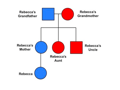
Case Study Conclusion: Cancer in the Family
Rebecca’s family tree, as illustrated in the pedigree above (Figure 5.18.1), shows a high incidence of cancer among close relatives. But are genes the cause of cancer in this family? Only genetic testing, which is the sequencing of specific genes in an individual, can reveal whether a cancer-causing gene is being inherited in this family.
Fortunately for Rebecca, the results of her genetic testing show that she does not have the mutations in the BRCA1 and BRCA2 genes that most commonly increase a person’s risk of getting cancer. This doesn't mean, however, that she doesn’t have other mutations in these genes that could increase her risk of getting cancer. There are many other mutations in BRCA genes whose effect on cancer risk is still not known — and there may be many more yet to be discovered. Figure 5.18.2 from the National Cancer Institute illustrates many of the different types of known mutations in the BRCA1 gene. It is important to continue to study the variations in genes such as BRCA in different people to better assess their possible contribution to the development of disease. As you now know from this chapter, many mutations are harmless, while others can cause significant health effects, depending on the specific mutation and the gene involved.
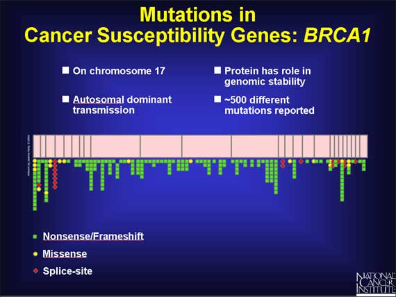
Mutations in BRCA genes are particularly likely to cause cancer because these genes encode for tumor-suppressor proteins that normally repair damaged DNA and control cell division. If these genes are mutated in a way that causes the proteins to not function properly, other mutations can accumulate and cell division can run out of control, which can cause cancer.
BRCA1 and BRCA2 are on chromosomes 17 and 13, respectively, which are autosomes. As Rebecca’s genetic counselor mentioned, mutations in these genes have a dominant inheritance pattern. Now that you know the pattern of inheritance of autosomal dominant genes, if Rebecca’s grandmother did have one copy of a mutated BRCA gene, what are the chances that Rebecca’s mother also has this mutation? Because it is dominant, only one copy of the gene is needed to increase the risk of cancer, and because it is on autosomes instead of sex chromosomes, the sex of the parent or offspring does not matter in the inheritance pattern. In this situation, Rebecca’s grandmother’s eggs would have had a 50 per cent chance of having a BRCA gene mutation (Mendel’s law of segregation). Therefore, Rebecca’s mother would have had a 50 per cent chance of inheriting this gene. Even though Rebecca does not have the most common BRCA mutations that increase the risk of cancer, it does not mean that her mother does not, because there would also only be a 50 per cent chance that she would pass it on to Rebecca. Rebecca’s mother, therefore, should consider getting tested for mutations in the BRCA genes, as well. Ideally, the individuals with cancer in a family should be tested first when a genetic cause is suspected, so that if there is a specific mutation being inherited, it can be identified, and the other family members can be tested for that same mutation.
Mutations in both BRCA1 and BRCA2 are often found in Ashkenazi Jewish families. However, these genes are not linked in the chromosomal sense, because they are on different chromosomes and are therefore inherited independently, in accordance with Mendel’s law of independent assortment. Why would certain gene mutations be prevalent in particular ethnic groups? If people within an ethnic group tend to produce offspring with each other, their genes will remain prevalent within the group. These may be genes for harmless variations such as skin, hair, or eye colour, or harmful variations such as the mutations in the BRCA genes. Other genetically based diseases and disorders are sometimes more commonly found in particular ethnic groups, such as cystic fibrosis in people of European descent, and sickle cell anemia in people of African descent. You will learn more about the prevalence of certain genes and traits in particular ethnic groups and populations in the chapter on Human Variation.
As you learned in this chapter, genetics is not the sole determinant of phenotype. The environment can also influence many traits, including adult height and skin colour. The environment plays a major role in the development of cancer, too. Ninety to 95 per cent of all cancers do not have an identified genetic cause, and are often caused by mutagens in the environment, such as UV radiation from the sun or toxic chemicals in cigarette smoke. But for families like Rebecca’s, knowing their family health history and genetic makeup may help them better prevent or treat diseases that are caused by their genetic inheritance. If a person knows they have a gene that can increase their risk of cancer, they can make lifestyle changes and have early and more frequent cancer screenings. They may even choose to have preventative surgeries that can help reduce their risk of getting cancer and increase their odds of long-term survival if cancer does occur. The next time you go to the doctor and they ask whether any members of your family have had cancer, you will have a deeper understanding why this information is so important to your health.
Chapter 5 Summary
In this chapter you learned about genetics — the science of heredity. Specifically you learned that:
- Chromosomes are structures made of DNA and proteins that are encoded with genetic instructions for making RNA and proteins. The instructions are organized into units called genes, which are segments of DNA that code for particular pieces of RNA. The RNA molecules can then act as a blueprint for proteins, or directly help regulate various cellular processes.
- Humans normally have 23 pairs of chromosomes. Of these, 22 pairs are autosomes, which contain genes for characteristics unrelated to sex. The other pair consists of sex chromosomes (XX in females, XY in males). Only the Y chromosome contains genes that determine sex.
- Humans have an estimated 20 thousand to 22 thousand genes. The majority of human genes have two or more possible versions, called alleles.
- Genes that are located on the same chromosome are called linked genes. Linkage explains why certain characteristics are frequently inherited together.
- Determining that DNA is the genetic material was an important milestone in biology.
-
- In the 1920s, Griffith showed that something in virulent bacteria could be transferred to nonvirulent bacteria, making them virulent, as well.
- In the 1940s, Avery and colleagues showed that the "something" Griffith found was DNA and not protein. This result was confirmed by Hershey and Chase, who demonstrated that viruses insert DNA into bacterial cells.
- In the 1950s, Chargaff showed that in DNA, the concentration of adenine is always the same as the concentration of thymine, and the concentration of guanine is always the same as the concentration of cytosine. These observations came to be known as Chargaff's rules.
- In the 1950s, James Watson and Francis Crick, building on the prior X-ray research of Rosalind Franklin and others, discovered the double helix structure of the DNA molecule.
- Knowledge of DNA's structure helped scientists understand how DNA replicates, which must occur before cell division. DNA replication is semi-conservative because each daughter molecule contains one strand from the parent molecule and one new strand that is complementary to it.
- The central dogma of molecular biology can be summed up as: DNA → RNA → Protein. This means that the genetic instructions encoded in DNA are transcribed to RNA. From RNA, they are translated into a protein.
- RNA is a nucleic acid. Unlike DNA, RNA consists of just one polynucleotide chain instead of two, contains the base uracil instead of thymine, and contains the sugar ribose instead of deoxyribose.
- The main function of RNA is to help make proteins. There are three main types of RNA: messenger RNA (mRNA), ribosomal RNA (rRNA), and transfer RNA (tRNA).
- According to the RNA world hypothesis, RNA was the first type of biochemical molecule to evolve, predating both DNA and proteins.
- The genetic code was cracked in the 1960s by Marshall Nirenberg. It consists of the sequence of nitrogen bases in a polynucleotide chain of DNA or RNA. The four bases make up the "letters" of the code. The letters are combined in groups of three to form code "words," or codons, each of which encodes for one amino acid or a start or stop signal.
-
- AUG is the start codon, and it establishes the reading frame of the code. After the start codon, the next three bases are read as the second codon, and so on until a stop codon is reached.
- The genetic code is universal, unambiguous, and redundant.
- Protein synthesis is the process in which cells make proteins. It occurs in two stages: transcription and translation.
-
- Transcription is the transfer of genetic instructions in DNA to mRNA in the nucleus. It includes the steps of initiation, elongation, and termination. After the mRNA is processed, it carries the instructions to a ribosome in the cytoplasm.
- Translation occurs at the ribosome, which consists of rRNA and proteins. In translation, the instructions in mRNA are read, and tRNA brings the correct sequence of amino acids to the ribosome. Then rRNA helps bonds form between the amino acids, producing a polypeptide chain.
- After a polypeptide chain is synthesized, it may undergo additional processing to form the finished protein.
- Mutations are random changes in the sequence of bases in DNA or RNA. They are the ultimate source of all new genetic variation in any species.
-
- Mutations may happen spontaneously during DNA replication or transcription. Other mutations are caused by environmental factors called mutagens.
- Germline mutations occur in gametes and may be passed on to offspring. Somatic mutations occur in cells other than gametes and cannot be passed on to offspring.
- Chromosomal alterations are mutations that change chromosome structure and usually affect the organism in multiple ways. Charcot-Marie-Tooth disease type 1 is an example of a chromosomal alteration.
- Point mutations are changes in a single nucleotide. The effects of point mutations depend on how they change the genetic code, and may range from no effects to very serious effects.
- Frameshift mutations change the reading frame of the genetic code and are likely to have a drastic effect on the encoded protein.
- Many mutations are neutral and have no effects on the organism in which they occur. Some mutations are beneficial and improve fitness, while others are harmful and decrease fitness.
- Using a gene to make a protein is called gene expression. Gene expression is regulated to ensure that the correct proteins are made when and where they are needed. Regulation may occur at any stage of protein synthesis or processing.
-
- The regulation of transcription is controlled by regulatory proteins that bind to regions of DNA called regulatory elements, which are usually located near promoters. Most regulatory proteins are either activators that promote transcription or repressors that impede transcription.
- A regulatory element common to almost all eukaryotic genes is the TATA box. A number of regulatory proteins must bind to the TATA box in the promoter before transcription can proceed.
- The regulation of gene expression is extremely important during the early development of an organism. Homeobox genes, which encode for chains of amino acids called homeodomains, are important genes that regulate development.
- Some types of cancer occur because of mutations in genes that control the cell cycle. Cancer-causing mutations most often occur in two types of regulatory genes, called tumor-suppressor genes and proto-oncogenes.
- Mendel experimented with the inheritance of traits in pea plants, which have two different forms of several visible characteristics. Mendel crossed pea plants with different forms of traits.
-
- In Mendel's first set of experiments, he crossed plants that only differed in one characteristic. The results led to Mendel's first law of inheritance, called the law of segregation. This law states that there are two factors controlling a given characteristic, one of which dominates the other, and these factors separate and go to different gametes when a parent reproduces.
- In Mendel's second set of experiments, he experimented with two characteristics at a time. The results led to Mendel's second law of inheritance, called the law of independent assortment. This law states that the factors controlling different characteristics are inherited independently of each other.
- Mendel's laws of inheritance, now expressed in terms of genes, form the basis of genetics, the science of heredity. Mendel is often called the father of genetics.
- The position of a gene on a chromosome is its locus. A given gene may have different versions called alleles. Paired chromosomes of the same type are called homologous chromosomes and they have the same genes at the same loci.
- The alleles an individual inherits for a given gene make up the individual's genotype. An organism with two of the same alleles is called a homozygote, and an individual with two different alleles is called a heterozygote.
- The expression of an organism's genotype is referred to as its phenotype. A dominant allele is always expressed in the phenotype, even when just one dominant allele has been inherited. A recessive allele is expressed in the phenotype only when two recessive alleles have been inherited.
- In sexual reproduction, two parents produce gametes that unite in the process of fertilization to form a single-celled zygote. Gametes are haploid cells with only one of each pair of homologous chromosomes, and the zygote is a diploid cell with two of each pair of chromosomes.
- Meiosis is the type of cell division that produces four haploid daughter cells that may become gametes. Meiosis occurs in two stages, called meiosis I and meiosis II, each of which occurs in four phases (prophase, metaphase, anaphase, and telophase).
- Meiosis is followed by gametogenesis, the process in which the haploid daughter cells change into mature gametes. Males produce gametes called sperm through spermatogenesis, and females produce gametes called eggs through oogenesis.
- Sexual reproduction produces offspring that are genetically unique. Crossing-over, independent alignment, and the random union of gametes result in a high degree of genetic variation.
- Mendelian inheritance refers to the inheritance of traits controlled by a single gene with two alleles, one of which may be completely dominant to the other. The pattern of inheritance of Mendelian traits depends on whether the traits are controlled by genes on autosomes or by genes on sex chromosomes.
-
- Examples of human autosomal Mendelian traits include albinism and Huntington's disease. Examples of human X-linked traits include red-green colour blindness and hemophilia.
- Two tools for studying inheritance are pedigrees and Punnett squares. A pedigree is a chart that shows how a trait is passed from generation to generation. A Punnett square is a chart that shows the expected ratios of possible genotypes in the offspring of two parents.
- Non-Mendelian inheritance refers to the inheritance of traits that have a more complex genetic basis than one gene with two alleles and complete dominance.
-
- Multiple allele traits are controlled by a single gene with more than two alleles. An example of a human multiple allele trait is ABO blood type.
- Codominance occurs when two alleles for a gene are expressed equally in the phenotype of heterozygotes. A human example of codominance occurs in the AB blood type, in which the A and B alleles are codominant.
- Incomplete dominance is the case in which the dominant allele for a gene is not completely dominant to a recessive allele, so an intermediate phenotype occurs in heterozygotes who inherit both alleles. A human example of incomplete dominance is Tay Sachs disease, in which heterozygotes produce half as much functional enzyme as normal homozygotes.
- Polygenic traits are controlled by more than one gene, each of which has a minor additive effect on the phenotype. This results in a continuum of phenotypes. Examples of human polygenic traits include skin colour and adult height. Many of these types of traits, as well as others, are affected by the environment, as well as by genes.
- Pleiotropy refers to the situation in which a gene affects more than one phenotypic trait. A human example of pleiotropy occurs with sickle cell anemia, which has multiple effects on the body.
- Epistasis is when one gene affects the expression of other genes. An example of epistasis is albinism, in which the albinism mutation negates the expression of skin colour genes.
- Genetic disorders are diseases, syndromes, or other abnormal conditions that are caused by mutations in one or more genes or by chromosomal alterations.
-
- Examples of genetic disorders caused by single-gene mutations include Marfan syndrome (autosomal dominant), sickle cell anemia (autosomal recessive), vitamin D-resistant rickets (X-linked dominant), and hemophilia A (X-linked recessive). Very few genetic disorders are caused by dominant mutations because these alleles are less likely to be passed on to successive generations.
- Nondisjunction is the failure of replicated chromosomes to separate properly during meiosis. This may result in genetic disorders caused by abnormal numbers of chromosomes. An example is Down syndrome, in which the individual inherits an extra copy of chromosome 21. Most chromosomal disorders involve the X chromosome. An example is Klinefelter's syndrome (XXY, XXXY).
- Prenatal genetic testing (by amniocentesis, for example) can detect chromosomal alterations in utero. The symptoms of some genetic disorders can be treated or prevented. For example, symptoms of phenylketonuria (PKU) can be prevented by following a low-phenylalanine diet throughout life.
- Cures for genetic disorders are still in the early stages of development. One potential cure is gene therapy, in which normal genes are introduced into cells by a vector such as a virus to compensate for mutated genes.
- Genetic engineering is the use of technology to change the genetic makeup of living things for human purposes.
-
- Genetic engineering methods include gene cloning and the polymerase chain reaction. Gene cloning is the process of isolating and making copies of a DNA segment, such as a gene. The polymerase chain reaction makes many copies of a gene or other DNA segment.
- Genetic engineering can be used to transform bacteria so they are able to make human proteins, such as insulin. It can also be used to create transgenic crops, such as crops that yield more food or resist insect pests.
- Genetic engineering has raised a number of ethical, legal, and social issues including health, environmental, and privacy concerns.
- The human genome refers to all of the DNA of the human species. It consists of more than 3.3 billion base pairs divided into 20,500 genes on 23 pairs of chromosomes.
- The Human Genome Project (HGP) was a multi-billion dollar international research project that began in 1990. By 2003, it had sequenced and mapped the location of all of the DNA base pairs in the human genome. It published the results as a human reference genome that is available to anyone on the Internet.
- Sequencing of the human genome is helping researchers better understand cancer and genetic diseases. It is also helping them tailor medications to individual patients, which is the focus of the new field of pharmacogenomics. In addition, it is helping researchers better understand human evolution.
Chapter 5 Review
-
- What are the differences between a sequence of DNA and the sequence of mature mRNA that it produces?
- Scientists sometimes sequence DNA that they “reverse transcribe” from the mRNA in an organism’s cells, which is called complementary DNA (cDNA). Why do you think this technique might be particularly useful for understanding an organism’s proteins versus sequencing the whole genome (i.e. nuclear DNA) of the organism?
- A person has a hypothetical A a genotype. Answer the following questions about this genotype:
- What do A and a represent?
- If the person expresses only the phenotype associated with A, is this an example of complete dominance, codominance, or incomplete dominance? Explain your answer. Also, describe what the observed phenotypes would be if it were either of the two incorrect answers.
- Explain how a mutation that occurs in a parent can result in a genetic disorder in their child. Be sure to include which type of cell or cells in the parent must be affected in order for this to happen.
- What is the term for an allele that is not expressed in a heterozygote?
- What might happen if codons encoded for more than one amino acid?
- Explain why a human gene can be inserted into bacteria and can still produce the correct human protein, despite being in a very different organism.
- What is gene therapy? Why is gene therapy considered a type of biotechnology?
Chapter 5 Attributions and References
Unit 5.18 Image Attributions
- Figure 5.18.1 Rebeccas Pedigree Cancer by CK-12 Foundation is used under a CC BY 3.0 (https://creativecommons.org/licenses/by/3.0/) license.
 ©CK-12 Foundation Licensed under
  • Terms of Use • Attribution
- Figure 5.18.2 Mutations_on_BRCA1 by National Cancer Institute (NCI) on Wikimedia Commons is in the public domain (https://en.wikipedia.org/wiki/Public_domain).
Reference
Brainard, J/ CK-12 Foundation. (2016). Figure 1 Pedigree for Rebecca's family, as described in the beginning of this chapter, [digital image]. In CK-12 College Human Biology (Section 5.17) [online Flexbook]. CK12.org. https://www.ck12.org/book/ck-12-college-human-biology/section/5.17/
Created by: CK-12/Adapted by Christine Miller

Case Study Conclusion: Cancer in the Family
Rebecca’s family tree, as illustrated in the pedigree above (Figure 5.18.1), shows a high incidence of cancer among close relatives. But are genes the cause of cancer in this family? Only genetic testing, which is the sequencing of specific genes in an individual, can reveal whether a cancer-causing gene is being inherited in this family.
Fortunately for Rebecca, the results of her genetic testing show that she does not have the mutations in the BRCA1 and BRCA2 genes that most commonly increase a person’s risk of getting cancer. This doesn't mean, however, that she doesn’t have other mutations in these genes that could increase her risk of getting cancer. There are many other mutations in BRCA genes whose effect on cancer risk is still not known — and there may be many more yet to be discovered. Figure 5.18.2 from the National Cancer Institute illustrates many of the different types of known mutations in the BRCA1 gene. It is important to continue to study the variations in genes such as BRCA in different people to better assess their possible contribution to the development of disease. As you now know from this chapter, many mutations are harmless, while others can cause significant health effects, depending on the specific mutation and the gene involved.

Mutations in BRCA genes are particularly likely to cause cancer because these genes encode for tumor-suppressor proteins that normally repair damaged DNA and control cell division. If these genes are mutated in a way that causes the proteins to not function properly, other mutations can accumulate and cell division can run out of control, which can cause cancer.
BRCA1 and BRCA2 are on chromosomes 17 and 13, respectively, which are autosomes. As Rebecca’s genetic counselor mentioned, mutations in these genes have a dominant inheritance pattern. Now that you know the pattern of inheritance of autosomal dominant genes, if Rebecca’s grandmother did have one copy of a mutated BRCA gene, what are the chances that Rebecca’s mother also has this mutation? Because it is dominant, only one copy of the gene is needed to increase the risk of cancer, and because it is on autosomes instead of sex chromosomes, the sex of the parent or offspring does not matter in the inheritance pattern. In this situation, Rebecca’s grandmother’s eggs would have had a 50 per cent chance of having a BRCA gene mutation (Mendel’s law of segregation). Therefore, Rebecca’s mother would have had a 50 per cent chance of inheriting this gene. Even though Rebecca does not have the most common BRCA mutations that increase the risk of cancer, it does not mean that her mother does not, because there would also only be a 50 per cent chance that she would pass it on to Rebecca. Rebecca’s mother, therefore, should consider getting tested for mutations in the BRCA genes, as well. Ideally, the individuals with cancer in a family should be tested first when a genetic cause is suspected, so that if there is a specific mutation being inherited, it can be identified, and the other family members can be tested for that same mutation.
Mutations in both BRCA1 and BRCA2 are often found in Ashkenazi Jewish families. However, these genes are not linked in the chromosomal sense, because they are on different chromosomes and are therefore inherited independently, in accordance with Mendel’s law of independent assortment. Why would certain gene mutations be prevalent in particular ethnic groups? If people within an ethnic group tend to produce offspring with each other, their genes will remain prevalent within the group. These may be genes for harmless variations such as skin, hair, or eye colour, or harmful variations such as the mutations in the BRCA genes. Other genetically based diseases and disorders are sometimes more commonly found in particular ethnic groups, such as cystic fibrosis in people of European descent, and sickle cell anemia in people of African descent. You will learn more about the prevalence of certain genes and traits in particular ethnic groups and populations in the chapter on Human Variation.
As you learned in this chapter, genetics is not the sole determinant of phenotype. The environment can also influence many traits, including adult height and skin colour. The environment plays a major role in the development of cancer, too. Ninety to 95 per cent of all cancers do not have an identified genetic cause, and are often caused by mutagens in the environment, such as UV radiation from the sun or toxic chemicals in cigarette smoke. But for families like Rebecca’s, knowing their family health history and genetic makeup may help them better prevent or treat diseases that are caused by their genetic inheritance. If a person knows they have a gene that can increase their risk of cancer, they can make lifestyle changes and have early and more frequent cancer screenings. They may even choose to have preventative surgeries that can help reduce their risk of getting cancer and increase their odds of long-term survival if cancer does occur. The next time you go to the doctor and they ask whether any members of your family have had cancer, you will have a deeper understanding why this information is so important to your health.
Chapter 5 Summary
In this chapter you learned about genetics — the science of heredity. Specifically you learned that:
- Chromosomes are structures made of DNA and proteins that are encoded with genetic instructions for making RNA and proteins. The instructions are organized into units called genes, which are segments of DNA that code for particular pieces of RNA. The RNA molecules can then act as a blueprint for proteins, or directly help regulate various cellular processes.
- Humans normally have 23 pairs of chromosomes. Of these, 22 pairs are autosomes, which contain genes for characteristics unrelated to sex. The other pair consists of sex chromosomes (XX in females, XY in males). Only the Y chromosome contains genes that determine sex.
- Humans have an estimated 20 thousand to 22 thousand genes. The majority of human genes have two or more possible versions, called alleles.
- Genes that are located on the same chromosome are called linked genes. Linkage explains why certain characteristics are frequently inherited together.
- Determining that DNA is the genetic material was an important milestone in biology.
-
- In the 1920s, Griffith showed that something in virulent bacteria could be transferred to nonvirulent bacteria, making them virulent, as well.
- In the 1940s, Avery and colleagues showed that the "something" Griffith found was DNA and not protein. This result was confirmed by Hershey and Chase, who demonstrated that viruses insert DNA into bacterial cells.
- In the 1950s, Chargaff showed that in DNA, the concentration of adenine is always the same as the concentration of thymine, and the concentration of guanine is always the same as the concentration of cytosine. These observations came to be known as Chargaff's rules.
- In the 1950s, James Watson and Francis Crick, building on the prior X-ray research of Rosalind Franklin and others, discovered the double helix structure of the DNA molecule.
- Knowledge of DNA's structure helped scientists understand how DNA replicates, which must occur before cell division. DNA replication is semi-conservative because each daughter molecule contains one strand from the parent molecule and one new strand that is complementary to it.
- The central dogma of molecular biology can be summed up as: DNA → RNA → Protein. This means that the genetic instructions encoded in DNA are transcribed to RNA. From RNA, they are translated into a protein.
- RNA is a nucleic acid. Unlike DNA, RNA consists of just one polynucleotide chain instead of two, contains the base uracil instead of thymine, and contains the sugar ribose instead of deoxyribose.
- The main function of RNA is to help make proteins. There are three main types of RNA: messenger RNA (mRNA), ribosomal RNA (rRNA), and transfer RNA (tRNA).
- According to the RNA world hypothesis, RNA was the first type of biochemical molecule to evolve, predating both DNA and proteins.
- The genetic code was cracked in the 1960s by Marshall Nirenberg. It consists of the sequence of nitrogen bases in a polynucleotide chain of DNA or RNA. The four bases make up the "letters" of the code. The letters are combined in groups of three to form code "words," or codons, each of which encodes for one amino acid or a start or stop signal.
-
- AUG is the start codon, and it establishes the reading frame of the code. After the start codon, the next three bases are read as the second codon, and so on until a stop codon is reached.
- The genetic code is universal, unambiguous, and redundant.
- Protein synthesis is the process in which cells make proteins. It occurs in two stages: transcription and translation.
-
- Transcription is the transfer of genetic instructions in DNA to mRNA in the nucleus. It includes the steps of initiation, elongation, and termination. After the mRNA is processed, it carries the instructions to a ribosome in the cytoplasm.
- Translation occurs at the ribosome, which consists of rRNA and proteins. In translation, the instructions in mRNA are read, and tRNA brings the correct sequence of amino acids to the ribosome. Then rRNA helps bonds form between the amino acids, producing a polypeptide chain.
- After a polypeptide chain is synthesized, it may undergo additional processing to form the finished protein.
- Mutations are random changes in the sequence of bases in DNA or RNA. They are the ultimate source of all new genetic variation in any species.
-
- Mutations may happen spontaneously during DNA replication or transcription. Other mutations are caused by environmental factors called mutagens.
- Germline mutations occur in gametes and may be passed on to offspring. Somatic mutations occur in cells other than gametes and cannot be passed on to offspring.
- Chromosomal alterations are mutations that change chromosome structure and usually affect the organism in multiple ways. Charcot-Marie-Tooth disease type 1 is an example of a chromosomal alteration.
- Point mutations are changes in a single nucleotide. The effects of point mutations depend on how they change the genetic code, and may range from no effects to very serious effects.
- Frameshift mutations change the reading frame of the genetic code and are likely to have a drastic effect on the encoded protein.
- Many mutations are neutral and have no effects on the organism in which they occur. Some mutations are beneficial and improve fitness, while others are harmful and decrease fitness.
- Using a gene to make a protein is called gene expression. Gene expression is regulated to ensure that the correct proteins are made when and where they are needed. Regulation may occur at any stage of protein synthesis or processing.
-
- The regulation of transcription is controlled by regulatory proteins that bind to regions of DNA called regulatory elements, which are usually located near promoters. Most regulatory proteins are either activators that promote transcription or repressors that impede transcription.
- A regulatory element common to almost all eukaryotic genes is the TATA box. A number of regulatory proteins must bind to the TATA box in the promoter before transcription can proceed.
- The regulation of gene expression is extremely important during the early development of an organism. Homeobox genes, which encode for chains of amino acids called homeodomains, are important genes that regulate development.
- Some types of cancer occur because of mutations in genes that control the cell cycle. Cancer-causing mutations most often occur in two types of regulatory genes, called tumor-suppressor genes and proto-oncogenes.
- Mendel experimented with the inheritance of traits in pea plants, which have two different forms of several visible characteristics. Mendel crossed pea plants with different forms of traits.
-
- In Mendel's first set of experiments, he crossed plants that only differed in one characteristic. The results led to Mendel's first law of inheritance, called the law of segregation. This law states that there are two factors controlling a given characteristic, one of which dominates the other, and these factors separate and go to different gametes when a parent reproduces.
- In Mendel's second set of experiments, he experimented with two characteristics at a time. The results led to Mendel's second law of inheritance, called the law of independent assortment. This law states that the factors controlling different characteristics are inherited independently of each other.
- Mendel's laws of inheritance, now expressed in terms of genes, form the basis of genetics, the science of heredity. Mendel is often called the father of genetics.
- The position of a gene on a chromosome is its locus. A given gene may have different versions called alleles. Paired chromosomes of the same type are called homologous chromosomes and they have the same genes at the same loci.
- The alleles an individual inherits for a given gene make up the individual's genotype. An organism with two of the same alleles is called a homozygote, and an individual with two different alleles is called a heterozygote.
- The expression of an organism's genotype is referred to as its phenotype. A dominant allele is always expressed in the phenotype, even when just one dominant allele has been inherited. A recessive allele is expressed in the phenotype only when two recessive alleles have been inherited.
- In sexual reproduction, two parents produce gametes that unite in the process of fertilization to form a single-celled zygote. Gametes are haploid cells with only one of each pair of homologous chromosomes, and the zygote is a diploid cell with two of each pair of chromosomes.
- Meiosis is the type of cell division that produces four haploid daughter cells that may become gametes. Meiosis occurs in two stages, called meiosis I and meiosis II, each of which occurs in four phases (prophase, metaphase, anaphase, and telophase).
- Meiosis is followed by gametogenesis, the process in which the haploid daughter cells change into mature gametes. Males produce gametes called sperm through spermatogenesis, and females produce gametes called eggs through oogenesis.
- Sexual reproduction produces offspring that are genetically unique. Crossing-over, independent alignment, and the random union of gametes result in a high degree of genetic variation.
- Mendelian inheritance refers to the inheritance of traits controlled by a single gene with two alleles, one of which may be completely dominant to the other. The pattern of inheritance of Mendelian traits depends on whether the traits are controlled by genes on autosomes or by genes on sex chromosomes.
-
- Examples of human autosomal Mendelian traits include albinism and Huntington's disease. Examples of human X-linked traits include red-green colour blindness and hemophilia.
- Two tools for studying inheritance are pedigrees and Punnett squares. A pedigree is a chart that shows how a trait is passed from generation to generation. A Punnett square is a chart that shows the expected ratios of possible genotypes in the offspring of two parents.
- Non-Mendelian inheritance refers to the inheritance of traits that have a more complex genetic basis than one gene with two alleles and complete dominance.
-
- Multiple allele traits are controlled by a single gene with more than two alleles. An example of a human multiple allele trait is ABO blood type.
- Codominance occurs when two alleles for a gene are expressed equally in the phenotype of heterozygotes. A human example of codominance occurs in the AB blood type, in which the A and B alleles are codominant.
- Incomplete dominance is the case in which the dominant allele for a gene is not completely dominant to a recessive allele, so an intermediate phenotype occurs in heterozygotes who inherit both alleles. A human example of incomplete dominance is Tay Sachs disease, in which heterozygotes produce half as much functional enzyme as normal homozygotes.
- Polygenic traits are controlled by more than one gene, each of which has a minor additive effect on the phenotype. This results in a continuum of phenotypes. Examples of human polygenic traits include skin colour and adult height. Many of these types of traits, as well as others, are affected by the environment, as well as by genes.
- Pleiotropy refers to the situation in which a gene affects more than one phenotypic trait. A human example of pleiotropy occurs with sickle cell anemia, which has multiple effects on the body.
- Epistasis is when one gene affects the expression of other genes. An example of epistasis is albinism, in which the albinism mutation negates the expression of skin colour genes.
- Genetic disorders are diseases, syndromes, or other abnormal conditions that are caused by mutations in one or more genes or by chromosomal alterations.
-
- Examples of genetic disorders caused by single-gene mutations include Marfan syndrome (autosomal dominant), sickle cell anemia (autosomal recessive), vitamin D-resistant rickets (X-linked dominant), and hemophilia A (X-linked recessive). Very few genetic disorders are caused by dominant mutations because these alleles are less likely to be passed on to successive generations.
- Nondisjunction is the failure of replicated chromosomes to separate properly during meiosis. This may result in genetic disorders caused by abnormal numbers of chromosomes. An example is Down syndrome, in which the individual inherits an extra copy of chromosome 21. Most chromosomal disorders involve the X chromosome. An example is Klinefelter's syndrome (XXY, XXXY).
- Prenatal genetic testing (by amniocentesis, for example) can detect chromosomal alterations in utero. The symptoms of some genetic disorders can be treated or prevented. For example, symptoms of phenylketonuria (PKU) can be prevented by following a low-phenylalanine diet throughout life.
- Cures for genetic disorders are still in the early stages of development. One potential cure is gene therapy, in which normal genes are introduced into cells by a vector such as a virus to compensate for mutated genes.
- Genetic engineering is the use of technology to change the genetic makeup of living things for human purposes.
-
- Genetic engineering methods include gene cloning and the polymerase chain reaction. Gene cloning is the process of isolating and making copies of a DNA segment, such as a gene. The polymerase chain reaction makes many copies of a gene or other DNA segment.
- Genetic engineering can be used to transform bacteria so they are able to make human proteins, such as insulin. It can also be used to create transgenic crops, such as crops that yield more food or resist insect pests.
- Genetic engineering has raised a number of ethical, legal, and social issues including health, environmental, and privacy concerns.
- The human genome refers to all of the DNA of the human species. It consists of more than 3.3 billion base pairs divided into 20,500 genes on 23 pairs of chromosomes.
- The Human Genome Project (HGP) was a multi-billion dollar international research project that began in 1990. By 2003, it had sequenced and mapped the location of all of the DNA base pairs in the human genome. It published the results as a human reference genome that is available to anyone on the Internet.
- Sequencing of the human genome is helping researchers better understand cancer and genetic diseases. It is also helping them tailor medications to individual patients, which is the focus of the new field of pharmacogenomics. In addition, it is helping researchers better understand human evolution.
Chapter 5 Review
-
- What are the differences between a sequence of DNA and the sequence of mature mRNA that it produces?
- Scientists sometimes sequence DNA that they “reverse transcribe” from the mRNA in an organism’s cells, which is called complementary DNA (cDNA). Why do you think this technique might be particularly useful for understanding an organism’s proteins versus sequencing the whole genome (i.e. nuclear DNA) of the organism?
- A person has a hypothetical A a genotype. Answer the following questions about this genotype:
- What do A and a represent?
- If the person expresses only the phenotype associated with A, is this an example of complete dominance, codominance, or incomplete dominance? Explain your answer. Also, describe what the observed phenotypes would be if it were either of the two incorrect answers.
- Explain how a mutation that occurs in a parent can result in a genetic disorder in their child. Be sure to include which type of cell or cells in the parent must be affected in order for this to happen.
- What is the term for an allele that is not expressed in a heterozygote?
- What might happen if codons encoded for more than one amino acid?
- Explain why a human gene can be inserted into bacteria and can still produce the correct human protein, despite being in a very different organism.
- What is gene therapy? Why is gene therapy considered a type of biotechnology?
Chapter 5 Attributions and References
Unit 5.18 Image Attributions
- Figure 5.18.1 Rebeccas Pedigree Cancer by CK-12 Foundation is used under a CC BY 3.0 (https://creativecommons.org/licenses/by/3.0/) license.
 ©CK-12 Foundation Licensed under
  • Terms of Use • Attribution
- Figure 5.18.2 Mutations_on_BRCA1 by National Cancer Institute (NCI) on Wikimedia Commons is in the public domain (https://en.wikipedia.org/wiki/Public_domain).
Reference
Brainard, J/ CK-12 Foundation. (2016). Figure 1 Pedigree for Rebecca's family, as described in the beginning of this chapter, [digital image]. In CK-12 College Human Biology (Section 5.17) [online Flexbook]. CK12.org. https://www.ck12.org/book/ck-12-college-human-biology/section/5.17/
Created by: CK-12/Adapted by Christine Miller
Mush!
These beautiful sled dogs are a metabolic marvel. While running up to 160 kilometres (about 99 miles) a day, they will each consume and burn about 12 thousand calories — about 240 calories per pound per day, which is the equivalent of about 24 Big Macs! A human endurance athlete, in contrast, typically burns only about 100 calories per pound (0.45 kg) each day. Scientists are intrigued by the amazing metabolism of sled dogs, although they still haven't determined how they use up so much energy. But one thing is certain: all living things need energy for everything they do, whether it's running a race or blinking an eye. In fact, every cell of your body constantly needs energy just to carry out basic life processes. You probably know that you get energy from the food you eat, but where does food come from? How does it come to contain energy? And how do your cells get the energy from food?
What Is Energy?
In the scientific world, energy is defined as the ability to do work. You can often see energy at work in living things — a bird flies through the air, a firefly glows in the dark, a dog wags its tail. These are obvious ways that living things use energy, but living things constantly use energy in less obvious ways, as well.
Why Living Things Need Energy
Inside every cell of all living things, energy is needed to carry out life processes. Energy is required to break down and build up molecules, and to transport many molecules across plasma membranes. All of life’s work needs energy. A lot of energy is also simply lost to the environment as heat. The story of life is a story of energy flow — its capture, its change of form, its use for work, and its loss as heat. Energy (unlike matter) cannot be recycled, so organisms require a constant input of energy. Life runs on chemical energy. Where do living organisms get this chemical energy?
How Organisms Get Energy
The chemical energy that organisms need comes from food. Food consists of organic molecules that store energy in their chemical bonds. In terms of obtaining food for energy, there are two types of organisms: autotrophs and heterotrophs.
Autotrophs
Autotrophs are organisms that capture energy from nonliving sources and transfer that energy into the living part of the ecosystem. They are also able to make their own food. Most autotrophs use the energy in sunlight to make food in the process of photosynthesis. Only certain organisms — such as plants, algae, and some bacteria — can make food through photosynthesis. Some photosynthetic organisms are shown in Figure 4.9.2.
 |
 |
 |
| Figure 4.9.2 Photosynthetic autotrophs, which make food using the energy in sunlight, include plants (left), algae (middle), and certain bacteria (right). |
Autotrophs are also called producers. They produce food not only for themselves, but for all other living things (known as consumers), as well. This is why autotrophs form the basis of food chains, such as the food chain shown In Figure 4.9.3.
A food chain shows how energy and matter flow from producers to consumers. Matter is recycled, but energy must keep flowing into the system. Where does this energy come from?
Watch the video "The simple story of photosynthesis and food - Amanda Ooten" from TED-Ed to learn more about photosynthesis:
https://www.youtube.com/watch?time_continue=39&v=eo5XndJaz-Y
The simple story of photosynthesis and food - Amanda Ooten, TED-Ed, 2013.
Heterotrophs
Heterotrophs are living things that cannot make their own food. Instead, they get their food by consuming other organisms, which is why they are also called consumers. They may consume autotrophs or other heterotrophs. Heterotrophs include all animals and fungi, as well as many single-celled organisms. In Figure 4.9.3, all of the organisms are consumers except for the grasses and phytoplankton. What do you think would happen to consumers if all producers were to vanish from Earth?
Energy Molecules: Glucose and ATP
Organisms mainly use two types of molecules for chemical energy: glucose and ATP. Both molecules are used as fuels throughout the living world. Both molecules are also key players in the process of photosynthesis.
Glucose
Glucose is a simple carbohydrate with the chemical formula C6H12O6. It stores chemical energy in a concentrated, stable form. In your body, glucose is the form of energy that is carried in your blood and taken up by each of your trillions of cells. Glucose is the end product of photosynthesis, and it is the nearly universal food for life. In Figure 4.9.4, you can see how photosynthesis stores energy from the sun in the glucose molecule and then how cellular respiration breaks the bonds in glucose to retrieve the energy.
ATP
If you remember from section 3.7 Nucleic Acids, ATP (adenosine triphosphate) is the energy-carrying molecule that cells use to power most cellular processes (nerve impulse conduction, protein synthesis and active transport are good examples of cell processes that rely on ATP as their energy source). ATP is made during the first half of photosynthesis and then used for energy during the second half of photosynthesis, when glucose is made. ATP releases energy when it gives up one of its three phosphate groups (Pi) and changes to ADP (adenosine diphosphate, which has two phosphate groups), as shown in Figure 4.9.5. Thus, the breakdown of ATP into ADP + Pi is a catabolic reaction that releases energy (exothermic). ATP is made from the combination of ADP and Pi, an anabolic reaction that takes in energy (endothermic).
Why Organisms Need Both Glucose and ATP
Why do living things need glucose if ATP is the molecule that cells use for energy? Why don’t autotrophs just make ATP and be done with it? The answer is in the “packaging.” A molecule of glucose contains more chemical energy in a smaller “package” than a molecule of ATP. Glucose is also more stable than ATP. Therefore, glucose is better for storing and transporting energy. Glucose, however, is too powerful for cells to use. ATP, on the other hand, contains just the right amount of energy to power life processes within cells. For these reasons, both glucose and ATP are needed by living things.
How Energy Flows Through Living Things
The flow of energy through living organisms begins with photosynthesis. This process stores energy from sunlight in the chemical bonds of glucose. By breaking the chemical bonds in glucose, cells release the stored energy and make the ATP they need. The process in which glucose is broken down and ATP is made is called cellular respiration.
Photosynthesis and cellular respiration are like two sides of the same coin. This is apparent in Figure 4.9.6. The products of one process are the reactants of the other. Together, the two processes store and release energy in living organisms. The two processes also work together to recycle oxygen in the Earth’s atmosphere.
4.9 Summary
- Energy is the ability to do work. It is needed by all living things and every living cell to carry out life processes, such as breaking down and building up molecules, and transporting many molecules across cell membranes.
- The form of energy that living things need for these processes is chemical energy, and it comes from food. Food consists of organic molecules that store energy in their chemical bonds.
- Autotrophs make their own food. Plants, for example, make food by photosynthesis. Autotrophs are also called producers.
- Heterotrophss obtain food by eating other organisms. Heterotrophs are also known as consumers.
- Organisms mainly use the molecules glucose and ATP for energy. Glucose is a compact, stable form of energy that is carried in the blood and taken up by cells. ATP contains less energy and is used to power cell processes.
- The flow of energy through living things begins with photosynthesis, which creates glucose. In a process called cellular respiration, organisms' cells break down glucose and make the ATP they need.
4.9 Review Questions
- Define energy.
- Why do living things need energy?
-
- Compare and contrast the two basic ways that organisms get energy.
- Describe the roles and relationships of the energy molecules glucose and ATP.
- Summarize how energy flows through living things.
- Why does the transformation of ATP to ADP release energy?
4.9 Explore More
https://www.youtube.com/watch?v=eDalQv7d2cs
Learn Biology: Autotrophs vs. Heterotrophs, Mahalodotcom, 2011.
https://www.youtube.com/watch?v=0glkXIj1DgE&feature=emb_logo
Energy Transfer in Trophic Levels, Teacher's Pet, 2015.
Attributions
Figure 4.9.1
Three Airmen participate in dog-sled expedition by U.S. Air Force photo by Tech. Sgt. Dan Rea is released into the public domain (https://en.wikipedia.org/wiki/Public_domain).
Figure 4.9.2
- Plant [photo] by Ren Ran on Unsplash is used under the Unsplash License (https://unsplash.com/license).
- Green Algae by Tristan Schmurr on Flickr is used under a CC BY 2.0 (https://creativecommons.org/licenses/by/2.0/) license.
- Cyanobacteria by Argon National Laboratory on Flickr is used under a CC BY-NC-SA 2.0 (https://creativecommons.org/licenses/by-nc-sa/2.0/) license.
Figure 4.9.3
Biomass_Pyramid by Swiggity.Swag.YOLO.Bro on Wikipedia is used and adapted by Christine Miller under a CC BY-SA 4.0 (https://creativecommons.org/licenses/by-sa/4.0/deed.en) license.
Figure 4.9.4
Photosynthesis and respiration by Christine Miller is used under a CC BY 4.0 (https://creativecommons.org/licenses/by/4.0/) license.
Figure 4.9.5
Photo synthesis and cellular respiration by Lady of Hats/ CK-12 Foundation is used under a CC BY-NC 3.0 (https://creativecommons.org/licenses/by-nc/3.0/) license.
References
LadyofHats/CK-12 Foundation. (2016, August 15). Figure 5: Photosynthesis and cellular respiration [digital image]. In Brainard, J., and Henderson, R., CK-12's College Human Biology FlexBook® (Section 4.9). CK-12 Foundation. https://www.ck12.org/book/ck-12-college-human-biology/section/4.9/
Mahalodotcom. (2011, January 14). Learn biology: Autotrophs vs. heterotrophs. YouTube. https://www.youtube.com/watch?v=eDalQv7d2cs
Teacher's Pet. (2015, March 23). Energy transfer in trophic levels. YouTube. https://www.youtube.com/watch?v=0glkXIj1DgE&feature=emb_logo
TED-Ed. (2013, March 5). The simple story of photosynthesis and food - Amanda Ooten. YouTube. https://www.youtube.com/watch?v=eo5XndJaz-Y&feature=youtu.be
Created by CK-12/Adapted by Christine Miller
The Thinker
![Auguste Rodin [CC0] The Thinker (French: Le Penseur) is a bronze sculpture by Auguste Rodin, usually placed on a stone pedestal. The work shows a nude male figure of over life-size sitting on a rock with his chin resting on one hand as though deep in thought, often used as an image to represent philosophy.](https://pressbooks.ccconline.org/acchumanbio/wp-content/uploads/sites/152/2019/06/The-thinker-2.jpg)
You've probably seen this famous statue created by the French sculptor Auguste Rodin. Rodin's skill as a sculptor is especially evident here because the statue — which is made of bronze — looks so lifelike. How does a bronze statue differ from a living, breathing human being or other living organism? What is life? What does it mean to be alive? Science has answers to these questions.
Characteristics of Living Things
To be classified as a living thing, most scientists agree that an object must have all seven of the traits listed below. Humans share these characteristics with other living things.
- Homeostasis
- Organization
- Metabolism
- Growth
- Adaptation
- Response to stimuli
- Reproduction
Homeostasis
All living things are able to maintain a more-or-less constant internal environment. Regardless of the conditions around them, they can keep things relatively stable on the inside. The condition in which a system is maintained in a more-or-less steady state is called homeostasis. Human beings, for example, maintain a stable internal body temperature. If you go outside when the air temperature is below freezing, your body doesn't freeze. Instead, by shivering and other means, it maintains a stable internal temperature.
Organization
Living things have multiple levels of organization. Their molecules are organized into one or more cells. A cell is the basic unit of the structure and function of living things. Cells are the building blocks of living organisms. An average adult human being, for example, consists of trillions of cells. Living things may appear very different from one another on the outside, but their cells are very similar. Compare the human cells and onion cells in Figures 2.2.3 and 2.2.4. What similarities do you see?
![Joseph Elsbernd [CC BY-SA 2.0 (https://creativecommons.org/licenses/by-sa/2.0)] Shows the image through a microscope of human cheek cells. The cells are oval in shape and light blue, with a darker blue spot close to the centre. The light blue shows the cell membrane and cytoplasm and the darker blue shows the nucleus of the cell.](https://pressbooks.ccconline.org/acchumanbio/wp-content/uploads/sites/152/2023/10/Cheek-Cells-2.jpg)
![kaibara87 [CC BY 2.0 (https://creativecommons.org/licenses/by/2.0)] Shows an image through a microscope of onion cells. The cells are packed together and are rectangular in shape. Their cell walls and nuclei are stained a darker blue and the cytoplasm is whitish.](https://pressbooks.ccconline.org/acchumanbio/wp-content/uploads/sites/152/2023/10/Onion-Cells-2.jpg)
Metabolism
All living things can use energy. They require energy to maintain internal conditions (homeostasis), to grow, and to execute other processes. Living cells use the "machinery" of metabolism, which is the building up and breaking down of chemical compounds. Living things can transform energy by building up large molecules from smaller ones. This form of metabolism is called anabolism. Living things can also break down, or decompose, large organic molecules into smaller ones. This form of metabolism is called catabolism.
Consider weight lifters who eat high-protein diets. A protein is a large molecule made up of several small amino acids. When we eat proteins, our digestive system breaks them down into amino acids (catabolism), so that they are small enough to be absorbed by the digestive system and into the blood. From there, amino acids are transported to muscles, where they are converted back to proteins (anabolism).

Growth
All living things have the capacity for growth. Growth is an increase in size that occurs when there is a higher rate of anabolism than catabolism. A human infant, for example, has changed dramatically in size by the time it reaches adulthood, as is apparent from the image below. In what other ways do we change as we grow from infancy to adulthood?
A human infant has a lot of growing to do before adulthood.
Adaptations and Evolution
An adaptation is a characteristic that helps living things survive and reproduce in a given environment. It comes about because living things have the ability to change over time in response to the environment. A change in the characteristics of living things over time is called evolution. It develops in a population of organisms through random genetic mutations and natural selection.
Response to Stimuli
All living things detect changes in their environment and respond to them. These stimuli can be internal or external, and the response can take many forms, from the movement of a unicellular organism in response to external chemicals (called chemotaxis) to complex reactions involving all the senses of a multicellular organism. A response is often expressed by motion; for example, the leaves of a plant turning toward the sun (called phototropism).
Click through the images below: the venus fly trap, the cat, and the flower are all showing response to a stimuli.
Figure 2.2.6 Examples of responses to environmental stimuli.
Reproduction
All living things are capable of reproduction, the process by which living things give rise to offspring. Reproduction may be as simple as a single cell dividing to form two daughter cells, which is how bacteria reproduce. Reproduction in human beings and many other organisms, of course, is much more complicated. Nonetheless, whether a living thing is a human being or a bacterium, it is normally capable of reproduction.
Feature: Myth vs. Reality
Myth: Viruses are living things.
Reality: The traditional scientific view of viruses is that they originate from bits of DNA or RNA shed from the cells of living things, but that they are not living things themselves. Scientists have long argued that viruses are not living things because they do not exhibit most of the defining traits of living organisms. A single virus, called a virion, consists of a set of genes (DNA or RNA) inside a protective protein coat, called a capsid. Viruses have organization, but they are not cells, and they do not possess the cellular "machinery" that living things use to carry out life processes. As a result, viruses cannot undertake metabolism, maintain homeostasis, or grow.
They do not seem to respond to their environment, and they can reproduce only by invading and using "tools" inside host cells to produce more virions. The only traits viruses seem to share with living things is the ability to evolve adaptations to their environment. In fact, some viruses evolve so quickly that it is difficult to design drugs and vaccines against them! That's why maintaining protection from the viral disease influenza, for example, requires a new flu vaccine each year.
Within the last decade, new discoveries in virology (the study of viruses) suggest that this traditional view about viruses may be incorrect, and that the "myth" that viruses are living things may be the reality. Researchers have discovered giant viruses that contain more genes than cellular life forms, such as bacteria. Some of the genes code for proteins needed to build new viruses, which suggests that these giant viruses may be able — or were once able — to reproduce without a host cell. Some of the strongest evidence that viruses are living things comes from studies of their proteins, which show that viruses and cellular life share a common ancestor in the distant past. Viruses may have once existed as primitive cells, but at some point they lost their cellular nature and became modern viruses that require host cells to reproduce. This idea is not so far-fetched when you consider that many other species require a host to complete their life cycle.
2.2 Summary
- To be classified as a living thing, most scientists agree that an object must exhibit seven characteristics. Humans share these traits with all other living things.
- All living things:
- Can maintain a more-or-less constant internal environment, which is called homeostasis.
- Have multiple levels of organization and consist of one or more cells.
- Can use energy and are capable of metabolism.
- Grow and develop.
- Can evolve adaptations to their environment.
- Can detect and respond to environmental stimuli.
- Are capable of reproduction, which is the process by which living things give rise to offspring.
2.2 Review Questions
- Identify the seven traits that most scientists agree are shared by all living things.
- What is homeostasis? What is one way humans fulfill this criterion of living things?
- Define reproduction and describe two different examples.
- Assume that you found an object that looks like a dead twig. You wonder if it might be a stick insect. How could you ethically determine if it is a living thing?
- Describe viruses and which traits they do and do not share with living things. Do you think viruses should be considered living things? Why or why not?
- People who are biologically unable to reproduce are certainly still considered alive. Discuss why this situation does not invalidate the criteria that living things must be capable of reproduction.
- What are the two types of metabolism described here. What are their differences?
- What are some similarities between the cells of different organisms? If you are not familiar with the specifics of cells, simply describe the similarities you see in the pictures above.
- What are two processes in a living thing that use energy?
- Give an example of a response to stimuli in humans.
- Do unicellular organisms (such as bacteria) have an internal environment that they maintain through homeostasis? Why or why not?
- Evolution occurs through natural ____________ .
- If alien life is found on other planets, do you think the aliens will have cells? Discuss your answer.
- Movement in response to an external chemical is called ___________, while movement towards light is called ___________ .
2.2 Explore More
https://www.youtube.com/watch?v=cQPVXrV0GNA&t=354s
Characteristics of Life, Ameoba Sisters, 2017.
Attributions
Figure 2.2.1
The Thinker MET 131262, by Auguste Rodin, 1910, from the Metropolitan Museum of Art, is in the public domain (https://en.wikipedia.org/wiki/Public_domain).
Figure 2.2.2
Homeostasis: Figure 4, by OpenStax College, Biology is used under a CC BY 4.0 (https://creativecommons.org/licenses/by/4.0) license. Download for free at http://cnx.org/contents/04fdb865-17a1-43d8-bb33-36f821ddd119@7.
Figure 2.2.3
Human cheek cells, by Joseph Elsbernd, 2012, on Flickr, is used under a CC BY-SA 2.0 (https://creativecommons.org/licenses/by-sa/2.0/) license.
Figure 2.2.4
Onion cells 2, by Umberto Salvagnin, 2009, on Flickr, is used under a CC BY 2.0 (https://creativecommons.org/licenses/by/2.0/) license.
Figure 2.2.5
Photo (family) by Jakob Owens on Unsplash is used under the Unsplash License (https://unsplash.com/license).
Figure 2.2.6
- Trap of Dionaea muscipula by che on Wikimedia Commons is used under a CC BY-SA 2.5 (https://creativecommons.org/licenses/by-sa/2.5/deed.en) license.
- Plants leaning towards the sunlight from Pxhere is used under a CC0 1.0 universal
public domain dedication license (https://creativecommons.org/publicdomain/zero/1.0/). - Surprised young cat by Watchduck (a.k.a. Tilman Piesk) on Wikimedia Commons is used under a CC BY 3.0 (https://creativecommons.org/licenses/by/3.0) license.
Figure 2.2.7
Bacteriophages, by Dr. Graham Beards, is used under a CC BY-SA 3.0 (https://creativecommons.org/licenses/by-sa/3.0) license.
References
Ameoba Sisters. (2017, October 26). Characteristics of life. YouTube. https://www.youtube.com/watch?v=cQPVXrV0GNA&feature=youtu.be
OpenStax. (2016, March 23). Figure 4 The body is able to regulate temperature in response to signals from the nervous system. In OpenStax, Biology (Section 33.3). OpenStax CNX. http://cnx.org/contents/185cbf87-c72e-48f5-b51e-f14f21b5eabd@10.8.
Wikipedia contributors. (2020, June 14). Adaptation. Wikipedia. https://en.wikipedia.org/w/index.php?title=Adaptation&oldid=962556016
Wikipedia contributors. (2020, June 21). Auguste Rodin. Wikipedia. https://en.wikipedia.org/w/index.php?title=Auguste_Rodin&oldid=963668399
Wikipedia contributors. (2020, June 22). Chemotaxis. Wikipedia. https://en.wikipedia.org/w/index.php?title=Chemotaxis&oldid=963884872
Wikipedia contributors. (2020, June 22). Evolution. Wikipedia. https://en.wikipedia.org/w/index.php?title=Evolution&oldid=963929880
Wikipedia contributors. (2020, June 20). Phototropism. Wikipedia. https://en.wikipedia.org/w/index.php?title=Phototropism&oldid=963567791
Wikipedia contributors. (2020, June 22). Virus. Wikipedia. https://en.wikipedia.org/w/index.php?title=Virus&oldid=963829311
Why Are Humans Such Sweaty Animals?
Combine exercise and a hot day, and you get sweat — and lots of it. Sweating is one of the adaptations humans have evolved to maintain homeostasis, or a constant internal environment. When sweat evaporates from the skin, it uses up some of the excess heat energy on the skin, thus helping to reduce the body's temperature. Humans are among the sweatiest of all species, with a fine-tuned ability to maintain a steady internal temperature, even at very high outside temperatures.
Unifying Principles of Biology
All living things have mechanisms for homeostasis. Homeostasis is one of four basic principles or theories that explain the structure and function of all species (including our own). Whether biologists are interested in ancient life, the life of bacteria, or how humans could live on Mars, they base their understanding of biology on these unifying principles:
Cell Theory
According to cell theory, all living things are made of cells, and living cells come only from other living cells. Each living thing begins life as a single cell. Some living things, including bacteria, remain single-celled. Other living things, including plants and animals, grow and develop into many cells. Your own body is made up of an amazing 100 trillion cells. But even you — like all other living things — began life as a single cell.
Watch this TED-Ed video about the origin of cell theory:
https://www.youtube.com/watch?v=4OpBylwH9DU
The Wacky History of Cell Theory - Lauren Royal-Woods, TED-Ed, 2012
Gene Theory
Gene theory is the idea that the characteristics of living things are controlled by genes, which are passed from parents to their offspring. Genes are located on larger structures called chromosomes. Chromosomes are found inside every cell, and they consist of molecules of DNA (deoxyribonucleic acid). Those molecules of DNA are encoded with instructions that "tell" cells how to behave.
Homeostasis
Homeostasis, or the condition in which a system is maintained in a more-or-less steady state, is a characteristic of individual living things, like the human ability to sweat. Homeostasis also applies to the entire biosphere, wherever life is found on Earth. Consider the concentration of oxygen in Earth's atmosphere. Oxygen makes up 21 per cent of the atmosphere, and this concentration is fairly constant. What maintains this homeostasis in the atmosphere? The answer is living things.
Most living things need oxygen to survive, so they remove oxygen from the air. On the other hand, many living things, including plants, give off oxygen when they convert carbon dioxide and water to food in the process of photosynthesis. These two processes balance out so the air maintains a constant level of oxygen.
Evolutionary Theory
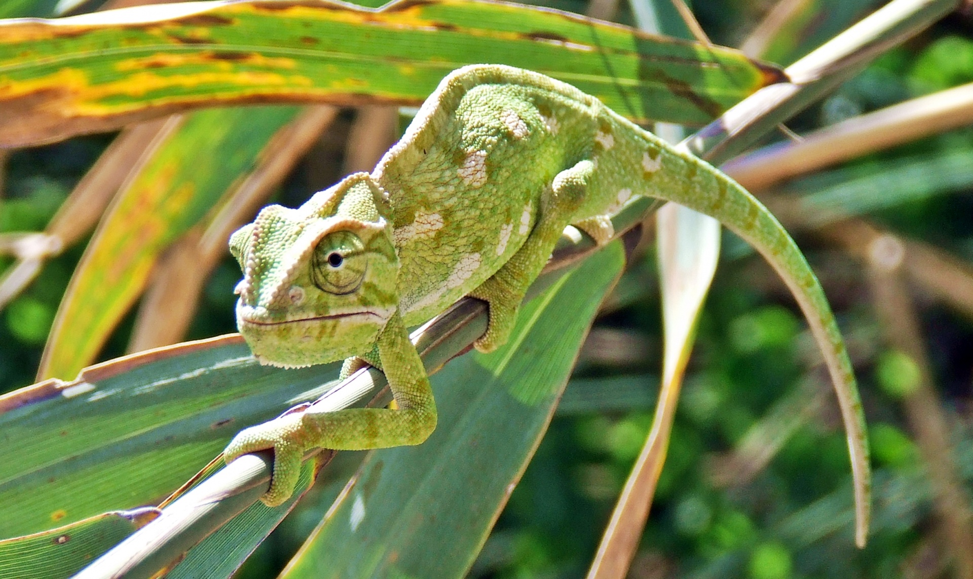
Evolution is a change in the characteristics of populations of living things over time. Evolution can occur by a process called natural selection, which results from random genetic mutations in a population. If these mutations lead to changes that allow the living things to better survive, then their chances of surviving and reproducing in a given environment increase. They will then pass more genes to the next generation. Over many generations, this can lead to major changes in the characteristics of those living things. Evolution explains how living things are changing today, as well as how modern living things descended from ancient life forms that no longer exist on Earth.
Traits that help living things survive and reproduce in a given environment are called adaptations. You can see an obvious adaptation in the image below. The chameleon is famous for its ability to change its colour to match its background as camouflage. Using camouflage, the chameleon can hide in plain sight.
Feature: Myth vs. Reality
Misconceptions about evolution are common. They include the following myths:
Myth |
Reality |
| "Evolution is "just" a theory or educated guess." | Scientists accept evolutionary theory as the best explanation for the diversity of life on Earth because of the large body of scientific evidence supporting it. Like any scientific theory, evolution is a broad, evidence-supported explanation for multiple phenomena. |
| "The theory of evolution explains how life on Earth began." | The theory of evolution explains how life changed on Earth after it began. |
| "The theory of evolution means that humans evolved from apes like those in zoos." | Humans and modern apes both evolved from a common ape-like ancestor millions of years ago. |
2.3 Summary
- Four basic principles or theories unify all fields of biology: cell theory, gene theory, homeostasis, and evolutionary theory.
- According to cell theory, all living things are made of cells and come from other living cells.
- Gene theory states that the characteristics of living things are controlled by genes that pass from parents to offspring.
- All living things strive to maintain internal balance, or homeostasis.
- The characteristics of populations of living things change over time through the process of micro-evolution as organisms acquire adaptations, or traits that better suit them to a given environment.
Use the flashcards below to review the four principles:
2.3 Review Questions
-
- How does sweating help the human body maintain homeostasis?
- Explain cell theory and gene theory.
- Describe an example of homeostasis in the atmosphere.
- Describe how you can apply the concepts of evolution,natural selection, adaptation, and homeostasis to the human ability to sweat.
- Which of the four unifying principles of biology is primarily concerned with:
- how DNA is passed down to offspring?
- how internal balance is maintained?
- _____________ are located on ______________.
- chromosomes; genes
- genes;chromosomes
- genes; traits
- none of the above
- Define an adaptation and give one example.
- Explain how gene theory and evolutionary theory relate to each other.
- Does evolution by natural selection occur within one generation? Why or why not?
- Explain why you think chameleons evolved the ability to change their colour to match their background, as well as how natural selection may have acted on the ancestors of chameleons to produce this adaptation.
2.3 Explore More
https://www.youtube.com/watch?v=Wg5DBH6uMCw&feature=emb_logo
Myths and misconceptions about evolution - Alex Gendler, TEDEd, 2013
Attributions
Figure 2.3.1
Photo(perspiration), by Hans Reniers on Unsplash. is used under the Unsplash license (https://unsplash.com/license).
Figure 2.3.2
Mediterranean Chameleon Reptile Lizard, by user:1588877 on Pixabay, is used under the Pixabay license (https://pixabay.com/de/service/license/).
References
TED-Ed. (2012, June 4). The wacky history of cell theory - Lauren Royal-Woods. YouTube. https://www.youtube.com/watch?v=4OpBylwH9DU&feature=youtu.be
TED-Ed. (2013, July 8). Myths and misconceptions about evolution - Alex Gendler. YouTube. https://www.youtube.com/watch?v=mZt1Gn0R22Q&t=10s
A body system including a series of hollow organs joined in a long, twisting tube from the mouth to the anus. The hollow organs that make up the GI tract are the mouth, esophagus, stomach, small intestine, large intestine, and anus. The liver, pancreas, and gallbladder are the solid organs of the digestive system.
Image shows a photograph of Gregor Mendel.
Case Study: Your Genes May Help You Save a Life
Like the little girl shown in Figure 6.1.1, seven-year-old Mateo is battling leukemia, a type of cancer that affects blood cells. Leukemia usually starts in the bone marrow where blood cells are produced. It causes the production of abnormal blood cells, most commonly white blood cells. Depending on the type of leukemia, it can also affect other types of blood cells. The abnormal blood cells replace the patient’s normal blood cells over time, which can lead to symptoms of fatigue, frequent infections, and easy bruising or bleeding. Leukemia can be fatal, but fortunately, there are some treatment options available that can prolong life — and may even cure the disease.
Mateo has undergone chemotherapy to kill the cancerous cells, but his doctors have told his parents that it is not enough. Mateo needs a bone marrow transplant in order to replace his abnormal bone marrow with healthy bone marrow. His family members are eager to donate bone marrow to him, but first they must be tested to see if they are a compatible match.
For blood transfusions, it is relatively easy to find a compatible blood donor, but bone marrow transplants require much more specific matching between donor and recipient. They must share several of the same type of proteins — called human leukocyte antigens (HLAs) — on the surface of their cells. One type of HLA protein is illustrated in Figure 6.1.3. Different people have different types of HLA proteins (or markers) depending on their specific genes. Typically, eight to ten HLA markers are tested and compared in the potential bone marrow donor and recipient. At least six or seven of these HLA markers must be identical between them in order for a match to be made.
If the match is not good, the patient’s body could reject the bone marrow transplant. Conversely, the transplanted bone marrow could produce immune cells that attack the patient’s body. A good match between donor and recipient is critical for bone marrow donation to be safe and effective.
A full sibling frequently provides the best match for bone marrow donation because they share many of the same genes from their parents. Mateo’s sister is tested, but unfortunately, she is not a match for him. This is not all that surprising since there is only about a 25 per cent chance that a sibling will be an identical HLA match. His parents and other family members are also tested, but none of them are a match, either. Mateo must join the 70 per cent of patients that need to look outside of their families for a bone marrow donor.
How do you find a bone marrow match outside of your family? Fortunately, people from all over the world have signed up to be potential bone marrow donors, usually by providing a simple swab of the inside of their cheek. DNA from the cells collected on the swab is then tested for HLA type. The potential donor’s HLA information is put into a donor registry, and doctors can then search national and international registries for compatible matches for their patients.
Patients are much more likely to be a match with a bone marrow donor of their same race or ethnic background. People with similar ancestry are more likely to share similar HLA genes. In Mateo’s case, his mother is African American, and his father is Japanese and Caucasian. His relatively rare combination of ethnic backgrounds may make it harder for him to find a match in the donor registries, as is the case for many multiethnic patients.
Read the rest of this chapter to learn more about the genetic and phenotypic variations that exist in humans, and how some of these differences came about due to differing natural selection pressures in different areas of the world. At the end of the chapter, learn more about Mateo’s quest for a bone marrow donor, the need for bone marrow donors from diverse ethnic backgrounds, and how you may be able to save someone’s life based on your genetic makeup!
Chapter Overview: Human Variation
In this chapter, you will learn about:
- The extent, types, and patterns of human genetic variation — within and between populations.
- How knowledge about human genetic variation can give insight into human origins and history, and how it may lead to treatments for diseases.
- The ways human variation has been classified, and how some classification methods contribute to racism.
- How gene flow and natural selection can result in a gradual change in the frequency of a trait over a geographic area.
- The ways in which humans can adapt to environmental stresses — genetically, physiologically, and culturally.
- Differences in human blood types (including the ABO and Rh groups), how they may have evolved, and their relationships to diseases.
- How malaria has caused humans to develop a variety of blood cell adaptations over the course of our evolution, including the trait that causes sickle cell anemia.
- Adaptations humans have evolved to deal with the stress of living at high altitudes and in extreme climates, and the ways people can temporarily acclimate to these environmental conditions.
- Human adaptations to our food supply, including lactose tolerance, and weight and blood sugar regulation.
As you read the chapter, think about the following questions:
- How similar are any two people genetically? Based on your answer, why do you think it is not easy to find an HLA match for bone marrow donation between people?
- What is the concept of race? What are its limitations? How does race or ethnicity relate to genetic variation?
- What is an antigen, such as the human leukocyte antigen? On a cellular and molecular level, what happens when there is not a good match between a tissue donor and recipient?
Attributions
Figure 6.1.1
Young_chemotherapy_patient_holds_teddy_bear by Bill Branson (Photographer) at National Cancer Institute/ National Institutes of Health, on Wikimedia Commons is in the public domain (https://en.wikipedia.org/wiki/Public_domain).
Figure 6.1.2
Acute_leukemia-ALL by VashiDonsk at English Wikipedia, now on Wikimedia Commons is used under a CC BY-SA 3.0 (https://creativecommons.org/licenses/by-sa/3.0/deed.en) license.
Figure 6.1.3
HLA_DQ_Illustration by Pdeitiker on Wikimedia Commons is released into the public domain (https://en.wikipedia.org/wiki/Public_domain).
All in the Family
This family photo (Figure 5.12.1) clearly illustrates an important point: children in a family resemble their parents and each other, but the children never look exactly the same, unless they are identical twins. Each of the daughters in the photo have inherited a unique combination of traits from the parents. In this concept, you will learn how this happens. It all begins with sex — sexual reproduction, that is.
Sexual Reproduction
Reproduction is the process by which organisms give rise to offspring. It is one of the defining characteristics of living things. Like many other organisms, human beings reproduce sexually. Sexual reproduction involves two parents. As you can see from Figure 5.12.2, in sexual reproduction, parents produce reproductive (sex) cells — called gametes — that unite to form an offspring. Gametes are haploid (or 1N) cells. This means they contain one copy of each chromosome in the nucleus. Gametes are produced by a type of cell division called meiosis, which is described in detail below. The process in which two gametes unite is called fertilization. The fertilized cell that results is referred to as a zygote. A zygote is a diploid (or 2N) cell, which means it contains two copies of each chromosome. Thus, it has twice the number of chromosomes as a gamete.
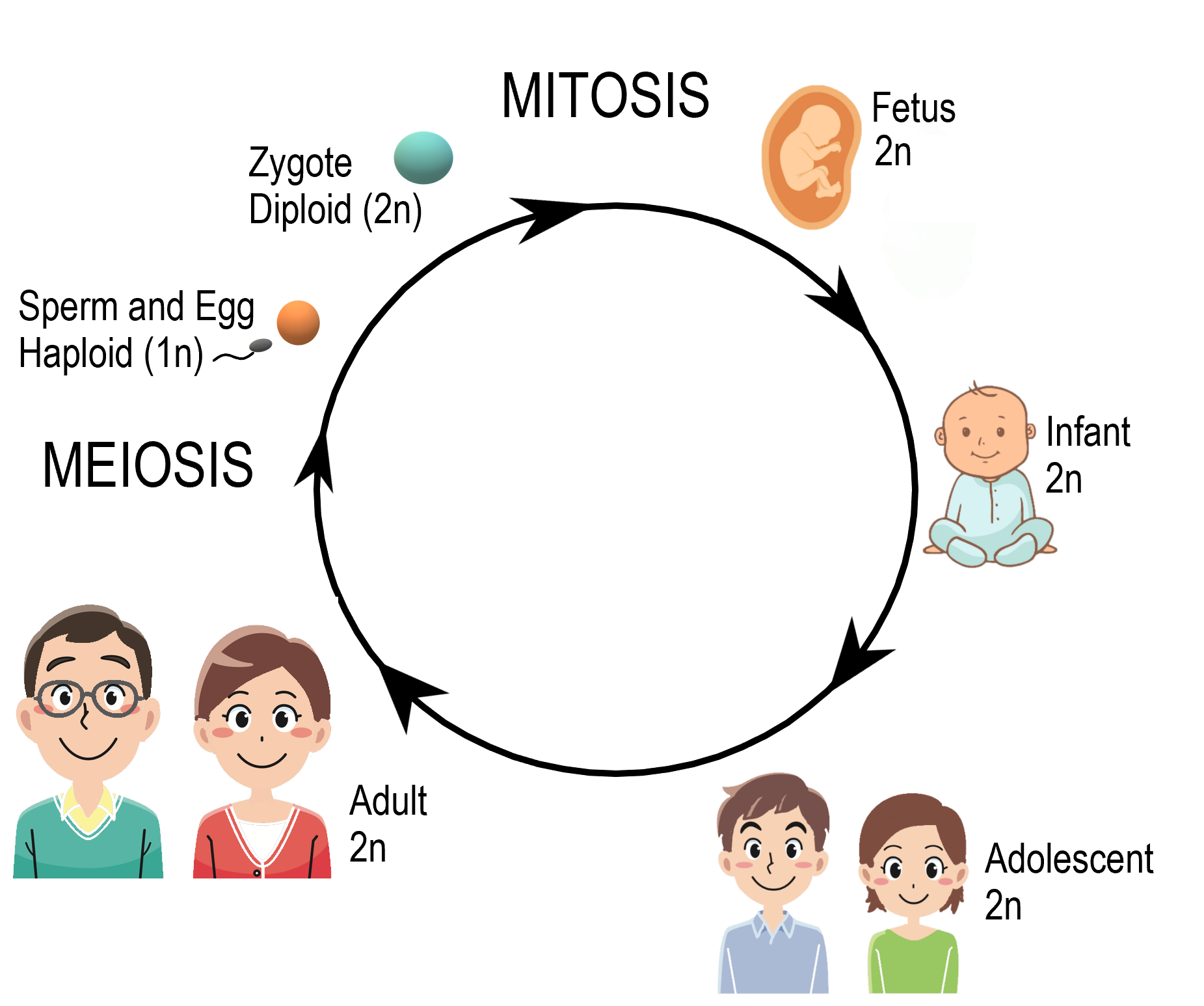
Meiosis
The process that produces haploid gametes is called meiosis. Meiosis is a type of cell division in which the number of chromosomes is reduced by half. It occurs only in certain special cells of an organism. During meiosis, homologous (paired) chromosomes separate, and four haploid cells form that have only one chromosome from each pair. The diagram (Figure 5.12.3) gives an overview of meiosis.
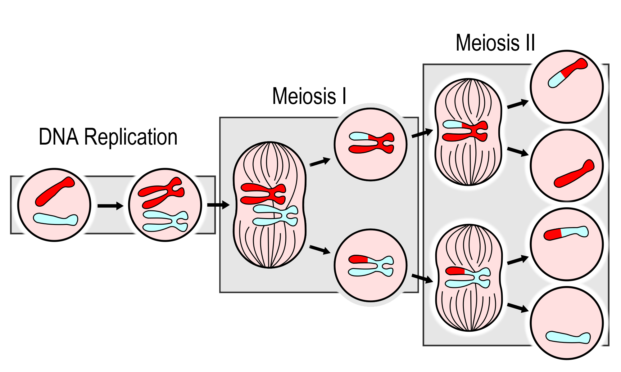
As you can see in the meiosis diagram, two cell divisions occur during the overall process, producing a total of four haploid cells from one parent cell. The two cell divisions are called meiosis I and meiosis II. Meiosis I begins after DNA replicates during interphase. Meiosis II follows meiosis I without DNA replicating again. Both meiosis I and meiosis II occur in four phases, called prophase, metaphase, anaphase, and telophase. You may recognize these four phases from mitosis, the division of the nucleus that takes place during routine cell division of eukaryotic cells.
Meiosis I- Increasing genetic variation
The phases of Meiosis I are:
- Prophase I: The nuclear envelope begins to break down, and the chromosomes condense. Centrioles start moving to opposite poles of the cell, and a spindle begins to form. Importantly, homologous chromosomes pair up, which is unique to prophase I. In prophase of mitosis and meiosis II, homologous chromosomes do not form pairs in this way. During prophase I, crossing-over occurs. The significance of crossing-over is discussed below.
- Metaphase I: Spindle fibres attach to the paired homologous chromosomes. The paired chromosomes line up along the equator of the cell, randomly aligning in a process called independent alignment. The significance of independent alignment is discussed below. This occurs only in metaphase I. In metaphase of mitosis and meiosis II, it is sister chromatids that line up along the equator of the cell.
- Anaphase I: Spindle fibres shorten, and the chromosomes of each homologous pair start to separate from each other. One chromosome of each pair moves toward one pole of the cell, and the other chromosome moves toward the opposite pole.
- Telophase I and Cytokinesis: The spindle breaks down, and new nuclear membranes form. The cytoplasm of the cell divides, and two haploid daughter cells result. The daughter cells each have a random assortment of chromosomes, with one from each homologous pair. Both daughter cells go on to meiosis II.
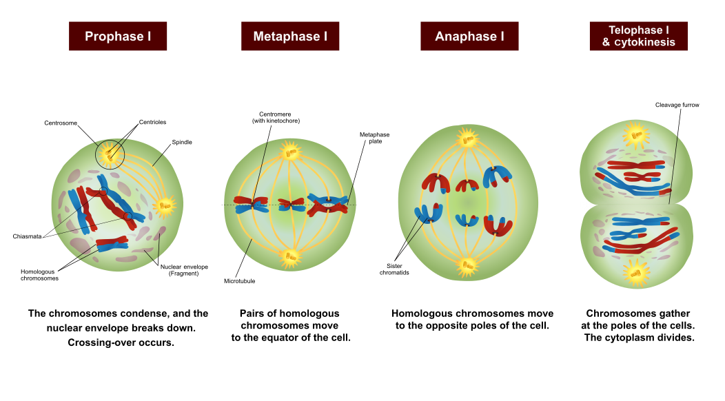
Meiosis II- Halfing the DNA
The phases of Meiosis II are:
- Prophase II: The nuclear envelope breaks down, and the spindle begins to form in each haploid daughter cell from meiosis I. The centrioles also start to separate.
- Metaphase II: Spindle fibres line up the sister chromatids of each chromosome along the equator of the cell.
- Anaphase II: Sister chromatids separate and move to opposite poles.
- Telophase II and Cytokinesis: The spindle breaks down, and new nuclear membranes form. The cytoplasm of each cell divides, and four haploid cells result. Each cell has a unique combination of chromosomes.
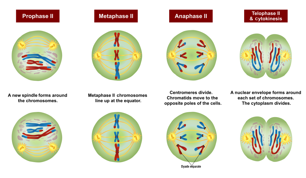
Sexual Reproduction and Genetic Variation
"It takes two to tango" might be a euphemism for sexual reproduction. Requiring two individuals to produce offspring, however, is also the main drawback of this way of reproducing, because it requires extra steps — and often a certain amount of luck — to successfully reproduce with a partner. On the other hand, sexual reproduction greatly increases the potential for genetic variation in offspring, which increases the likelihood that the resulting offspring will have genetic advantages. In fact, each offspring produced is almost guaranteed to be genetically unique, differing from both parents and from any other offspring. Sexual reproduction increases genetic variation in a number of ways:
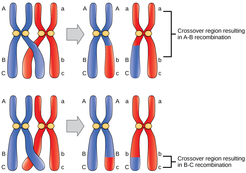
- When homologous chromosomes pair up during meiosis I, crossing-over can occur. Crossing-over is the exchange of genetic material between non-sister chromatids of homologous chromosomes. It results in new combinations of genes on each chromosome. This is called recombination. You can see how it happens in the figure to the right.
- When cells divide during meiosis, homologous chromosomes are randomly distributed to daughter cells, and different chromosomes segregate independently of each other. This is called independent alignment. It results in gametes that have unique combinations of chromosomes. You can see how it happens in Figure 5.12.7.
- In sexual reproduction, two gametes unite to produce an offspring. But which two of the millions of possible gametes will it be? This is a matter of chance, and it's obviously another source of genetic variation in offspring.
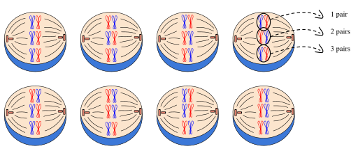
With all of this recombination of genes, there is a need for a new set of vocabulary. Remember, that sister chromatids are two identical pieces of DNA connected at a centromere. Once crossing over has occured, we can no longer call them sister chromatids since they are no longer identical; we term them dyads. In addition, once crossing over has occurred, the pair of homologous chromosomes can be referred to as tetrads.
All of these mechanisms — crossing over, independent assortment, and the random union of gametes — work together to result in an amazing range of potential genetic variation. Each human couple, for example, has the potential to produce more than 64 trillion genetically unique children. No wonder we are all different!
https://www.youtube.com/watch?v=VzDMG7ke69g
Meiosis (updated), Amoeba Sisters, 2017.
Gametogenesis
At the end of meiosis, four haploid cells have been produced, but the cells are not yet gametes. The cells need to develop before they become mature gametes capable of fertilization. The development of haploid cells into gametes is called gametogenesis. It differs between males and females.
- A gamete produced by a male is called a sperm, and the process that produces a mature sperm is called spermatogenesis. During this process, a sperm cell grows a tail and gains the ability to “swim,” like the human sperm cell shown in Figure 5.12.8.
- A gamete produced by a female is called anegg or ovum, and the process that produces a mature egg is called oogenesis, during which just one functional egg is produced. The other three haploid cells that result from meiosis are called polar bodies, and they disintegrate. The single egg is a very large cell, as you can see from the human egg also shown in Figure 5.12.8.
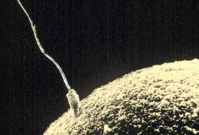
5.12 Summary
- In sexual reproduction, two parents produce gametes that unite in the process of fertilization to form a single-celled zygote. Gametes are haploid cells with one copy of each of the 23 chromosomes, and the zygote is a diploid cell with two copies of each of the 23 chromosomes.
- Meiosis is the type of cell division that produces four haploid daughter cells that may become gametes. Meiosis occurs in two stages, called meiosis I and meiosis II, each of which occurs in four phases (prophase, metaphase, anaphase, and telophase).
- Meiosis is followed by gametogenesis, the process during which the haploid daughter cells change into mature gametes. Males produce gametes called sperm in a process known as spermatogenesis, and females produce gametes called eggs in the process known as oogenesis.
- Sexual reproduction produces genetically unique offspring. Crossing-over, independent alignment, and the random union of gametes work together to result in an amazing range of potential genetic variation.
5.12 Review Questions
-
- Explain how sexual reproduction happens at the cellular level.
- Summarize what happens during Meiosis.
- Compare and contrast gametogenesis in males and females.
- Explain the mechanisms that increase genetic variation in the offspring produced by sexual reproduction.
- Why do gametes need to be haploid? What would happen to the chromosome number after fertilization if they were diploid?
- Describe one difference between Prophase I of Meiosis and Prophase of Mitosis.
- Do all of the chromosomes that you got from your mother go into one of your gametes? Why or why not?
5.12 Explore More
https://www.youtube.com/watch?v=qCLmR9-YY7o&feature=emb_logo
Meiosis: Where the Sex Starts - Crash Course Biology #13, CrashCourse, 2012.
https://www.youtube.com/watch?v=zrKdz93WlVk
Mitosis vs Meiosis Comparison, Amoeba Sisters, 2018.
Attributions
Figure 5.12.1
Family portrait by loly galina on Unsplash is used under the Unsplash License (https://unsplash.com/license).
Figure 5.12.2
Human Life Cycle by Christine Miller is used under a CC BY-NC-SA 4.0 (https://creativecommons.org/licenses/by-nc-sa/4.0/) license.
Figure 5.12.3
MajorEventsInMeiosis_variant_int by PatríciaR (internationalization) on Wikimedia Commons is used and adapted by Christine Miller. This image in the public domain. (Original image from NCBI; original vector version by Jakov.)
Figure 5.12.4
Meiosis 1/ Meiosis Stages by Ali Zifan on Wikimedia Commons is used and adapted by Christine Miller under a CC BY-SA 4.0 (https://creativecommons.org/licenses/by-sa/4.0) license.
Figure 5.12.5
Meiosis 2/ Meiosis Stages by Ali Zifan on Wikimedia Commons is used and adapted by Christine Miller under a CC BY-SA 4.0 (https://creativecommons.org/licenses/by-sa/4.0) license.
Figure 5.12.6
Crossover/ Figure 17 02 01 by CNX OpenStax on Wikimedia Commons is used under a CC BY 4.0 (https://creativecommons.org/licenses/by/4.0) license.
Figure 5.12.7
Independent_assortment by Mtian20 on Wikimedia Commons is used under a CC BY-SA 4.0 (https://creativecommons.org/licenses/by-sa/4.0) license.
Figure 5.12.8
sperm fertilizing egg by AndreaLaurel on Flickr is used under a CC BY 2.0 (https://creativecommons.org/licenses/by/2.0/) license.
References
Amoeba Sisters. (2017, July 11). Meiosis (updated). YouTube. https://www.youtube.com/watch?v=VzDMG7ke69g&feature=youtu.be
Amoeba Sisters. (2018, May 31). Mitosis vs meiosis comparison. YouTube. https://www.youtube.com/watch?v=zrKdz93WlVk&feature=youtu.be
CrashCourse, (2012, April 23). Meiosis: Where the sex starts - Crash Course Biology #13. YouTube. https://www.youtube.com/watch?v=qCLmR9-YY7o&feature=youtu.be
OpenStax CNX. (2016, May 27). Figure 1 Crossover may occur at different locations on the chromosome. In OpenStax, Biology (Section 17.2). http://cnx.org/contents/185cbf87-c72e-48f5-b51e-f14f21b5eabd@10.53.
Image shows a Lego (TM) representation of Gregor Mendel with his plants.
Image shows the process of crossing over. A pair of homologous chromosomes are side by side. They then overlap the ends of their structures and trades these sections, attaching some maternal DNA onto the paternal chromosome, and some paternal DNA onto the maternal chromosome. The result is that, within the homologous pair of chromosomes, each of the four pieces of DNA, destined to enter 4 separate cells, is now consists of a unique mix of genes.
A class of biological molecule consisting of linked monomers of amino acids and which are the most versatile macromolecules in living systems and serve crucial functions in essentially all biological processes.

