14.7 Case Study Conclusion: Flight Risk
Case Study Conclusion: Flight Risk
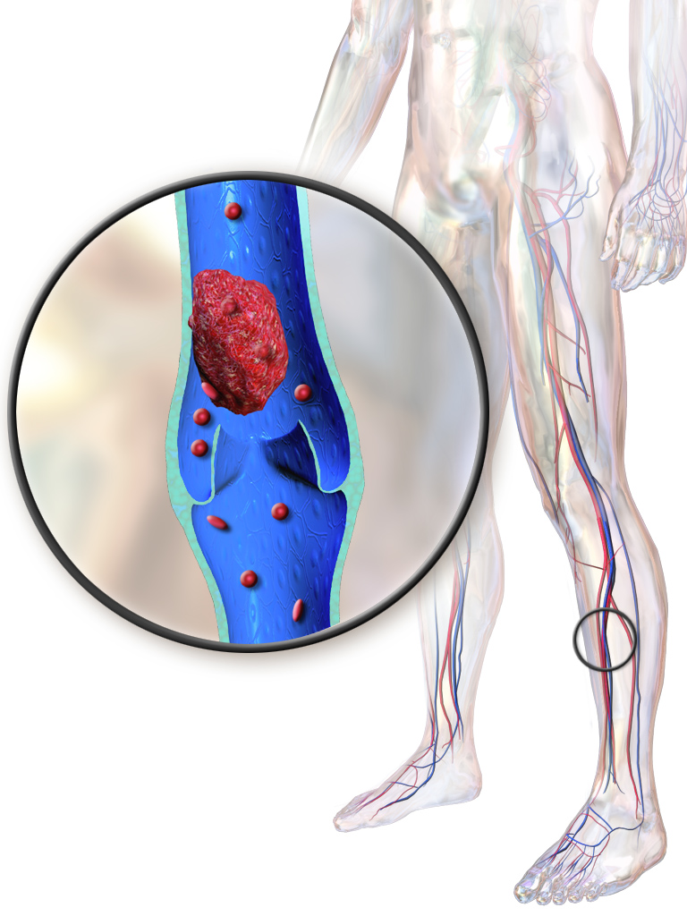
At the beginning of this chapter, you learned about Malcolm and Willie, who met while sitting next to each other on a plane. During the flight, Willie got up to take frequent walks, and was doing leg exercises to try to avoid the medical condition depicted in Figure 14.7.1 — deep vein thrombosis (DVT). DVT occurs when a blood clot forms in a deep vein, usually in the leg. It can be very dangerous — even deadly.
As you learned in this chapter, a blood clot is an aggregation of thrombocytes and proteins. Blood clots are helpful for preventing blood loss when a blood vessel is damaged. In some situations, though, they can be extremely dangerous. Blood clots can cause heart attacks or strokes by blocking the flow of blood to the heart or brain, respectively.
When DVT occurs, one of the major risks is pulmonary embolism (PE). PE is when the blood clot breaks off, travels through the blood vessels, and lodges in a pulmonary artery. Recall what the pulmonary arteries do — they carry deoxygenated blood from the heart to the lungs, where the blood picks up oxygen and releases carbon dioxide due to gas exchange between the capillaries and the alveoli of the lungs. Imagine what would happen if this flow of blood to the lungs was partially or completely blocked by a blood clot. Depending on the size of the blood clot and where it is lodged, a PE can cause a variety of serious consequences, ranging from lung damage to instant death, because of the disruption of the pulmonary circulation.
Willie has a higher risk of DVT and its consequences because he has heart failure. As you have learned, heart failure is a chronic condition in which the pumping action of the heart is impaired. One reason that heart failure is thought to increase the risk of DVT is because the blood is not being pushed strongly enough through the cardiovascular system, allowing blood clots to form more easily.
Willie needs to be particularly concerned about DVT while on a long plane flight. Why do you think this is? Think about how blood flows through arteries and veins. Blood is pushed through arteries mainly due to the pumping action of the heart. Veins, on the other hand, rely on the movement of the surrounding skeletal muscles to help push blood through them. Sitting still for long periods of time in cramped quarters (such as on a plane) can cause blood to pool in the deep veins of the legs, leading to the formation of a blood clot.
Even people who are generally healthy and don’t have heart disease can get DVT from sitting for too long on a long-distance flight, or in other situations when they are immobile for extended periods of time. Fortunately, walking periodically and doing some simple leg exercises can lower your risk of DVT by helping to push blood through your veins. If you are planning on taking a flight in the future, watch the short video below to learn some easy exercises that you can do right in your plane seat to help prevent DVT!
Chapter 14 Summary
In this chapter you learned about the structure, functions, and disorders of the cardiovascular system. Specifically, you learned that:
- The cardiovascular system is the organ system that transports materials to and from all the cells of the body. The main components of the cardiovascular system are the heart, blood vessels, and blood.
- The cardiovascular system has two interconnected circulations. The pulmonary circuit carries blood between the heart and lungs, where blood is oxygenated. The systemic circuit carries blood between the heart and the rest of the body, where it delivers oxygen.
- The heart is a muscular organ in the chest that consists mainly of cardiac muscle. It pumps blood through blood vessels by repeated, rhythmic contractions.
-
- The wall of the heart consists of three layers. The middle layer, the myocardium, is the thickest layer, and consists mainly of cardiac muscle.
- The interior of the heart consists of four chambers, with an upper atrium and lower ventricle on each side of the heart. Blood enters the heart through the atria, which pump it to the ventricles. Then, the ventricles pump blood out of the heart. Four valves in the heart keep blood flowing in the correct direction and prevent backflow.
-
- Deoxygenated blood flows into the right atrium through veins from the upper and lower body (superior and inferior vena cava, respectively), and oxygenated blood flows into the left atrium through four pulmonary veins from the lungs. Each atrium pumps the blood to the ventricle below it. From the right ventricle, deoxygenated blood is pumped to the lungs through the two pulmonary arteries. From the left ventricle, oxygenated blood is pumped to the rest of the body through the aorta.
- The coronary circulation consists of blood vessels that carry blood to and from the heart muscle cells. There are two coronary arteries that supply the two sides of the heart with oxygenated blood. Cardiac veins drain deoxygenated blood back into the heart.
- The cardiac cycle refers to a single complete heartbeat. It includes diastole — when the atria contract — and systole, when the ventricles contract.
- The normal, rhythmic beating of the heart is called sinus rhythm. It is established by the heart’s pacemaker cells in the sinoatrial node. Electrical signals from the pacemaker cells travel to the atria and cause them to contract. Then, the signals travel to the atrioventricular node, and from there to the ventricles via the Purkinje fibres, causing them to contract. Electrical stimulation from the autonomic nervous system and hormones from the endocrine system can also influence heartbeat.
- Blood vessels carry blood throughout the body. Major types of blood vessels are arteries, veins, and capillaries.
-
- Arteries are blood vessels that usually carry blood away from the heart (except for coronary arteries that supply the heart muscle with blood). Most arteries carry oxygenated blood. The largest artery is the aorta, which is connected to the heart and extends into the abdomen. Blood moves through arteries due to pressure from the beating of the heart.
- Veins are blood vessels that usually carry blood toward the heart. Most veins carry deoxygenated blood. The largest veins are the superior vena cava and inferior vena cava. Blood moves through veins by the squeezing action of surrounding skeletal muscles. Valves in veins prevent backflow of blood.
- Capillaries are the smallest blood vessels. They connect arterioles and venules. They form capillary beds where substances are exchanged between the blood and surrounding tissues.
- The walls of arteries and veins have three layers. The middle layer is thickest in arteries, in which it contains smooth muscle tissue that controls the diameter of the vessels. The outer layer is thickest in veins and consists mainly of connective tissue. The walls of capillaries consist of little more than a single layer of epithelial cells.
- Blood pressure is a measure of the force that blood exerts on the walls of arteries. It is expressed as a double number, with the higher number representing systolic pressure when the ventricles contract, and the lower number representing diastolic pressure when the ventricles relax. Normal blood pressure is generally defined as a pressure of 120/80 mm Hg or less.
- Vasoconstriction (narrowing) and vasodilation (widening) of arteries can occur to help regulate blood pressure or body temperature or to change blood flow as part of the fight-or-flight response.
- Blood is a fluid connective tissue that circulates throughout the body in blood vessels. Blood supplies tissues with oxygen and nutrients, and removes their metabolic wastes. Blood helps defend the body from pathogens and other threats, transports hormones and other substances, and helps to keep the body’s pH and temperature in homeostasis. Blood consists of a liquid part (called plasma) and cells, including erythrocytes, leukocytes and thrombocytes.
-
- Plasma makes up more than half of blood by volume. It consists of water and many dissolved substances. It also contains blood cells.
- Erythrocytes are the most numerous cells in blood. They consist mostly of hemoglobin, which carries oxygen. Red blood cells also carry antigens that determine blood types.
- Leukocytes are less numerous than red blood cells and are part of the body’s immune system. They protect the body from abnormal cells, microorganisms, and other harmful substances. There are several different types of white blood cells that differ in their specific immune functions.
- Thrombocytes are cell fragments that play important roles in blood clotting, or coagulation. They stick together at breaks in blood vessels to form a clot and stimulate the production of fibrin, which strengthens the clot.
- All blood cells form by proliferation of stem cells in red bone marrow in a process called hematopoiesis. When blood cells die, they are phagocytized by white blood cells and removed from the circulation.
- Disorders of the blood include leukemia, which is cancer of the bone-forming cells; hemophilia, which is any of several genetic blood-clotting disorders; carbon monoxide poisoning, which prevents erythrocytes from binding with oxygen and causes suffocation; HIV infection, which destroys certain leukocytes and can cause AIDS; and anemia, in which there are not enough erythrocytes to carry adequate oxygen to body tissues.
- Cardiovascular disease is a class of diseases that involve the cardiovascular system. Worldwide, it is the leading cause of death. Most cases occur in people over age 60, and onset typically occurs about a decade earlier in males than in females. Other risk factors include smoking, obesity, diabetes, high blood cholesterol, and lack of exercise.
-
- Two common conditions that lead to most cases of cardiovascular disease are hypertension and atherosclerosis. Hypertension is blood pressure that is persistently at or above 140/90 mm Hg. Atherosclerosis is a buildup of fatty, fibrous plaques in arteries that may reduce or block blood flow. Treating these conditions is important for preventing cardiovascular disease.
- Coronary artery disease is a group of diseases that result from atherosclerosis of coronary arteries. Two of the most common are angina and myocardial infarction (heart attack). In angina, cardiac cells receive inadequate oxygen, which causes chest pain. In a heart attack, cardiac cells die because blood flow to part of the heart is blocked. A heart attack may cause death, or lead to heart arrhythmias, heart failure, or cardiac arrest.
- Stroke occurs when blocked or broken arteries in the brain result in the death of brain cells. This may happen when an artery is blocked by a clot or plaque, or when an artery ruptures and bleeds in the brain. In both cases, part of the brain is damaged, and functions such as speech and controlled movements may be impaired, either temporarily or permanently.
- Peripheral artery disease occurs when atherosclerosis narrows peripheral arteries, usually in the legs, often causing pain when walking. It is important to diagnose this disease so the underlying atherosclerosis can be treated before it causes a heart attack or stroke.
In this chapter, you learned that the cardiovascular system carries nutrients to the cells of the body. Read the next chapter about the Digestive System to learn about how your body transforms your meals into the nutrients that cells need to function.
Chapter 14 Review Questions
-
- Alex goes to the doctor and learns that his blood pressure is 135/90 mm Hg. Answer the following questions about his blood pressure:
- Is this a normal blood pressure? Why or why not?
- Which number refers to the systolic pressure? Which number refers to the diastolic pressure?
- Describe what the atria and ventricles of Alex’s heart are doing when the pressure is at 135 mm Hg.
- Alex’s doctor would like him to lower his blood pressure. Why do you think he would like Alex to do this, and what are some ways in which he may be able to lower his blood pressure?
- What are three functions of the cardiovascular system?
- Which are the chambers of the heart that receive blood? Which are the chambers of the heart that pump
- Valves prevent blood from flowing backward in the cardiovascular system. Why do you think this is important?
- Compare the coronary arteries, pulmonary arteries, and arteries elsewhere in the body in terms of their target tissues (i.e. where they bring blood to) and whether they are carrying oxygenated or deoxygenated blood.
- Due to a reduction in the amount of oxygen that gets to the cells of the body, anemia causes weakness and fatigue. Explain how oxygen is transported to the cells of the body, and which blood cells are affected in anemia.
- What are the two conditions that are precursors to virtually all cases of cardiovascular disease?
- What are the main differences between the coronary circulation, pulmonary circulation, and systemic circulation?
- Define sinus rhythm.
- Generally speaking, which is a more serious and immediately life-threatening condition: heart failure or cardiac arrest? Explain your answer.
Attribution
Figure 14.7.1
Blausen_0290_DeepVeinThrombosis by BruceBlaus on Wikimedia Commons is used under a CC BY 3.0 (https://creativecommons.org/licenses/by/3.0) license.
Reference
Blausen.com Staff. (2014). Medical gallery of Blausen Medical 2014. WikiJournal of Medicine 1 (2). DOI:10.15347/wjm/2014.010. ISSN 2002-4436.
Created by: CK-12/Adapted by Christine Miller
A Deceptively Simple Model
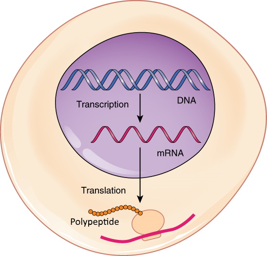
This simple model sums up one of the most important ideas in biology, which is called the central dogma of molecular biology (you'll read more about it below). You probably recognize the spiral-shaped structure in the nucleus. It represents a molecule of DNA, the biochemical molecule that stores genetic information in most living cells. The yellow chain represents a newly formed polypeptide — the beginning stage of creating a protein. Proteins are the class of biochemical molecules that carry out virtually all life processes. What is the structure in the center of the model? It appears to resemble DNA, but it is smaller and simpler. This molecule is the key to the central dogma, and it may have been the first type of biochemical molecule to evolve.
Central Dogma of Molecular Biology
DNA is found in chromosomes. In eukaryotic cells, chromosomes always remain in the nucleus, but proteins are made at ribosomes in the cytoplasm. How do the instructions in DNA get to the site of protein synthesis outside the nucleus?
Another type of nucleic acid is responsible. This nucleic acid is RNA, or ribonucleic acid. RNA is a small molecule that can squeeze through pores in the nuclear membrane. It carries the information from DNA in the nucleus to a ribosome in the cytoplasm and then helps assemble the protein. In short:
DNA → RNA → Protein
This expresses in words what the diagram in Figure 5.5.1 shows. The genetic instructions encoded in DNA in the nucleus are transcribed to RNA. Then, RNA carries the instructions to a ribosome in the cytoplasm, where they are translated into a protein. Discovering this sequence of events was a major milestone in molecular biology. It's called the central dogma of molecular biology.
Introducing RNA
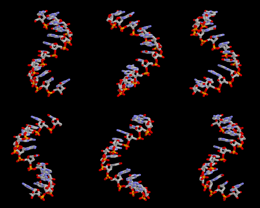
DNA alone cannot “tell” your cells how to make proteins. It needs the help of RNA, the other main player in the central dogma of molecular biology. Like DNA, RNA is a nucleic acid, so it consists of repeating nucleotides bonded together to form a polynucleotide chain. RNA differs from DNA in several ways: it exists as a single stranded molecule, contains the sugar ribose (as opposed to deoxyribose) and uses the base uracil instead of thymine.
Functions of RNA
The main function of RNA is to help make proteins. There are three main types of RNA involved in protein synthesis:
-
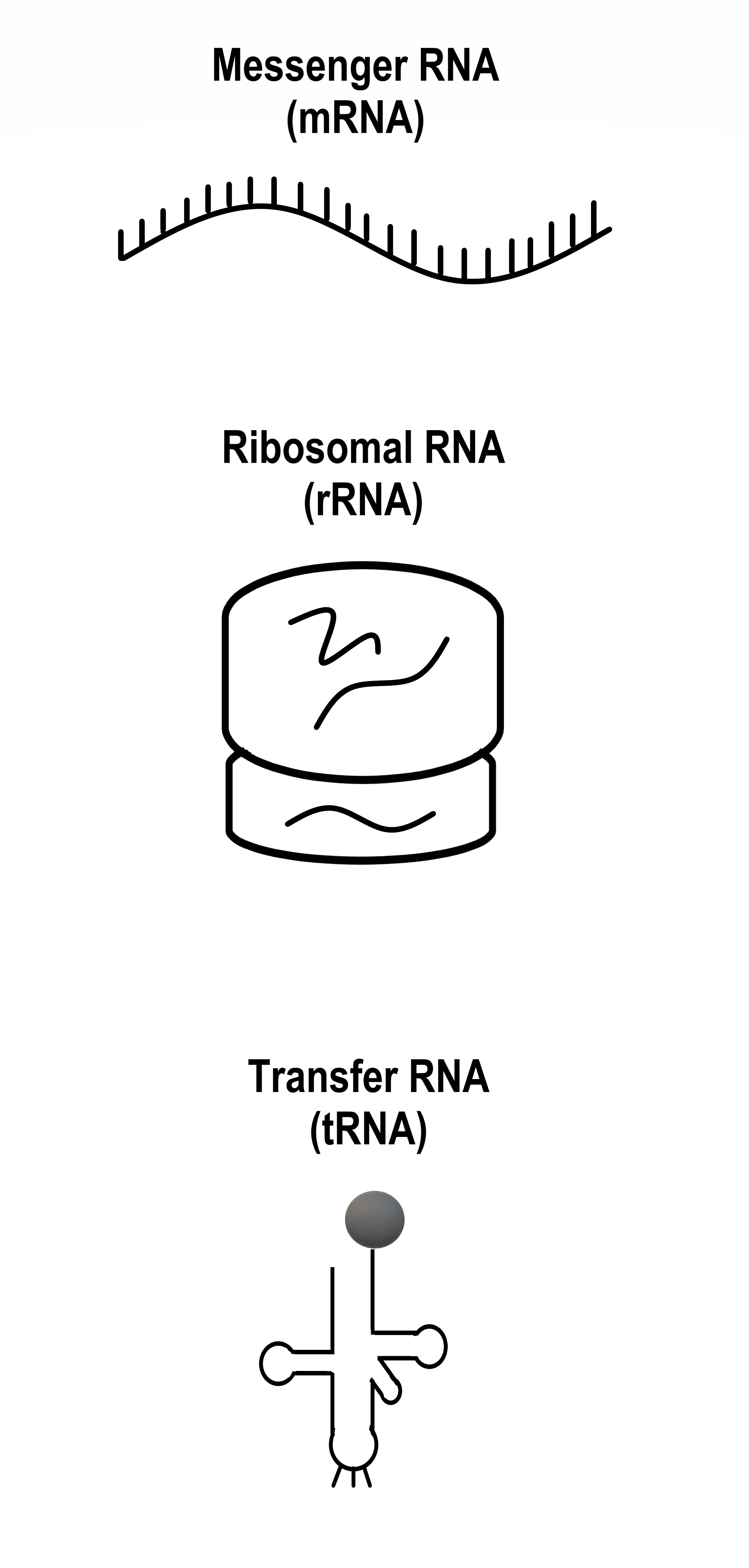
Figure 5.5.3 The three types of RNA take very different forms. Messenger RNA (mRNA) copies (or transcribes) the genetic instructions from DNA in the nucleus and carries them to the cytoplasm.
- Ribosomal RNA (rRNA) helps form ribosomes, where proteins are assembled. Ribosomes also contain proteins.
- Transfer RNA (tRNA) brings amino acids to ribosomes, where rRNA catalyzes the formation of chemical bonds between them to form a protein.
In section 5.7 Protein Synthesis, you can read in detail about how these three types of RNA build primary structure of proteins.
RNA is a very versatile molecule which plays multiple roles in living things. In addition to helping to make proteins, for example, there are RNA molecules that regulate the expression of genes, and RNA molecules that catalyze other biochemical reactions needed to sustain life. Because of the diversity of roles that RNA molecules play, they have been called the Swiss Army knives of the cellular world.
It's an RNA World
The function of DNA is to store genetic information inside cells. It does this job well, but that's about all it can do. DNA can't act as an enzyme, for example, to catalyze biochemical reactions that are needed to keep us alive. Proteins are needed for this and many other life functions. Proteins work exceptionally well to keep us alive, but they are unable to store genetic information. Proteins need DNA for that. Without DNA, proteins could not exist. On the other hand, without proteins, DNA could not survive. This poses a chicken-and-egg sort of problem: Which evolved first? DNA or proteins?
Some scientists think that the answer is neither. They speculate instead that RNA was the first biochemical to evolve. The reason? RNA can do more than one job. It can store information as DNA does, but it can also perform various jobs (such as catalysis) to keep cells alive, as proteins do. The idea that RNA was the first biochemical to evolve, predating both DNA and proteins, is called the RNA world hypothesis. According to this hypothesis, billions of years ago, RNA molecules evolved that could both survive and make copies of themselves. According to the hypothesis, early RNA molecules eventually evolved the ability to make proteins, and at some point RNA mutated to form DNA.
Feature: Reliable Sources
The RNA world hypothesis has not gained enough support in the scientific community to be accepted as a scientific theory. In fact, there are probably as many detractors as supporters of the hypothesis. Do a web search to learn more about the RNA world hypothesis and the evidence and arguments for and against it. When weighing the information you gather, consider the likely reliability of the different websites you visit. Based on what you determine are the most reliable sources and the most convincing arguments, form your own opinion about the hypothesis. You may decide to accept or reject the hypothesis. Alternatively, you may decide to reserve judgement until — or if — more evidence or arguments are forthcoming.
5.5 Summary
- The central dogma of molecular biology can be summed up as: DNA → RNA → Protein. This means that the genetic instructions encoded in DNA are first transcribed to RNA, and then from RNA they are translated into a protein.
- Like DNA, RNA is a nucleic acid. Unlike DNA, RNA consists of just one polynucleotide chain instead of two, contains the base uracil instead of thymine, and contains the sugar ribose instead of deoxyribose.
- The main function of RNA is helping to make proteins. There are three main types of RNA involved in protein synthesis: messenger RNA (mRNA), ribosomal RNA (rRNA), and transfer RNA (tRNA). RNA has additional functions, including regulating gene expression and catalyzing other biochemical reactions.
- According to the RNA world hypothesis, RNA was the first type of biochemical molecule to evolve, predating both DNA and proteins. The hypothesis is based mainly on the multiple functions of RNA, which can store genetic information like DNA and carry out life processes (like proteins).
5.5 Review Questions
- State the central dogma of molecular biology.
- Drag and drop to compare the structure and function of DNA and RNA:
3.
4.
5.5 Explore More
https://www.youtube.com/watch?time_continue=4&v=VYQQD0KNOis&feature=emb_logo
The RNA Origin of Life, NOVA PBS Official, 2014.
https://www.youtube.com/watch?v=JQByjprj_mA
DNA vs RNA (Updated), Amoeba Sisters, 2019.
Attributions
Figure 5.5.1
From DNA to Protein: Transcription through Translation by OpenStax College on Wikimedia Commons is used under a CC BY 4.0 (https://creativecommons.org/licenses/by/4.0/) license.
Figure 5.5.2
Molbio-Header by Squidonius on Wikimedia Commons is released into the public domain (https://en.wikipedia.org/wiki/Public_domain).
Figure 5.5.2
ARNm-Rasmol by Corentin Le Reun on Wikimedia Commons is is released into the public domain (https://en.wikipedia.org/wiki/Public_domain).ublic domain.
References
Amoeba Sisters. (2019, August 29). DNA vs RNA (Updated). YouTube. https://www.youtube.com/watch?v=JQByjprj_mA&feature=youtu.be
Betts, J. G., Young, K.A., Wise, J.A., Johnson, E., Poe, B., Kruse, D.H., Korol, O., Johnson, J.E., Womble, M., DeSaix, P. (2013, April 25). Figure 3.29 From DNA to Protein: Transcription through Translation [digital image]. In Anatomy and Physiology. OpenStax. https://openstax.org/books/anatomy-and-physiology/pages/3-4-protein-synthesis#fig-ch03_04_05
NOVA PBS Official. (2014, April 23). The RNA origin of life. YouTube. https://www.youtube.com/watch?v=VYQQD0KNOis&feature=youtu.be
Wikipedia contributors. (2020, June 28). RNA world. In Wikipedia. https://en.wikipedia.org/w/index.php?title=RNA_world&oldid=964998696
Image shows a section of DNA code - a specific sequence of the bases A, C, G, and T.
Refers to the body system consisting of the heart, blood vessels and the blood. Blood contains oxygen and other nutrients which your body needs to survive. The body takes these essential nutrients from the blood.
Created by: CK-12/Adapted by Christine Miller
The Blue Marble
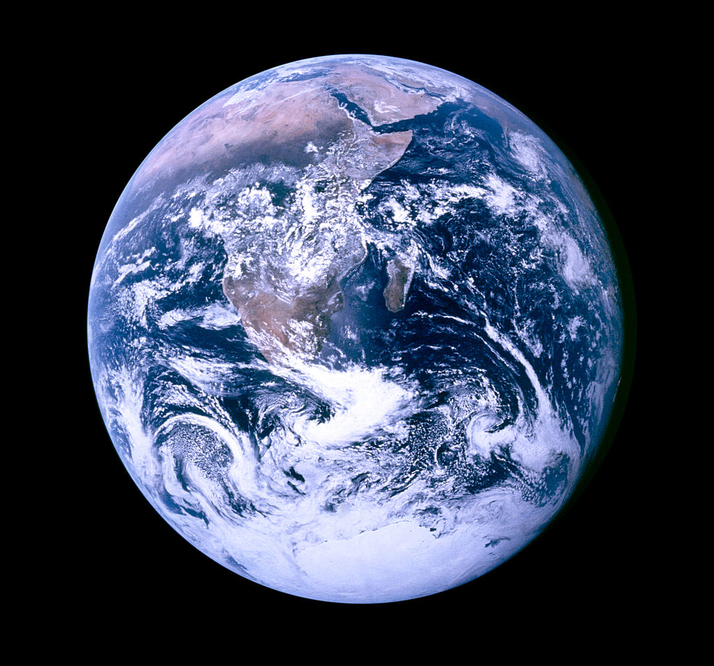
It's often called the "water planet," and it's been given the nickname "the blue marble." You probably just call it "home." Almost three-quarters of our home planet is covered by water, and without it, life as we know it could not exist on Earth. Water, like carbon, has a special role in living things: it is needed by all known forms of life. Although water consists of simple molecules, each containing just three atoms, its structure gives it unique properties that help explain why it is vital to all living organisms.
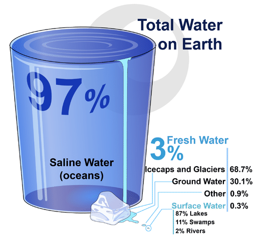
Water, Water Everywhere
If you look at Figure 3.11.2, you will see where Earth’s water is found. The term water generally refers to its liquid state, and water is a liquid over a wide range of temperatures on Earth. Water, however, also occurs on Earth as a solid (ice) and as a gas (water vapor).
Structure and Properties of Water
You are likely already aware of some of the properties of water. For example, you know that water is tasteless and odorless. You also probably know that water is transparent, which means that light can pass through it. This is important for organisms that live in the water, because some of them need sunlight to make food by photosynthesis.
Chemical Structure of Water
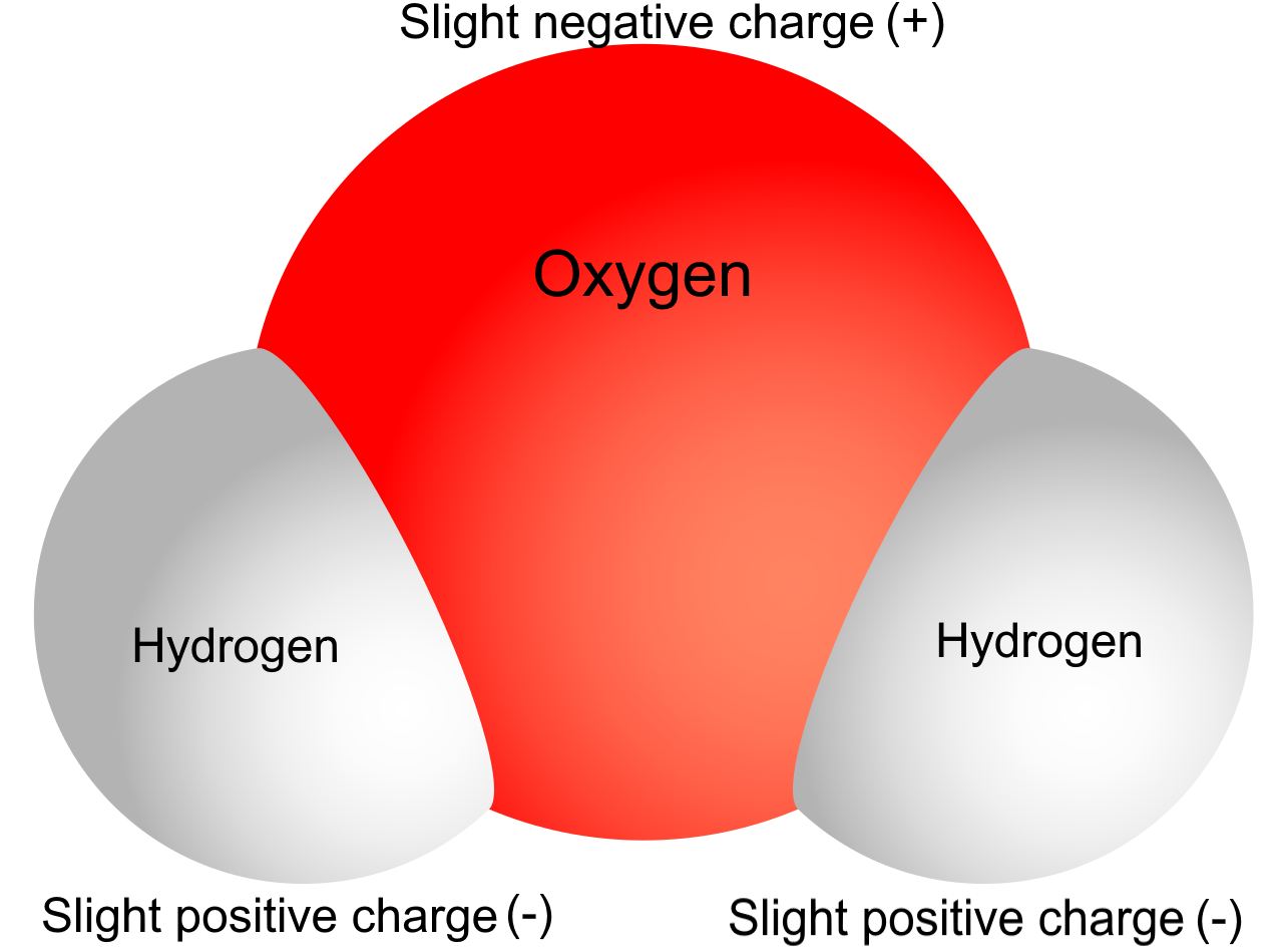
To understand some of water’s properties, you need to know more about its chemical structure. Each molecule of water consists of one atom of oxygen and two atoms of hydrogen. The oxygen atom in a water molecule attracts electrons more strongly than the hydrogen atoms do. As a result, the oxygen atom has a slightly negative charge, and the hydrogen atoms have a slightly positive charge. A difference in electrical charge between different parts of the same molecule is called polarity. The diagram in Figure 3.11.3 shows water’s polarity.
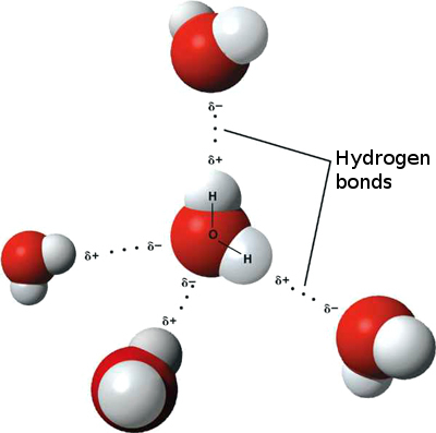
When it comes to charged molecules, opposites attract. In the case of water, the positive (hydrogen) end of one water molecule is attracted to the negative (oxygen) end of a nearby water molecule. Because of this attraction, weak bonds form between adjacent water molecules, as shown in Figure 3.11.4. The type of bond that forms between water molecules is called a hydrogen bond. Bonds between molecules are not as strong as bonds within molecules, but in water, they are strong enough to hold together nearby molecules.
How do you think hydrogen bonding affects water's properties?
Properties of Water
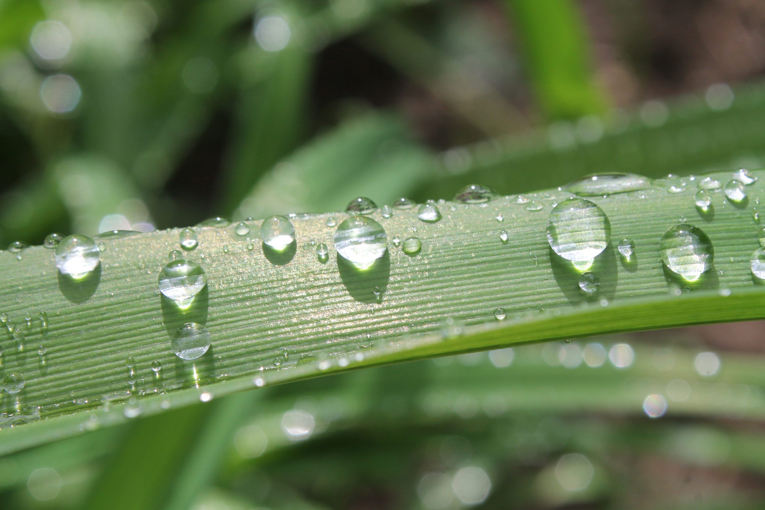
Hydrogen bonds between water molecules explain some of water’s properties — for example, why water molecules tend to "stick" together. Did you ever watch water drip from a leaky faucet or from a melting icicle? If you did, then you know that water always falls in drops, rather than as separate molecules. The dew drops pictured to the left are another example of water molecules sticking together.
Hydrogen bonds cause water to have a relatively high boiling point of 100°C (212°F). Extra energy is needed to break these bonds and separate water molecules so they can escape into the air as water vapor. Because of its high boiling point, most water on Earth is in a liquid state, rather than a gaseous state. Water in its liquid state is needed by all living things. Hydrogen bonds also cause water to expand when it freezes. This, in turn, causes ice to have a lower density (that is, less mass per unit volume) than liquid water. The lower density of ice means that it floats on water. In cold climates, ice floats on top of the water in lakes. This allows lake animals like fish to survive the winter by staying in the liquid water under the ice.
Watch the video below to hear more about hydrogen bonding and it's effects on the properties of water:
https://www.youtube.com/watch?v=UukRgqzk-KE
Why does ice float in water? - George Zaidan and Charles Morton, TED-ED, 2013.
Water and Living Things
The human body is about 70 per cent water (not counting the water in body fat, which varies from person to person). The body needs all this water to function normally. Just why is so much water required by human beings and other organisms? Water can dissolve many substances that organisms need. Water's polarity helps it dissolve other polar substances. Water is also necessary for many biochemical reactions. The examples below are among the most important biochemical processes that occur in living things, but they are just two of the many ways that water is involved in biochemical reactions.
- Photosynthesis: In this process, cells use the energy in sunlight to change carbon dioxide and water to glucose and oxygen. The reactions of photosynthesis can be represented by the chemical equation:
6CO2 + 6H2O + Energy → C6H12O6 + 6O2
- Cellular respiration: In this process, cells break down glucose in the presence of oxygen and release carbon dioxide, water, and energy. The reactions of cellular respiration can be represented by the chemical equation:
C6H12O6 + 6O2 → 6CO2 + 6H2O + Energy
Water is involved in many other biochemical reactions and almost all life processes depend on water.
Feature: My Human Body
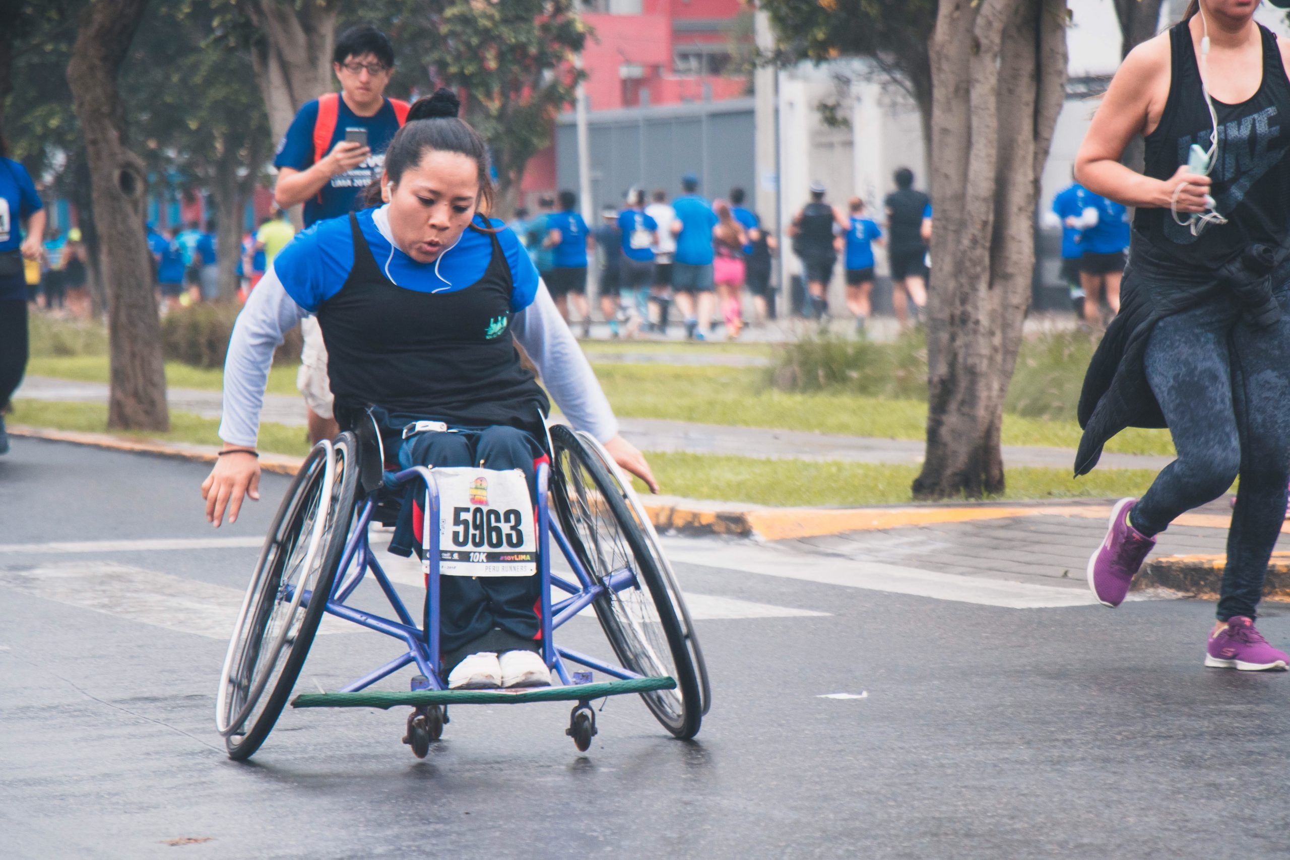
Are you a marathon runner or other endurance athlete? Do you live and work in a hot, humid climate? If you answered "yes" to either question, you may be at risk of water intoxication.
Water is considered the least toxic chemical compound, so it may surprise you to learn that drinking too much water can cause serious illness and even death. Water intoxication is a potentially fatal disturbance in brain functions. It results when the normal balance of sodium and other electrolytes in the body is pushed outside safe limits by overhydration, or taking in too much water. The condition is also called hyponatremia, which refers to a lower-than-normal level of sodium in the blood that occurs when more water is entering than leaving the body.
As excessive water is consumed, fluid outside the cells decreases in its concentration of sodium and other electrolytes relative to the concentration inside the cells. This causes fluid to enter the cells by osmosis to balance the electrolyte concentration. The extra fluid in the cells causes them to swell. In the brain, this swelling increases the pressure inside the skull. It is this increase in pressure that leads to the first observable symptoms of water intoxication, which typically include headache, confusion, irritability, and drowsiness. As the condition worsens, additional symptoms may occur, such as difficulty breathing during exertion, muscle weakness and pain, or nausea and vomiting. If the condition persists, the cells in the brain may swell to the point where blood flow is interrupted or pressure is applied to the brain stem. This is extremely dangerous and may lead to seizures, brain damage, coma, or even death.
Under normal circumstances, it is very rare to accidentally consume too much water. However, it is relatively common in athletes who participate in endurance activities, such as marathon running. A study conducted on participants of the 2002 Boston Marathon, for example, found that 13 per cent of the runners finished the race with water intoxication (Almond, et al., 2005). The study also found that water intoxication was just as likely to occur in runners who drank sports drinks containing electrolytes as those who drank plain water. Water intoxication is so common at marathon events that medical personnel who work at such events are trained to suspect water intoxication when runners collapse or show signs of confusion.
Because of the publicity water intoxication has received lately, sports experts have lowered their recommendations for water intake during endurance events. They now advise drinking only when thirsty rather than drinking to "stay ahead of thirst," which they recommended previously. Keeping water intake in line with water loss is the best way to prevent water intoxication. Mild water intoxication can be treated by restricting fluid intake. In more severe cases, treatment may require the use of diuretic drugs (which increase urination) or other types of drugs to reduce blood volume. Serious water intoxication should be considered a true medical emergency.
3.11 Summary
- Most water on Earth consists of salt water in the oceans. Only a tiny percentage of the Earth's water is fresh liquid water.
- Virtually all living things on Earth require liquid water. Water exists as a liquid over a wide range of temperatures and dissolves many substances. These properties depend on water's polarity, which causes water molecules to "stick" together.
- The human body is about 70 per cent water (outside of fat). Organisms need water to dissolve many substances and for most biochemical processes, including photosynthesis and cellular respiration.
3.11 Review Questions
- Where is most of Earth's fresh water found?
- Identify properties of water.
- What is polarity? Explain why water molecules are polar.
- Why do water molecules tend to "stick" together?
- What role does water play in photosynthesis and cellular respiration?
- Which do you think is stronger: the bonds between the hydrogen and oxygen atoms within a water molecule, or the bonds between the hydrogen and oxygen atoms between water molecules? Explain your answer.
- Given what you’ve learned about water intoxication (or hyponatremia), explain why you think drinking salt water would be bad for your cells.
- What is the name for the bonds that form between water molecules?
- Explain why water can dissolve other polar molecules.
- If there is pollution in the ocean that causes the water to become more cloudy or opaque, how do you think the ocean's photosynthetic organisms will be affected? Explain your answer.
- Describe one way in which your body gets rid of excess water.
- True or False: Ice floats on top of water because it is denser than water.
3.11 Explore More
https://www.youtube.com/watch?v=3jwAGWky98c&t=14s
Properties of Water, by The Amoeba Sisters, 2016.
https://www.youtube.com/watch?v=ASLUY2U1M-8&t=84s
How polarity makes water behave strangely - Christina Kleinberg, TED-Ed, 2013.
Attributions
Figure 3.11.1
Planet Earth by NASA (photo taken by either Harrison Schmitt or Ron Evans (of the Apollo 17 crew), on Wikimedia Commons, is released into the public domain (https://en.wikipedia.org/wiki/Public_domain).
Figure 3.11.2
Total water on earth by LadyofHats at CK12, is used under a CC BY-NC 3.0 (https://creativecommons.org/licenses/by-nc/3.0/) license.
Figure 3.11.3
Polarity of water by Christine Miller is released into the Public Domain (https://creativecommons.org/publicdomain/mark/1.0/).
Figure 3.11.4
Hydrogen bonds, translated by Michal Maňas (User:snek01) is released into the public domain (https://en.wikipedia.org/wiki/Public_domain). (Original uploader was Qwerter at Czech Wikipedia.)
Figure 3.11.5
Dew by Pascal Chanel on Unsplash is used under the Unsplash License (https://unsplash.com/license).
Figure 3.11.6
Woman in a wheelchair marathon by Kevin André on Unsplash is used under the Unsplash License (https://unsplash.com/license).
References
Almond, C.S., Shin, A.Y., Fortescue, E.B. et al. (2005, April). Hyponatremia among runners in the Boston Marathon. The New England Journal of Medicine, 352 (15), 1550–1624. doi:10.1056/NEJMoa043901. PMID 15829535.
Amoeba Sisters. (2016, July 26). Properties of Water. YouTube. https://www.youtube.com/watch?v=3jwAGWky98c&feature=youtu.be
Ruiz Villarreal, M. (LadyofHats). (2016, August 15). Figure 2. Total water on earth [digital image]. In Brainard, J., Henderson, R., CK-12's College Human Biology FlexBook® (section 3.11). CK12 Foundation. https://www.ck12.org/book/ck-12-college-human-biology/
TED-Ed. (2013, February 4). How polarity makes water behave strangely - Christina Kleinberg. YouTube. https://www.youtube.com/watch?v=ASLUY2U1M-8&feature=youtu.be
TED-Ed. (2013, October 22). Why does ice float in water? - George Zaidan and Charles Morton. YouTube. https://www.youtube.com/watch?v=UukRgqzk-KE&feature=youtu.be
Created by CK-12 Foundation/Adapted by Christine Miller
Case Study Conclusion: Needing to Relax
As you learned in the beginning of this chapter, botulinum toxin — one form of which is sold under the brand name Botox — does much more than smooth out wrinkles. It can be used to treat a number of disorders involving excessive muscle contraction, including cervical dystonia. You also learned that cervical dystonia, which Edward suffers from, causes abnormal, involuntary muscle contractions of the neck. This results in jerky movements of the head and neck, and/or a sustained abnormal tilt to the head. It is often painful and can significantly interfere with a person’s life.

How could a toxin actually help treat a muscular disorder? The botulinum toxin is produced by the soil bacterium, Clostridium botulinum, and it is the cause of the potentially deadly disease called botulism. Botulism is often a foodborne illness, commonly caused by foods that are improperly canned. Other forms of botulism are caused by wound infections, or occur when infants consume spores of the bacteria from soil or honey.
Botulism can be life-threatening, because it paralyzes muscles throughout the body, including those involved in breathing. When a very small amount of botulinum toxin is injected carefully into specific muscles by a trained medical professional, however, it can be useful in inhibiting unwanted muscle contractions.
For cosmetic purposes, botulinum toxin injected into the facial muscles relaxes them to reduce the appearance of wrinkles. When used to treat cervical dystonia, it is injected into the muscles of the neck to inhibit excessive muscle contractions. For many patients, this helps relieve the abnormal positioning, movements, and pain associated with the disorder. The effect is temporary, so the injections must be repeated every three to four months to keep the symptoms under control.
How does botulinum toxin inhibit muscle contraction? First, recall how skeletal muscle contraction works. A motor neuron instructs skeletal muscle fibres to contract at a synapse between them called the neuromuscular junction. A nerve impulse called an action potential travels down to the axon terminal of the motor neuron, where it causes the release of the neurotransmitter acetylcholine (ACh) from synaptic vesicles. The ACh travels across the synaptic cleft and binds to ACh receptors on the muscle fibre, signaling the muscle fibre to contract. According to the sliding filament theory, the contraction of the muscle fibre occurs due to the sliding of myosin and actin filaments across each other. This causes the Z discs of the sacromeres to move closer together, shortening the sacromeres and causing the muscle fibre to contract.
If you wanted to inhibit muscle contraction, at what points could you theoretically interfere with this process? Inhibiting the action potential in the motor neuron, the release of ACh, the activity of ACh receptors, or the sliding filament process in the muscle fibre would all theoretically impair this process and inhibit muscle contraction. For example, in the disease myasthenia gravis, the function of the ACh receptors is impaired, causing a lack of sufficient muscle contraction. As you have learned, this results in muscle weakness that can eventually become life-threatening. Botulinum toxin works by inhibiting the release of ACh from the motor neurons, thereby removing the signal instructing the muscles to contract.
Fortunately, Edward’s excessive muscle contractions and associated pain improved significantly thanks to botulinum toxin injections. Although cervical dystonia cannot currently be cured, botulinum toxin injections have improved the quality of life for many patients with this and other disorders involving excessive involuntary muscle contractions.
As you have learned in this chapter, our muscular system allows us to do things like make voluntary movements, digest our food, and pump blood through our bodies. Whether they are in your arm, heart, stomach, or blood vessels, muscle tissue works by contracting. But as you have seen here, too much contraction can be a very bad thing. Fortunately, scientists and physicians have found a way to put a potentially deadly toxin — and wrinkle-reducing treatment — to excellent use as a medical treatment for some muscular system disorders.
Chapter 12 Summary
In this chapter, you learned about the muscular system. Specifically, you learned that:
- The muscular system consists of all the muscles of the body. There are three types of muscle: skeletal muscle (which is attached to bones by tendons and enables voluntary body movements), cardiac muscle (which makes up the walls of the heart and makes it beat) and smooth muscle (which is found in the walls of internal organs and other internal structures and controls their movements).
- Muscles are organs composed mainly of muscle cells, which may also be called muscle fibres or myocytes. Muscle cells are specialized for the function of contracting, which occurs when protein filaments inside the cells slide over one another using energy from ATP. Muscle tissue is the only type of tissue that has cells with the ability to contract.
- Muscles can grow larger, or hypertrophy. This generally occurs through increased use, although hormonal or other influences can also play a role. Muscles can also grow smaller, or atrophy. This may occur through lack of use, starvation, certain diseases, or aging. In both hypertrophy and atrophy, the size — but not the number — of muscle fibres changes. The size of muscles is the main determinant of muscle strength.
- Skeletal muscles need the stimulus of motor neurons to contract, and to move the body, they need the skeletal system to act upon.
- Skeletal muscle is the most common type of muscle tissue in the human body. To move bones in opposite directions, skeletal muscles often consist of pairs of muscles that work in opposition to one another to move bones in different directions at joints.
- Skeletal muscle fibres are bundled together in units called muscle fascicles, which are bundled together to form individual skeletal muscles. Skeletal muscles also have connective tissue supporting and protecting the muscle tissue.
-
- Each skeletal muscle fibre consists of a bundle of myofibrils, which are bundles of protein filaments. The filaments are arranged in repeating units called sarcomeres, which are the basic functional units of skeletal muscles. Skeletal muscle tissue is striated, because of the pattern of sarcomeres in its fibres.
- Skeletal muscle fibres can be divided into two types, called slow-twitch and fast-twitch fibres. Slow-twitch fibres are used mainly in aerobic endurance activities (such as long-distance running). Fast-twitch fibres are used mainly for non-aerobic, strenuous activities (such as sprinting). Proportions of the two types of fibres vary from muscle to muscle and person to person.
- Smooth muscle tissue is found in the walls of internal organs and vessels. When smooth muscles contract, they help the organs and vessels carry out their functions. The pattern of smooth muscle contraction to move substances through body tubes is called peristalsis. Contractions of smooth muscles are involuntary and controlled by the autonomic nervous system, hormones, and other substances.
-
- Cells of smooth muscle tissue are not striated because they lack sarcomeres, but the cells contract in the same basic way as striated muscle cells. Unlike striated muscle, smooth muscle can sustain very long-term contractions and maintain its contractile function, even when stretched.
- Cardiac muscle tissue is found only in the wall of the heart. When cardiac muscle contracts, the heart beats and pumps blood. Contractions of cardiac muscle are involuntary, like those of smooth muscles. They are controlled by electrical impulses from specialized cardiac cells.
-
- Like skeletal muscle, cardiac muscle is striated because its filaments are arranged in sarcomeres. The exact arrangement, however, differs, making cardiac and skeletal muscle tissues look different from one another.
- The heart is the muscle that performs the greatest amount of physical work in the course of a lifetime. Its cells contain a great many mitochondria to produce ATP for energy and to help the heart resist fatigue.
- A muscle contraction is an increase in the tension or a decrease in the length of a muscle. A muscle contraction is isometric if muscle tension changes, but muscle length remains the same. It is isotonic if muscle length changes, but muscle tension remains the same.
-
- A skeletal muscle contraction begins with electrochemical stimulation of a muscle fibre by a motor neuron. This occurs at a chemical synapse called a neuromuscular junction. The neurotransmitter acetylcholine diffuses across the synaptic cleft and binds to receptors on the muscle fibre. This initiates a muscle contraction.
- Once stimulated, the protein filaments within the skeletal muscle fibre slide past each other to produce a contraction. The sliding filament theory is the most widely accepted explanation for how this occurs. According to this theory, thick myosin filaments repeatedly attach to and pull on thin actin filaments, thus shortening sarcomeres.
- Crossbridge cycling is a cycle of molecular events that underlies the sliding filament theory. Using energy in ATP, myosin heads repeatedly bind with and pull on actin filaments. This moves the actin filaments toward the center of a sarcomere, shortening the sarcomere and causing a muscle contraction.
- The ATP needed for a muscle contraction comes first from ATP already available in the cell, and more is generated from creatine phosphate. These sources are quickly used up. Glucose and glycogen can be broken down to form ATP and pyruvate. Pyruvate can then be used to produce ATP in aerobic respiration if oxygen is available, or it can be used in anaerobic respiration if oxygen is not available.
- Physical exercise is defined as any bodily activity that enhances or maintains physical fitness and overall health. Activities such as household chores may even count as physical exercise! Current recommendations for adults are 30 minutes of moderate exercise a day.
- Aerobic exercise is any physical activity that uses muscles at less than their maximum contraction strength, but for long periods of time. This type of exercise uses a relatively high percentage of slow-twitch muscle fibres that consume large amounts of oxygen. Aerobic exercises increase cardiovascular endurance, and include cycling and brisk walking.
- Anaerobic exercise is any physical activity that uses muscles at close to their maximum contraction strength, but for short periods of time. This type of exercise uses a relatively high percentage of fast-twitch muscle fibres that consume small amounts of oxygen. Anaerobic exercises increase muscle and bone mass and strength, and they include push-ups and sprinting.
- Flexibility exercise is any physical activity that stretches and lengthens muscles, thereby improving range of motion and reducing risk of injury. Examples include stretching and yoga.
- Many studies have shown that physical exercise is positively correlated with a diversity of physical, mental, and emotional health benefits. Physical exercise also increases quality of life and life expectancy.
-
- Many of the benefits of exercise may come about because contracting muscles release hormones called myokines, which promote tissue repair and growth and have anti-inflammatory effects.
- Physical exercise can reduce risk factors for cardiovascular disease, including hypertension and excess body weight. Physical exercise can also increase factors associated with cardiovascular health, such as mechanical efficiency of the heart.
- Physical exercise has been shown to offer protection from dementia and other cognitive problems, perhaps because it increases blood flow or neurotransmitters in the brain, among other potential effects.
- Numerous studies suggest that regular aerobic exercise works as well as pharmaceutical antidepressants in treating mild-to-moderate depression, possibly because it increases synthesis of natural euphoriants in the brain.
- Research shows that physical exercise generally improves sleep for most people, and helps sleep disorders, such as insomnia. Other health benefits of physical exercise include better immune system function and reduced risk of type 2 diabetes and obesity.
- There is great variation in individual responses to exercise, partly due to genetic differences in proportions of slow-twitch and fast-twitch muscle fibres. People with more slow-twitch fibres may be able to develop greater endurance from aerobic exercise, whereas people with more fast-twitch fibres may be able to develop greater muscle size and strength from anaerobic exercise.
- Some adverse effects may occur if exercise is extremely intense and the body is not given proper rest between exercise sessions. Many people who overwork their muscles develop delayed onset muscle soreness (DOMS), which may be caused by tiny tears in muscle fibres.
- Musculoskeletal disorders are injuries that occur in muscles or associated tissues (such as tendons) because of biomechanical stresses. The disorders may be caused by sudden exertion, over-exertion, repetitive motions, and similar stresses.
-
- A muscle strain is an injury in which muscle fibres tear as a result of overstretching. First aid for a muscle strain includes the five steps represented by the acronym PRICE (protection, rest, ice, compression, and elevation). Medications for inflammation and pain (such as NSAIDs) may also be used.
- Tendinitis is inflammation of a tendon that occurs when it is over-extended or worked too hard without rest. Tendinitis may also be treated with PRICE and NSAIDs.
- Carpal tunnel syndrome is a biomechanical problem that occurs in the wrist when the median nerve becomes compressed between carpal bones. It may occur with repetitive use, a tumor, or trauma to the wrist. It may cause pain, numbness, and eventually — if untreated — muscle wasting in the thumb and first two fingers of the hand.
- Neuromuscular disorders are systemic disorders that occur because of problems with the nervous control of muscle contractions, or with the muscle cells themselves.
-
- Muscular dystrophy is a genetic disorder caused by defective proteins in muscle cells. It is characterized by progressive skeletal muscle weakness and death of muscle tissues.
- Myasthenia gravis is a genetic neuromuscular disorder characterized by fluctuating muscle weakness and fatigue. More muscles are affected, and muscles become increasingly weakened as the disorder progresses. Myasthenia gravis most often occurs because immune system antibodies block acetylcholine receptors on muscle cells, and because of the actual loss of acetylcholine receptors.
- Parkinson’s disease is a degenerative disorder of the central nervous system that mainly affects the muscular system and movement. It occurs because of the death of neurons in the midbrain. Characteristic signs of the disorder are muscle tremor, muscle rigidity, slowness of movement, and postural instability. Dementia and depression also often characterize advanced stages of the disease.
As you saw in this chapter, muscles need oxygen to provide enough ATP for most of their activities. In fact, all of the body’s systems require oxygen, and also need to remove waste products, such as carbon dioxide. In the next chapter, you will learn about how the respiratory system obtains and distributes oxygen throughout the body, as well as how it removes wastes, such as carbon dioxide.
Chapter 12 Review
-
- What are tendons? Name a muscular system disorder involving tendons
- Describe the relationship between muscles, muscle fibres, and fascicles.
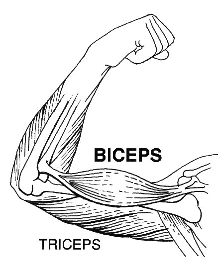
- The biceps and triceps muscles are shown above. Answer the following questions about these arm muscles.
- When the biceps contract and become shorter (as in the picture above), what kind of motion does this produce in the arm?
- Is the situation described in part (a) more likely to be an isometric or isotonic contraction? Explain your answer.
- If the triceps were to then contract, which way would the arm move?
- What are Z discs? What happens to them during muscle contraction?
- What is the function of mitochondria in muscle cells? Which type of muscle fibre has more mitochondria — slow-twitch or fast-twitch?
- What is the difference between primary and secondary Parkinson’s disease?
- Why can carpal tunnel syndrome cause muscle weakness in the hands?
Attributions
Figure 12.7.1
Botox, he whispered by Michael Reuter on Flickr is used under a CC BY 2.0 (https://creativecommons.org/licenses/by/2.0/) license.
Figure 12.7.2
botulism by jason wilson on Flickr is used under a CC BY 2.0 (https://creativecommons.org/licenses/by/2.0/) license.
Reference
Pearson Scott Foresman. (2020, April 14). File:Biceps (PSF).jpg [digital image]. Wikimedia Commons. https://commons.wikimedia.org/w/index.php?title=File:Biceps_(PSF).jpg&oldid=411251538. [Public Domain (https://en.wikipedia.org/wiki/Public_domain)]
Involuntary, striated muscle found only in the walls of the heart; also called myocardium.
Structures containing neuronal cell bodies in the peripheral nervous system.
A hollow, tube-like structure through which blood flows in the cardiovascular system; vein, artery, or capillary.
Created by CK-12 Foundation/Adapted by Christine Miller

Double Jointed?
Is this woman double jointed? No, there is actually no such thing — at least as far as humans are concerned. However, some people, like the woman pictured in Figure 11.6.1, are much more flexible than others, generally because they have looser ligaments. Physicians call this condition joint hypermobility. Regardless of what it’s called, the feats of people with highly mobile joints can be quite impressive.
What Are Joints?
Joints are locations at which bones of the skeleton connect with one another. A joint is also called an articulation. The majority of joints are structured in such a way that they allow movement. However, not all joints allow movement. Of joints that do allow movement, the extent and direction of the movements they allow also vary.
Classification of Joints
Joints can be classified structurally or functionally. The structural classification of joints depends on the manner in which the bones connect to each other. The functional classification of joints depends on the nature of the movement the joints allow. There is significant overlap between the two types of classifications, because function depends largely on structure.
Structural Classification of Joints
The structural classification of joints is based on the type of tissue that binds the bones to each other at the joint. There are three types of joints in the structural classification: fibrous, cartilaginous, and synovial joints.
- Fibrous joints are joints in which bones are joined by dense connective tissue that is rich in collagen fibres. These joints are also called sutures. The joints between bones of the cranium are fibrous joints.
- Cartilaginous joints are joints in which bones are joined by cartilage. The joints between most of the vertebrae in the spine are cartilaginous joints.
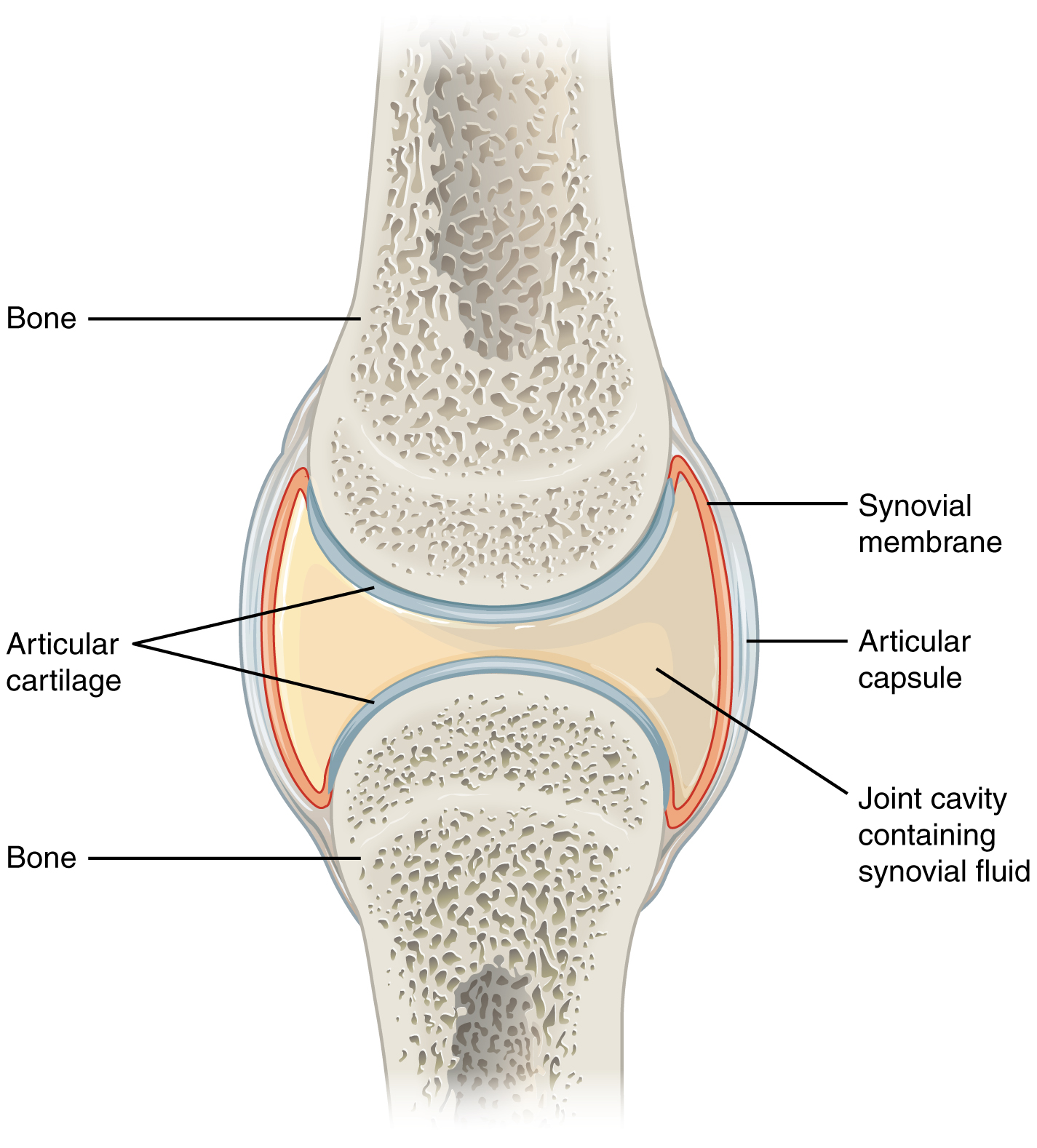
- Synovial joints are characterized by a fluid-filled space (called a synovial cavity) between the bones of the joints. You can see a drawing of a typical synovial joint in Figure 11.6.2. The cavity is enclosed by a membrane and filled with a fluid (called synovial fluid) that provides extra cushioning to the ends of the bones. Cartilage covers the articulating surfaces of the two bones, but the bones are actually held together by ligaments. The knee is a synovial joint.
Functional Classification of Joints
The functional classification of joints is based on the type and degree of movement that they allow. There are three types of joints in the functional classification: immovable, partly movable, and movable joints.
- Immovable joints allow little or no movement at the joint. Most immovable joints are fibrous joints. Besides the bones of the cranium, immovable joints include joints between the tibia and fibula in the lower leg, and between the radius and ulna in the lower arm.
- Partly movable joints permit slight movement. Most partly movable joints are cartilaginous joints. Besides the joints between vertebrae, they include the joints between the ribs and sternum (breast bone).
- Movable joints allow bones to move freely. All movable joints are synovial joints. Besides the knee, they include the shoulder, hip, and elbow. Movable joints are the most common type of joints in the body.
Types of Movable Joints
Movable joints can be classified further according to the type of movement they allow. There are six classes of movable joints: pivot, hinge, saddle, plane, condyloid, and ball-and-socket joints. An example of each class — as well as the type of movement it allows — is shown in Figure 11.6.3.
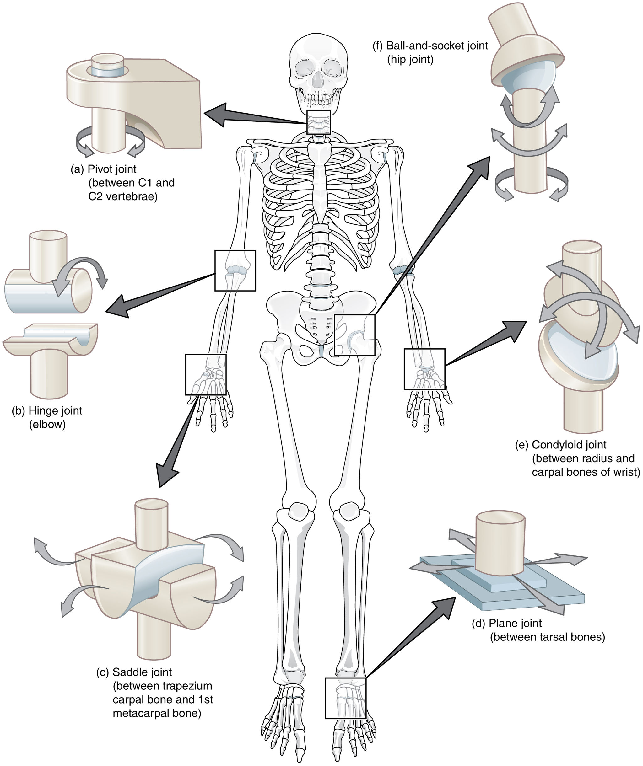
- A ball-and-socket joint allows the greatest range of movement of any movable joint. It allows forward and backward motion, as well as upward and downward movement. It also allows rotation in a circle. The hip and shoulder are the only two ball-and-socket joints in the human body.
- A pivot joint allows one bone to rotate around another. An example of a pivot joint is the joint between the first two vertebrae in the spine. This joint allows the head to rotate from left to right and back again.
- A hinge joint allows back and forth movement like the hinge of a door. An example of a hinge joint is the elbow. This joint allows the arm to bend back and forth.
- A saddle joint allows two different types of movement. An example of a saddle joint is the joint between the first metacarpal bone in the hand and one of the carpal bones in the wrist. This joint allows the thumb to move toward and away from the index finger, and also to cross over the palm toward the little finger.
- A plane joint (also called a gliding joint) allows two bones to glide over one another. The joints between the tarsals in the ankles and between the carpals in the wrists are mainly gliding joints. In the wrist, this type of joint allows the hand to bend upward at the wrist, and also to wave from side to side while the lower arm is held steady.
- A condyloid joint is one in which an oval-shaped head on one bone moves in an elliptical cavity in another bone, allowing movement in all directions, except rotation around an axis. The joint between the radius in the lower arm and carpal bones of the wrist is a condyloid joint, as is the joint at the base of the index finger.
Feature: My Human Body
Of all the parts of the skeletal system, the joints are generally the most fragile and subject to damage. If the cartilage that cushions bones at joints wears away, it does not grow back. Eventually, all of the cartilage may wear away. This causes osteoarthritis, which can be both painful and debilitating. In serious cases of osteoarthritis, people may lose the ability to climb stairs, walk long distances, perform routine daily activities, or participate in activities they love, such as gardening or playing sports. If you protect your joints, you can reduce your chances of joint damage, pain, and disability. If you already have joint damage, it is equally important to protect your joints and limit further damage. Follow these five tips:
- Maintain a normal, healthy weight. The more you weigh, the more force you exert on your joints. When you walk, each knee has to bear a force equal to as much as six times your body weight. If a person weighs 200 pounds, each knee bears more than half a ton of weight with every step. Seven in ten knee replacement surgeries for osteoarthritis can be attributed to obesity.
- Avoid too much high-impact exercise. Examples of high-impact activities include volleyball, basketball, and tennis. These activities generally involve running or jumping on hard surfaces, which puts tremendous stress on weight-bearing joints, especially the knees. Replace some or all of your high-impact activities with low-impact activities, such as biking, swimming, yoga, or lifting light weights.
- Reduce your risk of injury. Don’t be a weekend warrior, sitting at a desk all week and then crowding all your physical activity into two days. Get involved in a regular, daily exercise routine that keeps your body fit and your muscles toned. Building up muscles will make your joints more stable, allowing stress to spread across them. Be sure to do some stretching every day to keep the muscles around joints flexible and less prone to injury.
- Distribute work over your body, and use your largest, strongest joints. Use your shoulder, elbow, and wrist to lift heavy objects — not just your fingers. Hold small items in the palm of your hand, rather than by the fingers. Carry heavy items in a backpack, rather than in your hands. Hold weighty objects close to your body, instead of at arms’ length. Lift with your hips and knees, not your back.
- Respect pain. If it hurts, stop doing it. Take a break from the activity — at least until the pain stops. Try to use joints only to the point of mild fatigue, not pain.
11.6 Summary
- Joints are spots at which bones of the skeleton connect with one another. A joint is also called an articulation.
- Joints can be classified structurally or functionally, and there is significant overlap between the two types of classifications.
- The structural classification of joints depends on the type of tissue that binds the bones to each other at the joint. There are three types of joints in the structural classification: fibrous, cartilaginous, and synovial joints.
- The functional classification of joints is based on the type and degree of movement that they allow. There are three types of joints in the functional classification: immovable, partly movable, and movable joints.
- Movable joints can be classified further according to the type of movement they allow. There are six classes of movable joints: pivot, hinge, saddle, plane, condyloid, and ball-and-socket joints.
11.6 Review Questions
- What are joints?
- What are two ways that joints are commonly classified?
-
- How are joints classified structurally?
- Describe the functional classification of joints.
- How are movable joints classified?
- Name the six classes of movable joints. Describe how they move and give an example of each.
- Which specific type of moveable joint do you think your knee joint is? Explain your reasoning.
- Explain the difference between cartilage in a cartilaginous joint and cartilage in a synovial joint.
- Why are fibrous joints immovable?
- What is the function of synovial fluid?
11.6 Explore More
https://www.youtube.com/watch?v=IjiKUmfaZr4
Why do your knuckles pop? - Eleanor Nelsen, TED-Ed, 2015.
https://www.youtube.com/watch?v=FWsBm3hr3B0
Why haven’t we cured arthritis? - Kaitlyn Sadtler and Heather J. Faust, TED-Ed, 2019.
Attributions
Figure 11.6.1
Tags: Sports Gymnastics Fitness Woman Preparation by nastya_gepp on Pixabay is used under the Pixabay License (https://pixabay.com/de/service/license/).
Figure 11.6.2
Synovial_Joints by OpenStax College on Wikimedia Commons is used under a CC BY 3.0 (https://creativecommons.org/licenses/by/3.0) license.
Figure 11.6.3
Types_of_Synovial_Joints by OpenStax College on Wikimedia Commons is used under a CC BY 3.0 (https://creativecommons.org/licenses/by/3.0) license.
References
Betts, J. G., Young, K.A., Wise, J.A., Johnson, E., Poe, B., Kruse, D.H., Korol, O., Johnson, J.E., Womble, M., DeSaix, P. (2013, June 19). Figure 9.8 Synovial joints [digital image]. In Anatomy and Physiology (Section 9.4). OpenStax. https://openstax.org/books/anatomy-and-physiology/pages/9-4-synovial-joints
Betts, J. G., Young, K.A., Wise, J.A., Johnson, E., Poe, B., Kruse, D.H., Korol, O., Johnson, J.E., Womble, M., DeSaix, P. (2013, June 19). Figure 9.10 Types of synovial joints [digital image]. In Anatomy and Physiology (Section 9.4). OpenStax. https://openstax.org/books/anatomy-and-physiology/pages/9-4-synovial-joints
Mayo Clinic Staff. (n.d.). Osteoarthritis [online article]. MayoClinic.org. https://www.mayoclinic.org/diseases-conditions/osteoarthritis/symptoms-causes/syc-20351925
TED-Ed. (2015, May 5). Why do your knuckles pop? - Eleanor Nelsen. YouTube. https://www.youtube.com/watch?v=IjiKUmfaZr4
TED-Ed. (2019, November 7). Why haven’t we cured arthritis? - Kaitlyn Sadtler and Heather J. Faust. YouTube. https://www.youtube.com/watch?v=FWsBm3hr3B0
Case Study: More Than Just Tired
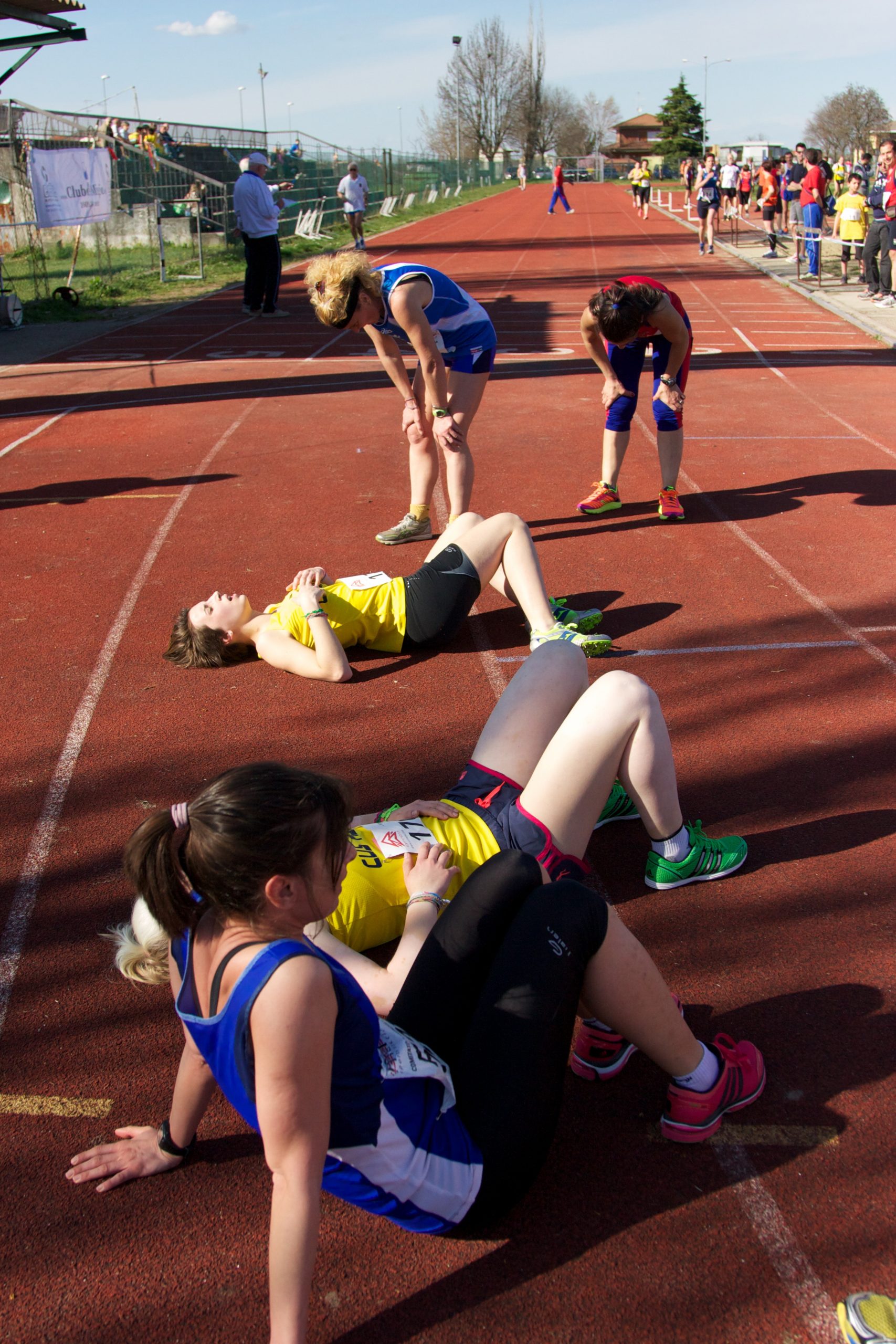
We all get tired sometimes, especially if we have been doing a lot of physical activity, like the athletes pictured in Figure 4.1.1. But for Jasmin (Figure 4.1.2), a 34-year-old former high school track star who is now a recreational runner, her tiredness was going far beyond what she thought should be normal for someone in generally good physical shape.

She was experiencing extreme fatigue after her runs, as well as muscle cramping, spasms, and an unusual sense of heaviness in her legs. At first, she just chalked it up to getting older, but her exhaustion and pain worsened to the point where the former athlete could no longer run for more than a few minutes at a time. She began to experience other unusual symptoms, such as blurry vision and vomiting for no apparent reason.
Concerned, Jasmin went to her doctor, who ran many tests and consulted with several specialists. After several months, she was finally diagnosed with a mitochondrial disease. Jasmin is surprised. She has an 8-year-old niece with a mitochondrial disease, but her niece’s symptoms started when she was very young, and they included seizures and learning disabilities. How can Jasmin have the same disease, but different symptoms? Why didn't she have problems until adulthood, while her niece experienced symptoms at an early age? And what are mitochondria, anyway?
Chapter Overview: The Importance of Cells
As you will learn in this chapter, mitochondria are important structures within our cells. This chapter will describe cells, which are the basic unit of structure and function in all living organisms. Specifically, you will learn:
- How cells were discovered, their common structures, and the principles of cell theory.
- The importance of size and shape to the functions of cells.
- The differences between eukaryotic cells (such as those in humans and other animals) and prokaryotic cells (such as bacteria).
- The structures and functions of cell parts, including mitochondria, the plasma membrane, cytoplasm, cytoskeleton, nucleus, ribosomes, Golgi apparatus, endoplasmic reticulum, vesicles, and vacuoles.
- The processes of passive and active transport to move substances into and out of cells and help maintain homeostasis.
- How organisms obtain the energy needed for life, including how the sugar glucose is broken down to produce ATP through the processes of anaerobic and aerobic cellular respiration.
- The phases of the cell cycle, how cells divide through mitosis, and how cancer can result from unregulated cell division.
As you read this chapter, think about the following questions related to Jasmin’s disease:
- What are mitochondria? What is their structure and function, and where did they come from during evolution?
- Why are fatigue and “exercise intolerance” (such as Jasmin’s extreme exhaustion after running) common symptoms of mitochondrial diseases?
- Why do you think Jasmin has symptoms that affect so many different parts of her body, including her legs, eyes, and digestive system?
- Why do you think mitochondrial diseases can run in families like Jasmin's?
Attributes
Figure 4.1.1
Difficult competition by Massimo Sartirana on Unsplash is used under the Unsplash License (https://unsplash.com/license).
Figure 4.1.2
Exhausted by Kevin Grieve on Unsplash is used under the Unsplash License (https://unsplash.com/license).
Created by: CK-12/Adapted by Christine Miller

Case Study: More Than Just Tired
We all get tired sometimes, especially if we have been doing a lot of physical activity, like the athletes pictured in Figure 4.1.1. But for Jasmin (Figure 4.1.2), a 34-year-old former high school track star who is now a recreational runner, her tiredness was going far beyond what she thought should be normal for someone in generally good physical shape.

She was experiencing extreme fatigue after her runs, as well as muscle cramping, spasms, and an unusual sense of heaviness in her legs. At first, she just chalked it up to getting older, but her exhaustion and pain worsened to the point where the former athlete could no longer run for more than a few minutes at a time. She began to experience other unusual symptoms, such as blurry vision and vomiting for no apparent reason.
Concerned, Jasmin went to her doctor, who ran many tests and consulted with several specialists. After several months, she was finally diagnosed with a mitochondrial disease. Jasmin is surprised. She has an 8-year-old niece with a mitochondrial disease, but her niece’s symptoms started when she was very young, and they included seizures and learning disabilities. How can Jasmin have the same disease, but different symptoms? Why didn't she have problems until adulthood, while her niece experienced symptoms at an early age? And what are mitochondria, anyway?
Chapter Overview: The Importance of Cells
As you will learn in this chapter, mitochondria are important structures within our cells. This chapter will describe cells, which are the basic unit of structure and function in all living organisms. Specifically, you will learn:
- How cells were discovered, their common structures, and the principles of cell theory.
- The importance of size and shape to the functions of cells.
- The differences between eukaryotic cells (such as those in humans and other animals) and prokaryotic cells (such as bacteria).
- The structures and functions of cell parts, including mitochondria, the plasma membrane, cytoplasm, cytoskeleton, nucleus, ribosomes, Golgi apparatus, endoplasmic reticulum, vesicles, and vacuoles.
- The processes of passive and active transport to move substances into and out of cells and help maintain homeostasis.
- How organisms obtain the energy needed for life, including how the sugar glucose is broken down to produce ATP through the processes of anaerobic and aerobic cellular respiration.
- The phases of the cell cycle, how cells divide through mitosis, and how cancer can result from unregulated cell division.
As you read this chapter, think about the following questions related to Jasmin’s disease:
- What are mitochondria? What is their structure and function, and where did they come from during evolution?
- Why are fatigue and “exercise intolerance” (such as Jasmin’s extreme exhaustion after running) common symptoms of mitochondrial diseases?
- Why do you think Jasmin has symptoms that affect so many different parts of her body, including her legs, eyes, and digestive system?
- Why do you think mitochondrial diseases can run in families like Jasmin's?
Attributes
Figure 4.1.1
Difficult competition by Massimo Sartirana on Unsplash is used under the Unsplash License (https://unsplash.com/license).
Figure 4.1.2
Exhausted by Kevin Grieve on Unsplash is used under the Unsplash License (https://unsplash.com/license).
Figure 4.2.1 Human cells viewed with a very powerful tool called a scanning electron microscope.
Amazing Cells
What are these incredible objects? Would it surprise you to learn that they are all human cells? Cells are actually too small to see with the unaided eye. It is visible here in such detail because it is being viewed with a very powerful tool called a scanning electron microscope. Cells may be small in size, but they are extremely important to life. Like all other living things, you are made of cells. Cells are the basis of life, and without cells, life as we know it would not exist. You will learn more about these amazing building blocks of life in this section.
What Are Cells?
If you look at living matter with a microscope — even a simple light microscope — you will see that it consists of cells. Cells are the basic units of the structure and function of living things. They are the smallest units that can carry out the processes of life. All organisms are made up of one or more cells, and all cells have many of the same structures and carry out the same basic life processes. Knowing the structure of cells and the processes they carry out is necessary to an understanding of life itself.
Discovery of Cells
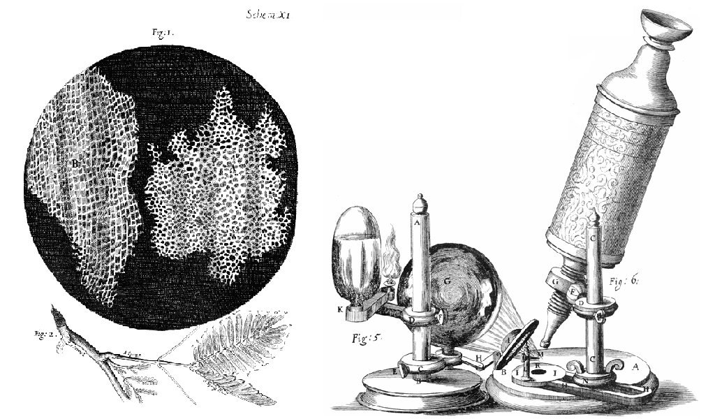
The first time the word cell was used to refer to these tiny units of life was in 1665 by a British scientist named Robert Hooke. Hooke was one of the earliest scientists to study living things under a microscope. The microscopes of his day were not very strong, but Hooke was still able to make an important discovery. When he looked at a thin slice of cork under his microscope, he was surprised to see what looked like a honeycomb. Hooke made the drawing in the figure to the right to show what he saw. As you can see, the cork was made up of many tiny units. Hooke called these units cells because they resembled cells in a monastery.
Soon after Robert Hooke discovered cells in cork, Anton van Leeuwenhoek in Holland made other important discoveries using a microscope. Leeuwenhoek made his own microscope lenses, and he was so good at it that his microscope was more powerful than other microscopes of his day. In fact, Leeuwenhoek’s microscope was almost as strong as modern light microscopes. Using his microscope, Leeuwenhoek was the first person to observe human cells and bacteria.
Cell Theory
By the early 1800s, scientists had observed cells of many different organisms. These observations led two German scientists named Theodor Schwann and Matthias Jakob Schleiden to propose cells as the basic building blocks of all living things. Around 1850, a German doctor named Rudolf Virchow was studying cells under a microscope, when he happened to see them dividing and forming new cells. He realized that living cells produce new cells through division. Based on this realization, Virchow proposed that living cells arise only from other living cells.
The ideas of all three scientists — Schwann, Schleiden, and Virchow — led to cell theory, which is one of the fundamental theories unifying all of biology.
Cell theory states that:
- All organisms are made of one or more cells.
- All the life functions of organisms occur within cells.
- All cells come from existing cells.
Seeing Inside Cells
Starting with Robert Hooke in the 1600s, the microscope opened up an amazing new world — a world of life at the level of the cell. As microscopes continued to improve, more discoveries were made about the cells of living things, but by the late 1800s, light microscopes had reached their limit. Objects much smaller than cells (including the structures inside cells) were too small to be seen with even the strongest light microscope.
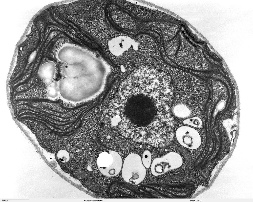
Then, in the 1950s, a new type of microscope was invented. Called the electron microscope, it used a beam of electrons instead of light to observe extremely small objects. With an electron microscope, scientists could finally see the tiny structures inside cells. They could even see individual molecules and atoms. The electron microscope had a huge impact on biology. It allowed scientists to study organisms at the level of their molecules, and it led to the emergence of the molecular biology field. With the electron microscope, many more cell discoveries were made.
Structures Shared By All Cells
Although cells are diverse, all cells have certain parts in common. These parts include a plasma membrane, cytoplasm, ribosomes, and DNA.
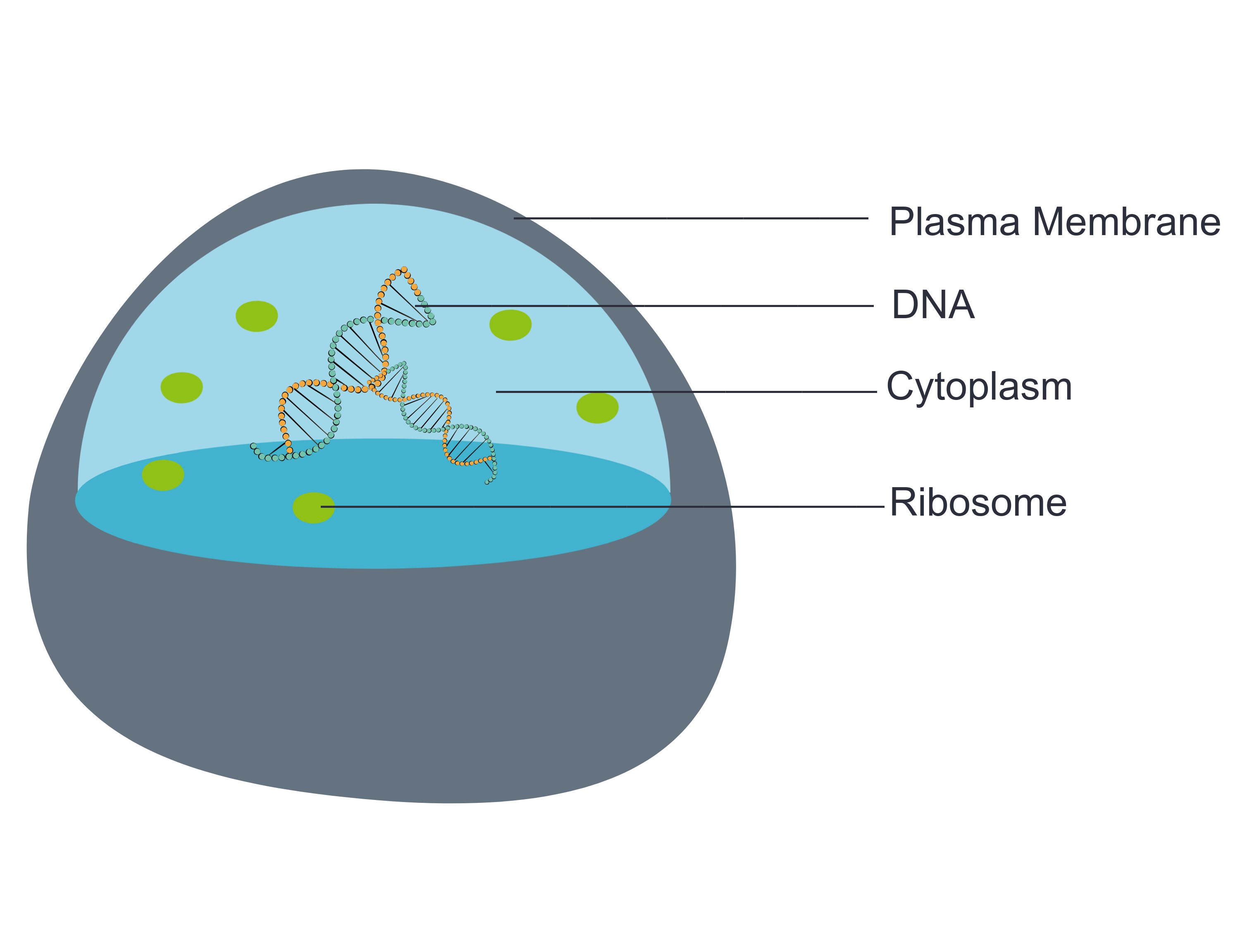
- The plasma membrane (a type of cell membrane) is a thin coat of lipids that surrounds a cell. It forms the physical boundary between the cell and its environment. You can think of it as the “skin” of the cell.
- Cytoplasm refers to all of the cellular material inside of the plasma membrane. Cytoplasm is made up of a watery substance called cytosol, and it contains other cell structures, such as ribosomes.
- Ribosomes are the structures in the cytoplasm in which proteins are made.
- DNA is a nucleic acid found in cells. It contains the genetic instructions that cells need to make proteins.
These four parts are common to all cells, from organisms as different as bacteria and human beings. How did all known organisms come to have such similar cells? The similarities show that all life on Earth has a common evolutionary history.
4.2 Summary
- Cells are the basic units of structure and function in living things. They are the smallest units that can carry out the processes of life.
- In the 1600s, Hooke was the first to observe cells from an organism (cork). Soon after, microscopist van Leeuwenhoek observed many other living cells.
- In the early 1800s, Schwann and Schleiden theorized that cells are the basic building blocks of all living things. Around 1850, Virchow observed cells dividing. To previous learnings, he added that living cells arise only from other living cells. These ideas led to cell theory, which states that all organisms are made of cells, that all life functions occur in cells, and that all cells come from other cells.
- It wasn't until the 1950s that scientists could see what was inside the cell. The invention of the electron microscope allowed them to see organelles and other structures smaller than cells.
- There is variation in cells, but all cells have a plasma membrane, cytoplasm, ribosomes, and DNA. These similarities show that all life on Earth has a common ancestor in the distant past.
4.2 Review Questions
- Describe cells.
- Explain how cells were discovered.
- Outline the development of cell theory.
-
- Identify the structures shared by all cells.
- Proteins are made on _____________ .
-
- Robert Hooke sketched what looked like honeycombs — or repeated circular or square units — when he observed plant cells under a microscope.
- What is each unit?
- Of the shared parts of all cells, what makes up the outer surface of each unit?
- Of the shared parts of all cells, what makes up the inside of each unit?
4.2 Explore More
https://www.youtube.com/watch?v=8IlzKri08kk
Introduction to Cells: The Grand Cell Tour, by The Amoeba Sisters, 2016.
Attributions
Figure 4.2.1
- A white blood cell (WBC) known as a neutrophil by National Institute of Allergy and Infectious Diseases (NIAID) on the CDC/ Public Health Image Library (PHIL) Photo ID# 18129. is in the public domain (https://en.wikipedia.org/wiki/Public_domain).
- Healthy Human T Cell by NIAID on Flickr. is used under a CC BY 2.0 (https://creativecommons.org/licenses/by/2.0/) license.
- Human natural killer cell by NIAID on Flickr. is used under a CC BY 2.0 (https://creativecommons.org/licenses/by/2.0/) license.
- Human blood with red blood cells, T cells (orange) and platelets (green) by ZEISS Microscopy on Flickr. is used under a CC BY-NC-ND 2.0 (https://creativecommons.org/licenses/by-nc-nd/2.0/) license.
- Developing nerve cells by ZEISS Microscopy on Flickr. is used under a CC BY-NC-ND 2.0 (https://creativecommons.org/licenses/by-nc-nd/2.0/) license.
Figure 4.2.2
Hooke-microscope-cork by Robert Hooke (1635-1702) [uploaded by Alejandro Porto] on Wikimedia Commons is released into the public domain (https://en.wikipedia.org/wiki/Public_domain).
Figure 4.2.3
Electron Microscope image of a cell by Dartmouth Electron Microscope Facility, Dartmouth College on Wikimedia Commons is released into the public domain (https://en.wikipedia.org/wiki/Public_domain).
Figure 4.2.4
Basic-Components-of-a-cell by Christine Miller is used under a CC0 1.0 (https://creativecommons.org/publicdomain/zero/1.0/) license.
References
Amoeba Sisters. (2016, November 1). Introduction to cells: The grand cell tour. YouTube. https://www.youtube.com/watch?v=8IlzKri08kk&feature=youtu.be
National Institute of Allergy and Infectious Diseases (NIAID). (2011). A white blood cell (WBC) known as a neutrophil, as it was in the process of ingesting a number of spheroid shaped, methicillin-resistant, Staphylococcus aureus (MRSA) bacteria [digital image]. CDC/ Public Health Image Library (PHIL) Photo ID# 18129. https://phil.cdc.gov/Details.aspx?pid=18129.
Wikipedia contributors. (2020, June 24). Antonie van Leeuwenhoek. In Wikipedia. https://en.wikipedia.org/w/index.php?title=Antonie_van_Leeuwenhoek&oldid=964339564
Wikipedia contributors. (2020, May 25). Matthias Jakob Schleiden. In Wikipedia. https://en.wikipedia.org/w/index.php?title=Matthias_Jakob_Schleiden&oldid=958819219
Wikipedia contributors. (2020, June 4). Rudolf Virchow. In Wikipedia,. https://en.wikipedia.org/w/index.php?title=Rudolf_Virchow&oldid=960708716
Wikipedia contributors. (2020, May 16). Theodor Schwann. In Wikipedia. https://en.wikipedia.org/w/index.php?title=Theodor_Schwann&oldid=956919239
Created by: CK-12/Adapted by Christine Miller

Letting in the Light
Look at the big windows in this house (Figure 4.7.1). Imagine all the light they must let in on a sunny day. Now imagine living in a house that has walls without any windows or doors. Nothing could enter or leave. Or imagine living in a house with holes in the walls instead of windows and doors. Things could enter or leave, but you couldn’t control what came in or went out. Only when a house has walls with windows and doors that can be opened or closed, can you control what enters or leaves. Windows and doors allow you to let in light and the family dog and keep out rain and bugs, for example.
Transport Across Membranes
If a cell were a house, the plasma membrane would be walls with windows and doors. Moving things in and out of the cell is an important function of the plasma membrane. It controls everything that enters and leaves the cell. There are two basic ways that substances can cross the plasma membrane: passive transport — which requires no energy expenditure by the cell — and active transport — which requires energy from the cell.
Transport Without Energy Expenditure By The Cell
Passive transport occurs when substances cross the plasma membrane without any input of energy from the cell. No energy is required because the substances are moving from an area where they have a higher concentration to an area where they have a lower concentration. Concentration refers to the number of particles of a substance per unit of volume. The more particles of a substance in a given volume, the higher the concentration. A substance always moves from an area where it is more concentrated to an area where it is less concentrated.
There are several different types of passive transport, including simple diffusion, osmosis, and facilitated diffusion. Each type is described below.
Simple Diffusion
Diffusion is the movement of a substance due to a difference in concentration. It happens without any help from other molecules. The substance simply moves from the area where it is more concentrated to the area where it is less concentrated. Picture someone spraying perfume in the corner of a room. Do the perfume molecules stay in the corner? No, they spread out, or diffuse throughout the room until they are evenly spread out. Figure 4.7.2 shows how diffusion works across a cell membrane. Substances that can squeeze between the lipid molecules in the plasma membrane by simple diffusion are generally very small, hydrophobic molecules, such as molecules of oxygen and carbon dioxide.
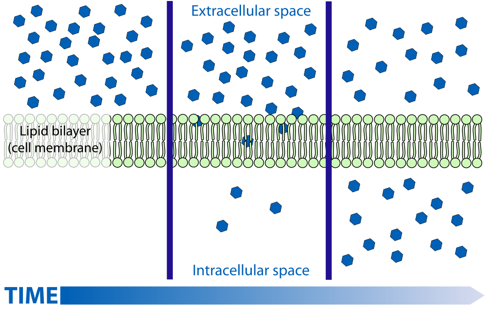
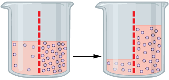
Osmosis
Osmosis is a special type of diffusion — the diffusion of water molecules across a membrane. Like other molecules, water moves from an area of higher concentration to an area of lower concentration. Water moves in or out of a cell until its concentration is the same on both sides of the plasma membrane. In Figure 4.7.3, the dotted red line shows a semi-permeable membrane. In the first beaker, there is an uneven concentration of solutes on either side of the membrane, but the solute cannot cross — diffusion of the solute can't occur. In this case, water will move to even out the concentration as has happened on the beaker on the right side. The water levels are uneven, but the process of osmosis has evened out the concentration gradient.
Facilitated Diffusion
Water and many other substances cannot simply diffuse across a membrane. Hydrophilic molecules, charged ions, and relatively large molecules (such as glucose) all need help with diffusion. This help comes from special proteins in the membrane known as transport proteins. Diffusion with the help of transport proteins is called facilitated diffusion. There are several types of transport proteins, including channel proteins and carrier proteins. Both are shown in Figure 4.7.4.
- Channel proteins form pores (or tiny holes) in the membrane. This allows water molecules and small ions to pass through the membrane without coming into contact with the hydrophobic tails of the lipid molecules in the interior of the membrane.
- Carrier proteins bind with specific ions or molecules. In doing so, they change shape. As carrier proteins change shape, they carry the ions or molecules across the membrane.
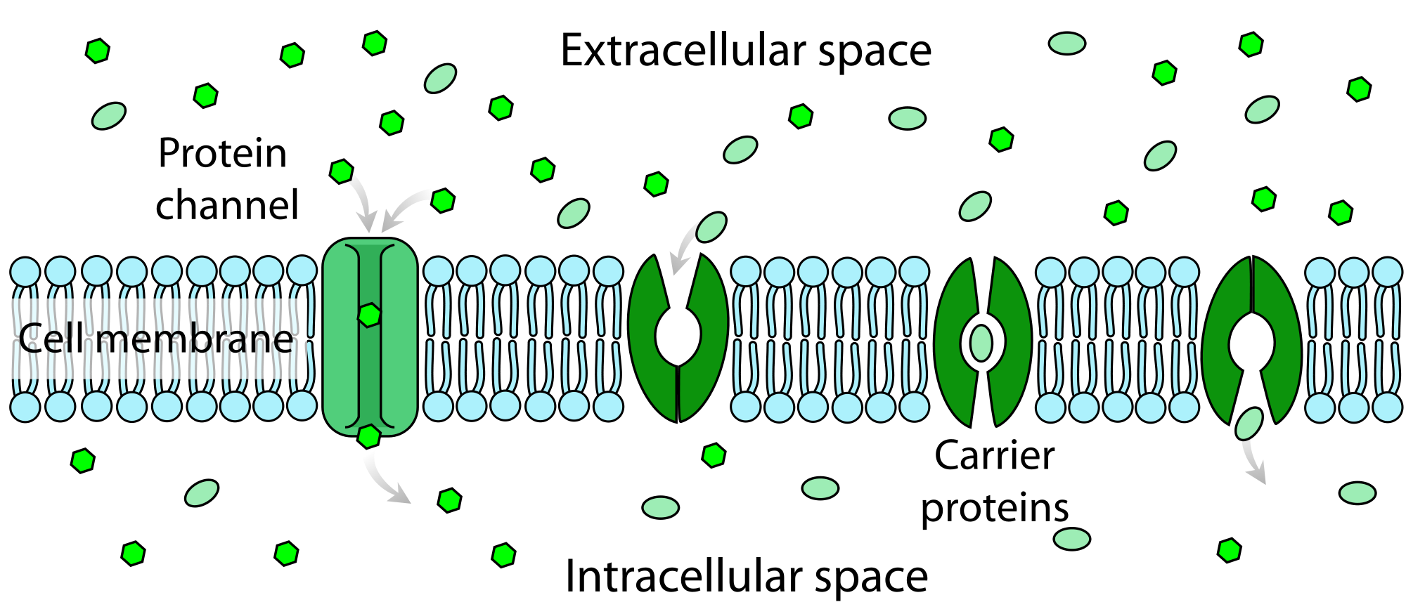
Transport and Homeostasis
For a cell to function normally, the inside of it must maintain a stable state. The concentrations of salts, nutrients, and other substances must be kept within certain ranges. The state in which stable conditions are maintained inside a cell (or an entire organism) is called homeostasis. Homeostasis requires constant adjustments, because conditions are always changing both inside and outside the cell. The transport of substances into and out of cells as described in this section plays an important role in homeostasis. By allowing the movement of substances into and out of cells, transport keeps conditions within normal ranges inside the cells and throughout the organism as a whole.
Watch this video "Cell Transport," by the Amoeba Sisters:
https://www.youtube.com/watch?v=Ptmlvtei8hw
Cell Transport with the Amoeba Sisters, 2016.
4.7 Summary
- Controlling the movement of things in and out of the cell is an important function of the plasma membrane. There are two basic ways that substances can cross the plasma membrane: passive transport — which requires no energy expenditure by the cell — and active transport — which requires energy.
- No energy is needed from the cell for passive transport because it occurs when substances move naturally from an area of higher concentration to an area of lower concentration.
- Simple diffusion is the movement of a substance due to differences in concentration. It happens without any help from other molecules. This is how very small, hydrophobic molecules (such as oxygen and carbon dioxide) enter and leave the cell.
- Osmosis is the diffusion of water molecules across a membrane. Water moves in or out of a cell by osmosis until its concentration is the same on both sides of the plasma membrane.
- Facilitated diffusion is the movement of a substance across a membrane due to differences in concentration, but it only occurs with the help of transport proteins (such as channel proteins or carrier proteins) in the membrane. This is how large or hydrophilic molecules and charged ions enter and leave the cell.
- Processes of passive transport play important roles in homeostasis. By allowing the movement of substances into and out of the cell, they keep conditions within normal ranges inside the cell and the organism as a whole.
4.7 Review Questions
- What is the main difference between passive and active transport?
- Summarize three different ways that passive transport can occur. Give an example of a substance that is transported in each way.
- Explain how transport across the plasma membrane is related to homeostasis of the cell.
- In general, why can only very small, hydrophobic molecules cross the cell membrane by simple diffusion?
- Explain how facilitated diffusion assists with osmosis in cells. Define osmosis and facilitated diffusion in your answer.
- Imagine a hypothetical cell with a higher concentration of glucose inside the cell than outside. Answer the following questions about this cell, assuming all transport across the membrane is passive, not active.
- Can the glucose simply diffuse across the cell membrane? Why or why not?
- Assuming that there are glucose transport proteins in the cell membrane, which way would glucose flow — into or out of the cell? Explain your answer.
- If the concentration of glucose was equal inside and outside of the cell, do you think there would be a net flow of glucose across the cell membrane in one direction or the other? Explain your answer.
- What are the similarities and differences between channel proteins and carrier proteins?
-
4.7 Explore More
https://www.youtube.com/watch?v=L-osEc07vMs&t=31s
Osmosis and Water Potential, Amoeba Sisters, 2018.
https://www.youtube.com/watch?v=AcrqIxt8am8
Structure Of The Cell Membrane - Active and Passive Transport, Professor Dave Explains, 2016.
Attributions
Figure 4.7.1
Windows/ The Oyster Suite in Eureka, CA by Drew Coffman on Unsplash is used under the Unsplash License https://unsplash.com/license).
Figure 4.7.2
Diffusion/ Scheme simple diffusion in cell membrane by Mariana Ruiz Villarreal [LadyofHats] is released into the public domain (https://en.wikipedia.org/wiki/Public_domain).
Figure 4.7.3
Osmosis by OpenStax on Wikimedia Commons is used under a CC BY 3.0 (https://creativecommons.org/licenses/by/3.0) license.
Figure 4.7.4
Scheme facilitated diffusion in cell membrane by Mariana Ruiz Villarreal [LadyofHats] is released into the public domain (https://en.wikipedia.org/wiki/Public_domain).
References
Amoeba Sisters. (2016, June 24). Cell transport. YouTube. https://www.youtube.com/watch?v=Ptmlvtei8hw&feature=youtu.be
Amoeba Sisters. (2018, June 27). Osmosis and water potential. YouTube. https://www.youtube.com/watch?v=L-osEc07vMs&feature=youtu.be
Betts, J. G., Young, K.A., Wise, J.A., Johnson, E., Poe, B., Kruse, D.H., Korol, O., Johnson, J.E., Womble, M., DeSaix, P. (2013, April 25). Figure 3.7 Osmosis [digital image]. In Anatomy and Physiology. OpenStax. https://openstax.org/books/anatomy-and-physiology/pages/3-1-the-cell-membrane
Professor Dave Explains. (2016, September 5). Structure of the cell membrane - Active and passive transport. https://www.youtube.com/watch?v=AcrqIxt8am8&feature=youtu.be
Created by CK-12 Foundation/Adapted by Christine Miller

Work Those Eye Muscles!
Imagine the man in Figure 12.3.1 turns his eyes in your direction. This is a very small movement, considering the conspicuously large and strong external eye muscles that control eyeball movements. These muscles have been called the strongest muscles in the human body relative to the work they do. However, the external eye muscles actually do a surprising amount of work. Eye movements occur almost constantly during waking hours, especially when we are scanning faces or reading. Eye muscles are also exercised nightly during the phase of sleep called rapid eye movement sleep. External eye muscles can move the eyes because they are made mainly of muscle tissue.
What is Muscle Tissue?
Muscle tissue is a soft tissue that makes up most of the tissues in the muscles of the human muscular system. Other tissues in muscles are connective tissues, such as tendons that attach skeletal muscles to bones and sheaths of connective tissues that cover or line muscle tissues. Only muscle tissue per se, has cells with the ability to contract.
There are three major types of muscle tissues in the human body: skeletal, smooth, and cardiac muscle tissues. Figure 12.3.2 shows how the three types of muscle tissues appear under magnification. When you read about each type below, you will learn why the three types appear as they do.
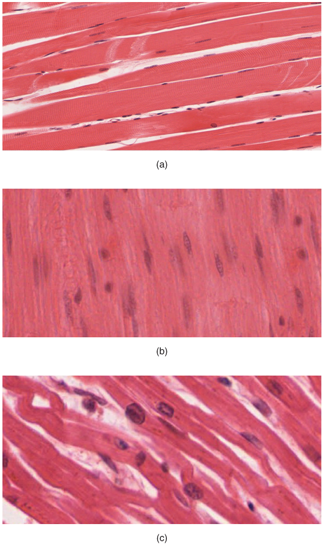
Skeletal Muscle Tissue
Skeletal muscle is muscle tissue that is attached to bones by tendons, which are bundles of collagen fibres. Whether you are moving your eyes or running a marathon, you are using skeletal muscles. Contractions of skeletal muscles are voluntary, or under conscious control of the central nervous system via the somatic nervous system. Skeletal muscle tissue is the most common type of muscle tissue in the human body. By weight, an average adult male is about 42% skeletal muscles, and the average adult female is about 36% skeletal muscles. Some of the major skeletal muscles in the human body are labeled in Figure 12.3.3 below.
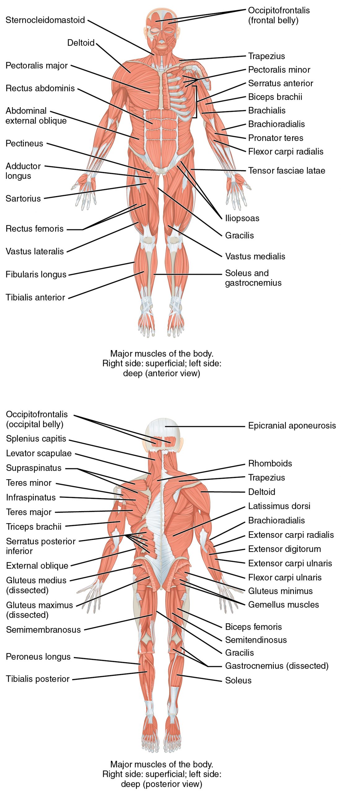
Skeletal Muscle Pairs
To move bones in opposite directions, skeletal muscles often consist of muscle pairs that work in opposition to one another, also called antagonistic muscle pairs. For example, when the biceps muscle (on the front of the upper arm) contracts, it can cause the elbow joint to flex or bend the arm, as shown in Figure 12.3.4. When the triceps muscle (on the back of the upper arm) contracts, it can cause the elbow to extend or straighten the arm. The biceps and triceps muscles, also shown in Figure 12.3.4, are an example of a muscle pair where the muscles work in opposition to each other.
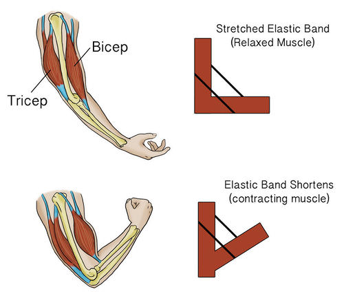
Skeletal Muscle Structure
Each skeletal muscle consists of hundreds — or even thousands — of skeletal muscle fibres, which are long, string-like cells. As shown in Figure 12.3.5 below, skeletal muscle fibres are individually wrapped in connective tissue called endomysium. The skeletal muscle fibres are bundled together in units called muscle fascicles, which are surrounded by sheaths of connective tissue called perimysium. Each fascicle contains between ten and 100 (or even more!) skeletal muscle fibres. Fascicles, in turn, are bundled together to form individual skeletal muscles, which are wrapped in connective tissue called epimysium. The connective tissues in skeletal muscles have a variety of functions. They support and protect muscle fibres, allowing them to withstand the forces of contraction by distributing the forces applied to the muscle. They also provide pathways for nerves and blood vessels to reach the muscles. In addition, the epimysium anchors the muscles to tendons.
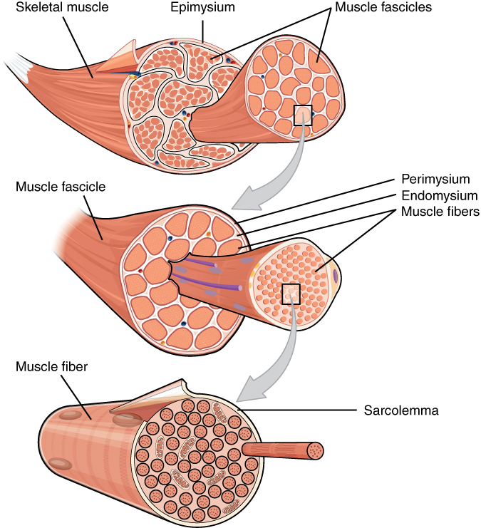
The same bundles-within-bundles structure is replicated within each muscle fibre. As shown in Figure 12.3.6, a muscle fibre consists of a bundle of myofibrils, which are themselves bundles of protein filaments. These protein filaments consist of thin filaments of the protein actin, which are anchored to structures called Z discs, and thick filaments of the protein myosin. The filaments are arranged together within a myofibril in repeating units called sarcomeres, which run from one Z disc to the next. The sarcomere is the basic functional unit of skeletal and cardiac muscles. It contracts as actin and myosin filaments slide over one another. Skeletal muscle tissue is said to be striated, because it appears striped. It has this appearance because of the regular, alternating A (dark) and I (light) bands of filaments arranged in sarcomeres inside the muscle fibres. Other components of a skeletal muscle fibre include multiple nuclei and mitochondria.
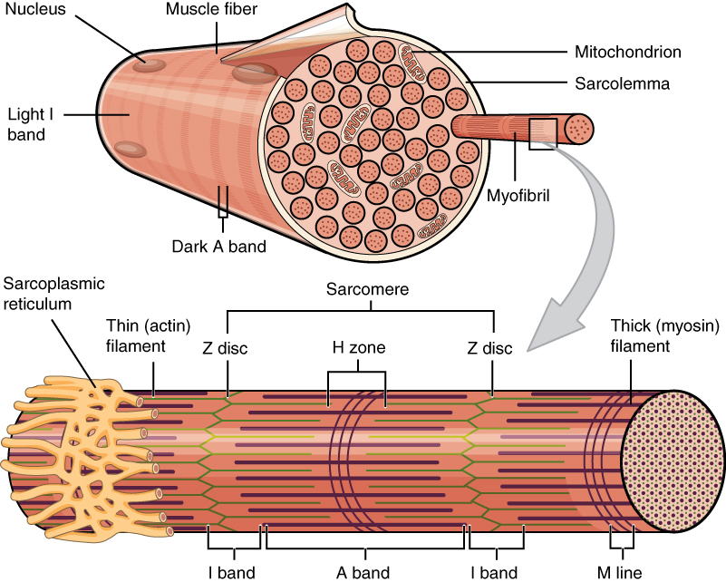
Slow- and Fast-Twitch Skeletal Muscle Fibres
Skeletal muscle fibres can be divided into two types, called slow-twitch (or type I) muscle fibres and fast-twitch (or type II) muscle fibres.
- Slow-twitch muscle fibres are dense with capillaries and rich in mitochondria and myoglobin, which is a protein that stores oxygen until needed for muscle activity. Relative to fast-twitch fibres, slow-twitch fibres can carry more oxygen and sustain aerobic (oxygen-using) activity. Slow-twitch fibres can contract for long periods of time, but not with very much force. They are relied upon primarily in endurance events, such as distance running or cycling.
- Fast-twitch muscle fibres contain fewer capillaries and mitochondria and less myoglobin. This type of muscle fibre can contract rapidly and powerfully, but it fatigues very quickly. Fast-twitch fibres can sustain only short, anaerobic (non-oxygen-using) bursts of activity. Relative to slow-twitch fibres, fast-twitch fibres contribute more to muscle strength and have a greater potential for increasing in mass. They are relied upon primarily in short, strenuous events, such as sprinting or weightlifting.
Proportions of fibre types vary considerably from muscle to muscle and from person to person. Individuals may be genetically predisposed to have a larger percentage of one type of muscle fibre than the other. Generally, an individual who has more slow-twitch fibres is better suited for activities requiring endurance, whereas an individual who has more fast-twitch fibres is better suited for activities requiring short bursts of power.
Smooth Muscle
Smooth muscle is muscle tissue in the walls of internal organs and other internal structures such as blood vessels. When smooth muscles contract, they help the organs and vessels carry out their functions. When smooth muscles in the stomach wall contract, for example, they squeeze the food inside the stomach, helping to mix and churn the food and break it into smaller pieces. This is an important part of digestion. Contractions of smooth muscles are involuntary, so they are not under conscious control. Instead, they are controlled by the autonomic nervous system, hormones, neurotransmitters, and other physiological factors.
Structure of Smooth Muscle
The cells that make up smooth muscle are generally called myocytes. Unlike the muscle fibres of striated muscle tissue, the myocytes of smooth muscle tissue do not have their filaments arranged in sarcomeres. Therefore, smooth tissue is not striated. However, the myocytes of smooth muscle do contain myofibrils, which in turn contain bundles of myosin and actin filaments. The filaments cause contractions when they slide over each other, as shown in Figure 12.3.7.

Functions of Smooth Muscle
Unlike striated muscle, smooth muscle can sustain very long-term contractions. Smooth muscle can also stretch and still maintain its contractile function, which striated muscle cannot. The elasticity of smooth muscle is enhanced by an extracellular matrix secreted by myocytes. The matrix consists of elastin, collagen, and other stretchy fibres. The ability to stretch and still contract is an important attribute of smooth muscle in organs such as the stomach and uterus (see Figures 12.3.8 and 12.3.9), both of which must stretch considerably as they perform their normal functions.
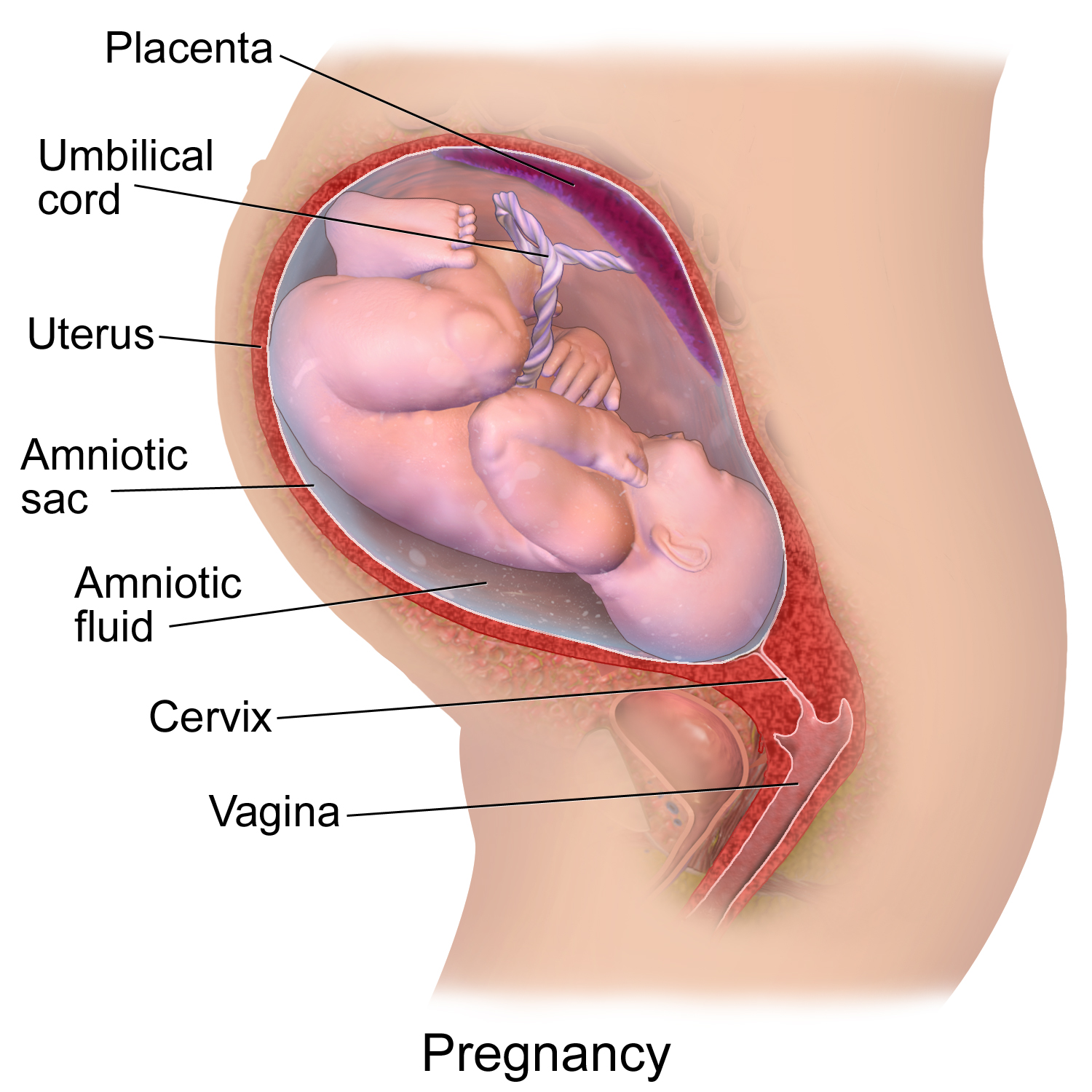 |
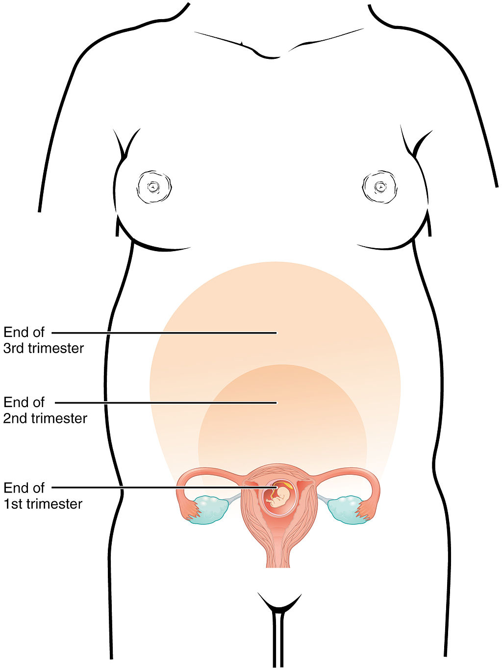 |
The following list indicates where many smooth muscles are found, along with some of their specific functions.
- Walls of organs of the gastrointestinal tract (such as the esophagus, stomach, and intestines), moving food through the tract by peristalsis
- Walls of air passages of the respiratory tract (such as the bronchi), controlling the diameter of the passages and the volume of air that can pass through them
- Walls of organs of the male and female reproductive tracts; in the uterus, for example, pushing a baby out of the uterus and into the birth canal
- Walls of structures of the urinary system, including the urinary bladder, allowing the bladder to expand so it can hold more urine, and then contract as urine is released
- Walls of blood vessels, controlling the diameter of the vessels and thereby affecting blood flow and blood pressure
- Walls of lymphatic vessels, squeezing the fluid called lymph through the vessels
- Iris of the eyes, controlling the size of the pupils and thereby the amount of light entering the eyes
- Arrector pili in the skin, raising hairs in hair follicles in the dermis
Cardiac Muscle
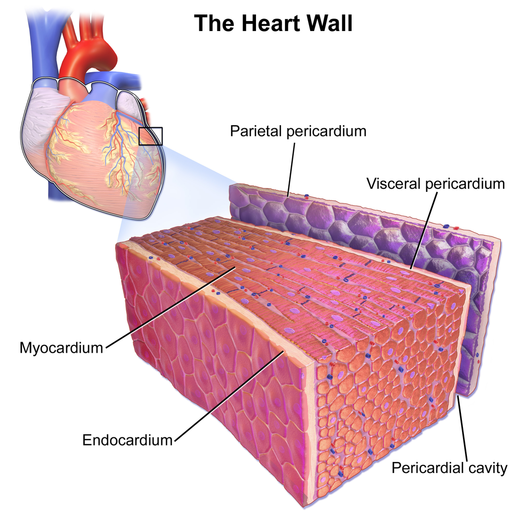
Cardiac muscle is found only in the wall of the heart. It is also called myocardium. As shown in Figure 12.3.10, myocardium is enclosed within connective tissues, including the endocardium on the inside of the heart and pericardium on the outside of the heart. When cardiac muscle contracts, the heart beats and pumps blood. Contractions of cardiac muscle are involuntary, like those of smooth muscles. They are controlled by electrical impulses from specialized cardiac muscle cells in an area of the heart muscle called the sinoatrial node.
Like skeletal muscle, cardiac muscle is striated because its filaments are arranged in sarcomeres inside the muscle fibres. However, in cardiac muscle, the myofibrils are branched at irregular angles rather than arranged in parallel rows (as they are in skeletal muscle). This explains why cardiac and skeletal muscle tissues look different from one another.
The cells of cardiac muscle tissue are arranged in interconnected networks. This arrangement allows rapid transmission of electrical impulses, which stimulate virtually simultaneous contractions of the cells. This enables the cells to coordinate contractions of the heart muscle.
The heart is the muscle that performs the greatest amount of physical work in the course of a lifetime. Although the power output of the heart is much less than the maximum power output of some other muscles in the human body, the heart does its work continuously over an entire lifetime without rest. Cardiac muscle contains a great many mitochondria, which produce ATP for energy and help the heart resist fatigue.
Feature: Human Biology in the News
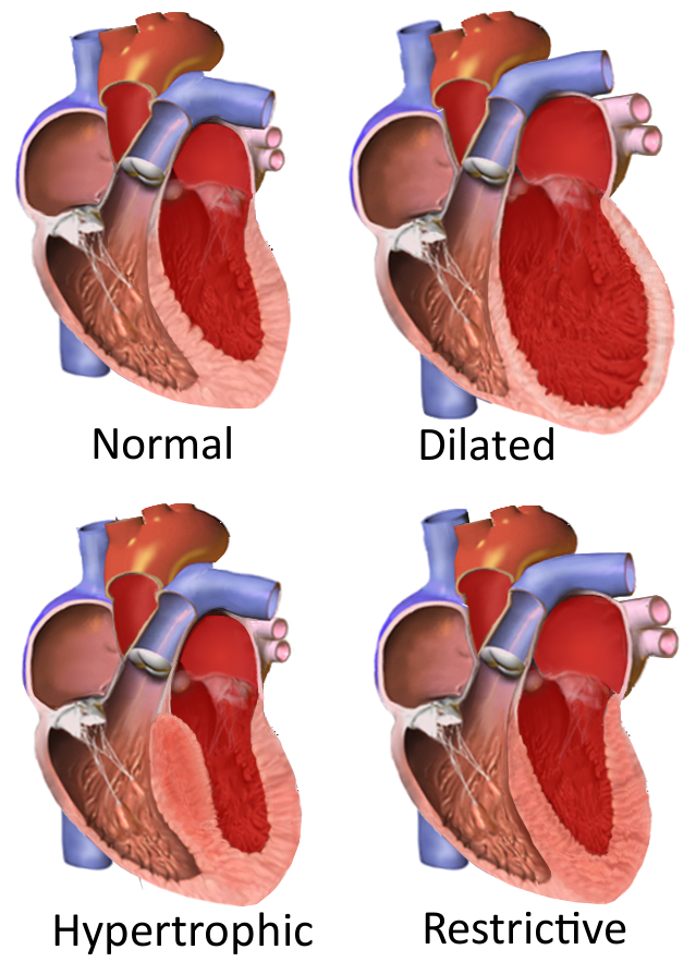
Cardiomyopathy is a disease in which the muscles of the heart are no longer able to effectively pump blood to the body — extreme forms of this disease can lead to heart failure. There are four main types of cardiomyopathy (also illustrated in Figure 12.3.11):
- Dilated (congestive) cardiomyopathy: the left ventricle (the chamber itself) of the heart becomes enlarged and can't pump blood our to the body. This is normally related to coronary artery disease and/or heart attack
- Hypertrophic cardiomyopathy: abnormal thickening of the muscular walls of the left ventricle make the chamber less able to work properly. This condition is more common in patients with a family history of the disease.
- Restrictive cardiomyopathy: the myocardium becomes abnormally rigid and inelastic and is unable to expand in between heartbeats to refill with blood. Restrictive cardiomyopathy typically affects older people.
- Arrhythmogenic right ventricular cardiomyopathy: the right ventricular muscle is replaced by adipose or scar tissue, reducing elasticity and interfering with normal heartbeat and rhythm. This disease is often caused by genetic mutations.
Cardiomyopathy is typically diagnosed with a physical exam supplemented by medical and family history, an angiogram, blood tests, chest x-rays and electrocardiograms. In some cases your doctor would also requisition a CT scan and/or genetic testing.
When treating cardiomyopathy, the goal is to reduce symptoms that affect everyday life. Certain medications can help regularize and slow heart rate, decrease chances of blood clots and cause vasodilation in the coronary arteries. If medication is not sufficient to manage symptoms, a pacemaker or even a heart transplant may be the best option. Lifestyle can also help manage the symptoms of cardiomyopathy — people living with this disease are encouraged to avoid drug and alcohol use, control high blood pressure, eat a healthy diet, get ample rest and exercise, as well as reduce stress levels.
12.3 Summary
- Muscle tissue is a soft tissue that makes up most of the tissues in the muscles of the human muscular system. It is the only type of tissue that has cells with the ability to contract.
- Skeletal muscle tissue is attached to bones by tendons. It allows voluntary body movements.
- Skeletal muscle is the most common type of muscle tissue in the human body. To move bones in opposite directions, skeletal muscles often consist of pairs of muscles that work in opposition to one another to move bones in different directions at joints.
- Skeletal muscle fibres are bundled together in units called muscle fascicles, which are bundled together to form individual skeletal muscles. Skeletal muscles also have connective tissue supporting and protecting the muscle tissue.
- Each skeletal muscle fibre consists of a bundle of myofibrils, which are bundles of protein filaments. The filaments are arranged in repeating units called sarcomeres, which are the basic functional units of skeletal muscles. Skeletal muscle tissue is striated because of the pattern of sarcomeres in its fibres.
- Skeletal muscle fibres can be divided into two types, called slow-twitch and fast-twitch fibres. Slow-twitch fibres are used mainly in aerobic endurance activities, such as long-distance running. Fast-twitch fibres are used mainly for non-aerobic, strenuous activities, such as sprinting. Proportions of the two types of fibres vary from muscle to muscle and person to person.
- Smooth muscle tissue is found in the walls of internal organs and vessels. When smooth muscles contract, they help the organs and vessels carry out their functions. Contractions of smooth muscles are involuntary and controlled by the autonomic nervous system, hormones, and other substances.
- Cells of smooth muscle tissue are not striated because they lack sarcomeres, but the cells contract in the same basic way as striated muscle cells. Unlike striated muscle, smooth muscle can sustain very long-term contractions and maintain its contractile function, even when stretched.
- Cardiac muscle tissue is found only in the wall of the heart. When cardiac muscle contracts, the heart beats and pumps blood. Contractions of cardiac muscle are involuntary, like those of smooth muscles. They are controlled by electrical impulses from specialized cardiac cells.
- Like skeletal muscle, cardiac muscle is striated because its filaments are arranged in sarcomeres inside the muscle fibres. However, the myofibrils are branched instead of arranged in parallel rows, making cardiac and skeletal muscle tissues look different from one another.
- The heart is the muscle that performs the greatest amount of physical work in the course of a lifetime. Its cells contain a great many mitochondria to produce ATP for energy and help the heart resist fatigue.
12.3 Review Questions
- What is muscle tissue?
-
-
- Where is skeletal muscle found, and what is its general function?
- Why do many skeletal muscles work in pairs?
- Describe the structure of a skeletal muscle.
- Relate muscle fibre structure to the functional units of muscles.
- Why is skeletal muscle tissue striated?
- Where is smooth muscle found? What controls the contraction of smooth muscle?
- Where is cardiac muscle found? What controls its contractions?
- The heart muscle is smaller and less powerful than some other muscles in the body. Why is the heart the muscle that performs the greatest amount of physical work in the course of a lifetime? How does the heart resist fatigue?
- Give one example of connective tissue that is found in muscles. Describe one of its functions.
12.3 Explore More
https://www.youtube.com/watch?v=3_PYnWVoUzM
What happens during a heart attack? - Krishna Sudhir, TED-Ed, 2017.
https://www.youtube.com/watch?v=bwOE1MEginA&feature=emb_logo
Three types of muscle | Circulatory system physiology | NCLEX-RN | KhanAcademyMedicine, 2012.
Attributions
Figure 12.3.1
Look by ali-yahya-155huuQwGvA [photo] by Ali Yahya on Unsplash is used under the Unsplash License (https://unsplash.com/license).
Figure 12.3.2
Skeletal_Smooth_Cardiac by OpenStax College on Wikimedia Commons is used under a CC BY 3.0 (https://creativecommons.org/licenses/by/3.0) license.
Figure 12.3.3
Anterior_and_Posterior_Views_of_Muscles by OpenStax on Wikimedia Commons is used under a CC BY 4.0 (https://creativecommons.org/licenses/by/4.0) license.
Figure 12.3.4
Antagonistic Muscle Pair by Laura Guerin at CK-12 Foundation on Wikimedia Commons is used under a CC BY-NC 3.0 (https://creativecommons.org/licenses/by-nc/3.0/) license.
Figure 12.3.5
Muscle_Fibes_(large) by OpenStax on Wikimedia Commons is used under a CC BY 4.0 (https://creativecommons.org/licenses/by/4.0) license.
Figure 12.3.6
Muscle_Fibers_(small) by OpenStax on Wikimedia Commons is used under a CC BY 4.0 (https://creativecommons.org/licenses/by/4.0) license.
Figure 12.3.7
Smooth_Muscle_Contraction by OpenStax on Wikimedia Commons is used under a CC BY 4.0 (https://creativecommons.org/licenses/by/4.0) license.
Figure 12.3.8
Blausen_0747_Pregnancy by BruceBlaus on Wikimedia Commons is used under a CC BY 3.0 (https://creativecommons.org/licenses/by/3.0) license.
Figure 12.3.9
Size_of_Uterus_Throughout_Pregnancy-02 by OpenStax College on Wikimedia Commons is used under a CC BY 3.0 (https://creativecommons.org/licenses/by/3.0) license.
Figure 12.3.10
1024px-Blausen_0470_HeartWall by BruceBlaus on Wikimedia Commons is used under a CC BY 3.0 (https://creativecommons.org/licenses/by/3.0) license.
Figure 12.3.11
Tipet_e_kardiomiopative by Npatchett at English Wikipedia on Wikimedia Commons is used under a CC BY-SA 3.0 (https://creativecommons.org/licenses/by-sa/3.0) license. (Work derived from Blausen 0165 Cardiomyopathy Dilated by BruceBlaus)
References
Betts, J. G., Young, K.A., Wise, J.A., Johnson, E., Poe, B., Kruse, D.H., Korol, O., Johnson, J.E., Womble, M., DeSaix, P. (2013, June 19). Figure 4.18 Muscle tissue [digital image]. In Anatomy and Physiology (Section 4.4). OpenStax. https://openstax.org/books/anatomy-and-physiology/pages/4-4-muscle-tissue-and-motion
Betts, J. G., Young, K.A., Wise, J.A., Johnson, E., Poe, B., Kruse, D.H., Korol, O., Johnson, J.E., Womble, M., DeSaix, P. (2013, June 19). Figure 28.18 Size of uterus throughout pregnancy [digital image]. In Anatomy and Physiology (Section 28.4). OpenStax. https://openstax.org/books/anatomy-and-physiology/pages/28-4-maternal-changes-during-pregnancy-labor-and-birth
Betts, J. G., Young, K.A., Wise, J.A., Johnson, E., Poe, B., Kruse, D.H., Korol, O., Johnson, J.E., Womble, M., DeSaix, P. (2016, May 18). Figure 10.3 The three connective tissue layers [digital image]. In Anatomy and Physiology (Section 10.2). OpenStax. https://openstax.org/books/anatomy-and-physiology/pages/10-2-skeletal-muscle
Betts, J. G., Young, K.A., Wise, J.A., Johnson, E., Poe, B., Kruse, D.H., Korol, O., Johnson, J.E., Womble, M., DeSaix, P. (2016, May 18). Figure 10.4 Muscle fiber [digital image]. In Anatomy and Physiology (Section 10.2). OpenStax. https://openstax.org/books/anatomy-and-physiology/pages/10-2-skeletal-muscle
Betts, J. G., Young, K.A., Wise, J.A., Johnson, E., Poe, B., Kruse, D.H., Korol, O., Johnson, J.E., Womble, M., DeSaix, P. (2016, May 18). Figure 10.24 Muscle contraction [digital image]. In Anatomy and Physiology (Section 10.8). OpenStax. https://openstax.org/books/anatomy-and-physiology/pages/10-8-smooth-muscle
Betts, J. G., Young, K.A., Wise, J.A., Johnson, E., Poe, B., Kruse, D.H., Korol, O., Johnson, J.E., Womble, M., DeSaix, P. (2016, May 18). Figure 11.5 Overview of the muscular system [digital image]. In Anatomy and Physiology (Section 11.2). OpenStax. https://openstax.org/books/anatomy-and-physiology/pages/11-2-naming-skeletal-muscles
Blausen.com staff. (2014). Medical gallery of Blausen Medical 2014. WikiJournal of Medicine 1 (2). DOI:10.15347/wjm/2014.010. ISSN 2002-4436.
Brainard, J/ CK-12 Foundation. (2012). Figure 5 Triceps and biceps muscles in the upper arm are opposing muscles. [digital image]. In CK-12 Biology (Section 21.3) [online Flexbook]. CK12.org. https://www.ck12.org/book/ck-12-biology/section/21.3/ (Last modified August 11, 2017.)
khanacademymedicine. (2012, October 19). Three types of muscle | Circulatory system physiology | NCLEX-RN | Khan Academy. YouTube.
TED-Ed. (2017, February 14). What happens during a heart attack? - Krishna Sudhir. YouTube. https://www.youtube.com/watch?v=3_PYnWVoUzM&feature=youtu.be
division of the peripheral nervous system that controls involuntary activities
A hormone is a signaling molecule produced by glands in multicellular organisms that target distant organs to regulate physiology and behavior.
The body system which acts as a chemical messenger system comprising feedback loops of the hormones released by internal glands of an organism directly into the circulatory system, regulating distant target organs. In humans, the major endocrine glands are the thyroid gland and the adrenal glands.
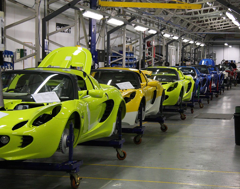
Created by: CK-12/Adapted by Christine Miller
Assembly Line
We stay alive because millions of different chemical reactions are taking place inside our bodies all the time. Each of our cells is like the busy auto assembly line pictured in Figure 3.10.1. Raw materials, half-finished products, and waste materials are constantly being used, produced, transported, and excreted. The "workers" on the cellular assembly line are mainly enzymes. These are the proteins that make biochemical reactions happen.
What Are Biochemical Reactions?
Chemical reactions that take place inside living things are called biochemical reactions. The sum of all the biochemical reactions in an organism is called metabolism. Metabolism includes both exothermic (energy-releasing) chemical reactions and endothermic (energy-absorbing) chemical reactions.
Catabolic Reactions
Exothermic reactions in organisms are called catabolic reactions. These reactions break down molecules into smaller units and release energy. An example of a catabolic reaction is the breakdown of glucose during cellular respiration, which releases energy that cells need to carry out life processes.
Anabolic Reactions
Endothermic reactions in organisms are called anabolic reactions. These reactions build up bigger molecules from smaller ones and absorb energy. An example of an anabolic reaction is the joining of amino acids to form a protein. Which type of reactions — catabolic or anabolic — do you think occur when your body digests food?
Enzymes
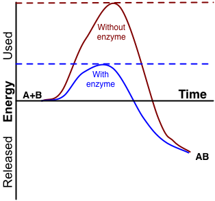
Most of the biochemical reactions that happen inside of living organisms require help. Why is this the case? For one thing, temperatures inside living things are usually too low for biochemical reactions to occur quickly enough to maintain life. The concentrations of reactants may also be too low for them to come together and react. Where do the biochemical reactions get the help they need to proceed? From the enzymes.
An enzyme is a protein that speeds up a biochemical reaction. It is a biological catalyst. An enzyme generally works by reducing the amount of activation energy needed to start the reaction. The graph in Figure 3.10.2 shows the activation energy needed for glucose to combine with oxygen. Less activation energy is needed when the correct enzyme is present than when it is not present.
An enzyme speeds up the reaction by lowering the required activation energy. Compare the activation energy needed with and without the enzyme.
How Well Enzymes Work
Enzymes are involved in most biochemical reactions, and they do their jobs extremely well. A typical biochemical reaction that would take several days or even several centuries to happen without an enzyme is likely to occur in just a split second with the proper enzyme! Without enzymes to speed up biochemical reactions, most organisms could not survive.
Enzymes are substrate-specific. The substrate of an enzyme is the specific substance it affects. Each enzyme works only with a particular substrate, which explains why there are so many different enzymes. In addition, for an enzyme to work, it requires specific conditions, such as the right temperature and pH. Some enzymes work best under acidic conditions, for example, while others work best in neutral environments.
Enzyme-Deficiency Disorders
There are hundreds of known inherited metabolic disorders in humans. In most of them, a single enzyme is either not produced by the body at all, or is otherwise produced in a form that doesn't work. The missing or defective enzyme is like an absentee worker on the cell's assembly line. Imagine the auto assembly line from the image at the start of this section. What if the worker who installed the steering wheel was absent? How would this impact the overall functioning of the vehicle? When an enzyme is missing, toxic chemicals build up, or an essential product isn't made. Generally, the normal enzyme is missing because the individual with the disorder inherited two copies of a gene mutation, which may have originated many generations previously.
Any given inherited metabolic disorder is generally quite rare in the general population. However, there are so many different metabolic disorders that a total of one in 1,000 to 2,500 newborns can be expected to have one.
3.10 Summary
- Biochemical reactions are chemical reactions that take place inside of living things. The sum of all of the biochemical reactions in an organism is called metabolism.
- Metabolism includes catabolic reactions, which are energy-releasing (exothermic) reactions, as well as anabolic reactions, which are energy-absorbing (endothermic) reactions.
- Most biochemical reactions need a biological catalyst called an enzyme to speed up the reaction. Enzymes reduce the amount of activation energy needed for the reaction to begin. Most enzymes are proteins that affect just one specific substance, which is called the enzyme's substrate.
- There are many inherited metabolic disorders in humans. Most of them are caused by a single defective or missing enzyme.
3.10 Review Questions
- What are biochemical reactions?
- Define metabolism.
- Compare and contrast catabolic and anabolic reactions.
- Explain the role of enzymes in biochemical reactions.
- What are enzyme-deficiency disorders?
- Explain why the relatively low temperature of living things, along with the low concentration of reactants, would cause biochemical reactions to occur very slowly in the body without enzymes.
- Answer the following questions about what happens after you eat a sandwich.
- Pieces of the sandwich go into your stomach, where there are digestive enzymes that break down the food. Which type of metabolic reaction is this? Explain your answer.
- During the process of digestion, some of the sandwich is broken down into glucose, which is then further broken down to release energy that your cells can use. Is this an exothermic endothermic reaction? Explain your answer.
- The proteins in the cheese, meat, and bread in the sandwich are broken down into their component amino acids. Then your body uses those amino acids to build new proteins. Which kind of metabolic reaction is represented by the building of these new proteins? Explain your answer.
- Explain why your body doesn’t just use one or two enzymes for all of its biochemical reactions.
- A ________ is the specific substance that an enzyme affects in a biochemical reaction.
- An enzyme is a biological _____________ .
- catabolism
- form of activation energy
- catalyst
- reactant
3.10 Explore More
https://www.youtube.com/watch?v=qgVFkRn8f10&feature=youtu.be
Enzymes (Updated), by The Amoeba Sisters, 2016.
https://www.youtube.com/watch?v=8m6RtOpqvtU&feature=youtu.be
What triggers a chemical reaction? - Kareem Jarrah, TED-Ed, 2015.
Figure 3.10.1
Auto Assembly line by Brian Snelson on Wikimedia Commons is used under a CC BY 2.0 (https://creativecommons.org/licenses/by/2.0) license.
Figure 3.10.2
Enzyme_activation_energy by G. Andruk [IMeowbot at the English language Wikipedia], is used under a CC BY-SA 3.0 (http://creativecommons.org/licenses/by-sa/3.0/) license.
References
Amoeba Sisters. (2016, August 28). Enzymes (updated). YouTube. https://www.youtube.com/watch?v=qgVFkRn8f10&feature=youtu.be
TED-Ed. (2015, January 15). What triggers a chemical reaction? - Kareem Jarrah. YouTube. https://www.youtube.com/watch?v=8m6RtOpqvtU&feature=youtu.be
The smallest type of blood vessel that connects arterioles and venules and that transfers substances between blood and tissues.
Created by CK-12 Foundation/Adapted by Christine Miller

Work Those Eye Muscles!
Imagine the man in Figure 12.3.1 turns his eyes in your direction. This is a very small movement, considering the conspicuously large and strong external eye muscles that control eyeball movements. These muscles have been called the strongest muscles in the human body relative to the work they do. However, the external eye muscles actually do a surprising amount of work. Eye movements occur almost constantly during waking hours, especially when we are scanning faces or reading. Eye muscles are also exercised nightly during the phase of sleep called rapid eye movement sleep. External eye muscles can move the eyes because they are made mainly of muscle tissue.
What is Muscle Tissue?
Muscle tissue is a soft tissue that makes up most of the tissues in the muscles of the human muscular system. Other tissues in muscles are connective tissues, such as tendons that attach skeletal muscles to bones and sheaths of connective tissues that cover or line muscle tissues. Only muscle tissue per se, has cells with the ability to contract.
There are three major types of muscle tissues in the human body: skeletal, smooth, and cardiac muscle tissues. Figure 12.3.2 shows how the three types of muscle tissues appear under magnification. When you read about each type below, you will learn why the three types appear as they do.
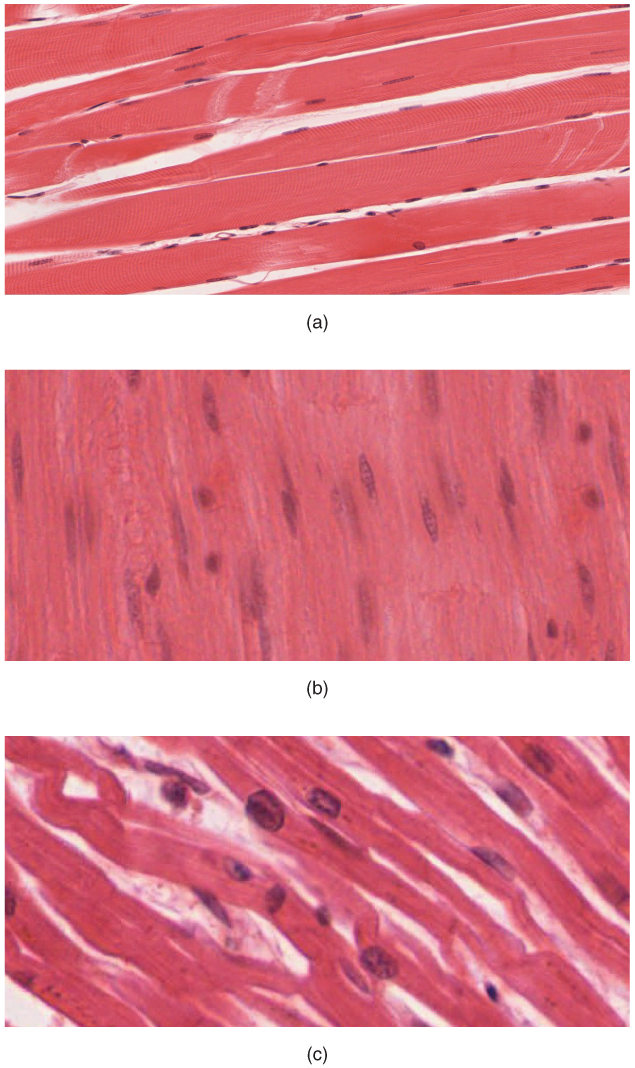
Skeletal Muscle Tissue
Skeletal muscle is muscle tissue that is attached to bones by tendons, which are bundles of collagen fibres. Whether you are moving your eyes or running a marathon, you are using skeletal muscles. Contractions of skeletal muscles are voluntary, or under conscious control of the central nervous system via the somatic nervous system. Skeletal muscle tissue is the most common type of muscle tissue in the human body. By weight, an average adult male is about 42% skeletal muscles, and the average adult female is about 36% skeletal muscles. Some of the major skeletal muscles in the human body are labeled in Figure 12.3.3 below.
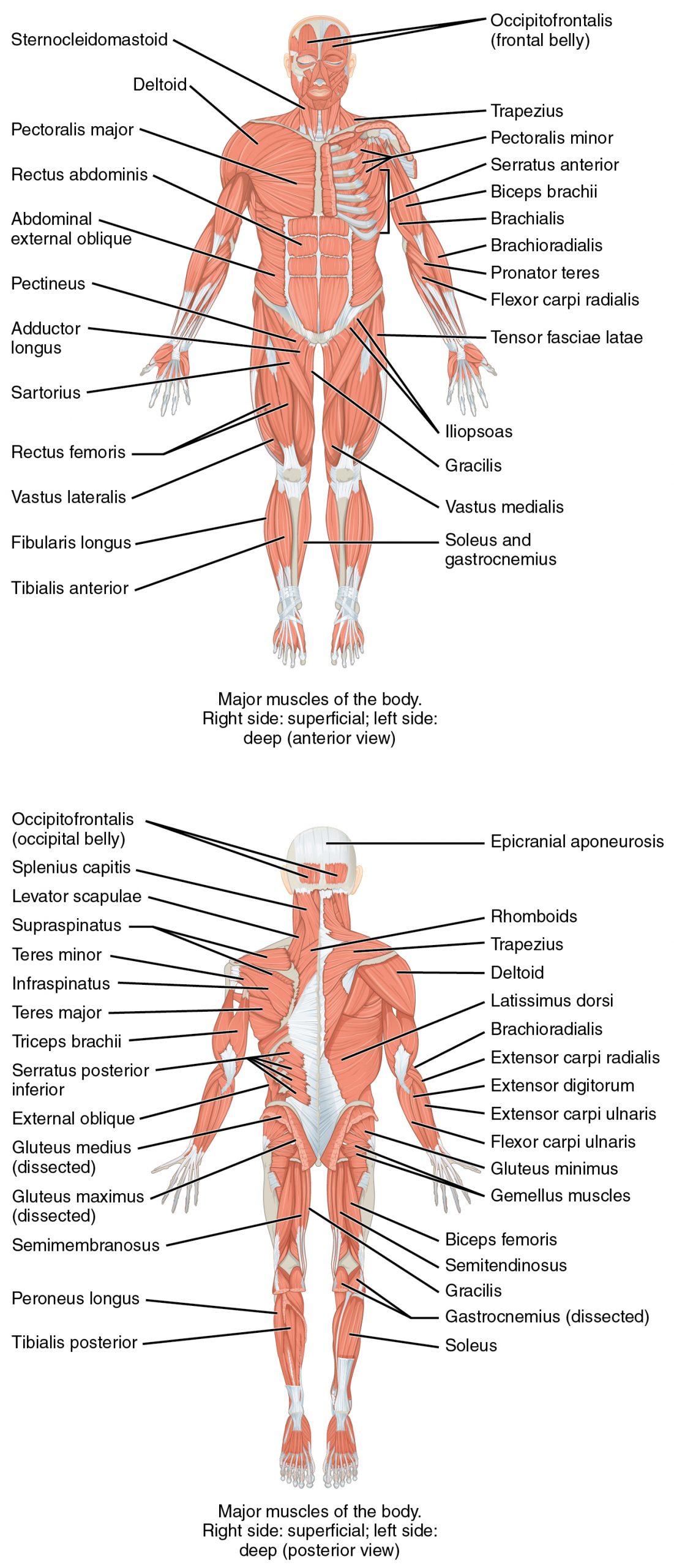
Skeletal Muscle Pairs
To move bones in opposite directions, skeletal muscles often consist of muscle pairs that work in opposition to one another, also called antagonistic muscle pairs. For example, when the biceps muscle (on the front of the upper arm) contracts, it can cause the elbow joint to flex or bend the arm, as shown in Figure 12.3.4. When the triceps muscle (on the back of the upper arm) contracts, it can cause the elbow to extend or straighten the arm. The biceps and triceps muscles, also shown in Figure 12.3.4, are an example of a muscle pair where the muscles work in opposition to each other.
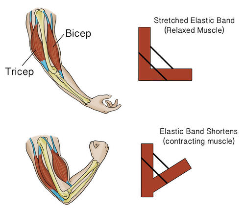
Skeletal Muscle Structure
Each skeletal muscle consists of hundreds — or even thousands — of skeletal muscle fibres, which are long, string-like cells. As shown in Figure 12.3.5 below, skeletal muscle fibres are individually wrapped in connective tissue called endomysium. The skeletal muscle fibres are bundled together in units called muscle fascicles, which are surrounded by sheaths of connective tissue called perimysium. Each fascicle contains between ten and 100 (or even more!) skeletal muscle fibres. Fascicles, in turn, are bundled together to form individual skeletal muscles, which are wrapped in connective tissue called epimysium. The connective tissues in skeletal muscles have a variety of functions. They support and protect muscle fibres, allowing them to withstand the forces of contraction by distributing the forces applied to the muscle. They also provide pathways for nerves and blood vessels to reach the muscles. In addition, the epimysium anchors the muscles to tendons.
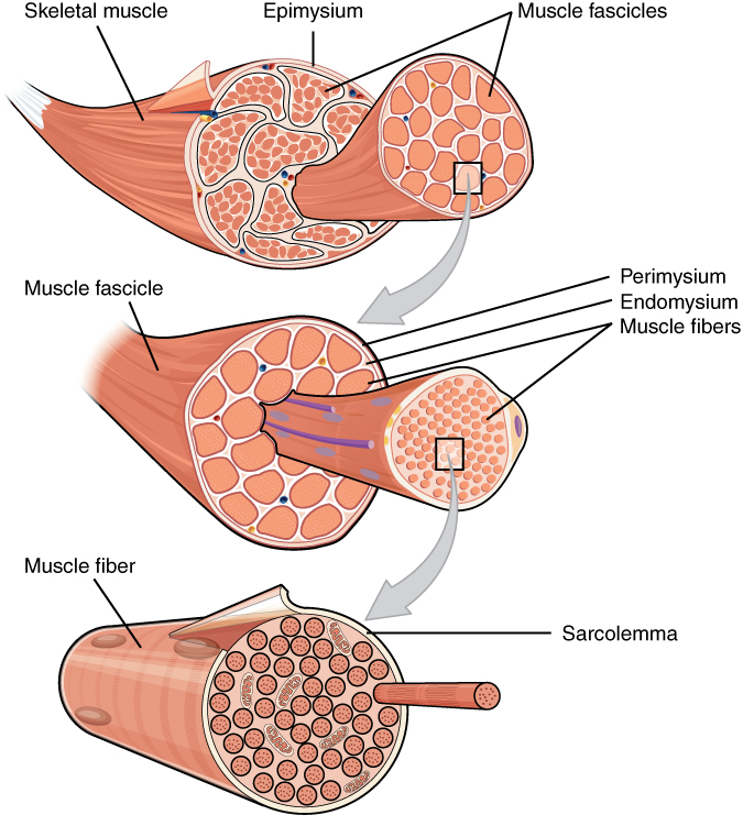
The same bundles-within-bundles structure is replicated within each muscle fibre. As shown in Figure 12.3.6, a muscle fibre consists of a bundle of myofibrils, which are themselves bundles of protein filaments. These protein filaments consist of thin filaments of the protein actin, which are anchored to structures called Z discs, and thick filaments of the protein myosin. The filaments are arranged together within a myofibril in repeating units called sarcomeres, which run from one Z disc to the next. The sarcomere is the basic functional unit of skeletal and cardiac muscles. It contracts as actin and myosin filaments slide over one another. Skeletal muscle tissue is said to be striated, because it appears striped. It has this appearance because of the regular, alternating A (dark) and I (light) bands of filaments arranged in sarcomeres inside the muscle fibres. Other components of a skeletal muscle fibre include multiple nuclei and mitochondria.
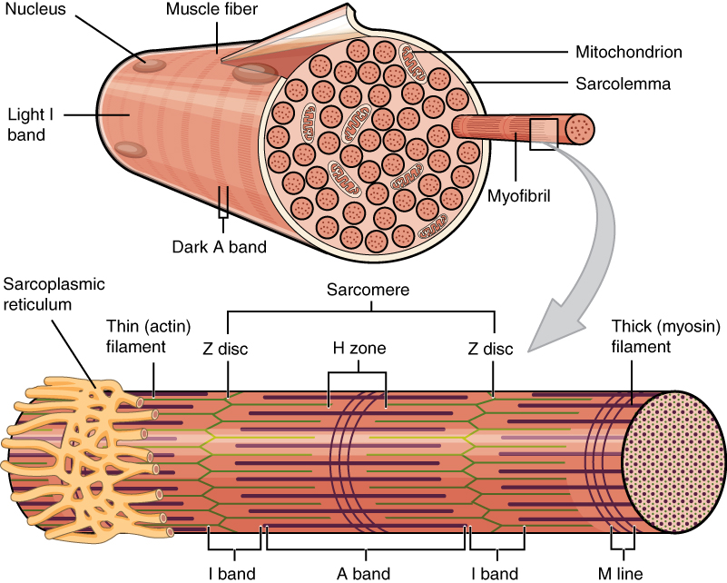
Slow- and Fast-Twitch Skeletal Muscle Fibres
Skeletal muscle fibres can be divided into two types, called slow-twitch (or type I) muscle fibres and fast-twitch (or type II) muscle fibres.
- Slow-twitch muscle fibres are dense with capillaries and rich in mitochondria and myoglobin, which is a protein that stores oxygen until needed for muscle activity. Relative to fast-twitch fibres, slow-twitch fibres can carry more oxygen and sustain aerobic (oxygen-using) activity. Slow-twitch fibres can contract for long periods of time, but not with very much force. They are relied upon primarily in endurance events, such as distance running or cycling.
- Fast-twitch muscle fibres contain fewer capillaries and mitochondria and less myoglobin. This type of muscle fibre can contract rapidly and powerfully, but it fatigues very quickly. Fast-twitch fibres can sustain only short, anaerobic (non-oxygen-using) bursts of activity. Relative to slow-twitch fibres, fast-twitch fibres contribute more to muscle strength and have a greater potential for increasing in mass. They are relied upon primarily in short, strenuous events, such as sprinting or weightlifting.
Proportions of fibre types vary considerably from muscle to muscle and from person to person. Individuals may be genetically predisposed to have a larger percentage of one type of muscle fibre than the other. Generally, an individual who has more slow-twitch fibres is better suited for activities requiring endurance, whereas an individual who has more fast-twitch fibres is better suited for activities requiring short bursts of power.
Smooth Muscle
Smooth muscle is muscle tissue in the walls of internal organs and other internal structures such as blood vessels. When smooth muscles contract, they help the organs and vessels carry out their functions. When smooth muscles in the stomach wall contract, for example, they squeeze the food inside the stomach, helping to mix and churn the food and break it into smaller pieces. This is an important part of digestion. Contractions of smooth muscles are involuntary, so they are not under conscious control. Instead, they are controlled by the autonomic nervous system, hormones, neurotransmitters, and other physiological factors.
Structure of Smooth Muscle
The cells that make up smooth muscle are generally called myocytes. Unlike the muscle fibres of striated muscle tissue, the myocytes of smooth muscle tissue do not have their filaments arranged in sarcomeres. Therefore, smooth tissue is not striated. However, the myocytes of smooth muscle do contain myofibrils, which in turn contain bundles of myosin and actin filaments. The filaments cause contractions when they slide over each other, as shown in Figure 12.3.7.

Functions of Smooth Muscle
Unlike striated muscle, smooth muscle can sustain very long-term contractions. Smooth muscle can also stretch and still maintain its contractile function, which striated muscle cannot. The elasticity of smooth muscle is enhanced by an extracellular matrix secreted by myocytes. The matrix consists of elastin, collagen, and other stretchy fibres. The ability to stretch and still contract is an important attribute of smooth muscle in organs such as the stomach and uterus (see Figures 12.3.8 and 12.3.9), both of which must stretch considerably as they perform their normal functions.
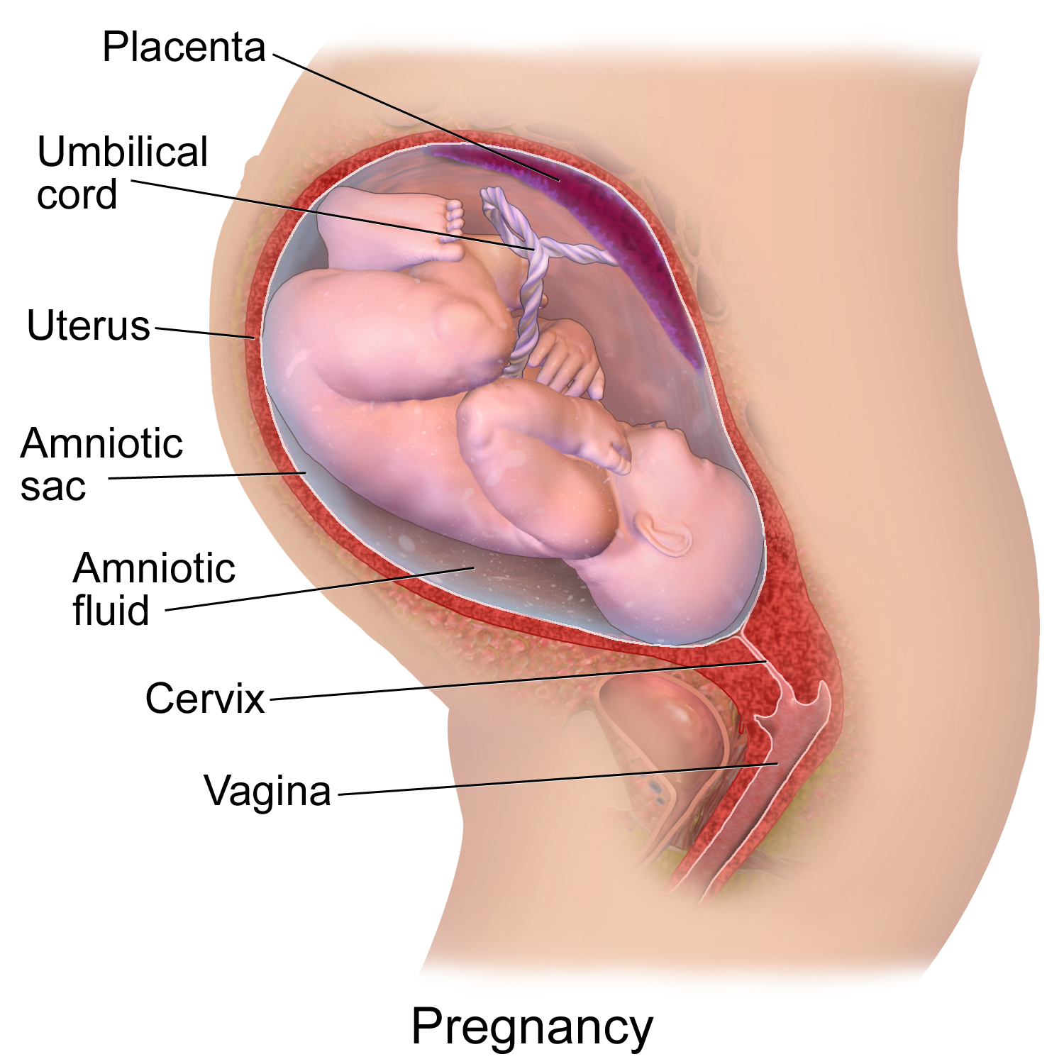 |
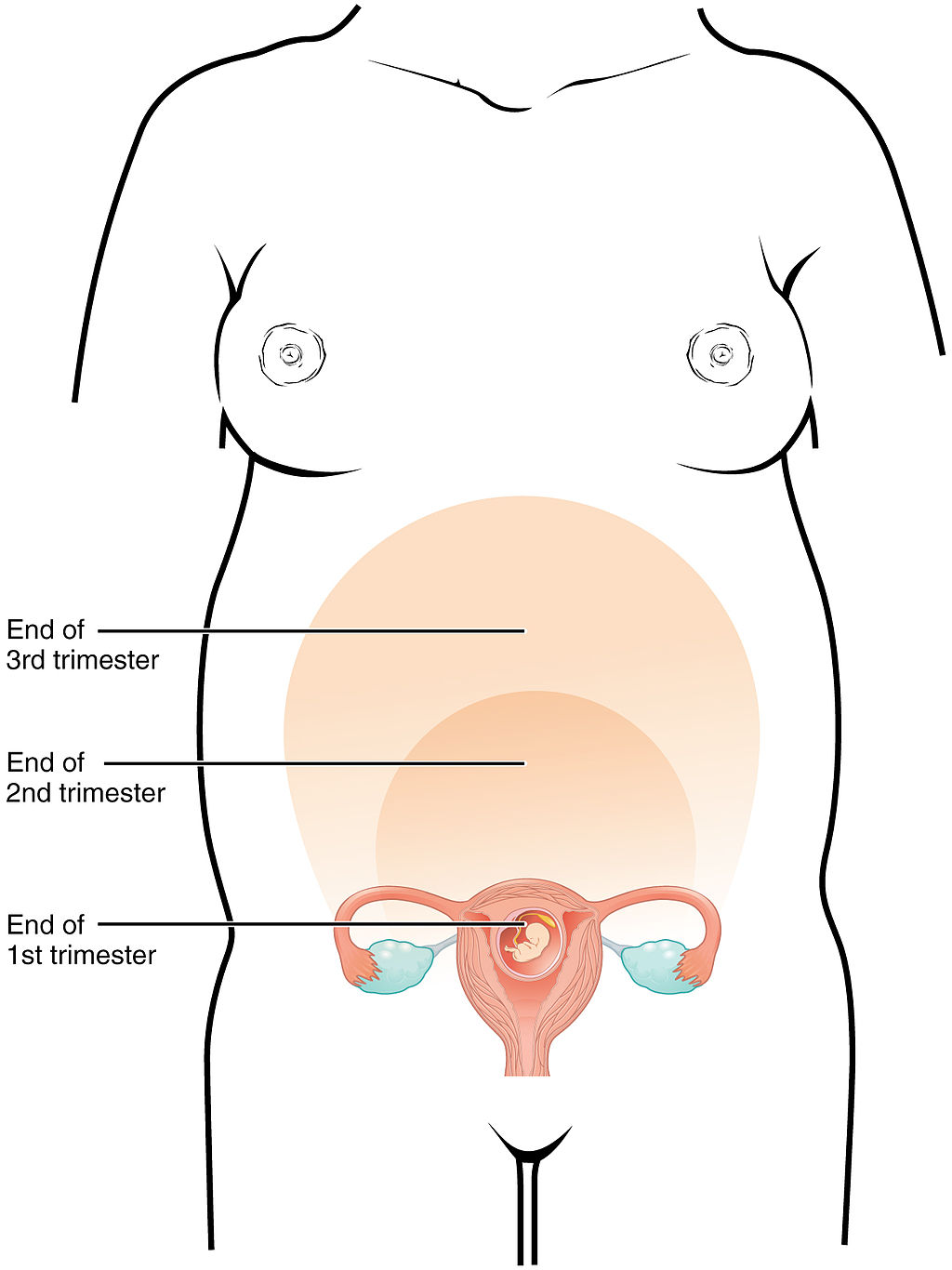 |
The following list indicates where many smooth muscles are found, along with some of their specific functions.
- Walls of organs of the gastrointestinal tract (such as the esophagus, stomach, and intestines), moving food through the tract by peristalsis
- Walls of air passages of the respiratory tract (such as the bronchi), controlling the diameter of the passages and the volume of air that can pass through them
- Walls of organs of the male and female reproductive tracts; in the uterus, for example, pushing a baby out of the uterus and into the birth canal
- Walls of structures of the urinary system, including the urinary bladder, allowing the bladder to expand so it can hold more urine, and then contract as urine is released
- Walls of blood vessels, controlling the diameter of the vessels and thereby affecting blood flow and blood pressure
- Walls of lymphatic vessels, squeezing the fluid called lymph through the vessels
- Iris of the eyes, controlling the size of the pupils and thereby the amount of light entering the eyes
- Arrector pili in the skin, raising hairs in hair follicles in the dermis
Cardiac Muscle
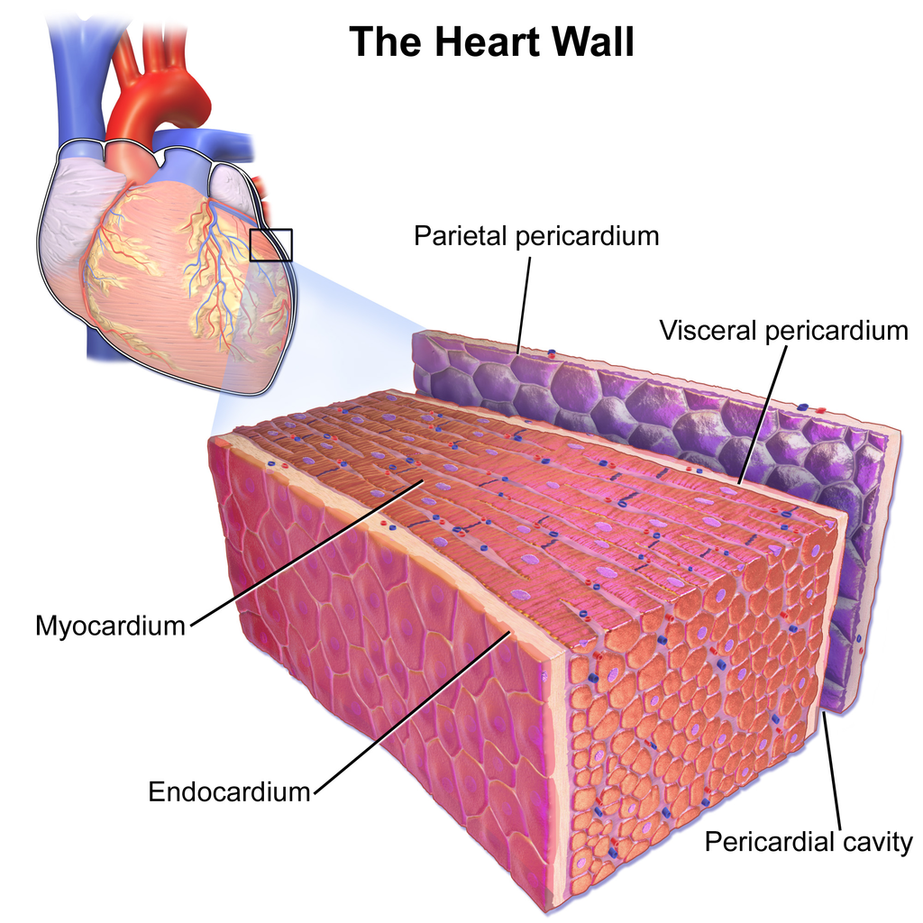
Cardiac muscle is found only in the wall of the heart. It is also called myocardium. As shown in Figure 12.3.10, myocardium is enclosed within connective tissues, including the endocardium on the inside of the heart and pericardium on the outside of the heart. When cardiac muscle contracts, the heart beats and pumps blood. Contractions of cardiac muscle are involuntary, like those of smooth muscles. They are controlled by electrical impulses from specialized cardiac muscle cells in an area of the heart muscle called the sinoatrial node.
Like skeletal muscle, cardiac muscle is striated because its filaments are arranged in sarcomeres inside the muscle fibres. However, in cardiac muscle, the myofibrils are branched at irregular angles rather than arranged in parallel rows (as they are in skeletal muscle). This explains why cardiac and skeletal muscle tissues look different from one another.
The cells of cardiac muscle tissue are arranged in interconnected networks. This arrangement allows rapid transmission of electrical impulses, which stimulate virtually simultaneous contractions of the cells. This enables the cells to coordinate contractions of the heart muscle.
The heart is the muscle that performs the greatest amount of physical work in the course of a lifetime. Although the power output of the heart is much less than the maximum power output of some other muscles in the human body, the heart does its work continuously over an entire lifetime without rest. Cardiac muscle contains a great many mitochondria, which produce ATP for energy and help the heart resist fatigue.
Feature: Human Biology in the News
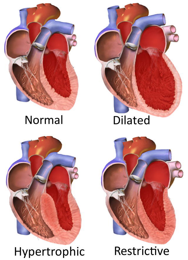
Cardiomyopathy is a disease in which the muscles of the heart are no longer able to effectively pump blood to the body — extreme forms of this disease can lead to heart failure. There are four main types of cardiomyopathy (also illustrated in Figure 12.3.11):
- Dilated (congestive) cardiomyopathy: the left ventricle (the chamber itself) of the heart becomes enlarged and can't pump blood our to the body. This is normally related to coronary artery disease and/or heart attack
- Hypertrophic cardiomyopathy: abnormal thickening of the muscular walls of the left ventricle make the chamber less able to work properly. This condition is more common in patients with a family history of the disease.
- Restrictive cardiomyopathy: the myocardium becomes abnormally rigid and inelastic and is unable to expand in between heartbeats to refill with blood. Restrictive cardiomyopathy typically affects older people.
- Arrhythmogenic right ventricular cardiomyopathy: the right ventricular muscle is replaced by adipose or scar tissue, reducing elasticity and interfering with normal heartbeat and rhythm. This disease is often caused by genetic mutations.
Cardiomyopathy is typically diagnosed with a physical exam supplemented by medical and family history, an angiogram, blood tests, chest x-rays and electrocardiograms. In some cases your doctor would also requisition a CT scan and/or genetic testing.
When treating cardiomyopathy, the goal is to reduce symptoms that affect everyday life. Certain medications can help regularize and slow heart rate, decrease chances of blood clots and cause vasodilation in the coronary arteries. If medication is not sufficient to manage symptoms, a pacemaker or even a heart transplant may be the best option. Lifestyle can also help manage the symptoms of cardiomyopathy — people living with this disease are encouraged to avoid drug and alcohol use, control high blood pressure, eat a healthy diet, get ample rest and exercise, as well as reduce stress levels.
12.3 Summary
- Muscle tissue is a soft tissue that makes up most of the tissues in the muscles of the human muscular system. It is the only type of tissue that has cells with the ability to contract.
- Skeletal muscle tissue is attached to bones by tendons. It allows voluntary body movements.
- Skeletal muscle is the most common type of muscle tissue in the human body. To move bones in opposite directions, skeletal muscles often consist of pairs of muscles that work in opposition to one another to move bones in different directions at joints.
- Skeletal muscle fibres are bundled together in units called muscle fascicles, which are bundled together to form individual skeletal muscles. Skeletal muscles also have connective tissue supporting and protecting the muscle tissue.
- Each skeletal muscle fibre consists of a bundle of myofibrils, which are bundles of protein filaments. The filaments are arranged in repeating units called sarcomeres, which are the basic functional units of skeletal muscles. Skeletal muscle tissue is striated because of the pattern of sarcomeres in its fibres.
- Skeletal muscle fibres can be divided into two types, called slow-twitch and fast-twitch fibres. Slow-twitch fibres are used mainly in aerobic endurance activities, such as long-distance running. Fast-twitch fibres are used mainly for non-aerobic, strenuous activities, such as sprinting. Proportions of the two types of fibres vary from muscle to muscle and person to person.
- Smooth muscle tissue is found in the walls of internal organs and vessels. When smooth muscles contract, they help the organs and vessels carry out their functions. Contractions of smooth muscles are involuntary and controlled by the autonomic nervous system, hormones, and other substances.
- Cells of smooth muscle tissue are not striated because they lack sarcomeres, but the cells contract in the same basic way as striated muscle cells. Unlike striated muscle, smooth muscle can sustain very long-term contractions and maintain its contractile function, even when stretched.
- Cardiac muscle tissue is found only in the wall of the heart. When cardiac muscle contracts, the heart beats and pumps blood. Contractions of cardiac muscle are involuntary, like those of smooth muscles. They are controlled by electrical impulses from specialized cardiac cells.
- Like skeletal muscle, cardiac muscle is striated because its filaments are arranged in sarcomeres inside the muscle fibres. However, the myofibrils are branched instead of arranged in parallel rows, making cardiac and skeletal muscle tissues look different from one another.
- The heart is the muscle that performs the greatest amount of physical work in the course of a lifetime. Its cells contain a great many mitochondria to produce ATP for energy and help the heart resist fatigue.
12.3 Review Questions
- What is muscle tissue?
-
-
- Where is skeletal muscle found, and what is its general function?
- Why do many skeletal muscles work in pairs?
- Describe the structure of a skeletal muscle.
- Relate muscle fibre structure to the functional units of muscles.
- Why is skeletal muscle tissue striated?
- Where is smooth muscle found? What controls the contraction of smooth muscle?
- Where is cardiac muscle found? What controls its contractions?
- The heart muscle is smaller and less powerful than some other muscles in the body. Why is the heart the muscle that performs the greatest amount of physical work in the course of a lifetime? How does the heart resist fatigue?
- Give one example of connective tissue that is found in muscles. Describe one of its functions.
12.3 Explore More
https://www.youtube.com/watch?v=3_PYnWVoUzM
What happens during a heart attack? - Krishna Sudhir, TED-Ed, 2017.
https://www.youtube.com/watch?v=bwOE1MEginA&feature=emb_logo
Three types of muscle | Circulatory system physiology | NCLEX-RN | KhanAcademyMedicine, 2012.
Attributions
Figure 12.3.1
Look by ali-yahya-155huuQwGvA [photo] by Ali Yahya on Unsplash is used under the Unsplash License (https://unsplash.com/license).
Figure 12.3.2
Skeletal_Smooth_Cardiac by OpenStax College on Wikimedia Commons is used under a CC BY 3.0 (https://creativecommons.org/licenses/by/3.0) license.
Figure 12.3.3
Anterior_and_Posterior_Views_of_Muscles by OpenStax on Wikimedia Commons is used under a CC BY 4.0 (https://creativecommons.org/licenses/by/4.0) license.
Figure 12.3.4
Antagonistic Muscle Pair by Laura Guerin at CK-12 Foundation on Wikimedia Commons is used under a CC BY-NC 3.0 (https://creativecommons.org/licenses/by-nc/3.0/) license.
Figure 12.3.5
Muscle_Fibes_(large) by OpenStax on Wikimedia Commons is used under a CC BY 4.0 (https://creativecommons.org/licenses/by/4.0) license.
Figure 12.3.6
Muscle_Fibers_(small) by OpenStax on Wikimedia Commons is used under a CC BY 4.0 (https://creativecommons.org/licenses/by/4.0) license.
Figure 12.3.7
Smooth_Muscle_Contraction by OpenStax on Wikimedia Commons is used under a CC BY 4.0 (https://creativecommons.org/licenses/by/4.0) license.
Figure 12.3.8
Blausen_0747_Pregnancy by BruceBlaus on Wikimedia Commons is used under a CC BY 3.0 (https://creativecommons.org/licenses/by/3.0) license.
Figure 12.3.9
Size_of_Uterus_Throughout_Pregnancy-02 by OpenStax College on Wikimedia Commons is used under a CC BY 3.0 (https://creativecommons.org/licenses/by/3.0) license.
Figure 12.3.10
1024px-Blausen_0470_HeartWall by BruceBlaus on Wikimedia Commons is used under a CC BY 3.0 (https://creativecommons.org/licenses/by/3.0) license.
Figure 12.3.11
Tipet_e_kardiomiopative by Npatchett at English Wikipedia on Wikimedia Commons is used under a CC BY-SA 3.0 (https://creativecommons.org/licenses/by-sa/3.0) license. (Work derived from Blausen 0165 Cardiomyopathy Dilated by BruceBlaus)
References
Betts, J. G., Young, K.A., Wise, J.A., Johnson, E., Poe, B., Kruse, D.H., Korol, O., Johnson, J.E., Womble, M., DeSaix, P. (2013, June 19). Figure 4.18 Muscle tissue [digital image]. In Anatomy and Physiology (Section 4.4). OpenStax. https://openstax.org/books/anatomy-and-physiology/pages/4-4-muscle-tissue-and-motion
Betts, J. G., Young, K.A., Wise, J.A., Johnson, E., Poe, B., Kruse, D.H., Korol, O., Johnson, J.E., Womble, M., DeSaix, P. (2013, June 19). Figure 28.18 Size of uterus throughout pregnancy [digital image]. In Anatomy and Physiology (Section 28.4). OpenStax. https://openstax.org/books/anatomy-and-physiology/pages/28-4-maternal-changes-during-pregnancy-labor-and-birth
Betts, J. G., Young, K.A., Wise, J.A., Johnson, E., Poe, B., Kruse, D.H., Korol, O., Johnson, J.E., Womble, M., DeSaix, P. (2016, May 18). Figure 10.3 The three connective tissue layers [digital image]. In Anatomy and Physiology (Section 10.2). OpenStax. https://openstax.org/books/anatomy-and-physiology/pages/10-2-skeletal-muscle
Betts, J. G., Young, K.A., Wise, J.A., Johnson, E., Poe, B., Kruse, D.H., Korol, O., Johnson, J.E., Womble, M., DeSaix, P. (2016, May 18). Figure 10.4 Muscle fiber [digital image]. In Anatomy and Physiology (Section 10.2). OpenStax. https://openstax.org/books/anatomy-and-physiology/pages/10-2-skeletal-muscle
Betts, J. G., Young, K.A., Wise, J.A., Johnson, E., Poe, B., Kruse, D.H., Korol, O., Johnson, J.E., Womble, M., DeSaix, P. (2016, May 18). Figure 10.24 Muscle contraction [digital image]. In Anatomy and Physiology (Section 10.8). OpenStax. https://openstax.org/books/anatomy-and-physiology/pages/10-8-smooth-muscle
Betts, J. G., Young, K.A., Wise, J.A., Johnson, E., Poe, B., Kruse, D.H., Korol, O., Johnson, J.E., Womble, M., DeSaix, P. (2016, May 18). Figure 11.5 Overview of the muscular system [digital image]. In Anatomy and Physiology (Section 11.2). OpenStax. https://openstax.org/books/anatomy-and-physiology/pages/11-2-naming-skeletal-muscles
Blausen.com staff. (2014). Medical gallery of Blausen Medical 2014. WikiJournal of Medicine 1 (2). DOI:10.15347/wjm/2014.010. ISSN 2002-4436.
Brainard, J/ CK-12 Foundation. (2012). Figure 5 Triceps and biceps muscles in the upper arm are opposing muscles. [digital image]. In CK-12 Biology (Section 21.3) [online Flexbook]. CK12.org. https://www.ck12.org/book/ck-12-biology/section/21.3/ (Last modified August 11, 2017.)
khanacademymedicine. (2012, October 19). Three types of muscle | Circulatory system physiology | NCLEX-RN | Khan Academy. YouTube.
TED-Ed. (2017, February 14). What happens during a heart attack? - Krishna Sudhir. YouTube. https://www.youtube.com/watch?v=3_PYnWVoUzM&feature=youtu.be
The transfer of genetic variation from one population to another. If the rate of gene flow is high enough, then two populations are considered to have equivalent allele frequencies and therefore effectively be a single population.
Created by: CK-12/Adapted by Christine Miller

Bring on the S'mores!
This inviting camp fire can be used for both heat and light. Heat and light are two forms of energy that are released when a fuel like wood is burned. The cells of living things also get energy by "burning." They "burn" glucose in a process called cellular respiration.
What Is Cellular Respiration?
Cellular respiration is the process by which living cells break down glucose molecules and release energy. The process is similar to burning, although it doesn’t produce light or intense heat as a campfire does. This is because cellular respiration releases the energy in glucose slowly and in many small steps. It uses the energy released to form molecules of ATP, the energy-carrying molecules that cells use to power biochemical processes. In this way, cellular respiration is an example of energy coupling: glucose is broken down in an exothermic reaction, and then the energy from this reaction powers the endothermic reaction of the formation of ATP. Cellular respiration involves many chemical reactions, but they can all be summed up with this chemical equation:
C6H12O6 6O2 → 6CO2 6H2O Chemical Energy (in ATP)
In words, the equation shows that glucose (C6H12O6) and oxygen (O2) react to form carbon dioxide (CO2) and water (H2O), releasing energy in the process. Because oxygen is required for cellular respiration, it is an aerobic process.
Cellular respiration occurs in the cells of all living things, both autotrophs and heterotrophs. All of them burn glucose to form ATP. The reactions of cellular respiration can be grouped into three stages: glycolysis, the Krebs cycle (also called the citric acid cycle), and electron transport. Figure 4.10.2 gives an overview of these three stages, which are also described in detail below.
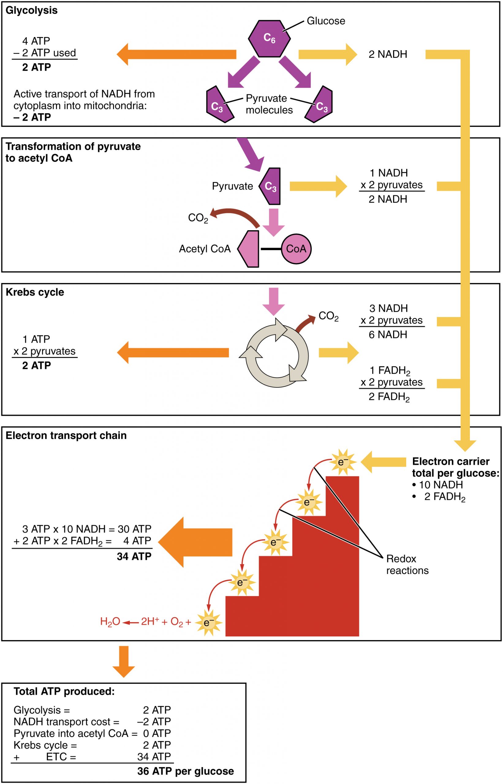
Cellular Respiration Stage I: Glycolysis
The first stage of cellular respiration is glycolysis, which happens in the cytosol of the cytoplasm.
Splitting Glucose
The word glycolysis literally means “glucose splitting,” which is exactly what happens in this stage. Enzymes split a molecule of glucose into two molecules of pyruvate (also known as pyruvic acid). This occurs in several steps, as summarized in the following diagram.
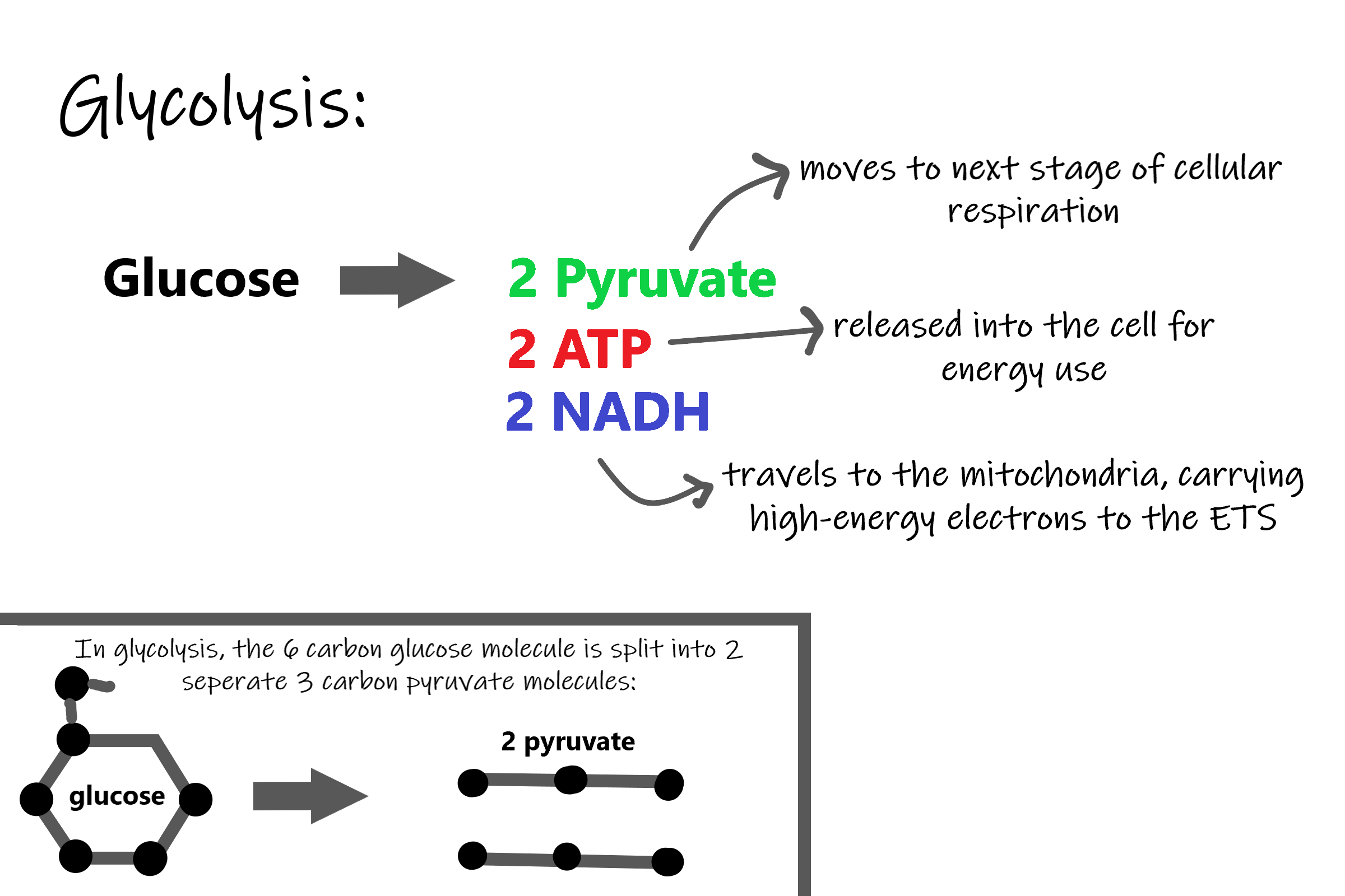
Results of Glycolysis
Energy is needed at the start of glycolysis to split the glucose molecule into two pyruvate molecules which go on to stage II of cellular respiration. The energy needed to split glucose is provided by two molecules of ATP; this is called the energy investment phase. As glycolysis proceeds, energy is released, and the energy is used to make four molecules of ATP; this is the energy harvesting phase. As a result, there is a net gain of two ATP molecules during glycolysis. During this stage, high-energy electrons are also transferred to molecules of NAD to produce two molecules of NADH, another energy-carrying molecule. NADH is used in stage III of cellular respiration to make more ATP.
Transition Reaction
Before pyruvate can enter the next stage of cellular respiration it needs to be modified slightly. The transition reaction is a very short reaction which converts the two molecules of pyruvate to two molecules of acetyl CoA, carbon dioxide, and two high energy electron pairs convert NAD to NADH. The carbon dioxide is released, the acetyl CoA moves to the mitochondria to enter the Kreb's Cycle (stage II), and the NADH carries the high energy electrons to the Electron Transport System (stage III).
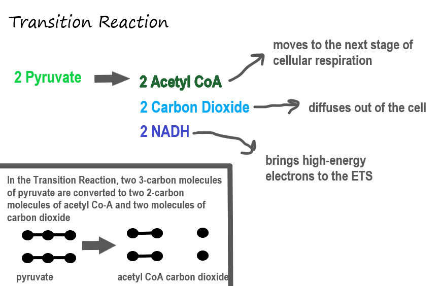
Structure of the Mitochondrion
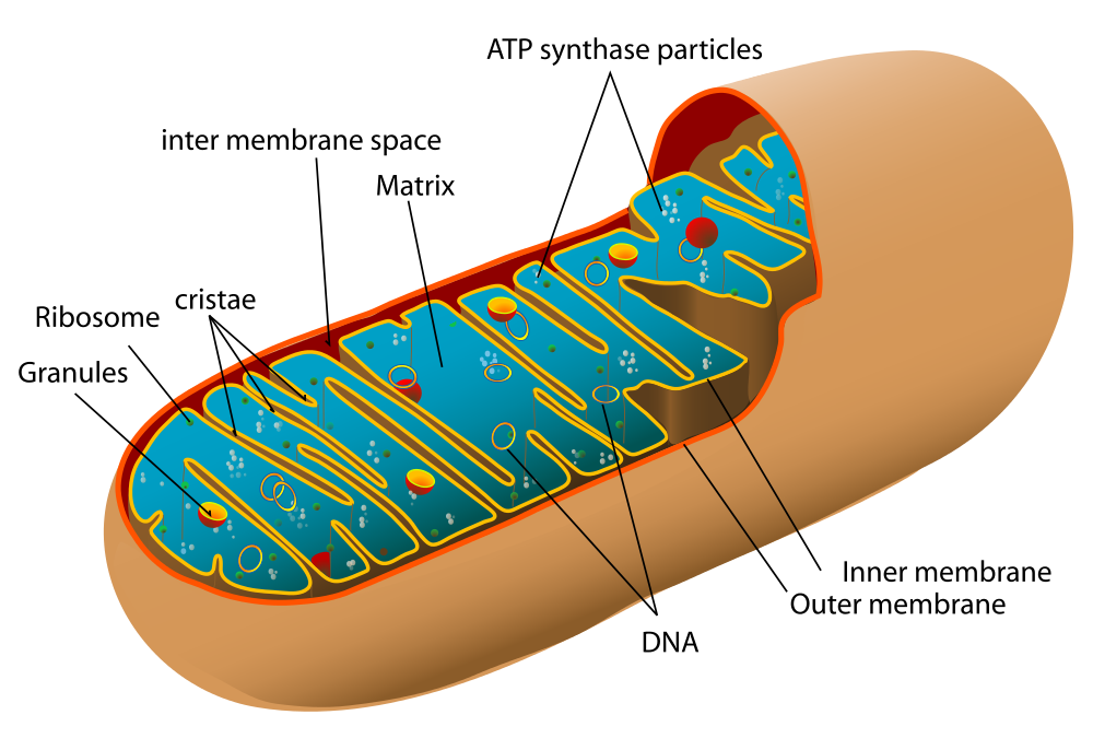
Before you read about the last two stages of cellular respiration, you need to know more about the mitochondrion, where these two stages take place. A diagram of a mitochondrion is shown in Figure 4.10.5.
The structure of a mitochondrion is defined by an inner and outer membrane. This structure plays an important role in aerobic respiration.
As you can see from the figure, a mitochondrion has an inner and outer membrane. The space between the inner and outer membrane is called the intermembrane space. The space enclosed by the inner membrane is called the matrix. The second stage of cellular respiration (the Krebs cycle) takes place in the matrix. The third stage (electron transport) happens on the inner membrane.
Cellular Respiration Stage II: The Krebs Cycle
Recall that glycolysis produces two molecules of pyruvate (pyruvic acid), which are then converted to acetyl CoA during the short transition reaction. These molecules enter the matrix of a mitochondrion, where they start the Krebs cycle (also known as the Citric Acid Cycle). The reason this stage is considered a cycle is because a molecule called oxaloacetate is present at both the beginning and end of this reaction and is used to break down the two molecules of acetyl CoA. The reactions that occur next are shown in Figure 4.10.6.
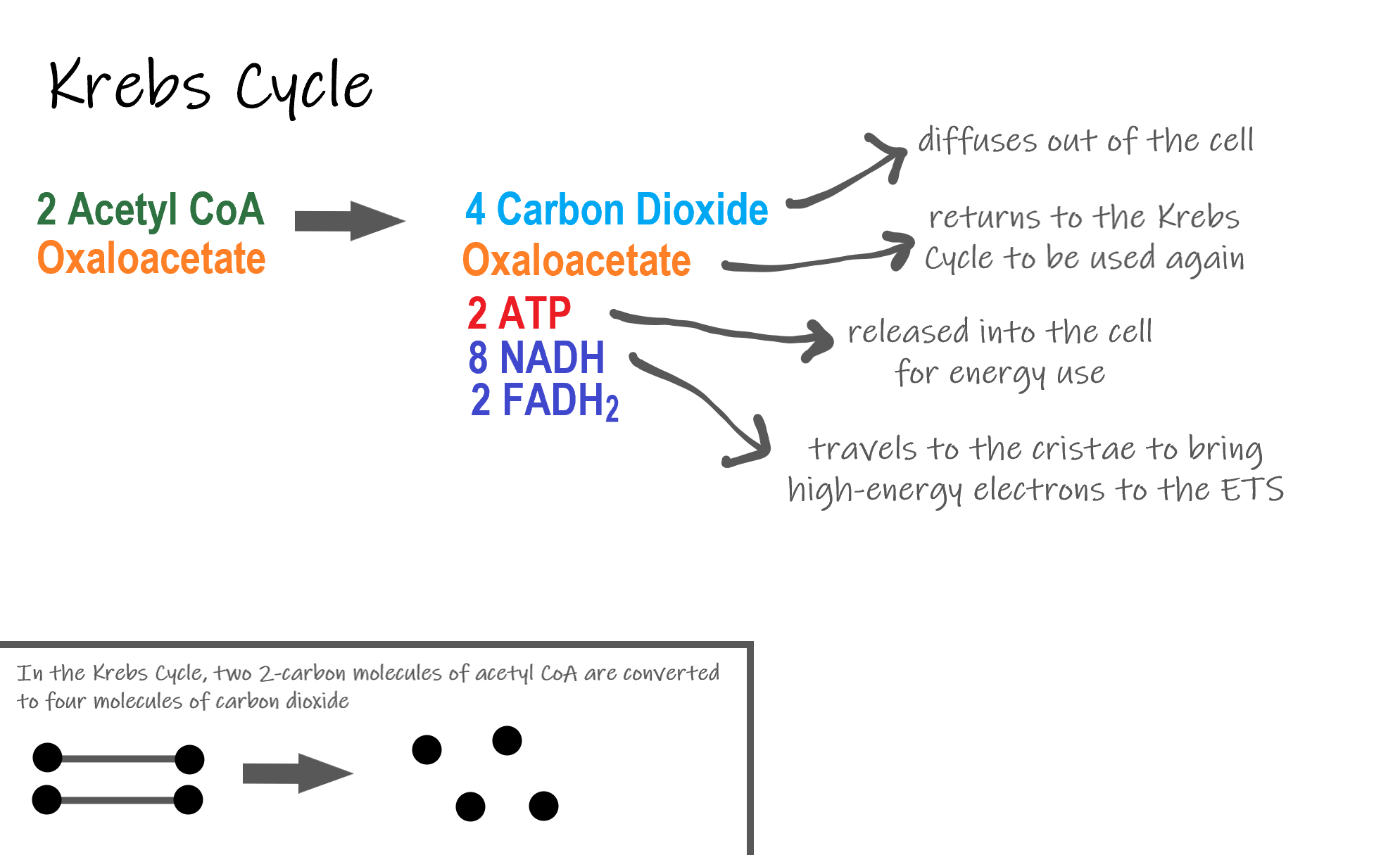
Steps of the Krebs Cycle
The Krebs cycle itself actually begins when acetyl-CoA combines with a four-carbon molecule called OAA (oxaloacetate) (see Figure 4.10.6). This produces citric acid, which has six carbon atoms. This is why the Krebs cycle is also called the citric acid cycle.
After citric acid forms, it goes through a series of reactions that release energy. The energy is captured in molecules of NADH, ATP, and FADH2, another energy-carrying coenzyme. Carbon dioxide is also released as a waste product of these reactions.
The final step of the Krebs cycle regenerates OAA, the molecule that began the Krebs cycle. This molecule is needed for the next turn through the cycle. Two turns are needed because glycolysis produces two pyruvic acid molecules when it splits glucose.
Results of the Glycolysis, Transition Reaction and Krebs Cycle
After glycolysis, transition reaction, and the Krebs cycle, the glucose molecule has been broken down completely. All six of its carbon atoms have combined with oxygen to form carbon dioxide. The energy from its chemical bonds has been stored in a total of 16 energy-carrier molecules. These molecules are:
- 4 ATP (2 from glycolysis, 2 from Krebs Cycle)
- 12 NADH (2 from glycolysis, 2 from transition reaction, and 8 from Krebs cycle)
- 2 FADH2 (both from the Krebs cycle)
The events of cellular respiration up to this point are exergonic reactions- they are releasing energy that had been stored in the bonds of the glucose molecule. This energy will be transferred to the third and final stage of cellular respiration: the Electron Transport System, which is an endergonic reaction. Using an exothermic reaction to power an endothermic reaction is known as energy coupling.
Cellular Respiration Stage III: Electron Transport Chain
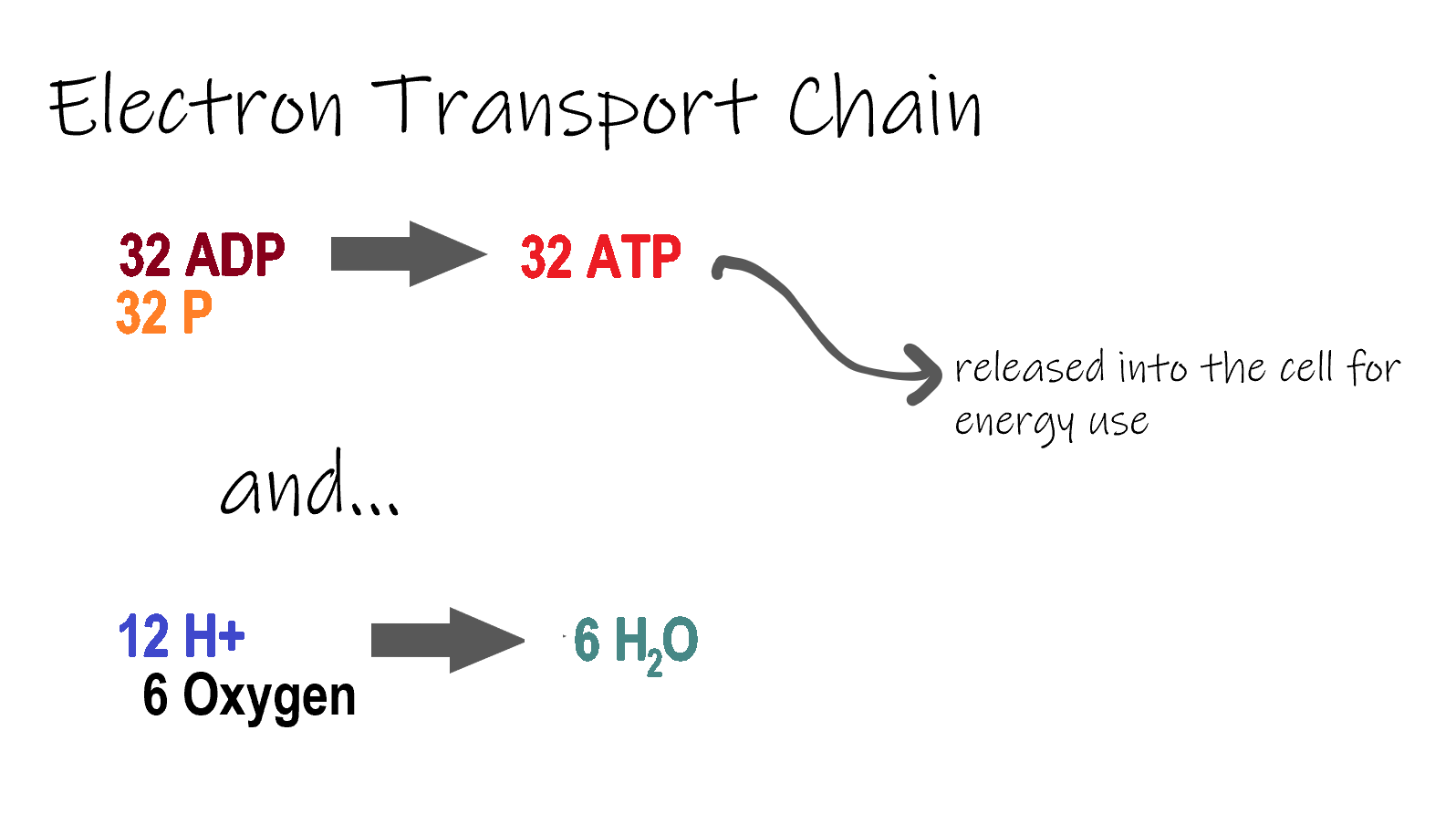
ETC, the final stage in cellular respiration produces 32 ATP. The Electron Transport Chain is the final stage of cellular respiration. In this stage, energy being transported by NADH and FADH2 is transferred to ATP. In addition, oxygen acts as the final proton acceptor for the hydrogens released from all the NADH and FADH2, forming water. Figure 4.10.8 shows the reactants and products of the ETC.
Transporting Electrons
The Electron transport chain is the third stage of cellular respiration and is illustrated in Figure 4.10.8. During this stage, high-energy electrons are released from NADH and FADH2, and they move along electron-transport chains on the inner membrane of the mitochondrion. An electron-transport chain is a series of molecules that transfer electrons from molecule to molecule by chemical reactions. Some of the energy from the electrons is used to pump hydrogen ions (H ) across the inner membrane, from the matrix into the intermembrane space. This ion transfer creates an electrochemical gradient that drives the synthesis of ATP.
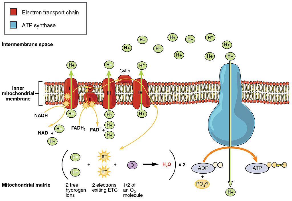
Making ATP
As shown in Figure 4.10.8, the pumping of hydrogen ions across the inner membrane creates a greater concentration of the ions in the intermembrane space than in the matrix. This gradient causes the ions to flow back across the membrane into the matrix, where their concentration is lower. ATP synthase acts as a channel protein, helping the hydrogen ions cross the membrane. It also acts as an enzyme, forming ATP from ADP and inorganic phosphate in a process called oxidative phosphorylation. After passing through the electron-transport chain, the “spent” electrons combine with oxygen to form water.
How Much ATP?
You have seen how the three stages of aerobic respiration use the energy in glucose to make ATP. How much ATP is produced in all three stages combined? Glycolysis produces two ATP molecules, and the Krebs cycle produces two more. Electron transport begins with several molecules of NADH and FADH2 from the Krebs cycle and transfers their energy into as many as 34 more ATP molecules. All told, then, up to 38 molecules of ATP can be produced from just one molecule of glucose in the process of cellular respiration.
4.10 Summary
- Cellular respiration is the aerobic process by which living cells break down glucose molecules, release energy, and form molecules of ATP. Generally speaking, this three-stage process involves glucose and oxygen reacting to form carbon dioxide and water.
- The first stage of cellular respiration, called glycolysis, takes place in the cytoplasm. In this step, enzymes split a molecule of glucose into two molecules of pyruvate, which releases energy that is transferred to ATP. Following glycolysis, a short reaction called the transition reaction converts the pyruvate into two molecules of acetyl CoA.
- The organelle called a mitochondrion is the site of the other two stages of cellular respiration. The mitochondrion has an inner and outer membrane separated by an intermembrane space, and the inner membrane encloses a space called the matrix.
- The second stage of cellular respiration, called the Krebs cycle, takes place in the matrix of a mitochondrion. During this stage, two turns through the cycle result in all of the carbon atoms from the two pyruvate molecules forming carbon dioxide and the energy from their chemical bonds being stored in a total of 16 energy-carrying molecules (including two from glycolysis and two from transition reaction).
- The third and final stage of cellular respiration, called electron transport, takes place on the inner membrane of the mitochondrion. Electrons are transported from molecule to molecule down an electron-transport chain. Some of the energy from the electrons is used to pump hydrogen ions across the membrane, creating an electrochemical gradient that drives the synthesis of many more molecules of ATP.
- In all three stages of cellular respiration combined, as many as 38 molecules of ATP are produced from just one molecule of glucose.
4.10 Review Questions
- What is the purpose of cellular respiration? Provide a concise summary of the process.
- State what happens during glycolysis.
- Describe the structure of a mitochondrion.
- What molecule is present at both the beginning and end of the Krebs cycle?
- What happens during the electron transport stage of cellular respiration?
- How many molecules of ATP can be produced from one molecule of glucose during all three stages of cellular respiration combined?
- Do plants undergo cellular respiration? Why or why not?
- Explain why the process of cellular respiration described in this section is considered aerobic.
- Name three energy-carrying molecules involved in cellular respiration.
-
- Which stage of aerobic cellular respiration produces the most ATP?
-
4.10 Explore More
https://www.youtube.com/watch?time_continue=2&v=00jbG_cfGuQ&feature=emb_logo
ATP & Respiration: Crash Course Biology #7, CrashCourse, 2012.
https://www.youtube.com/watch?v=4Eo7JtRA7lg&t=3s
Cellular Respiration and the Mighty Mitochondria, The Amoeba Sisters, 2014.
Attributions
Figure 4.10.1
Smores by Jessica Ruscello on Unsplash is used under the Unsplash License (https://unsplash.com/license).
Figure 4.10.2
Carbohydrate_Metabolism by OpenStax College on Wikimedia Commons is used under a CC BY 3.0 (https://creativecommons.org/licenses/by/3.0) license.
Figure 4.10.3
Glycolysis by Christine Miller is used under a CC BY 4.0 (https://creativecommons.org/licenses/by/4.0/) license.
Figure 4.10.4
Transition Reaction by Christine Miller is used under a CC BY 4.0 (https://creativecommons.org/licenses/by/4.0/) license.
Figure 4.10.5
Mitochondrion by Mariana Ruiz Villarreal [LadyofHats] on Wikimedia Commons is released into the public domain (https://en.wikipedia.org/wiki/Public_domain).
Figure 4.10.6
Krebs cycle by Christine Miller is used under a CC BY 4.0 (https://creativecommons.org/licenses/by/4.0/) license.
Figure 4.10.7
Electron Transport Chain (ETC) by Christine Miller is used under a CC BY 4.0 (https://creativecommons.org/licenses/by/4.0/) license.
Figure 4.10.8
The_Electron_Transport_Chain by OpenStax College on Wikimedia Commons is used under a CC BY 3.0 (https://creativecommons.org/licenses/by/3.0) license.
References
CrashCourse. (2012, March 12). ATP & Respiration: Crash Course Biology #7. YouTube. https://www.youtube.com/watch?time_continue=2&v=00jbG_cfGuQ&feature=emb_logo
Betts, J. G., Young, K.A., Wise, J.A., Johnson, E., Poe, B., Kruse, D.H., Korol, O., Johnson, J.E., Womble, M., DeSaix, P. (2013, April 25). Figure 24.8 Electron Transport Chain [digital image]. In Anatomy & Physiology, Connexions (Section ). OpenStax. https://openstax.org/books/anatomy-and-physiology/pages/24-2-carbohydrate-metabolism
Betts, J. G., Young, K.A., Wise, J.A., Johnson, E., Poe, B., Kruse, D.H., Korol, O., Johnson, J.E., Womble, M., DeSaix, P. (2013, April 25). Figure 24.9 Carbohydrate Metabolism [digital image]. In Anatomy & Physiology, Connexions (Section 24.2). OpenStax. https://openstax.org/books/anatomy-and-physiology/pages/24-2-carbohydrate-metabolism
The Amoeba Sisters. (2014, October 22). Cellular Respiration and the Mighty Mitochondria. YouTube. https://www.youtube.com/watch?v=4Eo7JtRA7lg&t=3s
An involuntary human body response mediated by the nervous and endocrine systems that prepares the body to fight or flee from perceived danger.
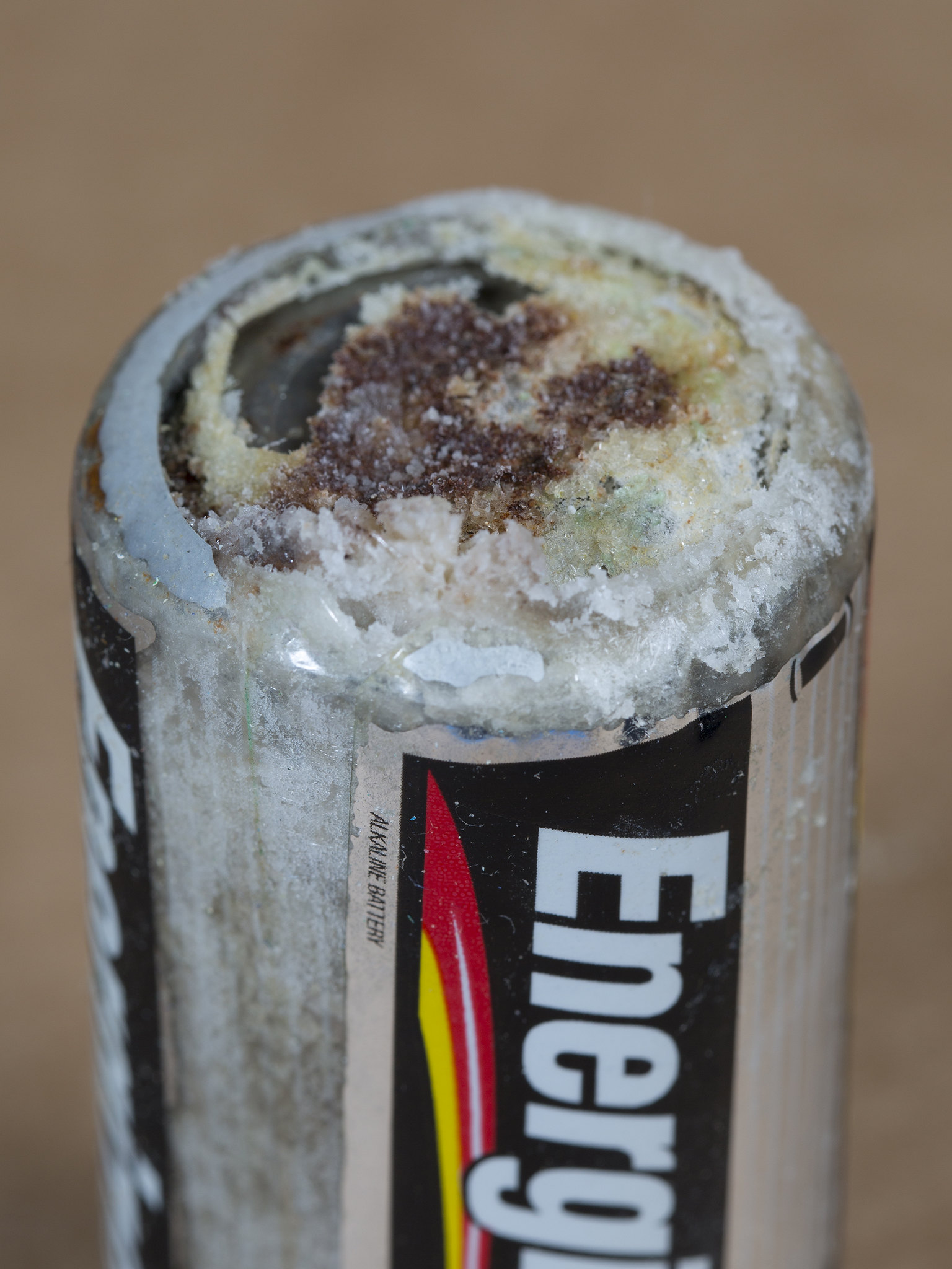
Danger! Acid!
You probably know that batteries contain dangerous chemicals, including strong acids. Strong acids can hurt you if they come into contact with your skin or eyes. Therefore, it may surprise you to learn that your life depends on acids. There are many acids inside your body, and some of them are as strong as battery acid. Acids are needed for digestion and some forms of energy production. Genes are made of nucleic acids, proteins of amino acids, and lipids of fatty acids.
Water and Solutions
Acids (such as battery acid) are solutions. A solution is a mixture of two or more substances that has the same composition throughout. Many solutions are a mixture of water and some other substance. Not all solutions are acids. Some are bases and some are neither acids nor bases. To understand acids and bases, you need to know more about pure water.
In pure water (such as distilled water), a tiny fraction of water molecules naturally breaks down to form ions. An ion is an electrically charged atom or molecule. The breakdown of water is represented by the chemical equation:
2 H2O → H3O+ + OH-
The products of this reaction are a hydronium ion (H3O+) and a hydroxide ion (OH-). The hydroxide ion, which has a negative charge, forms when a water molecule gives up a positively charged hydrogen ion (H+). The hydronium ion, which has a positive charge, forms when another water molecule accepts the hydrogen ion.
Acidity and pH
The concentration of hydronium ions in a solution is known as acidity. In pure water, the concentration of hydronium ions is very low; only about one in ten million water molecules naturally breaks down to form a hydronium ion. As a result, pure water is essentially neutral. Acidity is measured on a scale called pH, as shown in Figure 3.12.2. Pure water has a pH of 7, so the point of neutrality on the pH scale is 7.

This pH scale shows the acidity of many common substances. The lower the pH value, the more acidic a substance is.
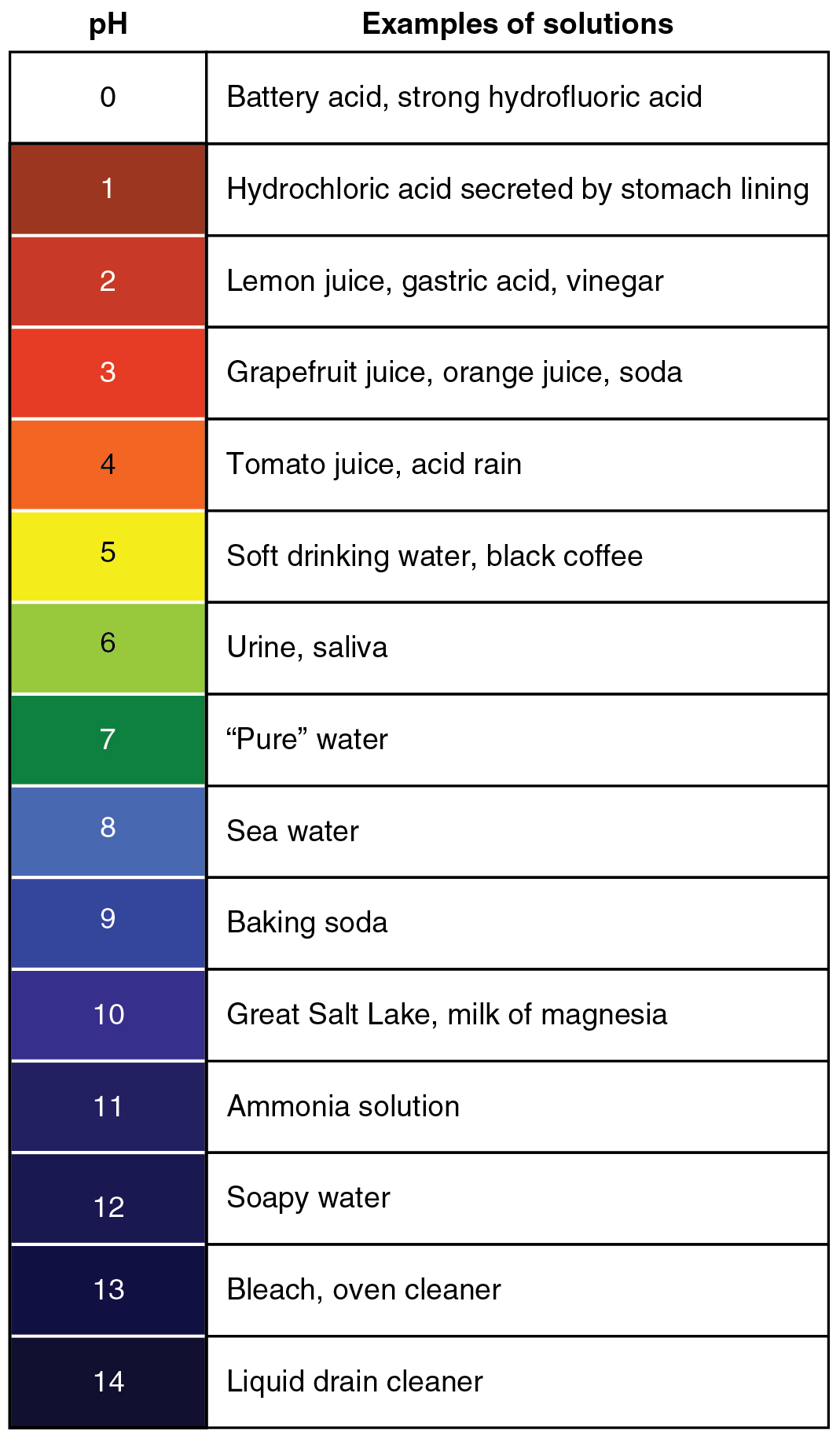
Acids
If a solution has a higher concentration of hydronium ions than pure water, it has a pH lower than 7. A solution with a pH lower than 7 is called an acid. As the hydronium ion concentration increases, the pH value decreases. Therefore, the more acidic a solution is, the lower its pH value is.
Did you ever taste vinegar? Like other acids, it tastes sour. Stronger acids can be harmful to organisms. Even stomach acid would eat through the stomach if it were not lined with a layer of mucus. Strong acids can also damage materials, even hard materials such as glass.
Bases
If a solution has a lower concentration of hydronium ions than pure water, it has a pH higher than 7. A solution with a pH higher than 7 is called a base. Bases, such as baking soda, have a bitter taste. Like strong acids, strong bases can harm organisms and damage materials. For example, lye can burn the skin, and bleach can remove the colour from clothing.
Buffers
A buffer is a solution that can resist changes in pH. Buffers are able to maintain a certain pH by by absorbing any H+ or OH- ions added to the solution. Buffers are extremely important in biological systems in order to maintain a pH conducive to life. Bicarbonate is an example of a buffer which is used to maintain pH of the blood. In this buffering system, if blood becomes too acidic, carbonic acid will convert to carbon dioxide and water. If the blood becomes too basic, carbonic acid will convert to bicarbonate and H+ ions:
CO2 + H2O ↔ H2CO3 ↔ HCO3- + H+
Acids, Bases, and Enzymes
Many acids and bases in living things provide the pH that enzymes need. Enzymes are biological catalysts that must work effectively for biochemical reactions to occur. Most enzymes can do their job only at a certain level of acidity. Cells secrete acids and bases to maintain the proper pH for enzymes to do their work.
Every time you digest food, acids and bases are at work in your digestive system. Consider the enzyme pepsin, which helps break down proteins in the stomach. Pepsin needs an acidic environment to do its job. The stomach secretes a strong acid called hydrochloric acid that allows pepsin to work. When stomach contents enter the small intestine, the acid must be neutralized, because enzymes in the small intestine need a basic environment in order to work. An organ called the pancreas secretes a base named bicarbonate into the small intestine, and this base neutralizes the acid.
Feature: My Human Body
Do you ever have heartburn? The answer is probably "yes." More than 60 million Americans have heartburn at least once a month, and more than 15 million suffer from it on a daily basis. Knowing more about heartburn may help you prevent it or know when it's time to seek medical treatment.
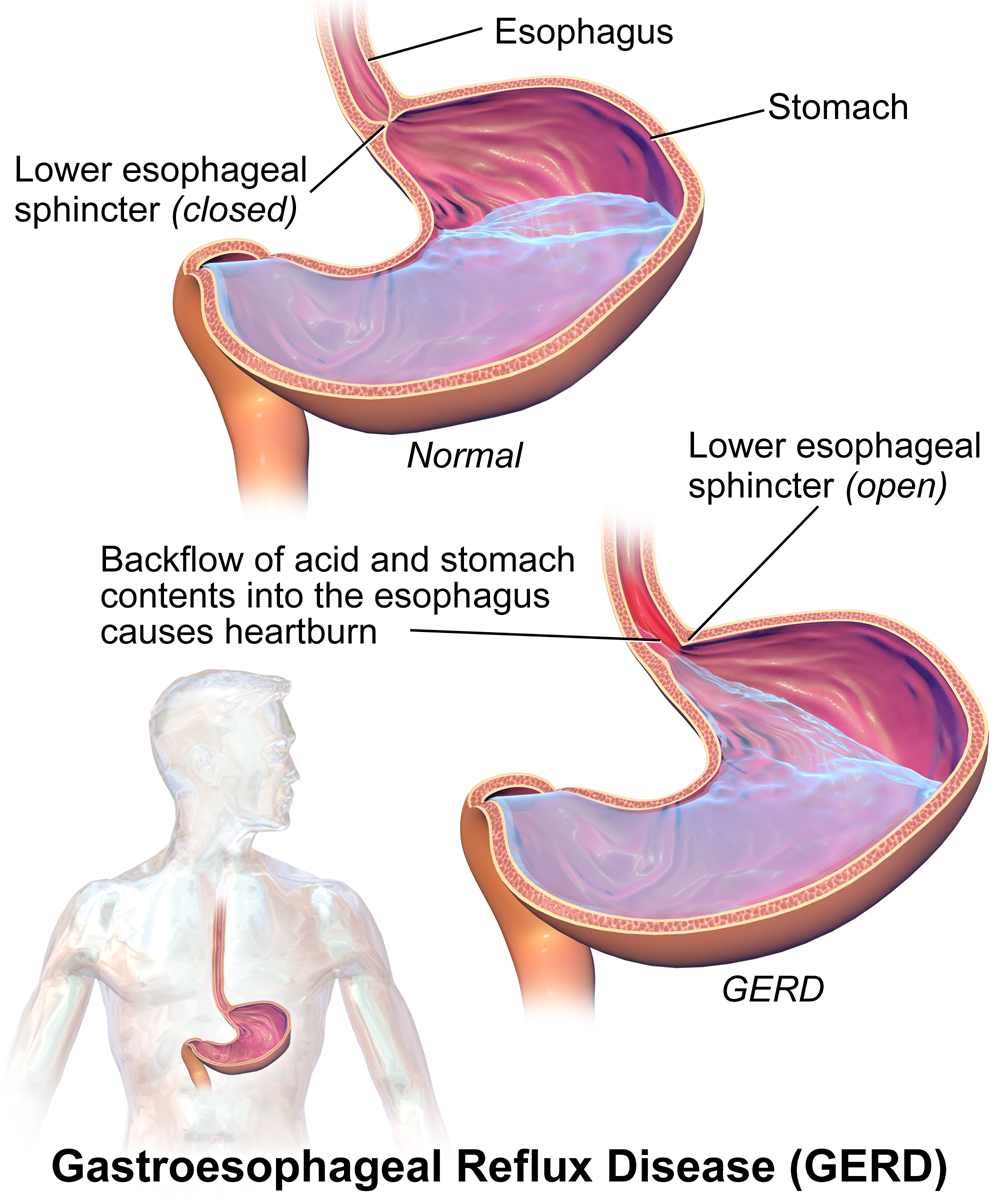
Heartburn doesn't have anything to do with the heart, but it does cause a burning sensation in the vicinity of the chest. Normally, the acid secreted into the stomach remains in the stomach where it is needed to allow pepsin to do its job of digesting proteins. A long tube called the esophagus carries food from the mouth to the stomach. A sphincter, or valve, between the esophagus and stomach opens to allow swallowed food to enter the stomach and then closes to prevent stomach contents from backflowing into the esophagus. If this sphincter is weak or relaxes inappropriately, stomach contents flow into the esophagus. Because stomach contents are usually acidic, this causes the burning sensation known as heartburn. People who are prone to heartburn and suffer from it often may be diagnosed with GERD, which stands for gastroesophageal reflux disease.
GERD — as well as occasional heartburn — often can be improved by dietary and other lifestyle changes that decrease the amount and acidity of reflux from the stomach into the esophagus.
- Some foods and beverages seem to contribute to GERD, so these should be avoided. Problematic foods include chocolate, fatty foods, peppermint, coffee, and alcoholic beverages.
- Decreasing portion size and eating the last meal of the day at least a couple of hours before bedtime may reduce the risk of reflux occurring.
- Smoking tends to weaken the lower esophageal sphincter, so quitting the habit may help control reflux.
- GERD is often associated with being overweight. Losing weight often brings improvement.
- Some people are helped by sleeping with the head of the bed elevated. This allows gravity to help control the backflow of acids into the esophagus from the stomach.
If you have frequent heartburn and lifestyle changes don't help, you may need medication to control the condition. Over-the-counter (OTC) antacids may be all that you need to control the occasional heartburn attack. OTC medications are usually bases that neutralize stomach acids. They may also create bubbles that help block stomach contents from entering the esophagus. For some people, OTC medications are not enough, and prescription medications are instead required for the control of GERD. These prescription medications generally work by inhibiting acid secretion in the stomach.
Be sure to see a doctor if you can't control your heartburn, or you have it often. Untreated GERD not only interferes with quality of life, it may also lead to more serious complications, ranging from esophageal bleeding to esophageal cancer.
3.12 Summary
- A solution is a mixture of two or more substances that has the same composition throughout. Many solutions consist of water and one or more dissolved substances.
- Acidity is a measure of the hydronium ion concentration in a solution. Pure water has a very low concentration and a pH of 7, which is the point of neutrality on the pH scale.
- Acids have a higher hydronium ion concentration than pure water and a pH lower than 7. Bases have a lower hydronium ion concentration than pure water and a pH higher than 7.
- Many acids and bases in living things are secreted to provide the proper pH for enzymes to work properly. Enzymes are the biological catalysts (like pepsin) needed to digest protein in the stomach. Pepsin requires an acidic environment.
3.12 Review Questions
-
- What is a solution?
- Define acidity.
- Explain how acidity is measured.
- Compare and contrast acids and bases.
- Hydrochloric acid is secreted by the stomach to provide an acidic environment for the enzyme pepsin. What is the pH of this acid? How strong of an acid is it compared with other acids?
- Define an ion. Identify the ions in the equation below, and explain what makes them ions:
- 2 H2O → H3O+ + OH-
- Explain why the pancreas secretes bicarbonate into the small intestine.
- Do you think pepsin would work in the small intestine? Why or why not?
- You may have mixed vinegar and baking soda and noticed that they bubble and react with each other. Explain why this happens. Explain also what happens to the pH of this solution after you mix the vinegar and baking soda.
- Pregnancy hormones can cause the lower esophageal sphincter to relax. What effect do you think this has on pregnant women? Explain your answer.
3.12 Explore More
https://www.youtube.com/watch?v=rIvEvwViJGk&feature=youtu.be
pH and Buffers by Bozeman Science, 2014.
https://www.youtube.com/watch?v=DupXDD87oHc&feature=youtu.be
The strengths and weaknesses of acids and bases - George Zaidan and Charles Morton, TED-Ed, 2013.
Attributions
Figure 3.12.1
Leaky battery by Carbon Arc on Flickr is used under a CC BY-NC-SA 2.0 (https://creativecommons.org/licenses/by-nc-sa/2.0/) license.
Figure 3.12.2
PH_Scale by Christinelmiller on Wikimedia Commons is used under a © CC0 1.0 (https://creativecommons.org/publicdomain/zero/1.0/) public domain dedication license.
Figure 3.12.3
Ph scale with examples by OpenStax College, on Wikimedia Commons, is used under a CC BY 3.0 (https://creativecommons.org/licenses/by/3.0) license.
Figure 3.12.4
GERD by BruceBlaus on Wikimedia Commons is used under a CC BY-SA 4.0 (https://creativecommons.org/licenses/by-sa/4.0) license.
References
Betts, J.G., Young, K.A., Wise, J.A., Johnson, E., Poe, B., Kruse, D.H., Korol, O., Johnson, J.E., Womble, M., DeSaix, P. (2013, April 25). Figure 26.15 The pH Scale [digital image]. In Anatomy and Physiology. OpenStax. https://openstax.org/books/anatomy-and-physiology/pages/26-4-acid-base-balance
Bozeman Science. (2014, February 22). pH and buffers. YouTube. https://www.youtube.com/watch?v=rIvEvwViJGk&feature=youtu.be
TED-Ed. (2013, October 24). The strengths and weaknesses of acids and bases - George Zaidan and Charles Morton. YouTube. https://www.youtube.com/watch?v=DupXDD87oHc&feature=youtu.be
a colorless cell that circulates in the blood and body fluids and is involved in counteracting foreign substances and disease; a white (blood) cell. There are several types, all amoeboid cells with a nucleus, including lymphocytes, granulocytes, monocytes, and macrophages.

Created by: CK-12/Adapted by Christine Miller
Danger! Acid!
You probably know that batteries contain dangerous chemicals, including strong acids. Strong acids can hurt you if they come into contact with your skin or eyes. Therefore, it may surprise you to learn that your life depends on acids. There are many acids inside your body, and some of them are as strong as battery acid. Acids are needed for digestion and some forms of energy production. Genes are made of nucleic acids, proteins of amino acids, and lipids of fatty acids.
Water and Solutions
Acids (such as battery acid) are solutions. A solution is a mixture of two or more substances that has the same composition throughout. Many solutions are a mixture of water and some other substance. Not all solutions are acids. Some are bases and some are neither acids nor bases. To understand acids and bases, you need to know more about pure water.
In pure water (such as distilled water), a tiny fraction of water molecules naturally breaks down to form ions. An ion is an electrically charged atom or molecule. The breakdown of water is represented by the chemical equation:
2 H2O → H3O+ + OH-
The products of this reaction are a hydronium ion (H3O+) and a hydroxide ion (OH-). The hydroxide ion, which has a negative charge, forms when a water molecule gives up a positively charged hydrogen ion (H+). The hydronium ion, which has a positive charge, forms when another water molecule accepts the hydrogen ion.
Acidity and pH
The concentration of hydronium ions in a solution is known as acidity. In pure water, the concentration of hydronium ions is very low; only about one in ten million water molecules naturally breaks down to form a hydronium ion. As a result, pure water is essentially neutral. Acidity is measured on a scale called pH, as shown in Figure 3.12.2. Pure water has a pH of 7, so the point of neutrality on the pH scale is 7.

This pH scale shows the acidity of many common substances. The lower the pH value, the more acidic a substance is.

Acids
If a solution has a higher concentration of hydronium ions than pure water, it has a pH lower than 7. A solution with a pH lower than 7 is called an acid. As the hydronium ion concentration increases, the pH value decreases. Therefore, the more acidic a solution is, the lower its pH value is.
Did you ever taste vinegar? Like other acids, it tastes sour. Stronger acids can be harmful to organisms. Even stomach acid would eat through the stomach if it were not lined with a layer of mucus. Strong acids can also damage materials, even hard materials such as glass.
Bases
If a solution has a lower concentration of hydronium ions than pure water, it has a pH higher than 7. A solution with a pH higher than 7 is called a base. Bases, such as baking soda, have a bitter taste. Like strong acids, strong bases can harm organisms and damage materials. For example, lye can burn the skin, and bleach can remove the colour from clothing.
Buffers
A buffer is a solution that can resist changes in pH. Buffers are able to maintain a certain pH by by absorbing any H+ or OH- ions added to the solution. Buffers are extremely important in biological systems in order to maintain a pH conducive to life. Bicarbonate is an example of a buffer which is used to maintain pH of the blood. In this buffering system, if blood becomes too acidic, carbonic acid will convert to carbon dioxide and water. If the blood becomes too basic, carbonic acid will convert to bicarbonate and H+ ions:
CO2 + H2O ↔ H2CO3 ↔ HCO3- + H+
Acids, Bases, and Enzymes
Many acids and bases in living things provide the pH that enzymes need. Enzymes are biological catalysts that must work effectively for biochemical reactions to occur. Most enzymes can do their job only at a certain level of acidity. Cells secrete acids and bases to maintain the proper pH for enzymes to do their work.
Every time you digest food, acids and bases are at work in your digestive system. Consider the enzyme pepsin, which helps break down proteins in the stomach. Pepsin needs an acidic environment to do its job. The stomach secretes a strong acid called hydrochloric acid that allows pepsin to work. When stomach contents enter the small intestine, the acid must be neutralized, because enzymes in the small intestine need a basic environment in order to work. An organ called the pancreas secretes a base named bicarbonate into the small intestine, and this base neutralizes the acid.
Feature: My Human Body
Do you ever have heartburn? The answer is probably "yes." More than 60 million Americans have heartburn at least once a month, and more than 15 million suffer from it on a daily basis. Knowing more about heartburn may help you prevent it or know when it's time to seek medical treatment.

Heartburn doesn't have anything to do with the heart, but it does cause a burning sensation in the vicinity of the chest. Normally, the acid secreted into the stomach remains in the stomach where it is needed to allow pepsin to do its job of digesting proteins. A long tube called the esophagus carries food from the mouth to the stomach. A sphincter, or valve, between the esophagus and stomach opens to allow swallowed food to enter the stomach and then closes to prevent stomach contents from backflowing into the esophagus. If this sphincter is weak or relaxes inappropriately, stomach contents flow into the esophagus. Because stomach contents are usually acidic, this causes the burning sensation known as heartburn. People who are prone to heartburn and suffer from it often may be diagnosed with GERD, which stands for gastroesophageal reflux disease.
GERD — as well as occasional heartburn — often can be improved by dietary and other lifestyle changes that decrease the amount and acidity of reflux from the stomach into the esophagus.
- Some foods and beverages seem to contribute to GERD, so these should be avoided. Problematic foods include chocolate, fatty foods, peppermint, coffee, and alcoholic beverages.
- Decreasing portion size and eating the last meal of the day at least a couple of hours before bedtime may reduce the risk of reflux occurring.
- Smoking tends to weaken the lower esophageal sphincter, so quitting the habit may help control reflux.
- GERD is often associated with being overweight. Losing weight often brings improvement.
- Some people are helped by sleeping with the head of the bed elevated. This allows gravity to help control the backflow of acids into the esophagus from the stomach.
If you have frequent heartburn and lifestyle changes don't help, you may need medication to control the condition. Over-the-counter (OTC) antacids may be all that you need to control the occasional heartburn attack. OTC medications are usually bases that neutralize stomach acids. They may also create bubbles that help block stomach contents from entering the esophagus. For some people, OTC medications are not enough, and prescription medications are instead required for the control of GERD. These prescription medications generally work by inhibiting acid secretion in the stomach.
Be sure to see a doctor if you can't control your heartburn, or you have it often. Untreated GERD not only interferes with quality of life, it may also lead to more serious complications, ranging from esophageal bleeding to esophageal cancer.
3.12 Summary
- A solution is a mixture of two or more substances that has the same composition throughout. Many solutions consist of water and one or more dissolved substances.
- Acidity is a measure of the hydronium ion concentration in a solution. Pure water has a very low concentration and a pH of 7, which is the point of neutrality on the pH scale.
- Acids have a higher hydronium ion concentration than pure water and a pH lower than 7. Bases have a lower hydronium ion concentration than pure water and a pH higher than 7.
- Many acids and bases in living things are secreted to provide the proper pH for enzymes to work properly. Enzymes are the biological catalysts (like pepsin) needed to digest protein in the stomach. Pepsin requires an acidic environment.
3.12 Review Questions
-
- What is a solution?
- Define acidity.
- Explain how acidity is measured.
- Compare and contrast acids and bases.
- Hydrochloric acid is secreted by the stomach to provide an acidic environment for the enzyme pepsin. What is the pH of this acid? How strong of an acid is it compared with other acids?
- Define an ion. Identify the ions in the equation below, and explain what makes them ions:
- 2 H2O → H3O+ + OH-
- Explain why the pancreas secretes bicarbonate into the small intestine.
- Do you think pepsin would work in the small intestine? Why or why not?
- You may have mixed vinegar and baking soda and noticed that they bubble and react with each other. Explain why this happens. Explain also what happens to the pH of this solution after you mix the vinegar and baking soda.
- Pregnancy hormones can cause the lower esophageal sphincter to relax. What effect do you think this has on pregnant women? Explain your answer.
3.12 Explore More
https://www.youtube.com/watch?v=rIvEvwViJGk&feature=youtu.be
pH and Buffers by Bozeman Science, 2014.
https://www.youtube.com/watch?v=DupXDD87oHc&feature=youtu.be
The strengths and weaknesses of acids and bases - George Zaidan and Charles Morton, TED-Ed, 2013.
Attributions
Figure 3.12.1
Leaky battery by Carbon Arc on Flickr is used under a CC BY-NC-SA 2.0 (https://creativecommons.org/licenses/by-nc-sa/2.0/) license.
Figure 3.12.2
PH_Scale by Christinelmiller on Wikimedia Commons is used under a © CC0 1.0 (https://creativecommons.org/publicdomain/zero/1.0/) public domain dedication license.
Figure 3.12.3
Ph scale with examples by OpenStax College, on Wikimedia Commons, is used under a CC BY 3.0 (https://creativecommons.org/licenses/by/3.0) license.
Figure 3.12.4
GERD by BruceBlaus on Wikimedia Commons is used under a CC BY-SA 4.0 (https://creativecommons.org/licenses/by-sa/4.0) license.
References
Betts, J.G., Young, K.A., Wise, J.A., Johnson, E., Poe, B., Kruse, D.H., Korol, O., Johnson, J.E., Womble, M., DeSaix, P. (2013, April 25). Figure 26.15 The pH Scale [digital image]. In Anatomy and Physiology. OpenStax. https://openstax.org/books/anatomy-and-physiology/pages/26-4-acid-base-balance
Bozeman Science. (2014, February 22). pH and buffers. YouTube. https://www.youtube.com/watch?v=rIvEvwViJGk&feature=youtu.be
TED-Ed. (2013, October 24). The strengths and weaknesses of acids and bases - George Zaidan and Charles Morton. YouTube. https://www.youtube.com/watch?v=DupXDD87oHc&feature=youtu.be
Created by CK-12/Adapted by Christine Miller
So Many Species!
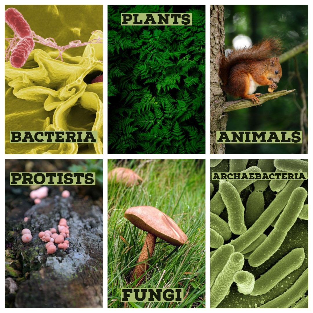
The collage shows a single species in each of the six kingdoms into which all of Earth's living things are commonly classified. How many species are there in each kingdom? In a word: millions. A total of almost two million living species have already been identified, and new species are being discovered all the time. Scientists estimate that there may be as many as 30 million unique species alive on Earth today! Clearly, there is a tremendous variety of life on Earth.
What Is Biodiversity?
Biological diversity, or biodiversity, refers to all of the variety of life that exists on Earth. Biodiversity can be described and measured at three different levels: species diversity, genetic diversity, and ecosystem diversity.
- Species diversity refers to the number of different species in an ecosystem or on Earth as a whole. This is the most common way to measure biodiversity. Current estimates for Earth's total number of living species range from 5 to 30 million species.
- Genetic diversity refers to the variation in genes within all of these species.
- Ecosystem diversity refers to the variety of ecosystems on Earth. An ecosystem is a system formed by populations of many different species interacting with each other and their environment.
https://www.youtube.com/watch?v=GK_vRtHJZu4
Why is Biodiversity So Important? - Kim Preshoff, TEDEd, 2015
Defining a Species
Biodiversity is most often measured by counting species, but what is a species? The answer to that question is not as straightforward as you might think. Formally, a species is defined as a group of actually or potentially interbreeding organisms. This means that members of the same species are similar enough to each other to produce fertile offspring together. By this definition of species, all human beings alive today belong to one species, Homo sapiens. All humans can potentially interbreed with each other, but not with members of any other species.
In the real world, it isn't always possible to make the observations necessary to determine whether or not different organisms can interbreed. For one thing, many species reproduce asexually, so individuals never interbreed — even with members of their own species. When studying extinct species represented by fossils, it is usually impossible to know if different organisms could interbreed. Keep in mind that 99 per cent of all species that have ever existed are now extinct! In practice, many biologists and virtually all paleontologists generally define species on the basis of morphology, rather than breeding behavior. Morphology refers to the form and structure of organisms. For classification purposes, it generally refers to relatively obvious physical traits. Typically, the more similar to one another different organisms appear, the greater the chance that they will be classified in the same species.
Classifying Living Things
People have been trying to classify the tremendous diversity of life on Earth for more than two thousand years. The science of classifying organisms is called taxonomy. Classification is an important step in understanding the present diversity and past evolutionary history of life on Earth. It helps us make sense of the overwhelming diversity of living things.
Linnaean Classification
All modern classification systems have their roots in the Linnaean classification system, which was developed by Swedish botanist Carolus Linnaeus in the 1700s. He tried to classify all living things known in his time by grouping together organisms that s
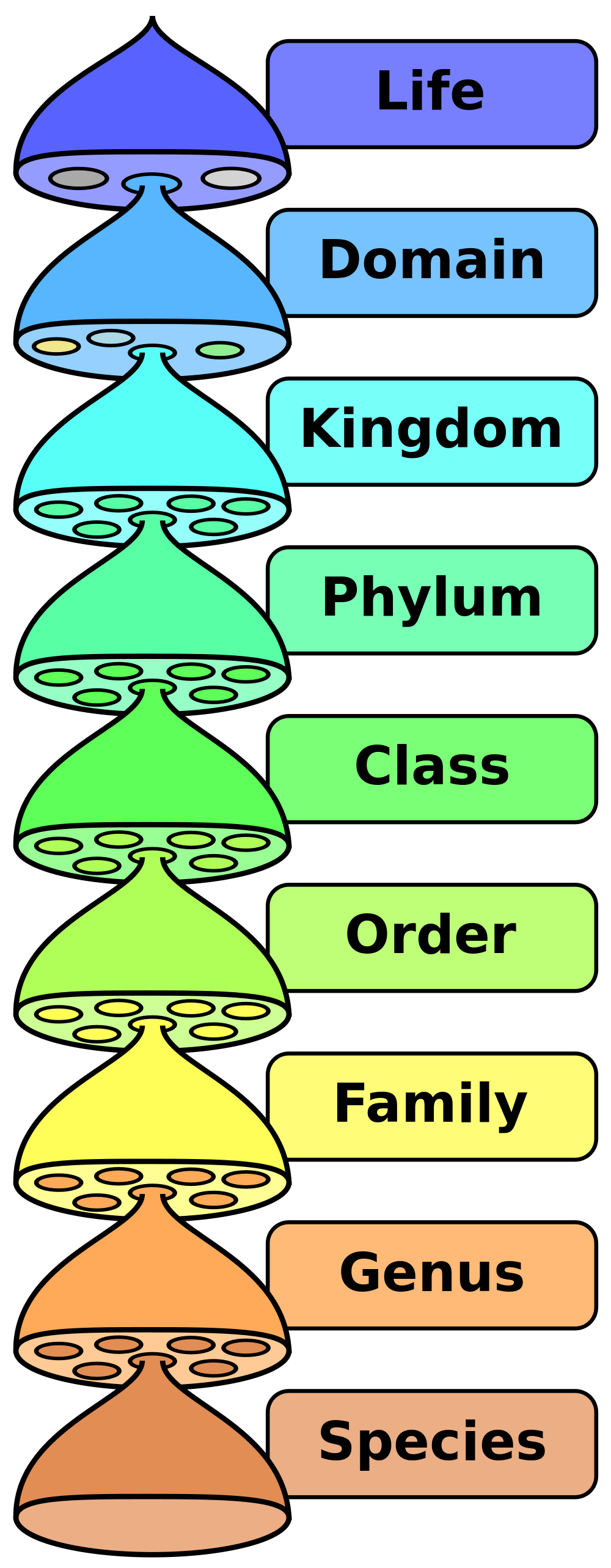
hared obvious morphological traits, such as number of legs or shape of leaves. For his contribution, Linnaeus is known as the “father of taxonomy.”
The Linnaean system of classification consists of a hierarchy of groupings, called taxa (singular, taxon). In the original system, taxa ranged from the kingdom to the species. The kingdom (ex. plant kingdom, animal kingdom) is the largest and most inclusive grouping. It consists of organisms that share just a few basic similarities. The species is the smallest and most exclusive grouping. Ideally, it consists of organisms that are similar enough to interbreed, as discussed above. Similar species are classified together in the same genus (plural, genera), then similar genera are classified together in the same family, and so on, all the way up to the kingdom.
A phrase to help you remember the order of the groupings is shown below. The first letter of each word is the first letter of the level of classification.
Dad Keeps Pots Clean Or Family Gets Sick
The hierarchy of taxa in the original Linnaean system of taxonomy included taxa from the species to the kingdom. The domain was added later.
Binomial Nomenclature
Perhaps the single greatest contribution Linnaeus made to science was his method of naming species. This method, called binomial nomenclature, gives each species a unique, two-word Latin name consisting of the genus name followed by a specific species identifier. An example is Homo sapiens, the two-word Latin name for humans. It literally means “wise human.” This is a reference to our big brains.
Why is having two names so important? It is similar to people having a first and a last name. You may know several people with the first name Michael, but adding Michael’s last name usually pins down exactly which Michael you mean. In the same way, having two names for a species helps to uniquely identify it.
Revisions in the Linnaean Classification
Linnaeus published his classification system in the 1700s. Since then, many new species have been discovered. Scientists can also now classify organisms on the basis of their biochemical and genetic similarities and differences, and not just their outward morphology. These changes have led to revisions in the original Linnaean system of classification.
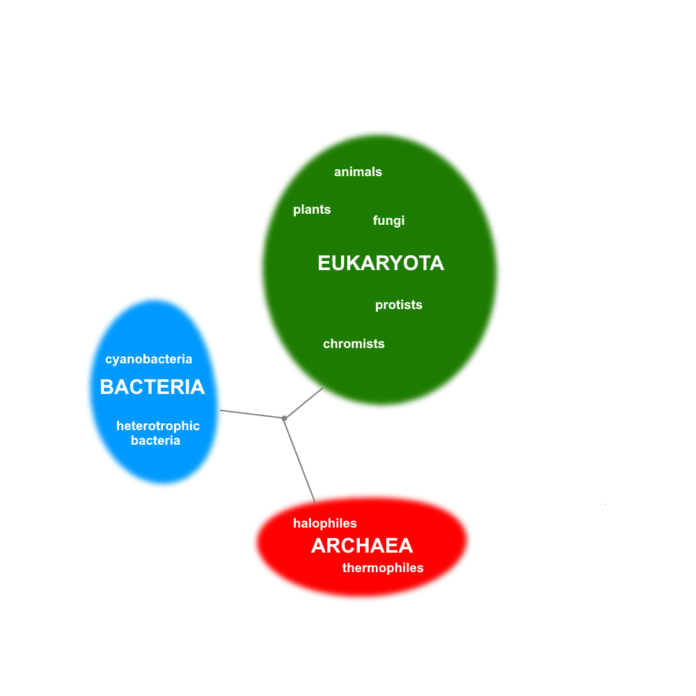
A major change to the Linnaean system is the addition of a new taxon called the domain. The domain is a taxon that is larger and more inclusive than the kingdom, as shown in the figure above. Most biologists agree that there are three domains of life on Earth: Bacteria, Archaea, and Eukarya . Both the Bacteria and the Archaea domains consist of single-celled organisms that lack a nucleus. This means that their genetic material is not enclosed within a membrane inside the cell. The Eukarya domain, in contrast, consists of all organisms whose cells do have a nucleus, so that their genetic material is enclosed within a membrane inside the cell. The Eukarya domain is made up of both single-celled and multicellular organisms. This domain includes several kingdoms, including the animal, plant, fungus, and protist kingdoms.
The three domains of life, as well as how they are related to each other and to a common ancestor. There are several theories about how the three domains are related and which arose first, or from another.
Phylogenetic Classification
Linnaeus classified organisms based on morphology. Basically, organisms were grouped together if they looked alike. After Darwin published his theory of evolution in the 1800s, scientists looked for a way to classify organisms that accounted for phylogeny. Phylogeny is the evolutionary history of a group of related organisms. It is represented by a phylogenetic tree, or some other tree-like diagram, like the one shown above to illustrate the three domains. A phylogenetic tree shows how closely related different groups of organisms are to one another. Each branching point represents a common ancestor of the branching groups.
2.4 Summary
- Biodiversity refers to the variety of life that exists on Earth. It includes species diversity, genetic diversity (within species), and ecosystem diversity.
- The formal biological definition of species is a group of actually or potentially interbreeding organisms. Our own species, Homo sapiens,is an example. In reality, organisms are often classified into species on the basis of morphology.
- A system for classifying living things was introduced by Linnaeus in the 1700s. It includes taxa from the species (least inclusive) to the kingdom (most inclusive). Linnaeus also introduced a system of naming species, which is called binomial nomenclature.
- The domain — a taxon higher than the kingdom — was later added to the Linnaean system. Living things are generally grouped into three domains: Bacteria, Archaea, and Eukarya. The human species and other animal species are placed in the Eukarya domain.
- Modern systems of classification take into account phylogenies, or evolutionary histories of related organisms, rather than just morphological similarities and differences. These relationships are often represented by phylogenetic trees or other tree-like diagrams
2.4 Review Questions
- What is biodiversity? Identify three ways that biodiversity may be measured.
- Define biological species. Why is this definition often difficult to apply?
- Explain why it is important to classify living things, and outline the Linnaean system of classification.
- What is binomial nomenclature? Give an example.
-
- Contrast the Linnaean and phylogenetic systems of classification.
- Describe the taxon called the domain, and compare the three widely recognized domains of living things.
- Based on the phylogenetic tree for the three domains of life above, explain whether you think Bacteria are more closely related to Archaea or Eukarya.
- A scientist discovers a new single-celled organism. Answer the following questions about this discovery.
- If this is all you know, can you place the organism into a particular domain? If so, what is the domain? If not, why not?
- What is one type of information that could help the scientist classify the organism?
- Define morphology. Give an example of a morphological trait in humans.
- Which type of biodiversity is represented in the differences between humans?
- Why do you think it is important to the definition of a species that members of a species can produce fertile offspring?
- Go to the A-Z Animals Animal Classification Page. In the search box, put in your favorite animal and write out it's classification.
2.4 Explore More
https://youtu.be/DVouQRAKxYo
Classification, Amoeba Sisters, 2013.
Attributions
Figure 2.4.1 (6 Kingdoms collage)
- Salmonella, by unknown/ NIAID on Wikimedia Commons is in the public domain (https://en.wikipedia.org/wiki/Public_domain).
- Fern from pxhere, is used under a CC0 1.0 universal public domain dedication license (https://creativecommons.org/publicdomain/zero/1.0/deed.en).
- Photo [squirrel] , by Radoslaw Prekurat on Unsplash is used under the Unsplash License (https://unsplash.com/license).
- Blood Milk Mushroom by Hans on Pixabay is used under the Pixabay License (https://pixabay.com/de/service/license/).
- Fungi by Ste Wright on Unsplash is used under the Unsplash License (https://unsplash.com/license).
- EscherichiaColi NIAID [adapted], by Rocky Mountain Laboratories, ca:NIAID, ca:NIH on Wikimedia Commons is in the public domain (https://en.wikipedia.org/wiki/Public_domain).
Figure 2.4.2
Biological classification, by Pengo [Peter Halasz] on Wikimedia Commons is in the public domain (https://en.wikipedia.org/wiki/Public_domain).
Figure 2.4.3
The three domains of life and major groups within, by C. Miller, 2019, is in the public domain (https://en.wikipedia.org/wiki/Public_domain).
References
Amoeba Sisters. (2017, March 8). Classification. YouTube. https://www.youtube.com/watch?v=DVouQRAKxYo&feature=youtu.be
A-Z Animals. (2008, December 1). Animal classification. https://a-z-animals.com/reference/animal-classification/
TED-Ed. (2015, April 20). Why is biodiversity so important? - Kim Preshoff. YouTube. https://www.youtube.com/watch?v=GK_vRtHJZu4
Wikipedia contributors. (2020, June 21). Carl Linnaeus. Wikipedia. https://en.wikipedia.org/w/index.php?title=Carl_Linnaeus&oldid=963767022
A group of diseases involving abnormal cell growth with the potential to invade or spread to other parts of the body.
Divide and Split
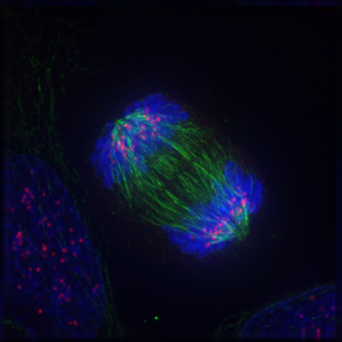
Can you guess what the colourful image in Figure 4.13.1 represents? It shows a eukaryotic cell during the process of cell division. In particular, the image shows the cell in a part of cell division called anaphase, where the DNA is being pulled to opposite ends of the cell. Normally, DNA is located in the nucleus of most human cells. The nucleus divides before the cell itself splits in two, and before the nucleus divides, the cell’s DNA is replicated (or copied). There must be two copies of the DNA so that each daughter cell will have a complete copy of the genetic material from the parent cell. How is the replicated DNA sorted and separated so that each daughter cell gets a complete set of the genetic material? To answer that question, you first need to know more about DNA and the forms it takes.
The Forms of DNA
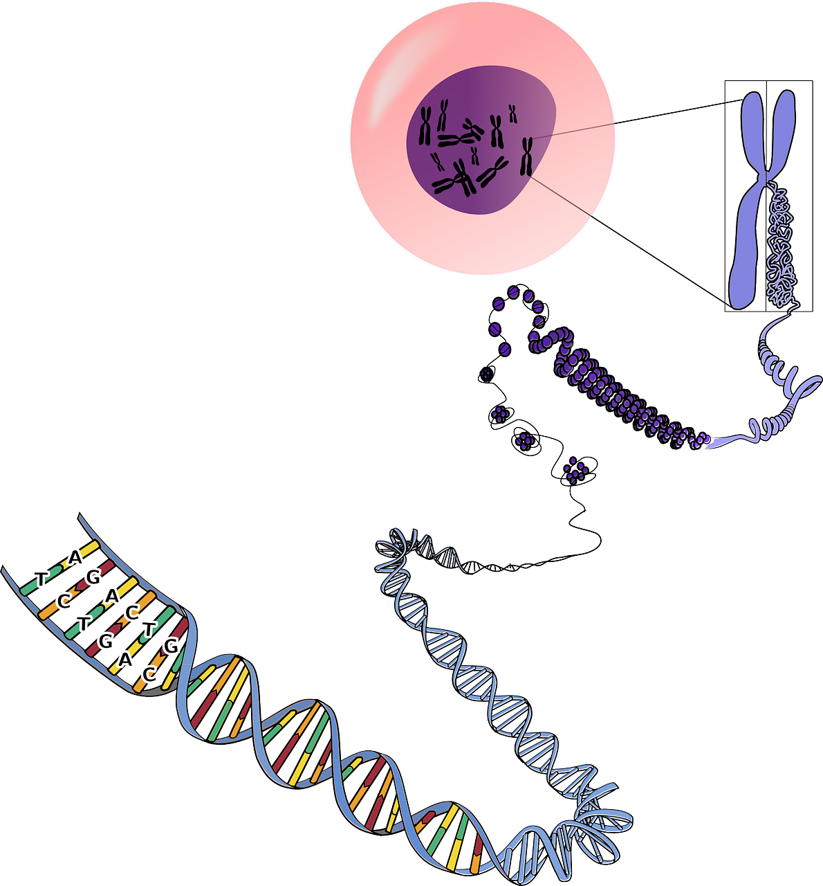
Except when a eukaryotic cell divides, its nuclear DNA exists as a grainy material called chromatin. Only once a cell is about to divide and its DNA has replicated does DNA condense and coil into the familiar X-shaped form of a chromosome, like the one shown below.
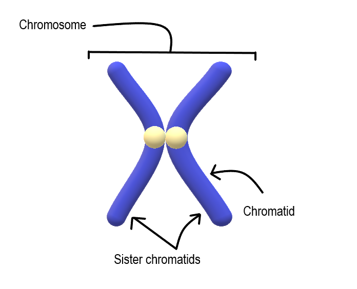
Most cells in the human body have two pairs of 23 different chromosomes, for a total of 46 chromosomes. Cells that have two pairs of chromosomes are called diploid. Because DNA has already replicated when it coils into a chromosome, each chromosome actually consists of two identical structures called sister chromatids. Sister chromatids are joined together at a region called a centromere.
Mitosis
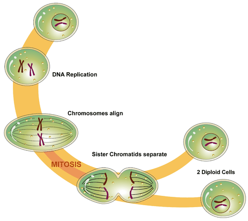
The process in which the nucleus of a eukaryotic cell divides is called mitosis. During mitosis, the two sister chromatids that make up each chromosome separate from each other and move to opposite poles of the cell. This is shown in the figure below.
Mitosis actually occurs in four phases. The phases are called prophase, metaphase, anaphase, and telophase.
Prophase
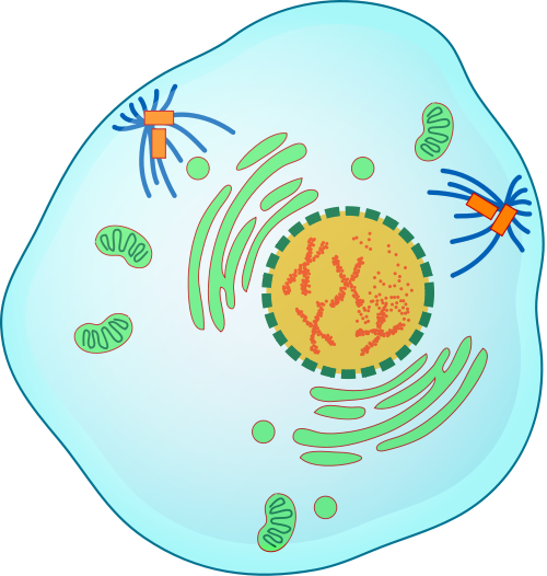
The first and longest phase of mitosis is prophase. During prophase, chromatin condenses into chromosomes, and the nuclear envelope (the membrane surrounding the nucleus) breaks down. In animal cells, the centrioles near the nucleus begin to separate and move to opposite poles of the cell. Centrioles are small organelles found only in eukaryotic cells. They help ensure that the new cells that form after cell division each contain a complete set of chromosomes. As the centrioles move apart, a spindle starts to form between them. The spindle consists of fibres made of microtubules.
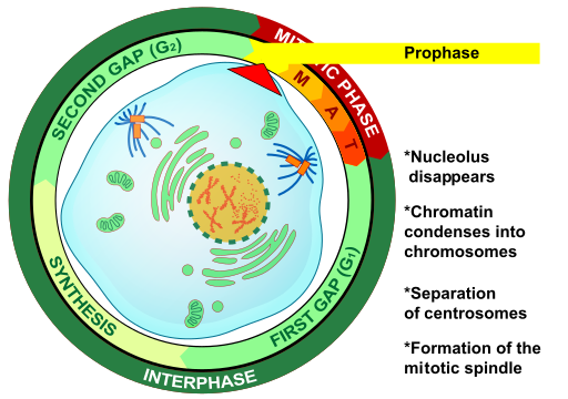
Metaphase
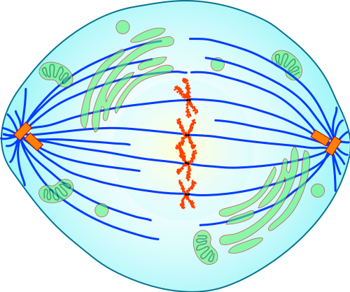
During metaphase, spindle fibres attach to the centromere of each pair of sister chromatids. As you can see in Figure 4.13.7, the sister chromatids line up at the equator (or center) of the cell. The spindle fibres ensure that sister chromatids will separate and go to different daughter cells when the cell divides.
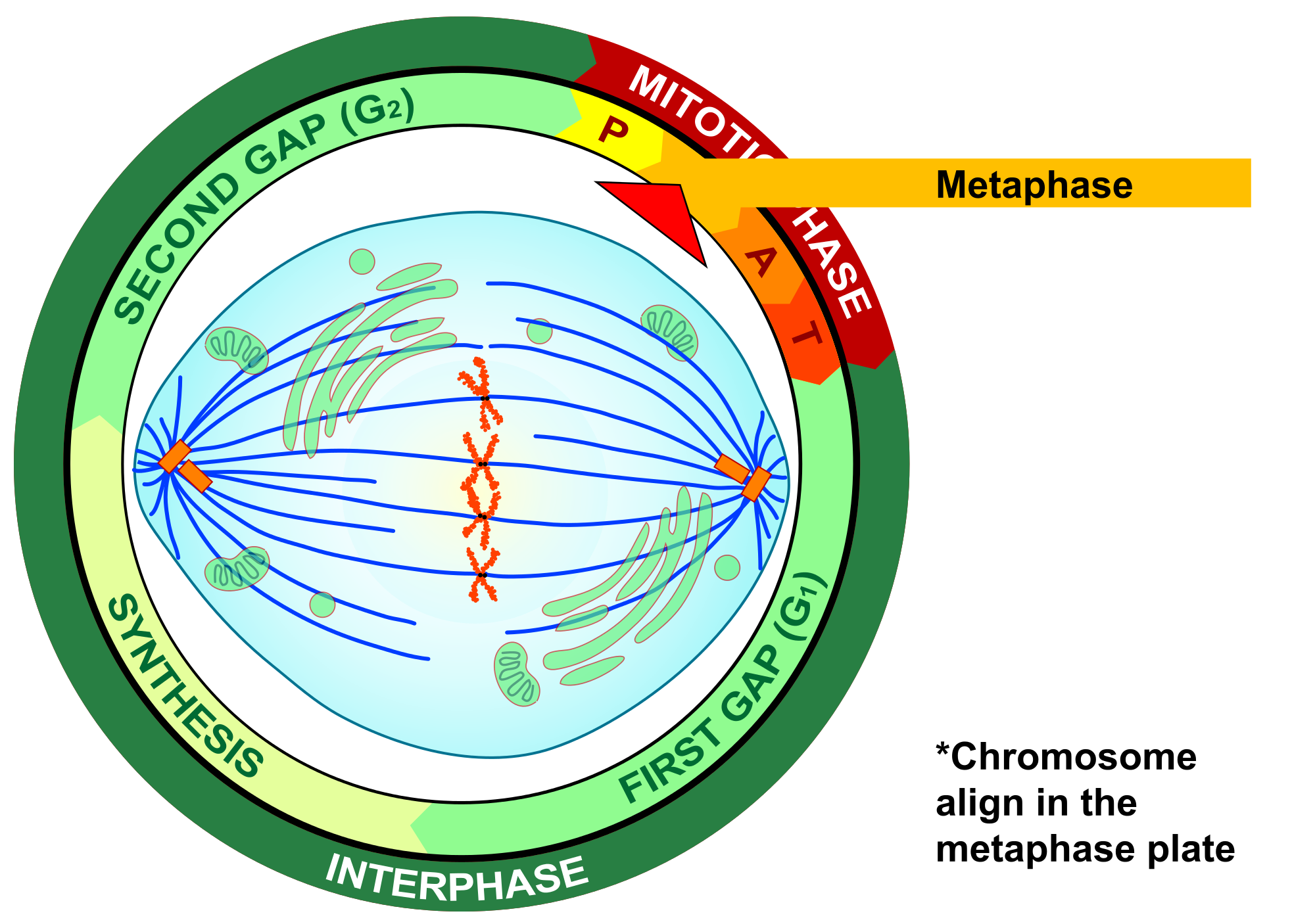
Anaphase
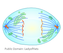
During anaphase, sister chromatids separate and the centromeres divide. The sister chromatids are pulled apart by the shortening of the spindle fibres. This is a little like reeling in a fish by shortening the fishing line. One sister chromatid moves to one pole of the cell, and the other sister chromatid moves to the opposite pole. At the end of anaphase, each pole of the cell has a complete set of chromosomes.
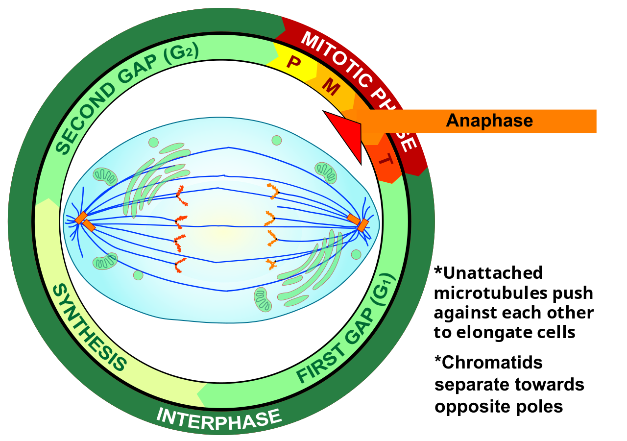
Telophase
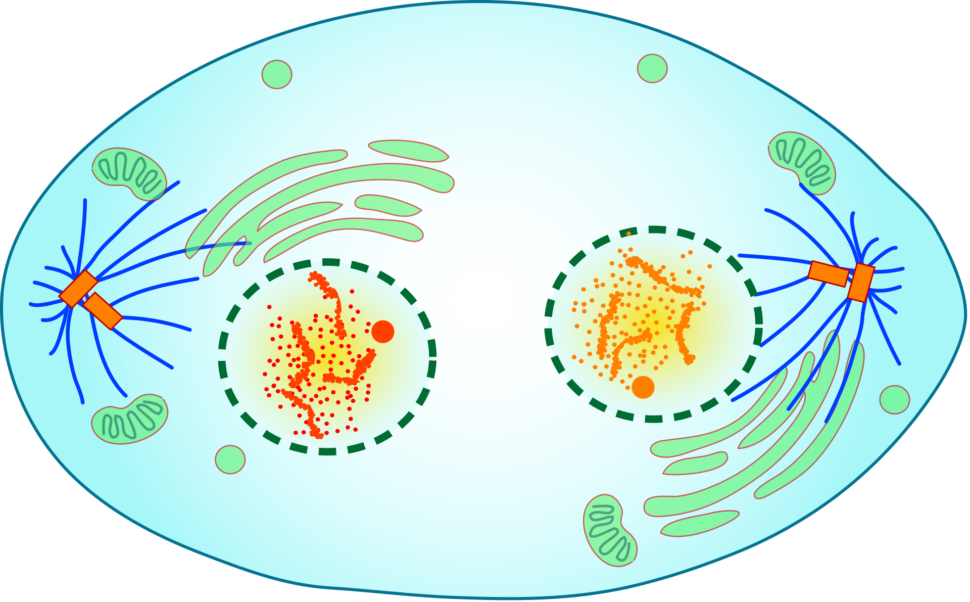
During telophase, the chromosomes begin to uncoil and form chromatin. This prepares the genetic material for directing the metabolic activities of the new cells. The spindle also breaks down, and new nuclear envelopes form.
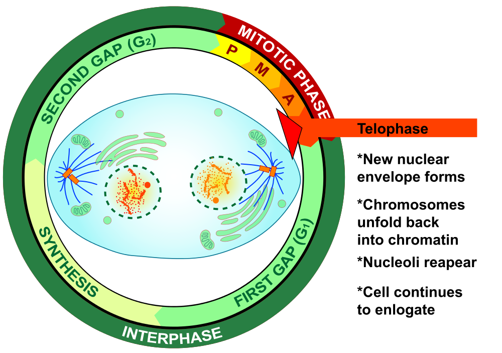
Cytokinesis

Cytokinesis is the final stage of cell division. During cytokinesis, the cytoplasm splits in two and the cell divides, as shown below. In animal cells, the plasma membrane of the parent cell pinches inward along the cell’s equator until two daughter cells form. Thus, the goal of mitosis and cytokinesis is now complete, because one parent cell has given rise to two daughter cells. The daughter cells have the same chromosomes as the parent cell.
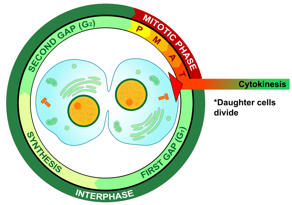
4.13 Summary
- Until a eukaryotic cell divides, its nuclear DNA exists as a grainy material called chromatin. After DNA replicates and the cell is about to divide, the DNA condenses and coils into the X-shaped form of a chromosome. Each chromosome actually consists of two sister chromatids, which are joined together at a centromere.
- Mitosis is the process during which the nucleus of a eukaryotic cell divides. During this process, sister chromatids separate from each other and move to opposite poles of the cell. This happens in four phases: prophase, metaphase, anaphase, and telophase.
- Cytokinesis is the final stage of cell division, during which the cytoplasm splits in two and two daughter cells form.
4.13 Review Questions
- Describe the different forms that DNA takes before and during cell division in a eukaryotic cell.
-
- Identify the four phases of mitosis in an animal cell, and summarize what happens during each phase.
- Order the diagrams of the stages of mitosis:
- Explain what happens during cytokinesis in an animal cell.
- What do you think would happen if the sister chromatids of one of the chromosomes did not separate during mitosis?
- True or False:
4.13 Explore More
https://www.youtube.com/watch?time_continue=3&v=C6hn3sA0ip0&feature=emb_logo
Mitosis, NDSU Virtual Cell Animations project (ndsuvirtualcell), 2012.
https://www.youtube.com/watch?time_continue=19&v=EA0qxhR2oOk&feature=emb_logo
Nondisjunction (Trisomy 21) - An Animated Tutorial, Kristen Koprowski, 2012.
Attributions
Figure 4.13.1
Anaphase_IF by Roy van Heesbeen on Wikimedia Commons is released into the public domain (https://en.wikipedia.org/wiki/Public_domain).
Figure 4.13.2
Chromosomes by OpenClipArt-Vectors on Pixabay is used under the Pixabay License (https://pixabay.com/service/license/).
Figure 4.13.3
Chromosome/ Chromatid/ Sister Chromatid by Christine Miller is released into the public domain (https://en.wikipedia.org/wiki/Public_domain).
Figure 4.13.4
Simple Mitosis by Mariana Ruiz Villarreal [LadyofHats] via CK-12 Foundation is used under a CC BY-NC 3.0 (https://creativecommons.org/licenses/by-nc/3.0/) license.
 ©CK-12 Foundation Licensed under
 ![]() • Terms of Use • Attribution
• Terms of Use • Attribution
Figure 4.13.5
Mitotic Prophase [tiny] by Mariana Ruiz Villarreal [LadyofHats] on Wikimedia Commons is released into the public domain (https://en.wikipedia.org/wiki/Public_domain).
Figure 4.13.6
Prophase Eukaryotic Mitosis by Mariana Ruiz Villarreal [LadyofHats] on Wikimedia Commons is released into the public domain (https://en.wikipedia.org/wiki/Public_domain).
Figure 4.13.7
Mitotic_Metaphase by Mariana Ruiz Villarreal [LadyofHats] on Wikimedia Commons is released into the public domain (https://en.wikipedia.org/wiki/Public_domain).
Figure 4.13.8
Metaphase Eukaryotic Mitosis by Mariana Ruiz Villarreal [LadyofHats] on Wikimedia Commons is released into the public domain (https://en.wikipedia.org/wiki/Public_domain).
Figure 4.13.9
Anaphase [adapted] by Mariana Ruiz Villarreal [LadyofHats] on Wikimedia Commons is released into the public domain (https://en.wikipedia.org/wiki/Public_domain).
Figure 4.13.10
Anaphase_eukaryotic_mitosis.svg by Mariana Ruiz Villarreal [LadyofHats] on Wikimedia Commons is released into the public domain (https://en.wikipedia.org/wiki/Public_domain).
Figure 4.13.11
Mitotic Telophase by Mariana Ruiz Villarreal [LadyofHats] on Wikimedia Commons is released into the public domain (https://en.wikipedia.org/wiki/Public_domain).
Figure 4.13.12
Telophase Eukaryotic Mitosis by Mariana Ruiz Villarreal [LadyofHats] on Wikimedia Commons is released into the public domain (https://en.wikipedia.org/wiki/Public_domain).
Figure 4.13.13
Mitotic Cytokinesis by Mariana Ruiz Villarreal [LadyofHats] on Wikimedia Commons is released into the public domain (https://en.wikipedia.org/wiki/Public_domain).
Figure 4.13.14
Cytokinesis Eukaryotic Mitosis by Mariana Ruiz Villarreal [LadyofHats] on Wikimedia Commons is released into the public domain (https://en.wikipedia.org/wiki/Public_domain).
References
Koprowski, K., Cabey, R. [Kristen Koprowski]. (2012). Nondisjunction (Trisomy 21) - An Animated Tutorial. YouTube. https://www.youtube.com/watch?v=EA0qxhR2oOk&feature=youtu.be
NDSU Virtual Cell Animations project [ndsuvirtualcell]. (2012). Mitosis. YouTube. https://www.youtube.com/watch?v=C6hn3sA0ip0&t=21s
Acquired Immunodeficiency Syndrome - a chronic, potentially life-threatening condition caused by the human immunodeficiency virus (HIV). By damaging your immune system, HIV interferes with your body's ability to fight infection and disease.
A condition in which you don't have enough healthy red blood cells to carry adequate oxygen to the body's tissues resulting in symptoms including weakness and fatigue.
Created by CK12/Adapted by Christine Miller
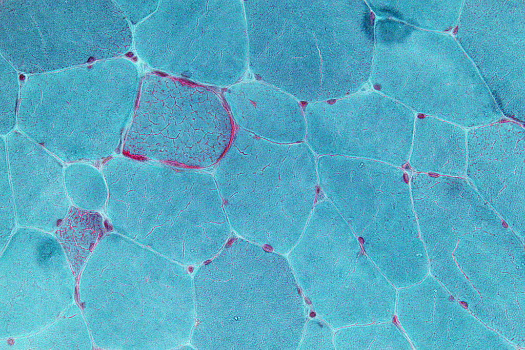
Jasmin discovered that her extreme fatigue, muscle pain, vision problems, and vomiting were due to problems in her mitochondria, like the damaged mitochondria shown in red in Figure 4.14.1. Mitochondria are small, membrane-bound organelles found in eukaryotic cells that provide energy for the cells of the body. They do this by carrying out the final two steps of aerobic cellular respiration: the Krebs cycle and electron transport. This is the major way that the human body breaks down the sugar glucose from food into a form of energy cells can use, namely the molecule ATP.
Because mitochondria provide energy for cells, you can understand why Jasmin was experiencing extreme fatigue, particularly after running. Her damaged mitochondria could not keep up with her need for energy, particularly after intense exercise, which requires a lot of additional energy. What is perhaps not so obvious are the reasons for her other symptoms, such as blurry vision, muscle spasms, and vomiting. All of the cells in the body require energy in order to function properly. Mitochondrial diseases can cause problems in mitochondria in any cell of the body, including muscle cells and cells of the nervous system, which includes the brain and nerves. The nervous system and muscles work together to control vision and digestive system functions, such as vomiting, so when they are not functioning properly, a variety of symptoms can emerge. This also explains why Jasmin’s niece, who has a similar mitochondrial disease, has symptoms related to brain function, such as seizures and learning disabilities. Our cells are microscopic, and mitochondria are even tinier — but they are essential for the proper functioning of our bodies. When they are damaged, serious health effects can occur.

One seemingly confusing aspect of mitochondrial diseases is that the type of symptoms, severity of symptoms, and age of onset can vary wildly between people — even within the same family! In Jasmin’s case, she did not notice symptoms until adulthood, while her niece had more severe symptoms starting at a much younger age. This makes sense when you know more about how mitochondrial diseases work.
Inherited mitochondrial diseases can be due to damage in either the DNA in the nucleus of cells or in the DNA in the mitochondria themselves. Recall that mitochondria are thought to have evolved from prokaryotic organisms that were once free-living, but were then infected or engulfed by larger cells. One of the pieces of evidence that supports this endosymbiotic theory is that mitochondria have their own, separate DNA. When the mitochondrial DNA is damaged (or mutated) it can result in some types of mitochondrial diseases. However, these mutations do not typically affect all of the mitochondria in a cell. During cell division, organelles such as mitochondria are replicated and passed down to the new daughter cells. If some of the mitochondria are damaged, and others are not, the daughter cells can have different amounts of damaged mitochondria. This helps explain the wide range of symptoms in people with mitochondrial diseases — even ones in the same family — because different cells in their bodies are affected in varying degrees. Jasmin’s niece was affected strongly and her symptoms were noticed early, while Jasmin’s symptoms were more mild and did not become apparent until adulthood.
There is still much more that needs to be discovered about the different types of mitochondrial diseases. But by learning about cells, their organelles, how they obtain energy, and how they divide, you should now have a better understanding of the biology behind these diseases.
Apply your understanding of cells to your own life. Can you think of other diseases that affect cellular structures or functions. Do they affect people you know? Since your entire body is made of cells, when cells are damaged or not functioning properly, it can cause a wide variety of health problems.
Chapter 4 Summary
Type your learning objectives here.
In this chapter you learned many facts about cells. Specifically, you learned that:
- Cells are the basic units of structure and function of living things.
- The first cells were observed from cork by Hooke in the 1600s. Soon after, van Leeuwenhoek observed other living cells.
- In the early 1800s, Schwann and Schleiden theorized that cells are the basic building blocks of all living things. Around 1850, Virchow saw cells dividing, and added his own theory that living cells arise only from other living cells. These ideas led to cell theory, which states that all organisms are made of cells, all life functions occur in cells, and all cells come from other cells.
- The invention of the electron microscope in the 1950s allowed scientists to see organelles and other structures inside cells for the first time.
- There is variation in cells, but all cells have a plasma membrane, cytoplasm, ribosomes, and DNA.
-
- The plasma membrane is composed mainly of a bilayer of phospholipid molecules and forms a barrier between the cytoplasm inside the cell and the environment outside the cell. It allows only certain substances to pass in or out of the cell. Some cells have extensions of their plasma membrane with other functions, such as flagella or cilia.
- Cytoplasm is a thick solution that fills a cell and is enclosed by the plasma membrane. It helps give the cell shape, holds organelles, and provides a site for many of the biochemical reactions inside the cell. The liquid part of the cytoplasm is called cytosol.
- Ribosomes are small structures where proteins are made.
- Cells are usually very small, so they have a large enough surface area-to-volume ratio to maintain normal cell processes. Cells with different functions often have different shapes.
- Prokaryotic cells do not have a nucleus. Eukaryotic cells have a nucleus, as well as other organelles. An organelle is a structure within the cytoplasm of a cell that is enclosed within a membrane and performs a specific job.
- The cytoskeleton is a highly organized framework of protein filaments and tubules that criss-cross the cytoplasm of a cell. It gives the cell shape and helps to hold cell structures (such as organelles) in place.
- The nucleus is the largest organelle in a eukaryotic cell. It is considered to be the cell's control center, and it contains DNA and controls gene expression, including which proteins the cell makes.
- The mitochondrion is an organelle that makes energy available to cells. According to the widely accepted endosymbiotic theory, mitochondria evolved from prokaryotic cells that were once free-living organisms that infected or were engulfed by larger prokaryotic cells.
- The endoplasmic reticulum (ER) is an organelle that helps make and transport proteins and lipids. Rough endoplasmic reticulum (RER) is studded with ribosomes. Smooth endoplasmic reticulum (SER) has no ribosomes.
- The Golgi apparatus is a large organelle that processes proteins and prepares them for use both inside and outside the cell. It is also involved in the transport of lipids around the cell.
- Vesicles and vacuoles are sac-like organelles that may be used to store and transport materials in the cell or as chambers for biochemical reactions. Lysosomes and peroxisomes are vesicles that break down foreign matter, dead cells, or poisons.
- Centrioles are organelles located near the nucleus that help organize the chromosomes before cell division so each daughter cell receives the correct number of chromosomes.
- There are two basic ways that substances can cross the cell’s plasma membrane: passive transport (which requires no energy expenditure by the cell) and active transport (which requires energy).
- No energy is needed from the cell for passive transport because it occurs when substances move naturally from an area of higher concentration to an area of lower concentration. Types of passive transport in cells include:
-
- Simple diffusion, which is the movement of a substance due to differences in concentration without any help from other molecules. This is how very small, hydrophobic molecules, such as oxygen and carbon dioxide, enter and leave the cell.
- Osmosis, which is the diffusion of water molecules across the membrane.
- Facilitated diffusion, which is the movement of a substance across a membrane due to differences in concentration, but only with the help of transport proteins in the membrane (such as channel proteins or carrier proteins). This is how large or hydrophilic molecules and charged ions enter and leave the cell.
- Active transport requires energy to move substances across the plasma membrane, often because the substances are moving from an area of lower concentration to an area of higher concentration or because of their large size. Two examples of active transport are the sodium-potassium pump and vesicle transport.
-
- The sodium-potassium pump moves sodium ions out of the cell and potassium ions into the cell, both against a concentration gradient, in order to maintain the proper concentrations of both ions inside and outside the cell and to thereby control membrane potential.
- Vesicle transport uses vesicles to move large molecules into or out of cells.
- Energy is the ability to do work. It is needed by every living cell to carry out life processes.
- The form of energy that living things need is chemical energy, and it comes from food. Food consists of organic molecules that store energy in their chemical bonds.
- Autotrophs (producers) make their own food. Think of plants that make food by photosynthesis. Heterotrophs (consumers) obtain food by eating other organisms.
- Organisms mainly use the molecules glucose and ATP for energy. Glucose is the compact, stable form of energy that is carried in the blood and taken up by cells. ATP contains less energy and is used to power cell processes.
- The flow of energy through living things begins with photosynthesis, which creates glucose. The cells of organisms break down glucose and make ATP.
- Cellular respiration is the aerobic process by which living cells break down glucose molecules, release energy, and form molecules of ATP. Overall, this three-stage process involves glucose and oxygen reacting to form carbon dioxide and water.
-
- Glycolysis, the first stage of cellular respiration, takes place in the cytoplasm. In this step, enzymes split a molecule of glucose into two molecules of pyruvate, which releases energy that is transferred to ATP.
- Transition Reaction takes place between glycolysis and Krebs Cycle. It is a very short reaction in which the pyruvate molecules from glycolysis are converted into Acetyl CoA in order to enter the Krebs Cycle.
- Krebs Cycle, the second stage of cellular respiration, takes place in the matrix of a mitochondrion. During this stage, two turns through the cycle result in all of the carbon atoms from the two pyruvate molecules forming carbon dioxide and the energy from their chemical bonds being stored in a total of 16 energy-carrying molecules (including four from glycolysis).
- The Electron Transport System, he third stage of cellular respiration, takes place on the inner membrane of the mitochondrion. Electrons are transported from molecule to molecule down an electron-transport chain. Some of the energy from the electrons is used to pump hydrogen ions across the membrane, creating an electrochemical gradient that drives the synthesis of many more molecules of ATP.
- In all three stages of aerobic cellular respiration combined, as many as 38 molecules of ATP are produced from just one molecule of glucose.
- Some organisms can produce ATP from glucose by anaerobic respiration, which does not require oxygen. Fermentation is an important type of anaerobic process. There are two types: alcoholic fermentation and lactic acid fermentation. Both start with glycolysis.
-
- Alcoholic fermentation is carried out by single-celled organisms, including yeasts and some bacteria. We use alcoholic fermentation in these organisms to make biofuels, bread, and wine.
- Lactic acid fermentation is undertaken by certain bacteria, including the bacteria in yogurt, and also by our muscle cells when they are worked hard and fast.
- Anaerobic respiration produces far less ATP (typically produces 2 ATP) than does aerobic cellular respiration, but it has the advantage of being much faster.
- The cell cycle is a repeating series of events that includes growth, DNA synthesis, and cell division.
- In a eukaryotic cell, the cell cycle has two major phases: interphase and mitotic phase. During interphase, the cell grows, performs routine life processes, and prepares to divide. During mitotic phase, first the nucleus divides (mitosis) and then the cytoplasm divides (cytokinesis), which produces two daughter cells.
-
- Until a eukaryotic cell divides, its nuclear DNA exists as a grainy material called chromatin. After DNA replicates and the cell is about to divide, the DNA condenses and coils into the X-shaped form of a chromosome. Each chromosome consists of two sister chromatids, which are joined together at a centromere.
- During mitosis, sister chromatids separate from each other and move to opposite poles of the cell. This happens in four phases: prophase, metaphase, anaphase, and telophase.
- The cell cycle is controlled mainly by regulatory proteins that signal the cell to either start or delay the next phase of the cycle at key checkpoints.
- Cancer is a disease that occurs when the cell cycle is no longer regulated, often because the cell's DNA has become damaged. Cancerous cells grow out of control and may form a mass of abnormal cells called a tumor.
In this chapter, you learned about cells and some of their functions, as well as how they pass genetic material in the form of DNA to their daughter cells. In the next chapter, you will learn how DNA is passed down to offspring, which causes traits to be inherited. These traits may be innocuous (such as eye colour) or detrimental (such as mutations that cause disease). The study of how genes are passed down to offspring is called genetics, and as you will learn in the next chapter, this is an interesting topic that is highly relevant to human health.
Chapter 4 Review
- Sequence:
- Drag and Drop:
- True or False:
- Multiple Choice:
- Briefly explain how the energy in the food you eat gets there, and how it provides energy for your neurons in the form necessary to power this process.
- Explain why the inside of the plasma membrane — the side that faces the cytoplasm of the cell — must be hydrophilic.
- Explain the relationships between interphase, mitosis, and cytokinesis.
Attributions
Figure 4.14.1
Mitochondrial Disease muscle sample by Nephron is used under a CC BY-SA 3.0 (https://creativecommons.org/licenses/by-sa/3.0) license.
Figure 4.14.2
Aunt and Niece by Tatiana Rodriguez on Unsplash is used under the Unsplash License (https://unsplash.com/license).
Reference
Wikipedia contributors. (2020, June 6). Mitochondrial disease. In Wikipedia. https://en.wikipedia.org/w/index.php?title=Mitochondrial_disease&oldid=961126371
Created by CK-12 Foundation/Adapted by Christine Miller

Fashion Statement
This colourful hairstyle makes quite a fashion statement. Many people spend a lot of time and money on their hair, even if they don’t have an exceptional hairstyle like this one. Besides its display value, hair actually has important physiological functions.
What is Hair?
Hair is a filament that grows from a hair follicle in the dermis of the skin. It consists mainly of tightly packed, keratin-filled cells called keratinocytes. The human body is covered with hair follicles, with the exception of a few areas, including the mucous membranes, lips, palms of the hands, and soles of the feet.
Structure of Hair
The part of the hair located within the follicle is called the hair root. The root is the only living part of the hair. The part of the hair that is visible above the surface of the skin is the hair shaft. The shaft of the hair has no biochemical activity and is considered dead.
Follicle and Root
Hair growth begins inside a follicle (see Figure 10.5.2 below). Each hair follicle contains stem cells that can keep dividing, which allows hair to grow. The stem cells can also regrow a new hair after one falls out. Another structure associated with a hair follicle is a sebaceous gland that produces oily sebum. The sebum lubricates and helps to waterproof the hair. A tiny arrector pili muscle is also attached to the follicle. When it contracts, the follicle moves, and the hair in the follicle stands up.
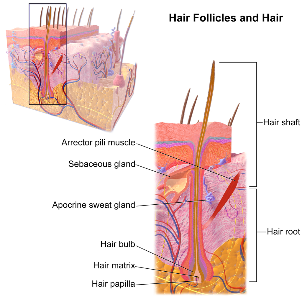
Shaft
The hair shaft is a hard filament that may grow very long. Hair normally grows in length by about half an inch a month. In cross-section, a hair shaft can be divided into three zones, called the cuticle, cortex, and medulla.
- The cuticle (or outer coat) is the outermost zone of the hair shaft. It consists of several layers of flat, thin keratinocytes that overlap one another like shingles on a roof. This arrangement helps the cuticle repel water. The cuticle is also covered with a layer of lipids, just one molecule thick, which increases its ability to repel water. This is the zone of the hair shaft that is visible to the eye.
- The cortex is the middle zone of the hair shaft, and it is also the widest part. The cortex is highly structured and organized, consisting of keratin bundles in rod-like structures. These structures give hair its mechanical strength. The cortex also contains melanin, which gives hair its colour.
- The medulla is the innermost zone of the hair shaft. This is a small, disorganized, and more open area at the center of the hair shaft. The medulla is not always present. When it is present, it contains highly pigmented cells full of keratin.
Characteristics of Hair
Two visible characteristics of hair are its colour and texture. In adult males, the extent of balding is another visible characteristic. All three characteristics are genetically controlled.
Hair Colour
All natural hair colours are the result of melanin, which is produced in hair follicles and packed into granules in the hair. Two forms of melanin are found in human hair: eumelanin and pheomelanin. Eumelanin is the dominant pigment in brown hair and black hair, and pheomelanin is the dominant pigment in red hair. Blond hair results when you have only a small amount of melanin in the hair. Gray and white hair occur when melanin production slows down, and eventually stops.
Figure 10.5.3 Variation in hair colouration. Which types of melanin are present for each hair colour shown?
Hair Texture
Hair exists in a variety of textures. The main aspects of hair texture are the curl pattern, thickness, and consistency.
- The shape of the hair follicle determines the shape of the hair shaft. The shape of the hair shaft, in turn, determines the curl pattern of the hair. Round hair shafts produce straight hair. Hair shafts that are oval or have other shapes produce wavy or curly hair .
- The size of the hair follicle determines the thickness of hair. Thicker hair has greater volume than thinner hair.
- The consistency of hair is determined by the hair follicle volume and the condition of the hair shaft. The consistency of hair is generally classified as fine, medium, or coarse. Fine hair has the smallest circumference, and coarse hair has the largest circumference. Medium hair falls in between these two extremes. Coarse hair also has a more open cuticle than thin or medium hair does, which causes it to be more porous.

Figure 10.5.4 Curly hair has a differently shaped shaft than straight hair.
Functions of Hair
In humans, one function of head hair is to provide insulation and help the head retain heat. Head hair also protects the skin on the head from damage by UV light.
The function of hair in other locations on the body is debated. One idea is that body hair helps keep us warm in cold weather. When the body is too cold, arrector pili muscles contract and cause hairs to stand up (shown in Figure 10.5.5), trapping a layer of warm air above the epidermis. However, this is more effective in mammals that have thick hair or fur than it is in relatively hairless human beings.
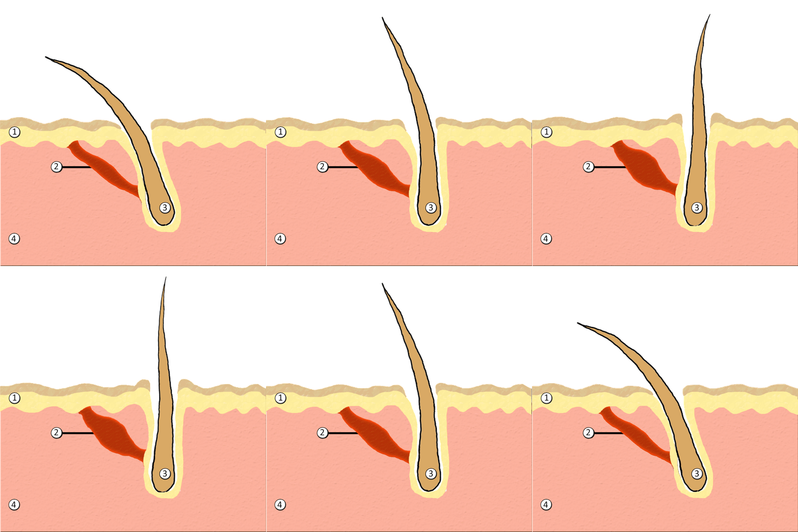
Human hair has an important sensory function, as well. Sensory receptors in the hair follicles can sense when the hair moves, whether it moves because of a breeze, or because of the touch of a physical object. The receptors may also provide sensory awareness of the presence of parasites on the skin.

Some hairs, such as the eyelashes, are especially sensitive to the presence of potentially harmful matter. The eyelashes grow at the edge of the eyelid and can sense when dirt, dust, or another potentially harmful object is too close to the eye. The eye reflexively closes as a result of this sensation. The eyebrows also provide some protection to the eyes. They protect the eyes from dirt, sweat, and rain. In addition, the eyebrows play a key role in nonverbal communication (see Figure 10.5.6). They help express emotions such as sadness, anger, surprise, and excitement.
Hair in Human Evolution
Among mammals, humans are nearly unique in having undergone significant loss of body hair during their evolution. Humans are also unlike most other mammals in having curly hair as one variation in hair texture. Even non-human primates (see Figure 10.5.7) all have straight hair. This suggests that curly hair evolved at some point during human evolution.
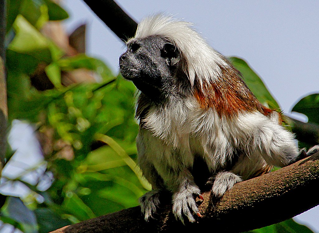
Loss of Body Hair
One hypothesis for the loss of body hair in the human lineage is that it would have facilitated cooling of the body by the evaporation of sweat. Humans also evolved far more sweat glands than other mammals, which is consistent with this hypothesis, because sweat evaporates more quickly from less hairy skin. Another hypothesis for human hair loss is that it would have led to fewer parasites on the skin. This might have been especially important when humans started living together in larger, more crowded social groups.
These hypotheses may explain why we lost body hair, but they can’t explain why we didn’t also lose head hair and hair in the pubic region and armpits. It is possible that head hair was retained because it protected the scalp from UV light. As our bipedal ancestors walked on the open savannas of equatorial Africa, the skin on the head would have been an area exposed to the most direct rays of sunlight in an upright hominid. Pubic and armpit hair may have been retained because they served as signs of sexual maturity, which would have been important for successful mating and reproduction.
Evolution of Curly Hair
Greater protection from UV light has also been posited as a possible selective agent favoring the evolution of curly hair. Researchers have found that straight hair allows more light to pass into the body through the hair shaft via the follicle than does curly hair. In this way, human hair is like a fibre optic cable. It allows light to pass through easily when it is straight, but it impedes the passage of light when it is kinked or coiled. This is indirect evidence that UV light may have been a selective agent leading to the evolution of curly hair.
Social and Cultural Significance of Hair
Hair has great social significance for human beings. Body hair is an indicator of biological sex, because hair distribution is sexually dimorphic. Adult males are generally hairier than adult females, and facial hair in particular is a notable secondary male sex characteristic. Hair may also be an indicator of age. White hair is a sign of older age in both males and females, and male pattern baldness is a sign of older age in males. In addition, hair colour and texture can be a sign of ethnic ancestry.
Hair also has great cultural significance. Hairstyle and colour may be an indicator of social group membership and for better or worse can be associated with specific stereotypes. Head shaving has been used in many times and places as a punishment, especially for women. On the other hand, in some cultures, cutting off one’s hair symbolizes liberation from one’s past. In other cultures, it is a sign of mourning. There are also many religious-based practices involving hair. For example, the majority of Muslim women hide their hair with a headscarf. Sikh men grow their hair long and cover it with a turban. Amish men (like the one pictured in Figure 10.5.8) grow facial hair only after they marry — but just a beard, and not a mustache.
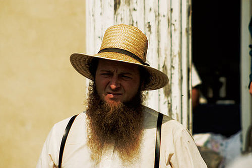
Unfortunately, sometimes hairstyle, colour and characteristics are used to apply stereotypes, particularly with respect to women. "Blonde jokes" are a good example of how negative stereotypes are maintained despite having no actual truth behind them. Many stereotypes related to hair are hidden, even from persons perpetrating the stereotype. Often a hairstyle is judged by another as having ties to gender, sexuality, worldview and/or socioeconomic status; even when these inferences are woefully inaccurate. It is important to be aware of our own biases and determine if these biases are appropriate - take a look at the collage in Figure 10.5.9. What are your initial reactions? Are these reactions founded in fact? Do you harbor an unfair bias?
Figure 10.5.9 What are your biases? Are they fair?
10.5 Summary
- Hair is a filament that grows from a hair follicle in the dermis of the skin. It consists mainly of tightly packed, keratin-filled cells called keratinocytes. The human body is almost completely covered with hair follicles.
- The part of a hair that is within the follicle is the hair root. This is the only living part of a hair. The part of a hair that is visible above the skin surface is the hair shaft. It consists of dead cells.
- Hair growth begins inside a follicle when stem cells within the follicle divide to produce new keratinocytes. An individual hair may grow to be very long.
- A hair shaft has three zones: the outermost zone called the cuticle; the middle zone called the cortex; and the innermost zone called the medulla.
- Genetically controlled, visible characteristics of hair include hair colour, hair texture, and the extent of balding in adult males. Melanin (eumelanin and/or pheomelanin) is the pigment that gives hair its colour. Aspects of hair texture include curl pattern, thickness, and consistency.
- Functions of head hair include providing insulation and protecting skin on the head from UV light. Hair everywhere on the body has an important sensory function. Hair in eyelashes and eyebrows protects the eyes from dust, dirt, sweat, and other potentially harmful substances. The eyebrows also play a role in nonverbal communication.
- Among mammals, humans are nearly unique in having undergone significant loss of body hair during their evolution, probably because sweat evaporates more quickly from less hairy skin. Curly hair also is thought to have evolved at some point during human evolution, perhaps because it provided better protection from UV light.
- Hair has social significance for human beings, because it is an indicator of biological sex, age, and ethnic ancestry. Human hair also has cultural significance. Hairstyle may be an indicator of social group membership, for example.
10.5 Review Questions
-
- Compare and contrast the hair root and hair shaft.
- Describe hair follicles.
-
-
- Explain variation in human hair colour.
- What factors determine the texture of hair?
- Describe two functions of human hair.
- What hypotheses have been proposed for the loss of body hair during human evolution?
- Discuss the social and cultural significance of human hair.
- Describe one way in which hair can be used as a method of communication in humans.
- Explain why waxing or tweezing body hair, which typically removes hair down to the root, generally keeps the skin hair-free for a longer period of time than shaving, which cuts hair off at the surface of the skin.
10.5 Explore More
https://www.youtube.com/watch?v=8diYLhl8bWU
Why do some people go bald? - Sarthak Sinha, TED-Ed, 2015.
https://www.youtube.com/watch?v=kNw8V_Fkw28
Hair Love | Oscar®-Winning Short Film (Full) | Sony Pictures Animation, 2019.
https://www.youtube.com/watch?v=hDW5e3NR1Cw
Why do we care about hair | Naomi Abigail | TEDxBaDinh, TEDx Talks, 2015.
Attributions
Figure 10.5.1
Hair by jessica-dabrowski-TETR8YLSqt4 [photo] by Jessica Dabrowski on Unsplash is used under the Unsplash License (https://unsplash.com/license).
Figure 10.5.2
Blausen_0438_HairFollicleAnatomy_02 by BruceBlaus on Wikimedia Commons is used under a CC BY 3.0 (https://creativecommons.org/licenses/by/3.0) license.
Figure 10.5.3
- Standing tall by Ilaya Raja on Unsplash is used under the Unsplash License (https://unsplash.com/license).
- Blond-haired woman smiling by Carlos Lindner on Unsplash is used under the Unsplash License (https://unsplash.com/license).
- Smith Mountain Lake redhead by Chris Ross Harris on Unsplash is used under the Unsplash License (https://unsplash.com/license).
- Through the look of experience by Laura Margarita Cedeño Peralta on Unsplash is used under the Unsplash License (https://unsplash.com/license).
Figure 10.5.4
Curly hair by chris-benson-clvEami9RN4 [photo] by Chris Benson on Unsplash is used under the Unsplash License (https://unsplash.com/license).
Figure 10.5.5
1024px-PilioerectionAnimation by AnthonyCaccese on Wikimedia Commons is used under a CC BY-SA 4.0 (https://creativecommons.org/licenses/by-sa/4.0/deed.en) license.
Figure 10.5.6
Pout by alexander-dummer-Em8I8Z_DwA4 [photo] by Alexander Dummer on Unsplash is used under the Unsplash License (https://unsplash.com/license).
Figure 10.5.7
Cotton_top_tamarin_monkey._(12046035746) by Bernard Spragg. NZ, from Christchurch, New Zealand on Wikimedia Commons is used under a CC0 1.0 Universal
Public Domain Dedication license (https://creativecommons.org/publicdomain/zero/1.0/deed.en).
Figure 10.5.8
Amish hairstyle by CK-12 Foundation is used under a CC BY-NC 3.0 (https://creativecommons.org/licenses/by-nc/3.0/) license.
 ©CK-12 Foundation Licensed under
 ![]() • Terms of Use • Attribution
• Terms of Use • Attribution
Figure 10.5.9
- Rainbow Hair Bubble Man by Behrouz Jafarnezhad on Unsplash is used under the Unsplash License (https://unsplash.com/license).
- Pink hair in Atlanta, United States by Tammie Allen on Unsplash is used under the Unsplash License (https://unsplash.com/license).
- Magdalena 2 by Valerie Elash on Unsplash is used under the Unsplash License (https://unsplash.com/license).
- Perfect Style by Daria Volkova on Unsplash is used under the Unsplash License (https://unsplash.com/license)
- Stay Classy by Fayiz Musthafa on Unsplash is used under the Unsplash License (https://unsplash.com/license)
- Take your time by Jan Tinneberg on Unsplash is used under the Unsplash License (https://unsplash.com/license)
References
Blausen.com staff. (2014). Medical gallery of Blausen Medical 2014. WikiJournal of Medicine 1 (2). DOI:10.15347/wjm/2014.010. ISSN 2002-4436.
Brainard, J/ CK-12 Foundation. (2016). Figure 7 This style of facial hair is adopted by most Amish men after they marry [digital image]. In CK-12 College Human Biology (Section 12.5) [online Flexbook]. CK12.org. https://www.ck12.org/book/ck-12-college-human-biology/section/12.5/
Sony Pictures Animation. (2019, December 5). Hair love | Oscar®-winning short film (Full) | Sony Pictures Animation. YouTube. https://www.youtube.com/watch?v=kNw8V_Fkw28
TED-Ed. (2015, August 25). Why do some people go bald? – Sarthak Sinha. YouTube. https://www.youtube.com/watch?v=8diYLhl8bWU
TEDx Talks. (2015, February 4). Why do we care about hair | Naomi Abigail | TEDxBaDinh. YouTube. https://www.youtube.com/watch?v=hDW5e3NR1Cw
Created by CK-12 Foundation/Adapted by Christine Miller
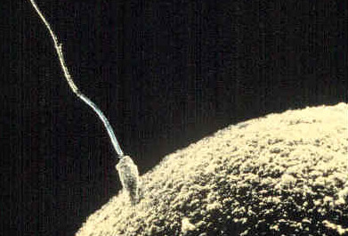
It’s All about Sex
A tiny sperm from dad breaks through the surface of a huge egg from mom. Voilà! In nine months, a new son or daughter will be born. Like most other multicellular organisms, human beings reproduce sexually. In human sexual reproduction, males produce sperm and females produce eggs, and a new offspring forms when a sperm unites with an egg. How do sperm and eggs form? And how do they arrive together at the right place and time so they can unite to form a new offspring? These are functions of the reproductive system.
What Is the Reproductive System?
The reproductive system is the human organ system responsible for the production and fertilization of gametes (sperm or eggs) and, in females, the carrying of a fetus. Both male and female reproductive systems have organs called gonads that produce gametes. A gamete is a haploid cell that combines with another haploid gamete during fertilization, forming a single diploid cell called a zygote. Besides producing gametes, the gonads also produce sex hormones. Sex hormones are endocrine hormones that control the development of sex organs before birth, sexual maturation at puberty, and reproduction once sexual maturation has occurred. Other reproductive system organs have various functions, such as maturing gametes, delivering gametes to the site of fertilization, and providing an environment for the development and growth of an offspring.
Sex Differences in the Reproductive System
The reproductive system is the only human organ system that is significantly different between males and females. Embryonic structures that will develop into the reproductive system start out the same in males and females, but by birth, the reproductive systems have differentiated. How does this happen?
Sex Differentiation
Starting around the seventh week after conception in genetically male (XY) embryos, a gene called SRY on the Y chromosome (shown in Figure 18.2.2) initiates the production of multiple proteins. These proteins cause undifferentiated gonadal tissue to develop into male gonads (testes). The male gonads then secrete hormones — including the male sex hormone testosterone — that trigger other changes in the developing offspring (now called a fetus), causing it to develop a complete male reproductive system. Without a Y chromosome, an embryo will develop female gonads (ovaries) that will produce the female sex hormone estrogen. Estrogen, in turn, will lead to the formation of the other organs of a normal female reproductive system.
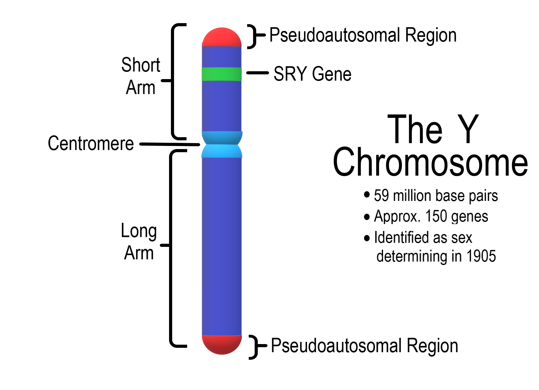
Homologous Structures
Undifferentiated embryonic tissues develop into different structures in male and female fetuses. Structures that arise from the same tissues in males and females are called homologous structures. The male testes and female ovaries, for example, are homologous structures that develop from the undifferentiated gonads of the embryo. Likewise, the male penis and female clitoris are homologous structures that develop from the same embryonic tissues.
Sex Hormones and Maturation
Male and female reproductive systems are different at birth, but they are immature and incapable of producing gametes or sex hormones. Maturation of the reproductive system occurs during puberty, when hormones from the hypothalamus and pituitary gland stimulate the testes or ovaries to start producing sex hormones again. The main sex hormones are testosterone in males and estrogen in females. Sex hormones, in turn, lead to the growth and maturation of the reproductive organs, rapid body growth, and the development of secondary sex characteristics. Secondary sex characteristics are traits that are different in mature males and females, but are not directly involved in reproduction. They include facial hair in males and breasts in females.
Male Reproductive System
The main structures of the male reproductive system are external to the body and illustrated in Figure 18.2.3. The two testes (singular, testis) hang between the thighs in a sac of skin called the scrotum. The testes produce both sperm and testosterone. Resting atop each testis is a coiled structure called the epididymis (plural, epididymes). The function of the epididymes is to mature and store sperm. The penis is a tubular organ that contains the urethra and has the ability to stiffen during sexual arousal. Sperm passes out of the body through the urethra during a sexual climax (orgasm). This release of sperm is called ejaculation.
In addition to these organs, the male reproductive system consists of several ducts and glands that are internal to the body. The ducts, which include the vas deferens (also called the ductus deferens), transport sperm from the epididymis to the urethra. The glands, which include the prostate gland and seminal vesicles, produce fluids that become part of semen. Semen is the fluid that carries sperm through the urethra and out of the body. It contains substances that control pH and provide sperm with nutrients for energy.
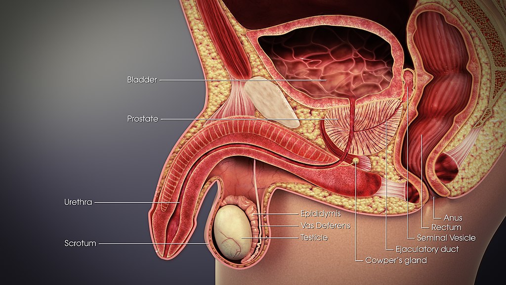
Female Reproductive System
The main structures of the female reproductive system are internal to the body and shown in the following figure. They include the paired ovaries, which are small, ovoid structures that produce ova and secrete estrogen. The two oviducts (sometimes called Fallopian tubes or uterine tubes) start near the ovaries and end at the uterus. Their function is to transport ova from the ovaries to the uterus. If an egg is fertilized, it usually occurs while it is traveling through an oviduct. The uterus is a pear-shaped muscular organ that functions to carry a fetus until birth. It can expand greatly to accommodate a growing fetus, and its muscular walls can contract forcefully during labour to push the baby out of the uterus and into the vagina. The vagina is a tubular tract connecting the uterus to the outside of the body. The vagina is where sperm are usually deposited during sexual intercourse and ejaculation. The vagina is also called the birth canal because a baby travels through the vagina to leave the body during birth.
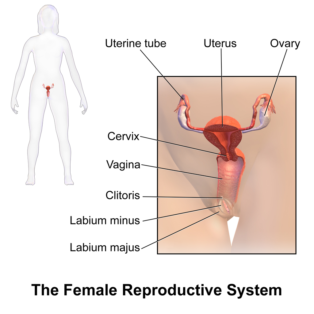
The external structures of the female reproductive system are referred to collectively as the vulva. They include the clitoris, which is homologous to the male penis. They also include two pairs of labia (singular, labium), which surround and protect the openings of the urethra and vagina.
18.2 Summary
- The reproductive system is the human organ system responsible for the production and fertilization of gametes and, in females, the carrying of a fetus.
- Both male and female reproductive systems have organs called gonads (testes in males, ovaries in females) that produce gametes (sperm or ova) and sex hormones (such as testosterone in males and estrogen in females). Sex hormones are endocrine hormones that control the prenatal development of reproductive organs, sexual maturation at puberty, and reproduction after puberty.
- The reproductive system is the only organ system that is significantly different between males and females. A Y-chromosome gene called SRY is responsible for undifferentiated embryonic tissues developing into a male reproductive system. Without a Y chromosome, the undifferentiated embryonic tissues develop into a female reproductive system.
- Structures such as testes and ovaries that arise from the same undifferentiated embryonic tissues in males and females are called homologous structures.
- Male and female reproductive systems are different at birth, but at that point, they are immature and nonfunctioning. Maturation of the reproductive system occurs during puberty, when hormones from the hypothalamus and pituitary gland stimulate the gonads to produce sex hormones again. The sex hormones, in turn, cause the changes of puberty.
- Male reproductive system organs include the testes, epididymis, penis, vas deferens, prostate gland, and seminal vesicles.
- Female reproductive system organs include the ovaries, oviducts, uterus, vagina, clitoris, and labia.
18.2 Review Questions
- What is the reproductive system?
-
- Explain the difference between the vulva and the vagina.
18.2 Explore More
https://youtu.be/kMWxuF9YW38
Sex Determination: More Complicated Than You Thought, TED-Ed, 2012.
https://youtu.be/vcPJkz-D5II
The evolution of animal genitalia - Menno Schilthuizen, TED-Ed, 2017.
https://youtu.be/l5knvmy1Z3s
Hormones and Gender Transition, Reactions, 2015.
Attributions
Figure 18.2.1
Sperm-egg by Unknown author on Wikimedia Commons is in the public domain (https://en.wikipedia.org/wiki/public_domain).
Figure 18.2.2
Y Chromosome by Christinelmiller on Wikimedia Commons is used under a CC BY-SA 4.0 (https://creativecommons.org/licenses/by-sa/4.0) license.
Figure 18.2.3
3D_Medical_Animation_Vas_Deferens by https://www.scientificanimations.com on Wikimedia Commons is used under a CC BY-SA 4.0 (https://creativecommons.org/licenses/by-sa/4.0) license.
Figure 18.2.4
Blausen_0399_FemaleReproSystem_01 by BruceBlaus on Wikimedia Commons is used under a CC BY 3.0 (https://creativecommons.org/licenses/by/3.0) license.
References
Blausen.com Staff. (2014). Medical gallery of Blausen Medical 2014. WikiJournal of Medicine 1 (2). DOI:10.15347/wjm/2014.010. ISSN 2002-4436.
Reactions. (2015, June 8). Hormones and gender transition. YouTube. https://www.youtube.com/watch?v=l5knvmy1Z3s&feature=youtu.be
TED-Ed. (2012, April 23). Sex determination: More complicated than you thought. YouTube. https://www.youtube.com/watch?v=kMWxuF9YW38&feature=youtu.be
TED-Ed. (2017, April 24). The evolution of animal genitalia - Menno Schilthuizen. YouTube. https://www.youtube.com/watch?v=vcPJkz-D5II&feature=youtu.be
Who's Who?
Figure 3.7.1 Identical twins show clearly the importance of genes in making us who we are. Genes would not be possible without nucleic acids.
What Are Nucleic Acids?
Nucleic acids are the class of biochemical compounds that includes DNA and RNA. These molecules are built of small monomers called nucleotides. Many nucleotides bind together to form a chain called a polynucleotide. The nucleic acid DNA (deoxyribonucleic acid) consists of two polynucleotide chains or strands. Thus, DNA is sometimes called double-stranded. The nucleic acid RNA (ribonucleic acid) consists of just one polynucleotide chain or strand, so RNA is sometimes called single-stranded.
Structure of Nucleic Acids
Each nucleotide consists of three smaller molecules:
- A sugar molecule (the sugar deoxyribose in DNA and the sugar ribose in RNA)
- A phosphate group
- A nitrogen base
The nitrogen bases in a nucleic acid stick out from the backbone. There are four different nitrogen bases: cytosine, adenine, guanine, and either thymine (in DNA) or uracil (in RNA). In DNA, bonds form between bases on the two nucleotide chains and hold the chains together. Each type of base binds with just one other type of base: cytosine always binds with guanine, and adenine always binds with thymine. These pairs of bases are called complementary base pairs.
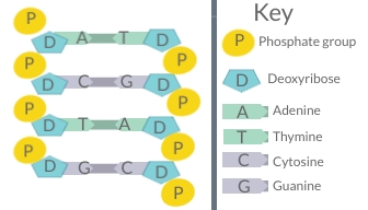
As you can see in Figure 3.7.2, sugars and phosphate groups form the backbone of a polynucleotide chain. Hydrogen bonds between complementary bases hold the two polynucleotide chains together.
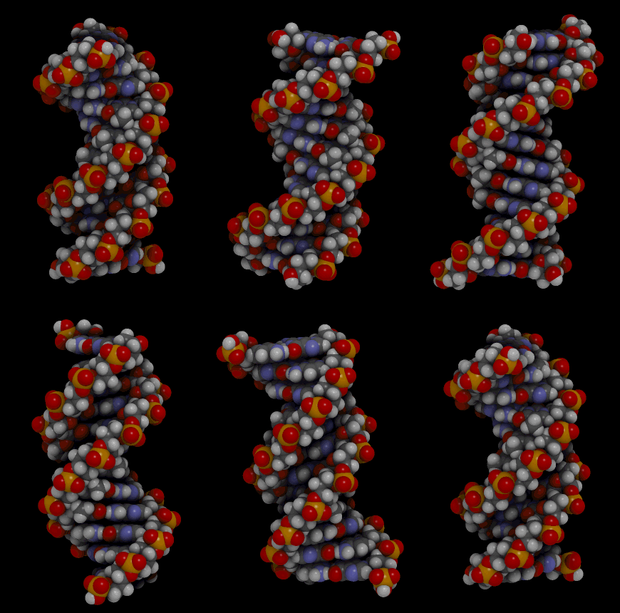
The binding of complementary bases causes DNA molecules automatically to take their well-known double helix shape, which is shown in the animation in Figure 3.7.3. A double helix is like a spiral staircase. It forms naturally and is very strong, making the two polynucleotide chains difficult to break apart.
DNA Molecule. Hydrogen bonds between complementary bases help form the double helix of a DNA molecule. The letters A, T, G, and C stand for the bases adenine, thymine, guanine, and cytosine. The sequence of these four bases in DNA is a code that carries instructions for making proteins. Shown is a representation of how the double helix folds into a chromosome.
Roles of Nucleic Acids
DNA makes up genes, and the sequence of bases in DNA makes up the genetic code. Between “starts” and “stops,” the code carries instructions for the correct sequence of amino acids in a protein. RNA uses the information in DNA to assemble the correct amino acids and help make the protein. The information in DNA is passed from parent cells to daughter cells whenever cells divide, and it is also passed from parents to offspring when organisms reproduce. This is how inherited characteristics are passed from one generation to the next.
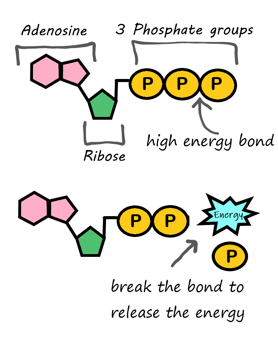
ATP is Energy
There is one type of specialized nucleic acid that exists only as a monomer. It stands apart from the other nucleic acids because it does not code for, or help create, proteins. This molecule is ATP, which stands for adenosine triphosphate. It consists of a sugar, adenosine, and three phosphate groups. It's primary role is as the basic energy currency in the cell. The way ATP works is all based on the phosphates. As shown in Figure 3.7.4, a large amount of energy is stored in the bond between the second and third phosphate group. When this bond is broken, it functions as an exothermic reaction and this energy can be used to power other processes taking place in the cell.
3.7 Summary
- Nucleic acids are the class of biochemical compounds that includes DNA and RNA. These molecules are built of small monomers called nucleotides, which bind together in long chains to form polynucleotides. DNA consists of two polynucleotides, and RNA consists of one polynucleotide.
- Each nucleotide consists of a sugar molecule, phosphate group, and nitrogen base. Sugars and phosphate groups of adjacent nucleotides bind together to form the "backbone" of the polynucleotide. Nitrogen bases jut out to the side of the sugar-phosphate backbone. Bonds between complementary bases hold together the two polynucleotide chains of DNA and cause it to take on its characteristic double helix shape.
- DNA makes up genes, and the sequence of nitrogen bases in DNA makes up the genetic code for the synthesis of proteins. RNA helps synthesize proteins in cells. The genetic code in DNA is also passed from parents to offspring during reproduction, which explains how inherited characteristics are passed from one generation to the next.
3.7 Review Questions
- What are nucleic acids?
- How does RNA differ structurally from DNA? Draw a picture of each.
- Describe a nucleotide. Explain how nucleotides bind together to form a polynucleotide.
- What role do nitrogen bases in nucleotides play in the structure and function of DNA?
- What is a function of RNA?
- Using what you learned in this article about nucleic acids, explain why twins look so similar.
-
- What are the nucleotides on the complementary strand of DNA below?
- Arrange the following in order from the smallest to the largest level of organization: DNA, nucleotide, polynucleotide.
- As part of the DNA replication process, the two polynucleotide chains are separated from each other, but each individual chain remains intact. What type of bonds are broken in this process?
- Adenine, guanine, cytosine, and thymine are _______________.
- Some diseases and disorders are caused by genes. Explain why these genetic disorders can be passed down from parents to their children.
- Are there any genetic disorders that run in your family?
3.7 Explore More
https://www.youtube.com/watch?v=aeAL6xThfL8
DNA: The book of you - Joe Hanson, TED-Ed, 2012.
Attributions
Figure 3.7.1
- Twins sitting next to each other by Craig Adderley on Pexels is used under the Pexels license (https://www.pexels.com/license/).
- Photograph Of Women Wearing Strip Shirt by Paul Bonafide Eferiano on Pexels is used under the Pexels license (https://www.pexels.com/license/).
- Two guys sitting on a beach by Daria Shevtsova on Pexels is used under the Pexels license (https://www.pexels.com/license/).
- Children Twins Girls Young Nicaraguan Portrait by skeeze on Pixabay is used under the Pixabay License (https://pixabay.com/service/license/).
Figure 3.7.2
DNA-diagram by Christine Miller [Christinelmiller] on Wikimedia Commons, is used under a CC BY 4.0 (https://creativecommons.org/licenses/by/4.0) license.
Figure 3.7.3
Bdna_cropped [gif] by Spiffistan, derivative work: Jahobr, on Wikimedia Commons, is released into the public domain (https://en.wikipedia.org/wiki/Public_domain).
Figure 3.7.4
ATP for energy by Christine Miller is used under a CC BY 4.0 (https://creativecommons.org/licenses/by/4.0/) license.
Reference
TED-Ed. (2012, November 26). DNA: The book of you - Joe Hanson. YouTube, 2012. https://www.youtube.com/watch?v=aeAL6xThfL8&feature=youtu.be

What Makes You...You?
This young woman has naturally red hair (Figure 5.3.1). Why is her hair red instead of some other colour? In general, what gives her the specific traits she has? There is a molecule in human beings and most other living things that is largely responsible for their traits. The molecule is large and has a spiral structure in eukaryotes. What molecule is it? With these hints, you probably know that the molecule is DNA.
Introducing DNA
Today, it is commonly known that DNA is the genetic material that is passed from parents to offspring and determines our traits. For a long time, scientists knew such molecules existed — that is, they were aware that genetic information is contained within biochemical molecules. What they didn’t know was which specific molecules play this role. In fact, for many decades, scientists thought that proteins were the molecules that contain genetic information.
Discovery that DNA is the Genetic Material
Determining that DNA is the genetic material was an important milestone in biology. It took many scientists undertaking creative experiments over several decades to show with certainty that DNA is the molecule that determines the traits of organisms. This research began in the early part of the 20th century.
Griffith's Experiments with Mice
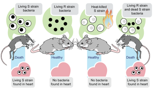
One of the first important discoveries was made in the 1920s by an American scientist named Frederick Griffith. Griffith was studying mice and two different strains of a bacterium, called R (rough)-strain and S (smooth)-strain. He injected the two bacterial strains into mice. The S-strain was virulent and killed the mice, whereas the R-strain was not virulent and did not kill the mice. You can see these details in Figure 5.3.2. Griffith also injected mice with S-strain bacteria that had been killed by heat. As expected, the dead bacteria did not harm the mice. However, when the dead S-strain bacteria were mixed with live R-strain bacteria and injected, the mice died.
Based on his observations, Griffith deduced that something in the dead S-strain was transferred to the previously harmless R-strain, making the R-strain deadly. What was this "something?" What type of substance could change the characteristics of the organism that received it?
Avery and His Colleagues Make a Major Contribution
In the early 1940s, a team of scientists led by Canadian-American Oswald Avery tried to answer the question raised by Griffith’s research results. First, they inactivated various substances in the S-strain bacteria. Then they killed the S-strain bacteria and mixed the remains with live R-strain bacteria. (Keep in mind that the R-strain bacteria normally did not harm the mice.) When they inactivated proteins, the R-strain was deadly to the injected mice. This ruled out proteins as the genetic material. Why? Even without the S-strain proteins, the R-strain was changed (or transformed) into the deadly strain. However, when the researchers inactivated DNA in the S-strain, the R-strain remained harmless. This led to the conclusion that DNA — and not protein — is the substance that controls the characteristics of organisms. In other words, DNA is the genetic material.
Hershey and Chase Confirm the Results
The conclusion that DNA is the genetic material was not widely accepted until it was confirmed by additional research. In the 1950s, Alfred Hershey and Martha Chase did experiments with viruses and bacteria. Viruses are not cells. Instead, they are basically DNA (or RNA) inside a protein coat. To reproduce, a virus must insert its own genetic material into a cell (such as a bacterium). Then, it uses the cell’s machinery to make more viruses. The researchers used different radioactive elements to label the DNA and proteins in DNA viruses. This allowed them to identify which molecule the viruses inserted into bacterial cells. DNA was the molecule they identified. This confirmed that DNA is the genetic material.
Chargaff Focuses on DNA Bases
Other important discoveries about DNA were made in the mid-1900s by Erwin Chargaff. He studied DNA from many different species and was especially interested in the four different nitrogen bases of DNA: adenine (A), guanine (G), cytosine (C), and thymine (T). Chargaff found that concentrations of the four bases differed between species. Within any given species, however, the concentration of adenine was always the same as the concentration of thymine, and the concentration of guanine was always the same as the concentration of cytosine. These observations came to be known as Chargaff’s rules. The significance of the rules would not be revealed until the double-helix structure of DNA was discovered.
Discovery of the Double Helix
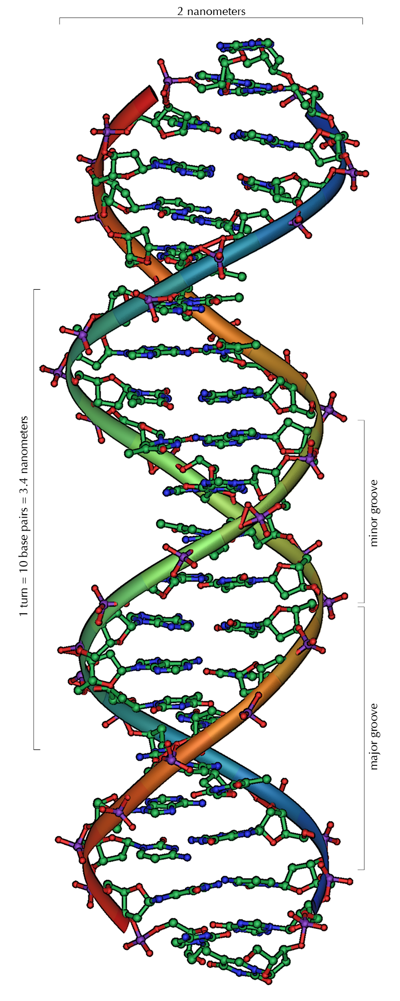
After DNA was shown to be the genetic material, scientists wanted to learn more about its structure and function. James Watson and Francis Crick are usually given credit for discovering that DNA has a double helix shape, as shown in Figure 5.3.3. In fact, Watson and Crick's discovery of the double helix depended heavily on the prior work of Rosalind Franklin and other scientists, who had used X-rays to learn more about DNA’s structure. Unfortunately, Franklin and these others have not always been given credit for their important contributions to the discovery of the double helix.
The DNA molecule has a double helix shape — the same basic shape as a spiral staircase. Do you see the resemblance? Which parts of the DNA molecule are like the steps of the spiral staircase?
The double helix shape of DNA, along with Chargaff’s rules, led to a better understanding of DNA. As a nucleic acid, DNA is made from nucleotide monomers. Long chains of nucleotides form polynucleotides, and the DNA double helix consists of two polynucleotide chains. Each nucleotide consists of a sugar (deoxyribose), a phosphate group, and one of the four bases (adenine, cytosine, guanine, or thymine). The sugar and phosphate molecules in adjacent nucleotides bond together and form the "backbone" of each polynucleotide chain.
Scientists concluded that bonds between the bases hold together the two polynucleotide chains of DNA. Moreover, adenine always bonds with thymine, and cytosine always bonds with guanine. That's why these pairs of bases are called complementary base pairs. Adenine and guanine have a two-ring structure, whereas cytosine and thymine have just one ring. If adenine were to bond with guanine, as well as thymine, for example, the distance between the two DNA chains would vary. When a one-ring molecule (like thymine) always bonds with a two-ring molecule (like adenine), however, the distance between the two chains remains constant. This maintains the uniform shape of the DNA double helix. The bonded base pairs (A-T and G-C) stick into the middle of the double helix, forming the "steps" of the spiral staircase.
5.3 Summary
- Determining that DNA is the genetic material was an important milestone in biology. One of the first important discoveries was made in the 1920s, when Griffith showed that something in virulent bacteria could be transferred to nonvirulent bacteria, making them virulent, as well.
- In the early 1940s, Avery and colleagues showed that the "something" Griffith found in his research was DNA and not protein. This result was confirmed by Hershey and Chase, who demonstrated that viruses insert DNA into bacterial cells so the cells will make copies of the viruses.
- In the mid-1950s, Chargaff showed that, within the DNA of any given species, the concentration of adenine is always the same as the concentration of thymine, and that the concentration of guanine is always the same as the concentration of cytosine. These observations came to be known as Chargaff's rules.
- Around the same time, James Watson and Francis Crick, building on the prior X-ray research of Rosalind Franklin and others, discovered the double-helix structure of the DNA molecule. Along with Chargaff's rules, this led to a better understanding of DNA's structure and function.
- Knowledge of DNA's structure helped scientists understand how DNA replicates, which must occur before cell division occurs so each daughter cell will have a complete set of chromosomes.
5.3 Review Questions
- Outline the discoveries that led to the determination that DNA (not protein) is the biochemical molecule that contains genetic information.
- State Chargaff's rules. Explain how the rules are related to the structure of the DNA molecule.
- Explain how the structure of a DNA molecule is like a spiral staircase. Which parts of the staircase represent the various parts of the molecule?
-
- Why do you think dead S-strain bacteria injected into mice did not harm the mice, but killed them when mixed with living (and normally harmless) R-strain bacteria?
- In Griffith’s experiment, do you think the heat treatment that killed the bacteria also inactivated the bacterial DNA? Why or why not?
- Give one example of the specific evidence that helped rule out proteins as genetic material.
5.3 Explore More
https://www.youtube.com/watch?v=V6bKn34nSbk
The Discovery of the Structure of DNA, OpenMind, 2017.
https://www.youtube.com/watch?time_continue=5&v=JiME-W58KpU&feature=emb_logo
Rosalind Franklin: Great Minds, SciShow, 2013.
Attributions
Figure 5.3.1
Redhead [photo] by Hichem Dahmani on Unsplash is used under the Unsplash License (https://unsplash.com/license).
Figure 5.3.2
Griffith’s mice by Mariana Ruiz Villarreal [LadyofHats] for CK-12 Foundation is used under a
CC BY-NC 3.0 (https://creativecommons.org/licenses/by-nc/3.0/) license.
 ©CK-12 Foundation Licensed under
 ![]() • Terms of Use • Attribution
• Terms of Use • Attribution
Figure 5.3.3
DNA_Overview by Michael Ströck [mstroeck] on Wikimedia Commons is used under a CC BY SA 3.0 (http://creativecommons.org/licenses/by-sa/3.0/) license.
References
Brainard, J/ CK-12. (2012). Concentration. In Physical Science [website]. CK12.org. https://www.ck12.org/c/physical-science/concentration/?referrer=crossref
OpenMind. (2017, September 11). The discovery of the structure of DNA. YouTube. https://www.youtube.com/watch?v=V6bKn34nSbk&feature=youtu.be
SciShow. (2013, July 9). Rosalind Franklin: Great minds. YouTube. https://www.youtube.com/watch?v=JiME-W58KpU&feature=youtu.be
Wikipedia contributors. (2020, June 27). Alfred Hershey. In Wikipedia. https://en.wikipedia.org/w/index.php?title=Alfred_Hershey&oldid=964789559
Wikipedia contributors. (2020, June 5). Erwin Chargaff. In Wikipedia. https://en.wikipedia.org/w/index.php?title=Erwin_Chargaff&oldid=960942873
Wikipedia contributors. (2020, June 29). Francis Crick. In Wikipedia. https://en.wikipedia.org/w/index.php?title=Francis_Crick&oldid=965135362
Wikipedia contributors. (2020, July 6). Frederick Griffith. In Wikipedia. https://en.wikipedia.org/w/index.php?title=Frederick_Griffith&oldid=966352134
Wikipedia contributors. (2020, July 5). James Watson. In Wikipedia. https://en.wikipedia.org/w/index.php?title=James_Watson&oldid=966111944
Wikipedia contributors. (2020, March 31). Martha Chase. In Wikipedia. https://en.wikipedia.org/w/index.php?title=Martha_Chase&oldid=948408219
Wikipedia contributors. (2020, July 2). Oswald Avery. In Wikipedia. https://en.wikipedia.org/w/index.php?title=Oswald_Avery&oldid=965632585
Wikipedia contributors. (2020, June 30). Rosalind Franklin. In Wikipedia. https://en.wikipedia.org/w/index.php?title=Rosalind_Franklin&oldid=965334881
Image shows a pictomicrograph of a protozoan parasite of the Giardia lamblia species. It is roughly cone-shaped, with several flagella trailing from the narrow end of it.

