14.4 Blood Vessels

Bulging Veins
Why do bodybuilders have such prominent veins? Bulging muscles push surface veins closer to the skin. Combine that with a virtual lack of subcutaneous fat, and you have bulging veins, as well as bulging muscles. Veins are one of three major types of blood vessels in the cardiovascular system.
Types of Blood Vessels
Blood vessels are the part of the cardiovascular system that transports blood throughout the human body. There are three major types of blood vessels. Besides veins, they include arteries and capillaries.
Arteries
Arteries are defined as blood vessels that carry blood away from the heart. Blood flows through arteries largely because it is under pressure from the pumping action of the heart. It should be noted that coronary arteries, which supply heart muscle cells with blood, travel toward the heart, but not as part of the blood flow that travels through the chambers of the heart. Most arteries, including coronary arteries, carry oxygenated blood, but there are a few exceptions, most notably the pulmonary artery. This artery carries deoxygenated blood from the heart to the lungs, where it picks up oxygen and releases carbon dioxide. In virtually all other arteries, the hemoglobin in red blood cells is highly saturated with oxygen (95–100 per cent). These arteries distribute oxygenated blood to tissues throughout the body.
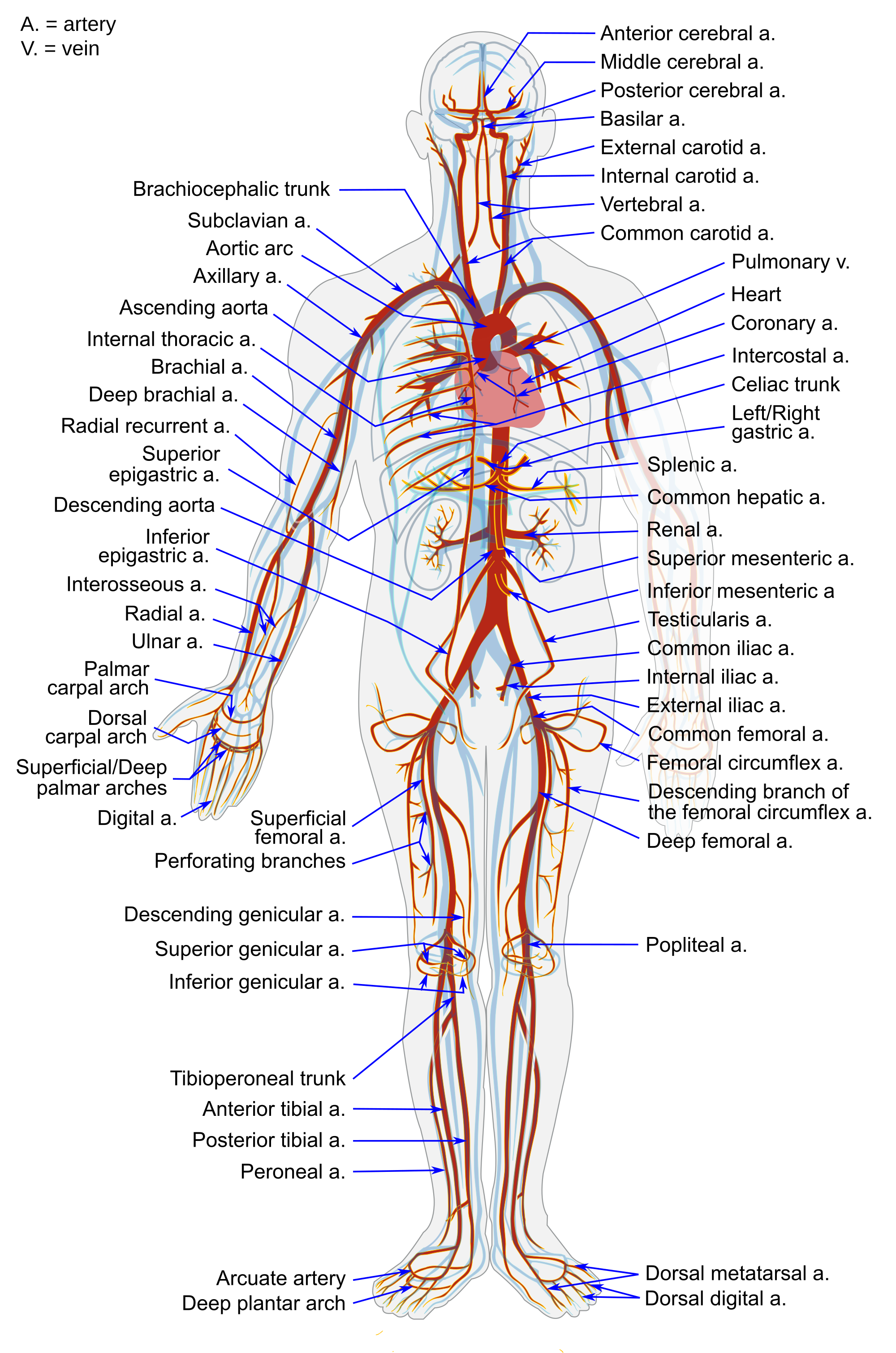
The largest artery in the body is the aorta, which is connected to the heart and extends down into the abdomen (see Figure 14.4.2). The aorta has high-pressure, oxygenated blood pumped directly into it from the left ventricle of the heart. The aorta has many branches, and the branches subdivide repeatedly, with the subdivisions growing smaller and smaller in diameter. The smallest arteries are called arterioles.
Veins
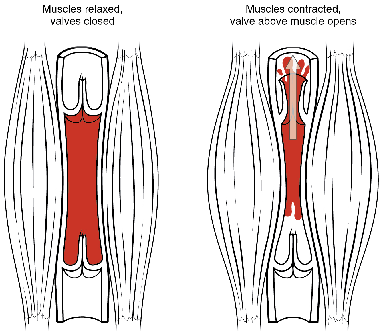
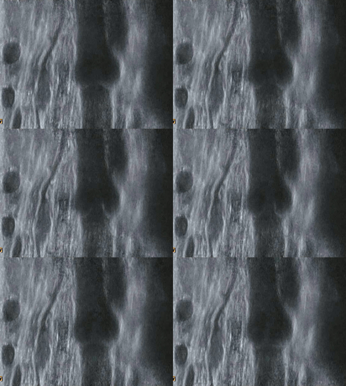
Veins are defined as blood vessels that carry blood toward the heart. Blood traveling through veins is not under pressure from the beating heart. It gets help moving along by the squeezing action of skeletal muscles, for example, when you walk or breathe. It is also prevented from flowing backward by valves in the larger veins, as illustrated in Figure 14.4.3. and as seen in the ultrasonography image in Figure 14.4.4. Veins are called capacitance blood vessels, because the majority of the body’s total volume of blood (about 60 per cent) is contained within veins.
Most veins carry deoxygenated blood, but there are a few exceptions, including the four pulmonary veins. These veins carry oxygenated blood from the lungs to the heart, which then pumps the blood to the rest of the body. In virtually all other veins, hemoglobin is relatively unsaturated with oxygen (about 75 per cent).
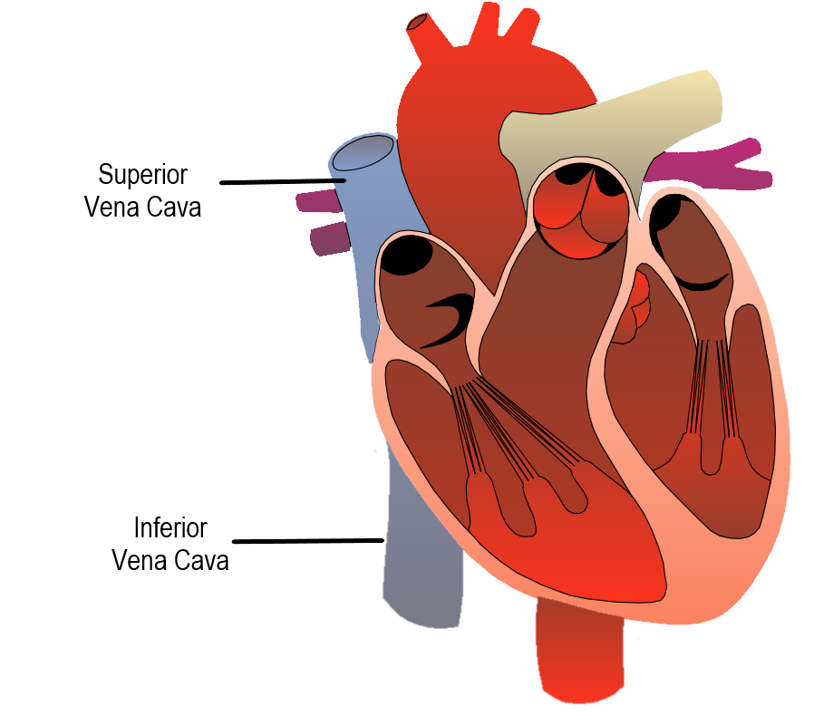
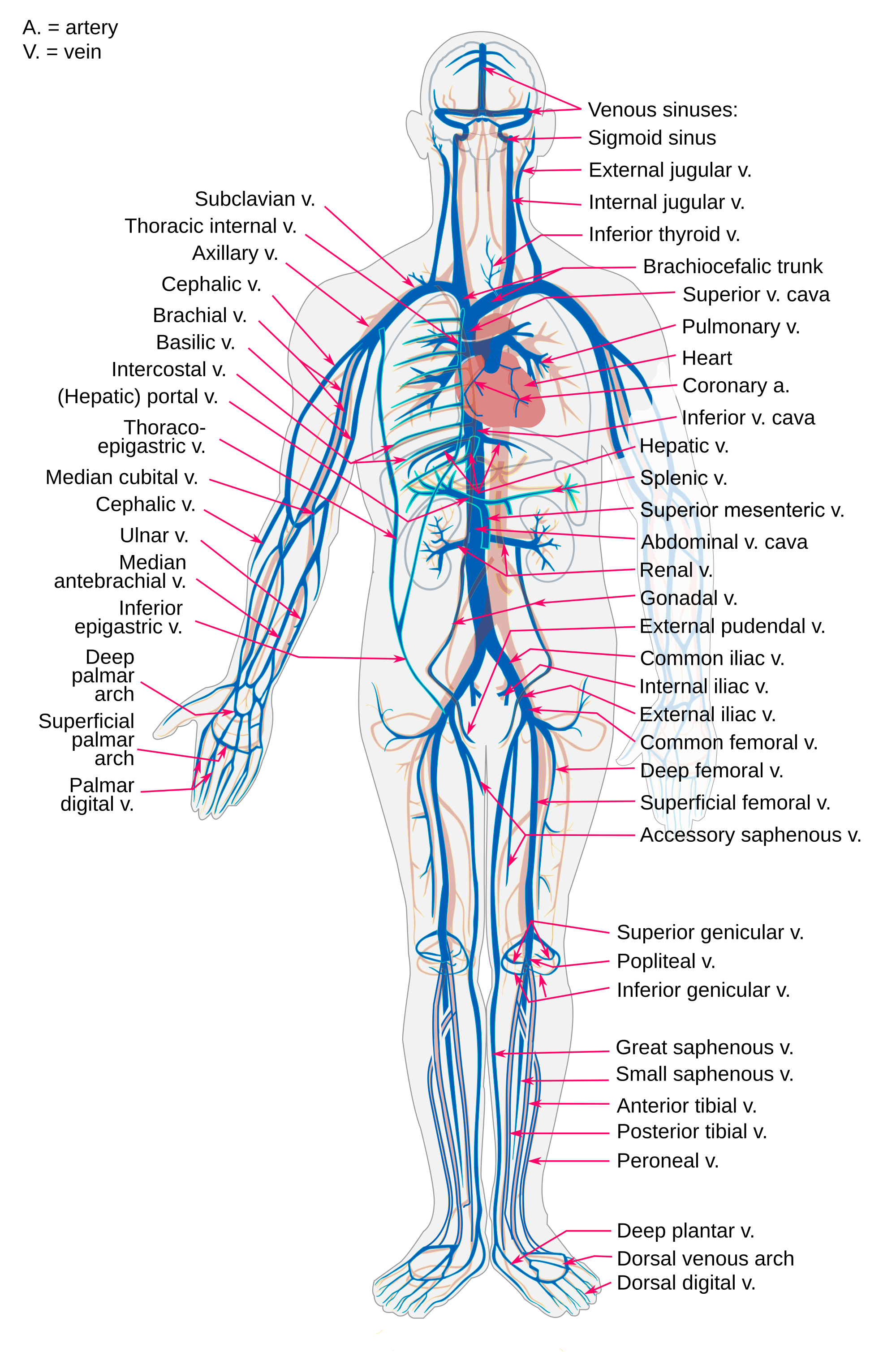
The two largest veins in the body are the superior vena cava — which carries blood from the upper body directly to the right atrium of the heart — and the inferior vena cava, which carries blood from the lower body directly to the right atrium (shown in Figure 14.4.5). Like arteries, veins form a complex, branching system of larger and smaller vessels. The smallest veins are called venules. They receive blood from capillaries and transport it to larger veins. Each venule receives blood from multiple capillaries. See the major veins of the human body in Figure 14.4.6.
Capillaries
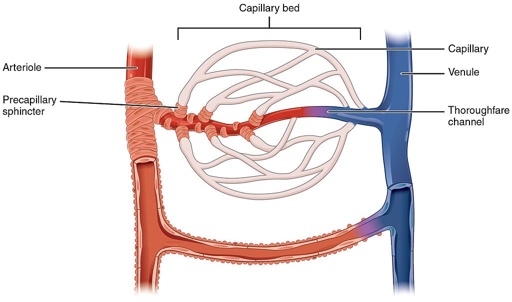
Capillaries are the smallest blood vessels in the cardiovascular system. They are so small that only one red blood cell at a time can squeeze through a capillary, and then only if the red blood cell deforms. Capillaries connect arterioles and venules, as shown in Figure 14.4.7. Capillaries generally form a branching network of vessels, called a capillary bed, that provides a large surface area for the exchange of substances between the blood and surrounding tissues.
Structure of Blood Vessels
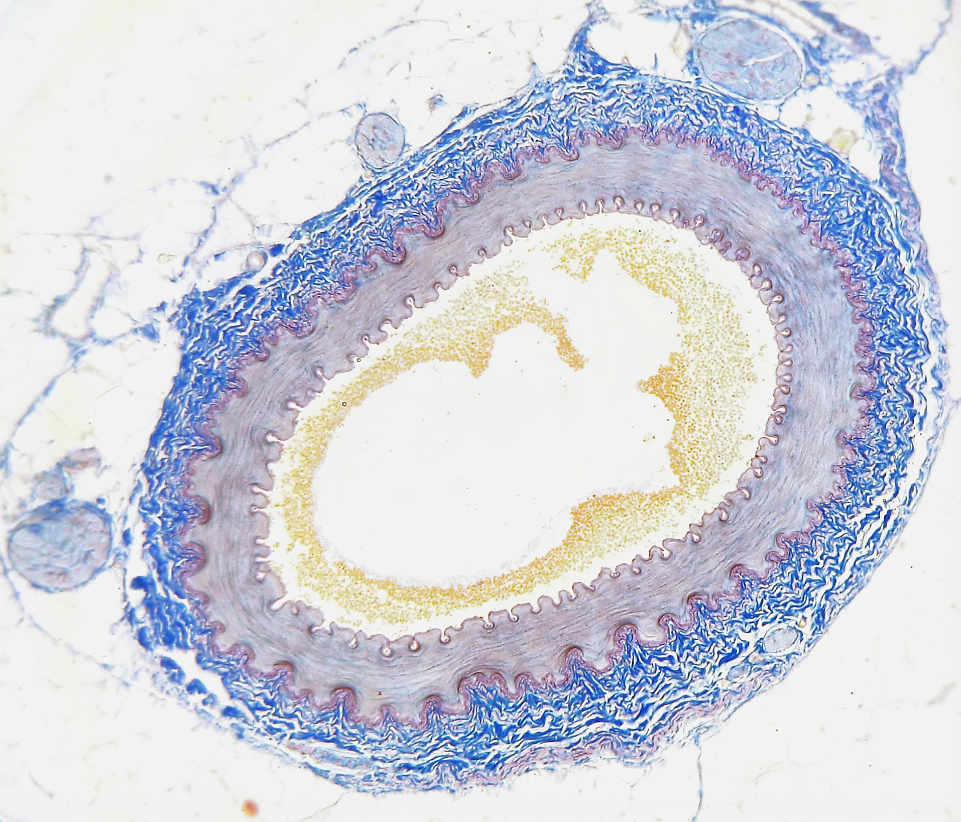
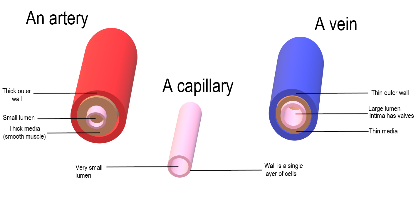
All blood vessels are basically hollow tubes with an internal space, called a lumen, through which blood flows. The lumen of an artery is shown in cross section in the photomicrograph (Figure 14.4.8). The width of blood vessels varies, but they all have a lumen. The walls of blood vessels differ depending on the type of vessel. In general, arteries and veins are more similar to one another than to capillaries in the structure of their walls.
Walls of Arteries and Veins
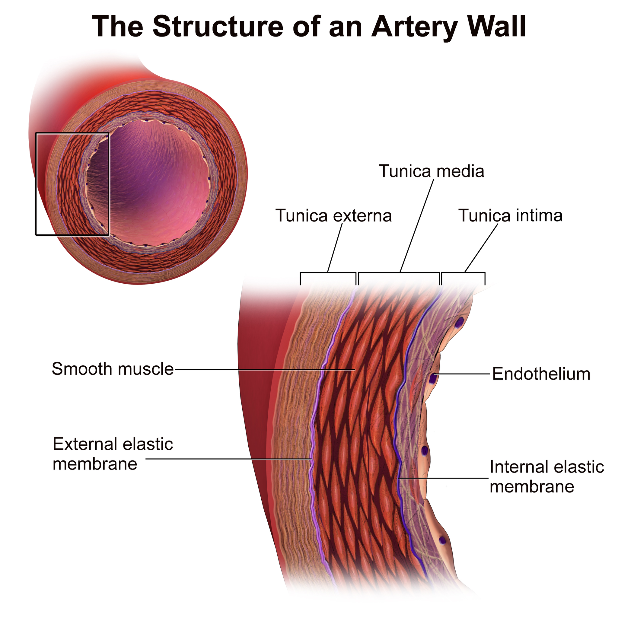
The walls of both arteries and veins have three layers: the tunica intima, tunica media, and tunica adventitia. You can see the three layers for an artery in the Figure 14.4.9.
- The tunica intima is the inner layer of arteries and veins. It is also the thinnest layer, consisting of a single layer of endothelial cells surrounded by a thin layer of connective tissues. It reduces friction between the blood and the inside of the blood vessel walls.
- The tunica media is the middle layer of arteries and veins. In arteries, this is the thickest layer. It consists mainly of elastic fibres and connective tissues. In arteries, this is the thickest layer, because it also contains smooth muscle tissues, which control the diameter of the vessels- as such, the width of the tunic media can be helpful in distinguishing arteries from veins.
- The tunica externa (also called tunica adventitia) is the outer layer of arteries and veins. It consists of connective tissue, and also contains nerves. In veins, this is the thickest layer. In general, the tunica externa protects and strengthens vessels, and attaches them to surrounding structures.
Capillary Walls
The walls of capillaries consist of little more than a single layer of epithelial cells. Being just one cell thick, the walls are well-suited for the exchange of substances between the blood inside them and the cells of surrounding tissues. Substances including water, oxygen, glucose, and other nutrients, as well as waste products (such as carbon dioxide), can pass quickly and easily through the extremely thin walls of capillaries. See figure 14.4.9 for a comparison of the structure of arteries, veins and capillaries.
Blood Pressure
The blood in arteries is normally under pressure because of the beating of the heart. The pressure is highest when the heart contracts and pumps out blood, and lowest when the heart relaxes and refills with blood. (You can feel this variation in pressure in your wrist or neck when you count your pulse.) Blood pressure is a measure of the force that blood exerts on the walls of arteries. It is generally measured in millimetres of mercury (mm Hg), and expressed as a double number — a higher number for systolic pressure when the ventricles contract, and a lower number for diastolic pressure when the ventricles relax. Normal blood pressure is generally defined as less than 120 mm Hg (systolic)/80 mm Hg (diastolic) when measured in the arm at the level of the heart. It decreases as blood flows farther away from the heart and into smaller arteries.
As arteries grow smaller, there is increasing resistance to blood flow through them, because of the blood’s friction against the arterial walls. This resistance restricts blood flow so that less blood reaches smaller, downstream vessels, thus reducing blood pressure before the blood flows into the tiniest vessels, the capillaries. Without this reduction in blood pressure, capillaries would not be able to withstand the pressure of the blood without bursting. By the time blood flows through the veins, it is under very little pressure. The pressure of blood against the walls of veins is always about the same — normally no more than 10 mm Hg.
Vasoconstriction and Vasodilation
Smooth muscles in the walls of arteries can contract or relax to cause vasoconstriction (narrowing of the lumen of blood vessels) or vasodilation (widening of the lumen of blood vessels). This allows the arteries — especially the arterioles — to contract or relax as needed to help regulate blood pressure. In this regard, the arterioles act like an adjustable nozzle on a garden hose. When they narrow, the increased friction with the arterial walls causes less blood to flow downstream from the narrowing, resulting in a drop in blood pressure. These actions are controlled by the autonomic nervous system in response to pressure-sensitive sensory receptors in the walls of larger arteries.
Arteries can also dilate or constrict to help regulate body temperature, by allowing more or less blood to flow from the warm body core to the body’s surface. In addition, vasoconstriction and vasodilation play roles in the fight-or-flight response, under control of the sympathetic nervous system. Vasodilation allows more blood to flow to skeletal muscles, and vasoconstriction reduces blood flow to digestive organs.
Feature: My Human Body
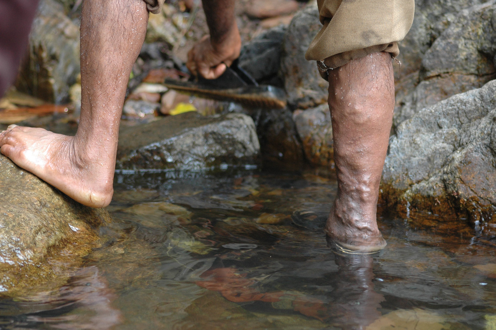
The lumpy appearance of this man’s leg (Figure 14.4.10) is caused by varicose veins. Do you have varicose veins? If you do, you may wonder whether they are a sign of a significant health problem. You may also wonder whether you should have them treated, and if so, what treatments are available. As is usually the case, when it comes to your health, knowledge is power.
Varicose veins are veins that have become enlarged and twisted, because their valves have become ineffective (see Figure 14.4.11). As a result, blood pools in the veins and stretches them out. Varicose veins occur most frequently in the superficial veins of the legs, but they may also occur in other parts of the body. They are most common in older adults, females, and people who have a family history of the condition. Obesity and pregnancy also increase the risk of developing varicose veins. A job that requires standing for long periods of time, chronic constipation, and long-term alcohol consumption are additional risk factors.
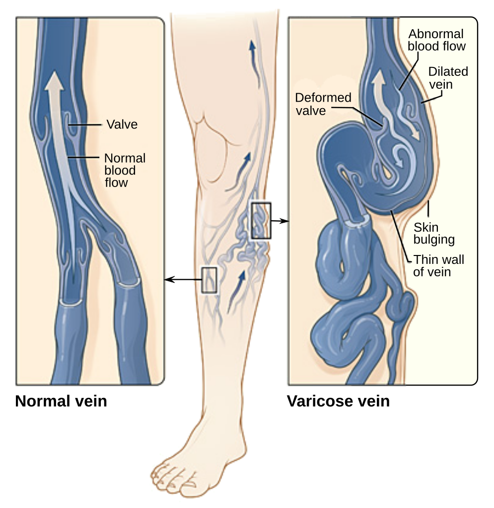
Varicose veins usually are not serious. For many people, they are only a cosmetic issue. In severe cases, however, varicose veins may cause pain and other problems. The affected leg(s) may feel heavy and achy, especially after long periods of standing. Ankles may become swollen by the end of the day. Minor injuries may bleed more than normal. The skin over the varicosity may become red, dry, and itchy. In very severe cases, skin ulcers may develop.
If you are concerned about varicose veins, call them to the attention of your doctor, who can determine the best course of action for your case. There are many potential treatments for varicose veins. Some of the treatments have potential adverse side effects, and with many of the treatments, varicose veins may return. The best treatment for a given patient depends in part on the severity of the condition.
- If varicose veins are not serious, conservative treatment options may be recommended. These include avoiding standing or sitting for long periods, frequently elevating the legs, and wearing graduated compression stockings.
- For more serious cases, less conservative, but non-surgical options may be advised. These include sclerotherapy, in which medicine is injected into the veins to make them shrink. Another non-surgical approach is endovenous thermal ablation. In this type of treatment, laser light, radio-frequency energy, or steam is used to heat the walls of the veins, causing them to shrink and collapse.
- For the most serious cases, surgery may be the best option. The most invasive surgery is vein stripping, in which all or part of the main trunk of a vein is tied off and removed from the leg while the patient is under general anesthesia. In a less invasive surgery, called ambulatory phlebectomy, short segments of a vein are removed through tiny incisions under local anesthesia.
14.4 Summary
- Blood vessels are the part of the cardiovascular system that carries blood throughout the human body. They are long, hollow,tube-like structures. There are three major types of blood vessels: arteries, veins, and capillaries.
- Arteries are blood vessels that carry blood away from the heart. Most arteries carry oxygenated blood. The largest artery is the aorta, which is connected to the heart and extends into the abdomen. Blood moves through arteries due to pressure from the beating of the heart.
- Veins are blood vessels that carry blood toward the heart. Most veins carry deoxygenated blood. The largest veins are the superior vena cava and inferior vena cava. Blood moves through veins by the squeezing action of surrounding skeletal muscles. Valves in veins prevent backflow of blood.
- Capillaries are the smallest blood vessels. They connect arterioles and venules. They form capillary beds, where substances are exchanged between the blood and surrounding tissues.
- The walls of arteries and veins have three layers. The middle layer is thickest in arteries, in which it contains smooth muscle tissue that controls the diameter of the vessels. The outer layer is thickest in veins, and consists mainly of connective tissue. The walls of capillaries consist of little more than a single layer of epithelial cells.
- Blood pressure is a measure of the force that blood exerts on the walls of arteries. It is expressed as a double number, with the higher number representing systolic pressure when the ventricles contract, and the lower number representing diastolic pressure when the ventricles relax. Normal blood pressure is generally defined as a pressure of less than 120/80 mm Hg.
- Vasoconstriction (narrowing) and vasodilation (widening) of arteries can occur to help regulate blood pressure or body temperature, or change blood flow as part of the fight-or-flight response.
14.4 Review Questions
- What are blood vessels? Name the three major types of blood vessels.
-
- Compare and contrast how blood moves through arteries and veins.
- What are capillaries, and what is their function?
- Does the blood in most veins have any oxygen at all? Explain your answer.
- Explain why it is important that the walls of capillaries are very thin.
14.4 Explore More
How blood pressure works – Wilfred Manzano, TED-Ed, 2015.
What are Varicose Veins? Cleveland Clinic, 2019.
Arteries vs Veins ( Circulatory System ), MooMooMath and Science, 2018.
Attributions
Figure 14.4.1
bodybuilding_PNG24 from pngimg.com is used under a CC BY-NC 4.0 (https://creativecommons.org/licenses/by-nc/4.0/) license.
Figure 14.4.2
Arterial_System_en.svg by Mariana Ruiz Villarreal [LadyofHats] on Wikimedia Commons is in the public domain (https://en.wikipedia.org/wiki/Public_domain).
Figure 14.4.3
Skeletal_Muscle_Vein_Pump by OpenStax College on Wikimedia Commons is used under a CC BY 3.0 (https://creativecommons.org/licenses/by/3.0) license.
Figure 14.4.4
Venous_valve_00013 by Nevit Dilmen on Wikimedia Commons is used under a CC BY-SA 3.0 (https://creativecommons.org/licenses/by-sa/3.0) license.
Figure 14.4.5
Superior and Inferior Vena Cava by ArtFavor (acquired from OCAL) from Freestockphotos.biz, is used under a CC0 1.0 Universal public domain dedication license (https://creativecommons.org/publicdomain/zero/1.0/). Work adapted by Christine Miller.
Figure 14.4.6
Venous_system_en.svg by Mariana Ruiz Villarreal [LadyofHats] on Wikimedia Commons is in the public domain (https://en.wikipedia.org/wiki/Public_domain).
Figure 14.4.7
1024px-2105_Capillary_Bed by OpenStax College on Wikimedia Commons is used under a CC BY 3.0 (https://creativecommons.org/licenses/by/3.0) license.
Figure 14.4.8
Artery by Lord of Konrad on Wikimedia Commons is used under a CC0 1.0 Universal public domain dedication license (https://creativecommons.org/publicdomain/zero/1.0/).
Figure 14.4.9
Blausen_0055_ArteryWallStructure by BruceBlaus on Wikimedia Commons is used under a CC BY 3.0 (https://creativecommons.org/licenses/by/3.0) license.
Figure 14.4.10
Artery Vein Capillary Comparison by Christinelmiller on Wikimedia Commons is used under a CC BY-SA 4.0 (https://creativecommons.org/licenses/by-sa/4.0) license.
Figure 14.4.11
Varicose-veins by Jackerhack at English Wikipedia on Wikimedia Commons is used under a CC BY-SA 2.5 (https://creativecommons.org/licenses/by-sa/2.5) license.
Figure 14.4.12
Varicose_veins-en.svg by Jmarchn on Wikimedia Commons is used under a CC BY-SA 3.0 (https://creativecommons.org/licenses/by-sa/3.0) license. [Work modified from Varicose veins.jpg on Wikimedia Commons from National Heart Lung and Blood Institute (NIH)]
References
Betts, J. G., Young, K.A., Wise, J.A., Johnson, E., Poe, B., Kruse, D.H., Korol, O., Johnson, J.E., Womble, M., DeSaix, P. (2013, June 19). Figure 20.6 Capillary bed [digital image]. In Anatomy and Physiology (Section 20.1). OpenStax. https://openstax.org/books/anatomy-and-physiology/pages/20-1-structure-and-function-of-blood-vessels
Betts, J. G., Young, K.A., Wise, J.A., Johnson, E., Poe, B., Kruse, D.H., Korol, O., Johnson, J.E., Womble, M., DeSaix, P. (2013, June 19). Figure 20.15 Skeletal muscle pump [digital image]. In Anatomy and Physiology (Section 20.2). OpenStax. https://openstax.org/books/anatomy-and-physiology/pages/20-2-blood-flow-blood-pressure-and-resistance
Blausen.com Staff. (2014). Medical gallery of Blausen Medical 2014. WikiJournal of Medicine 1 (2). DOI:10.15347/wjm/2014.010. ISSN 2002-4436.
Cleveland Clinic. (2019, December 30). What are varicose veins? YouTube. https://www.youtube.com/watch?v=9Wf8bLXVwFI&feature=youtu.be
MooMooMath and Science. (2018, April 5). Arteries vs veins ( Circulatory System ). YouTube. https://www.youtube.com/watch?v=hnjMdXSyA5o&feature=youtu.be
TED-Ed. (2015, July 23). How blood pressure works – Wilfred Manzano. YouTube. https://www.youtube.com/watch?v=Ab9OZsDECZw&feature=youtu.be
A hollow, tube-like structure through which blood flows in the cardiovascular system; vein, artery, or capillary.
Refers to the body system consisting of the heart, blood vessels and the blood. Blood contains oxygen and other nutrients which your body needs to survive. The body takes these essential nutrients from the blood.
Created by: CK-12/Adapted by Christine Miller
Assembly Line
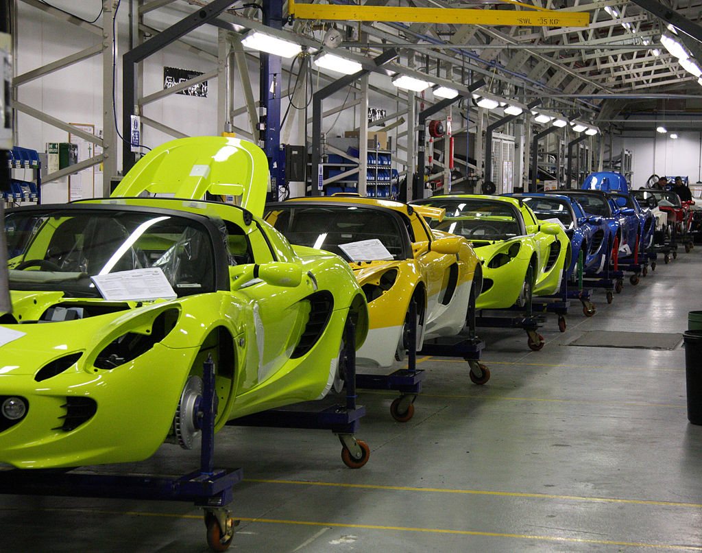
We stay alive because millions of different chemical reactions are taking place inside our bodies all the time. Each of our cells is like the busy auto assembly line pictured in Figure 3.10.1. Raw materials, half-finished products, and waste materials are constantly being used, produced, transported, and excreted. The "workers" on the cellular assembly line are mainly enzymes. These are the proteins that make biochemical reactions happen.
What Are Biochemical Reactions?
Chemical reactions that take place inside living things are called biochemical reactions. The sum of all the biochemical reactions in an organism is called metabolism. Metabolism includes both exothermic (energy-releasing) chemical reactions and endothermic (energy-absorbing) chemical reactions.
Catabolic Reactions
Exothermic reactions in organisms are called catabolic reactions. These reactions break down molecules into smaller units and release energy. An example of a catabolic reaction is the breakdown of glucose during cellular respiration, which releases energy that cells need to carry out life processes.
Anabolic Reactions
Endothermic reactions in organisms are called anabolic reactions. These reactions build up bigger molecules from smaller ones and absorb energy. An example of an anabolic reaction is the joining of amino acids to form a protein. Which type of reactions — catabolic or anabolic — do you think occur when your body digests food?
Enzymes
Most of the biochemical reactions that happen inside of living organisms require help. Why is this the case? For one thing, temperatures inside living things are usually too low for biochemical reactions to occur quickly enough to maintain life. The concentrations of reactants may also be too low for them to come together and react. Where do the biochemical reactions get the help they need to proceed? From the enzymes.
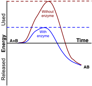
An enzyme is a protein that speeds up a biochemical reaction. It is a biological catalyst. An enzyme generally works by reducing the amount of activation energy needed to start the reaction. The graph in Figure 3.10.2 shows the activation energy needed for glucose to combine with oxygen. Less activation energy is needed when the correct enzyme is present than when it is not present.
An enzyme speeds up the reaction by lowering the required activation energy. Compare the activation energy needed with and without the enzyme.
How Well Enzymes Work
Enzymes are involved in most biochemical reactions, and they do their jobs extremely well. A typical biochemical reaction that would take several days or even several centuries to happen without an enzyme is likely to occur in just a split second with the proper enzyme! Without enzymes to speed up biochemical reactions, most organisms could not survive.
Enzymes are substrate-specific. The substrate of an enzyme is the specific substance it affects. Each enzyme works only with a particular substrate, which explains why there are so many different enzymes. In addition, for an enzyme to work, it requires specific conditions, such as the right temperature and pH. Some enzymes work best under acidic conditions, for example, while others work best in neutral environments.
Enzyme-Deficiency Disorders
There are hundreds of known inherited metabolic disorders in humans. In most of them, a single enzyme is either not produced by the body at all, or is otherwise produced in a form that doesn't work. The missing or defective enzyme is like an absentee worker on the cell's assembly line. Imagine the auto assembly line from the image at the start of this section. What if the worker who installed the steering wheel was absent? How would this impact the overall functioning of the vehicle? When an enzyme is missing, toxic chemicals build up, or an essential product isn't made. Generally, the normal enzyme is missing because the individual with the disorder inherited two copies of a gene mutation, which may have originated many generations previously.
Any given inherited metabolic disorder is generally quite rare in the general population. However, there are so many different metabolic disorders that a total of one in 1,000 to 2,500 newborns can be expected to have one.
3.10 Summary
- Biochemical reactions are chemical reactions that take place inside of living things. The sum of all of the biochemical reactions in an organism is called metabolism.
- Metabolism includes catabolic reactions, which are energy-releasing (exothermic) reactions, as well as anabolic reactions, which are energy-absorbing (endothermic) reactions.
- Most biochemical reactions need a biological catalyst called an enzyme to speed up the reaction. Enzymes reduce the amount of activation energy needed for the reaction to begin. Most enzymes are proteins that affect just one specific substance, which is called the enzyme's substrate.
- There are many inherited metabolic disorders in humans. Most of them are caused by a single defective or missing enzyme.
3.10 Review Questions
- What are biochemical reactions?
- Define metabolism.
- Compare and contrast catabolic and anabolic reactions.
- Explain the role of enzymes in biochemical reactions.
- What are enzyme-deficiency disorders?
- Explain why the relatively low temperature of living things, along with the low concentration of reactants, would cause biochemical reactions to occur very slowly in the body without enzymes.
- Answer the following questions about what happens after you eat a sandwich.
- Pieces of the sandwich go into your stomach, where there are digestive enzymes that break down the food. Which type of metabolic reaction is this? Explain your answer.
- During the process of digestion, some of the sandwich is broken down into glucose, which is then further broken down to release energy that your cells can use. Is this an exothermic endothermic reaction? Explain your answer.
- The proteins in the cheese, meat, and bread in the sandwich are broken down into their component amino acids. Then your body uses those amino acids to build new proteins. Which kind of metabolic reaction is represented by the building of these new proteins? Explain your answer.
- Explain why your body doesn’t just use one or two enzymes for all of its biochemical reactions.
- A ________ is the specific substance that an enzyme affects in a biochemical reaction.
- An enzyme is a biological _____________ .
- catabolism
- form of activation energy
- catalyst
- reactant
3.10 Explore More
https://www.youtube.com/watch?v=qgVFkRn8f10&feature=youtu.be
Enzymes (Updated), by The Amoeba Sisters, 2016.
https://www.youtube.com/watch?v=8m6RtOpqvtU&feature=youtu.be
What triggers a chemical reaction? - Kareem Jarrah, TED-Ed, 2015.
Figure 3.10.1
Auto Assembly line by Brian Snelson on Wikimedia Commons is used under a CC BY 2.0 (https://creativecommons.org/licenses/by/2.0) license.
Figure 3.10.2
Enzyme_activation_energy by G. Andruk [IMeowbot at the English language Wikipedia], is used under a CC BY-SA 3.0 (http://creativecommons.org/licenses/by-sa/3.0/) license.
References
Amoeba Sisters. (2016, August 28). Enzymes (updated). YouTube. https://www.youtube.com/watch?v=qgVFkRn8f10&feature=youtu.be
TED-Ed. (2015, January 15). What triggers a chemical reaction? - Kareem Jarrah. YouTube. https://www.youtube.com/watch?v=8m6RtOpqvtU&feature=youtu.be
Figure 4.2.1 Human cells viewed with a very powerful tool called a scanning electron microscope.
Amazing Cells
What are these incredible objects? Would it surprise you to learn that they are all human cells? Cells are actually too small to see with the unaided eye. It is visible here in such detail because it is being viewed with a very powerful tool called a scanning electron microscope. Cells may be small in size, but they are extremely important to life. Like all other living things, you are made of cells. Cells are the basis of life, and without cells, life as we know it would not exist. You will learn more about these amazing building blocks of life in this section.
What Are Cells?
If you look at living matter with a microscope — even a simple light microscope — you will see that it consists of cells. Cells are the basic units of the structure and function of living things. They are the smallest units that can carry out the processes of life. All organisms are made up of one or more cells, and all cells have many of the same structures and carry out the same basic life processes. Knowing the structure of cells and the processes they carry out is necessary to an understanding of life itself.
Discovery of Cells
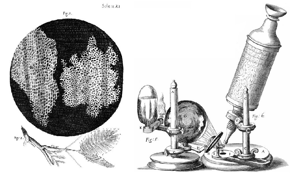
The first time the word cell was used to refer to these tiny units of life was in 1665 by a British scientist named Robert Hooke. Hooke was one of the earliest scientists to study living things under a microscope. The microscopes of his day were not very strong, but Hooke was still able to make an important discovery. When he looked at a thin slice of cork under his microscope, he was surprised to see what looked like a honeycomb. Hooke made the drawing in the figure to the right to show what he saw. As you can see, the cork was made up of many tiny units. Hooke called these units cells because they resembled cells in a monastery.
Soon after Robert Hooke discovered cells in cork, Anton van Leeuwenhoek in Holland made other important discoveries using a microscope. Leeuwenhoek made his own microscope lenses, and he was so good at it that his microscope was more powerful than other microscopes of his day. In fact, Leeuwenhoek’s microscope was almost as strong as modern light microscopes. Using his microscope, Leeuwenhoek was the first person to observe human cells and bacteria.
Cell Theory
By the early 1800s, scientists had observed cells of many different organisms. These observations led two German scientists named Theodor Schwann and Matthias Jakob Schleiden to propose cells as the basic building blocks of all living things. Around 1850, a German doctor named Rudolf Virchow was studying cells under a microscope, when he happened to see them dividing and forming new cells. He realized that living cells produce new cells through division. Based on this realization, Virchow proposed that living cells arise only from other living cells.
The ideas of all three scientists — Schwann, Schleiden, and Virchow — led to cell theory, which is one of the fundamental theories unifying all of biology.
Cell theory states that:
- All organisms are made of one or more cells.
- All the life functions of organisms occur within cells.
- All cells come from existing cells.
Seeing Inside Cells
Starting with Robert Hooke in the 1600s, the microscope opened up an amazing new world — a world of life at the level of the cell. As microscopes continued to improve, more discoveries were made about the cells of living things, but by the late 1800s, light microscopes had reached their limit. Objects much smaller than cells (including the structures inside cells) were too small to be seen with even the strongest light microscope.
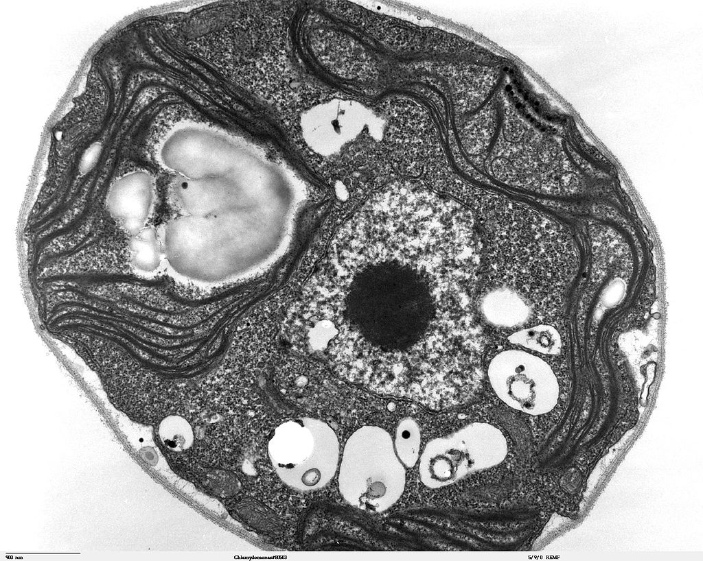
Then, in the 1950s, a new type of microscope was invented. Called the electron microscope, it used a beam of electrons instead of light to observe extremely small objects. With an electron microscope, scientists could finally see the tiny structures inside cells. They could even see individual molecules and atoms. The electron microscope had a huge impact on biology. It allowed scientists to study organisms at the level of their molecules, and it led to the emergence of the molecular biology field. With the electron microscope, many more cell discoveries were made.
Structures Shared By All Cells
Although cells are diverse, all cells have certain parts in common. These parts include a plasma membrane, cytoplasm, ribosomes, and DNA.
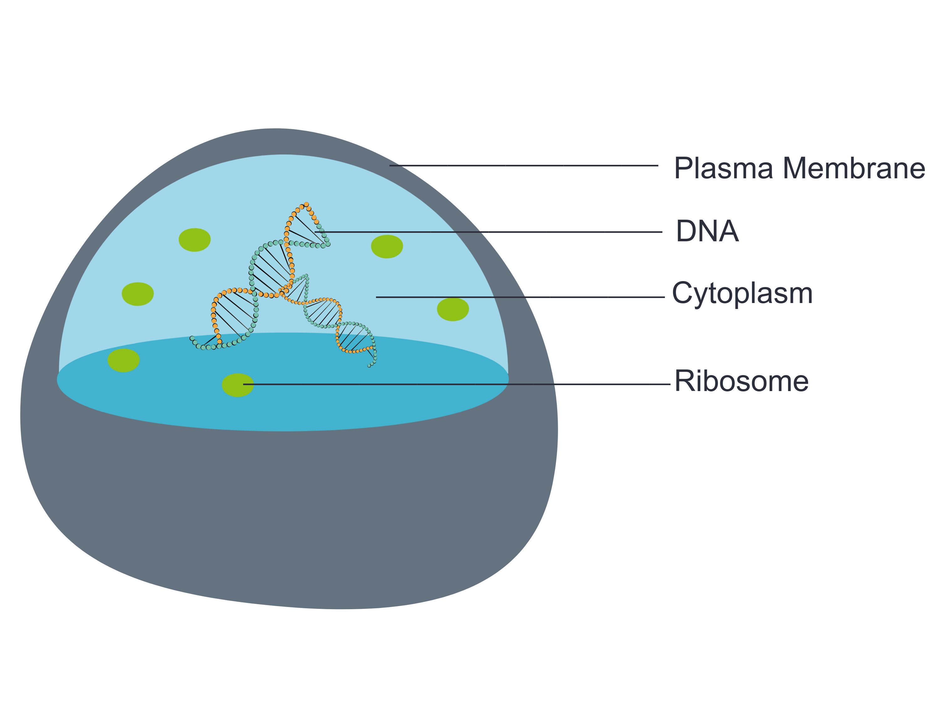
- The plasma membrane (a type of cell membrane) is a thin coat of lipids that surrounds a cell. It forms the physical boundary between the cell and its environment. You can think of it as the “skin” of the cell.
- Cytoplasm refers to all of the cellular material inside of the plasma membrane. Cytoplasm is made up of a watery substance called cytosol, and it contains other cell structures, such as ribosomes.
- Ribosomes are the structures in the cytoplasm in which proteins are made.
- DNA is a nucleic acid found in cells. It contains the genetic instructions that cells need to make proteins.
These four parts are common to all cells, from organisms as different as bacteria and human beings. How did all known organisms come to have such similar cells? The similarities show that all life on Earth has a common evolutionary history.
4.2 Summary
- Cells are the basic units of structure and function in living things. They are the smallest units that can carry out the processes of life.
- In the 1600s, Hooke was the first to observe cells from an organism (cork). Soon after, microscopist van Leeuwenhoek observed many other living cells.
- In the early 1800s, Schwann and Schleiden theorized that cells are the basic building blocks of all living things. Around 1850, Virchow observed cells dividing. To previous learnings, he added that living cells arise only from other living cells. These ideas led to cell theory, which states that all organisms are made of cells, that all life functions occur in cells, and that all cells come from other cells.
- It wasn't until the 1950s that scientists could see what was inside the cell. The invention of the electron microscope allowed them to see organelles and other structures smaller than cells.
- There is variation in cells, but all cells have a plasma membrane, cytoplasm, ribosomes, and DNA. These similarities show that all life on Earth has a common ancestor in the distant past.
4.2 Review Questions
- Describe cells.
- Explain how cells were discovered.
- Outline the development of cell theory.
-
- Identify the structures shared by all cells.
- Proteins are made on _____________ .
-
- Robert Hooke sketched what looked like honeycombs — or repeated circular or square units — when he observed plant cells under a microscope.
- What is each unit?
- Of the shared parts of all cells, what makes up the outer surface of each unit?
- Of the shared parts of all cells, what makes up the inside of each unit?
4.2 Explore More
https://www.youtube.com/watch?v=8IlzKri08kk
Introduction to Cells: The Grand Cell Tour, by The Amoeba Sisters, 2016.
Attributions
Figure 4.2.1
- A white blood cell (WBC) known as a neutrophil by National Institute of Allergy and Infectious Diseases (NIAID) on the CDC/ Public Health Image Library (PHIL) Photo ID# 18129. is in the public domain (https://en.wikipedia.org/wiki/Public_domain).
- Healthy Human T Cell by NIAID on Flickr. is used under a CC BY 2.0 (https://creativecommons.org/licenses/by/2.0/) license.
- Human natural killer cell by NIAID on Flickr. is used under a CC BY 2.0 (https://creativecommons.org/licenses/by/2.0/) license.
- Human blood with red blood cells, T cells (orange) and platelets (green) by ZEISS Microscopy on Flickr. is used under a CC BY-NC-ND 2.0 (https://creativecommons.org/licenses/by-nc-nd/2.0/) license.
- Developing nerve cells by ZEISS Microscopy on Flickr. is used under a CC BY-NC-ND 2.0 (https://creativecommons.org/licenses/by-nc-nd/2.0/) license.
Figure 4.2.2
Hooke-microscope-cork by Robert Hooke (1635-1702) [uploaded by Alejandro Porto] on Wikimedia Commons is released into the public domain (https://en.wikipedia.org/wiki/Public_domain).
Figure 4.2.3
Electron Microscope image of a cell by Dartmouth Electron Microscope Facility, Dartmouth College on Wikimedia Commons is released into the public domain (https://en.wikipedia.org/wiki/Public_domain).
Figure 4.2.4
Basic-Components-of-a-cell by Christine Miller is used under a CC0 1.0 (https://creativecommons.org/publicdomain/zero/1.0/) license.
References
Amoeba Sisters. (2016, November 1). Introduction to cells: The grand cell tour. YouTube. https://www.youtube.com/watch?v=8IlzKri08kk&feature=youtu.be
National Institute of Allergy and Infectious Diseases (NIAID). (2011). A white blood cell (WBC) known as a neutrophil, as it was in the process of ingesting a number of spheroid shaped, methicillin-resistant, Staphylococcus aureus (MRSA) bacteria [digital image]. CDC/ Public Health Image Library (PHIL) Photo ID# 18129. https://phil.cdc.gov/Details.aspx?pid=18129.
Wikipedia contributors. (2020, June 24). Antonie van Leeuwenhoek. In Wikipedia. https://en.wikipedia.org/w/index.php?title=Antonie_van_Leeuwenhoek&oldid=964339564
Wikipedia contributors. (2020, May 25). Matthias Jakob Schleiden. In Wikipedia. https://en.wikipedia.org/w/index.php?title=Matthias_Jakob_Schleiden&oldid=958819219
Wikipedia contributors. (2020, June 4). Rudolf Virchow. In Wikipedia,. https://en.wikipedia.org/w/index.php?title=Rudolf_Virchow&oldid=960708716
Wikipedia contributors. (2020, May 16). Theodor Schwann. In Wikipedia. https://en.wikipedia.org/w/index.php?title=Theodor_Schwann&oldid=956919239
The smallest type of blood vessel that connects arterioles and venules and that transfers substances between blood and tissues.
Created by: CK-12/Adapted by Christine Miller

Bring on the S'mores!
This inviting camp fire can be used for both heat and light. Heat and light are two forms of energy that are released when a fuel like wood is burned. The cells of living things also get energy by "burning." They "burn" glucose in a process called cellular respiration.
What Is Cellular Respiration?
Cellular respiration is the process by which living cells break down glucose molecules and release energy. The process is similar to burning, although it doesn’t produce light or intense heat as a campfire does. This is because cellular respiration releases the energy in glucose slowly and in many small steps. It uses the energy released to form molecules of ATP, the energy-carrying molecules that cells use to power biochemical processes. In this way, cellular respiration is an example of energy coupling: glucose is broken down in an exothermic reaction, and then the energy from this reaction powers the endothermic reaction of the formation of ATP. Cellular respiration involves many chemical reactions, but they can all be summed up with this chemical equation:
C6H12O6 6O2 → 6CO2 6H2O Chemical Energy (in ATP)
In words, the equation shows that glucose (C6H12O6) and oxygen (O2) react to form carbon dioxide (CO2) and water (H2O), releasing energy in the process. Because oxygen is required for cellular respiration, it is an aerobic process.
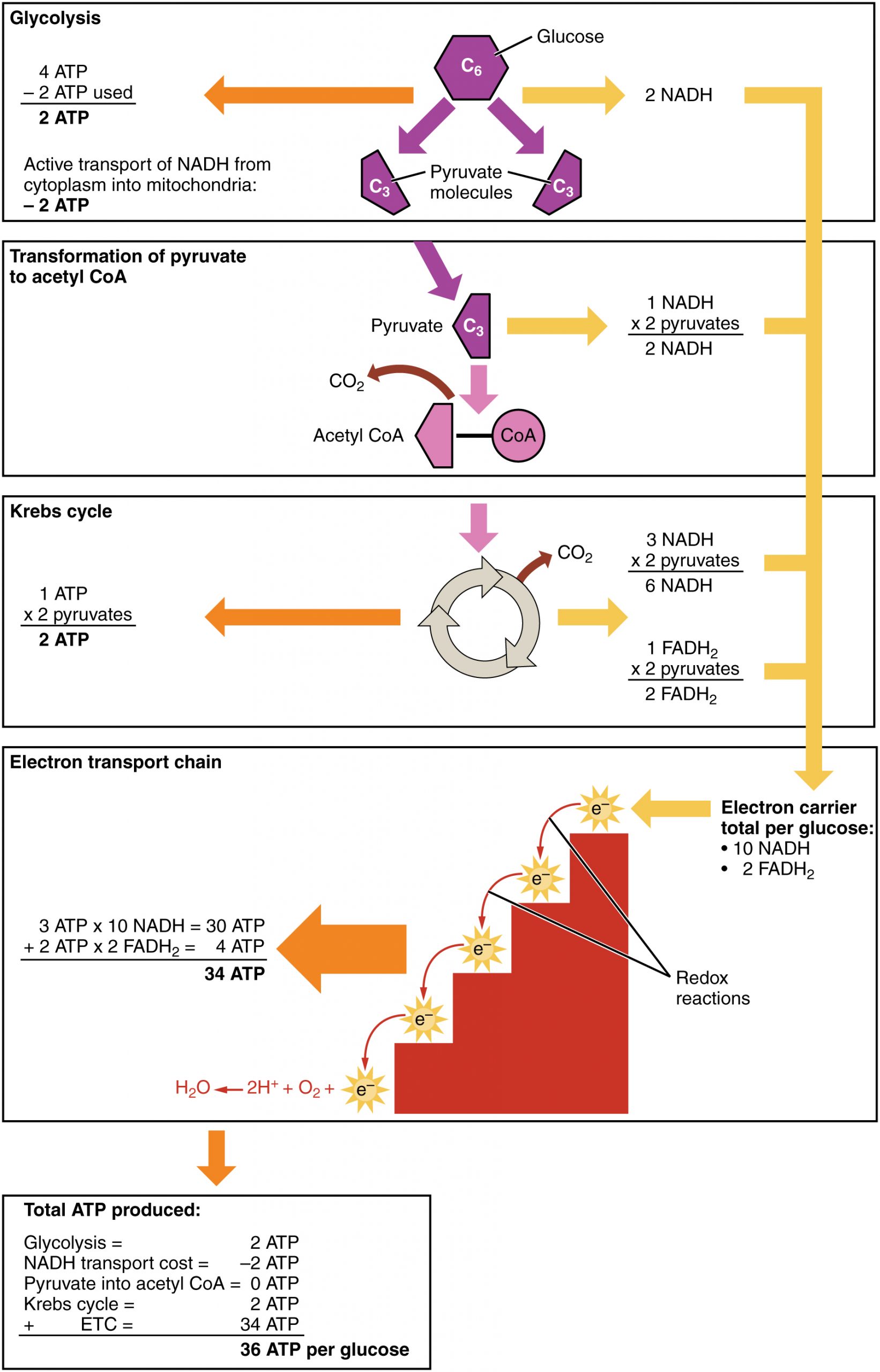
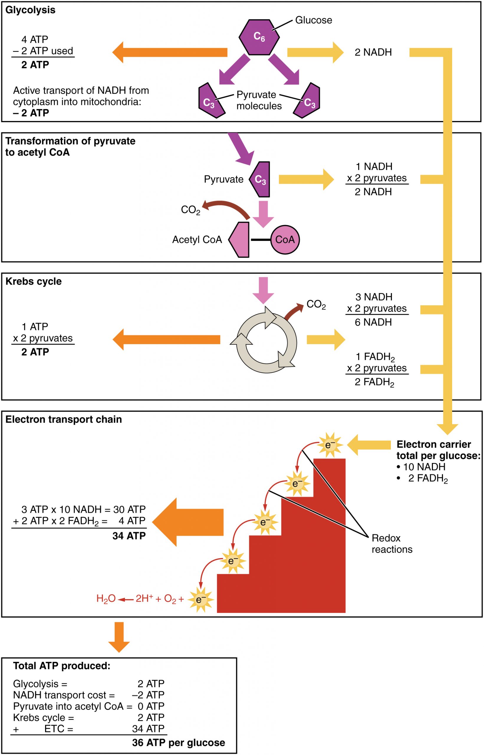
Cellular respiration occurs in the cells of all living things, both autotrophs and heterotrophs. All of them burn glucose to form ATP. The reactions of cellular respiration can be grouped into three stages: glycolysis, the Krebs cycle (also called the citric acid cycle), and electron transport. Figure 4.10.2 gives an overview of these three stages, which are also described in detail below.
Cellular Respiration Stage I: Glycolysis
The first stage of cellular respiration is glycolysis, which happens in the cytosol of the cytoplasm.
Splitting Glucose
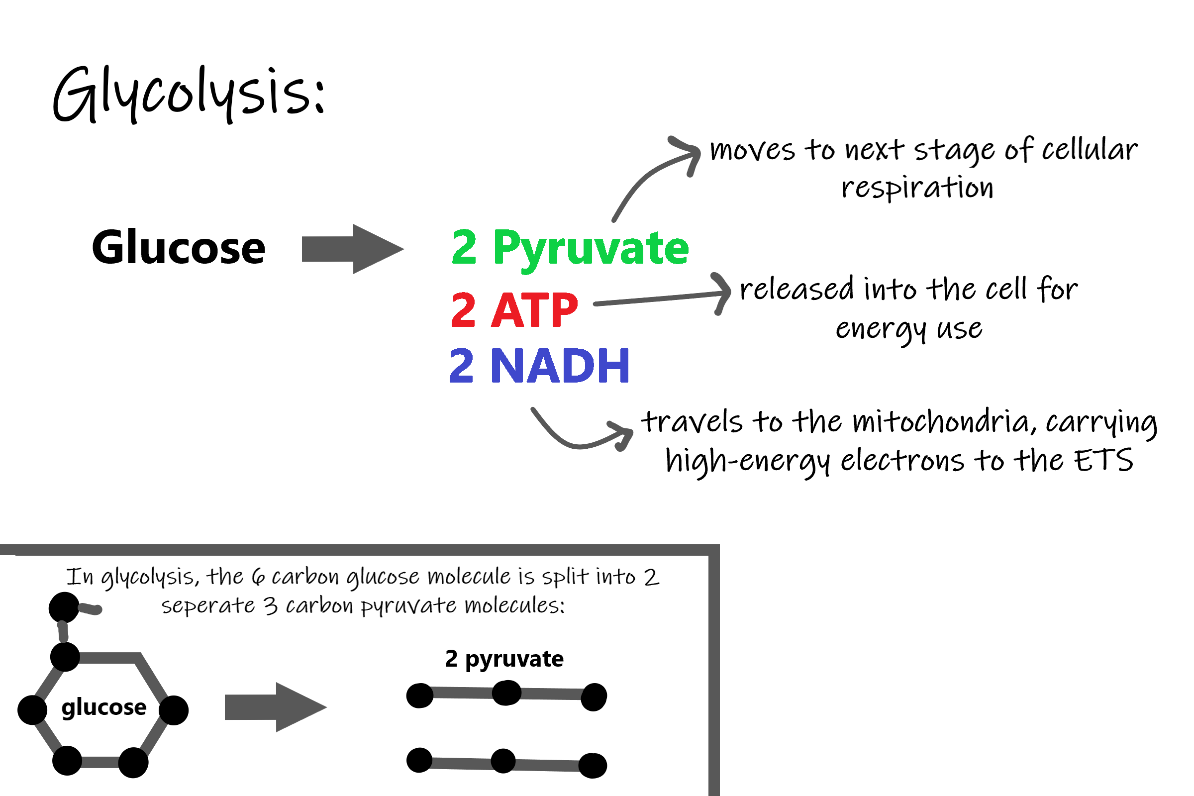
The word glycolysis literally means “glucose splitting,” which is exactly what happens in this stage. Enzymes split a molecule of glucose into two molecules of pyruvate (also known as pyruvic acid). This occurs in several steps, as summarized in the following diagram.
Results of Glycolysis
Energy is needed at the start of glycolysis to split the glucose molecule into two pyruvate molecules which go on to stage II of cellular respiration. The energy needed to split glucose is provided by two molecules of ATP; this is called the energy investment phase. As glycolysis proceeds, energy is released, and the energy is used to make four molecules of ATP; this is the energy harvesting phase. As a result, there is a net gain of two ATP molecules during glycolysis. During this stage, high-energy electrons are also transferred to molecules of NAD to produce two molecules of NADH, another energy-carrying molecule. NADH is used in stage III of cellular respiration to make more ATP.
Transition Reaction
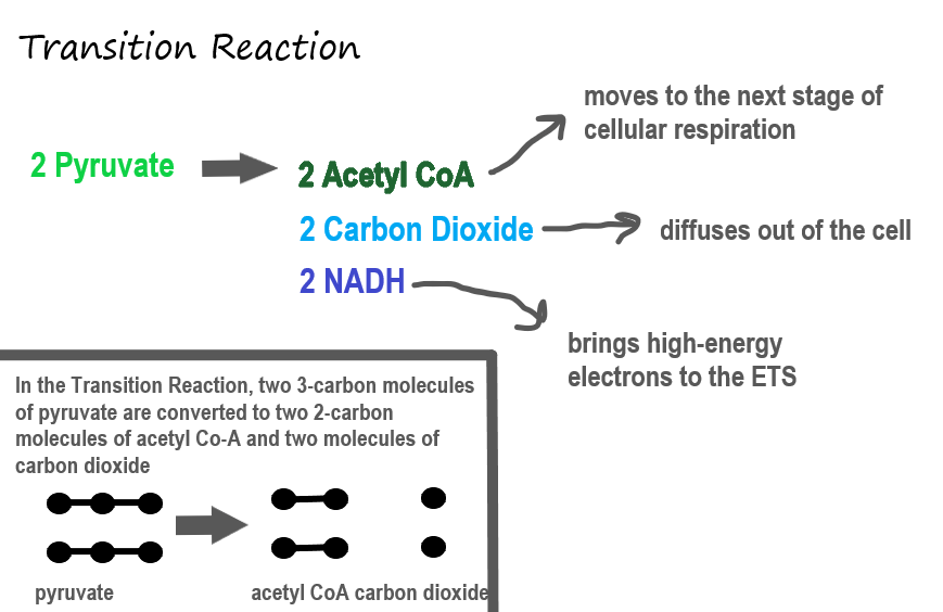
Before pyruvate can enter the next stage of cellular respiration it needs to be modified slightly. The transition reaction is a very short reaction which converts the two molecules of pyruvate to two molecules of acetyl CoA, carbon dioxide, and two high energy electron pairs convert NAD to NADH. The carbon dioxide is released, the acetyl CoA moves to the mitochondria to enter the Kreb's Cycle (stage II), and the NADH carries the high energy electrons to the Electron Transport System (stage III).
Structure of the Mitochondrion
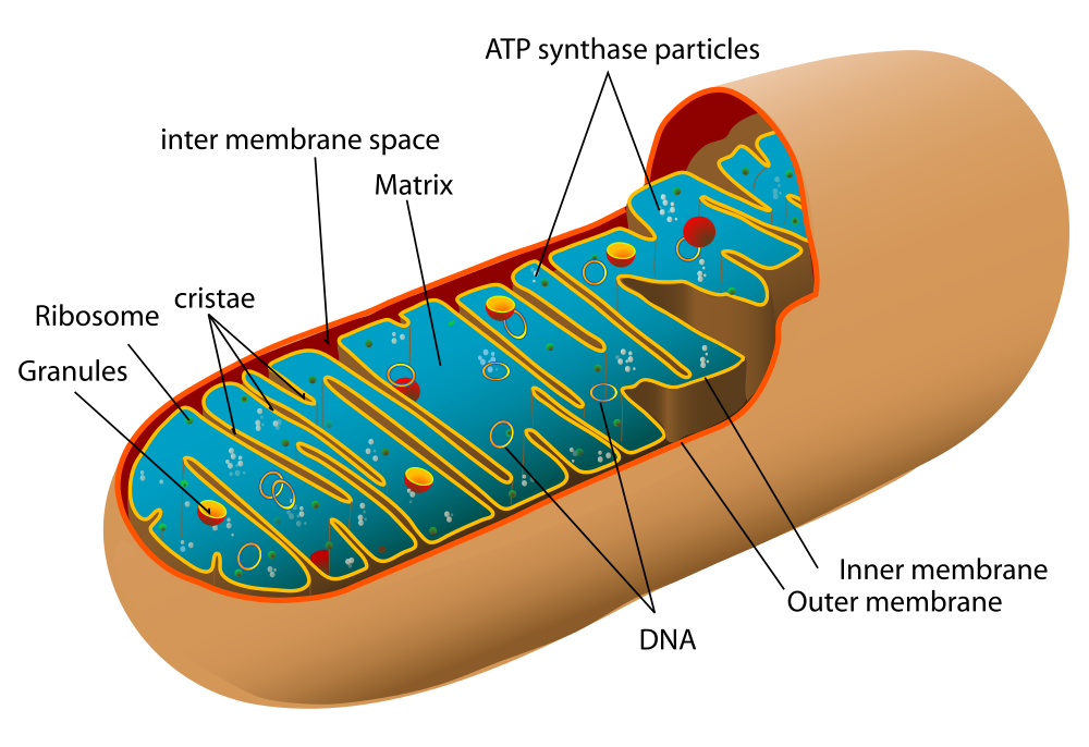
Before you read about the last two stages of cellular respiration, you need to know more about the mitochondrion, where these two stages take place. A diagram of a mitochondrion is shown in Figure 4.10.5.
The structure of a mitochondrion is defined by an inner and outer membrane. This structure plays an important role in aerobic respiration.
As you can see from the figure, a mitochondrion has an inner and outer membrane. The space between the inner and outer membrane is called the intermembrane space. The space enclosed by the inner membrane is called the matrix. The second stage of cellular respiration (the Krebs cycle) takes place in the matrix. The third stage (electron transport) happens on the inner membrane.
Cellular Respiration Stage II: The Krebs Cycle
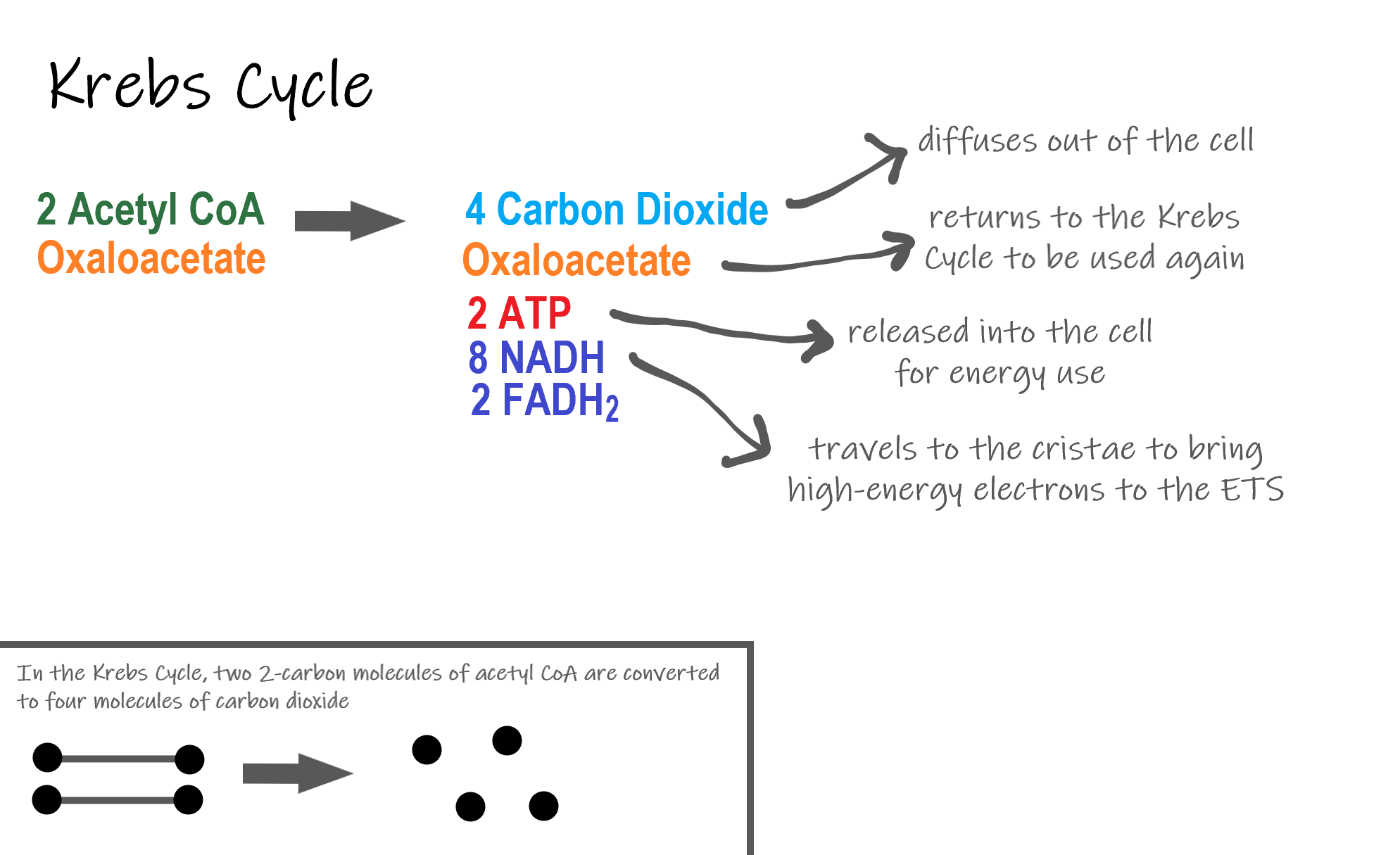
Recall that glycolysis produces two molecules of pyruvate (pyruvic acid), which are then converted to acetyl CoA during the short transition reaction. These molecules enter the matrix of a mitochondrion, where they start the Krebs cycle (also known as the Citric Acid Cycle). The reason this stage is considered a cycle is because a molecule called oxaloacetate is present at both the beginning and end of this reaction and is used to break down the two molecules of acetyl CoA. The reactions that occur next are shown in Figure 4.10.6.
Steps of the Krebs Cycle
The Krebs cycle itself actually begins when acetyl-CoA combines with a four-carbon molecule called OAA (oxaloacetate) (see Figure 4.10.6). This produces citric acid, which has six carbon atoms. This is why the Krebs cycle is also called the citric acid cycle.
After citric acid forms, it goes through a series of reactions that release energy. The energy is captured in molecules of NADH, ATP, and FADH2, another energy-carrying coenzyme. Carbon dioxide is also released as a waste product of these reactions.
The final step of the Krebs cycle regenerates OAA, the molecule that began the Krebs cycle. This molecule is needed for the next turn through the cycle. Two turns are needed because glycolysis produces two pyruvic acid molecules when it splits glucose.
Results of the Glycolysis, Transition Reaction and Krebs Cycle
After glycolysis, transition reaction, and the Krebs cycle, the glucose molecule has been broken down completely. All six of its carbon atoms have combined with oxygen to form carbon dioxide. The energy from its chemical bonds has been stored in a total of 16 energy-carrier molecules. These molecules are:
- 4 ATP (2 from glycolysis, 2 from Krebs Cycle)
- 12 NADH (2 from glycolysis, 2 from transition reaction, and 8 from Krebs cycle)
- 2 FADH2 (both from the Krebs cycle)
The events of cellular respiration up to this point are exergonic reactions- they are releasing energy that had been stored in the bonds of the glucose molecule. This energy will be transferred to the third and final stage of cellular respiration: the Electron Transport System, which is an endergonic reaction. Using an exothermic reaction to power an endothermic reaction is known as energy coupling.
Cellular Respiration Stage III: Electron Transport Chain
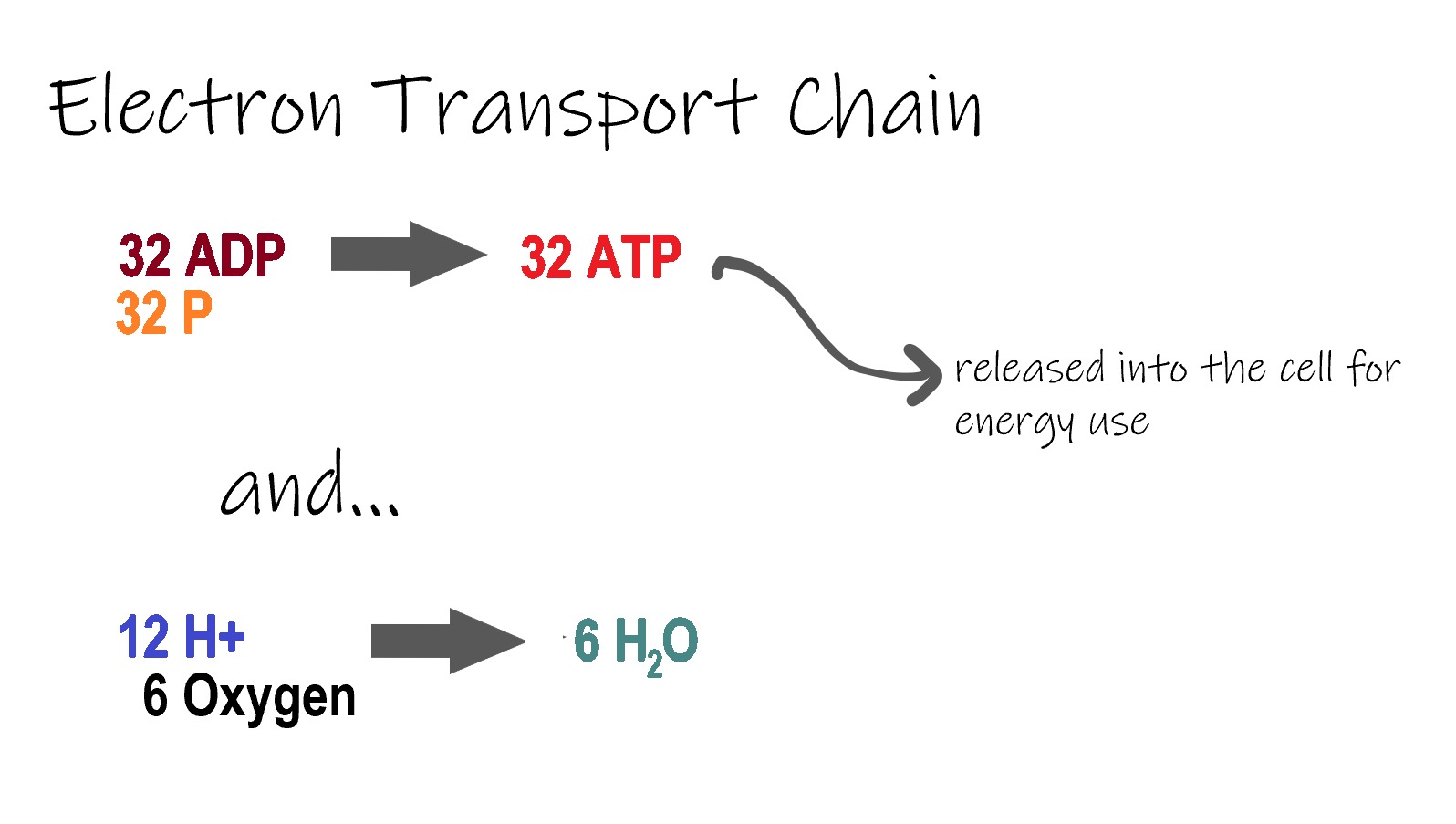
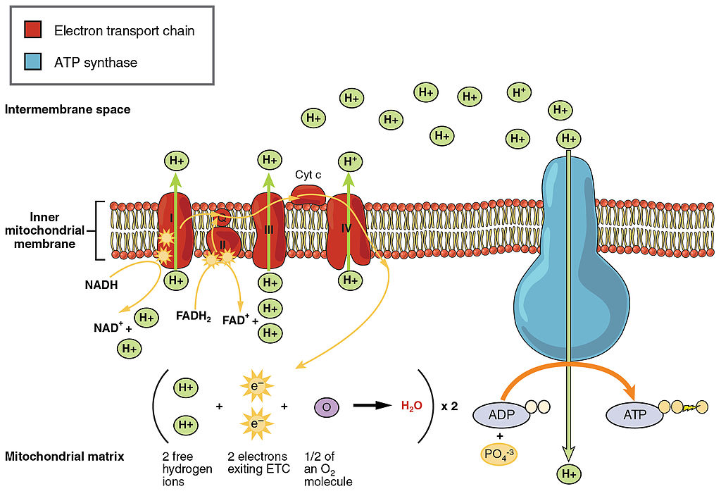
ETC, the final stage in cellular respiration produces 32 ATP. The Electron Transport Chain is the final stage of cellular respiration. In this stage, energy being transported by NADH and FADH2 is transferred to ATP. In addition, oxygen acts as the final proton acceptor for the hydrogens released from all the NADH and FADH2, forming water. Figure 4.10.8 shows the reactants and products of the ETC.
Transporting Electrons
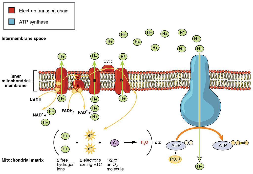
The Electron transport chain is the third stage of cellular respiration and is illustrated in Figure 4.10.8. During this stage, high-energy electrons are released from NADH and FADH2, and they move along electron-transport chains on the inner membrane of the mitochondrion. An electron-transport chain is a series of molecules that transfer electrons from molecule to molecule by chemical reactions. Some of the energy from the electrons is used to pump hydrogen ions (H ) across the inner membrane, from the matrix into the intermembrane space. This ion transfer creates an electrochemical gradient that drives the synthesis of ATP.
Making ATP
As shown in Figure 4.10.8, the pumping of hydrogen ions across the inner membrane creates a greater concentration of the ions in the intermembrane space than in the matrix. This gradient causes the ions to flow back across the membrane into the matrix, where their concentration is lower. ATP synthase acts as a channel protein, helping the hydrogen ions cross the membrane. It also acts as an enzyme, forming ATP from ADP and inorganic phosphate in a process called oxidative phosphorylation. After passing through the electron-transport chain, the “spent” electrons combine with oxygen to form water.
How Much ATP?
You have seen how the three stages of aerobic respiration use the energy in glucose to make ATP. How much ATP is produced in all three stages combined? Glycolysis produces two ATP molecules, and the Krebs cycle produces two more. Electron transport begins with several molecules of NADH and FADH2 from the Krebs cycle and transfers their energy into as many as 34 more ATP molecules. All told, then, up to 38 molecules of ATP can be produced from just one molecule of glucose in the process of cellular respiration.
4.10 Summary
- Cellular respiration is the aerobic process by which living cells break down glucose molecules, release energy, and form molecules of ATP. Generally speaking, this three-stage process involves glucose and oxygen reacting to form carbon dioxide and water.
- The first stage of cellular respiration, called glycolysis, takes place in the cytoplasm. In this step, enzymes split a molecule of glucose into two molecules of pyruvate, which releases energy that is transferred to ATP. Following glycolysis, a short reaction called the transition reaction converts the pyruvate into two molecules of acetyl CoA.
- The organelle called a mitochondrion is the site of the other two stages of cellular respiration. The mitochondrion has an inner and outer membrane separated by an intermembrane space, and the inner membrane encloses a space called the matrix.
- The second stage of cellular respiration, called the Krebs cycle, takes place in the matrix of a mitochondrion. During this stage, two turns through the cycle result in all of the carbon atoms from the two pyruvate molecules forming carbon dioxide and the energy from their chemical bonds being stored in a total of 16 energy-carrying molecules (including two from glycolysis and two from transition reaction).
- The third and final stage of cellular respiration, called electron transport, takes place on the inner membrane of the mitochondrion. Electrons are transported from molecule to molecule down an electron-transport chain. Some of the energy from the electrons is used to pump hydrogen ions across the membrane, creating an electrochemical gradient that drives the synthesis of many more molecules of ATP.
- In all three stages of cellular respiration combined, as many as 38 molecules of ATP are produced from just one molecule of glucose.
4.10 Review Questions
- What is the purpose of cellular respiration? Provide a concise summary of the process.
- State what happens during glycolysis.
- Describe the structure of a mitochondrion.
- What molecule is present at both the beginning and end of the Krebs cycle?
- What happens during the electron transport stage of cellular respiration?
- How many molecules of ATP can be produced from one molecule of glucose during all three stages of cellular respiration combined?
- Do plants undergo cellular respiration? Why or why not?
- Explain why the process of cellular respiration described in this section is considered aerobic.
- Name three energy-carrying molecules involved in cellular respiration.
-
- Which stage of aerobic cellular respiration produces the most ATP?
-
4.10 Explore More
https://www.youtube.com/watch?time_continue=2&v=00jbG_cfGuQ&feature=emb_logo
ATP & Respiration: Crash Course Biology #7, CrashCourse, 2012.
https://www.youtube.com/watch?v=4Eo7JtRA7lg&t=3s
Cellular Respiration and the Mighty Mitochondria, The Amoeba Sisters, 2014.
Attributions
Figure 4.10.1
Smores by Jessica Ruscello on Unsplash is used under the Unsplash License (https://unsplash.com/license).
Figure 4.10.2
Carbohydrate_Metabolism by OpenStax College on Wikimedia Commons is used under a CC BY 3.0 (https://creativecommons.org/licenses/by/3.0) license.
Figure 4.10.3
Glycolysis by Christine Miller is used under a CC BY 4.0 (https://creativecommons.org/licenses/by/4.0/) license.
Figure 4.10.4
Transition Reaction by Christine Miller is used under a CC BY 4.0 (https://creativecommons.org/licenses/by/4.0/) license.
Figure 4.10.5
Mitochondrion by Mariana Ruiz Villarreal [LadyofHats] on Wikimedia Commons is released into the public domain (https://en.wikipedia.org/wiki/Public_domain).
Figure 4.10.6
Krebs cycle by Christine Miller is used under a CC BY 4.0 (https://creativecommons.org/licenses/by/4.0/) license.
Figure 4.10.7
Electron Transport Chain (ETC) by Christine Miller is used under a CC BY 4.0 (https://creativecommons.org/licenses/by/4.0/) license.
Figure 4.10.8
The_Electron_Transport_Chain by OpenStax College on Wikimedia Commons is used under a CC BY 3.0 (https://creativecommons.org/licenses/by/3.0) license.
References
CrashCourse. (2012, March 12). ATP & Respiration: Crash Course Biology #7. YouTube. https://www.youtube.com/watch?time_continue=2&v=00jbG_cfGuQ&feature=emb_logo
Betts, J. G., Young, K.A., Wise, J.A., Johnson, E., Poe, B., Kruse, D.H., Korol, O., Johnson, J.E., Womble, M., DeSaix, P. (2013, April 25). Figure 24.8 Electron Transport Chain [digital image]. In Anatomy & Physiology, Connexions (Section ). OpenStax. https://openstax.org/books/anatomy-and-physiology/pages/24-2-carbohydrate-metabolism
Betts, J. G., Young, K.A., Wise, J.A., Johnson, E., Poe, B., Kruse, D.H., Korol, O., Johnson, J.E., Womble, M., DeSaix, P. (2013, April 25). Figure 24.9 Carbohydrate Metabolism [digital image]. In Anatomy & Physiology, Connexions (Section 24.2). OpenStax. https://openstax.org/books/anatomy-and-physiology/pages/24-2-carbohydrate-metabolism
The Amoeba Sisters. (2014, October 22). Cellular Respiration and the Mighty Mitochondria. YouTube. https://www.youtube.com/watch?v=4Eo7JtRA7lg&t=3s
The transfer of genetic variation from one population to another. If the rate of gene flow is high enough, then two populations are considered to have equivalent allele frequencies and therefore effectively be a single population.
Created by: CK-12/Adapted by Christine Miller

Bring on the S'mores!
This inviting camp fire can be used for both heat and light. Heat and light are two forms of energy that are released when a fuel like wood is burned. The cells of living things also get energy by "burning." They "burn" glucose in a process called cellular respiration.
What Is Cellular Respiration?
Cellular respiration is the process by which living cells break down glucose molecules and release energy. The process is similar to burning, although it doesn’t produce light or intense heat as a campfire does. This is because cellular respiration releases the energy in glucose slowly and in many small steps. It uses the energy released to form molecules of ATP, the energy-carrying molecules that cells use to power biochemical processes. In this way, cellular respiration is an example of energy coupling: glucose is broken down in an exothermic reaction, and then the energy from this reaction powers the endothermic reaction of the formation of ATP. Cellular respiration involves many chemical reactions, but they can all be summed up with this chemical equation:
C6H12O6 6O2 → 6CO2 6H2O Chemical Energy (in ATP)
In words, the equation shows that glucose (C6H12O6) and oxygen (O2) react to form carbon dioxide (CO2) and water (H2O), releasing energy in the process. Because oxygen is required for cellular respiration, it is an aerobic process.
Cellular respiration occurs in the cells of all living things, both autotrophs and heterotrophs. All of them burn glucose to form ATP. The reactions of cellular respiration can be grouped into three stages: glycolysis, the Krebs cycle (also called the citric acid cycle), and electron transport. Figure 4.10.2 gives an overview of these three stages, which are also described in detail below.
Cellular Respiration Stage I: Glycolysis
The first stage of cellular respiration is glycolysis, which happens in the cytosol of the cytoplasm.
Splitting Glucose
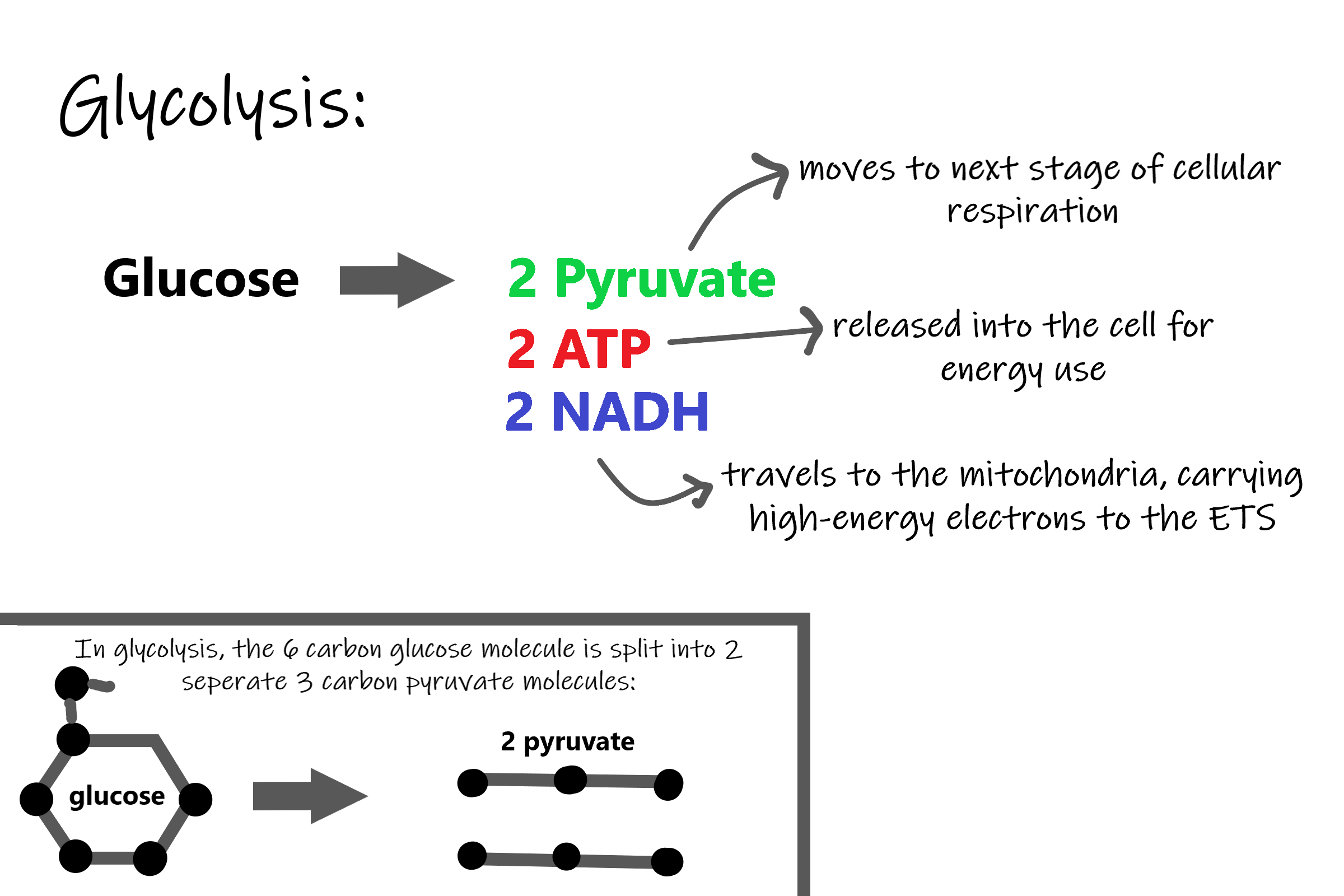
The word glycolysis literally means “glucose splitting,” which is exactly what happens in this stage. Enzymes split a molecule of glucose into two molecules of pyruvate (also known as pyruvic acid). This occurs in several steps, as summarized in the following diagram.
Results of Glycolysis
Energy is needed at the start of glycolysis to split the glucose molecule into two pyruvate molecules which go on to stage II of cellular respiration. The energy needed to split glucose is provided by two molecules of ATP; this is called the energy investment phase. As glycolysis proceeds, energy is released, and the energy is used to make four molecules of ATP; this is the energy harvesting phase. As a result, there is a net gain of two ATP molecules during glycolysis. During this stage, high-energy electrons are also transferred to molecules of NAD to produce two molecules of NADH, another energy-carrying molecule. NADH is used in stage III of cellular respiration to make more ATP.
Transition Reaction
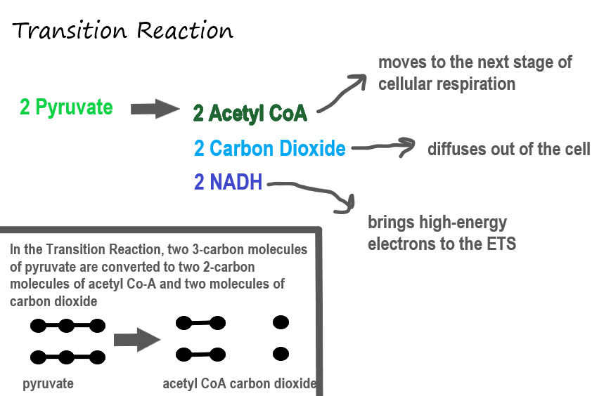
Before pyruvate can enter the next stage of cellular respiration it needs to be modified slightly. The transition reaction is a very short reaction which converts the two molecules of pyruvate to two molecules of acetyl CoA, carbon dioxide, and two high energy electron pairs convert NAD to NADH. The carbon dioxide is released, the acetyl CoA moves to the mitochondria to enter the Kreb's Cycle (stage II), and the NADH carries the high energy electrons to the Electron Transport System (stage III).
Structure of the Mitochondrion
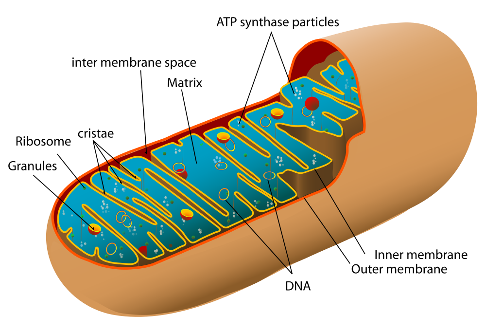
Before you read about the last two stages of cellular respiration, you need to know more about the mitochondrion, where these two stages take place. A diagram of a mitochondrion is shown in Figure 4.10.5.
The structure of a mitochondrion is defined by an inner and outer membrane. This structure plays an important role in aerobic respiration.
As you can see from the figure, a mitochondrion has an inner and outer membrane. The space between the inner and outer membrane is called the intermembrane space. The space enclosed by the inner membrane is called the matrix. The second stage of cellular respiration (the Krebs cycle) takes place in the matrix. The third stage (electron transport) happens on the inner membrane.
Cellular Respiration Stage II: The Krebs Cycle
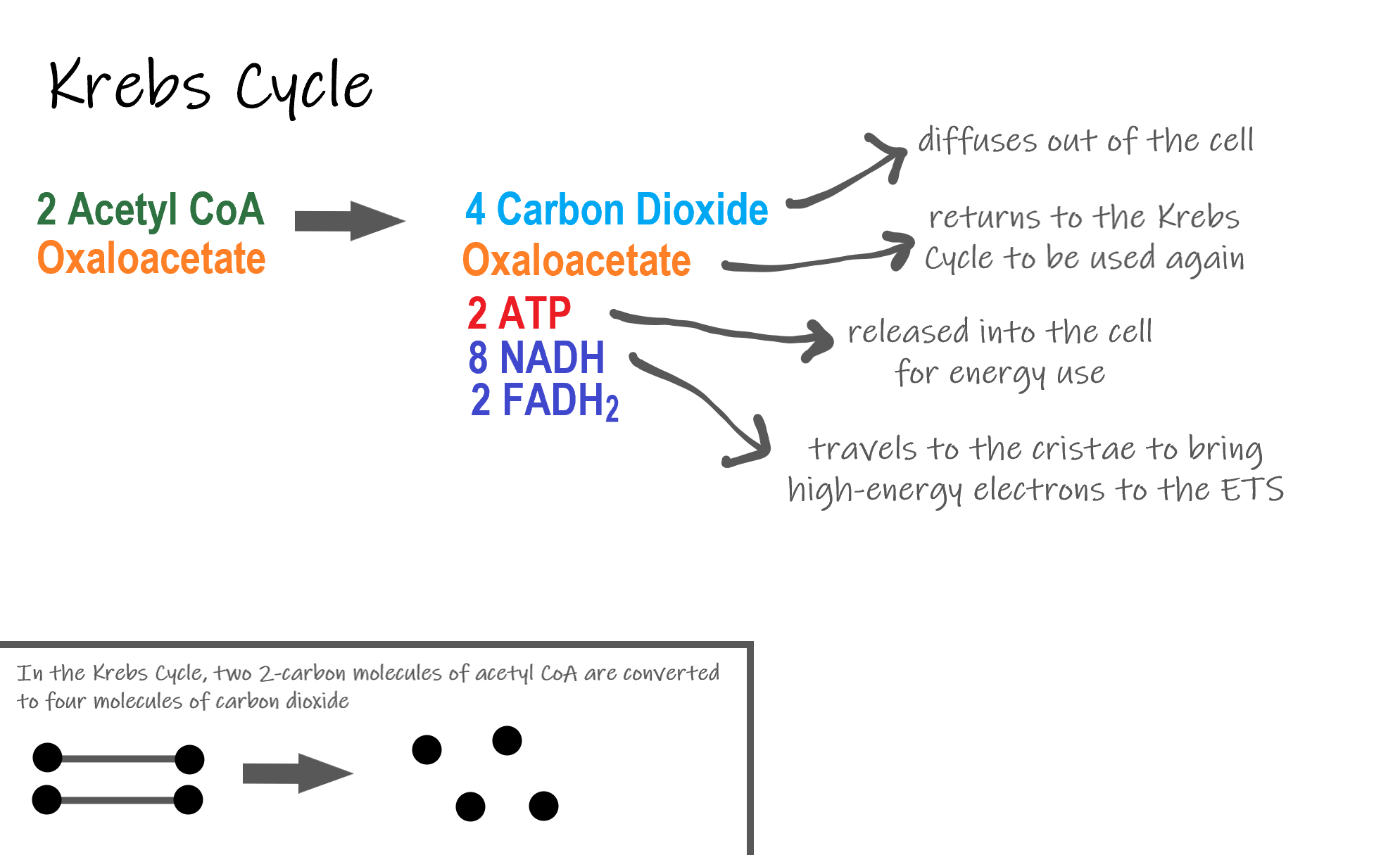
Recall that glycolysis produces two molecules of pyruvate (pyruvic acid), which are then converted to acetyl CoA during the short transition reaction. These molecules enter the matrix of a mitochondrion, where they start the Krebs cycle (also known as the Citric Acid Cycle). The reason this stage is considered a cycle is because a molecule called oxaloacetate is present at both the beginning and end of this reaction and is used to break down the two molecules of acetyl CoA. The reactions that occur next are shown in Figure 4.10.6.
Steps of the Krebs Cycle
The Krebs cycle itself actually begins when acetyl-CoA combines with a four-carbon molecule called OAA (oxaloacetate) (see Figure 4.10.6). This produces citric acid, which has six carbon atoms. This is why the Krebs cycle is also called the citric acid cycle.
After citric acid forms, it goes through a series of reactions that release energy. The energy is captured in molecules of NADH, ATP, and FADH2, another energy-carrying coenzyme. Carbon dioxide is also released as a waste product of these reactions.
The final step of the Krebs cycle regenerates OAA, the molecule that began the Krebs cycle. This molecule is needed for the next turn through the cycle. Two turns are needed because glycolysis produces two pyruvic acid molecules when it splits glucose.
Results of the Glycolysis, Transition Reaction and Krebs Cycle
After glycolysis, transition reaction, and the Krebs cycle, the glucose molecule has been broken down completely. All six of its carbon atoms have combined with oxygen to form carbon dioxide. The energy from its chemical bonds has been stored in a total of 16 energy-carrier molecules. These molecules are:
- 4 ATP (2 from glycolysis, 2 from Krebs Cycle)
- 12 NADH (2 from glycolysis, 2 from transition reaction, and 8 from Krebs cycle)
- 2 FADH2 (both from the Krebs cycle)
The events of cellular respiration up to this point are exergonic reactions- they are releasing energy that had been stored in the bonds of the glucose molecule. This energy will be transferred to the third and final stage of cellular respiration: the Electron Transport System, which is an endergonic reaction. Using an exothermic reaction to power an endothermic reaction is known as energy coupling.
Cellular Respiration Stage III: Electron Transport Chain
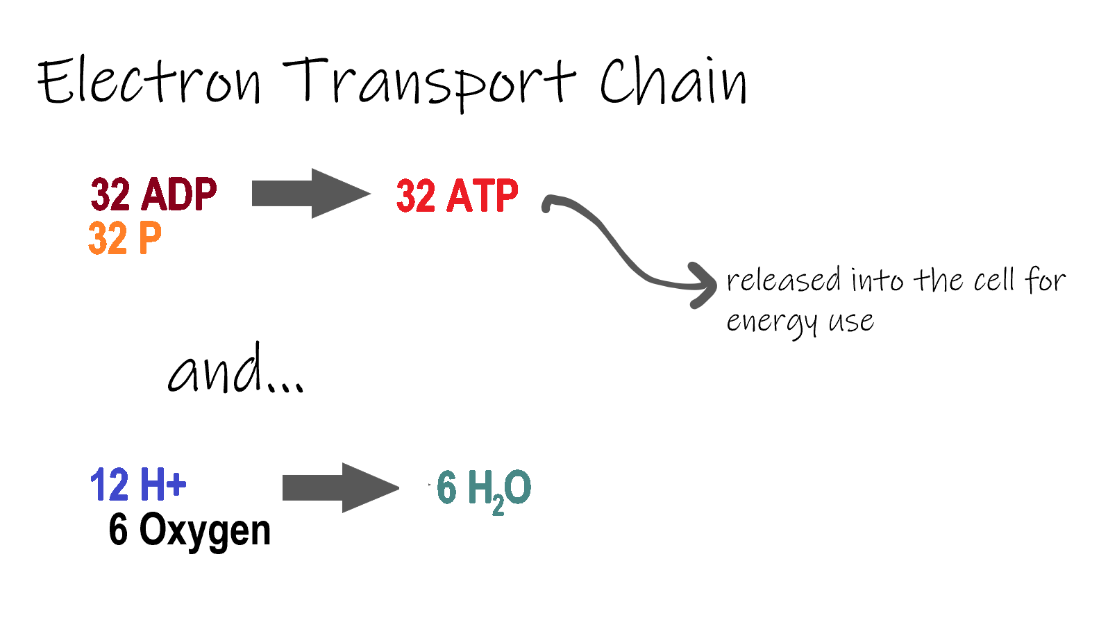
ETC, the final stage in cellular respiration produces 32 ATP. The Electron Transport Chain is the final stage of cellular respiration. In this stage, energy being transported by NADH and FADH2 is transferred to ATP. In addition, oxygen acts as the final proton acceptor for the hydrogens released from all the NADH and FADH2, forming water. Figure 4.10.8 shows the reactants and products of the ETC.
Transporting Electrons
The Electron transport chain is the third stage of cellular respiration and is illustrated in Figure 4.10.8. During this stage, high-energy electrons are released from NADH and FADH2, and they move along electron-transport chains on the inner membrane of the mitochondrion. An electron-transport chain is a series of molecules that transfer electrons from molecule to molecule by chemical reactions. Some of the energy from the electrons is used to pump hydrogen ions (H ) across the inner membrane, from the matrix into the intermembrane space. This ion transfer creates an electrochemical gradient that drives the synthesis of ATP.
Making ATP
As shown in Figure 4.10.8, the pumping of hydrogen ions across the inner membrane creates a greater concentration of the ions in the intermembrane space than in the matrix. This gradient causes the ions to flow back across the membrane into the matrix, where their concentration is lower. ATP synthase acts as a channel protein, helping the hydrogen ions cross the membrane. It also acts as an enzyme, forming ATP from ADP and inorganic phosphate in a process called oxidative phosphorylation. After passing through the electron-transport chain, the “spent” electrons combine with oxygen to form water.
How Much ATP?
You have seen how the three stages of aerobic respiration use the energy in glucose to make ATP. How much ATP is produced in all three stages combined? Glycolysis produces two ATP molecules, and the Krebs cycle produces two more. Electron transport begins with several molecules of NADH and FADH2 from the Krebs cycle and transfers their energy into as many as 34 more ATP molecules. All told, then, up to 38 molecules of ATP can be produced from just one molecule of glucose in the process of cellular respiration.
4.10 Summary
- Cellular respiration is the aerobic process by which living cells break down glucose molecules, release energy, and form molecules of ATP. Generally speaking, this three-stage process involves glucose and oxygen reacting to form carbon dioxide and water.
- The first stage of cellular respiration, called glycolysis, takes place in the cytoplasm. In this step, enzymes split a molecule of glucose into two molecules of pyruvate, which releases energy that is transferred to ATP. Following glycolysis, a short reaction called the transition reaction converts the pyruvate into two molecules of acetyl CoA.
- The organelle called a mitochondrion is the site of the other two stages of cellular respiration. The mitochondrion has an inner and outer membrane separated by an intermembrane space, and the inner membrane encloses a space called the matrix.
- The second stage of cellular respiration, called the Krebs cycle, takes place in the matrix of a mitochondrion. During this stage, two turns through the cycle result in all of the carbon atoms from the two pyruvate molecules forming carbon dioxide and the energy from their chemical bonds being stored in a total of 16 energy-carrying molecules (including two from glycolysis and two from transition reaction).
- The third and final stage of cellular respiration, called electron transport, takes place on the inner membrane of the mitochondrion. Electrons are transported from molecule to molecule down an electron-transport chain. Some of the energy from the electrons is used to pump hydrogen ions across the membrane, creating an electrochemical gradient that drives the synthesis of many more molecules of ATP.
- In all three stages of cellular respiration combined, as many as 38 molecules of ATP are produced from just one molecule of glucose.
4.10 Review Questions
- What is the purpose of cellular respiration? Provide a concise summary of the process.
- State what happens during glycolysis.
- Describe the structure of a mitochondrion.
- What molecule is present at both the beginning and end of the Krebs cycle?
- What happens during the electron transport stage of cellular respiration?
- How many molecules of ATP can be produced from one molecule of glucose during all three stages of cellular respiration combined?
- Do plants undergo cellular respiration? Why or why not?
- Explain why the process of cellular respiration described in this section is considered aerobic.
- Name three energy-carrying molecules involved in cellular respiration.
-
- Which stage of aerobic cellular respiration produces the most ATP?
-
4.10 Explore More
https://www.youtube.com/watch?time_continue=2&v=00jbG_cfGuQ&feature=emb_logo
ATP & Respiration: Crash Course Biology #7, CrashCourse, 2012.
https://www.youtube.com/watch?v=4Eo7JtRA7lg&t=3s
Cellular Respiration and the Mighty Mitochondria, The Amoeba Sisters, 2014.
Attributions
Figure 4.10.1
Smores by Jessica Ruscello on Unsplash is used under the Unsplash License (https://unsplash.com/license).
Figure 4.10.2
Carbohydrate_Metabolism by OpenStax College on Wikimedia Commons is used under a CC BY 3.0 (https://creativecommons.org/licenses/by/3.0) license.
Figure 4.10.3
Glycolysis by Christine Miller is used under a CC BY 4.0 (https://creativecommons.org/licenses/by/4.0/) license.
Figure 4.10.4
Transition Reaction by Christine Miller is used under a CC BY 4.0 (https://creativecommons.org/licenses/by/4.0/) license.
Figure 4.10.5
Mitochondrion by Mariana Ruiz Villarreal [LadyofHats] on Wikimedia Commons is released into the public domain (https://en.wikipedia.org/wiki/Public_domain).
Figure 4.10.6
Krebs cycle by Christine Miller is used under a CC BY 4.0 (https://creativecommons.org/licenses/by/4.0/) license.
Figure 4.10.7
Electron Transport Chain (ETC) by Christine Miller is used under a CC BY 4.0 (https://creativecommons.org/licenses/by/4.0/) license.
Figure 4.10.8
The_Electron_Transport_Chain by OpenStax College on Wikimedia Commons is used under a CC BY 3.0 (https://creativecommons.org/licenses/by/3.0) license.
References
CrashCourse. (2012, March 12). ATP & Respiration: Crash Course Biology #7. YouTube. https://www.youtube.com/watch?time_continue=2&v=00jbG_cfGuQ&feature=emb_logo
Betts, J. G., Young, K.A., Wise, J.A., Johnson, E., Poe, B., Kruse, D.H., Korol, O., Johnson, J.E., Womble, M., DeSaix, P. (2013, April 25). Figure 24.8 Electron Transport Chain [digital image]. In Anatomy & Physiology, Connexions (Section ). OpenStax. https://openstax.org/books/anatomy-and-physiology/pages/24-2-carbohydrate-metabolism
Betts, J. G., Young, K.A., Wise, J.A., Johnson, E., Poe, B., Kruse, D.H., Korol, O., Johnson, J.E., Womble, M., DeSaix, P. (2013, April 25). Figure 24.9 Carbohydrate Metabolism [digital image]. In Anatomy & Physiology, Connexions (Section 24.2). OpenStax. https://openstax.org/books/anatomy-and-physiology/pages/24-2-carbohydrate-metabolism
The Amoeba Sisters. (2014, October 22). Cellular Respiration and the Mighty Mitochondria. YouTube. https://www.youtube.com/watch?v=4Eo7JtRA7lg&t=3s
An involuntary human body response mediated by the nervous and endocrine systems that prepares the body to fight or flee from perceived danger.
Created by CK-12 Foundation/Adapted by Christine Miller
Tonsillitis
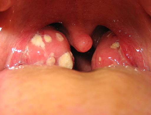
The white patches on either side of the throat in Figure 17.3.1 are signs of tonsillitis. The tonsils are small structures in the throat that are very common sites of infection. The white spots on the tonsils pictured here are evidence of infection. The patches consist of large amounts of dead bacteria, cellular debris, and white blood cells — in a word: pus. Children with recurrent tonsillitis may have their tonsils removed surgically to eliminate this type of infection. The tonsils are organs of the lymphatic system.
What Is the Lymphatic System?
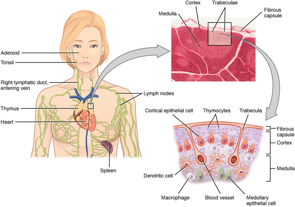
The lymphatic system is a collection of organs involved in the production, maturation, and harboring of white blood cells called lymphocytes. It also includes a network of vessels that transport or filter the fluid known as lymph in which lymphocytes circulate. Figure 17.3.2 shows major lymphatic vessels and other structures that make up the lymphatic system. Besides the tonsils, organs of the lymphatic system include the thymus, the spleen, and hundreds of lymph nodes distributed along the lymphatic vessels.
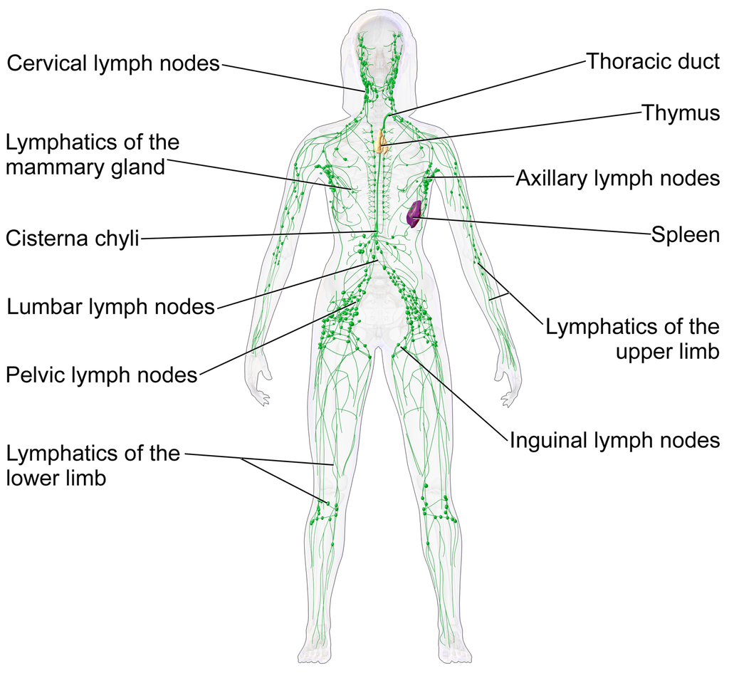
The lymphatic vessels form a transportation network similar in many respects to the blood vessels of the cardiovascular system. However, unlike the cardiovascular system, the lymphatic system is not a closed system. Instead, lymphatic vessels carry lymph in a single direction — always toward the upper chest, where the lymph empties from lymphatic vessels into blood vessels.
Cardiovascular Function of the Lymphatic System
The return of lymph to the bloodstream is one of the major functions of the lymphatic system. When blood travels through capillaries of the cardiovascular system, it is under pressure, which forces some of the components of blood (such as water, oxygen, and nutrients) through the walls of the capillaries and into the tissue spaces between cells, forming tissue fluid, also called interstitial fluid (see Figure 17.3.3). Interstitial fluid bathes and nourishes cells, and also absorbs their waste products. Much of the water from interstitial fluid is reabsorbed into the capillary blood by osmosis. Most of the remaining fluid is absorbed by tiny lymphatic vessels called lymph capillaries. Once interstitial fluid enters the lymphatic vessels, it is called lymph. Lymph is very similar in composition to blood plasma. Besides water, lymph may contain proteins, waste products, cellular debris, and pathogens. It also contains numerous white blood cells, especially the subset of white blood cells known as lymphocytes. In fact, lymphocytes are the main cellular components of lymph.
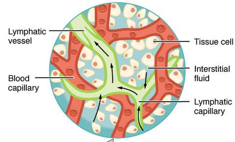
The lymph that enters lymph capillaries in tissues is transported through the lymphatic vessel network to two large lymphatic ducts in the upper chest. From there, the lymph flows into two major veins (called subclavian veins) of the cardiovascular system. Unlike blood, lymph is not pumped through its network of vessels. Instead, lymph moves through lymphatic vessels via a combination of contractions of the vessels themselves and the forces applied to the vessels externally by skeletal muscles, similarly to how blood moves through veins. Lymphatic vessels also contain numerous valves that keep lymph flowing in just one direction, thereby preventing backflow.
Digestive Function of the Lymphatic System
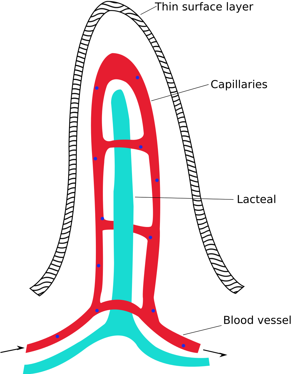
Lymphatic vessels called lacteals (see Figure 17.3.4) are present in the lining of the gastrointestinal tract, mainly in the small intestine. Each tiny villus in the lining of the small intestine has an internal bed of capillaries and lacteals. The capillaries absorb most nutrients from the digestion of food into the blood. The lacteals absorb mainly fatty acids from lipid digestion into the lymph, forming a fatty-acid-enriched fluid called chyle. Vessels of the lymphatic network then transport chyle from the small intestine to the main lymphatic ducts in the chest, from which it drains into the blood circulation. The nutrients in chyle then circulate in the blood to the liver, where they are processed along with the other nutrients that reach the liver directly via the bloodstream.
Immune Function of the Lymphatic System
The primary immune function of the lymphatic system is to protect the body against pathogens and cancerous cells. This function of the lymphatic system is centred on the production, maturation, and circulation of lymphocytes. Lymphocytes are leukocytes that are involved in the adaptive immune system. They are responsible for the recognition of — and tailored defense against — specific pathogens or tumor cells. Lymphocytes may also create a lasting memory of pathogens, so they can be attacked quickly and strongly if they ever invade the body again. In this way, lymphocytes bring about long-lasting immunity to specific pathogens.
There are two major types of lymphocytes, called B cells and T cells. Both B cells and T cells are involved in the adaptive immune response, but they play different roles.
Production and Maturation of Lymphocytes
Like all other types of blood cells (including erythrocytes), both B cells and T cells are produced from stem cells in the red marrow inside bones. After lymphocytes first form, they must go through a complicated maturation process before they are ready to search for pathogens. In this maturation process, they “learn” to distinguish self from non-self. Only those lymphocytes that successfully complete this maturation process go on to actually fight infections by pathogens.
B cells mature in the bone marrow, which is why they are called B cells. After they mature and leave the bone marrow, they travel first to the circulatory system and then enter the lymphatic system to search for pathogens. T cells, on the other hand, mature in the thymus, which is why they are called T cells. The thymus is illustrated in Figure 17.3.5. It is a small lymphatic organ in the chest that consists of an outer cortex and inner medulla, all surrounded by a fibrous capsule. After maturing in the thymus, T cells enter the rest of the lymphatic system to join B cells in the hunt for pathogens. The bone marrow and thymus are called primary lymphoid organs because of their role in the production and/or maturation of lymphocytes.
Lymphocytes in Secondary Lymphoid Organs
The tonsils, spleen, and lymph nodes are referred to as secondary lymphoid organs. These organs do not produce or mature lymphocytes. Instead, they filter lymph and store lymphocytes. It is in these secondary lymphoid organs that pathogens (or their antigens) activate lymphocytes and initiate adaptive immune responses. Activation leads to cloning of pathogen-specific lymphocytes, which then circulate between the lymphatic system and the blood, searching for and destroying their specific pathogens by producing antibodies against them.
Tonsils
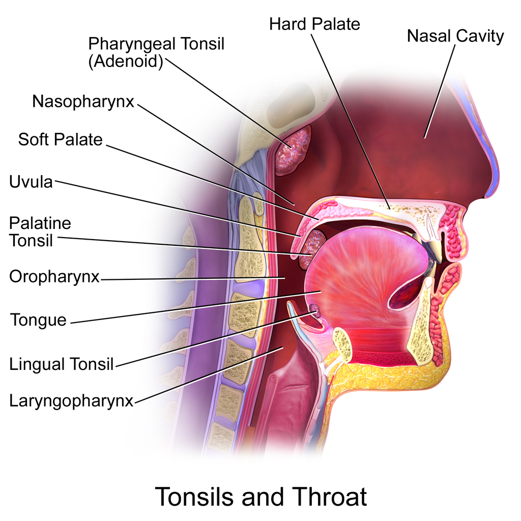
There are four pairs of human tonsils. Three of the four are shown in Figure 17.3.6. The fourth pair, called tubal tonsils, is located at the back of the nasopharynx. The palatine tonsils are the tonsils that are visible on either side of the throat. All four pairs of tonsils encircle a part of the anatomy where the respiratory and gastrointestinal tracts intersect, and where pathogens have ready access to the body. This ring of tonsils is called Waldeyer's ring.
Spleen
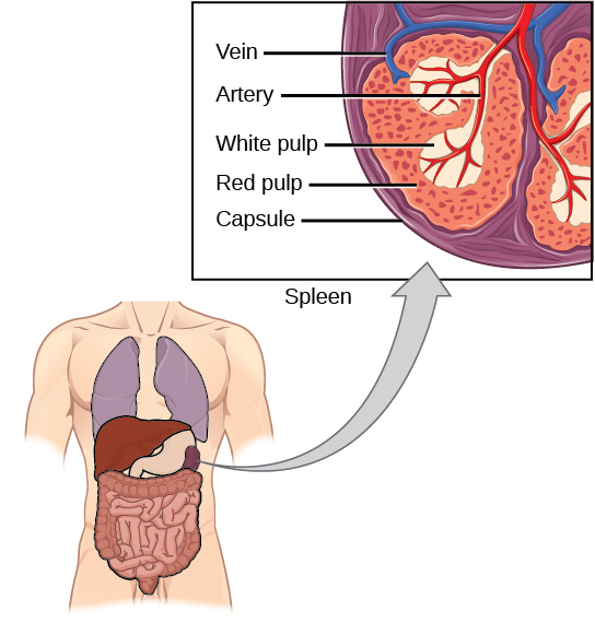
The spleen (Figure 17.3.7) is the largest of the secondary lymphoid organs, and is centrally located in the body. Besides harboring lymphocytes and filtering lymph, the spleen also filters blood. Most dead or aged erythrocytes are removed from the blood in the red pulp of the spleen. Lymph is filtered in the white pulp of the spleen. In the fetus, the spleen has the additional function of producing red blood cells. This function is taken over by bone marrow after birth.
Lymph Nodes
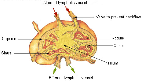
Each lymph node is a small, but organized collection of lymphoid tissue (see Figure 17.3.8) that contains many lymphocytes. Lymph nodes are located at intervals along the lymphatic vessels, and lymph passes through them on its way back to the blood.
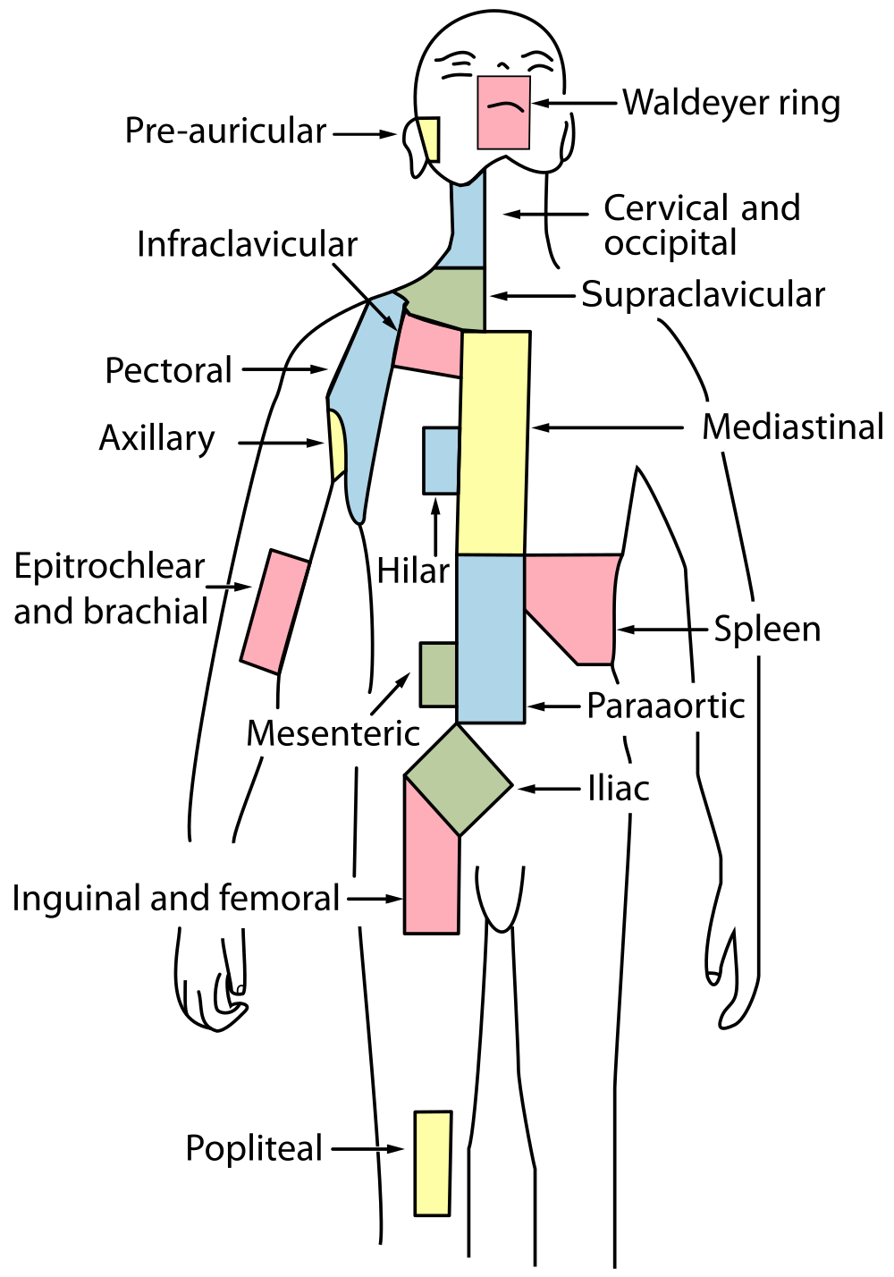
There are at least 500 lymph nodes in the human body. Many of them are clustered at the base of the limbs and in the neck. Figure 17.3.9 shows the major lymph node concentrations, and includes the spleen and the region named Waldeyer’s ring, which consists of the tonsils.
Feature: Myth vs. Reality
When lymph nodes become enlarged and tender to the touch, they are obvious signs of immune system activity. Because it is easy to see and feel swollen lymph nodes, they are one way an individual can monitor his or her own health. To be useful in this way, it is important to know the myths and realities about swollen lymph nodes.
Myth
|
Reality
|
| "You should see a doctor immediately whenever you have swollen lymph nodes." | Lymph nodes are constantly filtering lymph, so it is expected that they will change in size with varying amounts of debris or pathogens that may be present. A minor, unnoticed infection may cause swollen lymph nodes that may last for a few weeks. Generally, lymph nodes that return to their normal size within two or three weeks are not a cause for concern. |
| "Swollen lymph nodes mean you have a bacterial infection." | Although an infection is the most common cause of swollen lymph nodes, not all infections are caused by bacteria. Mononucleosis, for example, commonly causes swollen lymph nodes, and it is caused by viruses. There are also other causes of swollen lymph nodes besides infections, such as cancer and certain medications. |
| "A swollen lymph node means you have cancer." | Cancer is far less likely to be the cause of a swollen lymph node than is an infection. However, if a lymph node remains swollen longer than a few weeks — especially in the absence of an apparent infection — you should have your doctor check it. |
| "Cancer in a lymph node always originates somewhere else. There is no cancer of the lymph nodes." | Cancers do commonly spread from their site of origin to nearby lymph nodes and then to other organs, but cancer may also originate in the lymph nodes. This type of cancer is called lymphoma. |
17.3 Summary
- The lymphatic system is a collection of organs involved in the production, maturation, and harboring of leukocytes called lymphocytes. It also includes a network of vessels that transport or filter the fluid called lymph in which lymphocytes circulate.
- The return of lymph to the bloodstream is one of the functions of the lymphatic system. Lymph flows from tissue spaces — where it leaks out of blood vessels — to the subclavian veins in the upper chest, where it is returned to the cardiovascular system. Lymph is similar in composition to blood plasma. Its main cellular components are lymphocytes.
- Lymphatic vessels called lacteals are found in villi that line the small intestine. Lacteals absorb fatty acids from the digestion of lipids in the digestive system. The fatty acids are then transported through the network of lymphatic vessels to the bloodstream.
- The primary immune function of the lymphatic system is to protect the body against pathogens and cancerous cells. It is responsible for producing mature lymphocytes and circulating them in lymph. Lymphocytes, which include B cells and T cells, are the subset of white blood cells involved in adaptive immune responses. They may create a lasting memory of and immunity to specific pathogens.
- All lymphocytes are produced in bone marrow and then go through a process of maturation in which they “learn” to distinguish self from non-self. B cells mature in the bone marrow, and T cells mature in the thymus. Both the bone marrow and thymus are considered primary lymphatic organs.
- Secondary lymphatic organs include the tonsils, spleen, and lymph nodes. There are four pairs of tonsils that encircle the throat. The spleen filters blood, as well as lymph. There are hundreds of lymph nodes located in clusters along the lymphatic vessels. All of these secondary organs filter lymph and store lymphocytes, so they are sites where pathogens encounter and activate lymphocytes and initiate adaptive immune responses.
17.3 Review Questions
- What is the lymphatic system?
-
- Summarize the immune function of the lymphatic system.
- Explain the difference between lymphocyte maturation and lymphocyte activation.
17.3 Explore More
https://youtu.be/RMLPwOiYnII
What is Lymphoedema or Lymphedema? Compton Care, 2016.
https://youtu.be/ah74jT00jBA
Spleen physiology What does the spleen do in 2 minutes, Simple Nursing, 2015.
https://youtu.be/L4KexZZAdyA
How to check your lymph nodes, University Hospitals Bristol and Weston NHS FT, 2020.
Attributions
Figure 17.3.1
512px-Tonsillitis by Michaelbladon at English Wikipedia on Wikimedia Commons is in the public domain (https://en.wikipedia.org/wiki/Public_domain). (Transferred from en.wikipedia to Commons by Kauczuk)
Figure 17.3.2
Blausen_0623_LymphaticSystem_Female by BruceBlaus on Wikimedia Commons is used under a CC BY 3.0 (https://creativecommons.org/licenses/by/3.0) license.
Figure 17.3.3
2201_Anatomy_of_the_Lymphatic_System (cropped) by OpenStax College on Wikimedia Commons is used under a CC BY 3.0 (https://creativecommons.org/licenses/by/3.0) license.
Figure 17.3.4
1000px-Intestinal_villus_simplified.svg by Snow93 on Wikimedia Commons is in the public domain (https://en.wikipedia.org/wiki/Public_domain).
Figure 17.3.5
2206_The_Location_Structure_and_Histology_of_the_Thymus by OpenStax College on Wikimedia Commons is used under a CC BY 3.0 (https://creativecommons.org/licenses/by/3.0) license.
Figure 17.3.6
Blausen_0861_Tonsils&Throat_Anatomy2 by BruceBlaus on Wikimedia Commons is used under a CC BY 3.0 (https://creativecommons.org/licenses/by/3.0) license.
Figure 17.3.7
Figure_42_02_14 by CNX OpenStax on Wikimedia Commons is used under a CC BY 4.0 (https://creativecommons.org/licenses/by/4.0) license.
Figure 17.3.8
Illu_lymph_node_structure by NCI/ SEER Training on Wikimedia Commons is in the public domain (https://en.wikipedia.org/wiki/Public_domain). (Archives: https://web.archive.org/web/20070311015818/http://training.seer.cancer.gov/module_anatomy/unit8_2_lymph_compo1_nodes.html)
Figure 17.3.9
1000px-Lymph_node_regions.svg by Fred the Oyster (derivative work) on Wikimedia Commons is in the public domain (https://en.wikipedia.org/wiki/Public_domain). (Original by NCI/ SEER Training)
References
Betts, J. G., Young, K.A., Wise, J.A., Johnson, E., Poe, B., Kruse, D.H., Korol, O., Johnson, J.E., Womble, M., DeSaix, P. (2013, June 19). Figure 21.2 Anatomy of the lymphatic system [digital image]. In Anatomy and Physiology (Section 21.1). OpenStax. https://openstax.org/books/anatomy-and-physiology/pages/21-1-anatomy-of-the-lymphatic-and-immune-systems
Betts, J. G., Young, K.A., Wise, J.A., Johnson, E., Poe, B., Kruse, D.H., Korol, O., Johnson, J.E., Womble, M., DeSaix, P. (2013, June 19). Figure 21.7 Location, structure, and histology of the thymus [digital image]. In Anatomy and Physiology (Section 21.1). OpenStax. https://openstax.org/books/anatomy-and-physiology/pages/21-1-anatomy-of-the-lymphatic-and-immune-systems
Blausen.com Staff. (2014). Medical gallery of Blausen Medical 2014". WikiJournal of Medicine 1 (2). DOI:10.15347/wjm/2014.010. ISSN 2002-4436
Compton Care. (2016, March 7). What is lymphoedema or lymphedema? YouTube. https://www.youtube.com/watch?v=RMLPwOiYnII&feature=youtu.be
OpenStax. (2016, May 27) Figure 14. The spleen is similar to a lymph node but is much larger and filters blood instead of lymph [digital image]. In Open Stax, Biology (Section 42.2). OpenStax CNX. https://cnx.org/contents/GFy_h8cu@10.8:etZobsU-@6/Adaptive-Immune-Response
Simple Nursing. (2015, June 28). Spleen physiology What does the spleen do in 2 minutes. YouTube. https://www.youtube.com/watch?v=ah74jT00jBA&feature=youtu.be
University Hospitals Bristol and Weston NHS FT. (2020, May 13). How to check your lymph nodes. YouTube. https://www.youtube.com/watch?v=L4KexZZAdyA&feature=youtu.be

