14.3 Heart
Lub, Dub
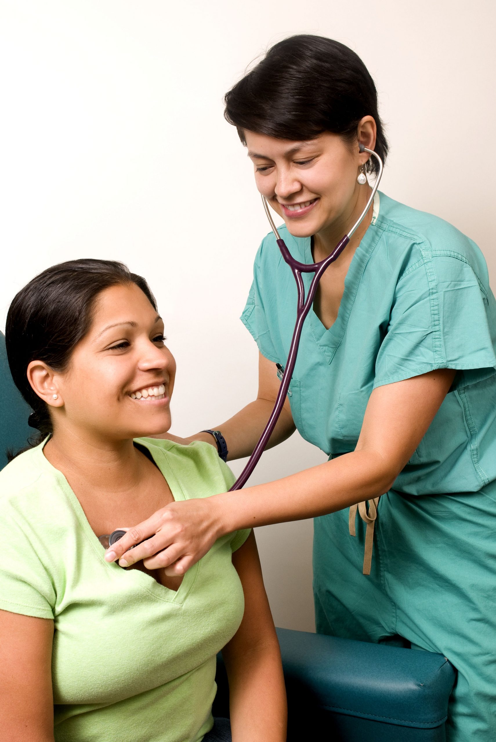
Lub dub, lub dub, lub dub… That’s how the sound of a beating heart is typically described. Those are also the only two sounds that should be audible when listening to a normal, healthy heart through a stethoscope, as in Figure 14.3.1. If a doctor hears something different from the normal lub dub sounds, it’s a sign of a possible heart abnormality. What causes the heart to produce the characteristic lub dub sounds? Read on to find out.
Introduction to the Heart
The heart is a muscular organ behind the sternum (breastbone), slightly to the left of the center of the chest. A normal adult heart is about the size of a fist. The function of the heart is to pump blood through blood vessels of the cardiovascular system. The continuous flow of blood through the system is necessary to provide all the cells of the body with oxygen and nutrients, and to remove their metabolic wastes.
Structure of the Heart
The heart has a thick muscular wall that consists of several layers of tissue. Internally, the heart is divided into four chambers through which blood flows. Because of heart valves, blood flows in just one direction through the chambers.
Heart Wall
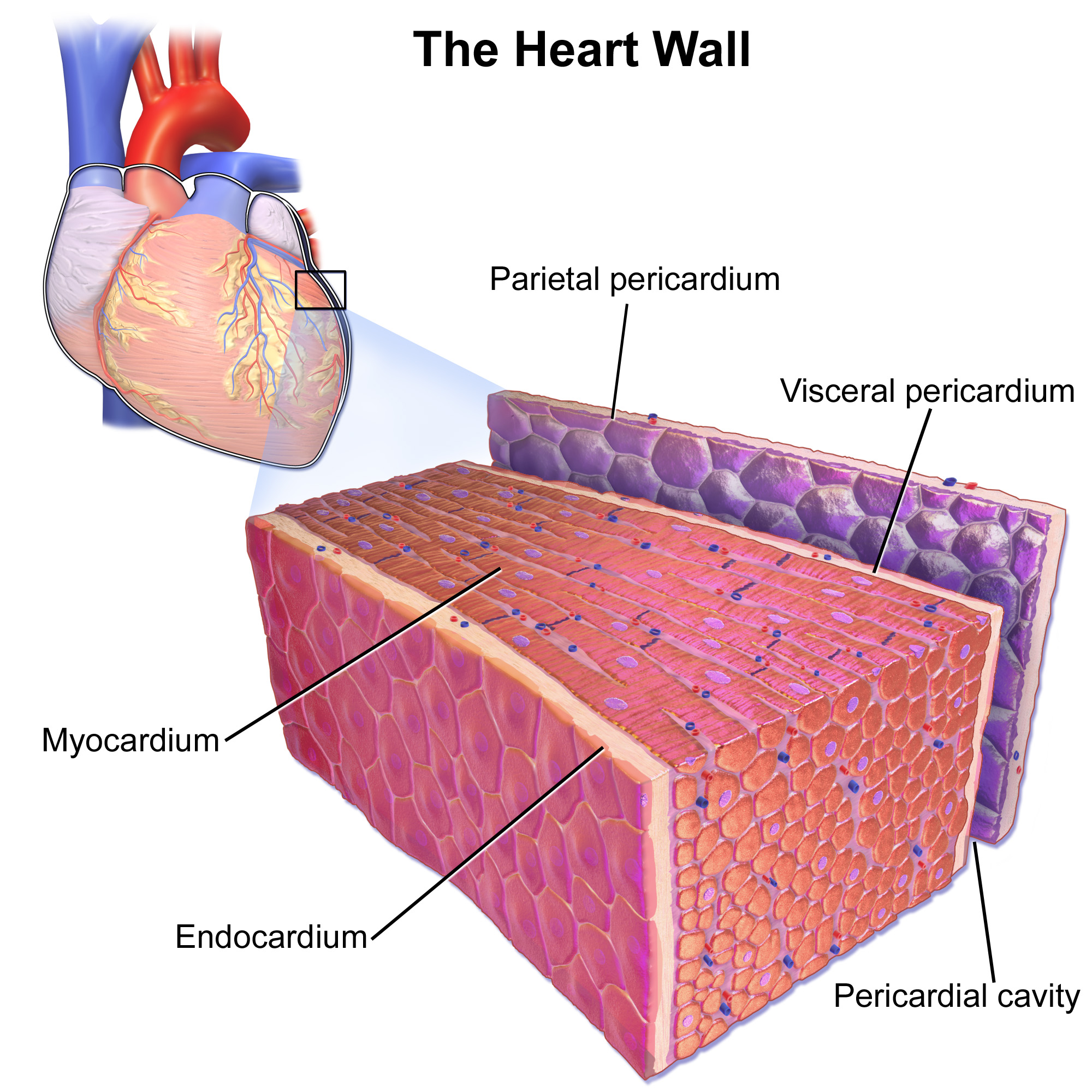
As shown in Figure 14.3.2, the wall of the heart is made up of three layers, called the endocardium, myocardium, and pericardium.
- The endocardium is the innermost layer of the heart wall. It is made up primarily of simple epithelial cells. It covers the heart chambers and valves. A thin layer of connective tissue joins the endocardium to the myocardium.
- The myocardium is the middle and thickest layer of the heart wall. It consists of cardiac muscle surrounded by a framework of collagen. There are two types of cardiac muscle cells in the myocardium: cardiomyocytes — which have the ability to contract easily — and pacemaker cells, which conduct electrical impulses that cause the cardiomyocytes to contract. About 99 per cent of cardiac muscle cells are cardiomyocytes, and the remaining one per cent is pacemaker cells. The myocardium is supplied with blood vessels and nerve fibres via the pericardium.
- The pericardium is a protective sac that encloses and protects the heart. The pericardium consists of two membranes (visceral pericardium and parietal pericardium), between which there is a fluid-filled cavity. The fluid helps to cushion the heart, and also lubricates its outer surface.
Heart Chambers
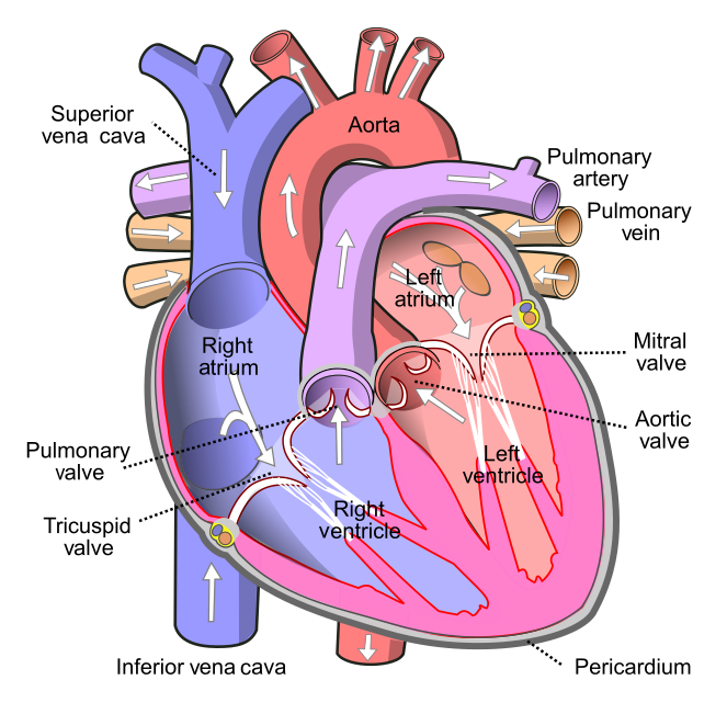
As shown in Figure 14.3.3 the four chambers of the heart include two upper chambers called atria (singular, atrium), and two lower chambers called ventricles. The atria are also referred to as receiving chambers, because blood coming into the heart first enters these two chambers. The right atrium receives deoxygenated blood from the upper and lower body through the superior vena cava and inferior vena cava, respectively. The left atrium receives oxygenated blood from the lungs through the pulmonary veins. The ventricles are also referred to as discharging chambers, because blood leaving the heart passes out through these two chambers. The right ventricle discharges blood to the lungs through the pulmonary artery, and the left ventricle discharges blood to the rest of the body through the aorta. The four chambers are separated from each other by dense connective tissue consisting mainly of collagen.
Heart Valves
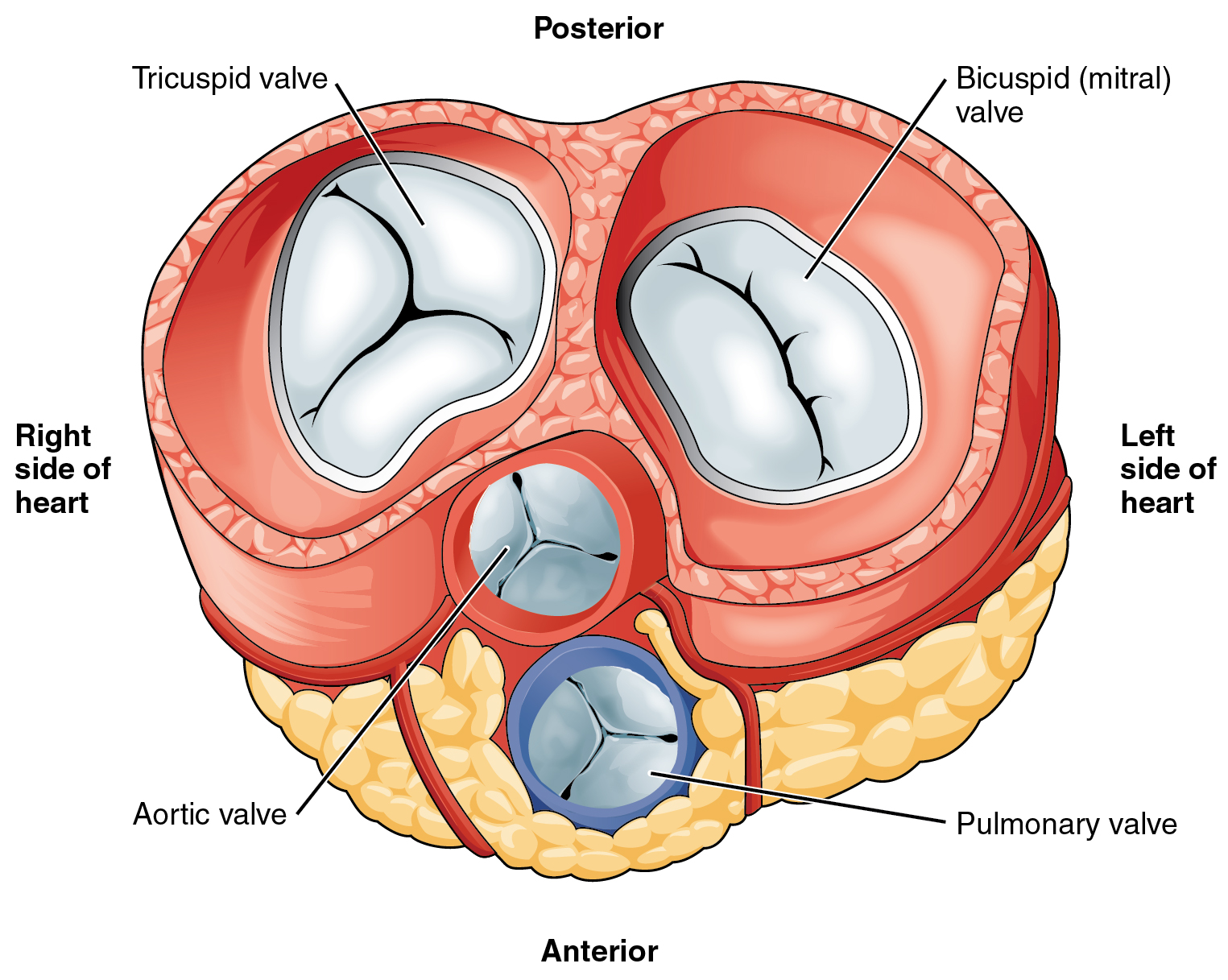
Figure 14.3.4 shows the location of the heart’s four valves in a top-down view, looking down at the heart as if the arteries and veins feeding into and out of the heart were removed. The heart valves allow blood to flow from the atria to the ventricles, and from the ventricles to the pulmonary artery and aorta. The valves are constructed in such a way that blood can flow through them in only one direction, thus preventing the backflow of blood. Figure 14.3.5 shows how valves open to let blood into the appropriate chamber, and then close to prevent blood from moving in the wrong direction and the next chamber contracts. The four valves are the:
- Tricuspid atrioventricular valve, (can be shortened to tricuspid AV valve) which allows blood to flow from the right atrium to the right ventricle.
- Bicuspid atrioventricular valve (also know as the mitral valve), which allows blood to flow from the left atrium to the left ventricle.
- Pulmonary semilunar valve, which allows blood to flow from the right ventricle to the pulmonary artery.
- Aortic semilunar valve, which allows blood to flow from the left ventricle to the aorta.
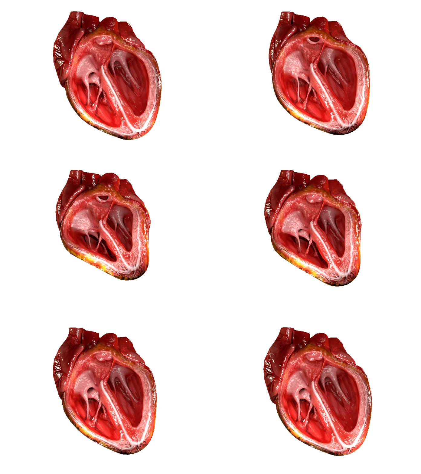
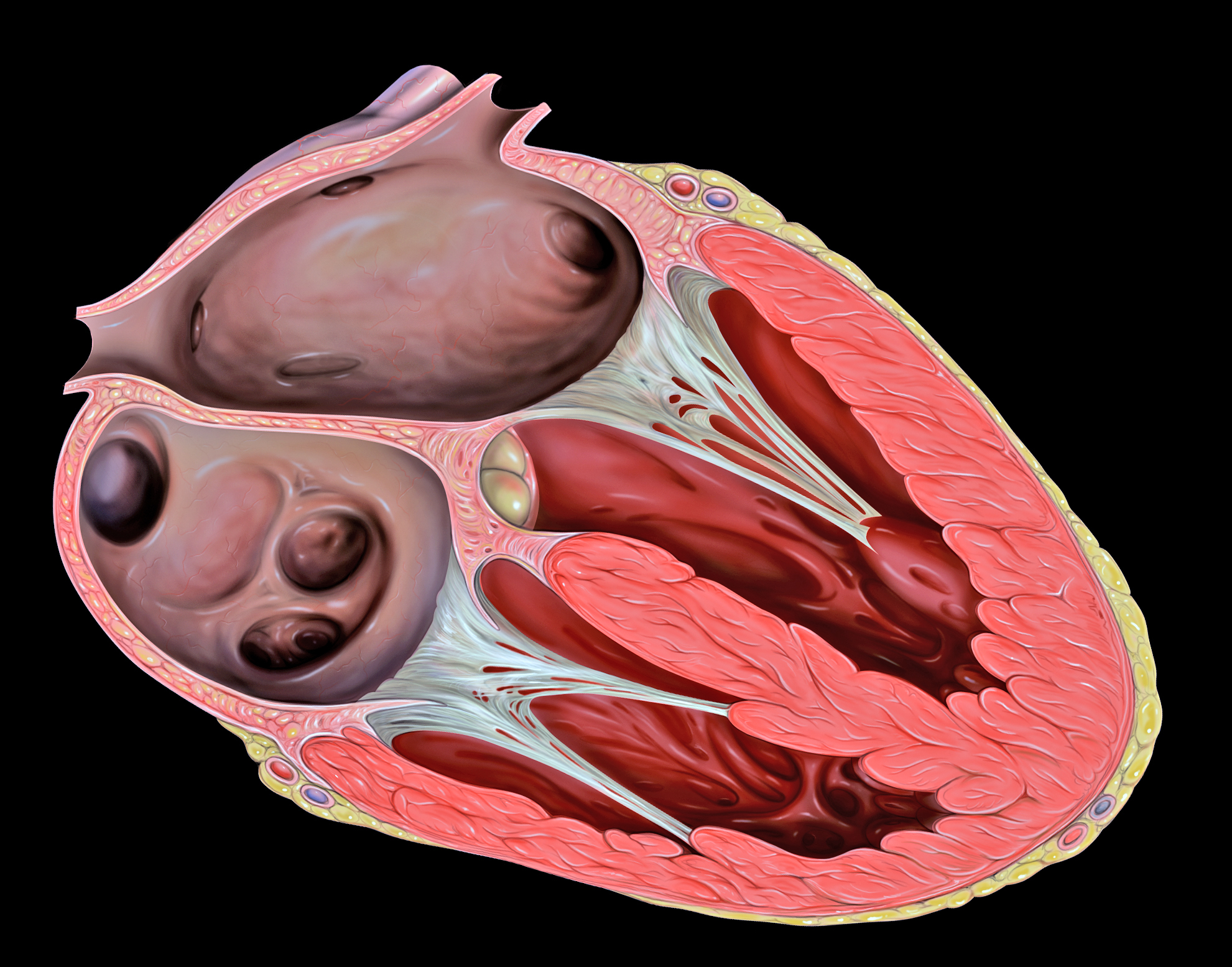
The two atrioventricular (AV) valves prevent backflow when the ventricles are contracting, while the semilunar valves prevent backflow from vessels. This means that the AV valves must withstand much more pressure than do the semilunar valves. In order to withstand the force of the ventricles contracting (to prevent blood from backflowing into the atria), the AV valves are reinforced with structures called chordae tendineae — tendon-like cords of connective tissue which anchor the valve and prevent it from prolapse. Figure 14.3.6 shows the structure and location of the chordae tendoneae.
The chordae tendoneae are under such force that they need special attachments to the interior of the ventricles where they anchor. Papillary muscles are specialized muscles in the interior of the ventricle that provide a strong anchor point for the chordae tendineae.
Coronary Circulation
The cardiomyocytes of the muscular walls of the heart are very active cells, because they are responsible for the constant beating of the heart. These cells need a continuous supply of oxygen and nutrients. The carbon dioxide and waste products they produce also must be continuously removed. The blood vessels that carry blood to and from the heart muscle cells make up the coronary circulation. Note that the blood vessels of the coronary circulation supply heart tissues with blood, and are different from the blood vessels that carry blood to and from the chambers of the heart as part of the general circulation. Coronary arteries supply oxygen-rich blood to the heart muscle cells. Coronary veins remove deoxygenated blood from the heart muscles cells.
- There are two coronary arteries — a right coronary artery that supplies the right side of the heart, and a left coronary artery that supplies the left side of the heart. These arteries branch repeatedly into smaller and smaller arteries and finally into capillaries, which exchange gases, nutrients, and waste products with cardiomyocytes.
- At the back of the heart, small cardiac veins drain into larger veins, and finally into the great cardiac vein, which empties into the right atrium. At the front of the heart, small cardiac veins drain directly into the right atrium.
Blood Circulation Through the Heart
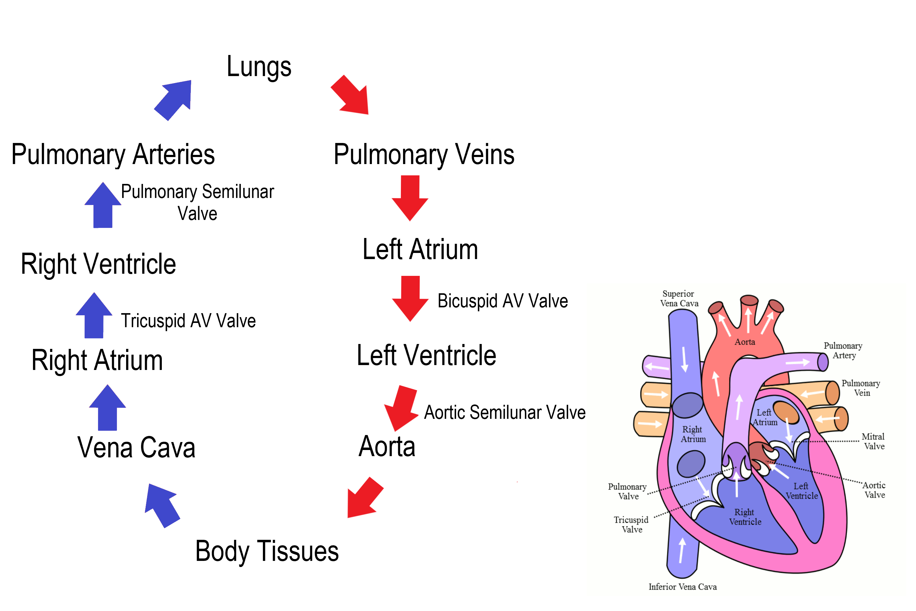
Figure 14.3.7 shows how blood circulates through the chambers of the heart. The right atrium collects blood from two large veins, the superior vena cava (from the upper body) and the inferior vena cava (from the lower body). The blood that collects in the right atrium is pumped through the tricuspid valve into the right ventricle. From the right ventricle, the blood is pumped through the pulmonary valve into the pulmonary artery. The pulmonary artery carries the blood to the lungs, where it enters the pulmonary circulation, gives up carbon dioxide, and picks up oxygen. The oxygenated blood travels back from the lungs through the pulmonary veins (of which there are four), and enters the left atrium of the heart. From the left atrium, the blood is pumped through the mitral valve into the left ventricle. From the left ventricle, the blood is pumped through the aortic valve into the aorta, which subsequently branches into smaller arteries that carry the blood throughout the rest of the body. After passing through capillaries and exchanging substances with cells, the blood returns to the right atrium via the superior vena cava and inferior vena cava, and the process begins anew.
Cardiac Cycle
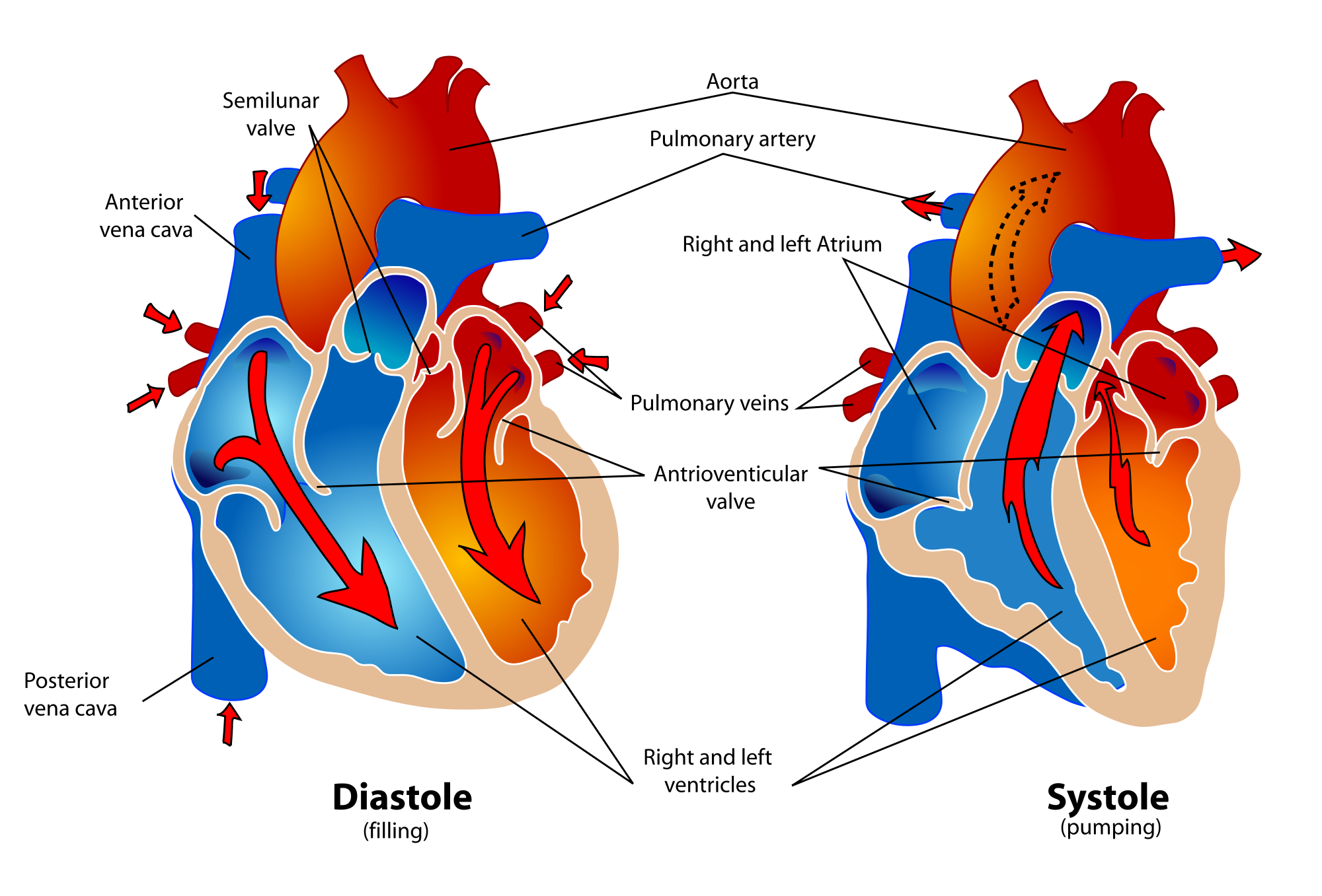
The cardiac cycle refers to a single complete heartbeat, which includes one iteration of the lub and dub sounds heard through a stethoscope. During the cardiac cycle, the atria and ventricles work in a coordinated fashion so that blood is pumped efficiently through and out of the heart. The cardiac cycle includes two parts, called diastole and systole, which are illustrated in the diagrams in Figure 14.3.8.
- During diastole, the atria contract and pump blood into the ventricles, while the ventricles relax and fill with blood from the atria.
- During systole, the atria relax and collect blood from the lungs and body, while the ventricles contract and pump blood out of the heart.
Electrical Stimulation of the Heart
The normal, rhythmical beating of the heart is called sinus rhythm. It is established by the heart’s pacemaker cells, which are located in an area of the heart called the sinoatrial node (shown in Figure 14.3.9). The pacemaker cells create electrical signals with the movement of electrolytes (sodium, potassium, and calcium ions) into and out of the cells. For each cardiac cycle, an electrical signal rapidly travels first from the sinoatrial node, to the right and left atria so they contract together. Then, the signal travels to another node, called the atrioventricular node (Figure 14.3.9), and from there to the right and left ventricles (which also contract together), just a split second after the atria contract.
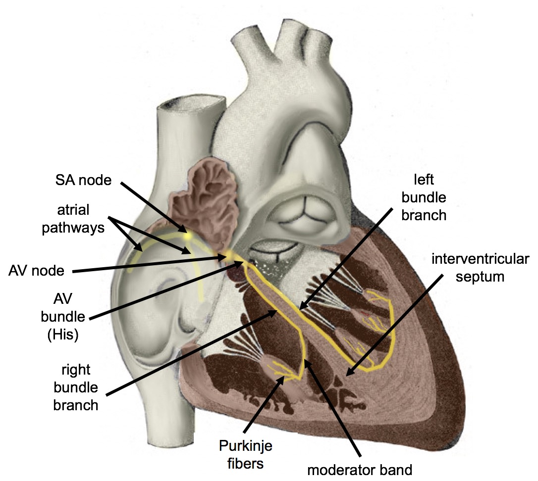
The normal sinus rhythm of the heart is influenced by the autonomic nervous system through sympathetic and parasympathetic nerves. These nerves arise from two paired cardiovascular centers in the medulla of the brainstem. The parasympathetic nerves act to decrease the heart rate, and the sympathetic nerves act to increase the heart rate. Parasympathetic input normally predominates. Without it, the pacemaker cells of the heart would generate a resting heart rate of about 100 beats per minute, instead of a normal resting heart rate of about 72 beats per minute. The cardiovascular centers receive input from receptors throughout the body, and act through the sympathetic nerves to increase the heart rate, as needed. Increased physical activity, for example, is detected by receptors in muscles, joints, and tendons. These receptors send nerve impulses to the cardiovascular centers, causing sympathetic nerves to increase the heart rate, and allowing more blood to flow to the muscles.
Besides the autonomic nervous system, other factors can also affect the heart rate. For example, thyroid hormones and adrenal hormones (such as epinephrine) can stimulate the heart to beat faster. The heart rate also increases when blood pressure drops or the body is dehydrated or overheated. On the other hand, cooling of the body and relaxation — among other factors — can contribute to a decrease in the heart rate.
Feature: Human Biology in the News
When a patient’s heart is too diseased or damaged to sustain life, a heart transplant is likely to be the only long-term solution. The first successful heart transplant was undertaken in South Africa in 1967. There are over 2,200 Canadians walking around today because of life-saving heart transplant surgery. Approximately 180 heart transplant surgeries are performed each year, but there are still so many Canadians on the transplant list that some die while waiting for a heart. The problem is that far too few hearts are available for transplant — there is more demand (people waiting for a heart transplant) than supply (organ donors). Sometimes, recipient hopefuls will receive a device called a Total Artificial Heart (see Figure 14.3.10), which can buy them some time until a donor heart becomes available.
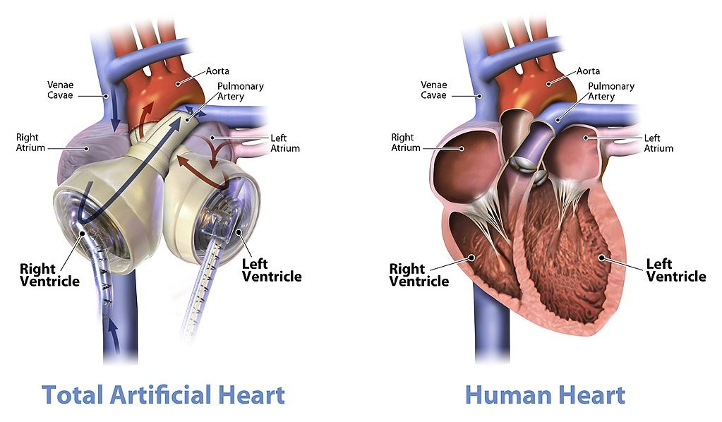
Watch the video below “Total artificial heart option…” from Stanford Health Care to see how it works:
Total artificial heart option at Stanford (Includes surgical graphic footage), Stanford Health Care, 2014.
14.3 Summary
- The heart is a muscular organ behind the sternum and slightly to the left of the center of the chest. Its function is to pump blood through the blood vessels of the cardiovascular system.
- The wall of the heart consists of three layers. The middle layer, the myocardium, is the thickest layer and consists mainly of cardiac muscle. The interior of the heart consists of four chambers, with an upper atrium and lower ventricle on each side of the heart. Blood enters the heart through the atria, which pump it to the ventricles. Then the ventricles pump blood out of the heart. Four valves in the heart keep blood flowing in the correct direction and prevent backflow.
- The coronary circulation consists of blood vessels that carry blood to and from the heart muscle cells, and is different from the general circulation of blood through the heart chambers. There are two coronary arteries that supply the two sides of the heart with oxygenated blood. Cardiac veins drain deoxygenated blood back into the heart.
- Deoxygenated blood flows into the right atrium through veins from the upper and lower body (superior and inferior vena cava, respectively), and oxygenated blood flows into the left atrium through four pulmonary veins from the lungs. Each atrium pumps the blood to the ventricle below it. From the right ventricle, deoxygenated blood is pumped to the lungs through the two pulmonary arteries. From the left ventricle, oxygenated blood is pumped to the rest of the body through the aorta.
- The cardiac cycle refers to a single complete heartbeat. It includes diastole — when the atria contract — and systole, when the ventricles contract.
- The normal, rhythmic beating of the heart is called sinus rhythm. It is established by the heart’s pacemaker cells in the sinoatrial node. Electrical signals from the pacemaker cells travel to the atria, and cause them to contract. Then, the signals travel to the atrioventricular node and from there to the ventricles, causing them to contract. Electrical stimulation from the autonomic nervous system and hormones from the endocrine system can also influence heartbeat.
14.3 Review Questions
- What is the heart, where is located, and what is its function?
-
- Describe the coronary circulation.
- Summarize how blood flows into, through, and out of the heart.
- Explain what controls the beating of the heart.
- What are the two types of cardiac muscle cells in the myocardium? What are the differences between these two types of cells?
- Explain why the blood from the cardiac veins empties into the right atrium of the heart. Focus on function (rather than anatomy) in your answer.
14.3 Explore More
Noel Bairey Merz: The single biggest health threat women face, TED, 2012.
Watch a Transcatheter Aortic Valve Replacement (TAVR) Procedure at St. Luke’s in Cedar Rapids, Iowa, UnityPoint Health – Cedar Rapids, 2018.
A Change of Heart: My Transplant Experience | Thomas Volk | TEDxUWLaCrosse, TEDx Talks, 2018.
Heart Transplant Recipient Meets Donor Family For The First Time, WMC Health, 2018.
Attributions
Figure 14.3.1
- Female clinician dressed in scrubs using a stethoscope by Amanda Mills, USCDCP, on Pixnio is used under a CC0 public domain certification license (https://creativecommons.org/licenses/publicdomain/).
- Human heart beating loud and strong (audio) by Daniel Simion on Soundbible.com is used under a CC BY 3.0 (https://creativecommons.org/licenses/by/3.0) license.
Figure 14.3.2
Blausen_0470_HeartWall by BruceBlaus on Wikimedia Commons is used under a CC BY 3.0 (https://creativecommons.org/licenses/by/3.0) license.
Figure 14.3.3
Diagram_of_the_human_heart_(cropped).svg by Wapcaplet on Wikimedia Commons is used under a CC BY-SA 3.0 (http://creativecommons.org/licenses/by-sa/3.0/) license.
Figure 14.3.4
Heart_Valves by OpenStax College on Wikimedia Commons is used under a CC BY 3.0 (https://creativecommons.org/licenses/by/3.0) license.
Figure 14.3.5
CG_Heart Valve Animation by DrJanaOfficial on Wikimedia Commons is used under a CC BY-SA 4.0 (https://creativecommons.org/licenses/by-sa/4.0) license.
Figure 14.3.6
Heart_tee_four_chamber_view by Patrick J. Lynch, medical illustrator from Yale University School of Medicine, on Wikimedia Commons is used under a CC BY 2.5 (https://creativecommons.org/licenses/by/2.5) license.
Figure 14.3.7
Circulation of blood through the heart by Christinelmiller on Wikimedia Commons is used under a CC BY-SA 4.0 (https://creativecommons.org/licenses/by-sa/4.0) license. [Original image in the bottom right is by Wapcaplet / CC BY-SA 3.0 (https://creativecommons.org/licenses/by-sa/3.0/)]
Figure 14.3.8
Human_healthy_pumping_heart_en.svg by Mariana Ruiz Villarreal [LadyofHats] on Wikimedia Common is released into the public domain (https://en.wikipedia.org/wiki/Public_domain).
Figure 14.3.9
Cardiac_Conduction_System by Cypressvine on Wikimedia Commons is used under a CC BY-SA 4.0 (https://creativecommons.org/licenses/by-sa/4.0) license.
References
Betts, J. G., Young, K.A., Wise, J.A., Johnson, E., Poe, B., Kruse, D.H., Korol, O., Johnson, J.E., Womble, M., DeSaix, P. (2013, June 19). Figure 19.12 Heart valves with the atria and major vessels removed [digital image]. In Anatomy and Physiology (Section 19.1). OpenStax. https://openstax.org/books/anatomy-and-physiology/pages/19-1-heart-anatomy#fig-ch20_01_04
Blausen.com Staff. (2014). Medical gallery of Blausen Medical 2014. WikiJournal of Medicine 1 (2). DOI:10.15347/wjm/2014.010. ISSN 2002-4436.
Heart and Stroke Foundation of Canada. (n.d.). https://www.heartandstroke.ca/
Sliwa, K., Zilla, P. (2017, December 7). 50th anniversary of the first human heart transplant—How is it seen today? European Heart Journal, 38(46):3402–3404. https://doi.org/10.1093/eurheartj/ehx695
Stanford Health Care. (2014, December 3). Total artificial heart option at Stanford (Includes surgical graphic footage). YouTube. https://www.youtube.com/watch?v=1PtxaxcPnGc&feature=youtu.be
TED. (2012, March 21). Noel Bairey Merz: The single biggest health threat women face. YouTube. https://www.youtube.com/watch?v=1bnzVjOJ6NM&feature=youtu.be
TEDx Talks. (2018, April 18). A change of heart: My transplant experience | Thomas Volk | TEDxUWLaCrosse. YouTube. https://www.youtube.com/watch?v=zU6mmix04PI&feature=youtu.be
UMagazine. (2015, Fall). The cutting edge: Patient first to bridge from experimental total artificial heart to transplant. UCLA Health. https://www.uclahealth.org/u-magazine/patient-first-to-bridge-from-experimental-total-artificial-heart-to-transplant
UnityPoint Health – Cedar Rapids. (2018, February 7). Watch a transcatheter aortic valve replacement (TAVR) Procedure at St. Luke’s in Cedar Rapids, Iowa. YouTube. https://www.youtube.com/watch?v=jJm7zBcN6-M&feature=youtu.be
WMC Health. (2018, September 13). Heart transplant recipient meets donor family for the first time. YouTube. https://www.youtube.com/watch?v=biGuwQhuAsk&feature=youtu.be
Created by CK-12 Foundation/Adapted by Christine Miller
Case Study Conclusion: Needing to Relax
As you learned in the beginning of this chapter, botulinum toxin — one form of which is sold under the brand name Botox — does much more than smooth out wrinkles. It can be used to treat a number of disorders involving excessive muscle contraction, including cervical dystonia. You also learned that cervical dystonia, which Edward suffers from, causes abnormal, involuntary muscle contractions of the neck. This results in jerky movements of the head and neck, and/or a sustained abnormal tilt to the head. It is often painful and can significantly interfere with a person’s life.

How could a toxin actually help treat a muscular disorder? The botulinum toxin is produced by the soil bacterium, Clostridium botulinum, and it is the cause of the potentially deadly disease called botulism. Botulism is often a foodborne illness, commonly caused by foods that are improperly canned. Other forms of botulism are caused by wound infections, or occur when infants consume spores of the bacteria from soil or honey.
Botulism can be life-threatening, because it paralyzes muscles throughout the body, including those involved in breathing. When a very small amount of botulinum toxin is injected carefully into specific muscles by a trained medical professional, however, it can be useful in inhibiting unwanted muscle contractions.
For cosmetic purposes, botulinum toxin injected into the facial muscles relaxes them to reduce the appearance of wrinkles. When used to treat cervical dystonia, it is injected into the muscles of the neck to inhibit excessive muscle contractions. For many patients, this helps relieve the abnormal positioning, movements, and pain associated with the disorder. The effect is temporary, so the injections must be repeated every three to four months to keep the symptoms under control.
How does botulinum toxin inhibit muscle contraction? First, recall how skeletal muscle contraction works. A motor neuron instructs skeletal muscle fibres to contract at a synapse between them called the neuromuscular junction. A nerve impulse called an action potential travels down to the axon terminal of the motor neuron, where it causes the release of the neurotransmitter acetylcholine (ACh) from synaptic vesicles. The ACh travels across the synaptic cleft and binds to ACh receptors on the muscle fibre, signaling the muscle fibre to contract. According to the sliding filament theory, the contraction of the muscle fibre occurs due to the sliding of myosin and actin filaments across each other. This causes the Z discs of the sacromeres to move closer together, shortening the sacromeres and causing the muscle fibre to contract.
If you wanted to inhibit muscle contraction, at what points could you theoretically interfere with this process? Inhibiting the action potential in the motor neuron, the release of ACh, the activity of ACh receptors, or the sliding filament process in the muscle fibre would all theoretically impair this process and inhibit muscle contraction. For example, in the disease myasthenia gravis, the function of the ACh receptors is impaired, causing a lack of sufficient muscle contraction. As you have learned, this results in muscle weakness that can eventually become life-threatening. Botulinum toxin works by inhibiting the release of ACh from the motor neurons, thereby removing the signal instructing the muscles to contract.
Fortunately, Edward’s excessive muscle contractions and associated pain improved significantly thanks to botulinum toxin injections. Although cervical dystonia cannot currently be cured, botulinum toxin injections have improved the quality of life for many patients with this and other disorders involving excessive involuntary muscle contractions.
As you have learned in this chapter, our muscular system allows us to do things like make voluntary movements, digest our food, and pump blood through our bodies. Whether they are in your arm, heart, stomach, or blood vessels, muscle tissue works by contracting. But as you have seen here, too much contraction can be a very bad thing. Fortunately, scientists and physicians have found a way to put a potentially deadly toxin — and wrinkle-reducing treatment — to excellent use as a medical treatment for some muscular system disorders.
Chapter 12 Summary
In this chapter, you learned about the muscular system. Specifically, you learned that:
- The muscular system consists of all the muscles of the body. There are three types of muscle: skeletal muscle (which is attached to bones by tendons and enables voluntary body movements), cardiac muscle (which makes up the walls of the heart and makes it beat) and smooth muscle (which is found in the walls of internal organs and other internal structures and controls their movements).
- Muscles are organs composed mainly of muscle cells, which may also be called muscle fibres or myocytes. Muscle cells are specialized for the function of contracting, which occurs when protein filaments inside the cells slide over one another using energy from ATP. Muscle tissue is the only type of tissue that has cells with the ability to contract.
- Muscles can grow larger, or hypertrophy. This generally occurs through increased use, although hormonal or other influences can also play a role. Muscles can also grow smaller, or atrophy. This may occur through lack of use, starvation, certain diseases, or aging. In both hypertrophy and atrophy, the size — but not the number — of muscle fibres changes. The size of muscles is the main determinant of muscle strength.
- Skeletal muscles need the stimulus of motor neurons to contract, and to move the body, they need the skeletal system to act upon.
- Skeletal muscle is the most common type of muscle tissue in the human body. To move bones in opposite directions, skeletal muscles often consist of pairs of muscles that work in opposition to one another to move bones in different directions at joints.
- Skeletal muscle fibres are bundled together in units called muscle fascicles, which are bundled together to form individual skeletal muscles. Skeletal muscles also have connective tissue supporting and protecting the muscle tissue.
-
- Each skeletal muscle fibre consists of a bundle of myofibrils, which are bundles of protein filaments. The filaments are arranged in repeating units called sarcomeres, which are the basic functional units of skeletal muscles. Skeletal muscle tissue is striated, because of the pattern of sarcomeres in its fibres.
- Skeletal muscle fibres can be divided into two types, called slow-twitch and fast-twitch fibres. Slow-twitch fibres are used mainly in aerobic endurance activities (such as long-distance running). Fast-twitch fibres are used mainly for non-aerobic, strenuous activities (such as sprinting). Proportions of the two types of fibres vary from muscle to muscle and person to person.
- Smooth muscle tissue is found in the walls of internal organs and vessels. When smooth muscles contract, they help the organs and vessels carry out their functions. The pattern of smooth muscle contraction to move substances through body tubes is called peristalsis. Contractions of smooth muscles are involuntary and controlled by the autonomic nervous system, hormones, and other substances.
-
- Cells of smooth muscle tissue are not striated because they lack sarcomeres, but the cells contract in the same basic way as striated muscle cells. Unlike striated muscle, smooth muscle can sustain very long-term contractions and maintain its contractile function, even when stretched.
- Cardiac muscle tissue is found only in the wall of the heart. When cardiac muscle contracts, the heart beats and pumps blood. Contractions of cardiac muscle are involuntary, like those of smooth muscles. They are controlled by electrical impulses from specialized cardiac cells.
-
- Like skeletal muscle, cardiac muscle is striated because its filaments are arranged in sarcomeres. The exact arrangement, however, differs, making cardiac and skeletal muscle tissues look different from one another.
- The heart is the muscle that performs the greatest amount of physical work in the course of a lifetime. Its cells contain a great many mitochondria to produce ATP for energy and to help the heart resist fatigue.
- A muscle contraction is an increase in the tension or a decrease in the length of a muscle. A muscle contraction is isometric if muscle tension changes, but muscle length remains the same. It is isotonic if muscle length changes, but muscle tension remains the same.
-
- A skeletal muscle contraction begins with electrochemical stimulation of a muscle fibre by a motor neuron. This occurs at a chemical synapse called a neuromuscular junction. The neurotransmitter acetylcholine diffuses across the synaptic cleft and binds to receptors on the muscle fibre. This initiates a muscle contraction.
- Once stimulated, the protein filaments within the skeletal muscle fibre slide past each other to produce a contraction. The sliding filament theory is the most widely accepted explanation for how this occurs. According to this theory, thick myosin filaments repeatedly attach to and pull on thin actin filaments, thus shortening sarcomeres.
- Crossbridge cycling is a cycle of molecular events that underlies the sliding filament theory. Using energy in ATP, myosin heads repeatedly bind with and pull on actin filaments. This moves the actin filaments toward the center of a sarcomere, shortening the sarcomere and causing a muscle contraction.
- The ATP needed for a muscle contraction comes first from ATP already available in the cell, and more is generated from creatine phosphate. These sources are quickly used up. Glucose and glycogen can be broken down to form ATP and pyruvate. Pyruvate can then be used to produce ATP in aerobic respiration if oxygen is available, or it can be used in anaerobic respiration if oxygen is not available.
- Physical exercise is defined as any bodily activity that enhances or maintains physical fitness and overall health. Activities such as household chores may even count as physical exercise! Current recommendations for adults are 30 minutes of moderate exercise a day.
- Aerobic exercise is any physical activity that uses muscles at less than their maximum contraction strength, but for long periods of time. This type of exercise uses a relatively high percentage of slow-twitch muscle fibres that consume large amounts of oxygen. Aerobic exercises increase cardiovascular endurance, and include cycling and brisk walking.
- Anaerobic exercise is any physical activity that uses muscles at close to their maximum contraction strength, but for short periods of time. This type of exercise uses a relatively high percentage of fast-twitch muscle fibres that consume small amounts of oxygen. Anaerobic exercises increase muscle and bone mass and strength, and they include push-ups and sprinting.
- Flexibility exercise is any physical activity that stretches and lengthens muscles, thereby improving range of motion and reducing risk of injury. Examples include stretching and yoga.
- Many studies have shown that physical exercise is positively correlated with a diversity of physical, mental, and emotional health benefits. Physical exercise also increases quality of life and life expectancy.
-
- Many of the benefits of exercise may come about because contracting muscles release hormones called myokines, which promote tissue repair and growth and have anti-inflammatory effects.
- Physical exercise can reduce risk factors for cardiovascular disease, including hypertension and excess body weight. Physical exercise can also increase factors associated with cardiovascular health, such as mechanical efficiency of the heart.
- Physical exercise has been shown to offer protection from dementia and other cognitive problems, perhaps because it increases blood flow or neurotransmitters in the brain, among other potential effects.
- Numerous studies suggest that regular aerobic exercise works as well as pharmaceutical antidepressants in treating mild-to-moderate depression, possibly because it increases synthesis of natural euphoriants in the brain.
- Research shows that physical exercise generally improves sleep for most people, and helps sleep disorders, such as insomnia. Other health benefits of physical exercise include better immune system function and reduced risk of type 2 diabetes and obesity.
- There is great variation in individual responses to exercise, partly due to genetic differences in proportions of slow-twitch and fast-twitch muscle fibres. People with more slow-twitch fibres may be able to develop greater endurance from aerobic exercise, whereas people with more fast-twitch fibres may be able to develop greater muscle size and strength from anaerobic exercise.
- Some adverse effects may occur if exercise is extremely intense and the body is not given proper rest between exercise sessions. Many people who overwork their muscles develop delayed onset muscle soreness (DOMS), which may be caused by tiny tears in muscle fibres.
- Musculoskeletal disorders are injuries that occur in muscles or associated tissues (such as tendons) because of biomechanical stresses. The disorders may be caused by sudden exertion, over-exertion, repetitive motions, and similar stresses.
-
- A muscle strain is an injury in which muscle fibres tear as a result of overstretching. First aid for a muscle strain includes the five steps represented by the acronym PRICE (protection, rest, ice, compression, and elevation). Medications for inflammation and pain (such as NSAIDs) may also be used.
- Tendinitis is inflammation of a tendon that occurs when it is over-extended or worked too hard without rest. Tendinitis may also be treated with PRICE and NSAIDs.
- Carpal tunnel syndrome is a biomechanical problem that occurs in the wrist when the median nerve becomes compressed between carpal bones. It may occur with repetitive use, a tumor, or trauma to the wrist. It may cause pain, numbness, and eventually — if untreated — muscle wasting in the thumb and first two fingers of the hand.
- Neuromuscular disorders are systemic disorders that occur because of problems with the nervous control of muscle contractions, or with the muscle cells themselves.
-
- Muscular dystrophy is a genetic disorder caused by defective proteins in muscle cells. It is characterized by progressive skeletal muscle weakness and death of muscle tissues.
- Myasthenia gravis is a genetic neuromuscular disorder characterized by fluctuating muscle weakness and fatigue. More muscles are affected, and muscles become increasingly weakened as the disorder progresses. Myasthenia gravis most often occurs because immune system antibodies block acetylcholine receptors on muscle cells, and because of the actual loss of acetylcholine receptors.
- Parkinson’s disease is a degenerative disorder of the central nervous system that mainly affects the muscular system and movement. It occurs because of the death of neurons in the midbrain. Characteristic signs of the disorder are muscle tremor, muscle rigidity, slowness of movement, and postural instability. Dementia and depression also often characterize advanced stages of the disease.
As you saw in this chapter, muscles need oxygen to provide enough ATP for most of their activities. In fact, all of the body’s systems require oxygen, and also need to remove waste products, such as carbon dioxide. In the next chapter, you will learn about how the respiratory system obtains and distributes oxygen throughout the body, as well as how it removes wastes, such as carbon dioxide.
Chapter 12 Review
-
- What are tendons? Name a muscular system disorder involving tendons
- Describe the relationship between muscles, muscle fibres, and fascicles.
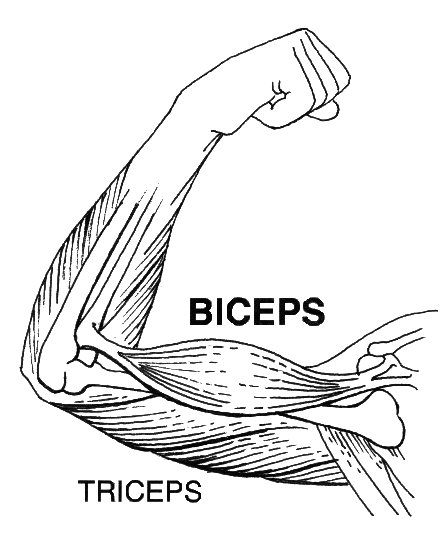
- The biceps and triceps muscles are shown above. Answer the following questions about these arm muscles.
- When the biceps contract and become shorter (as in the picture above), what kind of motion does this produce in the arm?
- Is the situation described in part (a) more likely to be an isometric or isotonic contraction? Explain your answer.
- If the triceps were to then contract, which way would the arm move?
- What are Z discs? What happens to them during muscle contraction?
- What is the function of mitochondria in muscle cells? Which type of muscle fibre has more mitochondria — slow-twitch or fast-twitch?
- What is the difference between primary and secondary Parkinson’s disease?
- Why can carpal tunnel syndrome cause muscle weakness in the hands?
Attributions
Figure 12.7.1
Botox, he whispered by Michael Reuter on Flickr is used under a CC BY 2.0 (https://creativecommons.org/licenses/by/2.0/) license.
Figure 12.7.2
botulism by jason wilson on Flickr is used under a CC BY 2.0 (https://creativecommons.org/licenses/by/2.0/) license.
Reference
Pearson Scott Foresman. (2020, April 14). File:Biceps (PSF).jpg [digital image]. Wikimedia Commons. https://commons.wikimedia.org/w/index.php?title=File:Biceps_(PSF).jpg&oldid=411251538. [Public Domain (https://en.wikipedia.org/wiki/Public_domain)]
Refers to the body system consisting of the heart, blood vessels and the blood. Blood contains oxygen and other nutrients which your body needs to survive. The body takes these essential nutrients from the blood.
Structures containing neuronal cell bodies in the peripheral nervous system.
Created by CK-12 Foundation/Adapted by Christine Miller
Case Study Conclusion: A Pain in the Foot
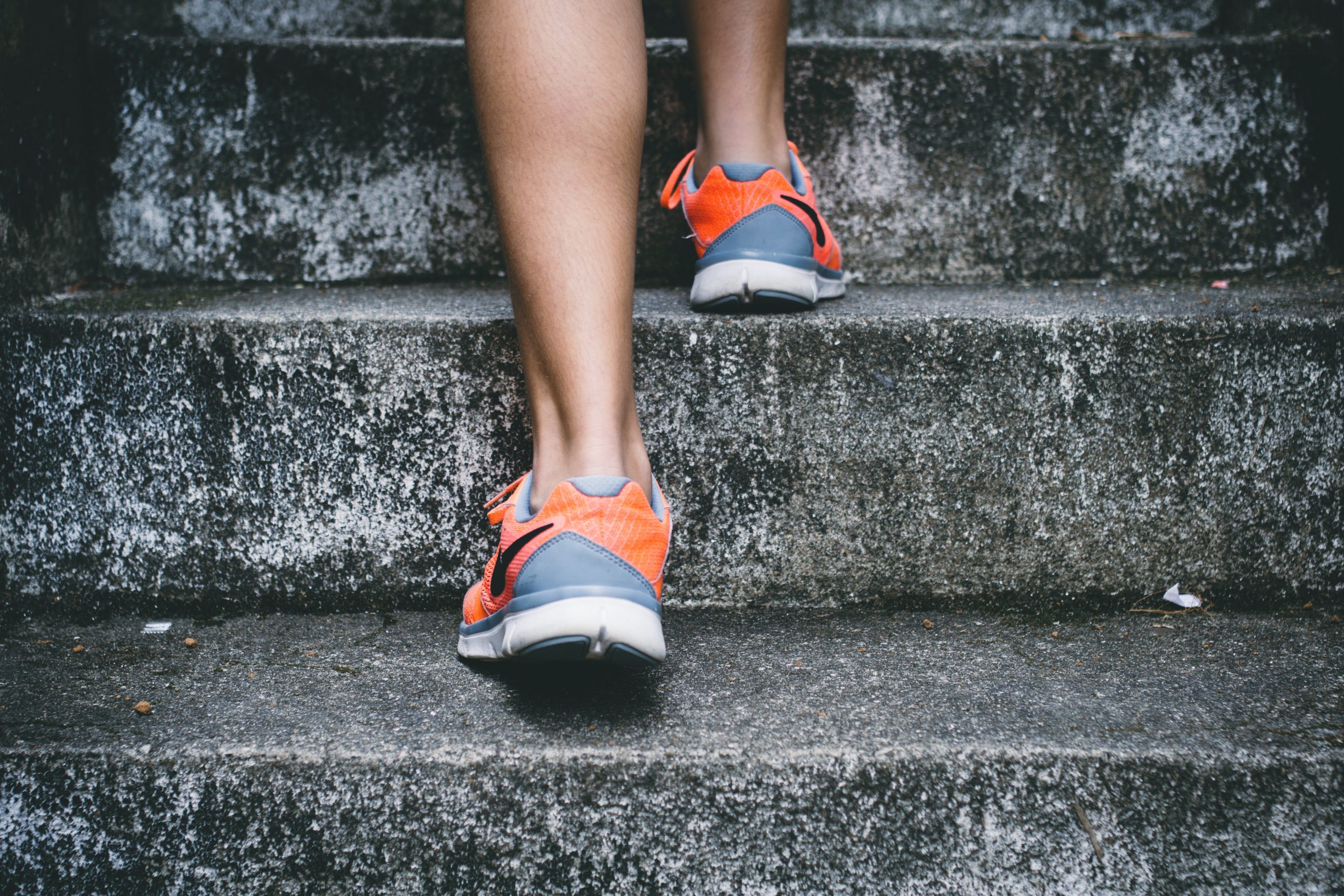
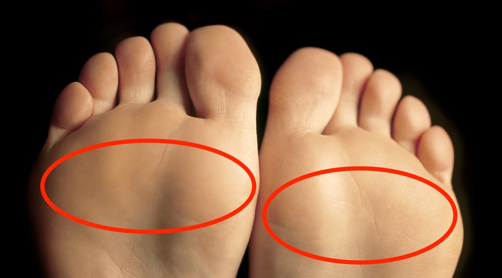
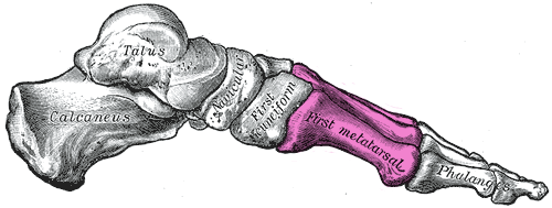
As Sophia discovered in the beginning of the chapter, wearing high heels can result in a condition called metatarsalgia. Metatarsalgia is named for the metatarsal bones, which are the five bones that run through the ball of the foot just behind the toes (highlighted in Figures 11.8.2 and 11.8.3). Wearing high heels causes excessive pressure on the ball of the foot, as described in the beginning of this chapter. Additionally, the toes are forced to pull upward in high heels, which moves the fleshy padding away from the ball of the foot and adds to the overall pressure placed on this region. Over time, this can cause inflammation and direct stress on the bones, resulting in the pain in the ball of the foot known as metatarsalgia. The pain occurs especially in weight-bearing positions, such as standing, walking, or running — which is what Sophia was experiencing. There may also be pain, numbness, or tingling in the toes associated with metatarsalgia.
 |
 |
Wearing high heels can also cause stress fractures in the feet, which are tiny breaks in bone that occur due to repeated mechanical stress. This is caused by the excessive pressure that high heels put on some of the bones of the feet. These fractures are somewhat similar to what occurs in osteoporosis when the bone mass decreases to the point where bones can fracture easily as a person goes about their daily activities. In both cases, a major noticeable injury is not necessary to create the tiny fractures. As you have learned, tiny fractures that accrue over time are the cause of dowager’s hump (or kyphosis), which is often seen in women with osteoporosis.
Don’t think you are immune to stress fractures just because you don’t wear high heels! This injury also commonly occurs in people who participate in sports involving repetitive striking of the foot on the ground, such as running, tennis, basketball, or gymnastics. They may be avoided by taking preventative measures. You should ramp up any increase in activity slowly, cross-train by engaging in a variety of different sports or activities, rest if you experience pain, and wear well-cushioned and supportive running shoes. It is important to know that your cardiovascular and muscular systems adapt to an increase in physical activity much more quickly than the skeletal system.
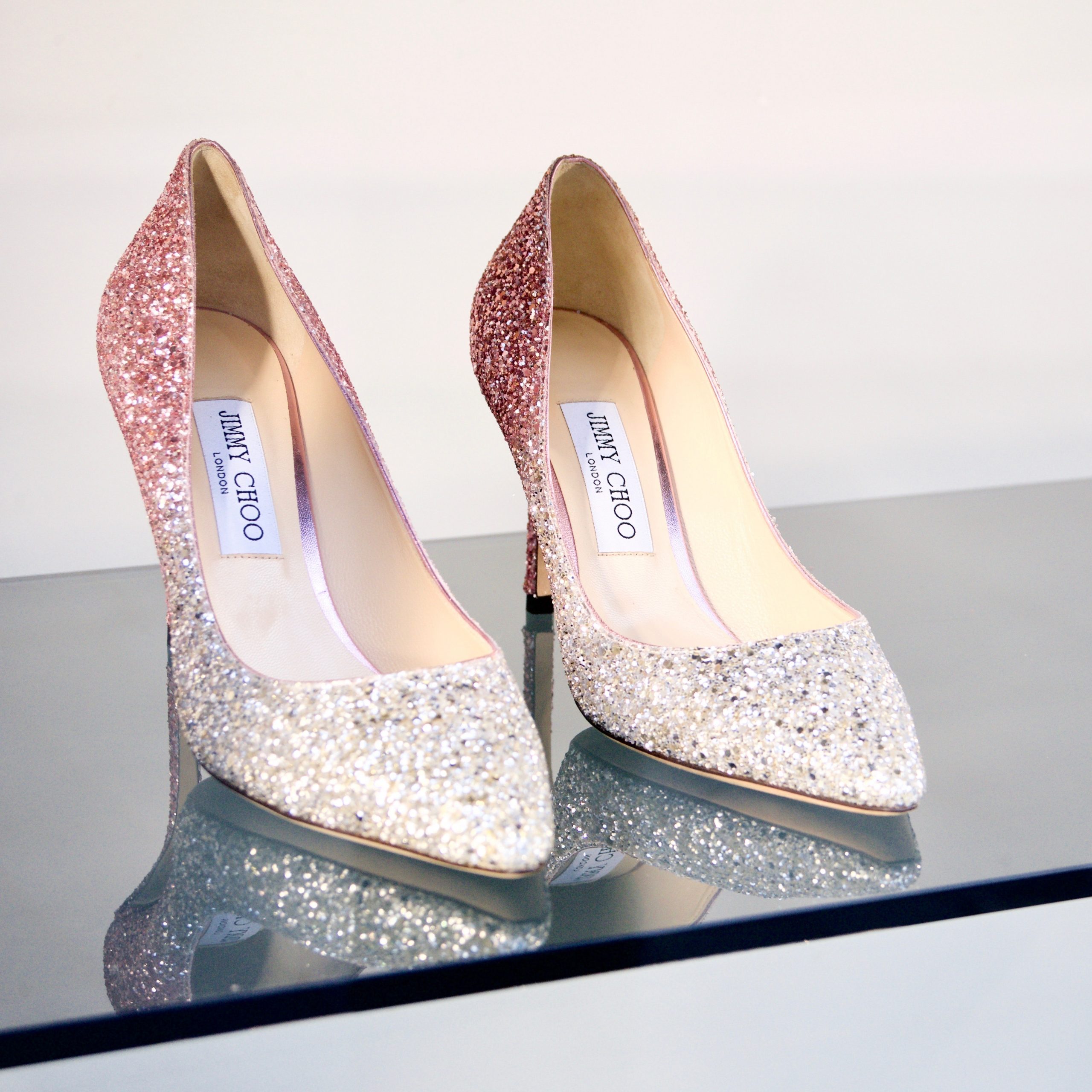
Sophia learned through her online research that wearing high heels can also lead to foot deformities, such as bunions and hammertoes. As you learned in an earlier chapter, a bunion is a protrusion on the side of the foot, most often at the base of the big toe. It can be caused by wearing shoes with a narrow, pointed toe box — a common shape for high heels (see Figure 11.8.4). The pressure of the shoes on the side of the foot causes an enlargement of bone or inflammation of other tissues in the region, which pushes the big toe toward the other toes.
Hammertoes are an abnormal bend in the middle joint of the second, third, or fourth toe (with the big toe being the first toe), causing the toe to be shaped similarly to a hammer. The narrow, pointed toe box of many high heels, combined with the way the toes are squished into the front of the shoe as a result of the height of the heel, can cause the toes to become deformed this way. Treatments for bunions and hammertoe include wearing shoes with a roomy toe box, padding or taping the toes, and toe exercises and stretches. If the bunion or hammertoe does not respond to these treatments, surgery may be necessary to correct the deformity.
Because the bones of the skeleton are connected and work together with other systems to support the body, wearing high heels can also cause physical problems in areas other than the feet. Wearing high heels shifts a person’s posture and alignment, and can put strain on tendons, muscles, and other joints in the body. Research published in 2014 from a team at Stanford University suggests that wearing high heels, particularly if the person is overweight or the heels are very high, may increase the risk of osteoarthritis (OA) in the knee, due to added stress on the knee joint as the person walks. As you have learned, OA results from the breakdown of cartilage and bone at the joint. Because it can only be treated to minimize symptoms — and not for a cure — OA could be an unfortunate long-term consequence of wearing high heels.
Sophia has decided that wearing high heels regularly is not worth the pain and potential long-term damage to her body. After consulting with her doctor, who confirmed she had metatarsalgia, she was able to successfully treat it with ice, rest, and wearing comfortable, supportive shoes instead of heels.
High heels are not the only kind of shoes that can cause problems. Flip-flops, worn-out sneakers, and shoes that are too tight can all cause foot issues. To prevent future problems from her shoe choices, Sophia is following guidelines recommended by medical experts. The guidelines include:
- Wearing shoes that fit well, have plenty of room in the toes, are supportive, and are comfortable right away. There should be no “break-in” period needed for shoes.
- Avoiding high heels, especially those with heels over two inches high, or those that have narrow, pointed toe boxes or very thin heels. The heels pictured in Figure 11.8.4 are an example of a type of shoe that should be avoided.
- If high heels must be worn, it should only be for a limited period of time.
As you have learned in this chapter, your skeletal system carries out a variety of important functions in your body, including physical support. But even though it is strong, your skeletal system can become damaged and deformed — even through such a seemingly innocuous act as wearing a certain type of shoe. Taking good care of your skeletal system is necessary to help it continue to take good care of the rest of you.
Chapter 11 Summary
In this chapter, you learned about the skeletal system. Specifically, you learned that:
- The skeletal system is the organ system that provides an internal framework for the human body. In adults, the skeletal system contains 206 bones.
- Bones are organs made of supportive connective tissues, mainly the tough protein collagen. Bones also contain blood vessels, nerves, and other tissues. Bones are hard and rigid, due to deposits of calcium and other mineral salts within their living tissues. Besides bones, the skeletal system includes cartilage and ligaments.
- The skeletal system has many different functions, including supporting the body and giving it shape, protecting internal organs, providing attachment surfaces for skeletal muscles, allowing body movements, producing blood cells, storing minerals, helping to maintain mineral homeostasis, and producing endocrine hormones.
- There is relatively little sexual dimorphism in the human skeleton, although the female skeleton tends to be smaller and less robust than the male skeleton. The greatest sex difference is in the pelvis, which is adapted for childbirth in females.
- The skeleton is traditionally divided into two major parts: the axial skeleton and the appendicular skeleton.
- The axial skeleton consists of a total of 80 bones. It includes the skull, vertebral column, and rib cage. It also includes the three tiny ossicles in the middle ear and the hyoid bone in the throat.
-
- The skull provides a bony framework for the head. It consists of 22 different bones: eight in the cranium, which encloses the brain, and 14 in the face, which includes the upper and lower jaw.
- The vertebral column is a flexible, S-shaped column of 33 vertebrae that connects the trunk with the skull and encloses the spinal cord. The vertebrae are divided into five regions: cervical, thoracic, lumbar, sacral, and coccygeal regions. The S shape of the vertebral column allows it to absorb shocks and distribute the weight of the body.
- The rib cage holds and protects the organs of the upper part of the trunk, including the heart and lungs. It includes the 12 thoracic vertebrae, the sternum, and 12 pairs of ribs.
- The appendicular skeleton consists of a total of 126 bones. It includes the bones of the four limbs, shoulder girdle, and pelvic girdle. The girdles attach the appendages to the axial skeleton.
-
- Each upper limb consists of 30 bones. There is one bone (called the humerus) in the upper arm, and two bones (called the ulna and radius) in the lower arm. The wrist contains eight carpal bones, the hand contains five metacarpals, and the fingers consist of 14 phalanges. The thumb is opposable to the palm and fingers of the same hand.
- Each lower limb also consists of 30 bones. There is one bone (called the femur) in the upper leg, and two bones (called the tibia and fibula) in the lower leg. The patella covers the knee joint. The ankle contains seven tarsal bones, and the foot contains five metatarsals. The tarsals and metatarsals form the heel and arch of the foot. The bones in the toes consist of 14 phalanges.
- The shoulder girdle attaches the upper limbs to the trunk of the body. It is connected to the axial skeleton only by muscles, allowing mobility of the upper limbs. Bones of the shoulder girdle include a right and left clavicle, and a right and left scapula.
- The pelvic girdle attaches the legs to the trunk of the body and supports the organs of the abdomen. It is connected to the axial skeleton by ligaments. The pelvic girdle consists of two halves that are fused together in adults. Each half consists of three bones: the ilium, pubis, and ischium.
- Bones are organs that consist mainly of bone (or osseous) tissue. Osseous tissue is a type of supportive connective tissue consisting of a collagen matrix that is mineralized with calcium and phosphorus crystals. The combination of flexible collagen and minerals makes bone hard, without making it brittle.
-
- There are two types of osseous tissues: compact bone tissue and spongy bone tissue. Compact bone tissue is smooth and dense. It forms the outer layer of bones. Spongy bone tissue is porous and light, and it is found inside many bones.
- Besides osseous tissues, bones also contain nerves, blood vessels, bone marrow, and periosteum.
- Bone tissue is composed of four different types of bone cells: osteoblasts, osteocytes, osteoclasts, and osteogenic cells. Osteoblasts form new collagen matrix and mineralize it, osteoclasts break down bone, osteocytes regulate the formation and breakdown of bone, and osteogenic cells divide and differentiate to form new osteoblasts. Bone is a very active tissue, constantly being remodeled by the work of osteoblasts and osteoclasts.
- There are six types of bones in the human body: long bones (such as the limb bones), short bones (such as the wrist bones), sesamoid bones (such as the patella), sutural bones in the skull, and irregular bones (such as the vertebrae).
- Early in the development of a human fetus, the skeleton is made almost entirely of cartilage. The relatively soft cartilage gradually turns into hard bone — a process that is called ossification. It begins at a primary ossification center in the middle of bone, and later also occurs at secondary ossification centers in the ends of bone. The bone can no longer grow in length after the areas of ossification meet and fuse at the time of skeletal maturity.
- Throughout life, bone is constantly being replaced in the process of bone remodeling. In this process, osteoclasts resorb bone and osteoblasts make new bone to replace it. Bone remodeling shapes the skeleton, repairs tiny flaws in bones, and helps maintain mineral homeostasis in the blood.
- Bone repair is the natural process in which a bone repairs itself following a bone fracture. This process may take several weeks. In the process, the periosteum produces cells that develop into osteoblasts, and the osteoblasts form new bone matrix to heal the fracture. Bone repair may be affected by diet, age, pre-existing bone disease, or other factors.
- Joints are locations at which bones of the skeleton connect with one another.
- Joints can be classified structurally or functionally, and there is significant overlap between the two types of classifications.
- The structural classification of joints depends on the type of tissue that binds the bones to each other at the joint. There are three types of joints in the structural classification: fibrous, cartilaginous, and synovial joints.
- The functional classification of joints is based on the type and degree of movement that they allow. There are three types of joints in the functional classification: immovable, partly movable, and movable joints.
- A number of disorders affect the skeletal system, including bone fractures and bone cancers. The two most common disorders of the skeletal system are osteoporosis and osteoarthritis.
- Osteoporosis is an age-related disorder in which bones lose mass, weaken, and break more easily than normal bones. The underlying mechanism in all cases of osteoporosis is an imbalance between bone formation and bone resorption in bone remodeling. Osteoporosis may also occur as a side effect of other disorders or certain medications.
-
- Osteoporosis is diagnosed by measuring a patient’s bone density and comparing it with the normal level of peak bone density. Fractures are the most dangerous aspect of osteoporosis. Osteoporosis is rarely fatal, but complications of fractures often are.
- Risk factors for osteoporosis include older age, female sex, European or Asian ancestry, family history of osteoporosis, short stature and small bones, smoking, alcohol consumption, lack of exercise, vitamin D deficiency, poor nutrition, and consumption of soft drinks.
- Osteoporosis is often treated with medications (such as bisphosphonates) that may slow or even reverse bone loss. Preventing osteoporosis includes eliminating any risk factors that can be controlled through changes of behavior, such as undertaking weight-bearing exercise.
- Osteoarthritis (OA) is a joint disease that results from the breakdown of joint cartilage and bone. The most common symptoms are joint pain and stiffness. OA is thought to be caused by mechanical stress on the joints with insufficient self-repair of cartilage, coupled with low-grade inflammation of the joints.
-
- Diagnosis of OA is typically made on the basis of signs and symptoms, such as joint deformities, pain, and stiffness. X-rays or other tests are sometimes used to either support the diagnosis or rule out other disorders. Age is the chief risk factor for OA. Other risk factors include joint injury, excess body weight, and a family history of OA.
- OA cannot be cured, but the symptoms can often be treated successfully. Treatments may include exercise, efforts to decrease stress on joints, pain medications, and surgery to replace affected hip or knee joints.
As you have learned in this chapter, one of the important functions of the skeletal system is to allow movement of the body. But it doesn’t do it alone. Movement is caused by the contraction of muscles, which pull on the bones, causing them to move. Read the next chapter to learn about this and other important functions of the muscular system.
Chapter 11 Review
-
-
- Why does the rib cage need to be flexible? Why can it be flexible?
- In general, what do “girdles” in the skeletal system do?
- Would swimming be more effective as an exercise for preventing osteoporosis or as a treatment for osteoarthritis? Explain your answer.
- Explain why some of the vertebrae become misshapen in the condition called dowager’s hump (or kyphosis).
- Explain why osteoarthritis often involves inflammation in the joints.
- Osteoporosis can involve excess bone resorption, as well as insufficient production of new bone tissue. What are the two main bone cell types that carry out these processes, respectively?
- Describe two roles that calcium in bones play in the body.
-
Attributions
Figure 11.8.1
Running Shoes by bruno-nascimento-PHIgYUGQPvU [photo] by Bruno Nascimento on Unsplash is used under the Unsplash License (https://unsplash.com/license).
Figure 11.8.2
Metatarsalgia/ Best Shoes for Metatarsalgia by Esther Max on Flickr is used under a CC BY 2.0 (https://creativecommons.org/licenses/by/2.0/) license.
Figure 11.8.3
Gray290_-_Mratatarsus (1) by Henry Vandyke Carter (1831-1897) (Revised by Warren H. Lewis, coloured by Was a bee) on Wikimedia Commons is in the public domain (https://en.wikipedia.org/wiki/Public_domain. (Bartleby.com: Gray’s Anatomy, Plate 290)
Figure 11.8.4
Heels by gavin-allanwood-ndpX28miBtE-unsplash by Photo by Gavin Allanwood on Unsplash is used under the Unsplash License (https://unsplash.com/license).
References
Mayo Clinic Staff. (n.d.). Hammertoe and mallet toe [online article]. MayoClinic.org. https://www.mayoclinic.org/diseases-conditions/hammertoe-and-mallet-toe/symptoms-causes/syc-20350839
VanDyke Carter, H. (1858). Illustration plate 290. In H. Gray, Anatomy of the Human Body. Lea & Febiger. Bartleby.com, 2000. www.bartleby.com/107/.
Created by CK-12 Foundation/Adapted by Christine Miller
Double Jointed?
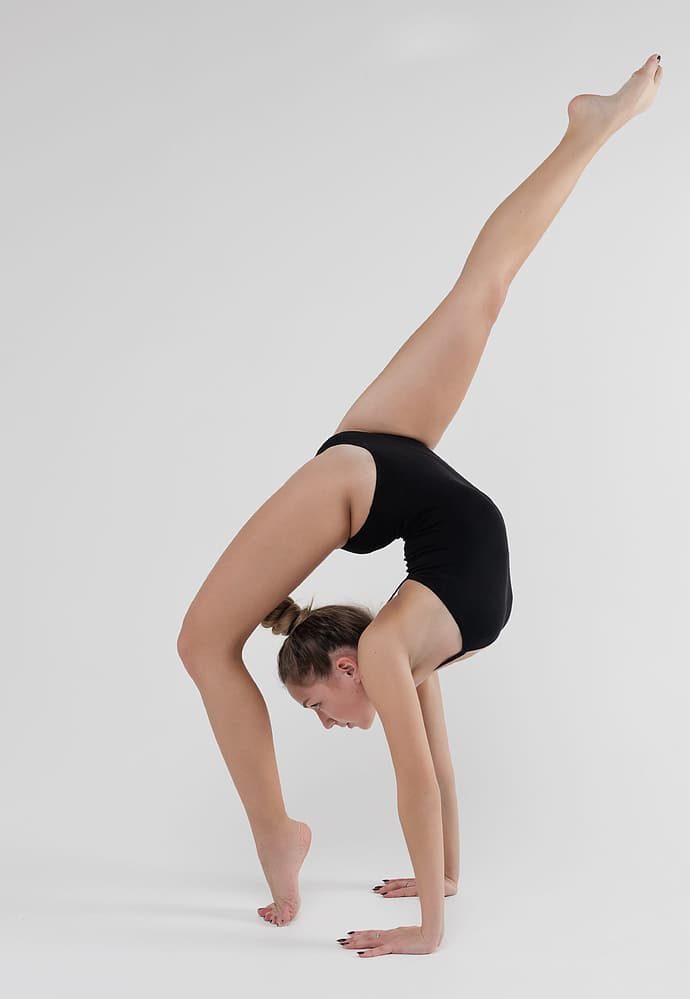
Is this woman double jointed? No, there is actually no such thing — at least as far as humans are concerned. However, some people, like the woman pictured in Figure 11.6.1, are much more flexible than others, generally because they have looser ligaments. Physicians call this condition joint hypermobility. Regardless of what it’s called, the feats of people with highly mobile joints can be quite impressive.
What Are Joints?
Joints are locations at which bones of the skeleton connect with one another. A joint is also called an articulation. The majority of joints are structured in such a way that they allow movement. However, not all joints allow movement. Of joints that do allow movement, the extent and direction of the movements they allow also vary.
Classification of Joints
Joints can be classified structurally or functionally. The structural classification of joints depends on the manner in which the bones connect to each other. The functional classification of joints depends on the nature of the movement the joints allow. There is significant overlap between the two types of classifications, because function depends largely on structure.
Structural Classification of Joints
The structural classification of joints is based on the type of tissue that binds the bones to each other at the joint. There are three types of joints in the structural classification: fibrous, cartilaginous, and synovial joints.
- Fibrous joints are joints in which bones are joined by dense connective tissue that is rich in collagen fibres. These joints are also called sutures. The joints between bones of the cranium are fibrous joints.
- Cartilaginous joints are joints in which bones are joined by cartilage. The joints between most of the vertebrae in the spine are cartilaginous joints.
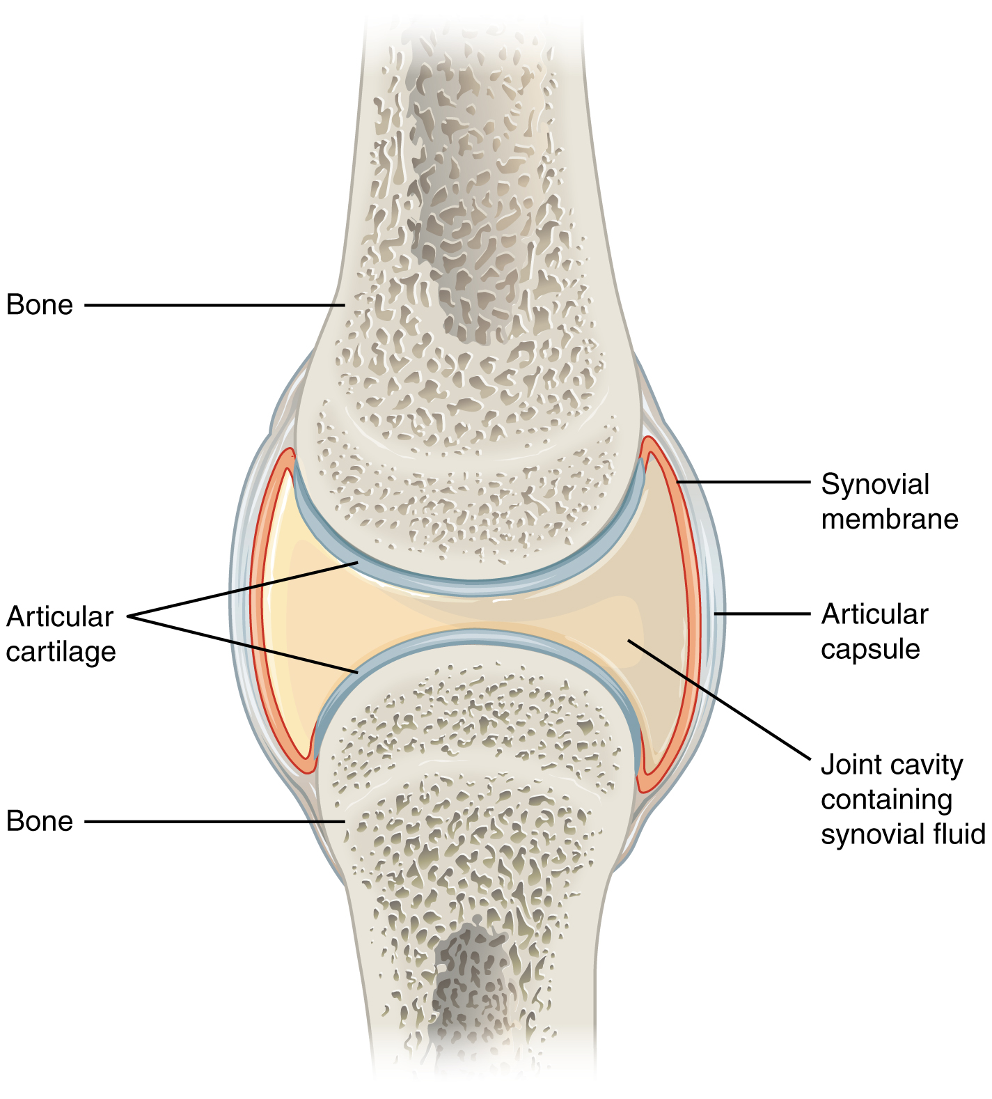
- Synovial joints are characterized by a fluid-filled space (called a synovial cavity) between the bones of the joints. You can see a drawing of a typical synovial joint in Figure 11.6.2. The cavity is enclosed by a membrane and filled with a fluid (called synovial fluid) that provides extra cushioning to the ends of the bones. Cartilage covers the articulating surfaces of the two bones, but the bones are actually held together by ligaments. The knee is a synovial joint.
Functional Classification of Joints
The functional classification of joints is based on the type and degree of movement that they allow. There are three types of joints in the functional classification: immovable, partly movable, and movable joints.
- Immovable joints allow little or no movement at the joint. Most immovable joints are fibrous joints. Besides the bones of the cranium, immovable joints include joints between the tibia and fibula in the lower leg, and between the radius and ulna in the lower arm.
- Partly movable joints permit slight movement. Most partly movable joints are cartilaginous joints. Besides the joints between vertebrae, they include the joints between the ribs and sternum (breast bone).
- Movable joints allow bones to move freely. All movable joints are synovial joints. Besides the knee, they include the shoulder, hip, and elbow. Movable joints are the most common type of joints in the body.
Types of Movable Joints
Movable joints can be classified further according to the type of movement they allow. There are six classes of movable joints: pivot, hinge, saddle, plane, condyloid, and ball-and-socket joints. An example of each class — as well as the type of movement it allows — is shown in Figure 11.6.3.
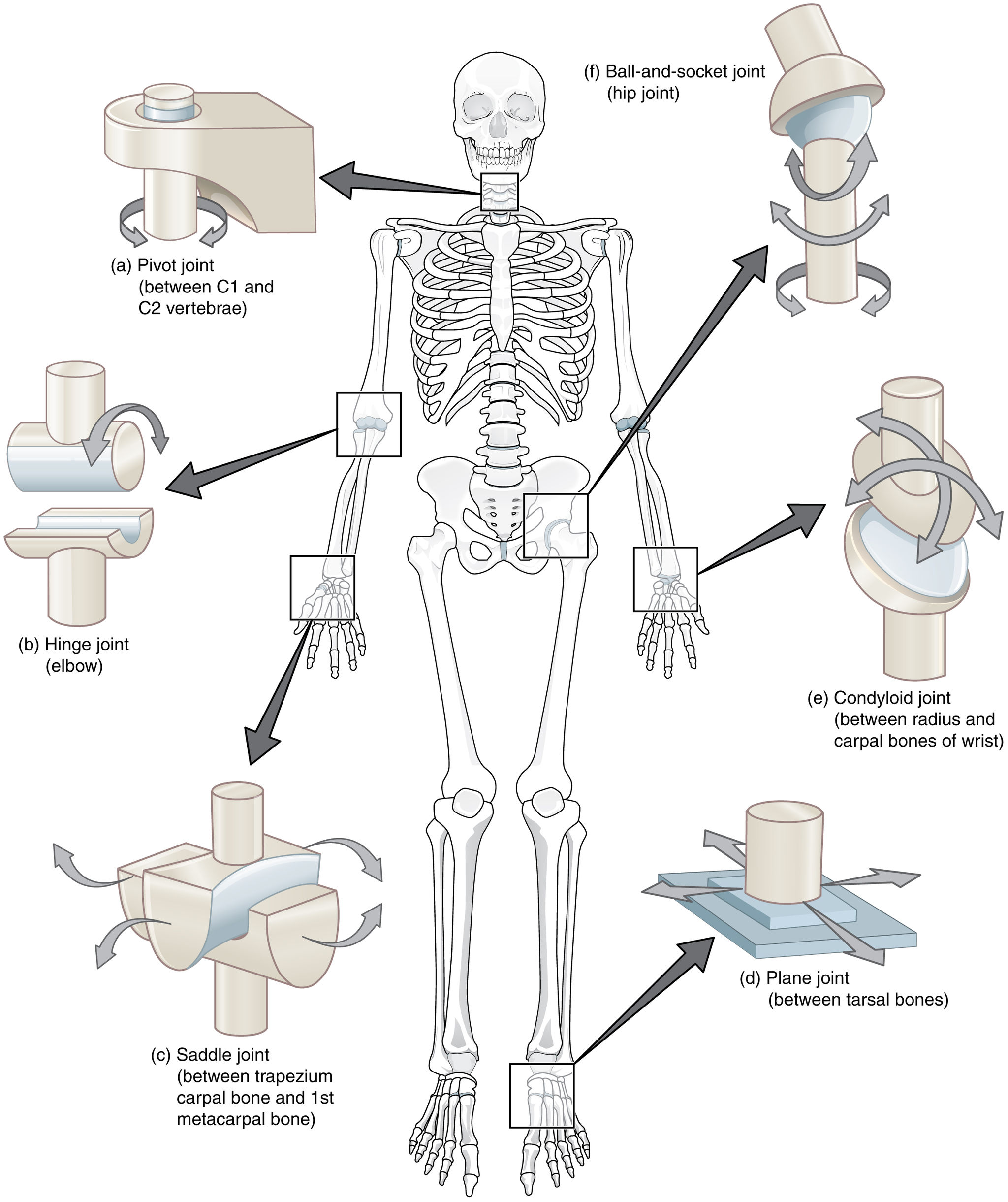
- A ball-and-socket joint allows the greatest range of movement of any movable joint. It allows forward and backward motion, as well as upward and downward movement. It also allows rotation in a circle. The hip and shoulder are the only two ball-and-socket joints in the human body.
- A pivot joint allows one bone to rotate around another. An example of a pivot joint is the joint between the first two vertebrae in the spine. This joint allows the head to rotate from left to right and back again.
- A hinge joint allows back and forth movement like the hinge of a door. An example of a hinge joint is the elbow. This joint allows the arm to bend back and forth.
- A saddle joint allows two different types of movement. An example of a saddle joint is the joint between the first metacarpal bone in the hand and one of the carpal bones in the wrist. This joint allows the thumb to move toward and away from the index finger, and also to cross over the palm toward the little finger.
- A plane joint (also called a gliding joint) allows two bones to glide over one another. The joints between the tarsals in the ankles and between the carpals in the wrists are mainly gliding joints. In the wrist, this type of joint allows the hand to bend upward at the wrist, and also to wave from side to side while the lower arm is held steady.
- A condyloid joint is one in which an oval-shaped head on one bone moves in an elliptical cavity in another bone, allowing movement in all directions, except rotation around an axis. The joint between the radius in the lower arm and carpal bones of the wrist is a condyloid joint, as is the joint at the base of the index finger.
Feature: My Human Body
Of all the parts of the skeletal system, the joints are generally the most fragile and subject to damage. If the cartilage that cushions bones at joints wears away, it does not grow back. Eventually, all of the cartilage may wear away. This causes osteoarthritis, which can be both painful and debilitating. In serious cases of osteoarthritis, people may lose the ability to climb stairs, walk long distances, perform routine daily activities, or participate in activities they love, such as gardening or playing sports. If you protect your joints, you can reduce your chances of joint damage, pain, and disability. If you already have joint damage, it is equally important to protect your joints and limit further damage. Follow these five tips:
- Maintain a normal, healthy weight. The more you weigh, the more force you exert on your joints. When you walk, each knee has to bear a force equal to as much as six times your body weight. If a person weighs 200 pounds, each knee bears more than half a ton of weight with every step. Seven in ten knee replacement surgeries for osteoarthritis can be attributed to obesity.
- Avoid too much high-impact exercise. Examples of high-impact activities include volleyball, basketball, and tennis. These activities generally involve running or jumping on hard surfaces, which puts tremendous stress on weight-bearing joints, especially the knees. Replace some or all of your high-impact activities with low-impact activities, such as biking, swimming, yoga, or lifting light weights.
- Reduce your risk of injury. Don’t be a weekend warrior, sitting at a desk all week and then crowding all your physical activity into two days. Get involved in a regular, daily exercise routine that keeps your body fit and your muscles toned. Building up muscles will make your joints more stable, allowing stress to spread across them. Be sure to do some stretching every day to keep the muscles around joints flexible and less prone to injury.
- Distribute work over your body, and use your largest, strongest joints. Use your shoulder, elbow, and wrist to lift heavy objects — not just your fingers. Hold small items in the palm of your hand, rather than by the fingers. Carry heavy items in a backpack, rather than in your hands. Hold weighty objects close to your body, instead of at arms’ length. Lift with your hips and knees, not your back.
- Respect pain. If it hurts, stop doing it. Take a break from the activity — at least until the pain stops. Try to use joints only to the point of mild fatigue, not pain.
11.6 Summary
- Joints are spots at which bones of the skeleton connect with one another. A joint is also called an articulation.
- Joints can be classified structurally or functionally, and there is significant overlap between the two types of classifications.
- The structural classification of joints depends on the type of tissue that binds the bones to each other at the joint. There are three types of joints in the structural classification: fibrous, cartilaginous, and synovial joints.
- The functional classification of joints is based on the type and degree of movement that they allow. There are three types of joints in the functional classification: immovable, partly movable, and movable joints.
- Movable joints can be classified further according to the type of movement they allow. There are six classes of movable joints: pivot, hinge, saddle, plane, condyloid, and ball-and-socket joints.
11.6 Review Questions
- What are joints?
- What are two ways that joints are commonly classified?
-
- How are joints classified structurally?
- Describe the functional classification of joints.
- How are movable joints classified?
- Name the six classes of movable joints. Describe how they move and give an example of each.
- Which specific type of moveable joint do you think your knee joint is? Explain your reasoning.
- Explain the difference between cartilage in a cartilaginous joint and cartilage in a synovial joint.
- Why are fibrous joints immovable?
- What is the function of synovial fluid?
11.6 Explore More
https://www.youtube.com/watch?v=IjiKUmfaZr4
Why do your knuckles pop? - Eleanor Nelsen, TED-Ed, 2015.
https://www.youtube.com/watch?v=FWsBm3hr3B0
Why haven’t we cured arthritis? - Kaitlyn Sadtler and Heather J. Faust, TED-Ed, 2019.
Attributions
Figure 11.6.1
Tags: Sports Gymnastics Fitness Woman Preparation by nastya_gepp on Pixabay is used under the Pixabay License (https://pixabay.com/de/service/license/).
Figure 11.6.2
Synovial_Joints by OpenStax College on Wikimedia Commons is used under a CC BY 3.0 (https://creativecommons.org/licenses/by/3.0) license.
Figure 11.6.3
Types_of_Synovial_Joints by OpenStax College on Wikimedia Commons is used under a CC BY 3.0 (https://creativecommons.org/licenses/by/3.0) license.
References
Betts, J. G., Young, K.A., Wise, J.A., Johnson, E., Poe, B., Kruse, D.H., Korol, O., Johnson, J.E., Womble, M., DeSaix, P. (2013, June 19). Figure 9.8 Synovial joints [digital image]. In Anatomy and Physiology (Section 9.4). OpenStax. https://openstax.org/books/anatomy-and-physiology/pages/9-4-synovial-joints
Betts, J. G., Young, K.A., Wise, J.A., Johnson, E., Poe, B., Kruse, D.H., Korol, O., Johnson, J.E., Womble, M., DeSaix, P. (2013, June 19). Figure 9.10 Types of synovial joints [digital image]. In Anatomy and Physiology (Section 9.4). OpenStax. https://openstax.org/books/anatomy-and-physiology/pages/9-4-synovial-joints
Mayo Clinic Staff. (n.d.). Osteoarthritis [online article]. MayoClinic.org. https://www.mayoclinic.org/diseases-conditions/osteoarthritis/symptoms-causes/syc-20351925
TED-Ed. (2015, May 5). Why do your knuckles pop? - Eleanor Nelsen. YouTube. https://www.youtube.com/watch?v=IjiKUmfaZr4
TED-Ed. (2019, November 7). Why haven’t we cured arthritis? - Kaitlyn Sadtler and Heather J. Faust. YouTube. https://www.youtube.com/watch?v=FWsBm3hr3B0
Involuntary, striated muscle found only in the walls of the heart; also called myocardium.
Created by CK-12 Foundation/Adapted by Christine Miller
Marvelous Muscles
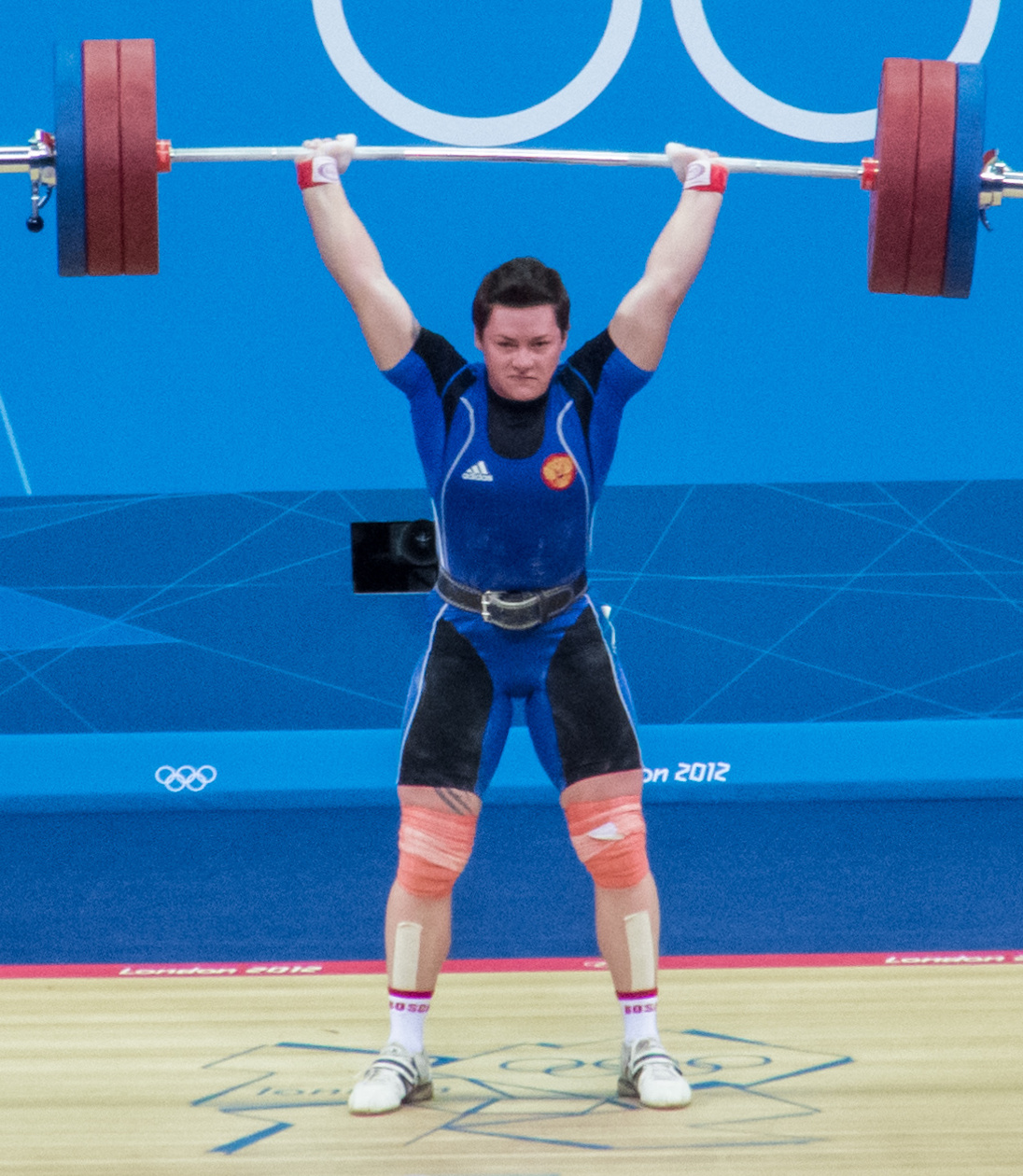
Does the word muscle make you think of the well-developed muscles of a weightlifter, like the woman in Figure 12.2.1? Her name is Natalia Zabolotnaya, and she’s a Russian Olympian. The muscles that are used to lift weights are easy to feel and see, but they aren’t the only muscles in the human body. Many muscles are deep within the body, where they form the walls of internal organs and other structures. You can flex your biceps at will, but you can’t control internal muscles like these. It’s a good thing that these internal muscles work without any conscious effort on your part, because movement of these muscles is essential for survival. Muscles are the organs of the muscular system.
What Is the Muscular System?
The muscular system consists of all the muscles of the body. The largest percentage of muscles in the muscular system consists of skeletal muscles, which are attached to bones and enable voluntary body movements (shown in Figure 12.2.2). There are almost 650 skeletal muscles in the human body, many of them shown in Figure 12.2.2. Besides skeletal muscles, the muscular system also includes cardiac muscle, which makes up the walls of the heart, and smooth muscles, which control movement in other internal organs and structures.
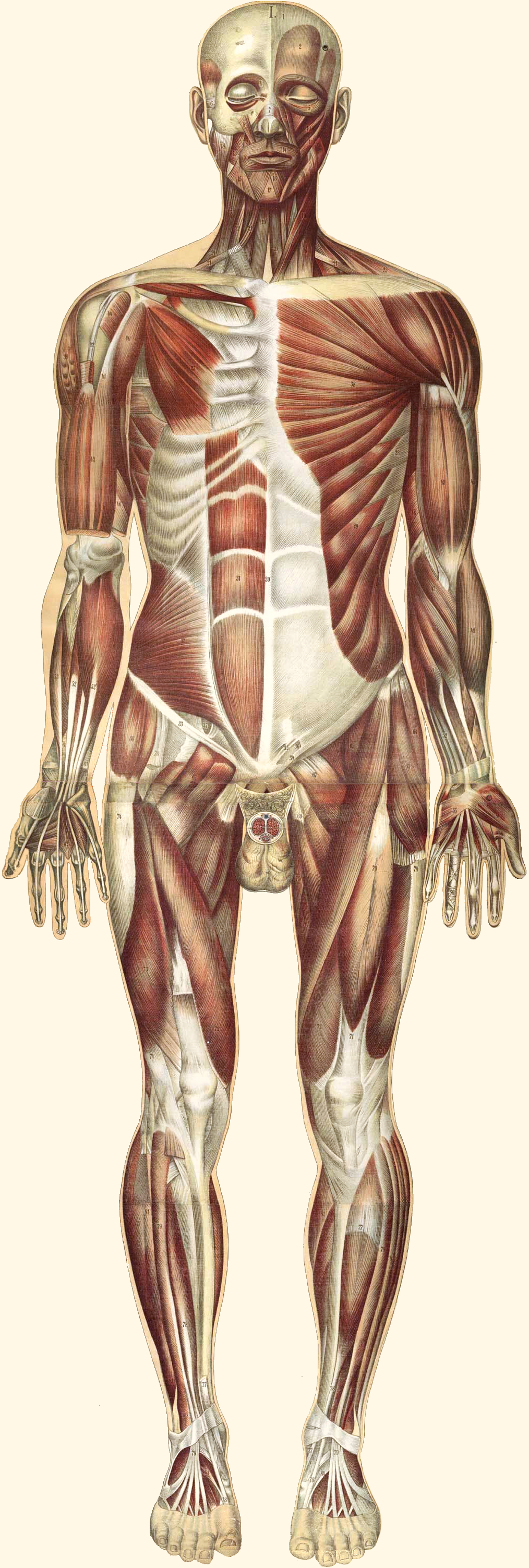
Muscle Structure and Function
Muscles are organs composed mainly of muscle cells, which are also called muscle fibres (mainly in skeletal and cardiac muscle) or myocytes (mainly in smooth muscle). Muscle cells are long, thin cells that are specialized for the function of contracting. They contain protein filaments that slide over one another using energy in ATP. The sliding filaments increase the tension in — or shorten the length of — muscle cells, causing a contraction. Muscle contractions are responsible for virtually all the movements of the body, both inside and out.
Skeletal muscles are attached to bones of the skeleton. When these muscles contract, they move the body. They allow us to use our limbs in a variety of ways, from walking to turning cartwheels. Skeletal muscles also maintain posture and help us to keep balance.
Smooth muscles in the walls of blood vessels contract to cause vasoconstriction, which may help conserve body heat. Relaxation of these muscles causes vasodilation, which may help the body lose heat. In the organs of the digestive system, smooth muscles squeeze food through the gastrointestinal tract by contracting in sequence to form a wave of muscle contractions called peristalsis. Think of squirting toothpaste through a tube by applying pressure in sequence from the bottom of the tube to the top, and you have a good idea of how food is moved by muscles through the digestive system. Peristalsis of smooth muscles also moves urine through the urinary tract.
Cardiac muscle tissue is found only in the walls of the heart. When cardiac muscle contracts, it makes the heart beat. The pumping action of the beating heart keeps blood flowing through the cardiovascular system.
Muscle Hypertrophy and Atrophy
Muscles can grow larger, or hypertrophy. This generally occurs through increased use, although hormonal or other influences can also play a role. The increase in testosterone that occurs in males during puberty, for example, causes a significant increase in muscle size. Physical exercise that involves weight bearing or resistance training can increase the size of skeletal muscles in virtually everyone. Exercises (such as running) that increase the heart rate may also increase the size and strength of cardiac muscle. The size of muscle, in turn, is the main determinant of muscle strength, which may be measured by the amount of force a muscle can exert.
Muscles can also grow smaller, or atrophy, which can occur through lack of physical activity or from starvation. People who are immobilized for any length of time — for example, because of a broken bone or surgery — lose muscle mass relatively quickly. People in concentration or famine camps may be so malnourished that they lose much of their muscle mass, becoming almost literally just “skin and bones.” Astronauts on the International Space Station may also lose significant muscle mass because of weightlessness in space (see Figure 12.2.3).
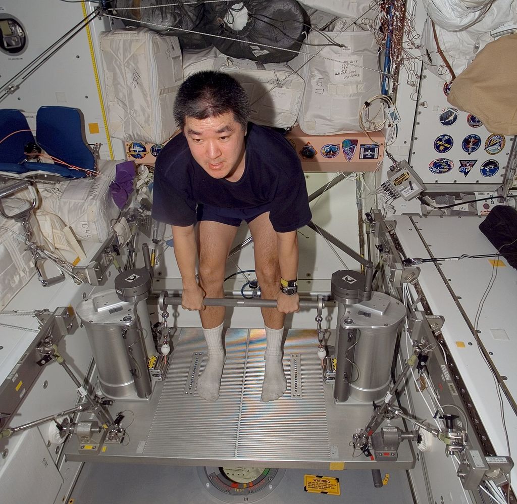
Many diseases, including cancer and AIDS, are often associated with muscle atrophy. Atrophy of muscles also happens with age. As people grow older, there is a gradual decrease in the ability to maintain skeletal muscle mass, known as sarcopenia. The exact cause of sarcopenia is not known, but one possible cause is a decrease in sensitivity to growth factors that are needed to maintain muscle mass. Because muscle size determines strength, muscle atrophy causes a corresponding decline in muscle strength.
In both hypertrophy and atrophy, the number of muscle fibres does not change. What changes is the size of the muscle fibres. When muscles hypertrophy, the individual fibres become wider. When muscles atrophy, the fibres become narrower.
Interactions with Other Body Systems
Muscles cannot contract on their own. Skeletal muscles need stimulation from motor neurons in order to contract. The point where a motor neuron attaches to a muscle is called a neuromuscular junction. Let’s say you decide to raise your hand in class. Your brain sends electrical messages through motor neurons to your arm and shoulder. The motor neurons, in turn, stimulate muscle fibres in your arm and shoulder to contract, causing your arm to rise.
Involuntary contractions of smooth and cardiac muscles are also controlled by electrical impulses, but in the case of these muscles, the impulses come from the autonomic nervous system (smooth muscle) or specialized cells in the heart (cardiac muscle). Hormones and some other factors also influence involuntary contractions of cardiac and smooth muscles. For example, the fight-or-flight hormone adrenaline increases the rate at which cardiac muscle contracts, thereby speeding up the heartbeat.
Muscles cannot move the body on their own. They need the skeletal system to act upon. The two systems together are often referred to as the musculoskeletal system. Skeletal muscles are attached to the skeleton by tough connective tissues called tendons. Many skeletal muscles are attached to the ends of bones that meet at a joint. The muscles span the joint and connect the bones. When the muscles contract, they pull on the bones, causing them to move. The skeletal system provides a system of levers that allow body movement. The muscular system provides the force that moves the levers.
12.2 Summary
- The muscular system consists of all the muscles of the body. There are three types of muscle: skeletal muscle (which is attached to bones and enables voluntary body movements), cardiac muscle (which makes up the walls of the heart and makes it beat), and smooth muscle (which is found in the walls of internal organs and other internal structures and controls their movements).
- Muscles are organs composed mainly of muscle cells, which may also be called muscle fibres or myocytes. Muscle cells are specialized for the function of contracting, which occurs when protein filaments inside the cells slide over one another using energy in ATP.
- Muscles can grow larger, or hypertrophy. This generally occurs through increased use (physical exercise), although hormonal or other influences can also play a role. Muscles can also grow smaller, or atrophy. This may occur through lack of use, starvation, certain diseases, or aging. In both hypertrophy and atrophy, the size — but not the number — of muscle fibres changes. The size of muscles is the main determinant of muscle strength.
- Skeletal muscles need the stimulus of motor neurons to contract, and to move the body, they need the skeletal system to act upon. Involuntary contractions of cardiac and smooth muscles are controlled by special cells in the heart, nerves of the autonomic nervous system, hormones, or other factors.
12.2 Review Questions
- What is the muscular system?
- Describe muscle cells and their function.
- Identify three types of muscle tissue and where each type is found.
- Define muscle hypertrophy and muscle atrophy.
- What are some possible causes of muscle hypertrophy?
- Give three reasons that muscle atrophy may occur.
- How do muscles change when they increase or decrease in size?
- How do changes in muscle size affect strength?
- Explain why astronauts can easily lose muscle mass in space.
- Describe how the terms muscle cells, muscle fibres, and myocytes relate to each other.
-
- Name two systems in the body that work together with the muscular system to carry out movements.
- Describe one way in which the muscular system is involved in regulating body temperature.
12.2 Explore More
https://www.youtube.com/watch?v=VVL-8zr2hk4
How your muscular system works - Emma Bryce, TED-Ed, 2017.
https://www.youtube.com/watch?v=Ujr0UAbyPS4&feature=emb_logo
3D Medical Animation - Peristalsis in Large Intestine/Bowel || ABP ©, AnimatedBiomedical, 2013.
https://www.youtube.com/watch?v=LkXwfTsqQgQ&feature=emb_logo
Muscle matters: Dr Brendan Egan at TEDxUCD, TEDx Talks, 2014.
Attributions
Figure 12.2.1
Natalia_Zabolotnaya_2012b by Simon Q on Wikimedia Commons is used under a CC BY 2.0 (https://creativecommons.org/licenses/by/2.0/deed.en) license.
Figure 12.2.2
Bougle_whole2_retouched by Bouglé, Julien from the National LIbrary of Medicine (NLM) on Wikimedia Commons is in the public domain (https://en.wikipedia.org/wiki/Public_domain).
Figure 12.2.3
Daniel_Tani_iss016e027910 by NASA/ International Space Station Imagery on Wikimedia Commons is in the public domain (https://en.wikipedia.org/wiki/Public_domain).
References
AnimatedBiomedical. (2013, January 30). 3D Medical animation - Peristalsis in large intestine/bowel || ABP ©. YouTube. https://www.youtube.com/watch?v=Ujr0UAbyPS4&feature=youtu.be
Bouglé, J. (1899). Le corps humain en grandeur naturelle : planches coloriées et superposées, avec texte explicatif. J. B. Baillière et fils. In Historical Anatomies on the Web. http://www.nlm.nih.gov/exhibition/historicalanatomies/bougle_home.html
TED-Ed. (2017, October 26). How your muscular system works - Emma Bryce. YouTube. https://www.youtube.com/watch?v=VVL-8zr2hk4&feature=youtu.be
TEDx Talks. (2014, June 27). Muscle matters: Dr Brendan Egan at TEDxUCD. YouTube. https://www.youtube.com/watch?v=LkXwfTsqQgQ&feature=youtu.be
Wikipedia contributors. (2020, June 15). Natalya Zabolotnaya. In Wikipedia. https://en.wikipedia.org/w/index.php?title=Natalya_Zabolotnaya&oldid=962630409
Case Study: More Than Just Tired
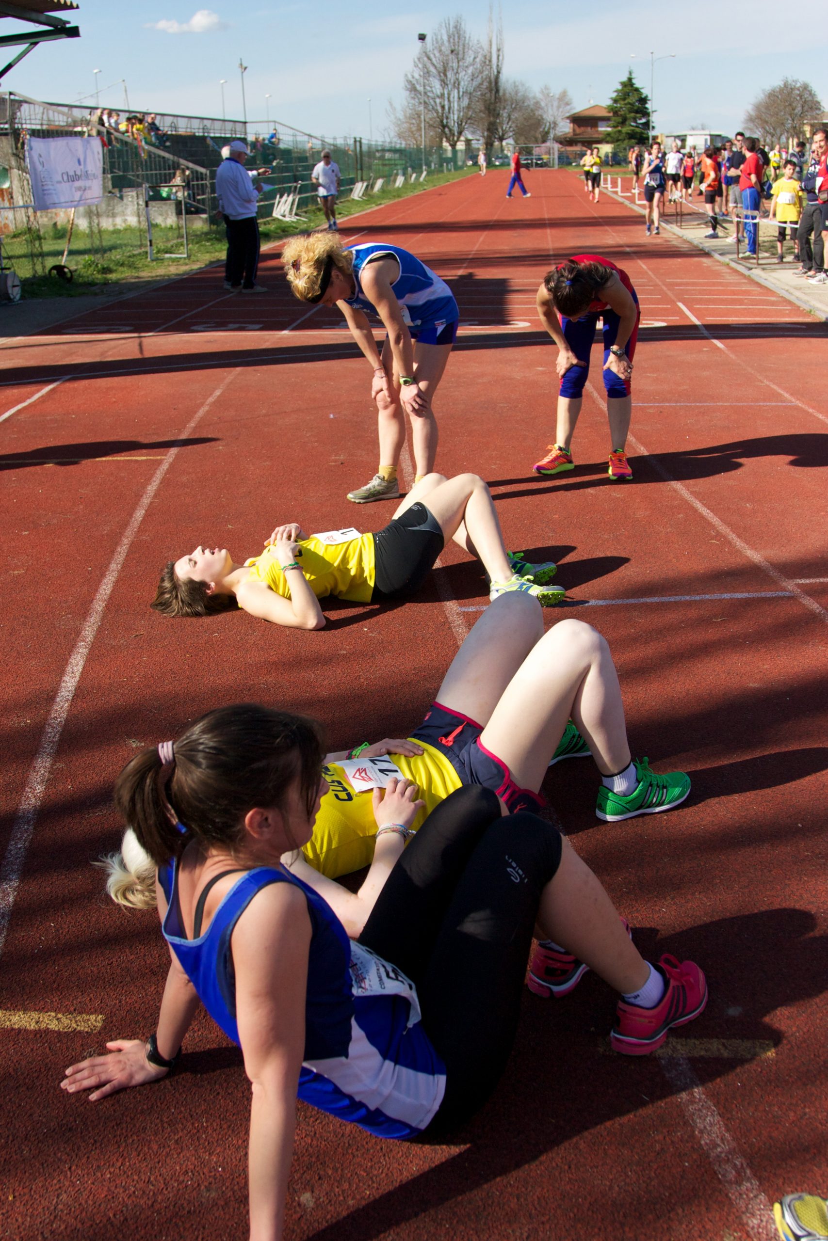
We all get tired sometimes, especially if we have been doing a lot of physical activity, like the athletes pictured in Figure 4.1.1. But for Jasmin (Figure 4.1.2), a 34-year-old former high school track star who is now a recreational runner, her tiredness was going far beyond what she thought should be normal for someone in generally good physical shape.
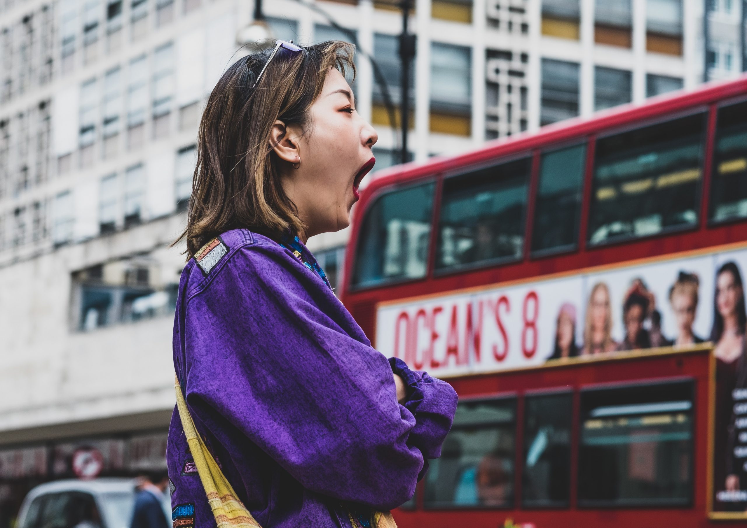
She was experiencing extreme fatigue after her runs, as well as muscle cramping, spasms, and an unusual sense of heaviness in her legs. At first, she just chalked it up to getting older, but her exhaustion and pain worsened to the point where the former athlete could no longer run for more than a few minutes at a time. She began to experience other unusual symptoms, such as blurry vision and vomiting for no apparent reason.
Concerned, Jasmin went to her doctor, who ran many tests and consulted with several specialists. After several months, she was finally diagnosed with a mitochondrial disease. Jasmin is surprised. She has an 8-year-old niece with a mitochondrial disease, but her niece’s symptoms started when she was very young, and they included seizures and learning disabilities. How can Jasmin have the same disease, but different symptoms? Why didn't she have problems until adulthood, while her niece experienced symptoms at an early age? And what are mitochondria, anyway?
Chapter Overview: The Importance of Cells
As you will learn in this chapter, mitochondria are important structures within our cells. This chapter will describe cells, which are the basic unit of structure and function in all living organisms. Specifically, you will learn:
- How cells were discovered, their common structures, and the principles of cell theory.
- The importance of size and shape to the functions of cells.
- The differences between eukaryotic cells (such as those in humans and other animals) and prokaryotic cells (such as bacteria).
- The structures and functions of cell parts, including mitochondria, the plasma membrane, cytoplasm, cytoskeleton, nucleus, ribosomes, Golgi apparatus, endoplasmic reticulum, vesicles, and vacuoles.
- The processes of passive and active transport to move substances into and out of cells and help maintain homeostasis.
- How organisms obtain the energy needed for life, including how the sugar glucose is broken down to produce ATP through the processes of anaerobic and aerobic cellular respiration.
- The phases of the cell cycle, how cells divide through mitosis, and how cancer can result from unregulated cell division.
As you read this chapter, think about the following questions related to Jasmin’s disease:
- What are mitochondria? What is their structure and function, and where did they come from during evolution?
- Why are fatigue and “exercise intolerance” (such as Jasmin’s extreme exhaustion after running) common symptoms of mitochondrial diseases?
- Why do you think Jasmin has symptoms that affect so many different parts of her body, including her legs, eyes, and digestive system?
- Why do you think mitochondrial diseases can run in families like Jasmin's?
Attributes
Figure 4.1.1
Difficult competition by Massimo Sartirana on Unsplash is used under the Unsplash License (https://unsplash.com/license).
Figure 4.1.2
Exhausted by Kevin Grieve on Unsplash is used under the Unsplash License (https://unsplash.com/license).
Created by: CK-12/Adapted by Christine Miller

Case Study: More Than Just Tired
We all get tired sometimes, especially if we have been doing a lot of physical activity, like the athletes pictured in Figure 4.1.1. But for Jasmin (Figure 4.1.2), a 34-year-old former high school track star who is now a recreational runner, her tiredness was going far beyond what she thought should be normal for someone in generally good physical shape.

She was experiencing extreme fatigue after her runs, as well as muscle cramping, spasms, and an unusual sense of heaviness in her legs. At first, she just chalked it up to getting older, but her exhaustion and pain worsened to the point where the former athlete could no longer run for more than a few minutes at a time. She began to experience other unusual symptoms, such as blurry vision and vomiting for no apparent reason.
Concerned, Jasmin went to her doctor, who ran many tests and consulted with several specialists. After several months, she was finally diagnosed with a mitochondrial disease. Jasmin is surprised. She has an 8-year-old niece with a mitochondrial disease, but her niece’s symptoms started when she was very young, and they included seizures and learning disabilities. How can Jasmin have the same disease, but different symptoms? Why didn't she have problems until adulthood, while her niece experienced symptoms at an early age? And what are mitochondria, anyway?
Chapter Overview: The Importance of Cells
As you will learn in this chapter, mitochondria are important structures within our cells. This chapter will describe cells, which are the basic unit of structure and function in all living organisms. Specifically, you will learn:
- How cells were discovered, their common structures, and the principles of cell theory.
- The importance of size and shape to the functions of cells.
- The differences between eukaryotic cells (such as those in humans and other animals) and prokaryotic cells (such as bacteria).
- The structures and functions of cell parts, including mitochondria, the plasma membrane, cytoplasm, cytoskeleton, nucleus, ribosomes, Golgi apparatus, endoplasmic reticulum, vesicles, and vacuoles.
- The processes of passive and active transport to move substances into and out of cells and help maintain homeostasis.
- How organisms obtain the energy needed for life, including how the sugar glucose is broken down to produce ATP through the processes of anaerobic and aerobic cellular respiration.
- The phases of the cell cycle, how cells divide through mitosis, and how cancer can result from unregulated cell division.
As you read this chapter, think about the following questions related to Jasmin’s disease:
- What are mitochondria? What is their structure and function, and where did they come from during evolution?
- Why are fatigue and “exercise intolerance” (such as Jasmin’s extreme exhaustion after running) common symptoms of mitochondrial diseases?
- Why do you think Jasmin has symptoms that affect so many different parts of her body, including her legs, eyes, and digestive system?
- Why do you think mitochondrial diseases can run in families like Jasmin's?
Attributes
Figure 4.1.1
Difficult competition by Massimo Sartirana on Unsplash is used under the Unsplash License (https://unsplash.com/license).
Figure 4.1.2
Exhausted by Kevin Grieve on Unsplash is used under the Unsplash License (https://unsplash.com/license).
Image shows an electron microscope image of a cell. There is a defined cell boundary and several structures inside of different sizes and textures.
Figure 4.2.1 Human cells viewed with a very powerful tool called a scanning electron microscope.
Amazing Cells
What are these incredible objects? Would it surprise you to learn that they are all human cells? Cells are actually too small to see with the unaided eye. It is visible here in such detail because it is being viewed with a very powerful tool called a scanning electron microscope. Cells may be small in size, but they are extremely important to life. Like all other living things, you are made of cells. Cells are the basis of life, and without cells, life as we know it would not exist. You will learn more about these amazing building blocks of life in this section.
What Are Cells?
If you look at living matter with a microscope — even a simple light microscope — you will see that it consists of cells. Cells are the basic units of the structure and function of living things. They are the smallest units that can carry out the processes of life. All organisms are made up of one or more cells, and all cells have many of the same structures and carry out the same basic life processes. Knowing the structure of cells and the processes they carry out is necessary to an understanding of life itself.
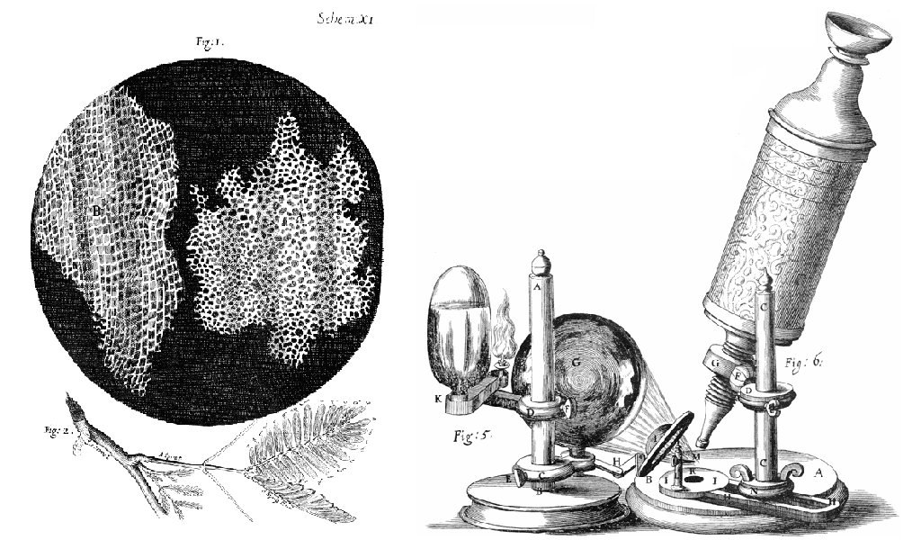
Discovery of Cells
The first time the word cell was used to refer to these tiny units of life was in 1665 by a British scientist named Robert Hooke. Hooke was one of the earliest scientists to study living things under a microscope. The microscopes of his day were not very strong, but Hooke was still able to make an important discovery. When he looked at a thin slice of cork under his microscope, he was surprised to see what looked like a honeycomb. Hooke made the drawing in the figure to the right to show what he saw. As you can see, the cork was made up of many tiny units. Hooke called these units cells because they resembled cells in a monastery.
Soon after Robert Hooke discovered cells in cork, Anton van Leeuwenhoek in Holland made other important discoveries using a microscope. Leeuwenhoek made his own microscope lenses, and he was so good at it that his microscope was more powerful than other microscopes of his day. In fact, Leeuwenhoek’s microscope was almost as strong as modern light microscopes. Using his microscope, Leeuwenhoek was the first person to observe human cells and bacteria.
Cell Theory
By the early 1800s, scientists had observed cells of many different organisms. These observations led two German scientists named Theodor Schwann and Matthias Jakob Schleiden to propose cells as the basic building blocks of all living things. Around 1850, a German doctor named Rudolf Virchow was studying cells under a microscope, when he happened to see them dividing and forming new cells. He realized that living cells produce new cells through division. Based on this realization, Virchow proposed that living cells arise only from other living cells.
The ideas of all three scientists — Schwann, Schleiden, and Virchow — led to cell theory, which is one of the fundamental theories unifying all of biology.
Cell theory states that:
- All organisms are made of one or more cells.
- All the life functions of organisms occur within cells.
- All cells come from existing cells.
Seeing Inside Cells
Starting with Robert Hooke in the 1600s, the microscope opened up an amazing new world — a world of life at the level of the cell. As microscopes continued to improve, more discoveries were made about the cells of living things, but by the late 1800s, light microscopes had reached their limit. Objects much smaller than cells (including the structures inside cells) were too small to be seen with even the strongest light microscope.
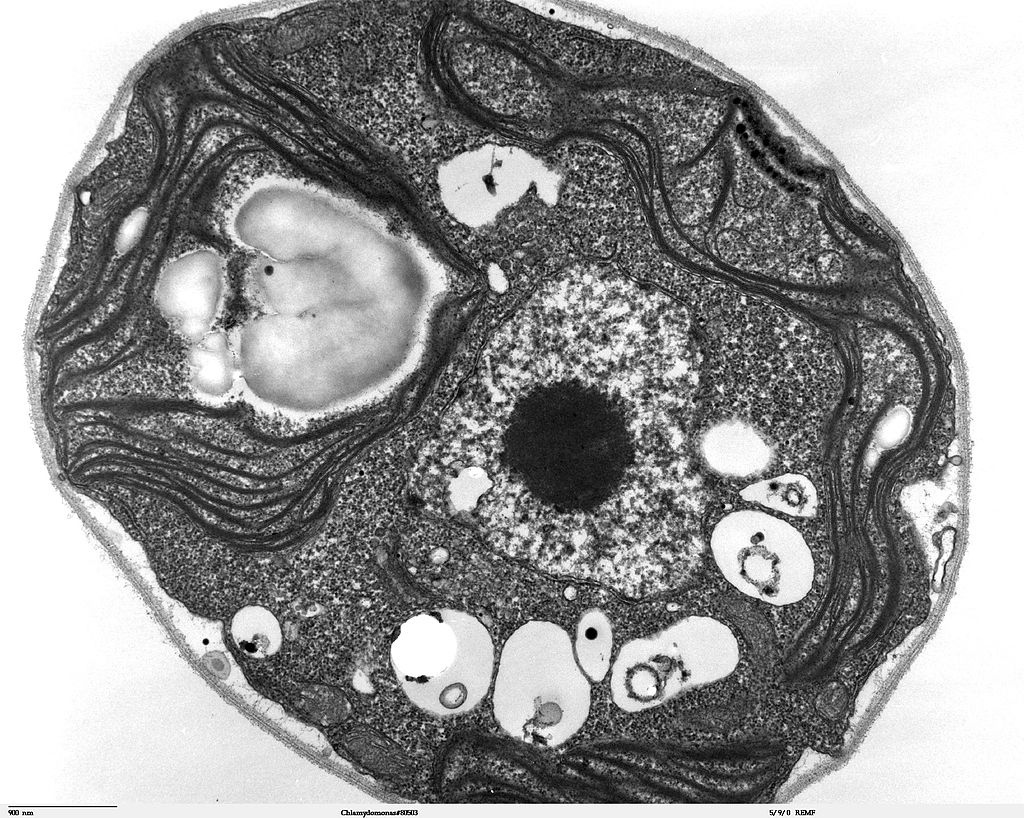
Then, in the 1950s, a new type of microscope was invented. Called the electron microscope, it used a beam of electrons instead of light to observe extremely small objects. With an electron microscope, scientists could finally see the tiny structures inside cells. They could even see individual molecules and atoms. The electron microscope had a huge impact on biology. It allowed scientists to study organisms at the level of their molecules, and it led to the emergence of the molecular biology field. With the electron microscope, many more cell discoveries were made.
Structures Shared By All Cells
Although cells are diverse, all cells have certain parts in common. These parts include a plasma membrane, cytoplasm, ribosomes, and DNA.
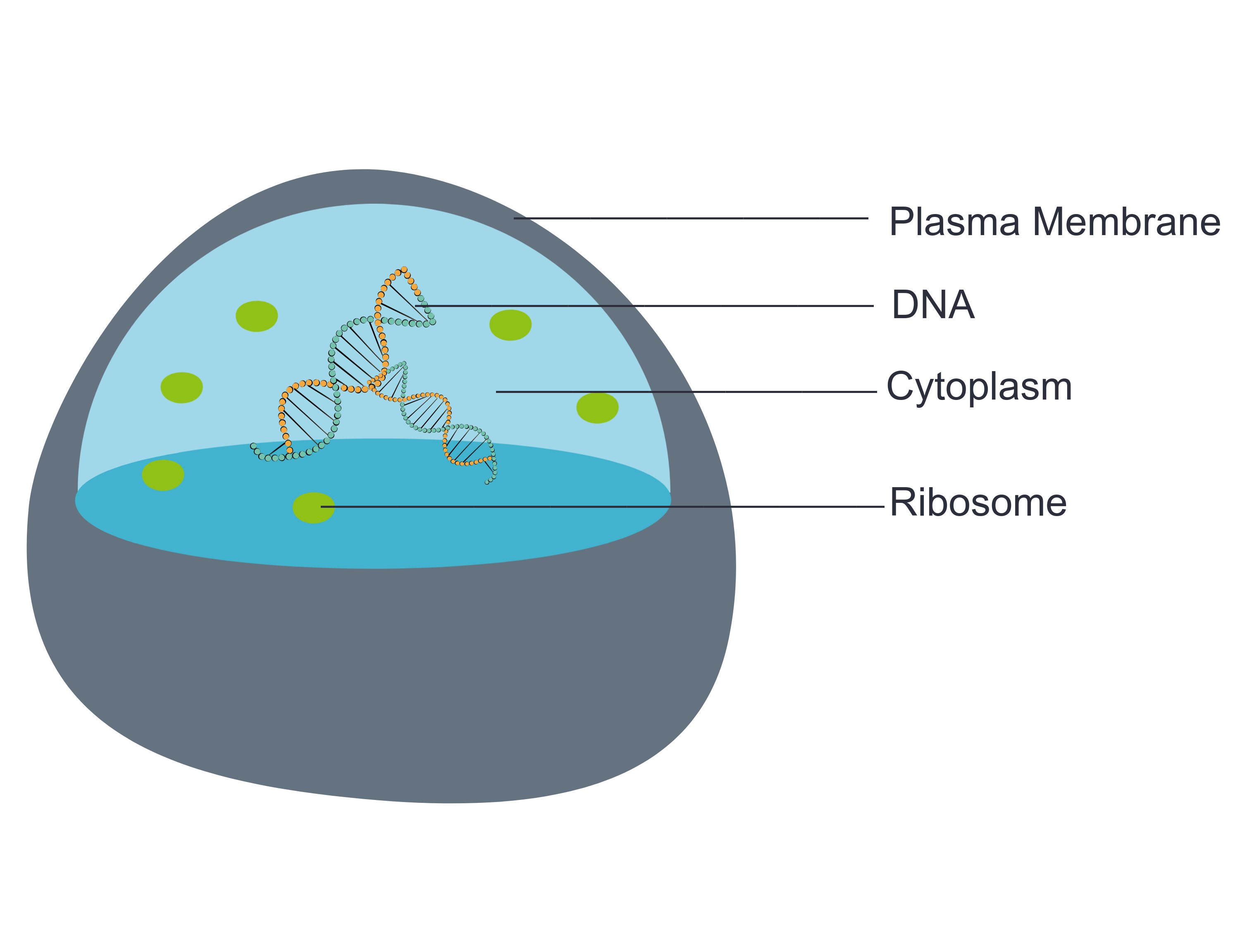
- The plasma membrane (a type of cell membrane) is a thin coat of lipids that surrounds a cell. It forms the physical boundary between the cell and its environment. You can think of it as the “skin” of the cell.
- Cytoplasm refers to all of the cellular material inside of the plasma membrane. Cytoplasm is made up of a watery substance called cytosol, and it contains other cell structures, such as ribosomes.
- Ribosomes are the structures in the cytoplasm in which proteins are made.
- DNA is a nucleic acid found in cells. It contains the genetic instructions that cells need to make proteins.
These four parts are common to all cells, from organisms as different as bacteria and human beings. How did all known organisms come to have such similar cells? The similarities show that all life on Earth has a common evolutionary history.
4.2 Summary
- Cells are the basic units of structure and function in living things. They are the smallest units that can carry out the processes of life.
- In the 1600s, Hooke was the first to observe cells from an organism (cork). Soon after, microscopist van Leeuwenhoek observed many other living cells.
- In the early 1800s, Schwann and Schleiden theorized that cells are the basic building blocks of all living things. Around 1850, Virchow observed cells dividing. To previous learnings, he added that living cells arise only from other living cells. These ideas led to cell theory, which states that all organisms are made of cells, that all life functions occur in cells, and that all cells come from other cells.
- It wasn't until the 1950s that scientists could see what was inside the cell. The invention of the electron microscope allowed them to see organelles and other structures smaller than cells.
- There is variation in cells, but all cells have a plasma membrane, cytoplasm, ribosomes, and DNA. These similarities show that all life on Earth has a common ancestor in the distant past.
4.2 Review Questions
- Describe cells.
- Explain how cells were discovered.
- Outline the development of cell theory.
-
- Identify the structures shared by all cells.
- Proteins are made on _____________ .
-
- Robert Hooke sketched what looked like honeycombs — or repeated circular or square units — when he observed plant cells under a microscope.
- What is each unit?
- Of the shared parts of all cells, what makes up the outer surface of each unit?
- Of the shared parts of all cells, what makes up the inside of each unit?
4.2 Explore More
https://www.youtube.com/watch?v=8IlzKri08kk
Introduction to Cells: The Grand Cell Tour, by The Amoeba Sisters, 2016.
Attributions
Figure 4.2.1
- A white blood cell (WBC) known as a neutrophil by National Institute of Allergy and Infectious Diseases (NIAID) on the CDC/ Public Health Image Library (PHIL) Photo ID# 18129. is in the public domain (https://en.wikipedia.org/wiki/Public_domain).
- Healthy Human T Cell by NIAID on Flickr. is used under a CC BY 2.0 (https://creativecommons.org/licenses/by/2.0/) license.
- Human natural killer cell by NIAID on Flickr. is used under a CC BY 2.0 (https://creativecommons.org/licenses/by/2.0/) license.
- Human blood with red blood cells, T cells (orange) and platelets (green) by ZEISS Microscopy on Flickr. is used under a CC BY-NC-ND 2.0 (https://creativecommons.org/licenses/by-nc-nd/2.0/) license.
- Developing nerve cells by ZEISS Microscopy on Flickr. is used under a CC BY-NC-ND 2.0 (https://creativecommons.org/licenses/by-nc-nd/2.0/) license.
Figure 4.2.2
Hooke-microscope-cork by Robert Hooke (1635-1702) [uploaded by Alejandro Porto] on Wikimedia Commons is released into the public domain (https://en.wikipedia.org/wiki/Public_domain).
Figure 4.2.3
Electron Microscope image of a cell by Dartmouth Electron Microscope Facility, Dartmouth College on Wikimedia Commons is released into the public domain (https://en.wikipedia.org/wiki/Public_domain).
Figure 4.2.4
Basic-Components-of-a-cell by Christine Miller is used under a CC0 1.0 (https://creativecommons.org/publicdomain/zero/1.0/) license.
References
Amoeba Sisters. (2016, November 1). Introduction to cells: The grand cell tour. YouTube. https://www.youtube.com/watch?v=8IlzKri08kk&feature=youtu.be
National Institute of Allergy and Infectious Diseases (NIAID). (2011). A white blood cell (WBC) known as a neutrophil, as it was in the process of ingesting a number of spheroid shaped, methicillin-resistant, Staphylococcus aureus (MRSA) bacteria [digital image]. CDC/ Public Health Image Library (PHIL) Photo ID# 18129. https://phil.cdc.gov/Details.aspx?pid=18129.
Wikipedia contributors. (2020, June 24). Antonie van Leeuwenhoek. In Wikipedia. https://en.wikipedia.org/w/index.php?title=Antonie_van_Leeuwenhoek&oldid=964339564
Wikipedia contributors. (2020, May 25). Matthias Jakob Schleiden. In Wikipedia. https://en.wikipedia.org/w/index.php?title=Matthias_Jakob_Schleiden&oldid=958819219
Wikipedia contributors. (2020, June 4). Rudolf Virchow. In Wikipedia,. https://en.wikipedia.org/w/index.php?title=Rudolf_Virchow&oldid=960708716
Wikipedia contributors. (2020, May 16). Theodor Schwann. In Wikipedia. https://en.wikipedia.org/w/index.php?title=Theodor_Schwann&oldid=956919239
Created by: CK-12/Adapted by Christine Miller
Is It Magic?
The harmless-looking bottle in Figure 3.8.1 contains a greenish-yellow, poisonous gas. The gas is chlorine, which is also used as bleach and to keep the water in pools and hot tubs free of germs. Chlorine can kill just about anything. Would you breathe in chlorine gas or drink liquid chlorine? Of course not, but you often eat a compound containing chlorine. You probably eat this chlorine compound just about every day. Can you guess what it is? It's table salt.
Table salt is actually sodium chloride (NaCl), which forms when chlorine and sodium (Na) combine in certain proportions. How does the toxic green chemical chlorine change into the harmless white compound we know as table salt? It isn't magic — it's chemistry, and it happens in a chemical reaction.
What Is a Chemical Reaction?
A chemical reaction is a process that changes some chemical substances into others. A substance that starts a chemical reaction is called a reactant, and a substance that forms as a result of a chemical reaction is called a product. During the reaction, the reactants are used up to create the products.
The burning of methane gas, as shown in the picture below, is a chemical reaction. In this reaction, the reactants are methane (CH4) and oxygen (O2), and the products are carbon dioxide (CO2) and water (H2O). As this example shows, a chemical reaction involves the breaking and forming of chemical bonds, which are forces that hold together the atoms of a molecule. When methane burns, for example, bonds break within the methane and oxygen molecules, and new bonds form in the molecules of carbon dioxide and water.
Chemical Equations
Chemical reactions can be represented by chemical equations. A chemical equation is a symbolic way of showing what happens during a chemical reaction. The burning of methane, for example, can be represented by the chemical equation:
CH4 + 2O2 → CO2 + 2H2O
The arrow in a chemical equation separates the reactants from the products, and shows the direction in which the reaction proceeds. If the reaction could occur in the opposite direction as well, two arrows pointing in opposite directions would be used. The number 2 in front of O2 and H2O, called the coefficient, shows that two oxygen molecules and two water molecules are involved in the reaction. If just one molecule is involved, no number is placed in front of the chemical symbol. Note the subscript of 2 for the oxygen (O) and hydrogen (H) atoms in the oxygen and water molecules, respectively. That tells you that each oxygen molecule is made up of two oxygen atoms. If there is no subscript, then there is a single atom. Thus, one water molecule is made up of two hydrogen atoms and one oxygen atom. In order for this chemical reaction to take place, one methane molecule reacts with two oxygen molecules to form one carbon dioxide molecule and two water molecules.
Conservation of Mass
In a chemical reaction, the quantity of each element does not change. There is the same amount of each element in the products as there was in the reactants. Mass is always conserved. According to the law of conservation of mass — which was first demonstrated convincingly by French chemist Antoine Lavoisier in 1785 — mass is neither created nor destroyed during a chemical reaction. Therefore, during a chemical reaction, the total mass of products is equal to the total mass of reactants. The conservation of mass is reflected in a reaction's chemical equation. The same number of atoms of each element appears on each side of the arrow. In the chemical equation above, there are four hydrogen atoms on each side of the arrow. Can you find all four of them on each side of the equation?
Chemical vs. Physical Changes
Many processes that happen all around us on a daily basis involve chemical reactions. Not every change, however, is a chemical change. Some changes are simply physical and do not involve chemical reactions. Physical changes include change in size of pieces and change in state. If you break an eggshell and pour out the egg into a pan, its chemical makeup and properties do not change. This is just a physical change. No chemical reactions have occurred, and no chemical bonds have broken or formed. Other examples of physical changes are cutting paper into smaller pieces and letting an ice cube melt. What if you put the egg in the pan over a hot flame? The egg turns to a rubbery solid and changes colour. The properties of the egg have changed because its chemical makeup has changed. Cooking the egg is a chemical change that involves chemical reactions.
Other common examples of chemical changes include a cake baking, metal rusting, and a candle burning. More practice is below.
Figure 3.8.5 Chemical changes often involve chemical reactions as well.
3.8 Summary
- A chemical reaction is a process that changes some chemical substances into others. A substance that starts a chemical reaction is called a reactant, and a substance that forms during a chemical reaction is called a product. During the chemical reaction, bonds break in reactants and new bonds form in products.
- Chemical reactions can be represented by chemical equations. According to the law of conservation of mass, mass is always conserved in a chemical reaction, so a chemical equation must be balanced, with the same number of atoms of each type of element in the products as in the reactants.
- Many chemical reactions — such as iron rusting and organic matter rotting — occur all around us each day, but not all changes are chemical processes. Some changes — like ice melting or paper being torn into smaller pieces — are physical processes that do not involve chemical reactions and the formation of new substances.
3.8 Review Questions
- What is a chemical reaction?
- Define the reactants and products in a chemical reaction.
- List three examples of common changes that involve chemical reactions.
- Define a chemical bond.
- What is a chemical equation? Give an example.
- What does it mean for a chemical equation to be balanced? Why must a chemical equation be balanced?
- Our cells use glucose (C6H12O6) to obtain energy in a chemical reaction called cellular respiration. In this reaction, six oxygen molecules (O2) react with one glucose molecule. Answer the following questions about this reaction:
- How many oxygen atoms are in one molecule of glucose?
- Write out what the reactant side of this equation would look like.
- In total, how many oxygen atoms are in the reactants? Explain how you calculated your answer.
- In total, how many oxygen atoms are in the products? Is it possible to answer this question without knowing what the products are? Why or why not?
- Answer the following questions about the following equation: CH4+ 2O2 → CO2 + 2H2O
- Can carbon dioxide (CO2)transform into methane (CH4) and oxygen (O2) in this reaction? Why or why not?
- How many molecules of carbon dioxide (CO2) are produced in this reaction?
- Is the evaporation of liquid water into water vapor a chemical reaction? Why or why not?
- Why do bonds break in the reactants during a chemical reaction?
3.8 Explore More
https://www.youtube.com/watch?v=2S6e11NBwiw&feature=emb_logo
The law of conservation of mass - Todd Ramsey, TED-Ed, 2015.
https://www.youtube.com/watch?v=37pir0ej_SE
Chemical Changes: Crash Course Kids #19.2, by Crash Course Kids, 2015.
Attributions
Figure 3.8.1
Chlorine_gas_in_high_concentration by Larenmclane on Wikimedia Commons, is used under a CC BY-SA 4.0 (https://creativecommons.org/licenses/by-sa/4.0/deed.en) license.
Figure 3.8.2
Tags: Salt Salt Shaker Spices Kitchen Spice Component; salt-4160306_1280 by katie175 from Pixabay is used under the Pixabay License (https://pixabay.com/de/service/license/).
Figure 3.8.3
Tags: Gas Flame Gas Stove Italy Gas Cook Kitchen by moerschy from Pixabay is used under the Pixabay License (https://pixabay.com/de/service/license/).
Figure 3.8.4
Antoine_lavoisier by unknown on Wikimedia Commons has been adapted by Christine Miller. The orginal work, believed to be from http://www.schuster-ingolstadt.de/Chemie.htm has been released into the public domain (https://en.wikipedia.org/wiki/Public_domain).
Figure 3.8.5
- Ice cream melting by Aron Visuals on Unsplash is used under the Unsplash License (https://unsplash.com/license).
- Kombucha [photo] by Klara Avsenik on Unsplash is used under the Unsplash License (https://unsplash.com/license).
- Grated cheese by Steve Buissinne on PublicDomainPictures is used under the CC0 1.0 Universal Public Domain Dedication license (https://creativecommons.org/publicdomain/zero/1.0/).
References
Crash Course Kids. (2015, July 16). Chemical changes: Crash Course Kids #19.2. YouTube. https://www.youtube.com/watch?v=37pir0ej_SE
TED-Ed. (2015, February 26 ). The law of conservation of mass - Todd Ramsey. YouTube. https://www.youtube.com/watch?v=2S6e11NBwiw&feature=emb_logo
Wikipedia contributors. (2020, June 15). Antoine Lavoisier. Wikipedia. https://en.wikipedia.org/w/index.php?title=Antoine_Lavoisier&oldid=962631283
A cardiac muscle cell. The cell is striated, containing thick and thin proteins arranged linearly. These filaments are composed, like other striated muscle cells, largely of actin and myosin. The cell has an abundant supply of mitochondria that supply the energy needed by the cell for regular muscular contraction.
Letting in the Light
Look at the big windows in this house (Figure 4.7.1). Imagine all the light they must let in on a sunny day. Now imagine living in a house that has walls without any windows or doors. Nothing could enter or leave. Or imagine living in a house with holes in the walls instead of windows and doors. Things could enter or leave, but you couldn’t control what came in or went out. Only when a house has walls with windows and doors that can be opened or closed, can you control what enters or leaves. Windows and doors allow you to let in light and the family dog and keep out rain and bugs, for example.
Transport Across Membranes
If a cell were a house, the plasma membrane would be walls with windows and doors. Moving things in and out of the cell is an important function of the plasma membrane. It controls everything that enters and leaves the cell. There are two basic ways that substances can cross the plasma membrane: passive transport — which requires no energy expenditure by the cell — and active transport — which requires energy from the cell.
Transport Without Energy Expenditure By The Cell
Passive transport occurs when substances cross the plasma membrane without any input of energy from the cell. No energy is required because the substances are moving from an area where they have a higher concentration to an area where they have a lower concentration. Concentration refers to the number of particles of a substance per unit of volume. The more particles of a substance in a given volume, the higher the concentration. A substance always moves from an area where it is more concentrated to an area where it is less concentrated.
There are several different types of passive transport, including simple diffusion, osmosis, and facilitated diffusion. Each type is described below.
Simple Diffusion
Diffusion is the movement of a substance due to a difference in concentration. It happens without any help from other molecules. The substance simply moves from the area where it is more concentrated to the area where it is less concentrated. Picture someone spraying perfume in the corner of a room. Do the perfume molecules stay in the corner? No, they spread out, or diffuse throughout the room until they are evenly spread out. Figure 4.7.2 shows how diffusion works across a cell membrane. Substances that can squeeze between the lipid molecules in the plasma membrane by simple diffusion are generally very small, hydrophobic molecules, such as molecules of oxygen and carbon dioxide.
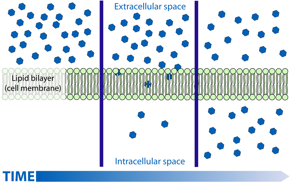
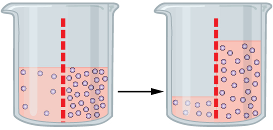
Osmosis
Osmosis is a special type of diffusion — the diffusion of water molecules across a membrane. Like other molecules, water moves from an area of higher concentration to an area of lower concentration. Water moves in or out of a cell until its concentration is the same on both sides of the plasma membrane. In Figure 4.7.3, the dotted red line shows a semi-permeable membrane. In the first beaker, there is an uneven concentration of solutes on either side of the membrane, but the solute cannot cross — diffusion of the solute can't occur. In this case, water will move to even out the concentration as has happened on the beaker on the right side. The water levels are uneven, but the process of osmosis has evened out the concentration gradient.
Facilitated Diffusion
Water and many other substances cannot simply diffuse across a membrane. Hydrophilic molecules, charged ions, and relatively large molecules (such as glucose) all need help with diffusion. This help comes from special proteins in the membrane known as transport proteins. Diffusion with the help of transport proteins is called facilitated diffusion. There are several types of transport proteins, including channel proteins and carrier proteins. Both are shown in Figure 4.7.4.
- Channel proteins form pores (or tiny holes) in the membrane. This allows water molecules and small ions to pass through the membrane without coming into contact with the hydrophobic tails of the lipid molecules in the interior of the membrane.
- Carrier proteins bind with specific ions or molecules. In doing so, they change shape. As carrier proteins change shape, they carry the ions or molecules across the membrane.
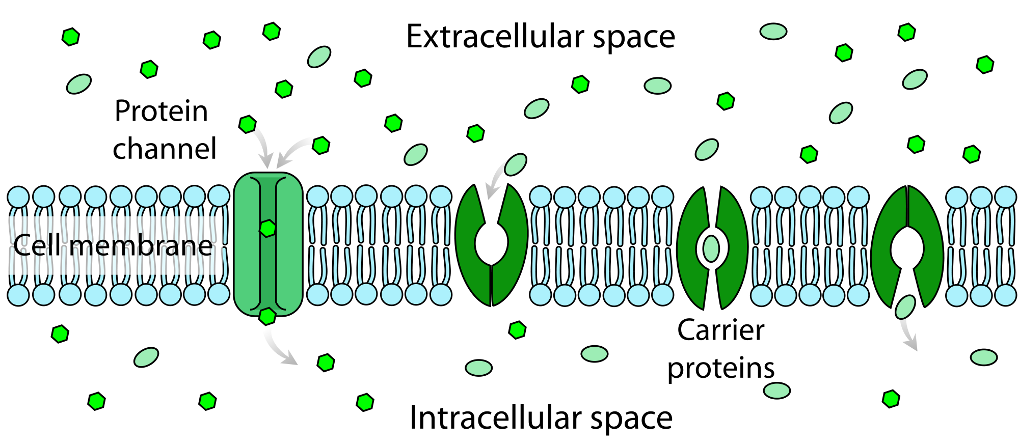
Transport and Homeostasis
For a cell to function normally, the inside of it must maintain a stable state. The concentrations of salts, nutrients, and other substances must be kept within certain ranges. The state in which stable conditions are maintained inside a cell (or an entire organism) is called homeostasis. Homeostasis requires constant adjustments, because conditions are always changing both inside and outside the cell. The transport of substances into and out of cells as described in this section plays an important role in homeostasis. By allowing the movement of substances into and out of cells, transport keeps conditions within normal ranges inside the cells and throughout the organism as a whole.
Watch this video "Cell Transport," by the Amoeba Sisters:
https://www.youtube.com/watch?v=Ptmlvtei8hw
Cell Transport with the Amoeba Sisters, 2016.
4.7 Summary
- Controlling the movement of things in and out of the cell is an important function of the plasma membrane. There are two basic ways that substances can cross the plasma membrane: passive transport — which requires no energy expenditure by the cell — and active transport — which requires energy.
- No energy is needed from the cell for passive transport because it occurs when substances move naturally from an area of higher concentration to an area of lower concentration.
- Simple diffusion is the movement of a substance due to differences in concentration. It happens without any help from other molecules. This is how very small, hydrophobic molecules (such as oxygen and carbon dioxide) enter and leave the cell.
- Osmosis is the diffusion of water molecules across a membrane. Water moves in or out of a cell by osmosis until its concentration is the same on both sides of the plasma membrane.
- Facilitated diffusion is the movement of a substance across a membrane due to differences in concentration, but it only occurs with the help of transport proteins (such as channel proteins or carrier proteins) in the membrane. This is how large or hydrophilic molecules and charged ions enter and leave the cell.
- Processes of passive transport play important roles in homeostasis. By allowing the movement of substances into and out of the cell, they keep conditions within normal ranges inside the cell and the organism as a whole.
4.7 Review Questions
- What is the main difference between passive and active transport?
- Summarize three different ways that passive transport can occur. Give an example of a substance that is transported in each way.
- Explain how transport across the plasma membrane is related to homeostasis of the cell.
- In general, why can only very small, hydrophobic molecules cross the cell membrane by simple diffusion?
- Explain how facilitated diffusion assists with osmosis in cells. Define osmosis and facilitated diffusion in your answer.
- Imagine a hypothetical cell with a higher concentration of glucose inside the cell than outside. Answer the following questions about this cell, assuming all transport across the membrane is passive, not active.
- Can the glucose simply diffuse across the cell membrane? Why or why not?
- Assuming that there are glucose transport proteins in the cell membrane, which way would glucose flow — into or out of the cell? Explain your answer.
- If the concentration of glucose was equal inside and outside of the cell, do you think there would be a net flow of glucose across the cell membrane in one direction or the other? Explain your answer.
- What are the similarities and differences between channel proteins and carrier proteins?
-
4.7 Explore More
https://www.youtube.com/watch?v=L-osEc07vMs&t=31s
Osmosis and Water Potential, Amoeba Sisters, 2018.
https://www.youtube.com/watch?v=AcrqIxt8am8
Structure Of The Cell Membrane - Active and Passive Transport, Professor Dave Explains, 2016.
Attributions
Figure 4.7.1
Windows/ The Oyster Suite in Eureka, CA by Drew Coffman on Unsplash is used under the Unsplash License https://unsplash.com/license).
Figure 4.7.2
Diffusion/ Scheme simple diffusion in cell membrane by Mariana Ruiz Villarreal [LadyofHats] is released into the public domain (https://en.wikipedia.org/wiki/Public_domain).
Figure 4.7.3
Osmosis by OpenStax on Wikimedia Commons is used under a CC BY 3.0 (https://creativecommons.org/licenses/by/3.0) license.
Figure 4.7.4
Scheme facilitated diffusion in cell membrane by Mariana Ruiz Villarreal [LadyofHats] is released into the public domain (https://en.wikipedia.org/wiki/Public_domain).
References
Amoeba Sisters. (2016, June 24). Cell transport. YouTube. https://www.youtube.com/watch?v=Ptmlvtei8hw&feature=youtu.be
Amoeba Sisters. (2018, June 27). Osmosis and water potential. YouTube. https://www.youtube.com/watch?v=L-osEc07vMs&feature=youtu.be
Betts, J. G., Young, K.A., Wise, J.A., Johnson, E., Poe, B., Kruse, D.H., Korol, O., Johnson, J.E., Womble, M., DeSaix, P. (2013, April 25). Figure 3.7 Osmosis [digital image]. In Anatomy and Physiology. OpenStax. https://openstax.org/books/anatomy-and-physiology/pages/3-1-the-cell-membrane
Professor Dave Explains. (2016, September 5). Structure of the cell membrane - Active and passive transport. https://www.youtube.com/watch?v=AcrqIxt8am8&feature=youtu.be
Created by CK-12 Foundation/Adapted by Christine Miller

Work Those Eye Muscles!
Imagine the man in Figure 12.3.1 turns his eyes in your direction. This is a very small movement, considering the conspicuously large and strong external eye muscles that control eyeball movements. These muscles have been called the strongest muscles in the human body relative to the work they do. However, the external eye muscles actually do a surprising amount of work. Eye movements occur almost constantly during waking hours, especially when we are scanning faces or reading. Eye muscles are also exercised nightly during the phase of sleep called rapid eye movement sleep. External eye muscles can move the eyes because they are made mainly of muscle tissue.
What is Muscle Tissue?
Muscle tissue is a soft tissue that makes up most of the tissues in the muscles of the human muscular system. Other tissues in muscles are connective tissues, such as tendons that attach skeletal muscles to bones and sheaths of connective tissues that cover or line muscle tissues. Only muscle tissue per se, has cells with the ability to contract.
There are three major types of muscle tissues in the human body: skeletal, smooth, and cardiac muscle tissues. Figure 12.3.2 shows how the three types of muscle tissues appear under magnification. When you read about each type below, you will learn why the three types appear as they do.
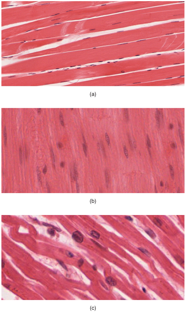
Skeletal Muscle Tissue
Skeletal muscle is muscle tissue that is attached to bones by tendons, which are bundles of collagen fibres. Whether you are moving your eyes or running a marathon, you are using skeletal muscles. Contractions of skeletal muscles are voluntary, or under conscious control of the central nervous system via the somatic nervous system. Skeletal muscle tissue is the most common type of muscle tissue in the human body. By weight, an average adult male is about 42% skeletal muscles, and the average adult female is about 36% skeletal muscles. Some of the major skeletal muscles in the human body are labeled in Figure 12.3.3 below.
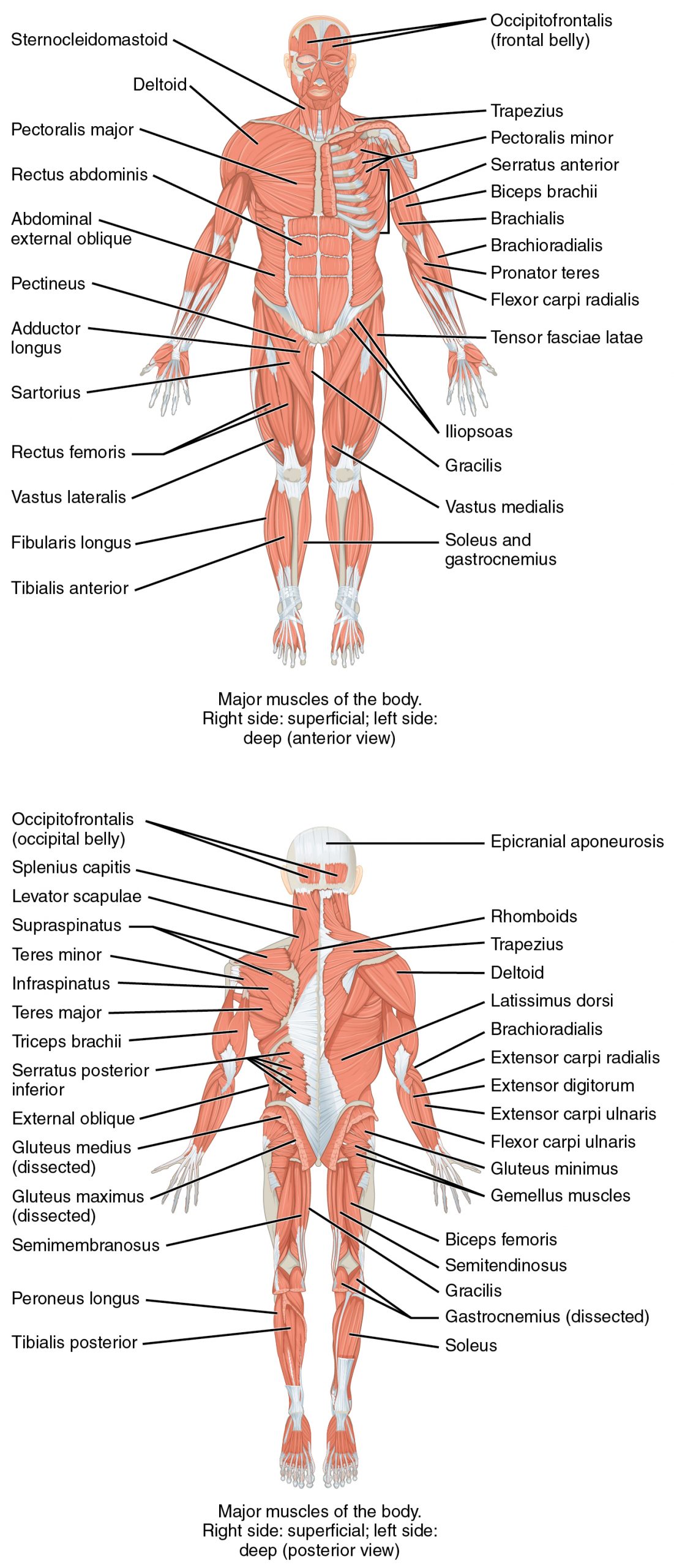
Skeletal Muscle Pairs
To move bones in opposite directions, skeletal muscles often consist of muscle pairs that work in opposition to one another, also called antagonistic muscle pairs. For example, when the biceps muscle (on the front of the upper arm) contracts, it can cause the elbow joint to flex or bend the arm, as shown in Figure 12.3.4. When the triceps muscle (on the back of the upper arm) contracts, it can cause the elbow to extend or straighten the arm. The biceps and triceps muscles, also shown in Figure 12.3.4, are an example of a muscle pair where the muscles work in opposition to each other.
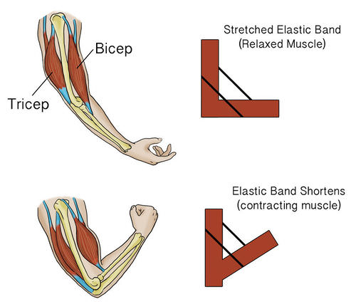
Skeletal Muscle Structure
Each skeletal muscle consists of hundreds — or even thousands — of skeletal muscle fibres, which are long, string-like cells. As shown in Figure 12.3.5 below, skeletal muscle fibres are individually wrapped in connective tissue called endomysium. The skeletal muscle fibres are bundled together in units called muscle fascicles, which are surrounded by sheaths of connective tissue called perimysium. Each fascicle contains between ten and 100 (or even more!) skeletal muscle fibres. Fascicles, in turn, are bundled together to form individual skeletal muscles, which are wrapped in connective tissue called epimysium. The connective tissues in skeletal muscles have a variety of functions. They support and protect muscle fibres, allowing them to withstand the forces of contraction by distributing the forces applied to the muscle. They also provide pathways for nerves and blood vessels to reach the muscles. In addition, the epimysium anchors the muscles to tendons.
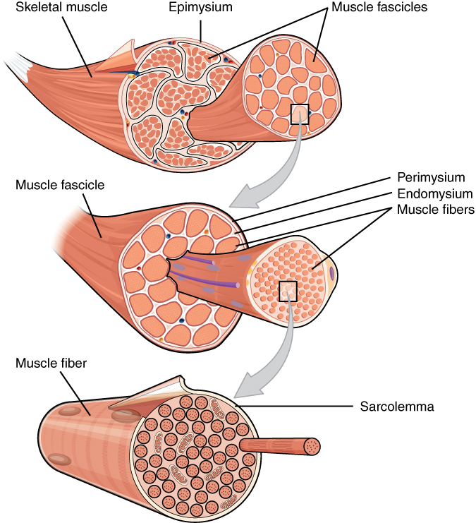
The same bundles-within-bundles structure is replicated within each muscle fibre. As shown in Figure 12.3.6, a muscle fibre consists of a bundle of myofibrils, which are themselves bundles of protein filaments. These protein filaments consist of thin filaments of the protein actin, which are anchored to structures called Z discs, and thick filaments of the protein myosin. The filaments are arranged together within a myofibril in repeating units called sarcomeres, which run from one Z disc to the next. The sarcomere is the basic functional unit of skeletal and cardiac muscles. It contracts as actin and myosin filaments slide over one another. Skeletal muscle tissue is said to be striated, because it appears striped. It has this appearance because of the regular, alternating A (dark) and I (light) bands of filaments arranged in sarcomeres inside the muscle fibres. Other components of a skeletal muscle fibre include multiple nuclei and mitochondria.
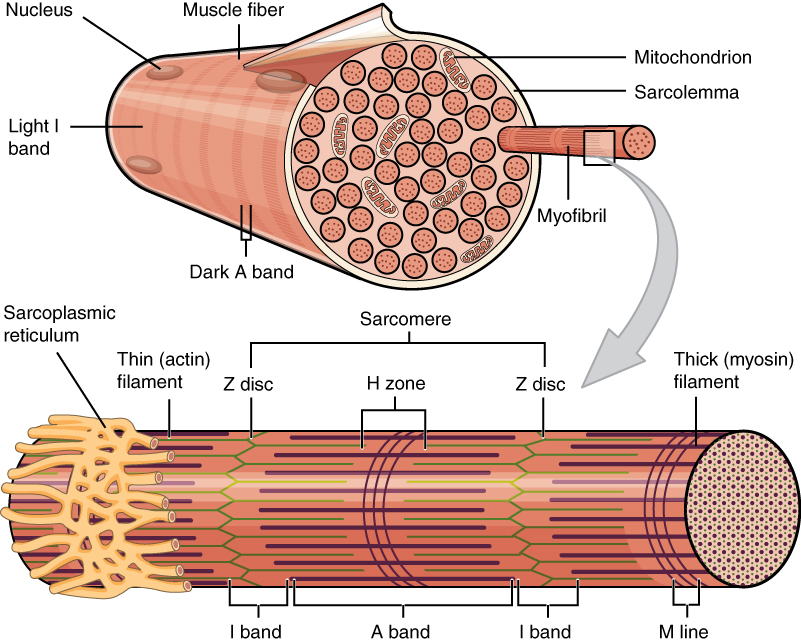
Slow- and Fast-Twitch Skeletal Muscle Fibres
Skeletal muscle fibres can be divided into two types, called slow-twitch (or type I) muscle fibres and fast-twitch (or type II) muscle fibres.
- Slow-twitch muscle fibres are dense with capillaries and rich in mitochondria and myoglobin, which is a protein that stores oxygen until needed for muscle activity. Relative to fast-twitch fibres, slow-twitch fibres can carry more oxygen and sustain aerobic (oxygen-using) activity. Slow-twitch fibres can contract for long periods of time, but not with very much force. They are relied upon primarily in endurance events, such as distance running or cycling.
- Fast-twitch muscle fibres contain fewer capillaries and mitochondria and less myoglobin. This type of muscle fibre can contract rapidly and powerfully, but it fatigues very quickly. Fast-twitch fibres can sustain only short, anaerobic (non-oxygen-using) bursts of activity. Relative to slow-twitch fibres, fast-twitch fibres contribute more to muscle strength and have a greater potential for increasing in mass. They are relied upon primarily in short, strenuous events, such as sprinting or weightlifting.
Proportions of fibre types vary considerably from muscle to muscle and from person to person. Individuals may be genetically predisposed to have a larger percentage of one type of muscle fibre than the other. Generally, an individual who has more slow-twitch fibres is better suited for activities requiring endurance, whereas an individual who has more fast-twitch fibres is better suited for activities requiring short bursts of power.
Smooth Muscle
Smooth muscle is muscle tissue in the walls of internal organs and other internal structures such as blood vessels. When smooth muscles contract, they help the organs and vessels carry out their functions. When smooth muscles in the stomach wall contract, for example, they squeeze the food inside the stomach, helping to mix and churn the food and break it into smaller pieces. This is an important part of digestion. Contractions of smooth muscles are involuntary, so they are not under conscious control. Instead, they are controlled by the autonomic nervous system, hormones, neurotransmitters, and other physiological factors.
Structure of Smooth Muscle
The cells that make up smooth muscle are generally called myocytes. Unlike the muscle fibres of striated muscle tissue, the myocytes of smooth muscle tissue do not have their filaments arranged in sarcomeres. Therefore, smooth tissue is not striated. However, the myocytes of smooth muscle do contain myofibrils, which in turn contain bundles of myosin and actin filaments. The filaments cause contractions when they slide over each other, as shown in Figure 12.3.7.

Functions of Smooth Muscle
Unlike striated muscle, smooth muscle can sustain very long-term contractions. Smooth muscle can also stretch and still maintain its contractile function, which striated muscle cannot. The elasticity of smooth muscle is enhanced by an extracellular matrix secreted by myocytes. The matrix consists of elastin, collagen, and other stretchy fibres. The ability to stretch and still contract is an important attribute of smooth muscle in organs such as the stomach and uterus (see Figures 12.3.8 and 12.3.9), both of which must stretch considerably as they perform their normal functions.
 |
 |
The following list indicates where many smooth muscles are found, along with some of their specific functions.
- Walls of organs of the gastrointestinal tract (such as the esophagus, stomach, and intestines), moving food through the tract by peristalsis
- Walls of air passages of the respiratory tract (such as the bronchi), controlling the diameter of the passages and the volume of air that can pass through them
- Walls of organs of the male and female reproductive tracts; in the uterus, for example, pushing a baby out of the uterus and into the birth canal
- Walls of structures of the urinary system, including the urinary bladder, allowing the bladder to expand so it can hold more urine, and then contract as urine is released
- Walls of blood vessels, controlling the diameter of the vessels and thereby affecting blood flow and blood pressure
- Walls of lymphatic vessels, squeezing the fluid called lymph through the vessels
- Iris of the eyes, controlling the size of the pupils and thereby the amount of light entering the eyes
- Arrector pili in the skin, raising hairs in hair follicles in the dermis
Cardiac Muscle
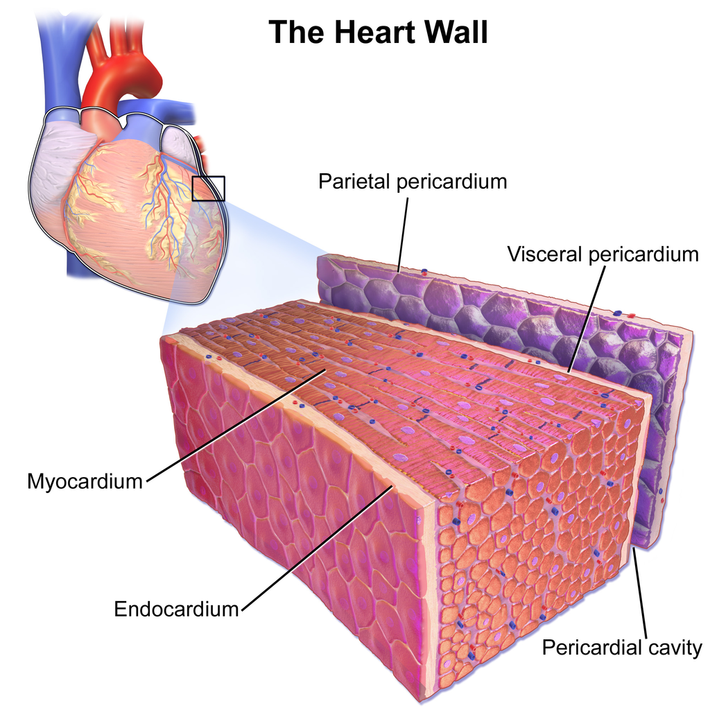
Cardiac muscle is found only in the wall of the heart. It is also called myocardium. As shown in Figure 12.3.10, myocardium is enclosed within connective tissues, including the endocardium on the inside of the heart and pericardium on the outside of the heart. When cardiac muscle contracts, the heart beats and pumps blood. Contractions of cardiac muscle are involuntary, like those of smooth muscles. They are controlled by electrical impulses from specialized cardiac muscle cells in an area of the heart muscle called the sinoatrial node.
Like skeletal muscle, cardiac muscle is striated because its filaments are arranged in sarcomeres inside the muscle fibres. However, in cardiac muscle, the myofibrils are branched at irregular angles rather than arranged in parallel rows (as they are in skeletal muscle). This explains why cardiac and skeletal muscle tissues look different from one another.
The cells of cardiac muscle tissue are arranged in interconnected networks. This arrangement allows rapid transmission of electrical impulses, which stimulate virtually simultaneous contractions of the cells. This enables the cells to coordinate contractions of the heart muscle.
The heart is the muscle that performs the greatest amount of physical work in the course of a lifetime. Although the power output of the heart is much less than the maximum power output of some other muscles in the human body, the heart does its work continuously over an entire lifetime without rest. Cardiac muscle contains a great many mitochondria, which produce ATP for energy and help the heart resist fatigue.
Feature: Human Biology in the News
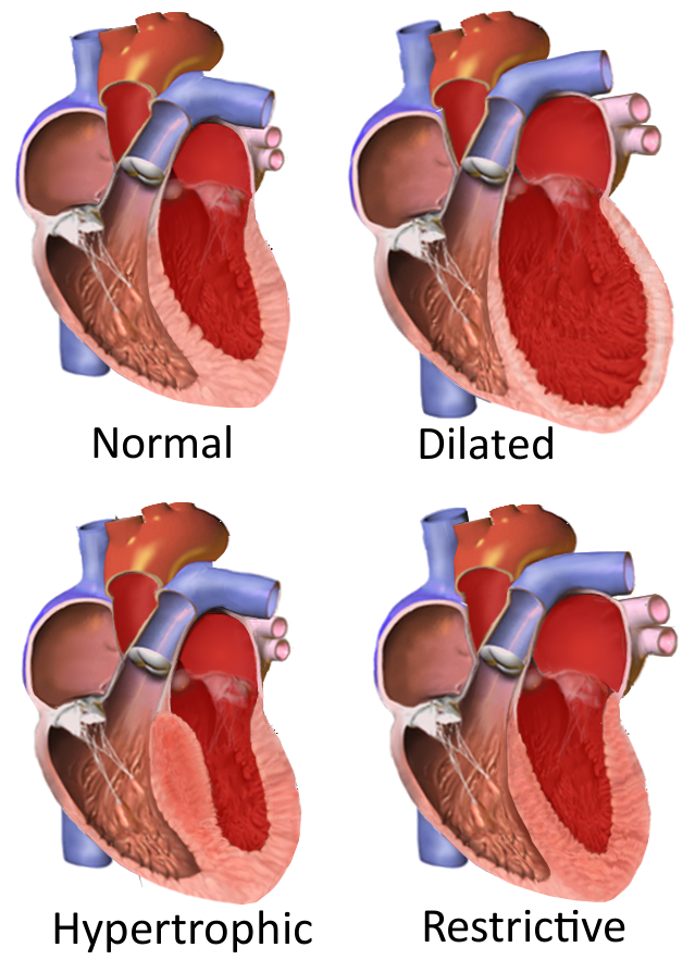
Cardiomyopathy is a disease in which the muscles of the heart are no longer able to effectively pump blood to the body — extreme forms of this disease can lead to heart failure. There are four main types of cardiomyopathy (also illustrated in Figure 12.3.11):
- Dilated (congestive) cardiomyopathy: the left ventricle (the chamber itself) of the heart becomes enlarged and can't pump blood our to the body. This is normally related to coronary artery disease and/or heart attack
- Hypertrophic cardiomyopathy: abnormal thickening of the muscular walls of the left ventricle make the chamber less able to work properly. This condition is more common in patients with a family history of the disease.
- Restrictive cardiomyopathy: the myocardium becomes abnormally rigid and inelastic and is unable to expand in between heartbeats to refill with blood. Restrictive cardiomyopathy typically affects older people.
- Arrhythmogenic right ventricular cardiomyopathy: the right ventricular muscle is replaced by adipose or scar tissue, reducing elasticity and interfering with normal heartbeat and rhythm. This disease is often caused by genetic mutations.
Cardiomyopathy is typically diagnosed with a physical exam supplemented by medical and family history, an angiogram, blood tests, chest x-rays and electrocardiograms. In some cases your doctor would also requisition a CT scan and/or genetic testing.
When treating cardiomyopathy, the goal is to reduce symptoms that affect everyday life. Certain medications can help regularize and slow heart rate, decrease chances of blood clots and cause vasodilation in the coronary arteries. If medication is not sufficient to manage symptoms, a pacemaker or even a heart transplant may be the best option. Lifestyle can also help manage the symptoms of cardiomyopathy — people living with this disease are encouraged to avoid drug and alcohol use, control high blood pressure, eat a healthy diet, get ample rest and exercise, as well as reduce stress levels.
12.3 Summary
- Muscle tissue is a soft tissue that makes up most of the tissues in the muscles of the human muscular system. It is the only type of tissue that has cells with the ability to contract.
- Skeletal muscle tissue is attached to bones by tendons. It allows voluntary body movements.
- Skeletal muscle is the most common type of muscle tissue in the human body. To move bones in opposite directions, skeletal muscles often consist of pairs of muscles that work in opposition to one another to move bones in different directions at joints.
- Skeletal muscle fibres are bundled together in units called muscle fascicles, which are bundled together to form individual skeletal muscles. Skeletal muscles also have connective tissue supporting and protecting the muscle tissue.
- Each skeletal muscle fibre consists of a bundle of myofibrils, which are bundles of protein filaments. The filaments are arranged in repeating units called sarcomeres, which are the basic functional units of skeletal muscles. Skeletal muscle tissue is striated because of the pattern of sarcomeres in its fibres.
- Skeletal muscle fibres can be divided into two types, called slow-twitch and fast-twitch fibres. Slow-twitch fibres are used mainly in aerobic endurance activities, such as long-distance running. Fast-twitch fibres are used mainly for non-aerobic, strenuous activities, such as sprinting. Proportions of the two types of fibres vary from muscle to muscle and person to person.
- Smooth muscle tissue is found in the walls of internal organs and vessels. When smooth muscles contract, they help the organs and vessels carry out their functions. Contractions of smooth muscles are involuntary and controlled by the autonomic nervous system, hormones, and other substances.
- Cells of smooth muscle tissue are not striated because they lack sarcomeres, but the cells contract in the same basic way as striated muscle cells. Unlike striated muscle, smooth muscle can sustain very long-term contractions and maintain its contractile function, even when stretched.
- Cardiac muscle tissue is found only in the wall of the heart. When cardiac muscle contracts, the heart beats and pumps blood. Contractions of cardiac muscle are involuntary, like those of smooth muscles. They are controlled by electrical impulses from specialized cardiac cells.
- Like skeletal muscle, cardiac muscle is striated because its filaments are arranged in sarcomeres inside the muscle fibres. However, the myofibrils are branched instead of arranged in parallel rows, making cardiac and skeletal muscle tissues look different from one another.
- The heart is the muscle that performs the greatest amount of physical work in the course of a lifetime. Its cells contain a great many mitochondria to produce ATP for energy and help the heart resist fatigue.
12.3 Review Questions
- What is muscle tissue?
-
-
- Where is skeletal muscle found, and what is its general function?
- Why do many skeletal muscles work in pairs?
- Describe the structure of a skeletal muscle.
- Relate muscle fibre structure to the functional units of muscles.
- Why is skeletal muscle tissue striated?
- Where is smooth muscle found? What controls the contraction of smooth muscle?
- Where is cardiac muscle found? What controls its contractions?
- The heart muscle is smaller and less powerful than some other muscles in the body. Why is the heart the muscle that performs the greatest amount of physical work in the course of a lifetime? How does the heart resist fatigue?
- Give one example of connective tissue that is found in muscles. Describe one of its functions.
12.3 Explore More
https://www.youtube.com/watch?v=3_PYnWVoUzM
What happens during a heart attack? - Krishna Sudhir, TED-Ed, 2017.
https://www.youtube.com/watch?v=bwOE1MEginA&feature=emb_logo
Three types of muscle | Circulatory system physiology | NCLEX-RN | KhanAcademyMedicine, 2012.
Attributions
Figure 12.3.1
Look by ali-yahya-155huuQwGvA [photo] by Ali Yahya on Unsplash is used under the Unsplash License (https://unsplash.com/license).
Figure 12.3.2
Skeletal_Smooth_Cardiac by OpenStax College on Wikimedia Commons is used under a CC BY 3.0 (https://creativecommons.org/licenses/by/3.0) license.
Figure 12.3.3
Anterior_and_Posterior_Views_of_Muscles by OpenStax on Wikimedia Commons is used under a CC BY 4.0 (https://creativecommons.org/licenses/by/4.0) license.
Figure 12.3.4
Antagonistic Muscle Pair by Laura Guerin at CK-12 Foundation on Wikimedia Commons is used under a CC BY-NC 3.0 (https://creativecommons.org/licenses/by-nc/3.0/) license.
Figure 12.3.5
Muscle_Fibes_(large) by OpenStax on Wikimedia Commons is used under a CC BY 4.0 (https://creativecommons.org/licenses/by/4.0) license.
Figure 12.3.6
Muscle_Fibers_(small) by OpenStax on Wikimedia Commons is used under a CC BY 4.0 (https://creativecommons.org/licenses/by/4.0) license.
Figure 12.3.7
Smooth_Muscle_Contraction by OpenStax on Wikimedia Commons is used under a CC BY 4.0 (https://creativecommons.org/licenses/by/4.0) license.
Figure 12.3.8
Blausen_0747_Pregnancy by BruceBlaus on Wikimedia Commons is used under a CC BY 3.0 (https://creativecommons.org/licenses/by/3.0) license.
Figure 12.3.9
Size_of_Uterus_Throughout_Pregnancy-02 by OpenStax College on Wikimedia Commons is used under a CC BY 3.0 (https://creativecommons.org/licenses/by/3.0) license.
Figure 12.3.10
1024px-Blausen_0470_HeartWall by BruceBlaus on Wikimedia Commons is used under a CC BY 3.0 (https://creativecommons.org/licenses/by/3.0) license.
Figure 12.3.11
Tipet_e_kardiomiopative by Npatchett at English Wikipedia on Wikimedia Commons is used under a CC BY-SA 3.0 (https://creativecommons.org/licenses/by-sa/3.0) license. (Work derived from Blausen 0165 Cardiomyopathy Dilated by BruceBlaus)
References
Betts, J. G., Young, K.A., Wise, J.A., Johnson, E., Poe, B., Kruse, D.H., Korol, O., Johnson, J.E., Womble, M., DeSaix, P. (2013, June 19). Figure 4.18 Muscle tissue [digital image]. In Anatomy and Physiology (Section 4.4). OpenStax. https://openstax.org/books/anatomy-and-physiology/pages/4-4-muscle-tissue-and-motion
Betts, J. G., Young, K.A., Wise, J.A., Johnson, E., Poe, B., Kruse, D.H., Korol, O., Johnson, J.E., Womble, M., DeSaix, P. (2013, June 19). Figure 28.18 Size of uterus throughout pregnancy [digital image]. In Anatomy and Physiology (Section 28.4). OpenStax. https://openstax.org/books/anatomy-and-physiology/pages/28-4-maternal-changes-during-pregnancy-labor-and-birth
Betts, J. G., Young, K.A., Wise, J.A., Johnson, E., Poe, B., Kruse, D.H., Korol, O., Johnson, J.E., Womble, M., DeSaix, P. (2016, May 18). Figure 10.3 The three connective tissue layers [digital image]. In Anatomy and Physiology (Section 10.2). OpenStax. https://openstax.org/books/anatomy-and-physiology/pages/10-2-skeletal-muscle
Betts, J. G., Young, K.A., Wise, J.A., Johnson, E., Poe, B., Kruse, D.H., Korol, O., Johnson, J.E., Womble, M., DeSaix, P. (2016, May 18). Figure 10.4 Muscle fiber [digital image]. In Anatomy and Physiology (Section 10.2). OpenStax. https://openstax.org/books/anatomy-and-physiology/pages/10-2-skeletal-muscle
Betts, J. G., Young, K.A., Wise, J.A., Johnson, E., Poe, B., Kruse, D.H., Korol, O., Johnson, J.E., Womble, M., DeSaix, P. (2016, May 18). Figure 10.24 Muscle contraction [digital image]. In Anatomy and Physiology (Section 10.8). OpenStax. https://openstax.org/books/anatomy-and-physiology/pages/10-8-smooth-muscle
Betts, J. G., Young, K.A., Wise, J.A., Johnson, E., Poe, B., Kruse, D.H., Korol, O., Johnson, J.E., Womble, M., DeSaix, P. (2016, May 18). Figure 11.5 Overview of the muscular system [digital image]. In Anatomy and Physiology (Section 11.2). OpenStax. https://openstax.org/books/anatomy-and-physiology/pages/11-2-naming-skeletal-muscles
Blausen.com staff. (2014). Medical gallery of Blausen Medical 2014. WikiJournal of Medicine 1 (2). DOI:10.15347/wjm/2014.010. ISSN 2002-4436.
Brainard, J/ CK-12 Foundation. (2012). Figure 5 Triceps and biceps muscles in the upper arm are opposing muscles. [digital image]. In CK-12 Biology (Section 21.3) [online Flexbook]. CK12.org. https://www.ck12.org/book/ck-12-biology/section/21.3/ (Last modified August 11, 2017.)
khanacademymedicine. (2012, October 19). Three types of muscle | Circulatory system physiology | NCLEX-RN | Khan Academy. YouTube.
TED-Ed. (2017, February 14). What happens during a heart attack? - Krishna Sudhir. YouTube. https://www.youtube.com/watch?v=3_PYnWVoUzM&feature=youtu.be
Created by: CK-12/Adapted by Christine Miller

Letting in the Light
Look at the big windows in this house (Figure 4.7.1). Imagine all the light they must let in on a sunny day. Now imagine living in a house that has walls without any windows or doors. Nothing could enter or leave. Or imagine living in a house with holes in the walls instead of windows and doors. Things could enter or leave, but you couldn’t control what came in or went out. Only when a house has walls with windows and doors that can be opened or closed, can you control what enters or leaves. Windows and doors allow you to let in light and the family dog and keep out rain and bugs, for example.
Transport Across Membranes
If a cell were a house, the plasma membrane would be walls with windows and doors. Moving things in and out of the cell is an important function of the plasma membrane. It controls everything that enters and leaves the cell. There are two basic ways that substances can cross the plasma membrane: passive transport — which requires no energy expenditure by the cell — and active transport — which requires energy from the cell.
Transport Without Energy Expenditure By The Cell
Passive transport occurs when substances cross the plasma membrane without any input of energy from the cell. No energy is required because the substances are moving from an area where they have a higher concentration to an area where they have a lower concentration. Concentration refers to the number of particles of a substance per unit of volume. The more particles of a substance in a given volume, the higher the concentration. A substance always moves from an area where it is more concentrated to an area where it is less concentrated.
There are several different types of passive transport, including simple diffusion, osmosis, and facilitated diffusion. Each type is described below.
Simple Diffusion
Diffusion is the movement of a substance due to a difference in concentration. It happens without any help from other molecules. The substance simply moves from the area where it is more concentrated to the area where it is less concentrated. Picture someone spraying perfume in the corner of a room. Do the perfume molecules stay in the corner? No, they spread out, or diffuse throughout the room until they are evenly spread out. Figure 4.7.2 shows how diffusion works across a cell membrane. Substances that can squeeze between the lipid molecules in the plasma membrane by simple diffusion are generally very small, hydrophobic molecules, such as molecules of oxygen and carbon dioxide.


Osmosis
Osmosis is a special type of diffusion — the diffusion of water molecules across a membrane. Like other molecules, water moves from an area of higher concentration to an area of lower concentration. Water moves in or out of a cell until its concentration is the same on both sides of the plasma membrane. In Figure 4.7.3, the dotted red line shows a semi-permeable membrane. In the first beaker, there is an uneven concentration of solutes on either side of the membrane, but the solute cannot cross — diffusion of the solute can't occur. In this case, water will move to even out the concentration as has happened on the beaker on the right side. The water levels are uneven, but the process of osmosis has evened out the concentration gradient.
Facilitated Diffusion
Water and many other substances cannot simply diffuse across a membrane. Hydrophilic molecules, charged ions, and relatively large molecules (such as glucose) all need help with diffusion. This help comes from special proteins in the membrane known as transport proteins. Diffusion with the help of transport proteins is called facilitated diffusion. There are several types of transport proteins, including channel proteins and carrier proteins. Both are shown in Figure 4.7.4.
- Channel proteins form pores (or tiny holes) in the membrane. This allows water molecules and small ions to pass through the membrane without coming into contact with the hydrophobic tails of the lipid molecules in the interior of the membrane.
- Carrier proteins bind with specific ions or molecules. In doing so, they change shape. As carrier proteins change shape, they carry the ions or molecules across the membrane.

Transport and Homeostasis
For a cell to function normally, the inside of it must maintain a stable state. The concentrations of salts, nutrients, and other substances must be kept within certain ranges. The state in which stable conditions are maintained inside a cell (or an entire organism) is called homeostasis. Homeostasis requires constant adjustments, because conditions are always changing both inside and outside the cell. The transport of substances into and out of cells as described in this section plays an important role in homeostasis. By allowing the movement of substances into and out of cells, transport keeps conditions within normal ranges inside the cells and throughout the organism as a whole.
Watch this video "Cell Transport," by the Amoeba Sisters:
https://www.youtube.com/watch?v=Ptmlvtei8hw
Cell Transport with the Amoeba Sisters, 2016.
4.7 Summary
- Controlling the movement of things in and out of the cell is an important function of the plasma membrane. There are two basic ways that substances can cross the plasma membrane: passive transport — which requires no energy expenditure by the cell — and active transport — which requires energy.
- No energy is needed from the cell for passive transport because it occurs when substances move naturally from an area of higher concentration to an area of lower concentration.
- Simple diffusion is the movement of a substance due to differences in concentration. It happens without any help from other molecules. This is how very small, hydrophobic molecules (such as oxygen and carbon dioxide) enter and leave the cell.
- Osmosis is the diffusion of water molecules across a membrane. Water moves in or out of a cell by osmosis until its concentration is the same on both sides of the plasma membrane.
- Facilitated diffusion is the movement of a substance across a membrane due to differences in concentration, but it only occurs with the help of transport proteins (such as channel proteins or carrier proteins) in the membrane. This is how large or hydrophilic molecules and charged ions enter and leave the cell.
- Processes of passive transport play important roles in homeostasis. By allowing the movement of substances into and out of the cell, they keep conditions within normal ranges inside the cell and the organism as a whole.
4.7 Review Questions
- What is the main difference between passive and active transport?
- Summarize three different ways that passive transport can occur. Give an example of a substance that is transported in each way.
- Explain how transport across the plasma membrane is related to homeostasis of the cell.
- In general, why can only very small, hydrophobic molecules cross the cell membrane by simple diffusion?
- Explain how facilitated diffusion assists with osmosis in cells. Define osmosis and facilitated diffusion in your answer.
- Imagine a hypothetical cell with a higher concentration of glucose inside the cell than outside. Answer the following questions about this cell, assuming all transport across the membrane is passive, not active.
- Can the glucose simply diffuse across the cell membrane? Why or why not?
- Assuming that there are glucose transport proteins in the cell membrane, which way would glucose flow — into or out of the cell? Explain your answer.
- If the concentration of glucose was equal inside and outside of the cell, do you think there would be a net flow of glucose across the cell membrane in one direction or the other? Explain your answer.
- What are the similarities and differences between channel proteins and carrier proteins?
-
4.7 Explore More
https://www.youtube.com/watch?v=L-osEc07vMs&t=31s
Osmosis and Water Potential, Amoeba Sisters, 2018.
https://www.youtube.com/watch?v=AcrqIxt8am8
Structure Of The Cell Membrane - Active and Passive Transport, Professor Dave Explains, 2016.
Attributions
Figure 4.7.1
Windows/ The Oyster Suite in Eureka, CA by Drew Coffman on Unsplash is used under the Unsplash License https://unsplash.com/license).
Figure 4.7.2
Diffusion/ Scheme simple diffusion in cell membrane by Mariana Ruiz Villarreal [LadyofHats] is released into the public domain (https://en.wikipedia.org/wiki/Public_domain).
Figure 4.7.3
Osmosis by OpenStax on Wikimedia Commons is used under a CC BY 3.0 (https://creativecommons.org/licenses/by/3.0) license.
Figure 4.7.4
Scheme facilitated diffusion in cell membrane by Mariana Ruiz Villarreal [LadyofHats] is released into the public domain (https://en.wikipedia.org/wiki/Public_domain).
References
Amoeba Sisters. (2016, June 24). Cell transport. YouTube. https://www.youtube.com/watch?v=Ptmlvtei8hw&feature=youtu.be
Amoeba Sisters. (2018, June 27). Osmosis and water potential. YouTube. https://www.youtube.com/watch?v=L-osEc07vMs&feature=youtu.be
Betts, J. G., Young, K.A., Wise, J.A., Johnson, E., Poe, B., Kruse, D.H., Korol, O., Johnson, J.E., Womble, M., DeSaix, P. (2013, April 25). Figure 3.7 Osmosis [digital image]. In Anatomy and Physiology. OpenStax. https://openstax.org/books/anatomy-and-physiology/pages/3-1-the-cell-membrane
Professor Dave Explains. (2016, September 5). Structure of the cell membrane - Active and passive transport. https://www.youtube.com/watch?v=AcrqIxt8am8&feature=youtu.be
Created by: CK-12/Adapted by Christine Miller
Jumping for Joy!
The people in Figure 6.2.1 illustrate some of the great phenotypic variation displayed in modern Homo sapiens. The lighter-skinned men in the photo are Euro-American tourists in Kenya (East Africa). The darker-skinned men are native Kenyans who belong to a tribal group named the Maasai. These men come from populations on different continents on opposite sides of the globe. Their populations have unique histories, environments, and cultures. Besides differences in skin colour, the men have different hair and eye colours, facial features, and body builds. Based on such obvious physical differences, you might think that our species is characterized by a high degree of genetic variation. In fact, there is much less genetic variation in the human species than there is in many other mammalian species, including our closest relatives — the chimpanzees.
Overview of Human Genetic Variation
No two human individuals are genetically identical unless they are monozygotic (identical) twins. Between any two people, DNA differs, on average, at about one in one thousand nucleotide base pairs. We each have a total of about three billion base pairs, so any two people differ by an average of about three million base pairs. That may sound like a lot, but it's only 0.1% of our total genetic makeup. This means that two people chosen at random are likely to be 99.9 per cent identical genetically, no matter where in the world they come from.
At an individual level, most human genetic variation is not very important biologically, because it has no apparent adaptive significance. It neither enhances nor detracts from individual fitness. Only a small percentage of DNA variations actually occur in coding regions of DNA — which are sequences that are translated into proteins — or in regulatory regions, which are sequences that control gene expression. Differences that occur in other regions of DNA have no impact on phenotype. Even variations in coding regions of DNA may or may not affect phenotype. Some DNA variations may alter the amino acid sequence of a protein, but not affect how the protein functions. Other DNA variations do not even change the amino acid sequence of the encoded protein.
At a population level, genetic variation is crucial if evolution is to occur. Genetically-based differences in fitness among individuals are the key to evolution by natural selection. Without genetic variation within populations, there can be no differential fitness by genotype, and natural selection cannot occur.
Patterns of Human Genetic Variation
Data comparing DNA sequences from around the world show that only about ten per cent of our total genetic variation occurs between people from different continents, like the American tourists and African Maasai pictured in Figure 6.1.1. The other 90 per cent of genetic variation occurs between people within continental populations, such as between North Americans or between Africans. Within any human population, many genes have two or more normal alleles that contribute to genetic differences among individuals. The case in which a gene has two or more alleles in a population at frequencies greater than one per cent is called a polymorphism. A single nucleotide polymorphism (SNP) involves variation in just one nucleotide in a DNA sequence. SNPs account for most of our genetic differences. Other types of variations (such as deletions and insertions of nucleotides in DNA sequences) account for a much smaller proportion of our overall genetic variation.
Different populations may have different allele frequencies for polymorphic genes. However, the distribution of allele frequencies in different populations around the world tends not to be discrete or distinct. Instead, the pattern is more often one of gradual geographic variations, or clines, in allele frequencies. You can see an example of a clinal distribution of allele frequencies in the map (Figure 6.2.2) below. Clinal distributions like this may be a reflection of natural selection pressures varying continuously over geographic space, or they may reflect a combination of genetic drift and gene flow of neutral alleles.
Although most genetic variation occurs within rather than between populations, certain alleles do seem to cluster in particular geographic areas. One example happens with the Duffy gene. Variations in this gene are the basis of the Duffy blood group, which is determined by the presence or absence of a red blood cell antigen, similar to the more familiar ABO blood group antigens. The genotype for having no antigen for the Duffy blood group is far higher in African populations and in people who have African ancestry than it is in non-African people, as indicated in the following table. Genes (such as the Duffy gene) may be useful as genetic markers to establish the ancestral populations of individuals.
Table 6.2.1
Population Frequencies for No Antigen in the Duffy Blood Group
| Population | Per cent of Population Lacking Duffy Antigen |
| African | 88-100 |
| African American | 68 |
| non-African American | <1 |
The reason for the different population frequencies for the Duffy antigen appears to be natural selection. People who lack the Duffy antigen are relatively resistant to malaria, which is one of the oldest and most devastating human diseases. Malaria has been a persistent and widespread disease in sub-Saharan Africa for tens of thousands of years. DNA analyses suggest that the allele associated with lack of the Duffy antigen evolved at least twice in Africa and was strongly selected for, causing it to increase in frequency. The Duffy gene is just one of many genes that have polymorphic alleles, because one of the alleles protects against malaria. In fact, a greater number of known genetic polymorphisms may be attributed to selection because of malaria than any other single selective agent.
Factors Influencing the Level of Human Genetic Variation
The age and size of a population increases the genetic variation within that population. You would expect an older, larger population to have more genetic variation. The older a population is, the longer it has been accumulating mutations. The larger a population is, the more people there are in which mutations can occur. Anatomically modern humans evolved less than a quarter million years ago, which is a relatively short period of time for mutations to accumulate. Our population was also quite small at some point in the past, perhaps consisting of no more than ten thousand adults, which reduced genetic variation even more. These factors explain why humans are relatively homogeneous genetically as a species.
What We Can Learn From Knowledge of Human Genetic Variation
Knowledge of genetic variation can help us understand our similarities and differences, our origins, and our evolutionary past. It can also help us understand human diseases and — hopefully — find new ways to treat them.
Human Origins
The data on human genetic variation generally supports the out-of-Africa hypothesis for human origins. According to this hypothesis, the common ancestor of all modern humans evolved in Africa around 200 thousand years ago. Then, starting no later than about 60 thousand years ago, part of the African population left Africa and migrated to Europe and Asia. As the migrants spread throughout the Old World, they replaced (and/or absorbed) the populations of archaic humans they encountered.
Most studies of human genetic variation find there is greater genetic diversity in African than non-African populations. This is consistent with the older age of the African population proposed by the out-of-Africa hypothesis. In addition, most of the genetic variation in non-African populations is a subset of the variation in African populations. This is consistent with the idea that part of the African population left Africa much later and migrated to other places in the Old World.
Recent comparisons of modern human and archaic human (including Neanderthal and Denisovan) DNA show that interbreeding occurred between their populations, but to differing degrees. The result of new DNA sequences entering a population’s gene pool through interbreeding is called admixture. There is greater admixture with archaic humans in modern European, Asian, and Oceanic populations than in modern African populations. Populations with the greatest admixture are those in Melanesia. About eight per cent of their DNA came from archaic Denisovans in East Asia.
Human Population History
Patterns of human genetic variation can be used to reconstruct population history. That history is literally recorded in our DNA. Any major population event (such as a significant reduction in population size or a high rate of migration) leaves a mark on a population’s genetic variation.
- Going through a dramatic size reduction decreases intra-population genetic variation (variation occurring within a population). As a case in point, DNA analyses suggest that there may have been drastic size reductions in the human populations that colonized the New World between 15 thousand and 20 thousand years ago. There were also size reductions in the human populations that first left Africa at least 60 thousand years ago, which helps explain the lower genetic diversity of modern non-African populations.
- A high rate of migration between populations may lead to gene flow, and this changes genetic variation in two ways. Gene flow decreases inter-population genetic variation (variation occurring between populations), while it increases intra-population variation. Gene flow — primarily between nearby populations — may contribute to the formation of clines in allele frequencies, as on the map in Figure 6.2.2.
Human Genetic Variation and Disease
An important benefit of studying human genetic variation is that we can learn more about the genetic basis of human diseases. The more we understand the causes of diseases, the more likely it is that we will be able to find effective treatments and cures for them.
Some disorders are caused by mutations in a single gene. Most of these disorders are generally rare, but some of them occur at significantly higher frequencies in certain populations. For example, Ellis-van Creveld syndrome has an unusually high frequency in Pennsylvania Amish populations, and Tay-Sachs disease has a relatively high frequency in Ashkenazi Jewish populations. Albinism is another single-gene disorder that has a variable frequency. In North America and Europe, rates of albinism are approximately 1:18,000. In Africa, in contrast, the rates range from 1:5,000 to 1:15,000. Some African populations have estimated albinism rates as high as 1:1000. The photo below (Figure 6.2.3) shows an African albino man from Mali, where there is a relatively high rate of albinism. High population-specific frequencies of single-gene disorders like these may be attributable to a variety of factors, such as small founding populations and a relative lack of gene flow.
It is likely that the majority of human diseases are caused by a complex mix of multiple genes (polygenic) and environmental factors (multifactorial). Examples of polygenic, multifactorial diseases are type II diabetes and heart disease. We do not typically think of these diseases as genetic diseases, because our genes do not predetermine whether we develop them. Our genes, however, do influence our chances of developing the diseases under certain environmental conditions. Even our chances of developing some infectious diseases are influenced by our genes. For example, a variant allele for a gene called CCR5 seems to confer resistance to infection with HIV, the virus that causes AIDS.
6.2 Summary
- No two human individuals are genetically identical (except for monozygotic twins), but the human species as a whole exhibits relatively little genetic diversity, relative to other mammalian species. Genetically, two people chosen at random are likely to be 99.9 per cent identical.
- Of the total genetic variation in humans, about 90 per cent occurs between people within continental populations. Only about 10 per cent occurs between people from different continents. Older, larger populations tend to have greater genetic variation, because there is more time and there are more people in which to accumulate mutations.
- Single nucleotide polymorphisms account for most human genetic differences. Allele frequencies for polymorphic genes generally have a clinal (rather than discrete) distribution. A minority of alleles seem to cluster in particular geographic areas, such as the allele for no antigen in the Duffy blood group. Such alleles may be useful as genetic markers to establish the ancestry of individuals.
- Knowledge of genetic variation can help us understand our similarities and differences. It can also help us reconstruct our evolutionary origins and history as a species. For example, the distribution of modern human genetic variation is consistent with the out-of-Africa hypothesis for the origin of modern humans.
- An important benefit of studying human genetic variation is learning more about the genetic basis of human diseases. This, in turn, should help us find more effective treatments and cures.
6.2 Review Questions
- Compare and contrast the significance of genetic variation at the individual and population levels.
- Describe genetic variation within and between human populations on different continents.
- Explain why allele frequencies for the Duffy gene may be used as a genetic marker for African ancestry.
- Identify factors that increase the level of genetic variation within populations.
-
- Discuss genetic evidence that supports the out-of-Africa hypothesis of modern human origins.
- What evidence suggests that modern humans interbred with archaic human populations after modern humans left Africa?
- How do population size reductions and gene flow impact the genetic variation of populations?
- Describe the role of genetic variation in human disease.
- Explain the reasons why variation in a DNA sequence can have no effect on the fitness of an individual.
- Explain why migration between populations decreases inter-population genetic variation. How does this relate to the development of clines in allele frequency?
- The amount of mixing of modern human DNA and archaic human DNA is an example of _________ .
6.2 Explore More
https://www.youtube.com/watch?v=RGtaq3PiIoU
The Journey of Your Past | National Geographic, National Geographic, 2013.
https://www.youtube.com/watch?v=kU0ei9ApmsY
Svante Pääbo: DNA clues to our inner neanderthal, TED, 2011.
https://www.youtube.com/watch?time_continue=2&v=cHRM2S_fBOk&feature=emb_logo
Why Are Some People Albino?, Seeker, 2015.
Attributions
Figure 6.2.1
Maasai_men_and_tourists_jumping by Christopher Michel on Wikimedia Commons is used under a CC BY 2.0 (https://creativecommons.org/licenses/by/2.0/deed.en) (license.
Figure 6.2.2
Geospatial_distribution_of_SNP_rs1426654-A_allele by Basu Mallick C, Iliescu FM, Möls M, Hill S, Tamang R, Chaubey G, et al. on Wikimedia Commons is used under a CC BY 2.5 (https://creativecommons.org/licenses/by/2.5/deed.en) license.
Figure 6.2.3
Mali_Salif_Keita2_400 [cropped] by unknown from The Department of State, Washington, DC. on Wikimedia Commons is in the public domain (https://en.wikipedia.org/wiki/Public_domain).
References
Basu Mallick C., Iliescu, F.M., Möls, M., Hill, S., Tamang, R., Chaubey, G., et al. (2013). The light skin allele of SLC24A5 in South Asians and Europeans shares identity by descent: Figure 2. Isofrequency map illustrating the geospatial distribution of SNP rs1426654-A allele across the world. PLoS Genetics, 9(11): e1003912. doi:10.1371/journal.pgen.1003912 http://journals.plos.org/plosgenetics/article?id=10.1371/journal.pgen.1003912
HealthLinkBC. (2019, November 5). Health topics: Malaria [online article]. BC Government (gov.bc.ca). https://www.healthlinkbc.ca/health-topics/hw119119
Mayo Clinic Staff. (n.d.). Albinism [online article]. MayoClinic.org. https://www.mayoclinic.org/diseases-conditions/albinism/symptoms-causes/syc-20369184
Mayo Clinic Staff. (n.d.). Heart disease [online article]. MayoClinic.org. https://www.mayoclinic.org/diseases-conditions/heart-disease/symptoms-causes/syc-20353118
Mayo Clinic Staff. (n.d.). HIV/AIDS [online article]. MayoClinic.org. https://www.mayoclinic.org/diseases-conditions/hiv-aids/symptoms-causes/syc-20373524
Mayo Clinic Staff. (n.d.). Type 2 diabetes [online article]. MayoClinic.org. https://www.mayoclinic.org/diseases-conditions/type-2-diabetes/symptoms-causes/syc-20351193
National Geographic. (2013, March 13). The journey of your past | National Geographic. YouTube. https://www.youtube.com/watch?v=RGtaq3PiIoU&feature=youtu.be
National Institutes of Health/ National Library of Medicine. (n.d.). Genes: CCR5 gene - C-C motif chemokine receptor 5 [online article]. US Government. https://ghr.nlm.nih.gov/gene/CCR5
National Organization for Rare Disorders (NORD). (2012). Ellis Van Creveld syndrome [online article]. RareDiseases.org. https://rarediseases.org/rare-diseases/ellis-van-creveld-syndrome/
National Organization for Rare Disorders (NORD). (2017). Tay Sachs disease [online article]. RareDiseases.org. https://rarediseases.org/rare-diseases/tay-sachs-disease/
Seeker. (2015, July 25). Why are some people albino?. YouTube. https://www.youtube.com/watch?v=cHRM2S_fBOk&feature=youtu.be
TED. (2011, August 30). Svante Pääbo: DNA clues to our inner neanderthal. YouTube. https://www.youtube.com/watch?v=kU0ei9ApmsY&feature=youtu.be
Wikipedia contributors. (2020, June 18). Melanesia. In Wikipedia. https://en.wikipedia.org/w/index.php?title=Melanesia&oldid=963224885
Wikipedia contributors. (2020, June 4). Old world. In Wikipedia. https://en.wikipedia.org/w/index.php?title=Old_World&oldid=960713597
image shows a diagram of the location of each type of tooth on the bottom jaw. Per side, there are three molars at the very back, just in front of the molars there are two submolars per side, and then one canine on each side. At the very front and center of the lower jaw are four incisors.
Image shows a diagram of Thrombocytes in their normal state and activated.
Thrombocytes (platelets) are typically ovoid during normal circulation, but when activated become super fibrous. The not activated platelets look like very smooth and the activated platelets look like sea anemones- lots of little projects sticking out of their surface.
one of a pair of glands located on top of the kidneys that secretes hormones such as cortisol and adrenaline
The body system which acts as a chemical messenger system comprising feedback loops of the hormones released by internal glands of an organism directly into the circulatory system, regulating distant target organs. In humans, the major endocrine glands are the thyroid gland and the adrenal glands.


