13.4 Gas Exchange
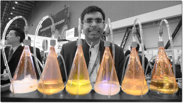
Oxygen Bar
Belly up to the bar and get your favorite… oxygen? That’s right — in some cities, you can get a shot of pure oxygen, with or without your choice of added flavors. Bar patrons inhale oxygen through a plastic tube inserted into their nostrils, paying up to a dollar per minute to inhale the pure gas. Proponents of the practice claim that breathing in extra oxygen will remove toxins from the body, strengthen the immune system, enhance concentration and alertness, increase energy, and even cure cancer! These claims, however, have not been substantiated by controlled scientific studies. Normally, blood leaving the lungs is almost completely saturated with oxygen, even without the use of extra oxygen, so it’s unlikely that a higher concentration of oxygen in air inside the lungs would lead to significantly greater oxygenation of the blood. Oxygen enters the blood in the lungs as part of the process of gas exchange.
What is Gas Exchange?
Gas exchange is the biological process through which gases are transferred across cell membranes to either enter or leave the blood. Oxygen is constantly needed by cells for aerobic cellular respiration, and the same process continually produces carbon dioxide as a waste product. Gas exchange takes place between the blood and cells throughout the body, with oxygen leaving the blood and entering the cells, and carbon dioxide leaving the cells and entering the blood. Gas exchange also takes place between the blood and the air in the lungs, with oxygen entering the blood from the inhaled air inside the lungs, and carbon dioxide leaving the blood and entering the air to be exhaled from the lungs.
Gas Exchange in the Lungs
Alveoli are the basic functional units of the lungs where gas exchange takes place between the air and the blood. Alveoli (singular, alveolus) are tiny air sacs that consist of connective and epithelial tissues. The connective tissue includes elastic fibres that allow alveoli to stretch and expand as they fill with air during inhalation. During exhalation, the fibres allow the alveoli to spring back and expel the air. Special cells in the walls of the alveoli secrete a film of fatty substances called surfactant. This substance prevents the alveolar walls from collapsing and sticking together when air is expelled. Other cells in alveoli include macrophages, which are mobile scavengers that engulf and destroy foreign particles that manage to reach the lungs in inhaled air.
As shown in Figure 13.4.2, alveoli are arranged in groups like clusters of grapes. Each alveolus is covered with epithelium that is just one cell thick. It is surrounded by a bed of pulmonary capillaries, each of which has a wall of epithelium just one cell thick. As a result, gases must cross through only two cells to pass between an alveolus and its surrounding capillaries.
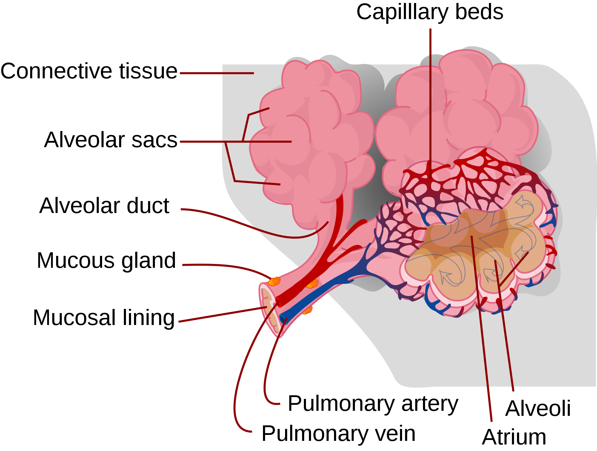
The pulmonary artery (also shown in Figure 13.4.2) carries deoxygenated blood from the heart to the lungs. Then, the blood travels through the pulmonary capillary beds, where it picks up oxygen and releases carbon dioxide. The oxygenated blood then leaves the lungs and travels back to the heart through pulmonary veins. There are four pulmonary veins (two for each lung), and all four carry oxygenated blood to the heart. From the heart, the oxygenated blood is then pumped to cells throughout the body.
Mechanism of Gas Exchange
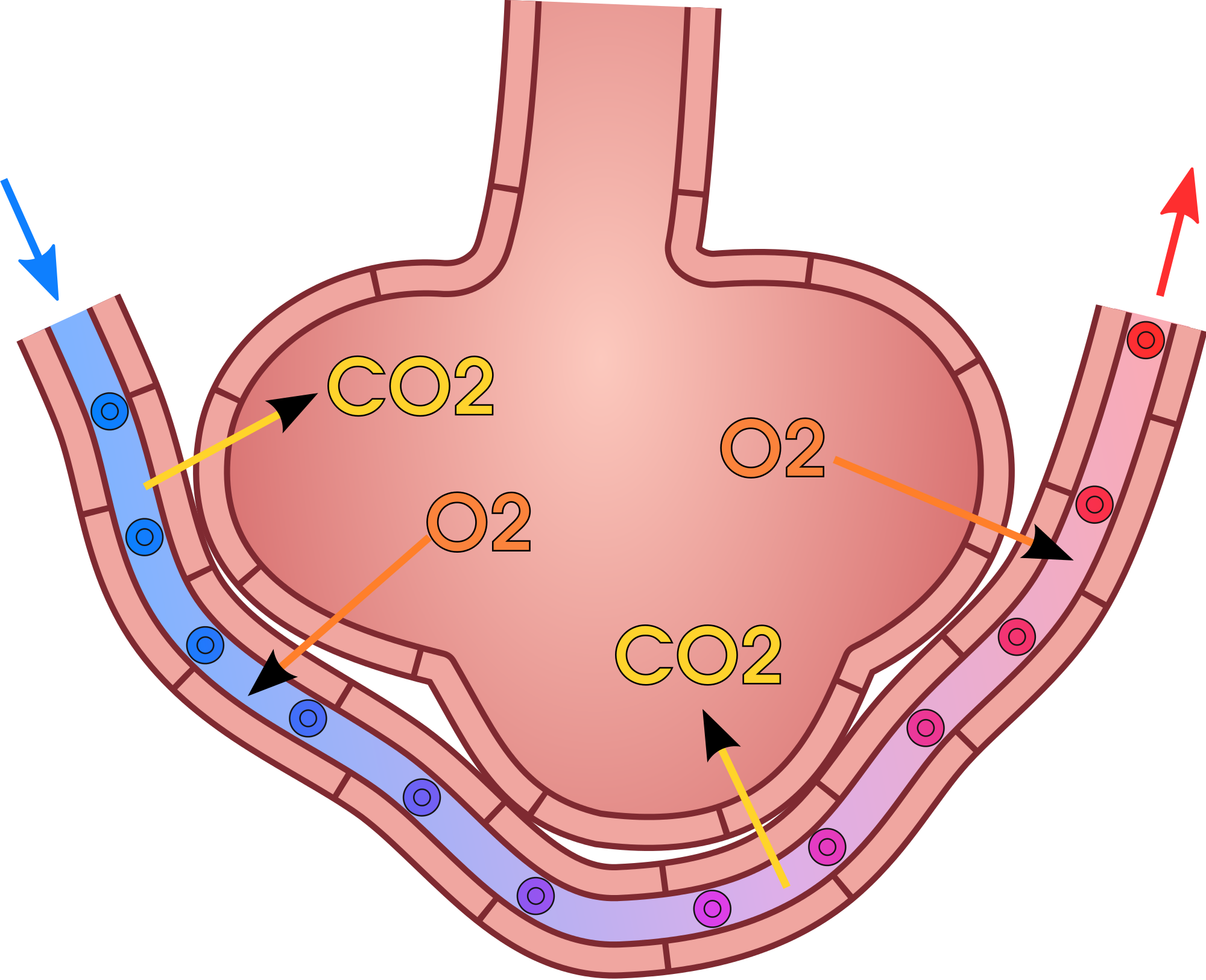
Gas exchange occurs by diffusion across cell membranes. Gas molecules naturally move down a concentration gradient from an area of higher concentration to an area of lower concentration. This is a passive process that requires no energy. To diffuse across cell membranes, gases must first be dissolved in a liquid. Oxygen and carbon dioxide are transported around the body dissolved in blood. Both gases bind to the protein hemoglobin in red blood cells, although oxygen does so more effectively than carbon dioxide. Some carbon dioxide also dissolves in blood plasma.
As shown in Figure 13.4.3, oxygen in inhaled air diffuses into a pulmonary capillary from the alveolus. Carbon dioxide in the blood diffuses in the opposite direction. The carbon dioxide can then be exhaled from the body.
Gas exchange by diffusion depends on having a large surface area through which gases can pass. Although each alveolus is tiny, there are hundreds of millions of them in the lungs of a healthy adult, so the total surface area for gas exchange is huge. It is estimated that this surface area may be as great as 100 m2 (or approximately 1,076 ft²). Often we think of lungs as balloons, but this type of structure would have very limited surface area and there wouldn’t be enough space for blood to interface with the air in the alveoli. The structure alveoli take in the lungs is more like a giant mass of soap bubbles — millions of tiny little chambers making up one large mass — this is what increases surface area giving blood lots of space to come into close enough contact to exchange gases by diffusion.
Gas exchange by diffusion also depends on maintaining a steep concentration gradient for oxygen and carbon dioxide. Continuous blood flow in the capillaries and constant breathing maintain this gradient.
- Each time you inhale, there is a greater concentration of oxygen in the air in the alveoli than there is in the blood in the pulmonary capillaries. As a result, oxygen diffuses from the air inside the alveoli into the blood in the capillaries. Carbon dioxide, in contrast, is more concentrated in the blood in the pulmonary capillaries than it is in the air inside the alveoli. As a result, carbon dioxide diffuses in the opposite direction.
- The cells of the body have a much lower concentration of oxygen than does the oxygenated blood that reaches them in peripheral capillaries, which are the capillaries that supply tissues throughout the body. As a result, oxygen diffuses from the peripheral capillaries into body cells. The opposite is true of carbon dioxide. It has a much higher concentration in body cells than it does in the blood of the peripheral capillaries. Thus, carbon dioxide diffuses from body cells into the peripheral capillaries.
13.4 Summary
- Gas exchange is the biological process through which gases are transferred across cell membranes to either enter or leave the blood. Gas exchange takes place continuously between the blood and cells throughout the body, and also between the blood and the air inside the lungs.
- Gas exchange in the lungs takes place in alveoli, which are tiny air sacs surrounded by networks of capillaries. The pulmonary artery carries deoxygenated blood from the heart to the lungs, where it travels through pulmonary capillaries, picking up oxygen and releasing carbon dioxide. The oxygenated blood then leaves the lungs through pulmonary veins.
- Gas exchange occurs by diffusion across cell membranes. Gas molecules naturally move down a concentration gradient from an area of higher concentration to an area of lower concentration. This is a passive process that requires no energy.
- Gas exchange by diffusion depends on the large surface area provided by the hundreds of millions of alveoli in the lungs. It also depends on a steep concentration gradient for oxygen and carbon dioxide. This gradient is maintained by continuous blood flow and constant breathing.
13.4 Review Questions
- What is gas exchange?
- Summarize the flow of blood into and out of the lungs for gas exchange.
-
- Describe the mechanism by which gas exchange takes place.
- Identify the two main factors upon which gas exchange by diffusion depends.
- If the concentration of oxygen were higher inside of a cell than outside of it, which way would the oxygen flow? Explain your answer.
- Why is it important that the walls of the alveoli are only one cell thick?
13.4 Explore More
Oxygen movement from alveoli to capillaries | NCLEX-RN | Khan Academy, khanacademymedicine, 2013.
About Carbon Monoxide and Carbon Monoxide Poisoning, EMDPrepare, 2009.
Oxygen’s surprisingly complex journey through your body – Enda Butler, TED-Ed, 2017.
Attributions
Figure 13.4.1
Oxygen Bar by Farrukh on Flickr is used under a CC BY-NC 2.0 (https://creativecommons.org/licenses/by-nc/2.0/) license.
Figure 13.4.2
Alveolus_diagram.svg by Mariana Ruiz Villarreal [LadyofHats] on Wikimedia Commons is released into the public domain (https://en.wikipedia.org/wiki/Public_domain).
Figure 13.4.3
Gas_exchange_in_the_aveolus.svg by domdomegg on Wikimedia Commons is used under a CC BY 4.0 (https://creativecommons.org/licenses/by/4.0) license.
References
EMDPrepare. (2009, December 21). About carbon monoxide and carbon monoxide poisoning. YouTube. https://www.youtube.com/watch?v=KmgIqVwytwA&feature=youtu.be
khanacademymedicine. (2013, February 25). Oxygen movement from alveoli to capillaries | NCLEX-RN | Khan Academy. YouTube. https://www.youtube.com/watch?v=nRpwdwm06Ic&feature=youtu.be
TED-Ed. (2017, April 13). Oxygen’s surprisingly complex journey through your body – Enda Butler. YouTube. https://www.youtube.com/watch?v=GVU_zANtroE&feature=youtu.be
Created by: CK-12/Adapted by Christine Miller
The original version of this chapter contained H5P content. You may want to remove or replace this element.
Figure 3.3.1 Carbo-licious!
Carbs Galore
What do all of these foods have in common? All of them consist mainly of large compounds called carbohydrates, often referred to as "carbs." Contrary to popular belief, carbohydrates are an important part of a healthy diet. They are also one of four major classes of biological macromolecules.
Chemical Compounds in Living Things
The compounds found in living things are known as biochemical compounds or biological molecules. Biochemical compounds make up the cells and other structures of organisms. They also carry out life processes. Carbon is the basis of all biochemical compounds, so carbon is essential to life on Earth. Without carbon, life as we know it could not exist.
Carbon is so basic to life because of its ability to form stable bonds with many elements, including itself. This property allows carbon to create a huge variety of very large and complex molecules. In fact, there are nearly 10 million carbon-based compounds in living things!
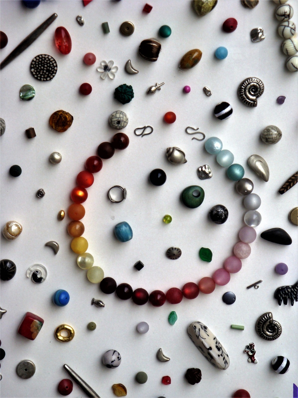
Most biochemical compounds are very large molecules called polymers. A polymer is built of repeating units of smaller compounds called monomers. Monomers are like the individual beads on a string of beads, and the whole string is the polymer. The individual beads (monomers) can do some jobs on their own, but sometimes you need a larger molecule, so the monomers can be connected to form polymers.
Classes of Biochemical Compounds
Although there are millions of different biochemical compounds in Earth's living things, all biochemical compounds contain the elements carbon, hydrogen, and oxygen. Some contain only these elements, while others contain additional elements, as well. The vast number of biochemical compounds can be grouped into just four major classes: carbohydrates, lipids, proteins, and nucleic acids.
Carbohydrates

Carbohydrates include sugars and starches. These compounds contain only the elements carbon, hydrogen, and oxygen. In living things, carbohydrates provide energy to cells, store energy, and form certain structures (such as the cell walls of plants). The monomer that makes up large carbohydrate compounds is called a monosaccharide. The sugar glucose, represented by the chemical model in Figure 3.3.2, is a monosaccharide. It contains six carbon atoms (C), along with several atoms of hydrogen (H) and oxygen (O). Thousands of glucose molecules can join together to form a polysaccharide, such as starch.
Lipids
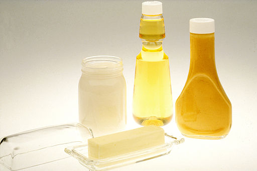
Lipids include fats and oils. They primarily contain the elements carbon, hydrogen, and oxygen, although some lipids contain additional elements, such as phosphorus. Lipids function in living things to store energy, form cell membranes, and carry messages. Lipids consist of repeating units that join together to form chains called fatty acids. Most naturally occurring fatty acids have an unbranched chain of an even number (generally between 4 and 28) of carbon atoms.
Proteins
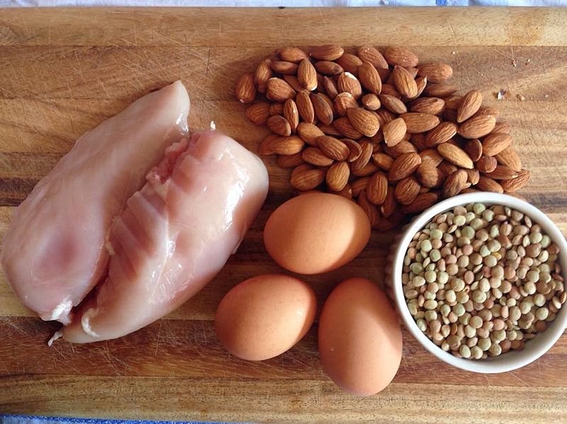
Proteins include enzymes, antibodies, and many other important compounds in living things. They contain the elements carbon, hydrogen, oxygen, nitrogen, and sulfur. Functions of proteins are very numerous. They help cells keep their shape, compose muscles, speed up chemical reactions, and carry messages and materials. The monomers that make up large protein compounds are called amino acids. There are 20 different amino acids that combine into long chains (called polypeptides) to form the building blocks of a vast array of proteins in living things.
Nucleic Acids
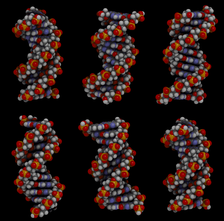
Nucleic acids include the molecules DNA (deoxyribonucleic acid) and RNA(ribonucleic acid). They contain the elements carbon, hydrogen, oxygen, nitrogen, and phosphorus. Their functions in living things are to encode instructions for making proteins, to help make proteins, and to pass instructions between parents and offspring. The monomer that makes up nucleic acids is the nucleotide. All nucleotides are the same, except for a component called a nitrogen base. There are four different nitrogen bases, and each nucleotide contains one of these four bases. The sequence of nitrogen bases in the chains of nucleotides in DNA and RNA makes up the code for protein synthesis, which is called the genetic code. The animation in Figure 3.3.5 represents the very complex structure of DNA, which consists of two chains of nucleotides.
3.3 Summary
- Biochemical compounds are carbon-based compounds found in living things. They make up cells and other structures of organisms and carry out life processes. Most biochemical compounds are large molecules called polymers that consist of many repeating units of smaller molecules, which are called monomers.
- There are millions of biochemical compounds, but all of them fall into four major classes: carbohydrates, lipids, proteins, and nucleic acids.
- Carbohydrates include sugars and starches. They provide cells with energy, store energy, and make up organic structures, such as the cell walls of plants.
- Lipids include fats and oils. They store energy, form cell membranes, and carry messages.
- Proteins include enzymes, antibodies, and numerous other important compounds in living things. They have many functions — helping cells keep their shape, making up muscles, speeding up chemical reactions, and carrying messages and materials.
- Nucleic acids include DNA and RNA. They encode instructions for making proteins, help make proteins, and pass encoded instructions from parents to offspring.
3.3 Review Questions
- Why is carbon so important to life on Earth?
- What are biochemical compounds?
- Describe the diversity of biochemical compounds and explain how they are classified.
- Identify two types of carbohydrates. What are the main functions of this class of biochemical compounds?
- What roles are played by lipids in living things?
- The enzyme amylase is found in saliva. It helps break down starches in foods into simpler sugar molecules. What type of biochemical compound do you think amylase is?
- Explain how DNA and RNA contain the genetic code.
- What are the three elements present in every class of biochemical compound?
- Classify each of the following terms as a monomer or a polymer:
- Nucleic acid
- Amino acid
- Monosaccharide
- Protein
- Nucleotide
- Polysaccharide
- Match each of the above monomers with its correct polymer and identify which class of biochemical compound is represented by each monomer/polymer pair.
- Is glucose a monomer or a polymer? Explain your answer.
- What is one element contained in proteins and nucleic acids, but not in carbohydrates?
- Describe the relationship between proteins and nucleic acids.
- Why do you think it is important to eat a diet that contains a balance of carbohydrates, proteins, and fats?
- Examine the picture of the meal in Figure 3.3.6. What types of biochemical compounds can you identify?

3.3 Explore More
https://youtu.be/YO244P1e9QM
Biomolecules (updated), by the Amoeba Sisters, 2016.
Attributions
Figure 3.3.1
- Cinnamon buns by adamkontor on Pixabay is used under the Pixabay License (https://pixabay.com/de/service/license/).
- Sugar by Bru-nO on Pixabay is used under the Pixabay License (https://pixabay.com/de/service/license/).
- Potatoes by HolgersFotografie on Pixabay is used under the Pixabay License (https://pixabay.com/de/service/license/).
- Blueberries; oatmeal by iha31 on Pixabay is used under the Pixabay License (https://pixabay.com/de/service/license/).
- Bread by pics_pd on Pixnio is used under a public domain certification (https://creativecommons.org/licenses/publicdomain/).
- Spaghetti by RitaE on Pixabay is used under the Pixabay License (https://pixabay.com/de/service/license/).
Figure 3.3.2
jewellery_beads_stones_necklace-1200668 on Pxhere, is used under a CC0 1.0 universal public domain dedication license (https://creativecommons.org/publicdomain/zero/1.0/).
Figure 3.3.3
Glucose; Structure of beta-D-glucopyranose (Haworth projection), by NEUROtiker on Wikimedia Commons, has been released into the public domain (https://en.wikipedia.org/wiki/Public_domain).
Figure 3.3.4
Lipid Examples; Butter and Oil, by Bill Branson (photographer), on Wikimedia Commons is released into the public domain (https://en.wikipedia.org/wiki/Public_domain).
Figure 3.3.5
Protein-rich_Foods, by Smastronardo on Wikimedia Commons, is used under a CC BY-SA 4.0 (https://creativecommons.org/licenses/by-sa/4.0) license.
Figure 3.3.6
Bdna_cropped [gif], by Jahobr on Wikimedia Commons, is released into the public domain (https://en.wikipedia.org/wiki/Public_domain) (This is a derivative work from Bdna.gif by Spiffistan.)
Figure 3.3.7Dinner by Quốc Trung [@boeing] on Unsplash is used under the Unsplash License (https://unsplash.com/license).
Reference
Amoeba Sisters. (2016, February 11). Biomolecules (updated). YouTube. https://www.youtube.com/watch?v=YO244P1e9QM&feature=youtu.be
Image shows the pathway of events in the activation of T Cells. This includes: 1) T Cells are activated when they encounter a foreign antigen on an MHC from an antigen-presenting cell. 2) Cytokines help the T cell to mature. Some T Cells become helper T cells and continue to produce cytokines. 3) Some T Cells become Killer T Cells and search out and destroy infected or cancerous cells.
a type of movement of substances across the cell membrane which does not require energy because the substances are moving with the concentration gradient (from high to low concentration).
The smallest type of blood vessel that connects arterioles and venules and that transfers substances between blood and tissues.

