13.2 Structure and Function of the Respiratory System

Seeing Your Breath
Why can you “see your breath” on a cold day? The air you exhale through your nose and mouth is warm like the inside of your body. Exhaled air also contains a lot of water vapor, because it passes over moist surfaces from the lungs to the nose or mouth. The water vapor in your breath cools suddenly when it reaches the much colder outside air. This causes the water vapor to condense into a fog of tiny droplets of liquid water. You release water vapor and other gases from your body through the process of respiration.
What is Respiration?
Respiration is the life-sustaining process in which gases are exchanged between the body and the outside atmosphere. Specifically, oxygen moves from the outside air into the body; and water vapor, carbon dioxide, and other waste gases move from inside the body to the outside air. Respiration is carried out mainly by the respiratory system. It is important to note that respiration by the respiratory system is not the same process as cellular respiration —which occurs inside cells — although the two processes are closely connected. Cellular respiration is the metabolic process in which cells obtain energy, usually by “burning” glucose in the presence of oxygen. When cellular respiration is aerobic, it uses oxygen and releases carbon dioxide as a waste product. Respiration by the respiratory system supplies the oxygen needed by cells for aerobic cellular respiration, and removes the carbon dioxide produced by cells during cellular respiration.
Respiration by the respiratory system actually involves two subsidiary processes. One process is ventilation, or breathing. Ventilation is the physical process of conducting air to and from the lungs. The other process is gas exchange. This is the biochemical process in which oxygen diffuses out of the air and into the blood, while carbon dioxide and other waste gases diffuse out of the blood and into the air. All of the organs of the respiratory system are involved in breathing, but only the lungs are involved in gas exchange.
Respiratory Organs
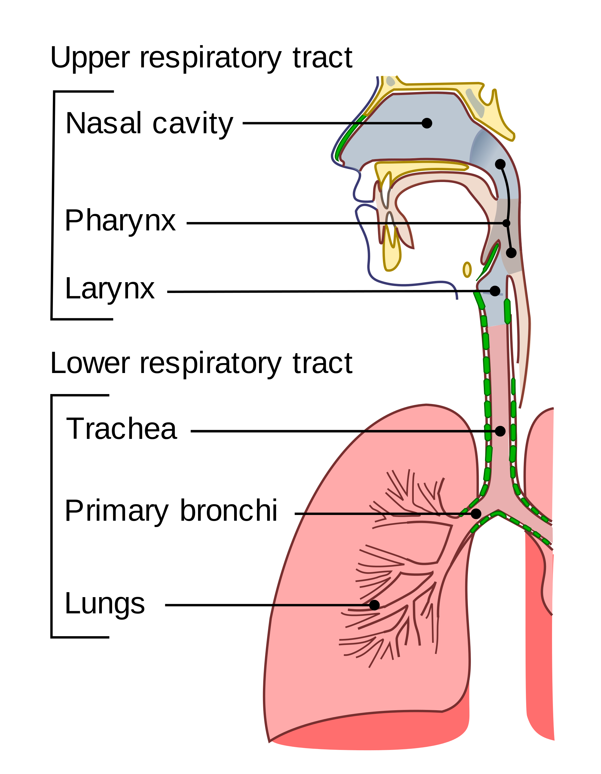
The organs of the respiratory system form a continuous system of passages, called the respiratory tract, through which air flows into and out of the body. The respiratory tract has two major divisions: the upper respiratory tract and the lower respiratory tract. The organs in each division are shown in Figure 13.2.2. In addition to these organs, certain muscles of the thorax (body cavity that fills the chest) are also involved in respiration by enabling breathing. Most important is a large muscle called the diaphragm, which lies below the lungs and separates the thorax from the abdomen. Smaller muscles between the ribs also play a role in breathing.
Upper Respiratory Tract
All of the organs and other structures of the upper respiratory tract are involved in conduction, or the movement of air into and out of the body. Upper respiratory tract organs provide a route for air to move between the outside atmosphere and the lungs. They also clean, humidify, and warm the incoming air. No gas exchange occurs in these organs.
Nasal Cavity
The nasal cavity is a large, air-filled space in the skull above and behind the nose in the middle of the face. It is a continuation of the two nostrils. As inhaled air flows through the nasal cavity, it is warmed and humidified by blood vessels very close to the surface of this epithelial tissue . Hairs in the nose and mucous produced by mucous membranes help trap larger foreign particles in the air before they go deeper into the respiratory tract. In addition to its respiratory functions, the nasal cavity also contains chemoreceptors needed for sense of smell, and contribution to the sense of taste.
Pharynx
The pharynx is a tube-like structure that connects the nasal cavity and the back of the mouth to other structures lower in the throat, including the larynx. The pharynx has dual functions — both air and food (or other swallowed substances) pass through it, so it is part of both the respiratory and the digestive systems. Air passes from the nasal cavity through the pharynx to the larynx (as well as in the opposite direction). Food passes from the mouth through the pharynx to the esophagus.
Larynx
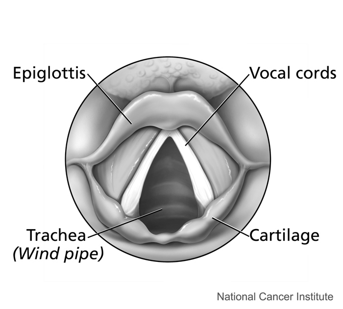
The larynx connects the pharynx and trachea, and helps to conduct air through the respiratory tract. The larynx is also called the voice box, because it contains the vocal cords, which vibrate when air flows over them, thereby producing sound. You can see the vocal cords in the larynx in Figures 13.2.3 and 13.2.4. Certain muscles in the larynx move the vocal cords apart to allow breathing. Other muscles in the larynx move the vocal cords together to allow the production of vocal sounds. The latter muscles also control the pitch of sounds and help control their volume.
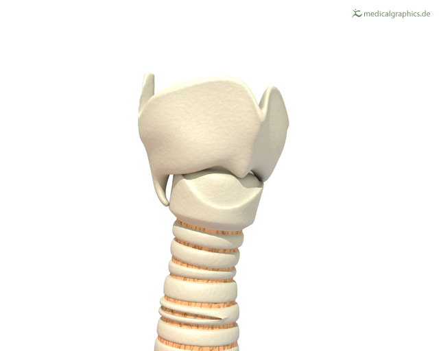 |
Figure 13.2.4 The larynx as viewed from the top. |
A very important function of the larynx is protecting the trachea from aspirated food. When swallowing occurs, the backward motion of the tongue forces a flap called the epiglottis to close over the entrance to the larynx. (You can see the epiglottis in both Figure 13.2.3 and 13.2.4.) This prevents swallowed material from entering the larynx and moving deeper into the respiratory tract. If swallowed material does start to enter the larynx, it irritates the larynx and stimulates a strong cough reflex. This generally expels the material out of the larynx, and into the throat.
Larynx Model – Respiratory System, Dr. Lotz, 2018.
Lower Respiratory Tract
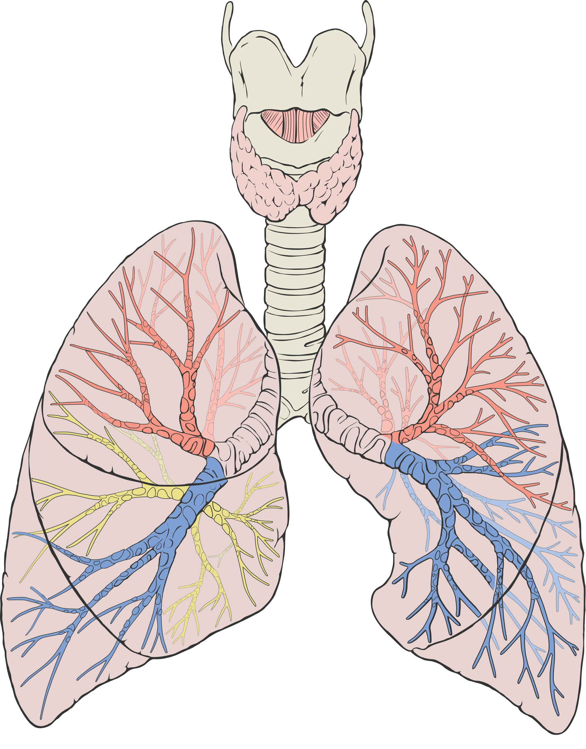
The trachea and other passages of the lower respiratory tract conduct air between the upper respiratory tract and the lungs. These passages form an inverted tree-like shape (Figure 13.2.5), with repeated branching as they move deeper into the lungs. All told, there are an astonishing 2,414 kilometres (1,500 miles) of airways conducting air through the human respiratory tract! It is only in the lungs, however, that gas exchange occurs between the air and the bloodstream.
Trachea
The trachea, or windpipe, is the widest passageway in the respiratory tract. It is about 2.5 cm wide and 10-15 cm long (approximately 1 inch wide and 4–6 inches long). It is formed by rings of cartilage, which make it relatively strong and resilient. The trachea connects the larynx to the lungs for the passage of air through the respiratory tract. The trachea branches at the bottom to form two bronchial tubes.
Bronchi and Bronchioles
There are two main bronchial tubes, or bronchi (singular, bronchus), called the right and left bronchi. The bronchi carry air between the trachea and lungs. Each bronchus branches into smaller, secondary bronchi; and secondary bronchi branch into still smaller tertiary bronchi. The smallest bronchi branch into very small tubules called bronchioles. The tiniest bronchioles end in alveolar ducts, which terminate in clusters of minuscule air sacs, called alveoli (singular, alveolus), in the lungs.
Lungs
The lungs are the largest organs of the respiratory tract. They are suspended within the pleural cavity of the thorax. The lungs are surrounded by two thin membranes called pleura, which secrete fluid that allows the lungs to move freely within the pleural cavity. This is necessary so the lungs can expand and contract during breathing. In Figure 13.2.6, you can see that each of the two lungs is divided into sections. These are called lobes, and they are separated from each other by connective tissues. The right lung is larger and contains three lobes. The left lung is smaller and contains only two lobes. The smaller left lung allows room for the heart, which is just left of the center of the chest.
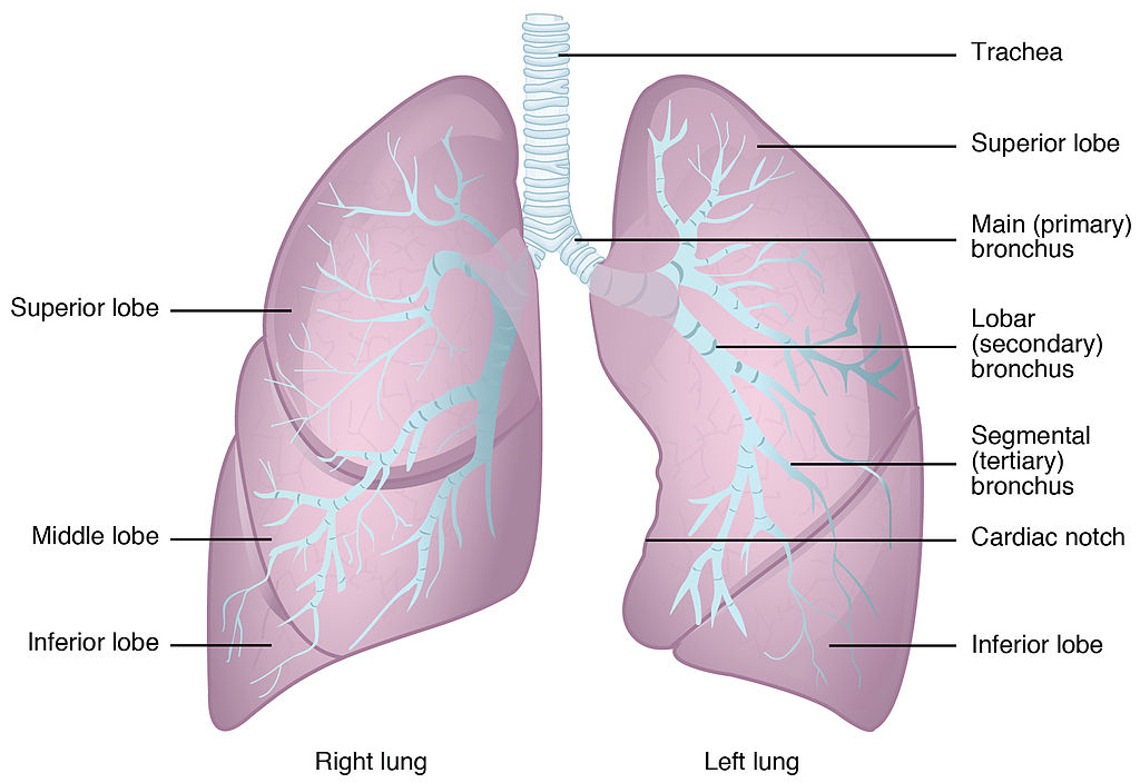
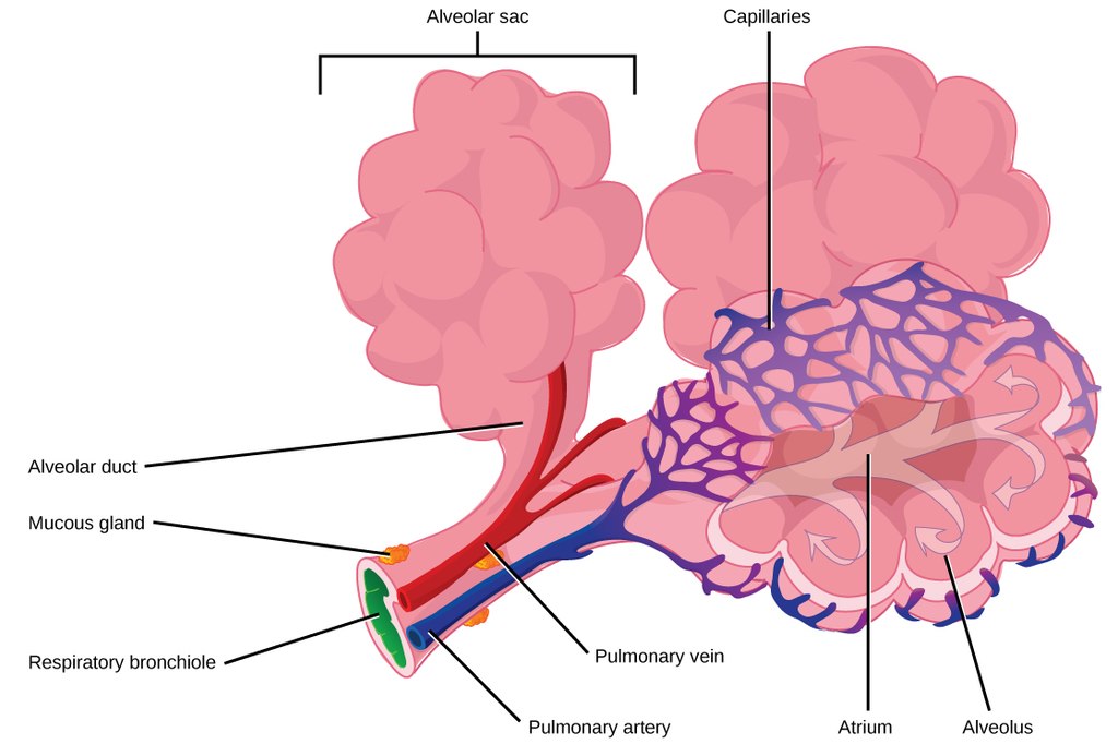
As mentioned previously, the bronchi terminate in bronchioles which feed air into alveoli, tiny sacs of simple squamous epithelial tissue which make up the bulk of the lung. The cross-section of lung tissue in the diagram below (Figure 13.2.7) shows the alveoli, in which gas exchange takes place with the capillary network that surrounds them.
 |
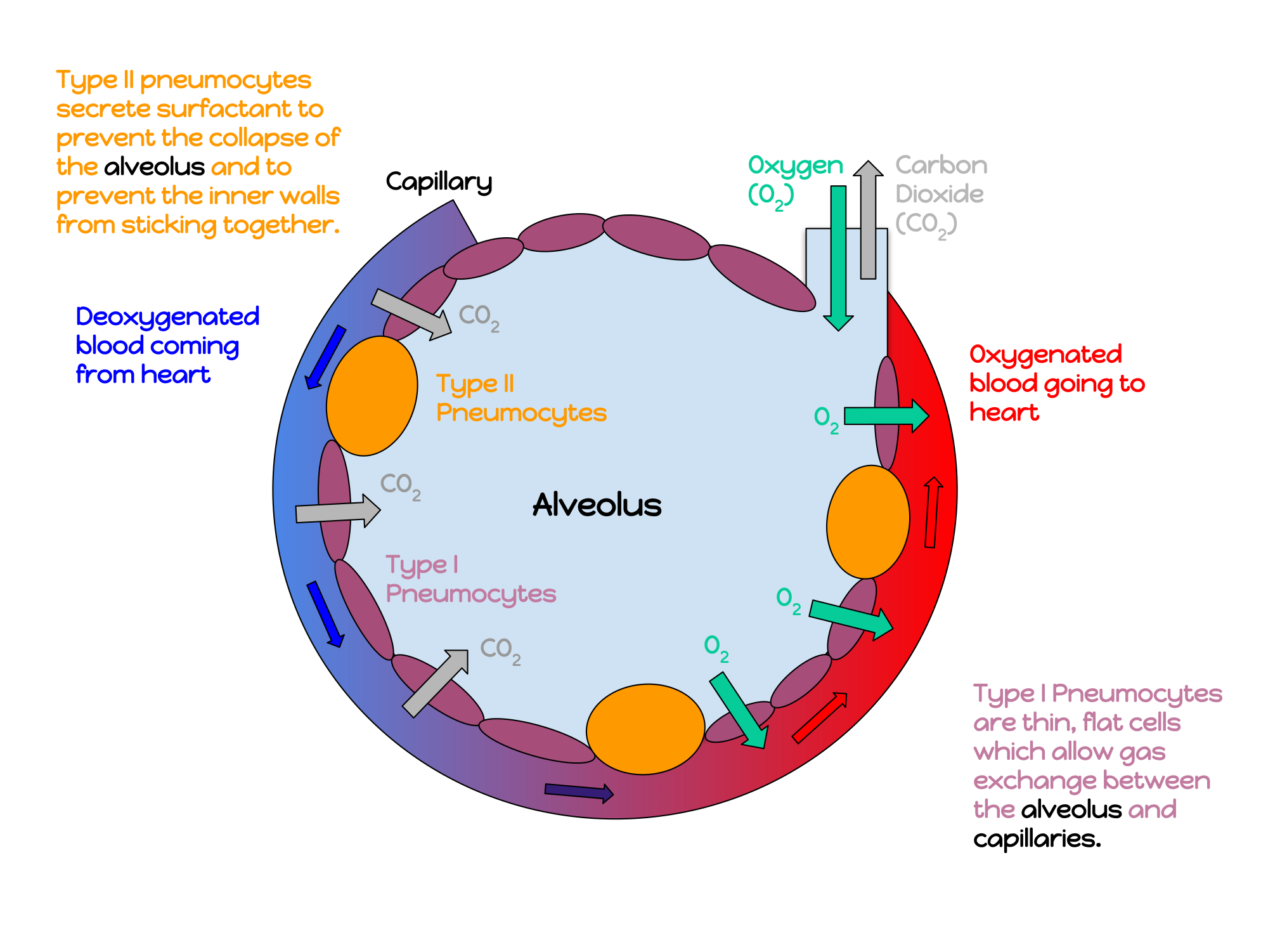 |
Lung tissue consists mainly of alveoli (see Figures 13.2.7 and 13.2.8). These tiny air sacs are the functional units of the lungs where gas exchange takes place. The two lungs may contain as many as 700 million alveoli, providing a huge total surface area for gas exchange to take place. In fact, alveoli in the two lungs provide as much surface area as half a tennis court! Each time you breathe in, the alveoli fill with air, making the lungs expand. Oxygen in the air inside the alveoli is absorbed by the blood via diffusion in the mesh-like network of tiny capillaries that surrounds each alveolus. The blood in these capillaries also releases carbon dioxide (also by diffusion) into the air inside the alveoli. Each time you breathe out, air leaves the alveoli and rushes into the outside atmosphere, carrying waste gases with it.
The lungs receive blood from two major sources. They receive deoxygenated blood from the right side of the heart. This blood absorbs oxygen in the lungs and carries it back to the left side heart to be pumped to cells throughout the body. The lungs also receive oxygenated blood from the heart that provides oxygen to the cells of the lungs for cellular respiration.
Protecting the Respiratory System
You may be able to survive for weeks without food and for days without water, but you can survive without oxygen for only a matter of minutes — except under exceptional circumstances — so protecting the respiratory system is vital. Ensuring that a patient has an open airway is the first step in treating many medical emergencies. Fortunately, the respiratory system is well protected by the ribcage of the skeletal system. The extensive surface area of the respiratory system, however, is directly exposed to the outside world and all its potential dangers in inhaled air. It should come as no surprise that the respiratory system has a variety of ways to protect itself from harmful substances, such as dust and pathogens in the air.
The main way the respiratory system protects itself is called the mucociliary escalator. From the nose through the bronchi, the respiratory tract is covered in epithelium that contains mucus-secreting goblet cells. The mucus traps particles and pathogens in the incoming air. The epithelium of the respiratory tract is also covered with tiny cell projections called cilia (singular, cilium), as shown in the animation. The cilia constantly move in a sweeping motion upward toward the throat, moving the mucus and trapped particles and pathogens away from the lungs and toward the outside of the body. The upward sweeping motion of cilia lining the respiratory tract helps keep it free from dust, pathogens, and other harmful substances.
Watch “Mucociliary clearance” by I-Hsun Wu to learn more:
Mucociliary clearance, I-Hsun Wu, 2015.
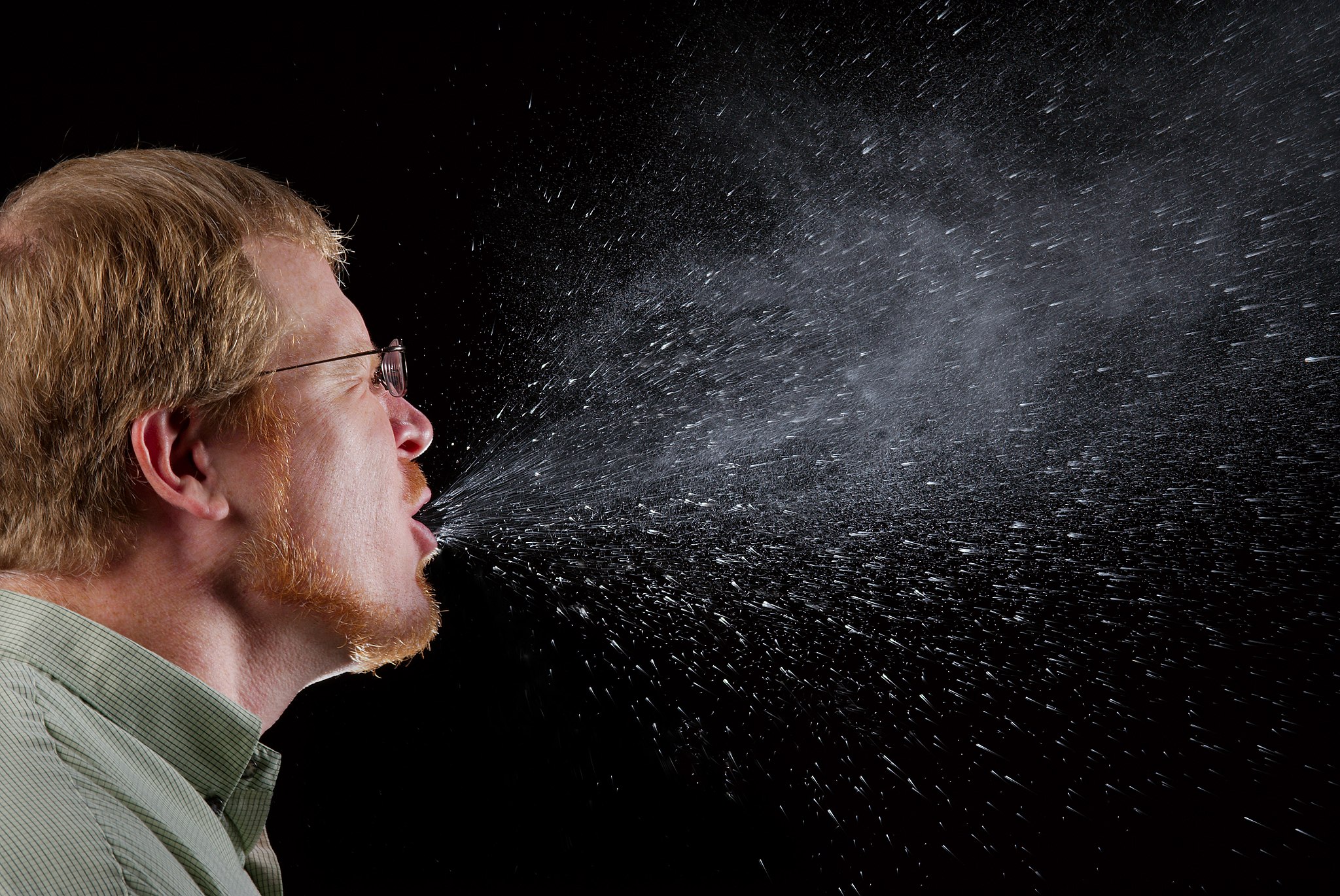
Sneezing is a similar involuntary response that occurs when nerves lining the nasal passage are irritated. It results in forceful expulsion of air from the mouth, which sprays millions of tiny droplets of mucus and other debris out of the mouth and into the air, as shown in Figure 13.2.9. This explains why it is so important to sneeze into a tissue (rather than the air) if we are to prevent the transmission of respiratory pathogens.
How the Respiratory System Works with Other Organ Systems
The amount of oxygen and carbon dioxide in the blood must be maintained within a limited range for survival of the organism. Cells cannot survive for long without oxygen, and if there is too much carbon dioxide in the blood, the blood becomes dangerously acidic (pH is too low). Conversely, if there is too little carbon dioxide in the blood, the blood becomes too basic (pH is too high). The respiratory system works hand-in-hand with the nervous and cardiovascular systems to maintain homeostasis in blood gases and pH.
It is the level of carbon dioxide — rather than the level of oxygen — that is most closely monitored to maintain blood gas and pH homeostasis. The level of carbon dioxide in the blood is detected by cells in the brain, which speed up or slow down the rate of breathing through the autonomic nervous system as needed to bring the carbon dioxide level within the normal range. Faster breathing lowers the carbon dioxide level (and raises the oxygen level and pH), while slower breathing has the opposite effects. In this way, the levels of carbon dioxide, oxygen, and pH are maintained within normal limits.
The respiratory system also works closely with the cardiovascular system to maintain homeostasis. The respiratory system exchanges gases with the outside air, but it needs the cardiovascular system to carry them to and from body cells. Oxygen is absorbed by the blood in the lungs and then transported through a vast network of blood vessels to cells throughout the body, where it is needed for aerobic cellular respiration. The same system absorbs carbon dioxide from cells and carries it to the respiratory system for removal from the body.
Feature: My Human Body
Choking due to a foreign object becoming lodged in the airway results in nearly 5 thousand deaths in Canada each year. In addition, choking accounts for almost 40% of unintentional injuries in infants under the age of one. For the sake of your own human body, as well as those of loved ones, you should be aware of choking risks, signs, and treatments.
Choking is the mechanical obstruction of the flow of air from the atmosphere into the lungs. It prevents breathing, and may be partial or complete. Partial choking allows some — though inadequate — air flow into the lungs. Prolonged or complete choking results in asphyxia, or suffocation, which is potentially fatal.
Obstruction of the airway typically occurs in the pharynx or trachea. Young children are more prone to choking than are older people, in part because they often put small objects in their mouth and do not understand the risk of choking that they pose. Young children may choke on small toys or parts of toys, or on household objects, in addition to food. Foods that are round (hotdogs, carrots, grapes) or can adapt their shape to that of the pharynx (bananas, marshmallows), are especially dangerous, and may cause choking in adults, as well as children.
How can you tell if a loved one is choking? The person cannot speak or cry out, or has great difficulty doing so. Breathing, if possible, is laboured, producing gasping or wheezing. The person may desperately clutch at his or her throat or mouth. If breathing is not soon restored, the person’s face will start to turn blue from lack of oxygen. This will be followed by unconsciousness, brain damage, and possibly death if oxygen deprivation continues beyond a few minutes.
If an infant is choking, turning the baby upside down and slapping him on the back may dislodge the obstructing object. To help an older person who is choking, first encourage the person to cough. Give them a few hard back slaps to help force the lodged object out of the airway. If these steps fail, perform the Heimlich maneuver on the person. See the series of videos below, from ProCPR, which demonstrate several ways to help someone who is choking based on age and consciousness.
Conscious Adult Choking, ProCPR, 2016.
Unconscious Adult Choking, ProCPR, 2011.
Conscious Child Choking, ProCPR, 2009.
Unconscious Child Choking, ProCPR, 2009.
Conscious Infant Choking, ProCPR, 2011.
Unconscious Infant Choking, ProCPR, 2011.
13.2 Summary
- Respiration is the process in which oxygen moves from the outside air into the body, and in which carbon dioxide and other waste gases move from inside the body into the outside air. It involves two subsidiary processes: ventilation and gas exchange. Respiration is carried out mainly by the respiratory system.
- The organs of the respiratory system form a continuous system of passages, called the respiratory tract. It has two major divisions: the upper respiratory tract and the lower respiratory tract.
- The upper respiratory tract includes the nasal cavity, pharynx, and larynx. All of these organs are involved in conduction, or the movement of air into and out of the body. Incoming air is also cleaned, humidified, and warmed as it passes through the upper respiratory tract. The larynx is called the voice box, because it contains the vocal cords, which are needed to produce vocal sounds.
- The lower respiratory tract includes the trachea, bronchi and bronchioles, and the lungs. The trachea, bronchi, and bronchioles are involved in conduction. Gas exchange takes place only in the lungs, which are the largest organs of the respiratory tract. Lung tissue consists mainly of tiny air sacs called alveoli, which is where gas exchange takes place between air in the alveoli and the blood in capillaries surrounding them.
- The respiratory system protects itself from potentially harmful substances in the air by the mucociliary escalator. This includes mucus-producing cells, which trap particles and pathogens in incoming air. It also includes tiny hair-like cilia that continually move to sweep the mucus and trapped debris away from the lungs and toward the outside of the body.
- The level of carbon dioxide in the blood is monitored by cells in the brain. If the level becomes too high, it triggers a faster rate of breathing, which lowers the level to the normal range. The opposite occurs if the level becomes too low. The respiratory system exchanges gases with the outside air, but it needs the cardiovascular system to carry the gases to and from cells throughout the body.
13.2 Review Questions
-
- What is respiration, as carried out by the respiratory system? Name the two subsidiary processes it involves.
- Describe the respiratory tract.
- Identify the organs of the upper respiratory tract. What are their functions?
- List the organs of the lower respiratory tract. Which organs are involved only in conduction?
- Where does gas exchange take place?
- How does the respiratory system protect itself from potentially harmful substances in the air?
- Explain how the rate of breathing is controlled.
- Why does the respiratory system need the cardiovascular system to help it perform its main function of gas exchange?
- Describe two ways in which the body prevents food from entering the lungs.
- What is the relationship between respiration and cellular respiration?
13.2 Explore More
How do lungs work? – Emma Bryce, TED-Ed, 2014.
Why Do Men Have Deeper Voices? BrainStuff – HowStuffWorks, 2015.
Why does your voice change as you get older? – Shaylin A. Schundler, TED-Ed, 2018.
Attributions
Figure 13.2.1
Exhale by pavel-lozovikov-HYovA7yPPvI [photo] by Pavel Lozovikov on Unsplash is used under the Unsplash License (https://unsplash.com/license).
Figure 13.2.2
Illu_conducting_passages.svg by Lord Akryl, Jmarchn from SEER Training Modules/ National Cancer Institute on Wikimedia Commons is in the public domain (https://en.wikipedia.org/wiki/Public_domain).
Figure 13.2.3
Larynx by www.medicalgraphics.de is used under a CC BY-ND 4.0 (https://creativecommons.org/licenses/by-nd/4.0/) license.
Figure 13.2.4
Larynx top view by Alan Hoofring (Illustrator) for National Cancer Institute is in the public domain (https://en.wikipedia.org/wiki/Public_domain).
Figure 13.2.5
2000px-Lungs_diagram_detailed.svg by Patrick J. Lynch, medical illustrator on Wikimedia Commons is used under a CC BY 2.5 (https://creativecommons.org/licenses/by/2.5) license. (Derivative work of Fruchtwasserembolie.png.)
Figure 13.2.6
Gross_Anatomy_of_the_Lungs by OpenStax College on Wikimedia Commons is used under a CC BY 3.0 (https://creativecommons.org/licenses/by/3.0) license.
Figure 13.2.7
Alveoli Structure by CNX OpenStax on Wikimedia Commons is used under a CC BY 4.0 (https://creativecommons.org/licenses/by/4.0) license.
Figure 13.2.8
annotated_diagram_of_an_alveolus.svg by Katherinebutler1331 on Wikimedia Commons is used under a CC BY-SA 4.0 (https://creativecommons.org/licenses/by-sa/4.0) license.
Figure 13.2.9
Sneeze by James Gathany at CDC Public Health Imagery Library (PHIL) #11162 on Wikimedia Commons is in the public domain (https://en.wikipedia.org/wiki/Public_domain).
References
Betts, J. G., Young, K.A., Wise, J.A., Johnson, E., Poe, B., Kruse, D.H., Korol, O., Johnson, J.E., Womble, M., DeSaix, P. (2013, June 19). Figure 22.2 Major respiratory structures [digital image]. In Anatomy and Physiology (Section 22.1). OpenStax. https://openstax.org/books/anatomy-and-physiology/pages/22-1-organs-and-structures-of-the-respiratory-system [CC BY 4.0 (https://creativecommons.org/licenses/by/4.0)].
Betts, J. G., Young, K.A., Wise, J.A., Johnson, E., Poe, B., Kruse, D.H., Korol, O., Johnson, J.E., Womble, M., DeSaix, P. (2013, June 19). Figure 22.13 Gross anatomy of the lungs [digital image]. In Anatomy and Physiology (Section 22.2). OpenStax. https://openstax.org/books/anatomy-and-physiology/pages/22-2-the-lungs
BrainStuff – HowStuffWorks. (2015, December 1). Why do men have deeper voices? YouTube. https://www.youtube.com/watch?v=6iFPs6JlSzY&feature=youtu.be
Dr. Lotz. (2018, January 25). Larynx model – Respiratory system. YouTube. https://www.youtube.com/watch?v=BsyB88mq5rQ&feature=youtu.be
I-Hsun Wu. (2015, March 31). Mucociliary clearance. YouTube. https://www.youtube.com/watch?v=HMB6flEaZwI&feature=youtu.be
OpenStax. (2016, May 27). Figure 9 Terminal bronchioles are connected by respiratory bronchioles to alveolar ducts and alveolar sacs [digital image]. In OpenStax, Biology (Section 39.1). OpenStax CNX. https://cnx.org/contents/GFy_h8cu@10.53:35-R0biq@11/Systems-of-Gas-Exchange
ProCPR. (2009, November 24). Conscious child choking. YouTube. https://www.youtube.com/watch?v=ZjmbD7aIaf0&feature=youtu.be
ProCPR. (2009, November 24).Unconscious child choking. YouTube. https://www.youtube.com/watch?v=Sba0T2XGIn4&feature=youtu.be
ProCPR. (2011, February 1). Conscious infant choking. YouTube. https://www.youtube.com/watch?v=axqIju9CLKA&feature=youtu.be
ProCPR. (2011, February 1). Unconscious adult choking. YouTube. https://www.youtube.com/watch?v=5kmsKNvKAvU&feature=youtu.be
ProCPR. (2011, February 1). Unconscious infant choking. YouTube. https://www.youtube.com/watch?v=_K7Dwy6b2wQ&feature=youtu.be
ProCPR. (2016, April 8). Conscious adult choking. YouTube. https://www.youtube.com/watch?v=XOTbjDGZ7wg&feature=youtu.be
TED-Ed. (2014, November 24). How do lungs work? – Emma Bryce. YouTube. https://www.youtube.com/watch?v=8NUxvJS-_0k&feature=youtu.be
TED-Ed. (2018, August 2). Why does your voice change as you get older? – Shaylin A. Schundler. YouTube. https://www.youtube.com/watch?v=rjibeBSnpJ0&feature=youtu.be
Case Study: Our Invisible Inhabitants
Lanying is suffering from a fever, body aches, and a painful sore throat that feels worse when she swallows. She visits her doctor, who examines her and performs a throat culture. When the results come back, he tells her that she has strep throat, which is caused by the bacteria Streptococcus pyogenes. He prescribes an antibiotic that will either kill the bacteria or stop it from reproducing, and advises her to take the full course of the treatment even if she is feeling better earlier. Stopping early can cause an increase in bacteria that are resistant to antibiotics.
Lanying takes the antibiotic as prescribed. Toward the end of the course, her throat is feeling much better — but she can’t say the same for other parts of her body! She has developed diarrhea and an itchy vaginal yeast infection. She calls her doctor, who suspects that the antibiotic treatment has caused both the digestive distress and the yeast infection. He explains that our bodies are home to many different kinds of microorganisms, some of which are actually beneficial to us because they help us digest our food and minimize the population of harmful microorganisms. When we take an antibiotic, many of these “good” bacteria are killed along with the “bad,” disease-causing bacteria, which can result in diarrhea and yeast infections.
Lanying's doctor prescribes an antifungal medication for her yeast infection. He also recommends that she eat yogurt with live cultures, which will help replace the beneficial bacteria in her gut. Our bodies contain a delicate balance of inhabitants that are invisible without a microscope, and changes in that balance can cause unpleasant health effects.
What Is Human Biology?
As you read the rest of this book, you'll learn more amazing facts about the human organism, and you'll get a better sense of how biology relates to your health. Human biology is the scientific study of the human species, which includes the fascinating story of human evolution and a detailed account of our genetics, anatomy, physiology, and ecology. In short, the study focuses on how we got here, how we function, and the role we play in the natural world. This helps us to better understand human health, because we can learn how to stay healthy and how diseases and injuries can be treated. Human biology should be of personal interest to you to the extent that it can benefit your own health, as well as the health of your friends and family. This branch of science also has broader implications for society and the human species as a whole.
Chapter Overview: Living Organisms and Human Biology
In the rest of this chapter, you'll learn about the traits shared by all living things, the basic principles that underlie all of biology, the vast diversity of living organisms, what it means to be human, and our place in the animal kingdom. Specifically, you'll learn:
- The seven traits shared by all living things: homeostasis, or the maintenance of a more-or-less constant internal environment; multiple levels of organization consisting of one or more cells; the use of energy and metabolism; the ability to grow and develop; the ability to evolve adaptations to the environment; the ability to detect and respond to environmental stimuli; and the ability to reproduce.
- The basic principles that unify all fields of biology, including gene theory, homeostasis, and evolutionary theory.
- The diversity of life (including the different kinds of biodiversity), the definition of a species, the classification and naming systems for living organisms, and how evolutionary relationships can be represented through diagrams, such as phylogenetic trees.
- How the human species is classified and how we've evolved from our close relatives and ancestors.
- The physical traits and social behaviors that humans share with other primates.
As you read this chapter, consider the following questions about Lanying's situation:
- What do single-celled organisms (such as the bacteria and yeast living in and on Lanying) have in common with humans?
- How are bacteria, yeast, and humans classified?
- How do the concepts of homeostasis and biodiversity apply to Lanying’s situation?
- Why can stopping antibiotics early cause the development of antibiotic-resistant bacteria?
Attribution
Figure 2.1.1
Photo (face mask) by Michael Amadeus, on Unsplash is used under the Unsplash license (https://unsplash.com/license).
Reference
Mayo Clinic Staff (n.d.). Strep throat [online article]. MayoClinic.org. https://www.mayoclinic.org/diseases-conditions/strep-throat/symptoms-causes/syc-20350338
Created by: CK-12/Adapted by Christine Miller
Mush!
These beautiful sled dogs are a metabolic marvel. While running up to 160 kilometres (about 99 miles) a day, they will each consume and burn about 12 thousand calories — about 240 calories per pound per day, which is the equivalent of about 24 Big Macs! A human endurance athlete, in contrast, typically burns only about 100 calories per pound (0.45 kg) each day. Scientists are intrigued by the amazing metabolism of sled dogs, although they still haven't determined how they use up so much energy. But one thing is certain: all living things need energy for everything they do, whether it's running a race or blinking an eye. In fact, every cell of your body constantly needs energy just to carry out basic life processes. You probably know that you get energy from the food you eat, but where does food come from? How does it come to contain energy? And how do your cells get the energy from food?
What Is Energy?
In the scientific world, energy is defined as the ability to do work. You can often see energy at work in living things — a bird flies through the air, a firefly glows in the dark, a dog wags its tail. These are obvious ways that living things use energy, but living things constantly use energy in less obvious ways, as well.
Why Living Things Need Energy
Inside every cell of all living things, energy is needed to carry out life processes. Energy is required to break down and build up molecules, and to transport many molecules across plasma membranes. All of life’s work needs energy. A lot of energy is also simply lost to the environment as heat. The story of life is a story of energy flow — its capture, its change of form, its use for work, and its loss as heat. Energy (unlike matter) cannot be recycled, so organisms require a constant input of energy. Life runs on chemical energy. Where do living organisms get this chemical energy?
How Organisms Get Energy
The chemical energy that organisms need comes from food. Food consists of organic molecules that store energy in their chemical bonds. In terms of obtaining food for energy, there are two types of organisms: autotrophs and heterotrophs.
Autotrophs
Autotrophs are organisms that capture energy from nonliving sources and transfer that energy into the living part of the ecosystem. They are also able to make their own food. Most autotrophs use the energy in sunlight to make food in the process of photosynthesis. Only certain organisms — such as plants, algae, and some bacteria — can make food through photosynthesis. Some photosynthetic organisms are shown in Figure 4.9.2.
 |
 |
 |
| Figure 4.9.2 Photosynthetic autotrophs, which make food using the energy in sunlight, include plants (left), algae (middle), and certain bacteria (right). |
Autotrophs are also called producers. They produce food not only for themselves, but for all other living things (known as consumers), as well. This is why autotrophs form the basis of food chains, such as the food chain shown In Figure 4.9.3.
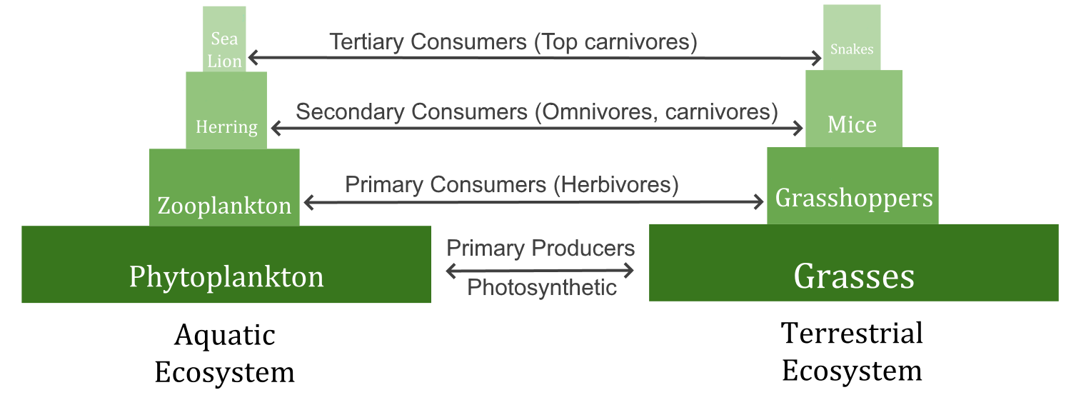
A food chain shows how energy and matter flow from producers to consumers. Matter is recycled, but energy must keep flowing into the system. Where does this energy come from?
Watch the video "The simple story of photosynthesis and food - Amanda Ooten" from TED-Ed to learn more about photosynthesis:
https://www.youtube.com/watch?time_continue=39&v=eo5XndJaz-Y
The simple story of photosynthesis and food - Amanda Ooten, TED-Ed, 2013.
Heterotrophs
Heterotrophs are living things that cannot make their own food. Instead, they get their food by consuming other organisms, which is why they are also called consumers. They may consume autotrophs or other heterotrophs. Heterotrophs include all animals and fungi, as well as many single-celled organisms. In Figure 4.9.3, all of the organisms are consumers except for the grasses and phytoplankton. What do you think would happen to consumers if all producers were to vanish from Earth?
Energy Molecules: Glucose and ATP
Organisms mainly use two types of molecules for chemical energy: glucose and ATP. Both molecules are used as fuels throughout the living world. Both molecules are also key players in the process of photosynthesis.
Glucose
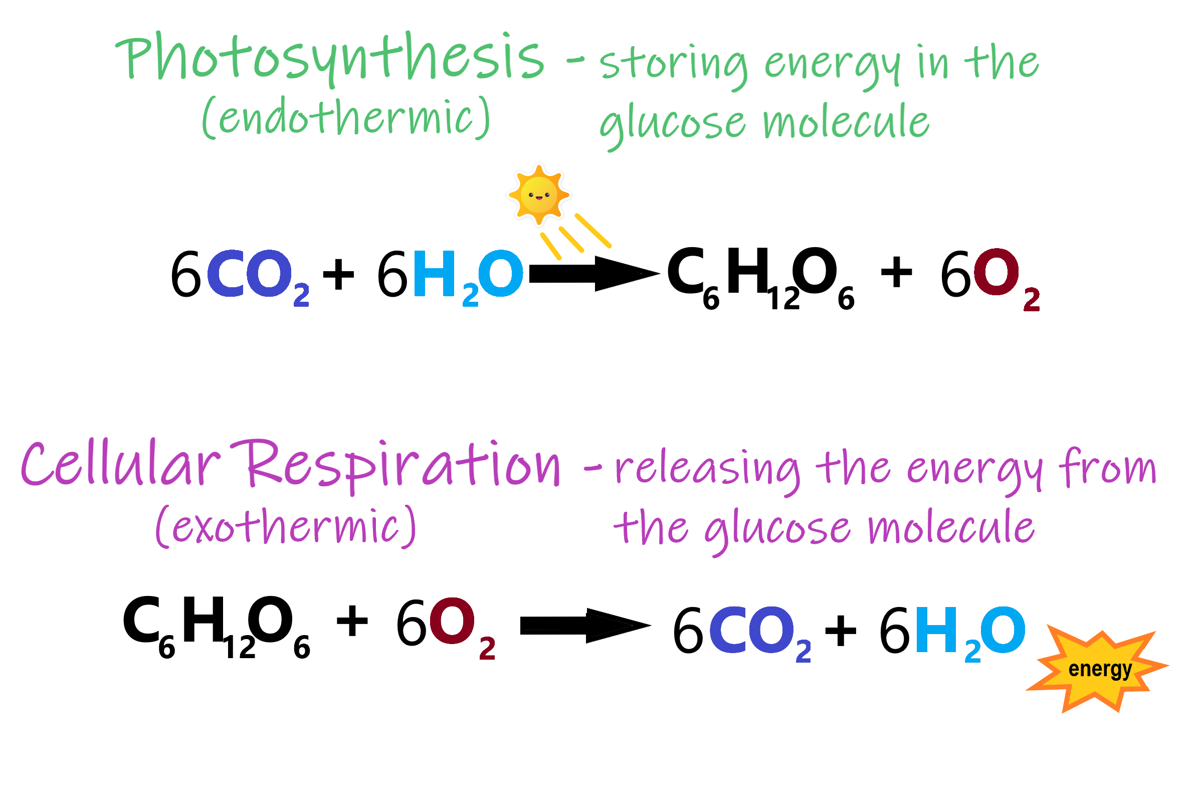
Glucose is a simple carbohydrate with the chemical formula C6H12O6. It stores chemical energy in a concentrated, stable form. In your body, glucose is the form of energy that is carried in your blood and taken up by each of your trillions of cells. Glucose is the end product of photosynthesis, and it is the nearly universal food for life. In Figure 4.9.4, you can see how photosynthesis stores energy from the sun in the glucose molecule and then how cellular respiration breaks the bonds in glucose to retrieve the energy.
ATP
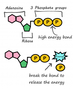
If you remember from section 3.7 Nucleic Acids, ATP (adenosine triphosphate) is the energy-carrying molecule that cells use to power most cellular processes (nerve impulse conduction, protein synthesis and active transport are good examples of cell processes that rely on ATP as their energy source). ATP is made during the first half of photosynthesis and then used for energy during the second half of photosynthesis, when glucose is made. ATP releases energy when it gives up one of its three phosphate groups (Pi) and changes to ADP (adenosine diphosphate, which has two phosphate groups), as shown in Figure 4.9.5. Thus, the breakdown of ATP into ADP + Pi is a catabolic reaction that releases energy (exothermic). ATP is made from the combination of ADP and Pi, an anabolic reaction that takes in energy (endothermic).
Why Organisms Need Both Glucose and ATP
Why do living things need glucose if ATP is the molecule that cells use for energy? Why don’t autotrophs just make ATP and be done with it? The answer is in the “packaging.” A molecule of glucose contains more chemical energy in a smaller “package” than a molecule of ATP. Glucose is also more stable than ATP. Therefore, glucose is better for storing and transporting energy. Glucose, however, is too powerful for cells to use. ATP, on the other hand, contains just the right amount of energy to power life processes within cells. For these reasons, both glucose and ATP are needed by living things.
How Energy Flows Through Living Things
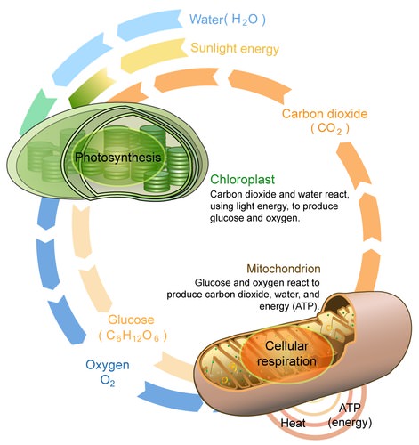
The flow of energy through living organisms begins with photosynthesis. This process stores energy from sunlight in the chemical bonds of glucose. By breaking the chemical bonds in glucose, cells release the stored energy and make the ATP they need. The process in which glucose is broken down and ATP is made is called cellular respiration.
Photosynthesis and cellular respiration are like two sides of the same coin. This is apparent in Figure 4.9.6. The products of one process are the reactants of the other. Together, the two processes store and release energy in living organisms. The two processes also work together to recycle oxygen in the Earth’s atmosphere.
4.9 Summary
- Energy is the ability to do work. It is needed by all living things and every living cell to carry out life processes, such as breaking down and building up molecules, and transporting many molecules across cell membranes.
- The form of energy that living things need for these processes is chemical energy, and it comes from food. Food consists of organic molecules that store energy in their chemical bonds.
- Autotrophs make their own food. Plants, for example, make food by photosynthesis. Autotrophs are also called producers.
- Heterotrophss obtain food by eating other organisms. Heterotrophs are also known as consumers.
- Organisms mainly use the molecules glucose and ATP for energy. Glucose is a compact, stable form of energy that is carried in the blood and taken up by cells. ATP contains less energy and is used to power cell processes.
- The flow of energy through living things begins with photosynthesis, which creates glucose. In a process called cellular respiration, organisms' cells break down glucose and make the ATP they need.
4.9 Review Questions
- Define energy.
- Why do living things need energy?
-
- Compare and contrast the two basic ways that organisms get energy.
- Describe the roles and relationships of the energy molecules glucose and ATP.
- Summarize how energy flows through living things.
- Why does the transformation of ATP to ADP release energy?
4.9 Explore More
https://www.youtube.com/watch?v=eDalQv7d2cs
Learn Biology: Autotrophs vs. Heterotrophs, Mahalodotcom, 2011.
https://www.youtube.com/watch?v=0glkXIj1DgE&feature=emb_logo
Energy Transfer in Trophic Levels, Teacher's Pet, 2015.
Attributions
Figure 4.9.1
Three Airmen participate in dog-sled expedition by U.S. Air Force photo by Tech. Sgt. Dan Rea is released into the public domain (https://en.wikipedia.org/wiki/Public_domain).
Figure 4.9.2
- Plant [photo] by Ren Ran on Unsplash is used under the Unsplash License (https://unsplash.com/license).
- Green Algae by Tristan Schmurr on Flickr is used under a CC BY 2.0 (https://creativecommons.org/licenses/by/2.0/) license.
- Cyanobacteria by Argon National Laboratory on Flickr is used under a CC BY-NC-SA 2.0 (https://creativecommons.org/licenses/by-nc-sa/2.0/) license.
Figure 4.9.3
Biomass_Pyramid by Swiggity.Swag.YOLO.Bro on Wikipedia is used and adapted by Christine Miller under a CC BY-SA 4.0 (https://creativecommons.org/licenses/by-sa/4.0/deed.en) license.
Figure 4.9.4
Photosynthesis and respiration by Christine Miller is used under a CC BY 4.0 (https://creativecommons.org/licenses/by/4.0/) license.
Figure 4.9.5
Photo synthesis and cellular respiration by Lady of Hats/ CK-12 Foundation is used under a CC BY-NC 3.0 (https://creativecommons.org/licenses/by-nc/3.0/) license.
References
LadyofHats/CK-12 Foundation. (2016, August 15). Figure 5: Photosynthesis and cellular respiration [digital image]. In Brainard, J., and Henderson, R., CK-12's College Human Biology FlexBook® (Section 4.9). CK-12 Foundation. https://www.ck12.org/book/ck-12-college-human-biology/section/4.9/
Mahalodotcom. (2011, January 14). Learn biology: Autotrophs vs. heterotrophs. YouTube. https://www.youtube.com/watch?v=eDalQv7d2cs
Teacher's Pet. (2015, March 23). Energy transfer in trophic levels. YouTube. https://www.youtube.com/watch?v=0glkXIj1DgE&feature=emb_logo
TED-Ed. (2013, March 5). The simple story of photosynthesis and food - Amanda Ooten. YouTube. https://www.youtube.com/watch?v=eo5XndJaz-Y&feature=youtu.be
A set of metabolic reactions and processes that take place in the cells of organisms to convert biochemical energy from nutrients into adenosine triphosphate (ATP).
Created by CK-12 Foundation/Adapted by Christine Miller
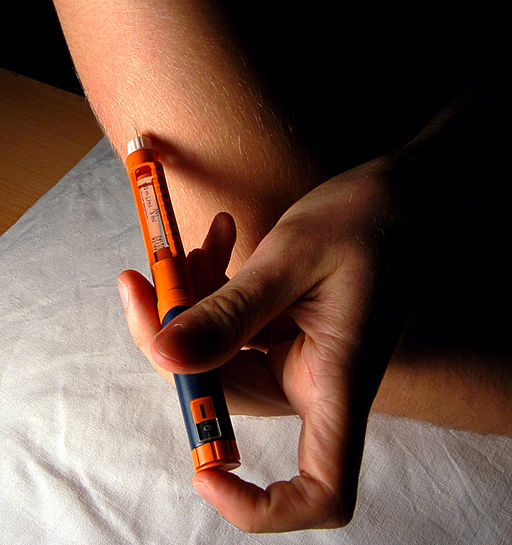
A Shot in the Arm
Giving yourself an injection can be difficult, but for someone with diabetes, it may be a matter of life or death. The person in the photo has diabetes and is injecting themselves with insulin, the hormone that helps control the level of glucose in the blood. Insulin is produced by the pancreas.
Introduction to the Pancreas
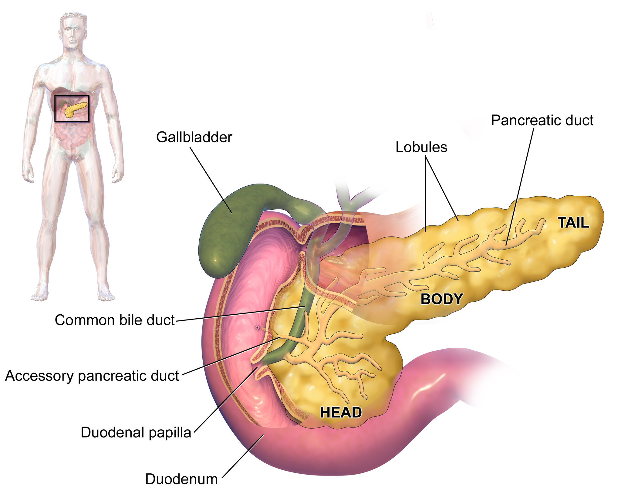
The pancreas is a large gland located in the upper left abdomen behind the stomach, as shown in Figure 9.7.2. The pancreas is about 15 cm (6 in) long, and it has a flat, oblong shape. Structurally, the pancreas is divided into a head, body, and tail. Functionally, the pancreas serves as both an endocrine gland and an exocrine gland.
- As an endocrine gland, the pancreas is part of the endocrine system. As such, it releases hormones (such as insulin) directly into the bloodstream for transport to cells throughout the body.
- As an exocrine gland, the pancreas is part of the digestive system. As such, it releases digestive enzymes into ducts that carry the enzymes to the gastrointestinal tract, where they assist with digestion. In this concept, the focus is on the pancreas as an endocrine gland. You can read about the pancreas as an exocrine gland in Chapter 15 Digestive System.
The Pancreas as an Endocrine Gland
The tissues within the pancreas that have an endocrine role exist as clusters of cells called pancreatic islets. They are also called the islets of Langerhans. You can see pancreatic tissue, including islets, in Figure 9.7.3. There are approximately three million pancreatic islets, and they are crisscrossed by a dense network of capillaries. The capillaries are lined by layers of islet cells that have direct contact with the blood vessels, into which they secrete their endocrine hormones.
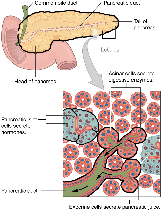
The pancreatic islets consist of four main types of cells, each of which secretes a different endocrine hormone. All of the hormones produced by the pancreatic islets, however, play crucial roles in glucose metabolism and the regulation of blood glucose levels, among other functions.
- Islet cells called alpha (α) cells secrete the hormone glucagon. The function of glucagon is to increase the level of glucose in the blood. It does this by stimulating the liver to convert stored glycogen into glucose, which is released into the bloodstream.
- Islets cells called beta (β) cells secrete the hormone insulin. The function of insulin is to decrease the level of glucose in the blood. It does this by promoting the absorption of glucose from the blood into fat, liver, and skeletal muscle cells. In these tissues, the absorbed glucose is converted into glycogen, fats (triglycerides), or both.
- Islet cells called delta (δ) cells secrete the hormone somatostatin. This hormone is also called growth hormone inhibiting hormone, because it inhibits the anterior lobe of the pituitary gland from producing growth hormone. Somatostatin also inhibits the secretion of pancreatic endocrine hormones and pancreatic exocrine enzymes.
- Islet cells called gamma (γ) cells secrete the hormone pancreatic polypeptide. The function of pancreatic polypeptide is to help regulate the secretion of both endocrine and exocrine substances by the pancreas.
Disorders of the Pancreas
There are a variety of disorders that affect the pancreas. They include pancreatitis, pancreatic cancer, and diabetes mellitus.
Pancreatitis
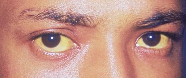
Pancreatitis is inflammation of the pancreas. It has a variety of possible causes, including gallstones, chronic alcohol use, infections (such as measles or mumps), and certain medications. Pancreatitis occurs when digestive enzymes produced by the pancreas damage the gland’s tissues, which causes problems with fat digestion. The disorder is usually associated with intense pain in the central abdomen, and the pain may radiate to the back. Yellowing of the skin and whites of the eyes (see Figure 9.7.4), which is called jaundice, is a common sign of pancreatitis. People with pancreatitis may also have pale stools and dark urine. Treatment of pancreatitis includes administering drugs to manage pain, and addressing the underlying cause of the disease, for example, by removing gallstones.
Pancreatic Cancer
There are several different types of pancreatic cancer that may affect either the endocrine or the exocrine tissues of the gland. Cancers affecting the endocrine tissues are all relatively rare. However, their incidence has been rising sharply. It is unclear to what extent this reflects increased detection, especially through medical imaging techniques. Unfortunately, pancreatic cancer is usually diagnosed at a relatively late stage when it is too late for surgery, which is the only way to cure the disorder. In 2020 it is estimated that 6,000 Canadians will be newly diagnosed with pancreatic cancer, and that during this same year, 5,300 will die of pancreatic cancer.
While it is rare before the age of 40, pancreatic cancer occurs most often after the age of 60. Factors that increase the risk of developing pancreatic cancer include smoking, obesity, diabetes, and a family history of the disease. About one in four cases of pancreatic cancer are attributable to smoking. Certain rare genetic conditions are also risk factors for pancreatic cancer.
Diabetes Mellitus
By far the most common type of pancreatic disorder is diabetes mellitus, more commonly called simply diabetes. There are many different types of diabetes, but diabetes mellitus is the most common. It occurs in two major types, type 1 diabetes and type 2 diabetes. The two types have different causes and may also have different treatments, but they generally produce the same initial symptoms, which include excessive urination and thirst. These symptoms occur because the kidneys excrete more urine in an attempt to rid the blood of excess glucose. Loss of water in urine stimulates greater thirst. Other signs and symptoms of diabetes are listed in Figure 9.7.5.
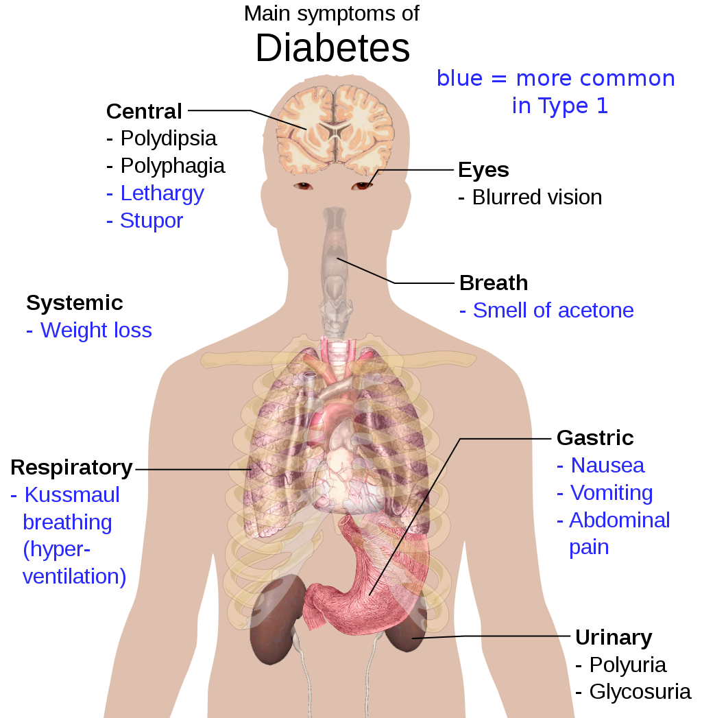
When diabetes is not well controlled, it is likely to have several serious long-term consequences. Most of these consequences are due to damage to small blood vessels caused by high glucose levels in the blood. Damage to blood vessels, in turn, may lead to increased risk of coronary artery disease and stroke. Damage to blood vessels in the retina of the eye can result in gradual vision loss and blindness. Damage to blood vessels in the kidneys can lead to chronic kidney disease, sometimes requiring dialysis or kidney transplant. Long-term consequences of diabetes may also include damage to the nerves of the body, known as diabetic neuropathy. In fact, this is the most common complication of diabetes. Symptoms of diabetic neuropathy may include numbness, tingling, and pain in the extremities.
Type 1 Diabetes
Type 1 diabetes is a chronic autoimmune disorder in which the immune system attacks the insulin-secreting beta cells of the pancreas. As a result, people with type 1 diabetes lack the insulin needed to keep blood glucose levels within the normal range. Type 1 diabetes may develop in people of any age, but is most often diagnosed before adulthood. For type 1 diabetics, insulin injections are critical for survival.
Type 2 Diabetes
Type 2 diabetes is the single most common form of diabetes. The cause of high blood glucose in this form of diabetes usually includes a combination of insulin resistance and impaired insulin secretion. Both genetic and environmental factors play roles in the development of type 2 diabetes. Type 2 diabetes can be managed with changes in diet and physical activity, which may increase insulin sensitivity and help reduce blood glucose levels to normal ranges. Medications may also be used as part of the treatment, as may insulin injections.
Feature: Human Biology in the News
Some patients with type 1 diabetes have been given pancreatic islet cells transplants from other human donors. If the transplanted cells are not rejected by the recipient’s immune system, they can cure the patient of diabetes. However, because of a shortage of appropriate human donors, only about one thousand such surgeries have been performed over the past ten years.
In June of 2016, a research team led by Dr. David K.C. Cooper at the Thomas E. Starzl Transplantation Institute in Pittsburgh, Pennsylvania, reported on their work developing pig islet cells for transplant into human diabetes patients. The researchers genetically engineered the pig islet cells to be protected from the human immune response. As a result, patients receiving the transplanted cells would require only minimal suppression of their immune system after the surgery. The pig islet cells would also be less likely to transmit pathogenic agents, because the animals could be raised in a controlled environment.
The researchers have successfully transplanted the pig islet cells into monkey models of type 1 diabetes. As of June 2016, the scientists were looking for funding to undertake clinical trials in humans with type 1 diabetes. Dr. Cooper predicted then that if the human trials go as well as expected, the pig islet cells could be available for curing patients in as little as two years.
9.7 Summary
- The pancreas is a gland located in the upper left abdomen behind the stomach that functions as both an endocrine gland and an exocrine gland. As an endocrine gland, the pancreas releases hormones (such as insulin) directly into the bloodstream. As an exocrine gland, the pancreas releases digestive enzymes into ducts that carry them to the gastrointestinal tract.
- Tissues in the pancreas that have an endocrine role exist as clusters of cells called pancreatic islets. The islets consist of four main types of cells, each of which secretes a different endocrine hormone. Alpha (α) cells secrete glucagon, beta (β) cells secrete insulin, delta (δ) cells secrete somatostatin, and gamma (γ) cells secrete pancreatic polypeptide.
- The endocrine hormones secreted by the pancreatic islets all play a role, either directly or indirectly, in glucose metabolism and homeostasis of blood glucose levels. For example, insulin stimulates the uptake of glucose by cells and decreases the level of glucose in the blood, whereas glucagon stimulates the conversion of glycogen to glucose and increases the level of glucose in the blood.
- Disorders of the pancreas include pancreatitis, pancreatic cancer, and diabetes mellitus. Pancreatitis is painful inflammation of the pancreas that has many possible causes. Pancreatic cancer of the endocrine tissues is rare, but increasing in frequency. It is generally discovered too late to cure surgically. Smoking is a major risk factor for pancreatic cancer.
- Diabetes mellitus is the most common type of pancreatic disorder. In diabetes, inadequate activity of insulin results in high blood levels of glucose. Type 1 diabetes is a chronic autoimmune disorder in which the immune system attacks the insulin-secreting beta cells of the pancreas. Type 2 diabetes is usually caused by a combination of insulin resistance and impaired insulin secretion due to a variety of environmental and genetic factors.
9.7 Review Questions
- Describe the structure and location of the pancreas.
- Distinguish between the endocrine and exocrine functions of the pancreas.
-
-
- What is pancreatitis? What are possible causes and effects of pancreatitis?
- Describe the incidence, prognosis, and risk factors of cancer of the endocrine tissues of the pancreas.
- Compare and contrast type 1 and type 2 diabetes.
- If the alpha islet cells of the pancreas were damaged to the point that they no longer functioned, how would this affect blood glucose levels? Assume that no outside regulation of this system is occurring and explain your answer. Further, would administration of insulin be more likely to help or hurt this condition? Explain your answer.
- Explain why diabetes causes excessive thirst.
9.7 Explore More
https://www.youtube.com/watch?v=8dgoeYPoE-0&t=2s
What does the pancreas do? - Emma Bryce, TED-Ed, 2015.
https://www.youtube.com/watch?v=qlzLSbAGMqA&feature=emb_logo
Type 2 diabetes in children, Children's Health, 2008.
https://www.youtube.com/watch?v=da1vvigy5tQ
Reversing Type 2 diabetes starts with ignoring the guidelines | Sarah Hallberg | TEDxPurdueU, TEDx Talks, 2015.
Attributions
Figure 9.7.1
Insulin_Application by Mr Hyde at Czech Wikipedia (Original text: moje foto) on Wikimedia Commons is released into the public domain (https://en.wikipedia.org/wiki/Public_domain).
Figure 9.7.2
Blausen_0699_PancreasAnatomy2 by BruceBlaus on Wikimedia Commons is used under a CC BY 3.0 (https://creativecommons.org/licenses/by/3.0) license.
Figure 9.7.3
Exocrine_and_Endocrine_Pancreas by OpenStax College is used under a CC BY 3.0 (https://creativecommons.org/licenses/by/3.0/deed.en) license.
Figure 9.7.4
Jaundice_eye_new by Info-farmer on Wikimedia Commons is in the public domain (https://en.wikipedia.org/wiki/Public_domain). (Original image, File:Jaundice eye.jpg, is from Centers for Disease Control and Prevention's Public Health Image Library (PHIL), with identification number #2860)
Figure 9.7.5
Main_symptoms_of_diabetes.svg by Mikael Häggström on Wikimedia Commons is released into the public domain (https://en.wikipedia.org/wiki/Public_domain).
Betts, J. G., Young, K.A., Wise, J.A., Johnson, E., Poe, B., Kruse, D.H., Korol, O., Johnson, J.E., Womble, M., DeSaix, P. (2013, July 19). Figure 23.26 Exocrine and endocrine pancreas [digital image]. In Anatomy and Physiology (Section 23.6). OpenStax. https://openstax.org/books/anatomy-and-physiology/pages/23-6-accessory-organs-in-digestion-the-liver-pancreas-and-gallbladder
Blausen.com Staff. (2014). Medical gallery of Blausen Medical 2014. WikiJournal of Medicine 1 (2). DOI:10.15347/wjm/2014.010. ISSN 2002-4436.
Children's Health. (2008, June 13). Type 2 diabetes in children. YouTube. https://www.youtube.com/watch?v=qlzLSbAGMqA&feature=youtu.be
, , , , , , . (2016, March 4). First update of the International Xenotransplantation Association consensus statement on conditions for undertaking clinical trials of porcine islet products in type 1 diabetes—Executive summary. Xenotransplantation 2016, 23: 3– 13. https://doi.org/10.1111/xen.12231
TED-Ed. (2015, February 19). What does the pancreas do? - Emma Bryce. YouTube. https://www.youtube.com/watch?v=8dgoeYPoE-0&feature=youtu.be
TEDx Talks. (2015, May 4). Reversing Type 2 diabetes starts with ignoring the guidelines | Sarah Hallberg | TEDxPurdueU. YouTube. https://www.youtube.com/watch?v=da1vvigy5tQ&feature=youtu.be
Created by CK-12/Adapted by Christine Miller
Case Study: Our Invisible Inhabitants

Lanying is suffering from a fever, body aches, and a painful sore throat that feels worse when she swallows. She visits her doctor, who examines her and performs a throat culture. When the results come back, he tells her that she has strep throat, which is caused by the bacteria Streptococcus pyogenes. He prescribes an antibiotic that will either kill the bacteria or stop it from reproducing, and advises her to take the full course of the treatment even if she is feeling better earlier. Stopping early can cause an increase in bacteria that are resistant to antibiotics.
Lanying takes the antibiotic as prescribed. Toward the end of the course, her throat is feeling much better — but she can’t say the same for other parts of her body! She has developed diarrhea and an itchy vaginal yeast infection. She calls her doctor, who suspects that the antibiotic treatment has caused both the digestive distress and the yeast infection. He explains that our bodies are home to many different kinds of microorganisms, some of which are actually beneficial to us because they help us digest our food and minimize the population of harmful microorganisms. When we take an antibiotic, many of these “good” bacteria are killed along with the “bad,” disease-causing bacteria, which can result in diarrhea and yeast infections.
Lanying's doctor prescribes an antifungal medication for her yeast infection. He also recommends that she eat yogurt with live cultures, which will help replace the beneficial bacteria in her gut. Our bodies contain a delicate balance of inhabitants that are invisible without a microscope, and changes in that balance can cause unpleasant health effects.
What Is Human Biology?
As you read the rest of this book, you'll learn more amazing facts about the human organism, and you'll get a better sense of how biology relates to your health. Human biology is the scientific study of the human species, which includes the fascinating story of human evolution and a detailed account of our genetics, anatomy, physiology, and ecology. In short, the study focuses on how we got here, how we function, and the role we play in the natural world. This helps us to better understand human health, because we can learn how to stay healthy and how diseases and injuries can be treated. Human biology should be of personal interest to you to the extent that it can benefit your own health, as well as the health of your friends and family. This branch of science also has broader implications for society and the human species as a whole.
Chapter Overview: Living Organisms and Human Biology
In the rest of this chapter, you'll learn about the traits shared by all living things, the basic principles that underlie all of biology, the vast diversity of living organisms, what it means to be human, and our place in the animal kingdom. Specifically, you'll learn:
- The seven traits shared by all living things: homeostasis, or the maintenance of a more-or-less constant internal environment; multiple levels of organization consisting of one or more cells; the use of energy and metabolism; the ability to grow and develop; the ability to evolve adaptations to the environment; the ability to detect and respond to environmental stimuli; and the ability to reproduce.
- The basic principles that unify all fields of biology, including gene theory, homeostasis, and evolutionary theory.
- The diversity of life (including the different kinds of biodiversity), the definition of a species, the classification and naming systems for living organisms, and how evolutionary relationships can be represented through diagrams, such as phylogenetic trees.
- How the human species is classified and how we've evolved from our close relatives and ancestors.
- The physical traits and social behaviors that humans share with other primates.
As you read this chapter, consider the following questions about Lanying's situation:
- What do single-celled organisms (such as the bacteria and yeast living in and on Lanying) have in common with humans?
- How are bacteria, yeast, and humans classified?
- How do the concepts of homeostasis and biodiversity apply to Lanying’s situation?
- Why can stopping antibiotics early cause the development of antibiotic-resistant bacteria?
Attribution
Figure 2.1.1
Photo (face mask) by Michael Amadeus, on Unsplash is used under the Unsplash license (https://unsplash.com/license).
Reference
Mayo Clinic Staff (n.d.). Strep throat [online article]. MayoClinic.org. https://www.mayoclinic.org/diseases-conditions/strep-throat/symptoms-causes/syc-20350338
The Thinker
![Auguste Rodin [CC0] The Thinker (French: Le Penseur) is a bronze sculpture by Auguste Rodin, usually placed on a stone pedestal. The work shows a nude male figure of over life-size sitting on a rock with his chin resting on one hand as though deep in thought, often used as an image to represent philosophy.](https://pressbooks.ccconline.org/acchumanbio/wp-content/uploads/sites/152/2019/06/The-thinker-2.jpg)
You've probably seen this famous statue created by the French sculptor Auguste Rodin. Rodin's skill as a sculptor is especially evident here because the statue — which is made of bronze — looks so lifelike. How does a bronze statue differ from a living, breathing human being or other living organism? What is life? What does it mean to be alive? Science has answers to these questions.
Characteristics of Living Things
To be classified as a living thing, most scientists agree that an object must have all seven of the traits listed below. Humans share these characteristics with other living things.
- Homeostasis
- Organization
- Metabolism
- Growth
- Adaptation
- Response to stimuli
- Reproduction
Homeostasis
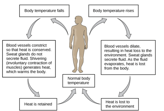
All living things are able to maintain a more-or-less constant internal environment. Regardless of the conditions around them, they can keep things relatively stable on the inside. The condition in which a system is maintained in a more-or-less steady state is called homeostasis. Human beings, for example, maintain a stable internal body temperature. If you go outside when the air temperature is below freezing, your body doesn't freeze. Instead, by shivering and other means, it maintains a stable internal temperature.
Organization
Living things have multiple levels of organization. Their molecules are organized into one or more cells. A cell is the basic unit of the structure and function of living things. Cells are the building blocks of living organisms. An average adult human being, for example, consists of trillions of cells. Living things may appear very different from one another on the outside, but their cells are very similar. Compare the human cells and onion cells in Figures 2.2.3 and 2.2.4. What similarities do you see?
![Joseph Elsbernd [CC BY-SA 2.0 (https://creativecommons.org/licenses/by-sa/2.0)] Shows the image through a microscope of human cheek cells. The cells are oval in shape and light blue, with a darker blue spot close to the centre. The light blue shows the cell membrane and cytoplasm and the darker blue shows the nucleus of the cell.](https://pressbooks.ccconline.org/acchumanbio/wp-content/uploads/sites/152/2023/10/Cheek-Cells-2.jpg)
![kaibara87 [CC BY 2.0 (https://creativecommons.org/licenses/by/2.0)] Shows an image through a microscope of onion cells. The cells are packed together and are rectangular in shape. Their cell walls and nuclei are stained a darker blue and the cytoplasm is whitish.](https://pressbooks.ccconline.org/acchumanbio/wp-content/uploads/sites/152/2023/10/Onion-Cells-2.jpg)
Metabolism
All living things can use energy. They require energy to maintain internal conditions (homeostasis), to grow, and to execute other processes. Living cells use the "machinery" of metabolism, which is the building up and breaking down of chemical compounds. Living things can transform energy by building up large molecules from smaller ones. This form of metabolism is called anabolism. Living things can also break down, or decompose, large organic molecules into smaller ones. This form of metabolism is called catabolism.
Consider weight lifters who eat high-protein diets. A protein is a large molecule made up of several small amino acids. When we eat proteins, our digestive system breaks them down into amino acids (catabolism), so that they are small enough to be absorbed by the digestive system and into the blood. From there, amino acids are transported to muscles, where they are converted back to proteins (anabolism).

Growth
All living things have the capacity for growth. Growth is an increase in size that occurs when there is a higher rate of anabolism than catabolism. A human infant, for example, has changed dramatically in size by the time it reaches adulthood, as is apparent from the image below. In what other ways do we change as we grow from infancy to adulthood?
A human infant has a lot of growing to do before adulthood.
Adaptations and Evolution
An adaptation is a characteristic that helps living things survive and reproduce in a given environment. It comes about because living things have the ability to change over time in response to the environment. A change in the characteristics of living things over time is called evolution. It develops in a population of organisms through random genetic mutations and natural selection.
Response to Stimuli
All living things detect changes in their environment and respond to them. These stimuli can be internal or external, and the response can take many forms, from the movement of a unicellular organism in response to external chemicals (called chemotaxis) to complex reactions involving all the senses of a multicellular organism. A response is often expressed by motion; for example, the leaves of a plant turning toward the sun (called phototropism).
Click through the images below: the venus fly trap, the cat, and the flower are all showing response to a stimuli.
Figure 2.2.6 Examples of responses to environmental stimuli.
Reproduction
All living things are capable of reproduction, the process by which living things give rise to offspring. Reproduction may be as simple as a single cell dividing to form two daughter cells, which is how bacteria reproduce. Reproduction in human beings and many other organisms, of course, is much more complicated. Nonetheless, whether a living thing is a human being or a bacterium, it is normally capable of reproduction.
Feature: Myth vs. Reality
Myth: Viruses are living things.
Reality: The traditional scientific view of viruses is that they originate from bits of DNA or RNA shed from the cells of living things, but that they are not living things themselves. Scientists have long argued that viruses are not living things because they do not exhibit most of the defining traits of living organisms. A single virus, called a virion, consists of a set of genes (DNA or RNA) inside a protective protein coat, called a capsid. Viruses have organization, but they are not cells, and they do not possess the cellular "machinery" that living things use to carry out life processes. As a result, viruses cannot undertake metabolism, maintain homeostasis, or grow.
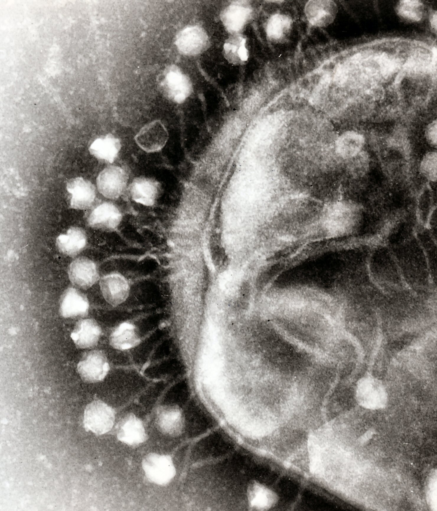
They do not seem to respond to their environment, and they can reproduce only by invading and using "tools" inside host cells to produce more virions. The only traits viruses seem to share with living things is the ability to evolve adaptations to their environment. In fact, some viruses evolve so quickly that it is difficult to design drugs and vaccines against them! That's why maintaining protection from the viral disease influenza, for example, requires a new flu vaccine each year.
Within the last decade, new discoveries in virology (the study of viruses) suggest that this traditional view about viruses may be incorrect, and that the "myth" that viruses are living things may be the reality. Researchers have discovered giant viruses that contain more genes than cellular life forms, such as bacteria. Some of the genes code for proteins needed to build new viruses, which suggests that these giant viruses may be able — or were once able — to reproduce without a host cell. Some of the strongest evidence that viruses are living things comes from studies of their proteins, which show that viruses and cellular life share a common ancestor in the distant past. Viruses may have once existed as primitive cells, but at some point they lost their cellular nature and became modern viruses that require host cells to reproduce. This idea is not so far-fetched when you consider that many other species require a host to complete their life cycle.
2.2 Summary
- To be classified as a living thing, most scientists agree that an object must exhibit seven characteristics. Humans share these traits with all other living things.
- All living things:
- Can maintain a more-or-less constant internal environment, which is called homeostasis.
- Have multiple levels of organization and consist of one or more cells.
- Can use energy and are capable of metabolism.
- Grow and develop.
- Can evolve adaptations to their environment.
- Can detect and respond to environmental stimuli.
- Are capable of reproduction, which is the process by which living things give rise to offspring.
2.2 Review Questions
- Identify the seven traits that most scientists agree are shared by all living things.
- What is homeostasis? What is one way humans fulfill this criterion of living things?
- Define reproduction and describe two different examples.
- Assume that you found an object that looks like a dead twig. You wonder if it might be a stick insect. How could you ethically determine if it is a living thing?
- Describe viruses and which traits they do and do not share with living things. Do you think viruses should be considered living things? Why or why not?
- People who are biologically unable to reproduce are certainly still considered alive. Discuss why this situation does not invalidate the criteria that living things must be capable of reproduction.
- What are the two types of metabolism described here. What are their differences?
- What are some similarities between the cells of different organisms? If you are not familiar with the specifics of cells, simply describe the similarities you see in the pictures above.
- What are two processes in a living thing that use energy?
- Give an example of a response to stimuli in humans.
- Do unicellular organisms (such as bacteria) have an internal environment that they maintain through homeostasis? Why or why not?
- Evolution occurs through natural ____________ .
- If alien life is found on other planets, do you think the aliens will have cells? Discuss your answer.
- Movement in response to an external chemical is called ___________, while movement towards light is called ___________ .
2.2 Explore More
https://www.youtube.com/watch?v=cQPVXrV0GNA&t=354s
Characteristics of Life, Ameoba Sisters, 2017.
Attributions
Figure 2.2.1
The Thinker MET 131262, by Auguste Rodin, 1910, from the Metropolitan Museum of Art, is in the public domain (https://en.wikipedia.org/wiki/Public_domain).
Figure 2.2.2
Homeostasis: Figure 4, by OpenStax College, Biology is used under a CC BY 4.0 (https://creativecommons.org/licenses/by/4.0) license. Download for free at http://cnx.org/contents/04fdb865-17a1-43d8-bb33-36f821ddd119@7.
Figure 2.2.3
Human cheek cells, by Joseph Elsbernd, 2012, on Flickr, is used under a CC BY-SA 2.0 (https://creativecommons.org/licenses/by-sa/2.0/) license.
Figure 2.2.4
Onion cells 2, by Umberto Salvagnin, 2009, on Flickr, is used under a CC BY 2.0 (https://creativecommons.org/licenses/by/2.0/) license.
Figure 2.2.5
Photo (family) by Jakob Owens on Unsplash is used under the Unsplash License (https://unsplash.com/license).
Figure 2.2.6
- Trap of Dionaea muscipula by che on Wikimedia Commons is used under a CC BY-SA 2.5 (https://creativecommons.org/licenses/by-sa/2.5/deed.en) license.
- Plants leaning towards the sunlight from Pxhere is used under a CC0 1.0 universal
public domain dedication license (https://creativecommons.org/publicdomain/zero/1.0/). - Surprised young cat by Watchduck (a.k.a. Tilman Piesk) on Wikimedia Commons is used under a CC BY 3.0 (https://creativecommons.org/licenses/by/3.0) license.
Figure 2.2.7
Bacteriophages, by Dr. Graham Beards, is used under a CC BY-SA 3.0 (https://creativecommons.org/licenses/by-sa/3.0) license.
References
Ameoba Sisters. (2017, October 26). Characteristics of life. YouTube. https://www.youtube.com/watch?v=cQPVXrV0GNA&feature=youtu.be
OpenStax. (2016, March 23). Figure 4 The body is able to regulate temperature in response to signals from the nervous system. In OpenStax, Biology (Section 33.3). OpenStax CNX. http://cnx.org/contents/185cbf87-c72e-48f5-b51e-f14f21b5eabd@10.8.
Wikipedia contributors. (2020, June 14). Adaptation. Wikipedia. https://en.wikipedia.org/w/index.php?title=Adaptation&oldid=962556016
Wikipedia contributors. (2020, June 21). Auguste Rodin. Wikipedia. https://en.wikipedia.org/w/index.php?title=Auguste_Rodin&oldid=963668399
Wikipedia contributors. (2020, June 22). Chemotaxis. Wikipedia. https://en.wikipedia.org/w/index.php?title=Chemotaxis&oldid=963884872
Wikipedia contributors. (2020, June 22). Evolution. Wikipedia. https://en.wikipedia.org/w/index.php?title=Evolution&oldid=963929880
Wikipedia contributors. (2020, June 20). Phototropism. Wikipedia. https://en.wikipedia.org/w/index.php?title=Phototropism&oldid=963567791
Wikipedia contributors. (2020, June 22). Virus. Wikipedia. https://en.wikipedia.org/w/index.php?title=Virus&oldid=963829311
Created by CK-12/Adapted by Christine Miller
The Thinker
![Auguste Rodin [CC0] The Thinker (French: Le Penseur) is a bronze sculpture by Auguste Rodin, usually placed on a stone pedestal. The work shows a nude male figure of over life-size sitting on a rock with his chin resting on one hand as though deep in thought, often used as an image to represent philosophy.](https://pressbooks.ccconline.org/acchumanbio/wp-content/uploads/sites/152/2019/06/The-thinker-2.jpg)
You've probably seen this famous statue created by the French sculptor Auguste Rodin. Rodin's skill as a sculptor is especially evident here because the statue — which is made of bronze — looks so lifelike. How does a bronze statue differ from a living, breathing human being or other living organism? What is life? What does it mean to be alive? Science has answers to these questions.
Characteristics of Living Things
To be classified as a living thing, most scientists agree that an object must have all seven of the traits listed below. Humans share these characteristics with other living things.
- Homeostasis
- Organization
- Metabolism
- Growth
- Adaptation
- Response to stimuli
- Reproduction
Homeostasis
All living things are able to maintain a more-or-less constant internal environment. Regardless of the conditions around them, they can keep things relatively stable on the inside. The condition in which a system is maintained in a more-or-less steady state is called homeostasis. Human beings, for example, maintain a stable internal body temperature. If you go outside when the air temperature is below freezing, your body doesn't freeze. Instead, by shivering and other means, it maintains a stable internal temperature.

Organization
Living things have multiple levels of organization. Their molecules are organized into one or more cells. A cell is the basic unit of the structure and function of living things. Cells are the building blocks of living organisms. An average adult human being, for example, consists of trillions of cells. Living things may appear very different from one another on the outside, but their cells are very similar. Compare the human cells and onion cells in Figures 2.2.3 and 2.2.4. What similarities do you see?
![Joseph Elsbernd [CC BY-SA 2.0 (https://creativecommons.org/licenses/by-sa/2.0)] Shows the image through a microscope of human cheek cells. The cells are oval in shape and light blue, with a darker blue spot close to the centre. The light blue shows the cell membrane and cytoplasm and the darker blue shows the nucleus of the cell.](https://pressbooks.ccconline.org/acchumanbio/wp-content/uploads/sites/152/2023/10/Cheek-Cells-2.jpg)
![kaibara87 [CC BY 2.0 (https://creativecommons.org/licenses/by/2.0)] Shows an image through a microscope of onion cells. The cells are packed together and are rectangular in shape. Their cell walls and nuclei are stained a darker blue and the cytoplasm is whitish.](https://pressbooks.ccconline.org/acchumanbio/wp-content/uploads/sites/152/2023/10/Onion-Cells-2.jpg)
Metabolism
All living things can use energy. They require energy to maintain internal conditions (homeostasis), to grow, and to execute other processes. Living cells use the "machinery" of metabolism, which is the building up and breaking down of chemical compounds. Living things can transform energy by building up large molecules from smaller ones. This form of metabolism is called anabolism. Living things can also break down, or decompose, large organic molecules into smaller ones. This form of metabolism is called catabolism.
Consider weight lifters who eat high-protein diets. A protein is a large molecule made up of several small amino acids. When we eat proteins, our digestive system breaks them down into amino acids (catabolism), so that they are small enough to be absorbed by the digestive system and into the blood. From there, amino acids are transported to muscles, where they are converted back to proteins (anabolism).

Growth
All living things have the capacity for growth. Growth is an increase in size that occurs when there is a higher rate of anabolism than catabolism. A human infant, for example, has changed dramatically in size by the time it reaches adulthood, as is apparent from the image below. In what other ways do we change as we grow from infancy to adulthood?
A human infant has a lot of growing to do before adulthood.
Adaptations and Evolution
An adaptation is a characteristic that helps living things survive and reproduce in a given environment. It comes about because living things have the ability to change over time in response to the environment. A change in the characteristics of living things over time is called evolution. It develops in a population of organisms through random genetic mutations and natural selection.
Response to Stimuli
All living things detect changes in their environment and respond to them. These stimuli can be internal or external, and the response can take many forms, from the movement of a unicellular organism in response to external chemicals (called chemotaxis) to complex reactions involving all the senses of a multicellular organism. A response is often expressed by motion; for example, the leaves of a plant turning toward the sun (called phototropism).
Click through the images below: the venus fly trap, the cat, and the flower are all showing response to a stimuli.
Figure 2.2.6 Examples of responses to environmental stimuli.
Reproduction
All living things are capable of reproduction, the process by which living things give rise to offspring. Reproduction may be as simple as a single cell dividing to form two daughter cells, which is how bacteria reproduce. Reproduction in human beings and many other organisms, of course, is much more complicated. Nonetheless, whether a living thing is a human being or a bacterium, it is normally capable of reproduction.
Feature: Myth vs. Reality
Myth: Viruses are living things.
Reality: The traditional scientific view of viruses is that they originate from bits of DNA or RNA shed from the cells of living things, but that they are not living things themselves. Scientists have long argued that viruses are not living things because they do not exhibit most of the defining traits of living organisms. A single virus, called a virion, consists of a set of genes (DNA or RNA) inside a protective protein coat, called a capsid. Viruses have organization, but they are not cells, and they do not possess the cellular "machinery" that living things use to carry out life processes. As a result, viruses cannot undertake metabolism, maintain homeostasis, or grow.

They do not seem to respond to their environment, and they can reproduce only by invading and using "tools" inside host cells to produce more virions. The only traits viruses seem to share with living things is the ability to evolve adaptations to their environment. In fact, some viruses evolve so quickly that it is difficult to design drugs and vaccines against them! That's why maintaining protection from the viral disease influenza, for example, requires a new flu vaccine each year.
Within the last decade, new discoveries in virology (the study of viruses) suggest that this traditional view about viruses may be incorrect, and that the "myth" that viruses are living things may be the reality. Researchers have discovered giant viruses that contain more genes than cellular life forms, such as bacteria. Some of the genes code for proteins needed to build new viruses, which suggests that these giant viruses may be able — or were once able — to reproduce without a host cell. Some of the strongest evidence that viruses are living things comes from studies of their proteins, which show that viruses and cellular life share a common ancestor in the distant past. Viruses may have once existed as primitive cells, but at some point they lost their cellular nature and became modern viruses that require host cells to reproduce. This idea is not so far-fetched when you consider that many other species require a host to complete their life cycle.
2.2 Summary
- To be classified as a living thing, most scientists agree that an object must exhibit seven characteristics. Humans share these traits with all other living things.
- All living things:
- Can maintain a more-or-less constant internal environment, which is called homeostasis.
- Have multiple levels of organization and consist of one or more cells.
- Can use energy and are capable of metabolism.
- Grow and develop.
- Can evolve adaptations to their environment.
- Can detect and respond to environmental stimuli.
- Are capable of reproduction, which is the process by which living things give rise to offspring.
2.2 Review Questions
- Identify the seven traits that most scientists agree are shared by all living things.
- What is homeostasis? What is one way humans fulfill this criterion of living things?
- Define reproduction and describe two different examples.
- Assume that you found an object that looks like a dead twig. You wonder if it might be a stick insect. How could you ethically determine if it is a living thing?
- Describe viruses and which traits they do and do not share with living things. Do you think viruses should be considered living things? Why or why not?
- People who are biologically unable to reproduce are certainly still considered alive. Discuss why this situation does not invalidate the criteria that living things must be capable of reproduction.
- What are the two types of metabolism described here. What are their differences?
- What are some similarities between the cells of different organisms? If you are not familiar with the specifics of cells, simply describe the similarities you see in the pictures above.
- What are two processes in a living thing that use energy?
- Give an example of a response to stimuli in humans.
- Do unicellular organisms (such as bacteria) have an internal environment that they maintain through homeostasis? Why or why not?
- Evolution occurs through natural ____________ .
- If alien life is found on other planets, do you think the aliens will have cells? Discuss your answer.
- Movement in response to an external chemical is called ___________, while movement towards light is called ___________ .
2.2 Explore More
https://www.youtube.com/watch?v=cQPVXrV0GNA&t=354s
Characteristics of Life, Ameoba Sisters, 2017.
Attributions
Figure 2.2.1
The Thinker MET 131262, by Auguste Rodin, 1910, from the Metropolitan Museum of Art, is in the public domain (https://en.wikipedia.org/wiki/Public_domain).
Figure 2.2.2
Homeostasis: Figure 4, by OpenStax College, Biology is used under a CC BY 4.0 (https://creativecommons.org/licenses/by/4.0) license. Download for free at http://cnx.org/contents/04fdb865-17a1-43d8-bb33-36f821ddd119@7.
Figure 2.2.3
Human cheek cells, by Joseph Elsbernd, 2012, on Flickr, is used under a CC BY-SA 2.0 (https://creativecommons.org/licenses/by-sa/2.0/) license.
Figure 2.2.4
Onion cells 2, by Umberto Salvagnin, 2009, on Flickr, is used under a CC BY 2.0 (https://creativecommons.org/licenses/by/2.0/) license.
Figure 2.2.5
Photo (family) by Jakob Owens on Unsplash is used under the Unsplash License (https://unsplash.com/license).
Figure 2.2.6
- Trap of Dionaea muscipula by che on Wikimedia Commons is used under a CC BY-SA 2.5 (https://creativecommons.org/licenses/by-sa/2.5/deed.en) license.
- Plants leaning towards the sunlight from Pxhere is used under a CC0 1.0 universal
public domain dedication license (https://creativecommons.org/publicdomain/zero/1.0/). - Surprised young cat by Watchduck (a.k.a. Tilman Piesk) on Wikimedia Commons is used under a CC BY 3.0 (https://creativecommons.org/licenses/by/3.0) license.
Figure 2.2.7
Bacteriophages, by Dr. Graham Beards, is used under a CC BY-SA 3.0 (https://creativecommons.org/licenses/by-sa/3.0) license.
References
Ameoba Sisters. (2017, October 26). Characteristics of life. YouTube. https://www.youtube.com/watch?v=cQPVXrV0GNA&feature=youtu.be
OpenStax. (2016, March 23). Figure 4 The body is able to regulate temperature in response to signals from the nervous system. In OpenStax, Biology (Section 33.3). OpenStax CNX. http://cnx.org/contents/185cbf87-c72e-48f5-b51e-f14f21b5eabd@10.8.
Wikipedia contributors. (2020, June 14). Adaptation. Wikipedia. https://en.wikipedia.org/w/index.php?title=Adaptation&oldid=962556016
Wikipedia contributors. (2020, June 21). Auguste Rodin. Wikipedia. https://en.wikipedia.org/w/index.php?title=Auguste_Rodin&oldid=963668399
Wikipedia contributors. (2020, June 22). Chemotaxis. Wikipedia. https://en.wikipedia.org/w/index.php?title=Chemotaxis&oldid=963884872
Wikipedia contributors. (2020, June 22). Evolution. Wikipedia. https://en.wikipedia.org/w/index.php?title=Evolution&oldid=963929880
Wikipedia contributors. (2020, June 20). Phototropism. Wikipedia. https://en.wikipedia.org/w/index.php?title=Phototropism&oldid=963567791
Wikipedia contributors. (2020, June 22). Virus. Wikipedia. https://en.wikipedia.org/w/index.php?title=Virus&oldid=963829311
Created by: CK-12/Adapted by: Christine Miller
What Is Pseudoscience?
Pseudoscience is a claim, belief, or practice that is presented as scientific but does not adhere to the standards and methods of science. True science is based on repeated evidence-gathering and testing of falsifiable hypotheses. Pseudoscience does not adhere to these criteria. In addition to phrenology, some other examples of pseudoscience include astrology, extrasensory perception (ESP), reflexology, reincarnation, and Scientology,
Characteristics of Pseudoscience
Whether a field is actually science or just pseudoscience is not always clear. However, pseudoscience generally exhibits certain common characteristics. Indicators of pseudoscience include:
- The use of vague, exaggerated, or untestable claims: Many claims made by pseudoscience cannot be tested with evidence. As a result, they cannot be falsified, even if they are not true.
- An over-reliance on confirmation rather than refutation: Any incident that appears to justify a pseudoscience claim is treated as proof of the claim. Claims are assumed true until proven otherwise, and the burden of disproof is placed on skeptics of the claim.
- A lack of openness to testing by other experts: Practitioners of pseudoscience avoid subjecting their ideas to peer review. They may refuse to share their data and justify the need for secrecy with claims of proprietary or privacy.
- An absence of progress in advancing knowledge: In pseudoscience, ideas are not subjected to repeated testing followed by rejection or refinement, as hypotheses are in true science. Ideas in pseudoscience may remain unchanged for hundreds — or even thousands — of years. In fact, the older an idea is, the more it tends to be trusted in pseudoscience.
- Personalization of issues: Proponents of pseudoscience adopt beliefs that have little or no rational basis, so they may try to confirm their beliefs by treating critics as enemies. Instead of arguing to support their own beliefs, they attack the motives and character of their critics.
- The use of misleading language: Followers of pseudoscience may use scientific-sounding terms to make their ideas sound more convincing. For example, they may use the formal name dihydrogen monoxide to refer to plain old water.
Persistence of Pseudoscience
Despite failing to meet scientific standards, many pseudosciences survive. Some pseudosciences remain very popular with large numbers of believers. A good example is astrology.
Astrology is the study of the movements and relative positions of celestial objects as a means for divining information about human affairs and terrestrial events. Many ancient cultures attached importance to astronomical events, and some developed elaborate systems for predicting terrestrial events from celestial observations. Throughout most of its history in the West, astrology was considered a scholarly tradition and was common in academic circles. With the advent of modern Western science, astrology was called into question. It was challenged on both theoretical and experimental grounds, and it was eventually shown to have no scientific validity or explanatory power.

Today, astrology is considered a pseudoscience, yet it continues to have many devotees. Most people know their astrological sign, and many people are familiar with the personality traits supposedly associated with their sign. Astrological readings and horoscopes are readily available online and in print media, and a lot of people read them, even if only occasionally. About a third of all adult Americans actually believe that astrology is scientific. Studies suggest that the persistent popularity of pseudosciences such as astrology reflects a high level of scientific illiteracy. It seems that many Americans do not have an accurate understanding of scientific principles and methodology. They are not convinced by scientific arguments against their beliefs.
Dangers of Pseudoscience
Belief in astrology is unlikely to cause a person harm, but belief in some other pseudosciences might — especially in health care-related areas. Treatments that seem scientific but are not may be ineffective, expensive, and even dangerous to patients. Seeking out pseudoscientific treatments may also delay or preclude patients from seeking scientifically-based medical treatments that have been tested and found safe and effective. In short, irrational health care may not be harmless.
Scientific Hoaxes, Frauds, and Fallacies
Pseudoscience is not the only way that science may be misused. Scientific hoaxes, frauds, and fallacies may misdirect the pursuit of science, put patients at risk, or mislead and confuse the public. An example of each of these misuses of science and its negative effects is described below.
The Piltdown Hoax
![Image by By James Howard McGregor [Public domain], via Wikimedia Commons A side profile view of an artists rendition of what the Piltdown Man may have looked like, had he been real.](https://pressbooks.ccconline.org/acchumanbio/wp-content/uploads/sites/152/2023/10/Piltdown-Man-1.jpg)
Piltdown Man (see picture left) was a paleontological hoax in which bone fragments were presented as the fossilized remains of a previously unknown early human. These fragments consisted of parts of a skull and jawbone, reported to have been found in 1908 in a gravel pit at Piltdown, East Sussex, England. The significance of the specimen remained the subject of controversy until it was exposed in 1953 as a hoax. It eventually came to light that the specimen consisted of the lower jawbone of an orangutan deliberately combined with skull bones of a modern human. The Piltdown hoax is perhaps the most infamous paleontological hoax ever perpetrated, both for its impact on the direction of research on human evolution and for the length of time between its "discovery" and its full exposure as a forgery.
![Photo by Anrie [CC BY-SA 3.0 (https://creativecommons.org/licenses/by-sa/3.0)], from Wikimedia Commons A replica of the infamous Piltdown skull. The skull is encased in a glass sphere. The replica shows portions of the skull which were bone in white, and the portions of the skull which were inferred in black.](https://pressbooks.ccconline.org/acchumanbio/wp-content/uploads/sites/152/2023/10/Sterkfontein_Piltdown_man-1.jpg)
In 1912, the head of the geological department at the British Museum proposed that Piltdown man represented an evolutionary missing link between apes and humans. With its human-like cranium and ape-like jaw, it seemed to support the idea then prevailing in England that human evolution began with the brain. The Piltdown specimen led scientists down a blind alley in the belief that the human brain increased in size before the jaw underwent size reductions to become more like the modern human jaw. This belief confused and misdirected the study of human evolution for decades, and actual fossils of early humans were ignored because they didn't support the accepted paradigm.
The Vaccine-Autism Fraud
You may have heard that certain vaccines put the health of young children at risk. This persistent idea is not supported by scientific evidence or accepted by the vast majority of experts in the field. It stems largely from an elaborate medical research fraud that was reported in a 1998 article published in the respected British medical journal, The Lancet. The main author of the article was a British physician named Andrew Wakefield. In the article, Wakefield and his colleagues described case histories of 12 children, most of whom were reported to have developed autism soon after the administration of the MMR (measles, mumps, rubella) vaccine.
Several subsequent peer-reviewed studies failed to show any association between the MMR vaccine and autism. It also later emerged that Wakefield had received research funding from a group of people who were suing vaccine manufacturers. In 2004, ten of Wakefield's 12 coauthors formally retracted the conclusions in their paper. In 2010, editors of The Lancetretracted the entire paper. That same year, Wakefield was charged with deliberate falsification of research and barred from practicing medicine in the United Kingdom. Unfortunately, by then, the damage had already been done. Parents afraid that their children would develop autism had refrained from having them vaccinated. British MMR vaccination rates fell from nearly 100 per cent to 80 per cent in the years following the study. The consensus of medical experts today is that Wakefield's fraud put hundreds of thousands of children at risk because of the lower vaccination rates and also diverted research efforts and funding away from finding the true cause of autism.
Correlation-Causation Fallacy
Many statistical tests used in scientific research calculate correlations between variables. Correlation refers to how closely related two data sets are, which may be a useful starting point for further investigation. Correlation, however, is also one of the most misused types of evidence, primarily because of the logical fallacy that correlation implies causation. In reality, just because two variables are correlated does not necessarily mean that either variable causes the other.
A few simple examples, illustrated by the graphs below, can be used to demonstrate the correlation-causation fallacy. Assume a study found that both per capita consumption of mozzarella cheese and the number of Civil Engineering doctorates awarded are correlated; that is, rates of both events increase together. If correlation really did imply causation, then you could conclude from the second example that the increase in age of Miss America causes an increase in murders of a specific type or vice versa.
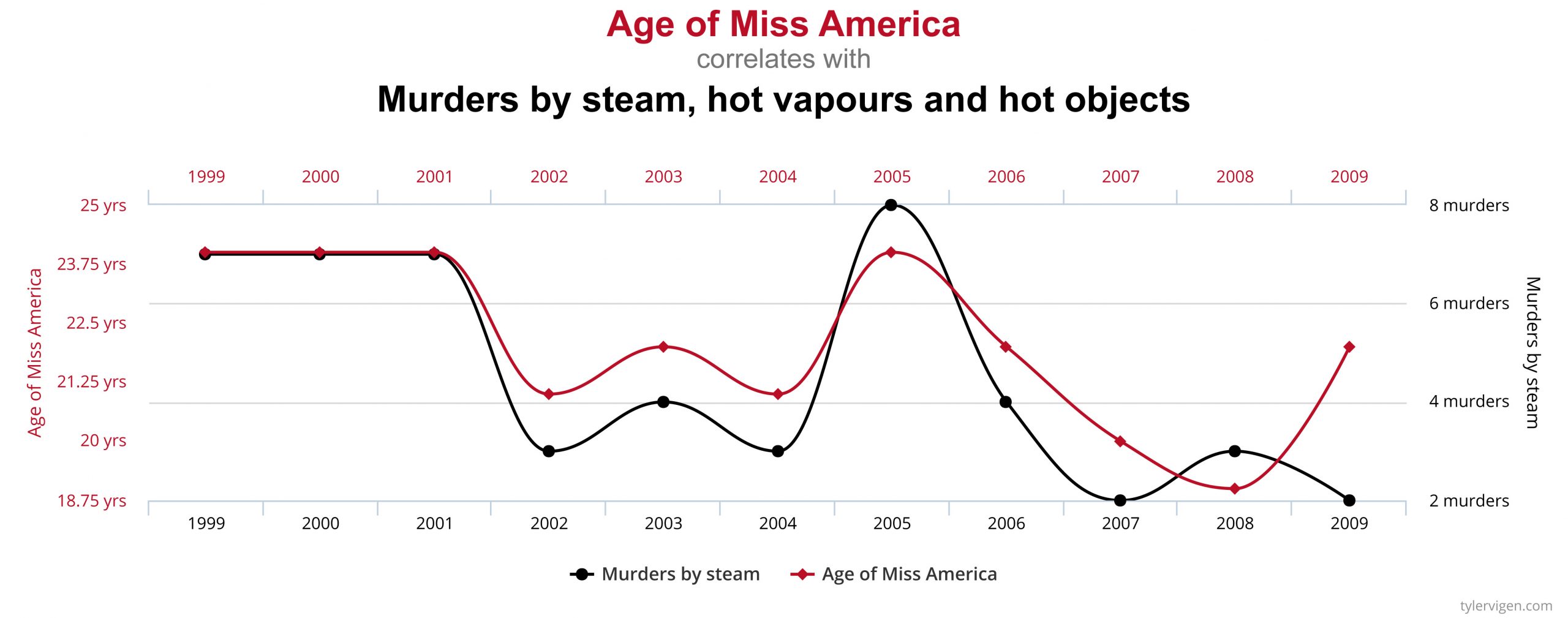
An actual example of the correlation-causation fallacy occurred during the latter half of the 20th century. Numerous studies showed that women taking hormone replacement therapy (HRT) to treat menopausal symptoms also had a lower-than-average incidence of coronary heart disease (CHD). This correlation was misinterpreted as evidence that HRT protects women against CHD. Subsequent studies that controlled other factors related to CHD disproved this presumed causal connection. The studies found that women taking HRT were more likely to come from higher socio-economic groups, with better-than-average diets and exercise regimens. Rather than HRT causing lower CHD incidence, these studies concluded that HRT and lower CHD were both effects of higher socio-economic status and related lifestyle factors.
Check out this “Rough Guide to Spotting Bad Science” infographic from Compound Interest:
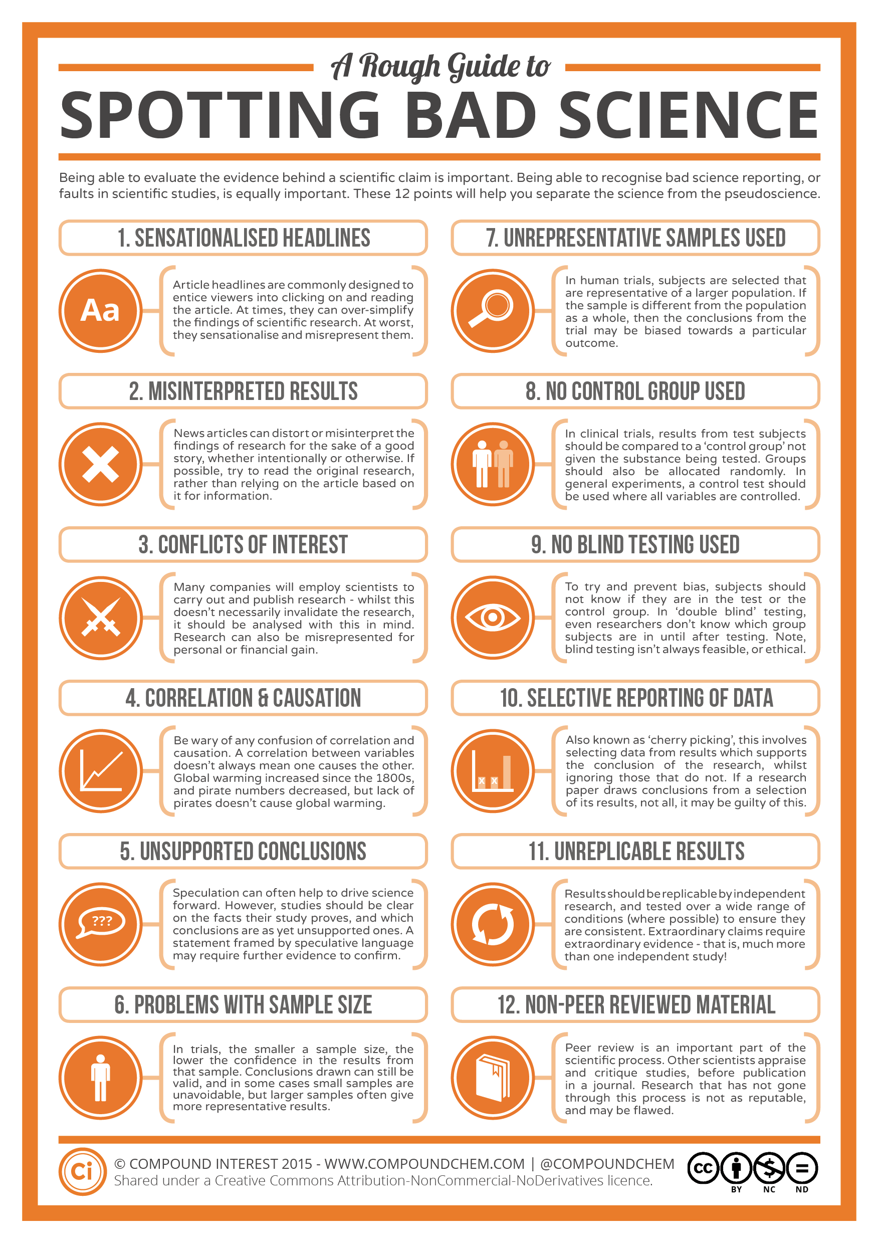
1.7 Summary
- Pseudoscience is a claim, belief, or practice that is presented as scientific, but does not adhere to scientific standards and methods.
- Indicators of pseudoscience include untestable claims, lack of openness to testing by experts, absence of progress in advancing knowledge, and attacks on the motives and character of critics.
- Some pseudosciences, including astrology, remain popular. This suggests that many people do not possess the scientific literacy needed to distinguish pseudoscience from true science, or to be convinced by scientific arguments against them.
- Belief in a pseudoscience such as astrology is unlikely to cause harm, but belief in pseudoscientific medical treatments may be harmful.
- In addition to pseudoscience, other examples of the misuse of science include scientific hoaxes (such as the Piltdown hoax), scientific frauds (such as the MMR vaccine-autism fraud), and scientific fallacies (such as the correlation-causation fallacy).
1.7 Review Questions
- Define pseudoscience. Give three examples.
- What are some indicators that a claim, belief, or practice might be pseudoscience rather than true science?
- Astrology was once considered a science, and it was common in academic circles. Why did its status change from a science to a pseudoscience?
- What are possible reasons that some pseudosciences remain popular even after they have been shown to have no scientific validity or explanatory power?
- List three other ways besides pseudoscience that science can be misused, and identify an example of each.
- Explain how misuses of science may waste money and effort. How can they potentially cause harm to the public?
- Many claims made by pseudoscience cannot be tested with evidence. From a scientific perspective, why is it important that claims be testable?
- What do you think is the difference between pseudoscience and belief?
- If you see a website that claims that an herbal supplement causes weight loss and they use a lot of scientific terms to explain how it works, can you be assured that the drug is scientifically proven to work? If not, what are some steps you can take to determine whether or not the drug does in fact work?
- Why do you think it was problematic that Andrew Wakefield received funding from a group of people who were suing vaccine manufacturers?
- What do you think it says about the 1998 Wakefield paper that ten of the 12 coauthors formally retracted their conclusions?
1.7 Explore More
https://www.youtube.com/watch?v=E91bGT9BjYk
How to spot a misleading graph - Lea Gaslowitz, TED-Ed, 2017.
https://www.youtube.com/watch?v=sxYrzzy3cq8
How statistics can be misleading - Mark Liddell, TED-Ed, 2016.
Attributions
Figure 1.7.1
Zodiac Signs Cancer Aquarius Aries Gemini Leo from Max Pixel, is used under a CC0 1.0 Universal Public Domain Dedication license (https://creativecommons.org/publicdomain/zero/1.0/deed.en).
Figure 1.7.2
Piltdown Man - McGregor model, by James Howard McGregor on Wikimedia Commons is in the public domain (https://en.wikipedia.org/wiki/Public_domain).
Figure 1.7.3
Sterkfontein Piltdown man, by Anrie on Wikimedia Commons is used under a CC BY-SA 3.0 (https://creativecommons.org/licenses/by-sa/3.0) license.
Figure 1.7.4
Spurious Correlations (Causation Fallacy) - Consumption of mozzarella cheese and awarded Doctorates by Tyler Vigen on Tylervigen.com is used under a CC BY 4.0 (https://creativecommons.org/licenses/by/4.0/) license.
Figure 1.7.5
Spurious Correlations (Causation Fallacy) - Miss America and Murder, by Tyler Vigen, is used under a CC BY 4.0 (https://creativecommons.org/licenses/by/4.0/) license.
Figure 1.7.6
A rough guide to spotting bad science, by Compound Interest, is used under a CC BY-NC-ND 2.0 (https://creativecommons.org/licenses/by-nc-nd/2.0/ca/) license
References
TED-Ed. (2017, July 6). How to spot a misleading graph - Lea Gaslowitz. YouTube. https://www.youtube.com/watch?v=E91bGT9BjYk&feature=youtu.be
Wakefield, A.J., Murch, S.H., Anthony, A., Linnell, J., Casson, D.M., Malik, M., et al. (1998). Ileal-lymphoid-nodular hyperplasia, non-specific colitis, and pervasive developmental disorder in children. Lancet, 351: 637–41.
Wikipedia contributors. (2020, June 18). Andrew Wakefield. Wikipedia. https://en.wikipedia.org/w/index.php?title=Andrew_Wakefield&oldid=963243135
The space occurring between two or more membranes. In cell biology, it's most commonly described as the region between the inner membrane and the outer membrane of a mitochondrion or a chloroplast.
Image shows a freshly baked Steak and Kidney Pie.
Created by CK-12 Foundation/Adapted by Christine Miller

Case Study Conclusion: Drink and Flush
You are probably aware that, because of its effects on the brain, drinking alcohol can cause visual disturbances, slurred speech, drowsiness, impaired judgment, and loss of coordination. Although it may be less obvious, alcohol also can have serious effects on the functioning of the excretory system.
As you learned from the conversation between Talia and Shae — who were in line for the restroom at the beginning of this chapter — alcohol consumption inhibits a hormone that causes our bodies to retain water. As a result, more water is released in urine, increasing the frequency of restroom trips, as well as the risk of dehydration.
Which hormone discussed in this chapter does this? If you answered antidiuretic hormone (ADH; also called vasopressin) — you are correct! ADH is secreted by the posterior pituitary gland and acts on the kidneys. As you have learned, the kidneys filter the blood, reabsorb needed substances, and produce urine. ADH helps the body conserve water by influencing this process. ADH makes the collecting ducts in the kidneys permeable to water, allowing water molecules to be reabsorbed from the urine back into the blood through osmosis into capillaries.
Alcohol is thought to produce more dilute urine by inhibiting the release of ADH. This causes the collecting ducts to be more impermeable to water, so less water can be reabsorbed, and more is excreted in urine. Because the volume of urine is increased, the bladder fills up more quickly, and the urge to urinate occurs more frequently. This is part of the reason why you often see a long line for the restroom in situations where many people are drinking alcohol. In addition to producing more dilute urine, simply consuming many beverages can also increase urine output.
In most cases, moderate drinking causes only a minor and temporary effect on kidney function. However, when people consume a large quantity of alcohol in a short period of time, or abuse alcohol over long time periods, there can be serious effects on the kidney. Binge drinking (consuming roughly four to five drinks in two hours) can cause a condition called “acute kidney injury,” a serious and sudden impairment of kidney function that requires immediate medical attention. As with the other cases of kidney failure that you learned about in this chapter, the treatment is to artificially filter the blood using hemodialysis. While normal kidney function may eventually return, acute kidney injury can sometimes cause long-term damage to the kidneys.
In cases where people abuse alcohol, particularly for an extended period of time, there can be many serious effects on the kidneys and other parts of the excretory system. The dehydrating effect of alcohol on the body can impair the function of many organs, including the kidneys themselves. Additionally, because of alcohol’s effect on kidney function, water balance, and ion balance, chronic alcohol consumption can cause abnormalities in blood ion concentration and acid-base balance, which can be very dangerous.
Drinking more than two alcoholic beverages a day can increase your risk for high blood pressure, too. As you have learned, high blood pressure is a risk factor for some kidney disorders, as well as a common cause of kidney failure. Drinking too much alcohol can damage the kidneys by raising blood pressure.
Finally, chronic excessive consumption of alcohol can cause liver disease. The liver is an important organ of the excretory system that breaks down toxic substances in the blood. The liver and kidneys work together to remove wastes from the bloodstream. You may remember, for example, the liver transforms ammonia into urea, which is then filtered and excreted by the kidneys. When the liver is not functioning normally, it puts added strain on the kidneys, which can result in kidney dysfunction. This association between alcohol, liver disease, and kidney dysfunction is so strong that most of the patients in Canada with both liver disease and related kidney dysfunction are alcoholics.
As you have learned, the excretory system is essential in removing toxic wastes from the body and regulating homeostasis. Having an occasional drink can temporarily alter these functions, but excessive alcohol exposure can seriously and permanently damage this system in many ways. Limiting alcohol consumption can help preserve the normal functioning of the excretory system, so that it can protect your health.
Chapter 16 Summary
In this chapter you learned about the excretory system. Specifically, you learned that:
- Excretion is the process of removing wastes and excess water from the body. It is an essential process in all living things, and a major way in which the human body maintains homeostasis.
- Organs of the excretory system include the skin, liver, large intestine, lungs, and kidneys.
-
- The skin plays a role in excretion through the production of sweat by sweat glands. Sweating eliminates excess water and salts, as well as a small amount of urea, a byproduct of protein catabolism.
- The liver is a very important organ of excretion. The liver breaks down many substances — including toxins — in the blood. The liver also excretes bilirubin (a waste product of hemoglobin catabolism) in bile. Bile then travels to the small intestine and is eventually excreted in feces by the large intestine.
- The main excretory function of the large intestine is to eliminate solid waste that remains after food is digested and water is extracted from the indigestible matter. The large intestine also collects and excretes wastes from throughout the body.
- The lungs are responsible for the excretion of gaseous wastes — primarily carbon dioxide — from cellular respiration in cells throughout the body. Exhaled air also contains water vapor and trace levels of some other waste gases.
- The paired kidneys are often considered the main organs of excretion. Their primary function is the elimination of excess water and wastes from the bloodstream by the production of urine. The kidneys filter many substances out of blood, allow the blood to reabsorb needed materials, and use the remaining materials to form urine.
-
- The two bean-shaped kidneys are located high in the back of the abdominal cavity on either side of the spine. A renal artery connects each kidney with the aorta, and transports unfiltered blood to the kidney. A renal vein connects each kidney with the inferior vena cava and transports filtered blood back to the circulation.
- The kidney has two main layers involved in the filtration of blood and formation of urine: the outer cortex and inner medulla. At least a million nephrons — which are the tiny functional units of the kidney — span the cortex and medulla. The entire kidney is surrounded by a fibrous capsule and protective fat layers.
- As blood flows through a nephron, many materials are filtered out of the blood, needed materials are returned to the blood, and the remaining materials are used to form urine.
-
-
- In each nephron, the glomerulus and the surrounding glomerular capsule form the unit that filters blood. From the glomerular capsule, the material filtered from blood (called filtrate) passes through the long renal tubule. As it does, some substances are reabsorbed into the blood, and other substances are secreted from the blood into the filtrate, finally forming urine. The urine empties into collecting ducts, where more water may be reabsorbed.
-
- The kidneys are part of the urinary system, which also includes the ureters, urinary bladder, and urethra. The main function of the urinary system is to eliminate the waste products of metabolism from the body by forming and excreting urine. After urine forms in the kidneys, it is transported through the ureters to the bladder. The bladder stores the urine until urination, when urine is transported by the urethra to be excreted outside the body.
-
- Besides the elimination of waste products such as urea, uric acid, excess water, and mineral ions, the urinary system has other vital functions. These include maintaining homeostasis of mineral ions in extracellular fluid, regulating acid-base balance in the blood, regulating the volume of extracellular fluids, and controlling blood pressure.
-
-
- The formation of urine must be closely regulated to maintain body-wide homeostasis. Several endocrine hormones help control this function of the urinary system, including antidiuretic hormone secreted from the posterior pituitary gland, parathyroid hormone from the parathyroid glands, and aldosterone from the adrenal glands.
-
-
-
-
- For example, the kidneys are part of the renin-angiotensin-aldosterone system that regulates the concentration of sodium in the blood to control blood pressure. In this system, the enzyme renin secreted by the kidneys works with hormones from the liver and adrenal gland to stimulate nephrons to reabsorb more sodium and water from urine.
- The kidneys also secrete endocrine hormones, including calcitriol — which helps control the level of calcium in the blood — and erythropoietin, which stimulates bone marrow to produce red blood cells.
-
- The process of urination is controlled by both the autonomic and the somatic nervous systems. The autonomic system causes the detrusor muscle in the bladder wall to relax as the bladder fills with urine, but conscious contraction of the detrusor muscle expels urine from the bladder during urination.
- Ureters are tube-like structures that connect the kidneys with the urinary bladder. Each ureter arises at the renal pelvis of a kidney and travels down through the abdomen to the urinary bladder. The walls of the ureter contain smooth muscle that can contract to push urine through the ureter by peristalsis. The walls are lined with transitional epithelium that can expand and stretch.
- The urinary bladder is a hollow, muscular organ that rests on the pelvic floor. It is also lined with transitional epithelium. The function of the bladder is to collect and store urine from the kidneys before the urine is eliminated through urination. Filling of the bladder triggers the autonomic nervous system to stimulate the detrusor muscle in the bladder wall to contract. This forces urine out of the bladder and into the urethra.
- The urethra is a tube that connects the urinary bladder to the external urethral orifice. Somatic nerves control the sphincter at the distal end of the urethra. This allows the opening of the sphincter for urination to be under voluntary control.
-
- Diabetic nephropathy is a progressive kidney disease caused by damage to the capillaries in the glomeruli of the kidneys due to long-standing diabetes mellitus. Years of capillary damage may occur before symptoms first appear.
- Polycystic kidney disease (PKD) is a genetic disorder (autosomal dominant or recessive) in which multiple abnormal cysts grow in the kidneys.
- Diabetic nephropathy, PKD, or chronic hypertension may lead to kidney failure, in which the kidneys are no longer able to adequately filter metabolic wastes from the blood. Kidneys may fail to such a degree that kidney transplantation or repeated, frequent hemodialysis is needed to support life. In hemodialysis, the patient’s blood is filtered artificially through a machine and then returned to the patient’s circulation.
- A kidney stone is a solid crystal that forms in a kidney from minerals in urine. A small stone may pass undetected through the ureters and the rest of the urinary tract. A larger stone may cause pain when it passes or be too large to pass, causing blockage of a ureter. Large kidney stones may be shattered with high-intensity ultrasound into pieces small enough to pass through the urinary tract, or they may be removed surgically.
- A bladder infection is generally caused by bacteria that reach the bladder from the GI tract and multiply. Bladder infections are much more common in females than males because the female urethra is much shorter and closer to the anus. Treatment generally includes antibiotic drugs.
- Urinary incontinence is a chronic problem of uncontrolled leakage of urine. It is very common, especially at older ages and in women. In men, urinary incontinence is usually caused by an enlarged prostate gland. In women, it is usually caused by stretching of pelvic floor muscles during childbirth (stress incontinence) or by an “overactive bladder” that empties without warning (urge incontinence).
You have learned that, through the removal of toxic wastes and the maintenance of homeostasis, the excretory system protects your body. But how does your body protect itself against pathogens and other threats? Read the next chapter on the immune system to find out.
Chapter 16 Review
-
- In what ways can the alveoli of the lungs be considered analogous to the nephrons of the kidney?
- What is urea? Where is urea produced, and what is it produced from? How is urea excreted from the body?
- If a person has a large kidney stone preventing urine that has left the kidney from reaching the bladder, where do you think this kidney stone is located? Explain your answer.
- As it relates to urine production, explain what is meant by “Excretion = Filtration – Reabsorption + Secretion."
- Which disease discussed in the chapter specifically affects the glomerular capillaries of the kidneys? Where are the glomerular capillaries located within the kidneys, and what is their function?
- Describe one way in which the excretory system helps maintain homeostasis in the body.
- High blood pressure can both contribute to the development of kidney disorders and be a symptom of kidney disorders. What is a kidney disorder that can be caused by high blood pressure? What is a kidney disorder that has high blood pressure as a symptom? How does blood pressure generally relate to the function of the kidney?
- If the body is dehydrated, what do the kidneys do? What does this do to the appearance of the urine produced?
- Identify three risk factors for the development of kidney stones.
Attribution
Figure 16.7.1
Tags: Alcohol Drink Alkolismus Bottles Glass Container by Gerd Altmann [geralt] on Pixabay is used under the Pixabay License (https://pixabay.com/service/license/).
A chameleon on a branch, surrounded by foliage. The chameleon is camouflaged to blend into its surroundings.
Why Are Humans Such Sweaty Animals?
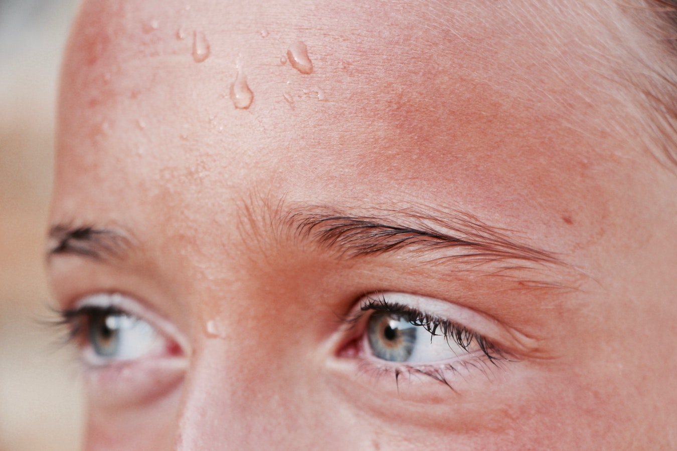
Combine exercise and a hot day, and you get sweat — and lots of it. Sweating is one of the adaptations humans have evolved to maintain homeostasis, or a constant internal environment. When sweat evaporates from the skin, it uses up some of the excess heat energy on the skin, thus helping to reduce the body's temperature. Humans are among the sweatiest of all species, with a fine-tuned ability to maintain a steady internal temperature, even at very high outside temperatures.
Unifying Principles of Biology
All living things have mechanisms for homeostasis. Homeostasis is one of four basic principles or theories that explain the structure and function of all species (including our own). Whether biologists are interested in ancient life, the life of bacteria, or how humans could live on Mars, they base their understanding of biology on these unifying principles:
Cell Theory
According to cell theory, all living things are made of cells, and living cells come only from other living cells. Each living thing begins life as a single cell. Some living things, including bacteria, remain single-celled. Other living things, including plants and animals, grow and develop into many cells. Your own body is made up of an amazing 100 trillion cells. But even you — like all other living things — began life as a single cell.
Watch this TED-Ed video about the origin of cell theory:
https://www.youtube.com/watch?v=4OpBylwH9DU
The Wacky History of Cell Theory - Lauren Royal-Woods, TED-Ed, 2012
Gene Theory
Gene theory is the idea that the characteristics of living things are controlled by genes, which are passed from parents to their offspring. Genes are located on larger structures called chromosomes. Chromosomes are found inside every cell, and they consist of molecules of DNA (deoxyribonucleic acid). Those molecules of DNA are encoded with instructions that "tell" cells how to behave.
Homeostasis
Homeostasis, or the condition in which a system is maintained in a more-or-less steady state, is a characteristic of individual living things, like the human ability to sweat. Homeostasis also applies to the entire biosphere, wherever life is found on Earth. Consider the concentration of oxygen in Earth's atmosphere. Oxygen makes up 21 per cent of the atmosphere, and this concentration is fairly constant. What maintains this homeostasis in the atmosphere? The answer is living things.
Most living things need oxygen to survive, so they remove oxygen from the air. On the other hand, many living things, including plants, give off oxygen when they convert carbon dioxide and water to food in the process of photosynthesis. These two processes balance out so the air maintains a constant level of oxygen.
Evolutionary Theory
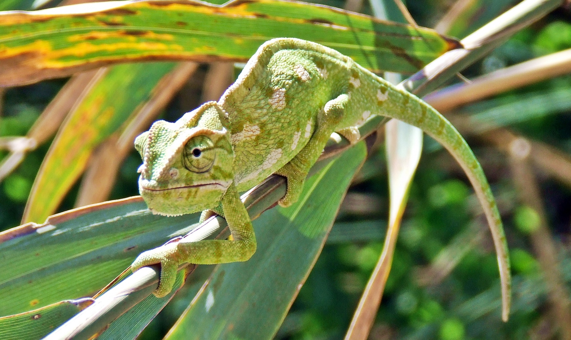
Evolution is a change in the characteristics of populations of living things over time. Evolution can occur by a process called natural selection, which results from random genetic mutations in a population. If these mutations lead to changes that allow the living things to better survive, then their chances of surviving and reproducing in a given environment increase. They will then pass more genes to the next generation. Over many generations, this can lead to major changes in the characteristics of those living things. Evolution explains how living things are changing today, as well as how modern living things descended from ancient life forms that no longer exist on Earth.
Traits that help living things survive and reproduce in a given environment are called adaptations. You can see an obvious adaptation in the image below. The chameleon is famous for its ability to change its colour to match its background as camouflage. Using camouflage, the chameleon can hide in plain sight.
Feature: Myth vs. Reality
Misconceptions about evolution are common. They include the following myths:
Myth |
Reality |
| "Evolution is "just" a theory or educated guess." | Scientists accept evolutionary theory as the best explanation for the diversity of life on Earth because of the large body of scientific evidence supporting it. Like any scientific theory, evolution is a broad, evidence-supported explanation for multiple phenomena. |
| "The theory of evolution explains how life on Earth began." | The theory of evolution explains how life changed on Earth after it began. |
| "The theory of evolution means that humans evolved from apes like those in zoos." | Humans and modern apes both evolved from a common ape-like ancestor millions of years ago. |
2.3 Summary
- Four basic principles or theories unify all fields of biology: cell theory, gene theory, homeostasis, and evolutionary theory.
- According to cell theory, all living things are made of cells and come from other living cells.
- Gene theory states that the characteristics of living things are controlled by genes that pass from parents to offspring.
- All living things strive to maintain internal balance, or homeostasis.
- The characteristics of populations of living things change over time through the process of micro-evolution as organisms acquire adaptations, or traits that better suit them to a given environment.
Use the flashcards below to review the four principles:
2.3 Review Questions
-
- How does sweating help the human body maintain homeostasis?
- Explain cell theory and gene theory.
- Describe an example of homeostasis in the atmosphere.
- Describe how you can apply the concepts of evolution,natural selection, adaptation, and homeostasis to the human ability to sweat.
- Which of the four unifying principles of biology is primarily concerned with:
- how DNA is passed down to offspring?
- how internal balance is maintained?
- _____________ are located on ______________.
- chromosomes; genes
- genes;chromosomes
- genes; traits
- none of the above
- Define an adaptation and give one example.
- Explain how gene theory and evolutionary theory relate to each other.
- Does evolution by natural selection occur within one generation? Why or why not?
- Explain why you think chameleons evolved the ability to change their colour to match their background, as well as how natural selection may have acted on the ancestors of chameleons to produce this adaptation.
2.3 Explore More
https://www.youtube.com/watch?v=Wg5DBH6uMCw&feature=emb_logo
Myths and misconceptions about evolution - Alex Gendler, TEDEd, 2013
Attributions
Figure 2.3.1
Photo(perspiration), by Hans Reniers on Unsplash. is used under the Unsplash license (https://unsplash.com/license).
Figure 2.3.2
Mediterranean Chameleon Reptile Lizard, by user:1588877 on Pixabay, is used under the Pixabay license (https://pixabay.com/de/service/license/).
References
TED-Ed. (2012, June 4). The wacky history of cell theory - Lauren Royal-Woods. YouTube. https://www.youtube.com/watch?v=4OpBylwH9DU&feature=youtu.be
TED-Ed. (2013, July 8). Myths and misconceptions about evolution - Alex Gendler. YouTube. https://www.youtube.com/watch?v=mZt1Gn0R22Q&t=10s
Created by CK-12/Adapted by Christine Miller
Why Are Humans Such Sweaty Animals?

Combine exercise and a hot day, and you get sweat — and lots of it. Sweating is one of the adaptations humans have evolved to maintain homeostasis, or a constant internal environment. When sweat evaporates from the skin, it uses up some of the excess heat energy on the skin, thus helping to reduce the body's temperature. Humans are among the sweatiest of all species, with a fine-tuned ability to maintain a steady internal temperature, even at very high outside temperatures.
Unifying Principles of Biology
All living things have mechanisms for homeostasis. Homeostasis is one of four basic principles or theories that explain the structure and function of all species (including our own). Whether biologists are interested in ancient life, the life of bacteria, or how humans could live on Mars, they base their understanding of biology on these unifying principles:
Cell Theory
According to cell theory, all living things are made of cells, and living cells come only from other living cells. Each living thing begins life as a single cell. Some living things, including bacteria, remain single-celled. Other living things, including plants and animals, grow and develop into many cells. Your own body is made up of an amazing 100 trillion cells. But even you — like all other living things — began life as a single cell.
Watch this TED-Ed video about the origin of cell theory:
https://www.youtube.com/watch?v=4OpBylwH9DU
The Wacky History of Cell Theory - Lauren Royal-Woods, TED-Ed, 2012
Gene Theory
Gene theory is the idea that the characteristics of living things are controlled by genes, which are passed from parents to their offspring. Genes are located on larger structures called chromosomes. Chromosomes are found inside every cell, and they consist of molecules of DNA (deoxyribonucleic acid). Those molecules of DNA are encoded with instructions that "tell" cells how to behave.
Homeostasis
Homeostasis, or the condition in which a system is maintained in a more-or-less steady state, is a characteristic of individual living things, like the human ability to sweat. Homeostasis also applies to the entire biosphere, wherever life is found on Earth. Consider the concentration of oxygen in Earth's atmosphere. Oxygen makes up 21 per cent of the atmosphere, and this concentration is fairly constant. What maintains this homeostasis in the atmosphere? The answer is living things.
Most living things need oxygen to survive, so they remove oxygen from the air. On the other hand, many living things, including plants, give off oxygen when they convert carbon dioxide and water to food in the process of photosynthesis. These two processes balance out so the air maintains a constant level of oxygen.
Evolutionary Theory

Evolution is a change in the characteristics of populations of living things over time. Evolution can occur by a process called natural selection, which results from random genetic mutations in a population. If these mutations lead to changes that allow the living things to better survive, then their chances of surviving and reproducing in a given environment increase. They will then pass more genes to the next generation. Over many generations, this can lead to major changes in the characteristics of those living things. Evolution explains how living things are changing today, as well as how modern living things descended from ancient life forms that no longer exist on Earth.
Traits that help living things survive and reproduce in a given environment are called adaptations. You can see an obvious adaptation in the image below. The chameleon is famous for its ability to change its colour to match its background as camouflage. Using camouflage, the chameleon can hide in plain sight.
Feature: Myth vs. Reality
Misconceptions about evolution are common. They include the following myths:
Myth |
Reality |
| "Evolution is "just" a theory or educated guess." | Scientists accept evolutionary theory as the best explanation for the diversity of life on Earth because of the large body of scientific evidence supporting it. Like any scientific theory, evolution is a broad, evidence-supported explanation for multiple phenomena. |
| "The theory of evolution explains how life on Earth began." | The theory of evolution explains how life changed on Earth after it began. |
| "The theory of evolution means that humans evolved from apes like those in zoos." | Humans and modern apes both evolved from a common ape-like ancestor millions of years ago. |
2.3 Summary
- Four basic principles or theories unify all fields of biology: cell theory, gene theory, homeostasis, and evolutionary theory.
- According to cell theory, all living things are made of cells and come from other living cells.
- Gene theory states that the characteristics of living things are controlled by genes that pass from parents to offspring.
- All living things strive to maintain internal balance, or homeostasis.
- The characteristics of populations of living things change over time through the process of micro-evolution as organisms acquire adaptations, or traits that better suit them to a given environment.
Use the flashcards below to review the four principles:
2.3 Review Questions
-
- How does sweating help the human body maintain homeostasis?
- Explain cell theory and gene theory.
- Describe an example of homeostasis in the atmosphere.
- Describe how you can apply the concepts of evolution,natural selection, adaptation, and homeostasis to the human ability to sweat.
- Which of the four unifying principles of biology is primarily concerned with:
- how DNA is passed down to offspring?
- how internal balance is maintained?
- _____________ are located on ______________.
- chromosomes; genes
- genes;chromosomes
- genes; traits
- none of the above
- Define an adaptation and give one example.
- Explain how gene theory and evolutionary theory relate to each other.
- Does evolution by natural selection occur within one generation? Why or why not?
- Explain why you think chameleons evolved the ability to change their colour to match their background, as well as how natural selection may have acted on the ancestors of chameleons to produce this adaptation.
2.3 Explore More
https://www.youtube.com/watch?v=Wg5DBH6uMCw&feature=emb_logo
Myths and misconceptions about evolution - Alex Gendler, TEDEd, 2013
Attributions
Figure 2.3.1
Photo(perspiration), by Hans Reniers on Unsplash. is used under the Unsplash license (https://unsplash.com/license).
Figure 2.3.2
Mediterranean Chameleon Reptile Lizard, by user:1588877 on Pixabay, is used under the Pixabay license (https://pixabay.com/de/service/license/).
References
TED-Ed. (2012, June 4). The wacky history of cell theory - Lauren Royal-Woods. YouTube. https://www.youtube.com/watch?v=4OpBylwH9DU&feature=youtu.be
TED-Ed. (2013, July 8). Myths and misconceptions about evolution - Alex Gendler. YouTube. https://www.youtube.com/watch?v=mZt1Gn0R22Q&t=10s
Created by CK-12/Adapted by Christine Miller
So Many Species!
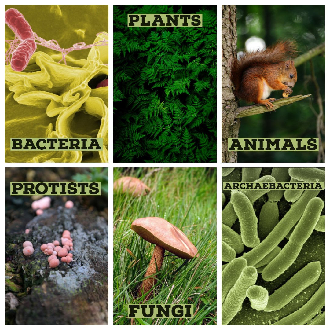
The collage shows a single species in each of the six kingdoms into which all of Earth's living things are commonly classified. How many species are there in each kingdom? In a word: millions. A total of almost two million living species have already been identified, and new species are being discovered all the time. Scientists estimate that there may be as many as 30 million unique species alive on Earth today! Clearly, there is a tremendous variety of life on Earth.
What Is Biodiversity?
Biological diversity, or biodiversity, refers to all of the variety of life that exists on Earth. Biodiversity can be described and measured at three different levels: species diversity, genetic diversity, and ecosystem diversity.
- Species diversity refers to the number of different species in an ecosystem or on Earth as a whole. This is the most common way to measure biodiversity. Current estimates for Earth's total number of living species range from 5 to 30 million species.
- Genetic diversity refers to the variation in genes within all of these species.
- Ecosystem diversity refers to the variety of ecosystems on Earth. An ecosystem is a system formed by populations of many different species interacting with each other and their environment.
https://www.youtube.com/watch?v=GK_vRtHJZu4
Why is Biodiversity So Important? - Kim Preshoff, TEDEd, 2015
Defining a Species
Biodiversity is most often measured by counting species, but what is a species? The answer to that question is not as straightforward as you might think. Formally, a species is defined as a group of actually or potentially interbreeding organisms. This means that members of the same species are similar enough to each other to produce fertile offspring together. By this definition of species, all human beings alive today belong to one species, Homo sapiens. All humans can potentially interbreed with each other, but not with members of any other species.
In the real world, it isn't always possible to make the observations necessary to determine whether or not different organisms can interbreed. For one thing, many species reproduce asexually, so individuals never interbreed — even with members of their own species. When studying extinct species represented by fossils, it is usually impossible to know if different organisms could interbreed. Keep in mind that 99 per cent of all species that have ever existed are now extinct! In practice, many biologists and virtually all paleontologists generally define species on the basis of morphology, rather than breeding behavior. Morphology refers to the form and structure of organisms. For classification purposes, it generally refers to relatively obvious physical traits. Typically, the more similar to one another different organisms appear, the greater the chance that they will be classified in the same species.
Classifying Living Things
People have been trying to classify the tremendous diversity of life on Earth for more than two thousand years. The science of classifying organisms is called taxonomy. Classification is an important step in understanding the present diversity and past evolutionary history of life on Earth. It helps us make sense of the overwhelming diversity of living things.
Linnaean Classification
All modern classification systems have their roots in the Linnaean classification system, which was developed by Swedish botanist Carolus Linnaeus in the 1700s. He tried to classify all living things known in his time by grouping together organisms that s
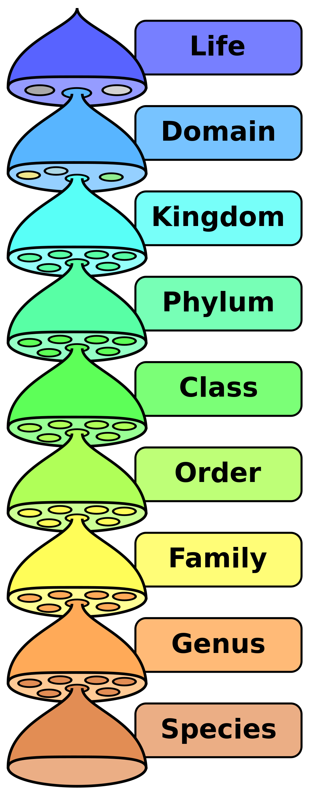
hared obvious morphological traits, such as number of legs or shape of leaves. For his contribution, Linnaeus is known as the “father of taxonomy.”
The Linnaean system of classification consists of a hierarchy of groupings, called taxa (singular, taxon). In the original system, taxa ranged from the kingdom to the species. The kingdom (ex. plant kingdom, animal kingdom) is the largest and most inclusive grouping. It consists of organisms that share just a few basic similarities. The species is the smallest and most exclusive grouping. Ideally, it consists of organisms that are similar enough to interbreed, as discussed above. Similar species are classified together in the same genus (plural, genera), then similar genera are classified together in the same family, and so on, all the way up to the kingdom.
A phrase to help you remember the order of the groupings is shown below. The first letter of each word is the first letter of the level of classification.
Dad Keeps Pots Clean Or Family Gets Sick
The hierarchy of taxa in the original Linnaean system of taxonomy included taxa from the species to the kingdom. The domain was added later.
Binomial Nomenclature
Perhaps the single greatest contribution Linnaeus made to science was his method of naming species. This method, called binomial nomenclature, gives each species a unique, two-word Latin name consisting of the genus name followed by a specific species identifier. An example is Homo sapiens, the two-word Latin name for humans. It literally means “wise human.” This is a reference to our big brains.
Why is having two names so important? It is similar to people having a first and a last name. You may know several people with the first name Michael, but adding Michael’s last name usually pins down exactly which Michael you mean. In the same way, having two names for a species helps to uniquely identify it.
Revisions in the Linnaean Classification
Linnaeus published his classification system in the 1700s. Since then, many new species have been discovered. Scientists can also now classify organisms on the basis of their biochemical and genetic similarities and differences, and not just their outward morphology. These changes have led to revisions in the original Linnaean system of classification.
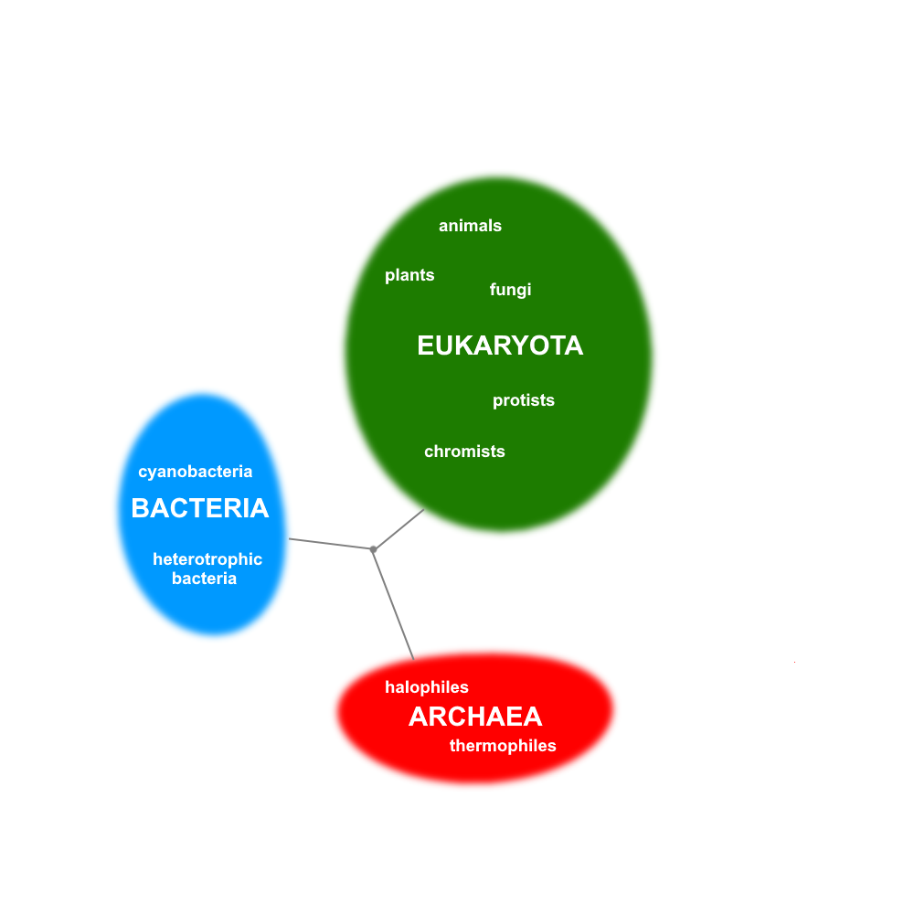
A major change to the Linnaean system is the addition of a new taxon called the domain. The domain is a taxon that is larger and more inclusive than the kingdom, as shown in the figure above. Most biologists agree that there are three domains of life on Earth: Bacteria, Archaea, and Eukarya . Both the Bacteria and the Archaea domains consist of single-celled organisms that lack a nucleus. This means that their genetic material is not enclosed within a membrane inside the cell. The Eukarya domain, in contrast, consists of all organisms whose cells do have a nucleus, so that their genetic material is enclosed within a membrane inside the cell. The Eukarya domain is made up of both single-celled and multicellular organisms. This domain includes several kingdoms, including the animal, plant, fungus, and protist kingdoms.
The three domains of life, as well as how they are related to each other and to a common ancestor. There are several theories about how the three domains are related and which arose first, or from another.
Phylogenetic Classification
Linnaeus classified organisms based on morphology. Basically, organisms were grouped together if they looked alike. After Darwin published his theory of evolution in the 1800s, scientists looked for a way to classify organisms that accounted for phylogeny. Phylogeny is the evolutionary history of a group of related organisms. It is represented by a phylogenetic tree, or some other tree-like diagram, like the one shown above to illustrate the three domains. A phylogenetic tree shows how closely related different groups of organisms are to one another. Each branching point represents a common ancestor of the branching groups.
2.4 Summary
- Biodiversity refers to the variety of life that exists on Earth. It includes species diversity, genetic diversity (within species), and ecosystem diversity.
- The formal biological definition of species is a group of actually or potentially interbreeding organisms. Our own species, Homo sapiens,is an example. In reality, organisms are often classified into species on the basis of morphology.
- A system for classifying living things was introduced by Linnaeus in the 1700s. It includes taxa from the species (least inclusive) to the kingdom (most inclusive). Linnaeus also introduced a system of naming species, which is called binomial nomenclature.
- The domain — a taxon higher than the kingdom — was later added to the Linnaean system. Living things are generally grouped into three domains: Bacteria, Archaea, and Eukarya. The human species and other animal species are placed in the Eukarya domain.
- Modern systems of classification take into account phylogenies, or evolutionary histories of related organisms, rather than just morphological similarities and differences. These relationships are often represented by phylogenetic trees or other tree-like diagrams
2.4 Review Questions
- What is biodiversity? Identify three ways that biodiversity may be measured.
- Define biological species. Why is this definition often difficult to apply?
- Explain why it is important to classify living things, and outline the Linnaean system of classification.
- What is binomial nomenclature? Give an example.
-
- Contrast the Linnaean and phylogenetic systems of classification.
- Describe the taxon called the domain, and compare the three widely recognized domains of living things.
- Based on the phylogenetic tree for the three domains of life above, explain whether you think Bacteria are more closely related to Archaea or Eukarya.
- A scientist discovers a new single-celled organism. Answer the following questions about this discovery.
- If this is all you know, can you place the organism into a particular domain? If so, what is the domain? If not, why not?
- What is one type of information that could help the scientist classify the organism?
- Define morphology. Give an example of a morphological trait in humans.
- Which type of biodiversity is represented in the differences between humans?
- Why do you think it is important to the definition of a species that members of a species can produce fertile offspring?
- Go to the A-Z Animals Animal Classification Page. In the search box, put in your favorite animal and write out it's classification.
2.4 Explore More
https://youtu.be/DVouQRAKxYo
Classification, Amoeba Sisters, 2013.
Attributions
Figure 2.4.1 (6 Kingdoms collage)
- Salmonella, by unknown/ NIAID on Wikimedia Commons is in the public domain (https://en.wikipedia.org/wiki/Public_domain).
- Fern from pxhere, is used under a CC0 1.0 universal public domain dedication license (https://creativecommons.org/publicdomain/zero/1.0/deed.en).
- Photo [squirrel] , by Radoslaw Prekurat on Unsplash is used under the Unsplash License (https://unsplash.com/license).
- Blood Milk Mushroom by Hans on Pixabay is used under the Pixabay License (https://pixabay.com/de/service/license/).
- Fungi by Ste Wright on Unsplash is used under the Unsplash License (https://unsplash.com/license).
- EscherichiaColi NIAID [adapted], by Rocky Mountain Laboratories, ca:NIAID, ca:NIH on Wikimedia Commons is in the public domain (https://en.wikipedia.org/wiki/Public_domain).
Figure 2.4.2
Biological classification, by Pengo [Peter Halasz] on Wikimedia Commons is in the public domain (https://en.wikipedia.org/wiki/Public_domain).
Figure 2.4.3
The three domains of life and major groups within, by C. Miller, 2019, is in the public domain (https://en.wikipedia.org/wiki/Public_domain).
References
Amoeba Sisters. (2017, March 8). Classification. YouTube. https://www.youtube.com/watch?v=DVouQRAKxYo&feature=youtu.be
A-Z Animals. (2008, December 1). Animal classification. https://a-z-animals.com/reference/animal-classification/
TED-Ed. (2015, April 20). Why is biodiversity so important? - Kim Preshoff. YouTube. https://www.youtube.com/watch?v=GK_vRtHJZu4
Wikipedia contributors. (2020, June 21). Carl Linnaeus. Wikipedia. https://en.wikipedia.org/w/index.php?title=Carl_Linnaeus&oldid=963767022
Image shows a squirrel monkey perching in the branches of a tree.
Created by CK-12 Foundation/Adapted by Christine Miller
Oxygen Bar
Belly up to the bar and get your favorite... oxygen? That’s right — in some cities, you can get a shot of pure oxygen, with or without your choice of added flavors. Bar patrons inhale oxygen through a plastic tube inserted into their nostrils, paying up to a dollar per minute to inhale the pure gas. Proponents of the practice claim that breathing in extra oxygen will remove toxins from the body, strengthen the immune system, enhance concentration and alertness, increase energy, and even cure cancer! These claims, however, have not been substantiated by controlled scientific studies. Normally, blood leaving the lungs is almost completely saturated with oxygen, even without the use of extra oxygen, so it’s unlikely that a higher concentration of oxygen in air inside the lungs would lead to significantly greater oxygenation of the blood. Oxygen enters the blood in the lungs as part of the process of gas exchange.
What is Gas Exchange?
Gas exchange is the biological process through which gases are transferred across cell membranes to either enter or leave the blood. Oxygen is constantly needed by cells for aerobic cellular respiration, and the same process continually produces carbon dioxide as a waste product. Gas exchange takes place between the blood and cells throughout the body, with oxygen leaving the blood and entering the cells, and carbon dioxide leaving the cells and entering the blood. Gas exchange also takes place between the blood and the air in the lungs, with oxygen entering the blood from the inhaled air inside the lungs, and carbon dioxide leaving the blood and entering the air to be exhaled from the lungs.
Gas Exchange in the Lungs
Alveoli are the basic functional units of the lungs where gas exchange takes place between the air and the blood. Alveoli (singular, alveolus) are tiny air sacs that consist of connective and epithelial tissues. The connective tissue includes elastic fibres that allow alveoli to stretch and expand as they fill with air during inhalation. During exhalation, the fibres allow the alveoli to spring back and expel the air. Special cells in the walls of the alveoli secrete a film of fatty substances called surfactant. This substance prevents the alveolar walls from collapsing and sticking together when air is expelled. Other cells in alveoli include macrophages, which are mobile scavengers that engulf and destroy foreign particles that manage to reach the lungs in inhaled air.
As shown in Figure 13.4.2, alveoli are arranged in groups like clusters of grapes. Each alveolus is covered with epithelium that is just one cell thick. It is surrounded by a bed of pulmonary capillaries, each of which has a wall of epithelium just one cell thick. As a result, gases must cross through only two cells to pass between an alveolus and its surrounding capillaries.
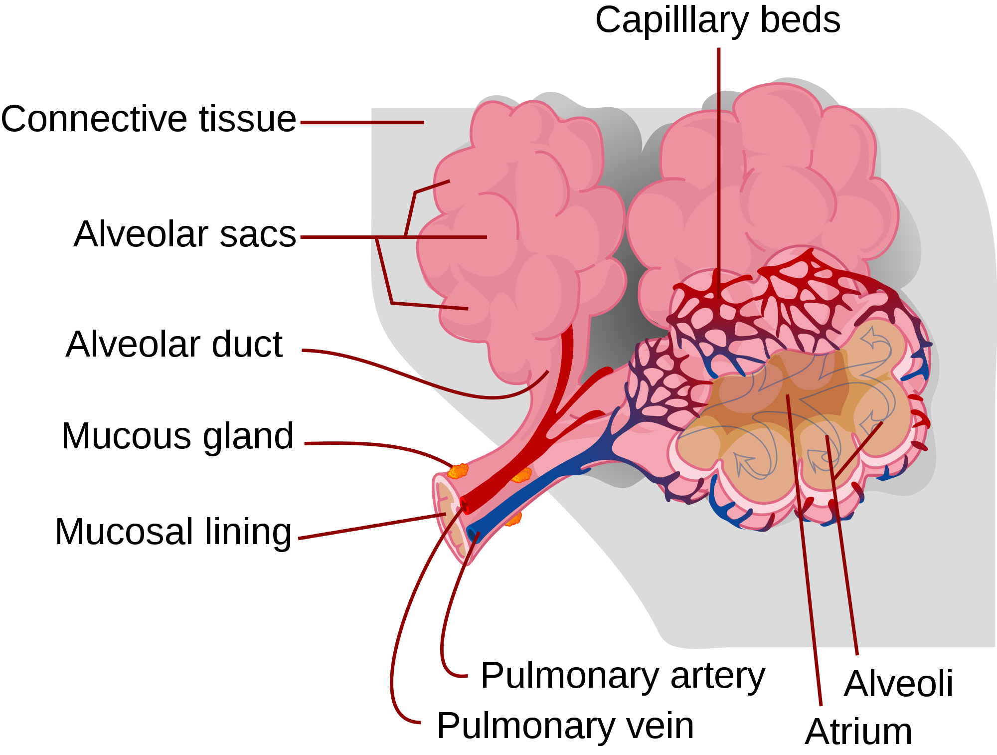
The pulmonary artery (also shown in Figure 13.4.2) carries deoxygenated blood from the heart to the lungs. Then, the blood travels through the pulmonary capillary beds, where it picks up oxygen and releases carbon dioxide. The oxygenated blood then leaves the lungs and travels back to the heart through pulmonary veins. There are four pulmonary veins (two for each lung), and all four carry oxygenated blood to the heart. From the heart, the oxygenated blood is then pumped to cells throughout the body.
Mechanism of Gas Exchange
Gas exchange occurs by diffusion across cell membranes. Gas molecules naturally move down a concentration gradient from an area of higher concentration to an area of lower concentration. This is a passive process that requires no energy. To diffuse across cell membranes, gases must first be dissolved in a liquid. Oxygen and carbon dioxide are transported around the body dissolved in blood. Both gases bind to the protein hemoglobin in red blood cells, although oxygen does so more effectively than carbon dioxide. Some carbon dioxide also dissolves in blood plasma.
As shown in Figure 13.4.3, oxygen in inhaled air diffuses into a pulmonary capillary from the alveolus. Carbon dioxide in the blood diffuses in the opposite direction. The carbon dioxide can then be exhaled from the body.
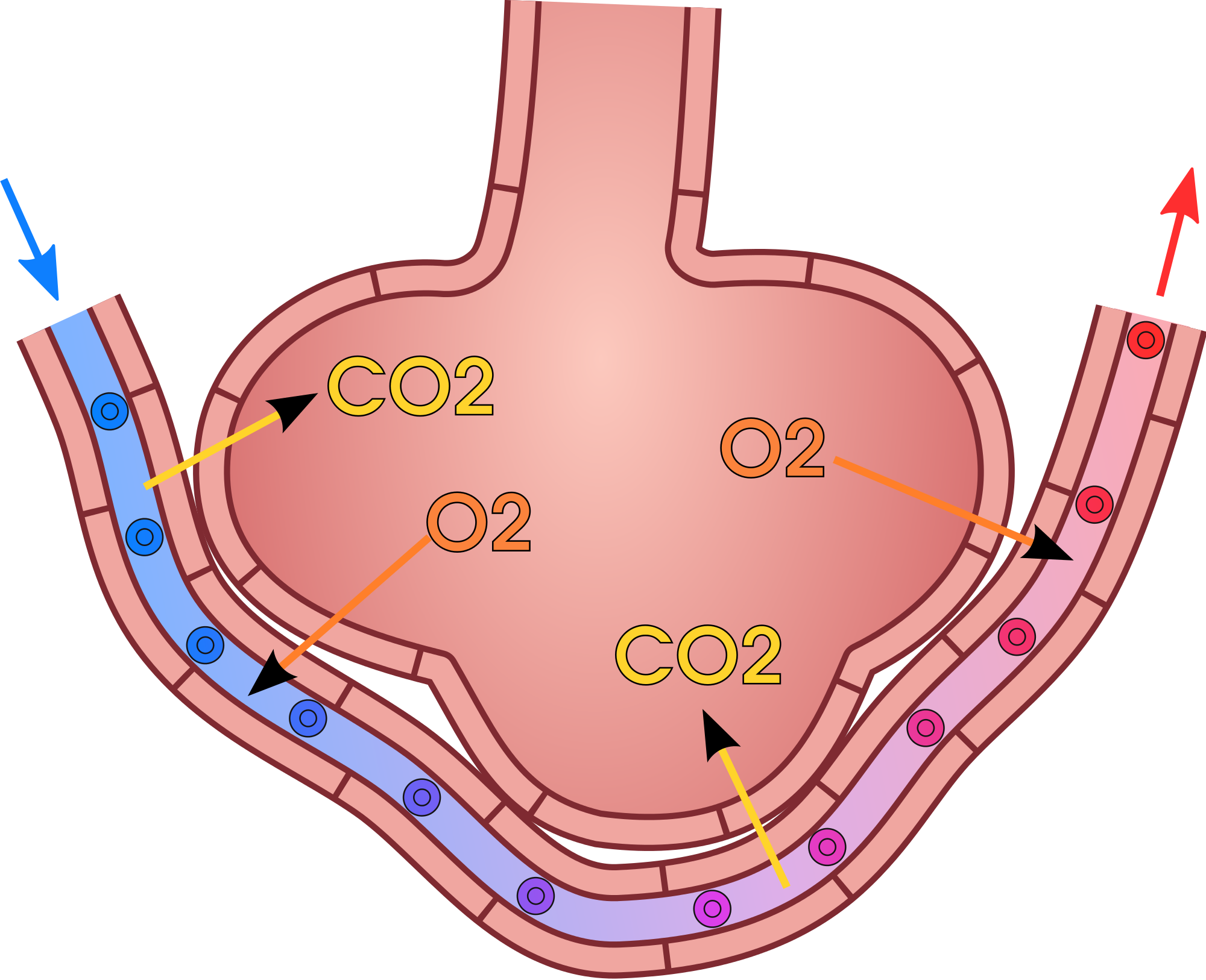
Gas exchange by diffusion depends on having a large surface area through which gases can pass. Although each alveolus is tiny, there are hundreds of millions of them in the lungs of a healthy adult, so the total surface area for gas exchange is huge. It is estimated that this surface area may be as great as 100 m2 (or approximately 1,076 ft²). Often we think of lungs as balloons, but this type of structure would have very limited surface area and there wouldn't be enough space for blood to interface with the air in the alveoli. The structure alveoli take in the lungs is more like a giant mass of soap bubbles — millions of tiny little chambers making up one large mass — this is what increases surface area giving blood lots of space to come into close enough contact to exchange gases by diffusion.
Gas exchange by diffusion also depends on maintaining a steep concentration gradient for oxygen and carbon dioxide. Continuous blood flow in the capillaries and constant breathing maintain this gradient.
- Each time you inhale, there is a greater concentration of oxygen in the air in the alveoli than there is in the blood in the pulmonary capillaries. As a result, oxygen diffuses from the air inside the alveoli into the blood in the capillaries. Carbon dioxide, in contrast, is more concentrated in the blood in the pulmonary capillaries than it is in the air inside the alveoli. As a result, carbon dioxide diffuses in the opposite direction.
- The cells of the body have a much lower concentration of oxygen than does the oxygenated blood that reaches them in peripheral capillaries, which are the capillaries that supply tissues throughout the body. As a result, oxygen diffuses from the peripheral capillaries into body cells. The opposite is true of carbon dioxide. It has a much higher concentration in body cells than it does in the blood of the peripheral capillaries. Thus, carbon dioxide diffuses from body cells into the peripheral capillaries.
13.4 Summary
- Gas exchange is the biological process through which gases are transferred across cell membranes to either enter or leave the blood. Gas exchange takes place continuously between the blood and cells throughout the body, and also between the blood and the air inside the lungs.
- Gas exchange in the lungs takes place in alveoli, which are tiny air sacs surrounded by networks of capillaries. The pulmonary artery carries deoxygenated blood from the heart to the lungs, where it travels through pulmonary capillaries, picking up oxygen and releasing carbon dioxide. The oxygenated blood then leaves the lungs through pulmonary veins.
- Gas exchange occurs by diffusion across cell membranes. Gas molecules naturally move down a concentration gradient from an area of higher concentration to an area of lower concentration. This is a passive process that requires no energy.
- Gas exchange by diffusion depends on the large surface area provided by the hundreds of millions of alveoli in the lungs. It also depends on a steep concentration gradient for oxygen and carbon dioxide. This gradient is maintained by continuous blood flow and constant breathing.
13.4 Review Questions
- What is gas exchange?
- Summarize the flow of blood into and out of the lungs for gas exchange.
-
- Describe the mechanism by which gas exchange takes place.
- Identify the two main factors upon which gas exchange by diffusion depends.
- If the concentration of oxygen were higher inside of a cell than outside of it, which way would the oxygen flow? Explain your answer.
- Why is it important that the walls of the alveoli are only one cell thick?
13.4 Explore More
https://www.youtube.com/watch?v=nRpwdwm06Ic&feature=emb_logo
Oxygen movement from alveoli to capillaries | NCLEX-RN | Khan Academy, khanacademymedicine, 2013.
https://www.youtube.com/watch?v=KmgIqVwytwA&feature=emb_logo
About Carbon Monoxide and Carbon Monoxide Poisoning, EMDPrepare, 2009.
https://www.youtube.com/watch?v=GVU_zANtroE
Oxygen’s surprisingly complex journey through your body - Enda Butler, TED-Ed, 2017.
Attributions
Figure 13.4.1
Oxygen Bar by Farrukh on Flickr is used under a CC BY-NC 2.0 (https://creativecommons.org/licenses/by-nc/2.0/) license.
Figure 13.4.2
Alveolus_diagram.svg by Mariana Ruiz Villarreal [LadyofHats] on Wikimedia Commons is released into the public domain (https://en.wikipedia.org/wiki/Public_domain).
Figure 13.4.3
Gas_exchange_in_the_aveolus.svg by domdomegg on Wikimedia Commons is used under a CC BY 4.0 (https://creativecommons.org/licenses/by/4.0) license.
References
EMDPrepare. (2009, December 21). About carbon monoxide and carbon monoxide poisoning. YouTube. https://www.youtube.com/watch?v=KmgIqVwytwA&feature=youtu.be
khanacademymedicine. (2013, February 25). Oxygen movement from alveoli to capillaries | NCLEX-RN | Khan Academy. YouTube. https://www.youtube.com/watch?v=nRpwdwm06Ic&feature=youtu.be
TED-Ed. (2017, April 13). Oxygen’s surprisingly complex journey through your body - Enda Butler. YouTube. https://www.youtube.com/watch?v=GVU_zANtroE&feature=youtu.be
Turkana Boy, also called Nariokotome Boy, is the name given to fossil KNM-WT 15000, a nearly complete skeleton of a Homo erectus (Homo ergaster) youth who lived at c. 1.5 to 1.6 million years ago. This specimen is the most complete early human skeleton ever found.
bony “cage” enclosing the thoracic cavity and consisting of the ribs, thoracic vertebrae, and sternum
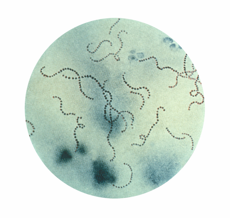
As you may recall from the beginning of the chapter, Lanying’s strep throat was caused by Streptococcus pyogenes bacteria, the species shown in the photomicrograph in Figure 2.6.1. She took antibiotics to treat the S. pyogenes infection, but this also affected her “good” bacteria, throwing off the balance of microorganisms living inside her and resulting in diarrhea and a yeast infection.
After reading this chapter, you should know that microorganisms such as bacteria and yeast that live in humans are also similar to us in many ways. They are living organisms, so we share the traits of homeostasis, organization, metabolism, growth, adaptation, response to stimuli, and reproduction. Like us, microorganisms contain genes, consist of cells, and have the ability to evolve. Lanying’s beneficial gut bacteria help digest her food as part of their metabolic processes. Lanying got a yeast infection likely because the growth and reproductive rates of the yeast living on her body were not held in check by beneficial bacteria after she took the antibiotics. You can see there are many ways in which an understanding of the basic characteristics of all life can directly apply to your own.
You also learned how living organisms are classified, from bacteria that are in the Bacteria domain, to yeast (fungus kingdom) and humans (animal kingdom) which are both in the Eukarya domain. You probably now recognize that Streptococcus pyogenes is the binomial nomenclature for this species, and the fact that Streptococcus refers to the genus name.
As Lanying’s doctor told her, there are many different species of microorganisms living in the human digestive system. You should recognize this type of biodiversity is called species diversity. This diversity is maintained in a balance, or homeostasis, that can be upset when one type of organism is killed, for instance, by antibiotics.
Lanying’s doctor advised her to complete the entire course of antibiotics because stopping too early would kill or inhibit the bacteria that are most susceptible to the antibiotic, while leaving the bacteria that are more resistant to the antibiotic alive. This difference in susceptibility to antibiotics is an example of genetic diversity. Over time, the surviving antibiotic-resistant bacteria will have increased survival and reproductive rates compared to the more susceptible bacteria, and the trait of antibiotic resistance will become more common in the population. In this way, bacteria can evolve and become better adapted to its environment — at a major cost to our health, because our antibiotics will no longer be effective! Improper use of antibiotics leading to increased antibiotic resistance is an issue of major concern to public health experts.

Lanying's doctor suggested that she also take some probiotics — food or supplements that contain "good microorganisms." These good bacteria help our bodies digest food and keep out "bad microorganisms." Specifically, lactobacillus acidophilus could help combat her yeast infection and help restore normal gut function.
After reading this chapter, you know how humans are classified, and you've learned some characteristics of humans and other closely related species. Beyond our more obvious features of big brains, intelligence, and the ability to walk upright, we also serve as a home to many different microorganisms that may be invisible to the naked eye but play a big role in maintaining our health.
Chapter 2 Summary
In this chapter, you learned about the basic principles of biology and how humans are situated among other living organisms. Specifically, you learned:
- To be classified as a living thing, most scientists agree that an object must exhibit seven characteristics, including:
- Maintaining a more-or-less constant internal environment, which is called homeostasis.
- Having multiple levels of organization and consisting of one or more cells.
- Using energy and being capable of metabolism.
- Being able to grow and develop.
- Being capable of evolving adaptations to the environment.
- Being able to detect and respond to environmental stimuli.
- Being capable of reproducing, which is the process by which living things give rise to offspring.
- Four basic principles or theories unify all the fields of biology: cell theory, gene theory, homeostasis, and evolutionary theory.
- According to cell theory, all living things are made of cells and come from other living cells.
- Gene theory states that the characteristics of living things are controlled by genes that pass from parents to offspring.
- All living things — and even the entire biosphere — strive to maintain homeostasis.
- The characteristics of living things change over time as they evolve, and some acquire adaptations or traits that better suit them to a given environment.
- Biodiversity refers to the variety of life that exists on Earth. It includes species diversity, genetic diversity within species, and ecosystem diversity.
- The formal biological definition of "species" is a group of actually or potentially interbreeding organisms. In reality, organisms are often classified into species on the basis of morphology.
- A system for classifying living things was introduced by Linnaeus in the 1700s. It includes taxa from the species (least inclusive) to the kingdom (most inclusive). Linnaeus also introduced a system of naming species, called binomial nomenclature.
- The domain, a taxon higher than the kingdom, was later added to the Linnaean system. Living things are generally grouped into three domains: Bacteria, Archaea, and Eukarya. Humans and other animal species are placed in the Eukarya domain.
- Modern systems of classification take into account phylogenies, or evolutionary histories of related organisms, rather than just morphological similarities and differences. These relationships are often represented by phylogenetic trees or other tree-like diagrams.
- The human species, Homo sapiens, is placed in the primate order of the class of mammals, which are chordates in the animal kingdom.
- Traits that humans share with other primates include: five digits with nails and opposable thumbs; an excellent sense of vision, including stereoscopic vision and the ability to see in colour; and a large brain, high degree of intelligence, and complex behaviors. Like most other primates, we also live in social groups. Many of our primate traits are adaptations to life in the trees.
- Within the primate order, our species is placed in the hominid family, which also includes chimpanzees, gorillas, and orangutans.
- The genus Homo first evolved about 2.8 million years ago. Early Homo species were fully bipedal but had small brains. All are now extinct.
- During the last 800 thousand years, Homo sapiens evolved, with smaller faces, jaws, and front teeth, but much bigger brains than earlier Homo species.
Now you understand the basic principles of biology and some of the characteristics of living organisms. In the next chapter, you will learn about the molecules that make up living organisms, as well as the chemistry that allows organisms to exist and function.
Chapter 2 Review
- What are the four basic unifying principles of biology?
- A scientist is exploring in a remote area with many unidentified species. He finds an unknown object that does not appear to be living. What is one way he could tell whether it is a dead organism that was once alive or an inanimate object that was never living?
- Cows are dependent on bacteria living in their digestive systems to help break down cellulose in the plant material that they eat. Explain what characteristics these bacteria must have to be considered living organisms themselves (and not just part of the cow).
- What is the basic unit of structure and function in living things?
- Give one example of homeostasis that occurs in humans.
- Can a living thing exist without using energy? Why or why not?
-
-
- Give an example of a response to stimuli that occurs in a unicellular organism.
- A scientist discovers two types of similar looking insects that have not been previously identified. Answer the following questions about this discovery.
- What is one way she can try to determine whether the two types are the same species?
- If they are not the same species, what are some ways she can try to determine how closely related they are to each other?
- What is the name for a type of diagram she can create to demonstrate their evolutionary relationship to each other and to other insects?
- If she determines that the two types are different species but the same genus, create your own names for them using binomial nomenclature. You can be creative and make up the genus and species names, but be sure to put them in the format of binomial nomenclature.
- If they are the same species but have different colours, what kind of biodiversity does this most likely reflect?
- If they are the same species, but one type of insect has a better sense of smell for their limited food source than the other type, what do you think will happen over time? Assume the insects will experience natural selection.
- Amphibians, such as frogs, have a backbone, but no hair. What is the most specific taxon that they share with humans?
- What is one characteristic of extinct Homo species that was larger than that of modern humans?
- What is one characteristic of modern humans that is larger than that of extinct Homo species?
- How does the long period of dependency (of infants on adults) in primates relate to learning?
- Name one type of primate in the hominid family, other than humans.
- Why do you think that scientists compare the bones of structures (such as the feet) of extinct Homo species to ours?
- Some mammals other than primates — such as cats — also have their eyes placed in the front of their face. How do you think the vision of a cat compares to that of a mouse, where the eyes are placed more at the sides?
- Living sponges are animals. Are we in the same kingdom as sponges? Explain your answer.
Attributions
Figure 2.6.1
A photomicrograph of Streptococcus pyogenes bacteria, by Centers for Disease Control and Prevention, Public Health Image Library (PHIL) ID#2109, is in the public domain (https://en.wikipedia.org/wiki/Public_domain).
Figure 2.6.2
Yogurt, by Sara Cervera, 2019, is used under the Unsplash License (https://unsplash.com/license).
References
Wikipedia contributors. (2020, April 3). Lactobacillus acidophilus. Wikipedia. https://en.wikipedia.org/w/index.php?title=Lactobacillus_acidophilus&oldid=948947925
Image shows an operating room. There are several surgeons in gowns, masks and gloves. They are operating on a patient.
Having a higher proportion of hydronium ions than hydroxide ions; having the properties of an acid; having a pH below 7.
Created by: CK-12/Adapted by Christine Miller
Ribosome Review
The 25-metre long sculpture shown in Figure 4.6.1 is a recognition of the beauty of one of the metabolic functions that takes place in the cells in your body. This artwork brings to life an important structure in living cells: the ribosome, the cell structure where proteins are synthesized. The slender silver strand is the messenger RNA(mRNA) bringing the code for a protein out into the cytoplasm. The purple and green structures are ribosomal subunits (which together form a single ribosome), which can "read" the code on the mRNA and direct the bonding of the correct sequence of amino acids to create a protein. All living cells — whether they are prokaryotic or eukaryotic — contain ribosomes, but only eukaryotic cells also contain a nucleus and several other types of organelles.
What Are Organelles?
An organelle is a structure within the cytoplasm of a eukaryotic cell that is enclosed within a membrane and performs a specific job. Organelles are involved in many vital cell functions. Organelles in animal cells include the nucleus, mitochondria, endoplasmic reticulum, Golgi apparatus, vesicles, and vacuoles. Ribosomes are not enclosed within a membrane, but they are still commonly referred to as organelles in eukaryotic cells.
The Nucleus
The nucleus is the largest organelle in a eukaryotic cell, and it's considered the cell’s control center. It contains most of the cell’s DNA(which makes up chromosomes), and it is encoded with the genetic instructions for making proteins. The function of the nucleus is to regulate gene expression, including controlling which proteins the cell makes. In addition to DNA, the nucleus contains a thick liquid called nucleoplasm, which is similar in composition to the cytosol found in the cytoplasm outside the nucleus. Most eukaryotic cells contain just a single nucleus, but some types of cells (such as red blood cells) contain no nucleus and a few other types of cells (such as muscle cells) contain multiple nuclei.
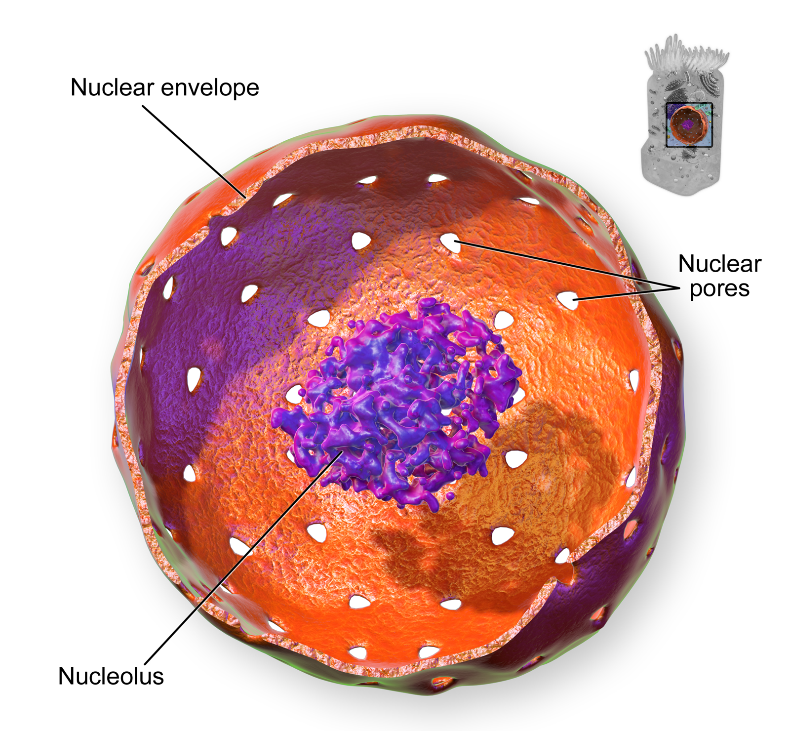
As you can see in the model pictured in Figure 4.6.2, the membrane enclosing the nucleus is called the nuclear envelope. This is actually a double membrane that encloses the entire organelle and isolates its contents from the cellular cytoplasm. Tiny holes called nuclear pores allow large molecules to pass through the nuclear envelope, with the help of special proteins. Large proteins and RNA molecules must be able to pass through the nuclear envelope so proteins can be synthesized in the cytoplasm and the genetic material can be maintained inside the nucleus. The nucleolus shown in the model below is mainly involved in the assembly of ribosomes. After being produced in the nucleolus, ribosomes are exported to the cytoplasm, where they are involved in the synthesis of proteins.
Mitochondria
The mitochondrion (plural, mitochondria) is an organelle that makes energy available to the cell. This is why mitochondria are sometimes referred to as the "power plants of the cell." They use energy from organic compounds (such as glucose) to make molecules of ATP (adenosine triphosphate), an energy-carrying molecule that is used almost universally inside cells for energy.
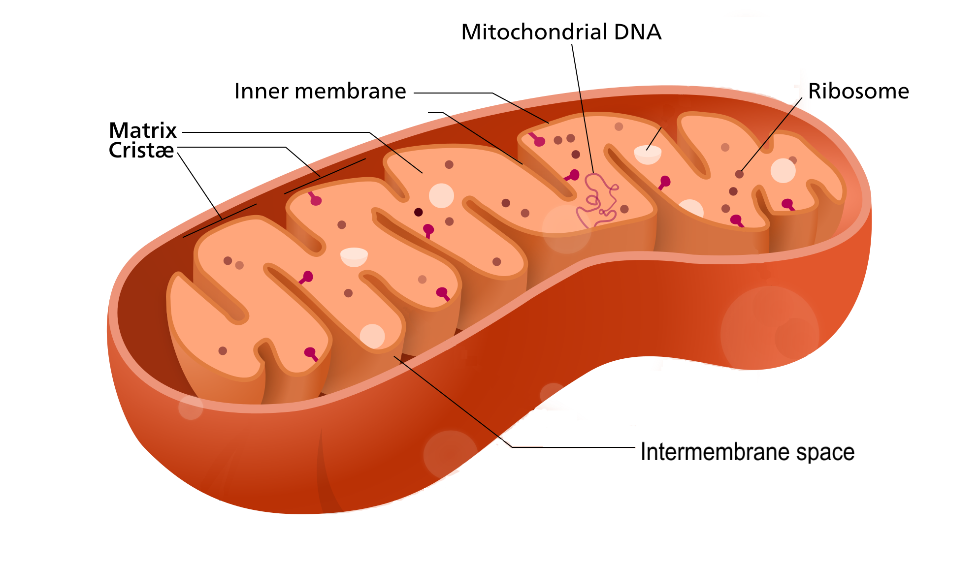
Mitochondria (as in the Figure 4.6.3 diagram) have a complex structure including an inner and out membrane. In addition, mitochondria have their own DNA, ribosomes, and a version of cytoplasm, called matrix. Does this sound similar to the requirements to be considered a cell? That's because they are!
Scientists think that mitochondria were once free-living organisms because they contain their own DNA. They theorize that ancient prokaryotes infected (or were engulfed by) larger prokaryotic cells, and the two organisms evolved a symbiotic relationship that benefited both of them. The larger cells provided the smaller prokaryotes with a place to live. In return, the larger cells got extra energy from the smaller prokaryotes. Eventually, the smaller prokaryotes became permanent guests of the larger cells, as organelles inside them. This theory is called endosymbiotic theory, and it is widely accepted by biologists today. (See the video in section 4.3 to learn all about endosymbiotic theory.)
Endoplasmic Reticulum
The endoplasmic reticulum (ER) is an organelle that helps make and transport proteins and lipids. There are two types of endoplasmic reticulum: rough endoplasmic reticulum (rER) and smooth endoplasmic reticulum (sER). Both types are shown in Figure 4.6.4.
- rER looks rough because it is studded with ribosomes. It provides a framework for the ribosomes, which make proteins. Bits of its membrane pinch off to form tiny sacs called vesicles, which carry proteins away from the ER.
- sER looks smooth because it does not have ribosomes. sER makes lipids, stores substances, and plays other roles.
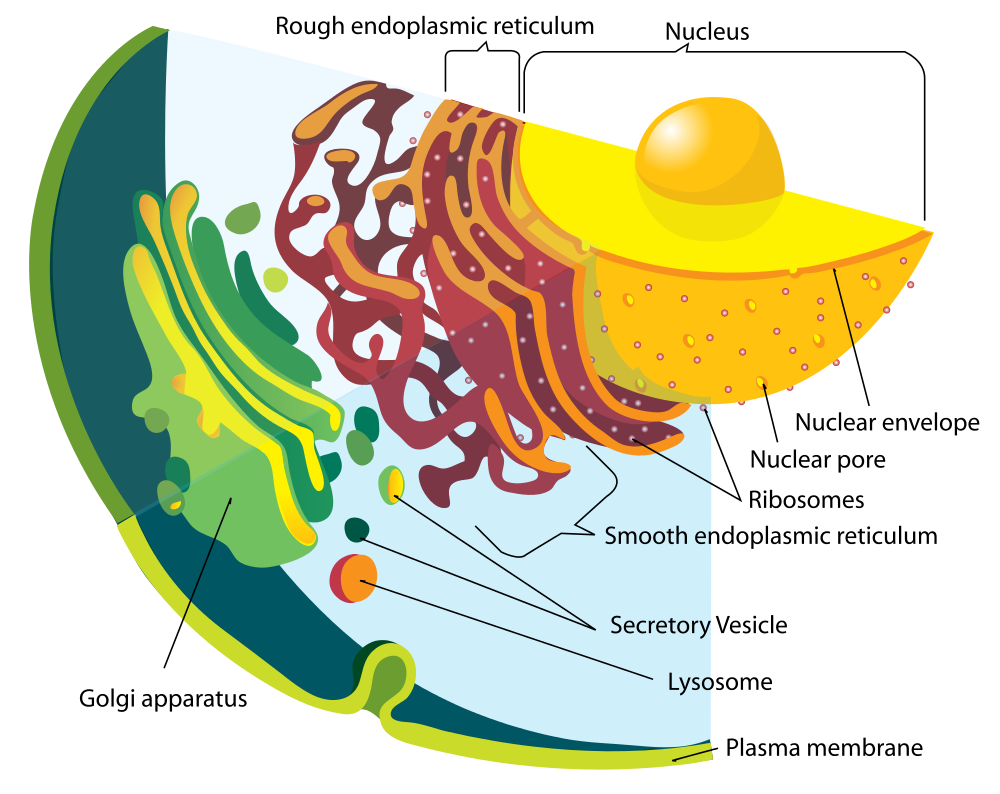
The Figure 4.6.4 drawing includes the nucleus, rER, sER, and Golgi apparatus. From the drawing, you can see how all these organelles work together to make and transport proteins.
Golgi Apparatus
The Golgi apparatus (shown in the Figure 4.6.4 diagram) is a large organelle that processes proteins and prepares them for use both inside and outside the cell. You can see the Golgi apparatus in the figure above. The Golgi apparatus is something like a post office. It receives items (proteins from the ER), then packages and labels them before sending them on to their destinations (to different parts of the cell or to the cell membrane for transport out of the cell). The Golgi apparatus is also involved in the transport of lipids around the cell.
Vesicles and Vacuoles
Both vesicles and vacuoles are sac-like organelles made of phospholipid bilayer that store and transport materials in the cell. Vesicles are much smaller than vacuoles and have a variety of functions. The vesicles that pinch off from the membranes of the ER and Golgi apparatus store and transport protein and lipid molecules. You can see an example of this type of transport vesicle in the Figure 4.6.4. Some vesicles are used as chambers for biochemical reactions.
There are some vesicles which are specialized to carry out specific functions. Lysosomes, which use enzymes to break down foreign matter and dead cells, have a double membrane to make sure their contents don't leak into the rest of the cell. Peroxisomes are another type of specialized vesicle with the main function of breaking down fatty acids and some toxins.
Centrioles
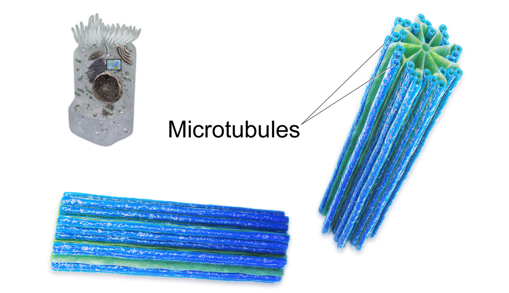
Centrioles are organelles involved in cell division. The function of centrioles is to help organize the chromosomes before cell division occurs so that each daughter cell has the correct number of chromosomes after the cell divides. Centrioles are found only in animal cells, and are located near the nucleus. Each centriole is made mainly of a protein named tubulin. The centriole is cylindrical in shape and consists of many microtubules, as shown in the model pictured in Figure 4.6.5.
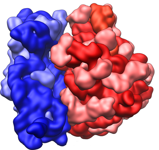
Ribosomes
Ribosomes are small structures where proteins are made. Although they are not enclosed within a membrane, they are frequently considered organelles. Each ribosome is formed of two subunits, like the ones pictured at the beginning of this section (Figure 4.6.1) and in Figure 4.6.6. Both subunits consist of proteins and RNA. mRNA from the nucleus carries the genetic code, copied from DNA, which remains in the nucleus. At the ribosome, the genetic code in mRNA is used to assemble and join together amino acids to make proteins. Ribosomes can be found alone or in groups within the cytoplasm, as well as on the rER.
4.6 Summary
- An organelle is a structure within the cytoplasm of a eukaryotic cell that is enclosed within a membrane and performs a specific job. Although ribosomes are not enclosed within a membrane, they are still commonly referred to as organelles in eukaryotic cells.
- The nucleus is the largest organelle in a eukaryotic cell, and it is considered to be the cell's control center. It controls gene expression, including controlling which proteins the cell makes.
- The mitochondrion (plural, mitochondria) is an organelle that makes energy available to the cells. It is like the power plant of the cell. According to the widely accepted endosymbiotic theory, mitochondria evolved from prokaryotic cells that were once free-living organisms that infected or were engulfed by larger prokaryotic cells.
- The endoplasmic reticulum (ER) is an organelle that helps make and transport proteins and lipids. Rough endoplasmic reticulum (rER) is studded with ribosomes. Smooth endoplasmic reticulum (sER) has no ribosomes.
- The Golgi apparatus is a large organelle that processes proteins and prepares them for use both inside and outside the cell. It is also involved in the transport of lipids around the cell.
- Both vesicles and vacuoles are sac-like organelles that may be used to store and transport materials in the cell or as chambers for biochemical reactions. Lysosomes and peroxisomes are special types of vesicles that break down foreign matter, dead cells, or poisons.
- Centrioles are organelles located near the nucleus that help organize the chromosomes before cell division so each daughter cell receives the correct number of chromosomes.
- Ribosomes are small structures where proteins are made. They are found in both prokaryotic and eukaryotic cells. They may be found alone or in groups within the cytoplasm or on the rER.
4.6 Review Questions
- What is an organelle?
- Describe the structure and function of the nucleus.
- Explain how the nucleus, ribosomes, rough endoplasmic reticulum, and Golgi apparatus work together to make and transport proteins.
- Why are mitochondria referred to as the "power plants of the cell"?
- What roles are played by vesicles and vacuoles?
- Why do all cells need ribosomes — even prokaryotic cells that lack a nucleus and other cell organelles?
- Explain endosymbiotic theory as it relates to mitochondria. What is one piece of evidence that supports this theory?
-
4.6 Explore More
https://www.youtube.com/watch?v=URUJD5NEXC8&t=121s
Biology: Cell Structure I Nucleus Medical Media, Nucleus Medical Media, 2015.
https://www.youtube.com/watch?v=Id2rZS59xSE&feature=youtu.be
David Bolinsky: Visualizing the wonder of a living cell, TED, 2007.
Attributes
Figure 4.6.1
Ribosomes at Work by Pedrik on Flickr is used under a CC BY-NC-SA 2.0 (https://creativecommons.org/licenses/by-nc-sa/2.0/) license.
Figure 4.6.2
Nucleus by BruceBlaus on Wikimedia Commons is used under a CC BY 3.0 (https://creativecommons.org/licenses/by/3.0) license.
Figure 4.6.3
Mitochondrion_structure.svg by Kelvinsong; modified by Sowlos on Wikimedia Commons is used and adapted by Christine Miller under a CC BY-SA 3.0 (https://creativecommons.org/licenses/by-sa/3.0) license.
Figure 4.6.4
Endomembrane_system_diagram_en.svg by Mariana Ruiz [LadyofHats] on Wikimedia Commons is released into the public domain (https://en.wikipedia.org/wiki/Public_domain).
Figure 4.6.5
Centrioles by BruceBlaus on Wikimedia Commons is used under a CC BY 3.0 (https://creativecommons.org/licenses/by/3.0) license.
Figure 4.6.6
Ribosome_shape by Vossman on Wikimedia Commons is used and adapted by Christine Miller under a CC BY-SA 3.0 (https://creativecommons.org/licenses/by-sa/3.0) license.
References
Blausen.com staff. (2014). Nucleus - Medical gallery of Blausen Medical 2014. WikiJournal of Medicine 1 (2). DOI:10.15347/wjm/2014.010. ISSN 2002-4436. https://en.wikiversity.org/wiki/WikiJournal_of_Medicine/Medical_gallery_of_Blausen_Medical_2014
Blausen.com staff (2014). Centrioles - Medical gallery of Blausen Medical 2014. WikiJournal of Medicine 1 (2). DOI:10.15347/wjm/2014.010. ISSN 2002-4436.https://en.wikiversity.org/wiki/WikiJournal_of_Medicine/Medical_gallery_of_Blausen_Medical_2014
Nucleus Medical Media. (2015, March 18). Biology: Cell structure I Nucleus Medical Media. YouTube. https://www.youtube.com/watch?v=URUJD5NEXC8&feature=youtu.be
TED. (2007, July 24). David Bolinsky: Visualizing the wonder of a living cell. YouTube. https://www.youtube.com/watch?v=Id2rZS59xSE&feature=youtu.be
Refers to the body system consisting of the heart, blood vessels and the blood. Blood contains oxygen and other nutrients which your body needs to survive. The body takes these essential nutrients from the blood.
The ability of an organism to maintain constant internal conditions despite external changes.
The central nervous system organ inside the skull that is the control center of the nervous system.

