12.2 Introduction to the Muscular System
Marvelous Muscles
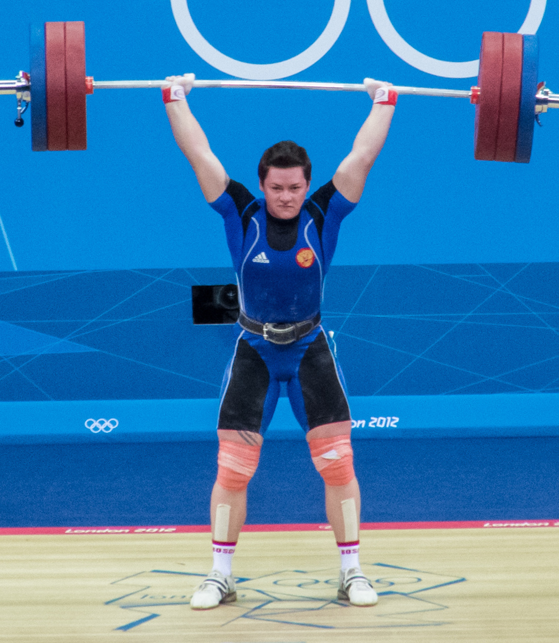
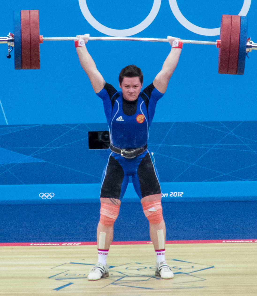
Does the word muscle make you think of the well-developed muscles of a weightlifter, like the woman in Figure 12.2.1? Her name is Natalia Zabolotnaya, and she’s a Russian Olympian. The muscles that are used to lift weights are easy to feel and see, but they aren’t the only muscles in the human body. Many muscles are deep within the body, where they form the walls of internal organs and other structures. You can flex your biceps at will, but you can’t control internal muscles like these. It’s a good thing that these internal muscles work without any conscious effort on your part, because movement of these muscles is essential for survival. Muscles are the organs of the muscular system.
What Is the Muscular System?
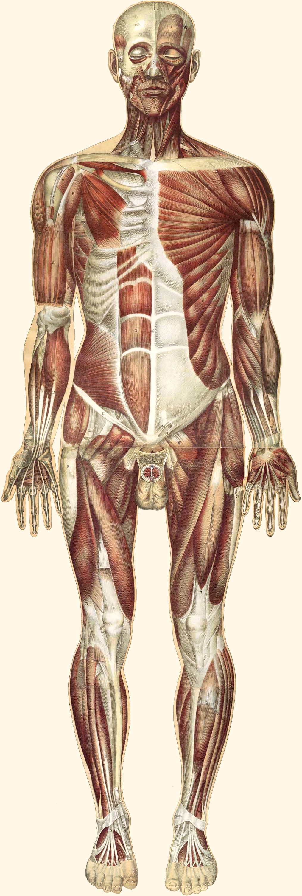
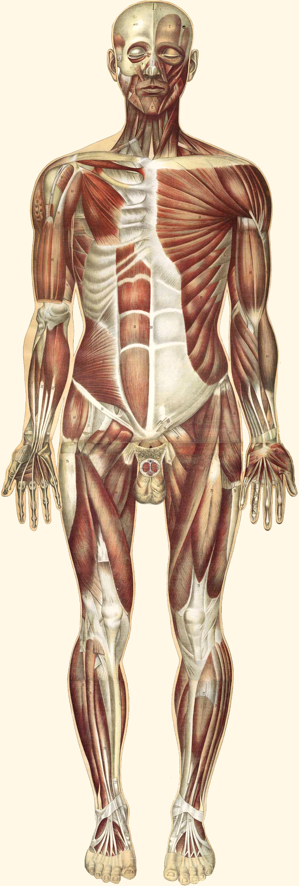
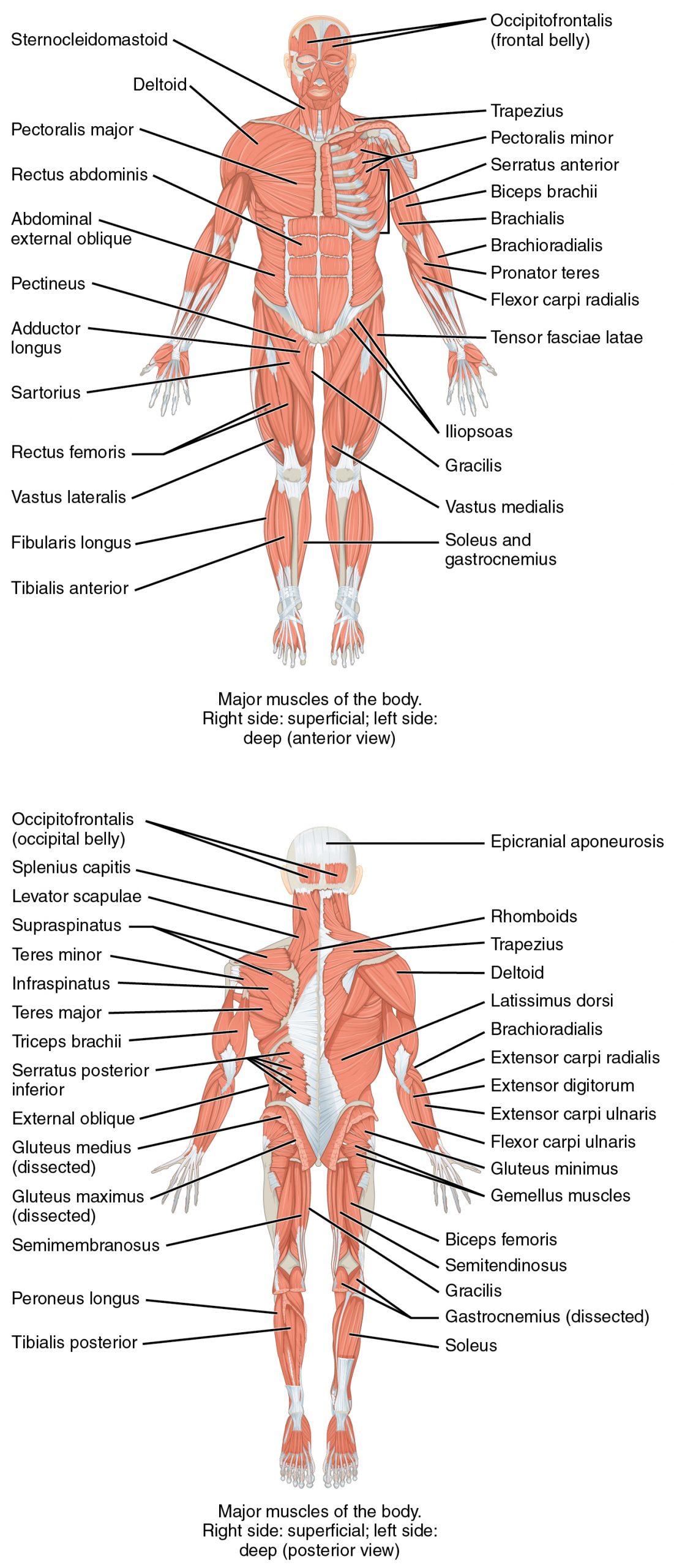
The muscular system consists of all the muscles of the body. The largest percentage of muscles in the muscular system consists of skeletal muscles, which are attached to bones and enable voluntary body movements (shown in Figure 12.2.2). There are almost 650 skeletal muscles in the human body, many of them shown in Figure 12.2.2. Besides skeletal muscles, the muscular system also includes cardiac muscle, which makes up the walls of the heart, and smooth muscles, which control movement in other internal organs and structures.
Muscle Structure and Function
Muscles are organs composed mainly of muscle cells, which are also called muscle fibres (mainly in skeletal and cardiac muscle) or myocytes (mainly in smooth muscle). Muscle cells are long, thin cells that are specialized for the function of contracting. They contain protein filaments that slide over one another using energy in ATP. The sliding filaments increase the tension in — or shorten the length of — muscle cells, causing a contraction. Muscle contractions are responsible for virtually all the movements of the body, both inside and out.
Skeletal muscles are attached to bones of the skeleton. When these muscles contract, they move the body. They allow us to use our limbs in a variety of ways, from walking to turning cartwheels. Skeletal muscles also maintain posture and help us to keep balance.
Smooth muscles in the walls of blood vessels contract to cause vasoconstriction, which may help conserve body heat. Relaxation of these muscles causes vasodilation, which may help the body lose heat. In the organs of the digestive system, smooth muscles squeeze food through the gastrointestinal tract by contracting in sequence to form a wave of muscle contractions called peristalsis. Think of squirting toothpaste through a tube by applying pressure in sequence from the bottom of the tube to the top, and you have a good idea of how food is moved by muscles through the digestive system. Peristalsis of smooth muscles also moves urine through the urinary tract.
Cardiac muscle tissue is found only in the walls of the heart. When cardiac muscle contracts, it makes the heart beat. The pumping action of the beating heart keeps blood flowing through the cardiovascular system.
Muscle Hypertrophy and Atrophy
Muscles can grow larger, or hypertrophy. This generally occurs through increased use, although hormonal or other influences can also play a role. The increase in testosterone that occurs in males during puberty, for example, causes a significant increase in muscle size. Physical exercise that involves weight bearing or resistance training can increase the size of skeletal muscles in virtually everyone. Exercises (such as running) that increase the heart rate may also increase the size and strength of cardiac muscle. The size of muscle, in turn, is the main determinant of muscle strength, which may be measured by the amount of force a muscle can exert.
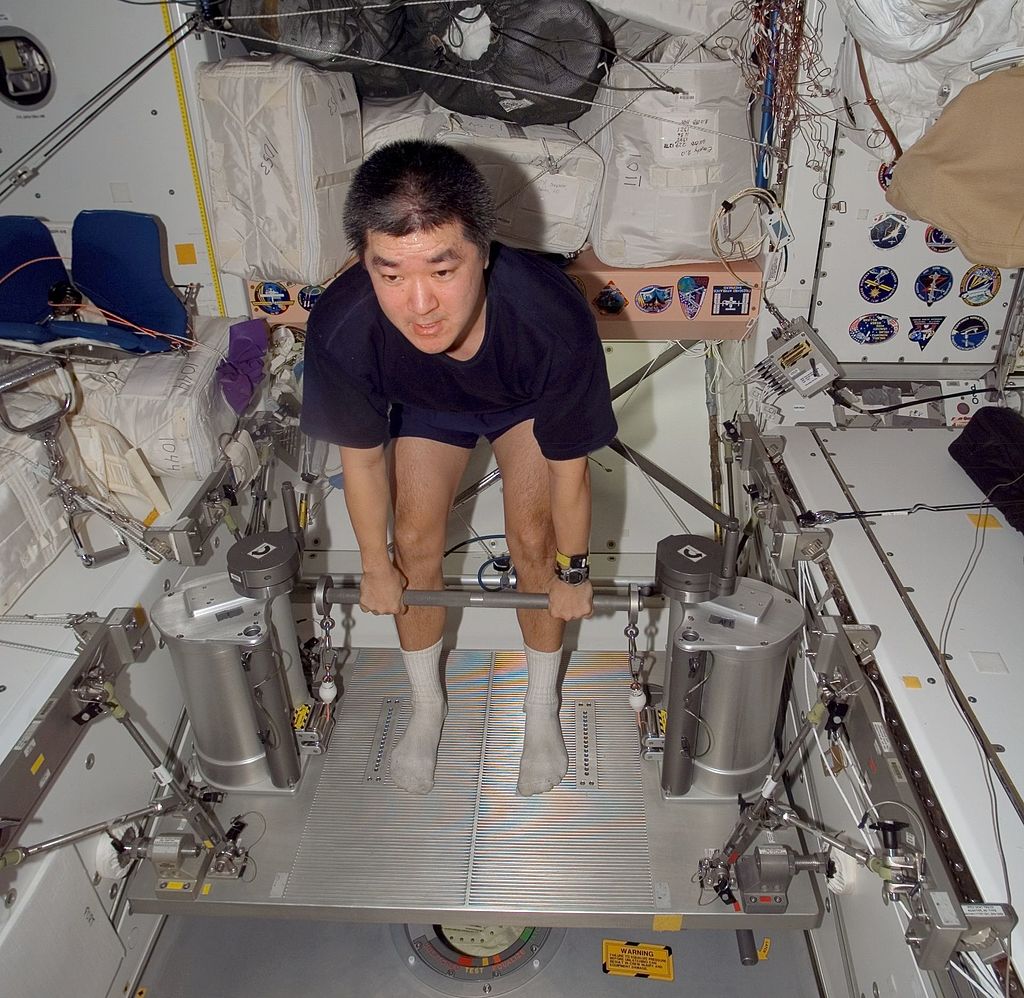
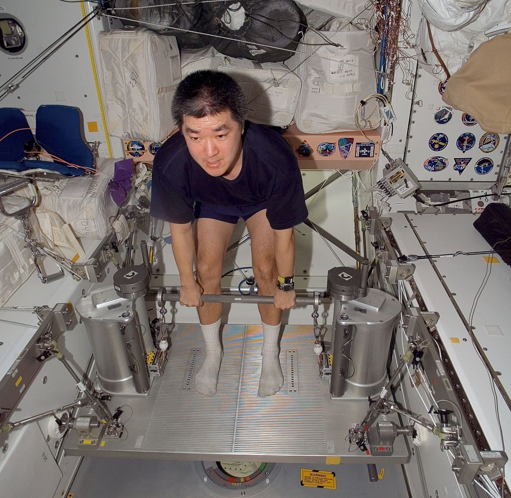
Muscles can also grow smaller, or atrophy, which can occur through lack of physical activity or from starvation. People who are immobilized for any length of time — for example, because of a broken bone or surgery — lose muscle mass relatively quickly. People in concentration or famine camps may be so malnourished that they lose much of their muscle mass, becoming almost literally just “skin and bones.” Astronauts on the International Space Station may also lose significant muscle mass because of weightlessness in space (see Figure 12.2.3).
Many diseases, including cancer and AIDS, are often associated with muscle atrophy. Atrophy of muscles also happens with age. As people grow older, there is a gradual decrease in the ability to maintain skeletal muscle mass, known as sarcopenia. The exact cause of sarcopenia is not known, but one possible cause is a decrease in sensitivity to growth factors that are needed to maintain muscle mass. Because muscle size determines strength, muscle atrophy causes a corresponding decline in muscle strength.
In both hypertrophy and atrophy, the number of muscle fibres does not change. What changes is the size of the muscle fibres. When muscles hypertrophy, the individual fibres become wider. When muscles atrophy, the fibres become narrower.
Interactions with Other Body Systems
Muscles cannot contract on their own. Skeletal muscles need stimulation from motor neurons in order to contract. The point where a motor neuron attaches to a muscle is called a neuromuscular junction. Let’s say you decide to raise your hand in class. Your brain sends electrical messages through motor neurons to your arm and shoulder. The motor neurons, in turn, stimulate muscle fibres in your arm and shoulder to contract, causing your arm to rise.
Involuntary contractions of smooth and cardiac muscles are also controlled by electrical impulses, but in the case of these muscles, the impulses come from the autonomic nervous system (smooth muscle) or specialized cells in the heart (cardiac muscle). Hormones and some other factors also influence involuntary contractions of cardiac and smooth muscles. For example, the fight-or-flight hormone adrenaline increases the rate at which cardiac muscle contracts, thereby speeding up the heartbeat.
Muscles cannot move the body on their own. They need the skeletal system to act upon. The two systems together are often referred to as the musculoskeletal system. Skeletal muscles are attached to the skeleton by tough connective tissues called tendons. Many skeletal muscles are attached to the ends of bones that meet at a joint. The muscles span the joint and connect the bones. When the muscles contract, they pull on the bones, causing them to move. The skeletal system provides a system of levers that allow body movement. The muscular system provides the force that moves the levers.
12.2 Summary
- The muscular system consists of all the muscles of the body. There are three types of muscle: skeletal muscle (which is attached to bones and enables voluntary body movements), cardiac muscle (which makes up the walls of the heart and makes it beat), and smooth muscle (which is found in the walls of internal organs and other internal structures and controls their movements).
- Muscles are organs composed mainly of muscle cells, which may also be called muscle fibres or myocytes. Muscle cells are specialized for the function of contracting, which occurs when protein filaments inside the cells slide over one another using energy in ATP.
- Muscles can grow larger, or hypertrophy. This generally occurs through increased use (physical exercise), although hormonal or other influences can also play a role. Muscles can also grow smaller, or atrophy. This may occur through lack of use, starvation, certain diseases, or aging. In both hypertrophy and atrophy, the size — but not the number — of muscle fibres changes. The size of muscles is the main determinant of muscle strength.
- Skeletal muscles need the stimulus of motor neurons to contract, and to move the body, they need the skeletal system to act upon. Involuntary contractions of cardiac and smooth muscles are controlled by special cells in the heart, nerves of the autonomic nervous system, hormones, or other factors.
12.2 Review Questions
- What is the muscular system?
- Describe muscle cells and their function.
- Identify three types of muscle tissue and where each type is found.
- Define muscle hypertrophy and muscle atrophy.
- What are some possible causes of muscle hypertrophy?
- Give three reasons that muscle atrophy may occur.
- How do muscles change when they increase or decrease in size?
- How do changes in muscle size affect strength?
- Explain why astronauts can easily lose muscle mass in space.
- Describe how the terms muscle cells, muscle fibres, and myocytes relate to each other.
-
- Name two systems in the body that work together with the muscular system to carry out movements.
- Describe one way in which the muscular system is involved in regulating body temperature.
12.2 Explore More
How your muscular system works – Emma Bryce, TED-Ed, 2017.
3D Medical Animation – Peristalsis in Large Intestine/Bowel || ABP ©, AnimatedBiomedical, 2013.
Muscle matters: Dr Brendan Egan at TEDxUCD, TEDx Talks, 2014.
Attributions
Figure 12.2.1
Natalia_Zabolotnaya_2012b by Simon Q on Wikimedia Commons is used under a CC BY 2.0 (https://creativecommons.org/licenses/by/2.0/deed.en) license.
Figure 12.2.2
Bougle_whole2_retouched by Bouglé, Julien from the National LIbrary of Medicine (NLM) on Wikimedia Commons is in the public domain (https://en.wikipedia.org/wiki/Public_domain).
Figure 12.2.3
Daniel_Tani_iss016e027910 by NASA/ International Space Station Imagery on Wikimedia Commons is in the public domain (https://en.wikipedia.org/wiki/Public_domain).
References
AnimatedBiomedical. (2013, January 30). 3D Medical animation – Peristalsis in large intestine/bowel || ABP ©. YouTube. https://www.youtube.com/watch?v=Ujr0UAbyPS4&feature=youtu.be
Bouglé, J. (1899). Le corps humain en grandeur naturelle : planches coloriées et superposées, avec texte explicatif. J. B. Baillière et fils. In Historical Anatomies on the Web. http://www.nlm.nih.gov/exhibition/historicalanatomies/bougle_home.html
TED-Ed. (2017, October 26). How your muscular system works – Emma Bryce. YouTube. https://www.youtube.com/watch?v=VVL-8zr2hk4&feature=youtu.be
TEDx Talks. (2014, June 27). Muscle matters: Dr Brendan Egan at TEDxUCD. YouTube. https://www.youtube.com/watch?v=LkXwfTsqQgQ&feature=youtu.be
Wikipedia contributors. (2020, June 15). Natalya Zabolotnaya. In Wikipedia. https://en.wikipedia.org/w/index.php?title=Natalya_Zabolotnaya&oldid=962630409
Created by: CK-12/Adapted by Christine Miller

A Bag Full of Jell-O
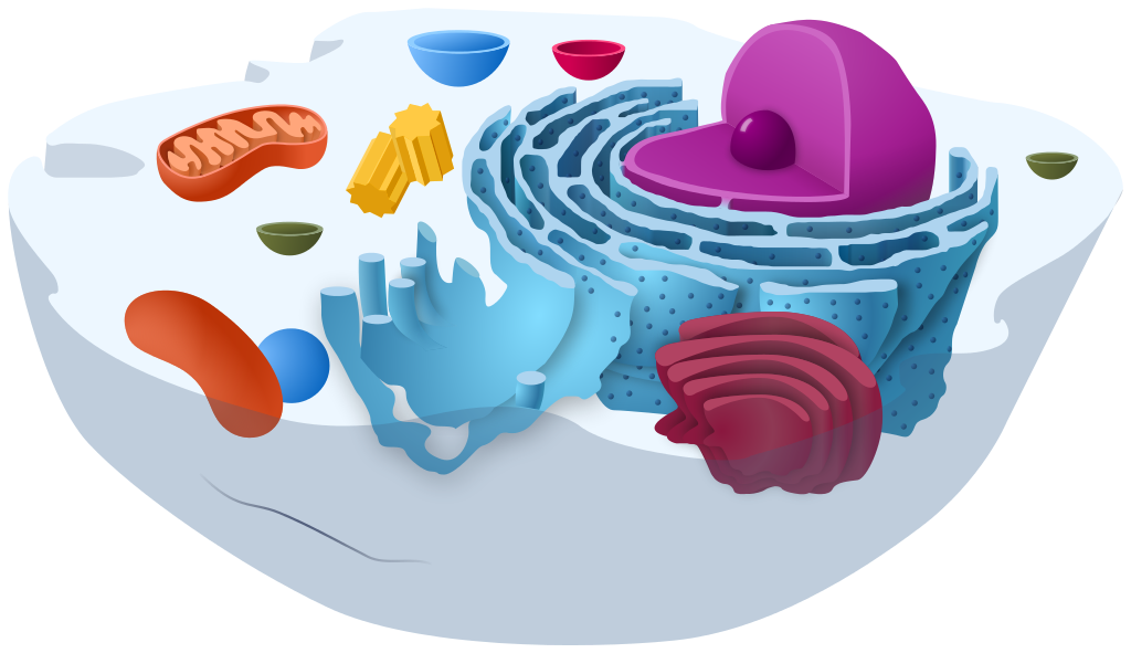
The simple cut-away model of an animal cell (Figure 4.4.1) shows that a cell resembles a plastic bag full of Jell-O. Its basic structure is a plasma membrane filled with cytoplasm. Like Jell-O containing mixed fruit (Figure 4.4.2), the cytoplasm of the cell also contains various structures, including a nucleus and other organelles. Your body is composed of trillions of cells, but all of them perform the same basic life functions. They all obtain and use energy, respond to the environment, and reproduce. How do your cells carry out these basic functions and keep themselves — and you — alive? To answer these questions, you need to know more about the structures that make up cells, starting with the plasma membrane.
What is the Plasma Membrane?
The plasma membrane is a structure that forms a barrier between the cytoplasm inside the cell and the environment outside the cell. Without the plasma membrane, there would be no cell. Although it is very thin and flexible, the plasma membrane protects and supports the cell by controlling everything that enters and leaves it. It allows only certain substances to pass through, while keeping others in or out. To understand how the plasma membrane controls what passes into or out of the cell, you need to know its basic structure.
Phospholipid Bilayer
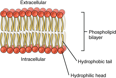
The plasma membrane is composed mainly of phospholipids, which consist of fatty acids and alcohol. The phospholipids in the plasma membrane are arranged in two layers, called a phospholipid bilayer. As shown in the simplified diagram in Figure 4.4.3, each individual phospholipid molecule has a phosphate group head (in red) and two fatty acid tails (in yellow). The head “loves” water (hydrophilic) and the tails “hate” water (hydrophobic). The water-hating tails are on the interior of the membrane, whereas the water-loving heads point outward, toward either the cytoplasm (intracellular) or the fluid that surrounds the cell (extracellular).
Hydrophobic molecules can easily pass through the plasma membrane if they are small enough, because they are water-hating like the interior of the membrane. Hydrophilic molecules, on the other hand, cannot pass through the plasma membrane — at least not without help — because they are water-loving like the exterior of the membrane.
Other Molecules in the Plasma Membrane
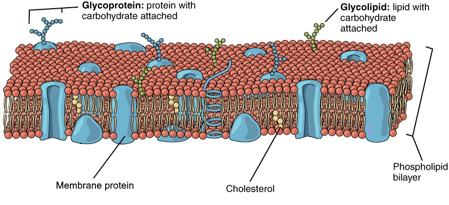
The plasma membrane also contains other molecules, primarily other lipids and proteins. The yellow molecules in the diagram here, for example, are the lipid cholesterol. Molecules of the steroid lipid cholesterol help the plasma membrane keep its shape. Proteins in the plasma membrane (shown blue in Figure 4.4.4) include: transport proteins that assist other substances in crossing the cell membrane, receptors that allow the cell to respond to chemical signals in its environment, and cell-identity markers that indicate what type of cell it is and whether it belongs in the body.
Additional Functions of the Plasma Membrane
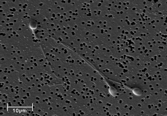
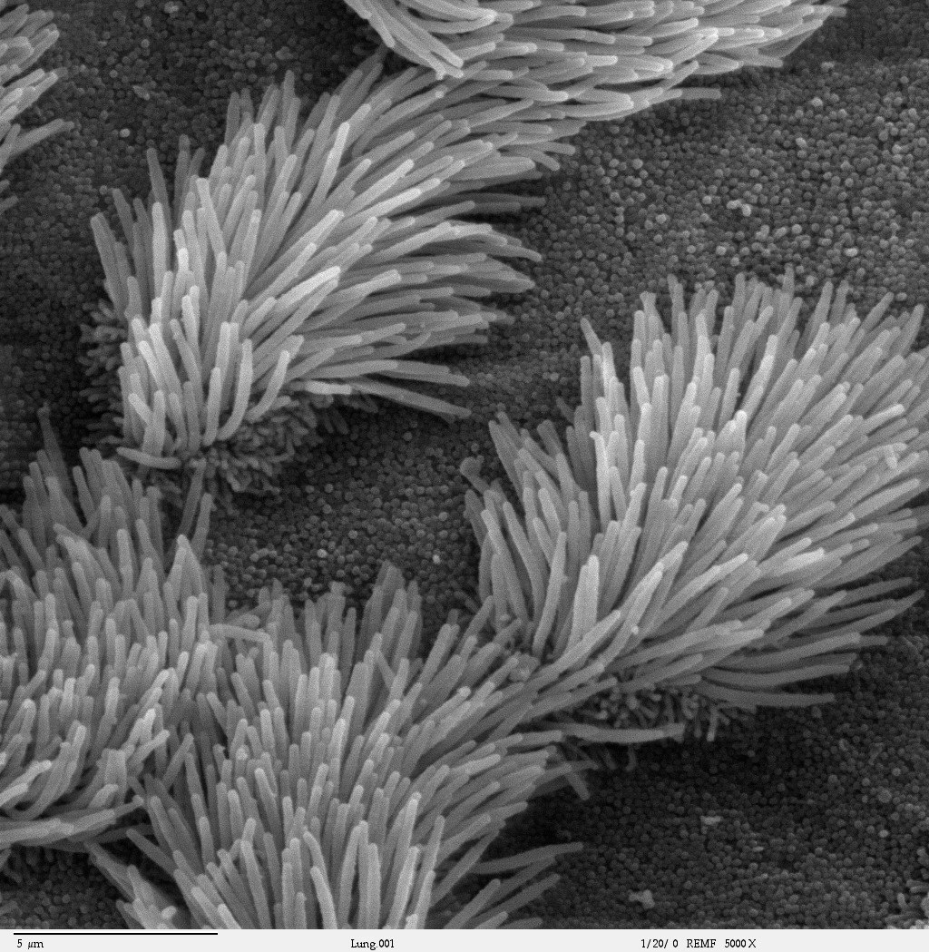
The plasma membrane may have extensions, such as whip-like flagella (singular flagellum) or brush-like cilia (singular cilium), shown below (Figure 4.4.5), that give it other functions. In single-celled organisms, these membrane extensions may help the organisms move. In multicellular organisms, the extensions have different functions. For example, the cilia on human lung cells sweep foreign particles and mucus toward the mouth and nose, while the flagellum on a human sperm cell allows it to swim.
Feature: My Human Body
If you smoke or use e-cigarettes (vaping) and need another reason to quit, here's a good one. We usually think of lung cancer as the major disease caused by smoking. But smoking and vaping can have devastating effects on the body's ability to protect itself from repeated, serious respiratory infections, such as bronchitis and pneumonia.
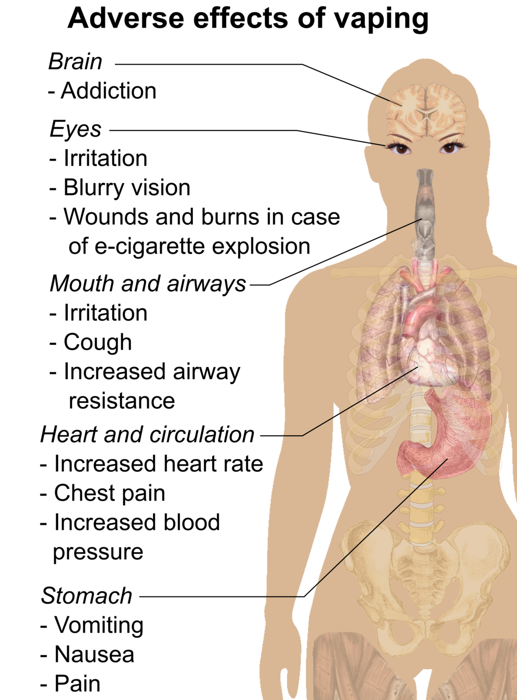
Cilia are microscopic, hair-like projects on cells that line the respiratory, reproductive, and digestive systems. Cilia in the respiratory system line most of your airways, where they have the job of trapping and removing dust, germs, and other foreign particles before they can make you sick. Cilia secrete mucus that traps particles, and they move in a continuous wave-like motion that sweeps the mucus and particles upward toward the throat, where they can be expelled from the body. When you are sick and cough up phlegm, that's what you are doing.
Smoking prevents cilia from performing these important functions. Chemicals in tobacco smoke paralyze the cilia so they can't sweep mucus out of the airways. Those chemicals also inhibit the cilia from producing mucus. Fortunately, these effects start to wear off soon after the most recent exposure to tobacco smoke. If you stop smoking, your cilia will return to normal. Even if prolonged smoking has destroyed cilia, they will regrow and resume functioning in a matter of months after you stop smoking.
4.4 Summary
- The plasma membrane is a structure that forms a barrier between the cytoplasm inside the cell and the environment outside the cell. It allows only certain substances to pass in or out of the cell.
- The plasma membrane is composed mainly of a bilayer of phospholipid molecules. It also contains other molecules, such as the steroid cholesterol, which helps the membrane keep its shape, and transport proteins, which help substances pass through the membrane.
- The plasma membranes of some cells have extensions that have other functions, like flagella to help sperm move, or cilia to help keep our airways clear.
4.4 Review Questions
- What are the general functions of the plasma membrane?
- Describe the phospholipid bilayer of the plasma membrane.
- Identify other molecules in the plasma membrane. State their functions.
- Why do some cells have plasma membrane extensions, like flagella and cilia?
- Explain why hydrophilic molecules cannot easily pass through the cell membrane. What type of molecule in the cell membrane might help hydrophilic molecules pass through it?
- Which part of a phospholipid molecule in the plasma membrane is made of fatty acid chains? Is this part hydrophobic or hydrophilic?
- The two layers of phospholipids in the plasma membrane are called a phospholipid ____________.
-
- Steroid hormones can pass directly through cell membranes. Why do you think this is the case?
- Some antibiotics work by making holes in the plasma membrane of bacterial cells. How do you think this kills the cells?
- What is the name of the long, whip-like extensions of the plasma membrane that helps some single-celled organisms move?
4.4 Explore More
https://www.youtube.com/watch?v=yAXnYcUjn5k&feature=emb_logo
Insights into cell membranes via dish detergent - Ethan Perlstein, TED-Ed, 2013.
https://www.youtube.com/watch?v=qBCVVszQQNs
Inside the cell membrane, by The Amoeba Sisters, 2018.
Attributions
Figure 4.4.1
Animal Cell Unannotated, by Kelvin Song on Wikimedia Commons is used under a CC0 1.0 (https://creativecommons.org/publicdomain/zero/1.0/deed.en) public domain dedication license.
Figure 4.4.2
Jello mold at the mexican bakery photo by Aimée Knight on Flickr is used under a CC BY 2.0 (https://creativecommons.org/licenses/by/2.0/) license.
Figure 4.4.3
Phospholipid_Bilayer by OpenStax on Wikimedia Commons is used under a CC BY 4.0 (https://creativecommons.org/licenses/by/4.0) license.
Figure 4.4.4
Lipid bilayer by OpenStax on Wikimedia Commons is used under a CC BY 4.0 (https://creativecommons.org/licenses/by/4.0) license.
Figure 4.4.5
Spermatozoa-human-3140x by No specific author on Wikimedia Commons is released into the public domain (https://en.wikipedia.org/wiki/Public_domain).
Figure 4.4.6
Cilia/ Bronchiolar epithelium 3 - SEM by Charles Daghlian on Wikimedia Commons is released into the public domain (https://en.wikipedia.org/wiki/Public_domain).
Figure 4.4.7
Adverse effects of vaping (raster) by Mikael Häggström on Wikimedia Commons is released into the public domain (https://en.wikipedia.org/wiki/Public_domain).
References
Amoeba Sisters. (2018, February 27). Inside the cell membrane. YouTube. https://www.youtube.com/watch?v=qBCVVszQQNs&feature=youtu.be
Betts, J.G., Young, K.A., Wise, J.A., Johnson, E., Poe, B., Kruse, D.H., Korol, O., Johnson, J.E.. Womble, M., DeSaix. P. (2013, April 25). Figure 3.3 Phospolipid Bilayer [digital image]. In Anatomy and Physiology. OpenStax. https://openstax.org/books/anatomy-and-physiology/pages/3-1-the-cell-membrane
Betts, J.G., Young, K.A., Wise, J.A., Johnson, E., Poe, B., Kruse, D.H., Korol, O., Johnson, J.E.. Womble, M., DeSaix. P. (2013, April 25). Figure 3.4 Cell Membrane [digital image]. In Anatomy and Physiology. OpenStax. https://openstax.org/books/anatomy-and-physiology/pages/3-1-the-cell-membrane
Ghosh, A., Coakley, R. C., Mascenik, T., Rowell, T. R., Davis, E. S., Rogers, K., Webster, M. J., Dang, H., Herring, L. E., Sassano, M. F., Livraghi-Butrico, A., Van Buren, S. K., Graves, L. M., Herman, M. A., Randell, S. H., Alexis, N. E., & Tarran, R. (n.d.). Chronic E-Cigarette Exposure Alters the Human Bronchial Epithelial Proteome. American Journal of Respiratory and Critical /Care Medicine, 198(1), 67–76. https://doi-org.ezproxy.tru.ca/10.1164/rccm.201710-2033OC
TED-Ed. (2013, February 26). Insights into cell membranes via dish detergent - Ethan Perlstein. YouTube. https://www.youtube.com/watch?v=yAXnYcUjn5k&feature=youtu.be
Created by CK-12 Foundation/Adapted by Christine Miller
Marvelous Muscles
Does the word muscle make you think of the well-developed muscles of a weightlifter, like the woman in Figure 12.2.1? Her name is Natalia Zabolotnaya, and she’s a Russian Olympian. The muscles that are used to lift weights are easy to feel and see, but they aren’t the only muscles in the human body. Many muscles are deep within the body, where they form the walls of internal organs and other structures. You can flex your biceps at will, but you can’t control internal muscles like these. It’s a good thing that these internal muscles work without any conscious effort on your part, because movement of these muscles is essential for survival. Muscles are the organs of the muscular system.
What Is the Muscular System?
The muscular system consists of all the muscles of the body. The largest percentage of muscles in the muscular system consists of skeletal muscles, which are attached to bones and enable voluntary body movements (shown in Figure 12.2.2). There are almost 650 skeletal muscles in the human body, many of them shown in Figure 12.2.2. Besides skeletal muscles, the muscular system also includes cardiac muscle, which makes up the walls of the heart, and smooth muscles, which control movement in other internal organs and structures.
Muscle Structure and Function
Muscles are organs composed mainly of muscle cells, which are also called muscle fibres (mainly in skeletal and cardiac muscle) or myocytes (mainly in smooth muscle). Muscle cells are long, thin cells that are specialized for the function of contracting. They contain protein filaments that slide over one another using energy in ATP. The sliding filaments increase the tension in — or shorten the length of — muscle cells, causing a contraction. Muscle contractions are responsible for virtually all the movements of the body, both inside and out.
Skeletal muscles are attached to bones of the skeleton. When these muscles contract, they move the body. They allow us to use our limbs in a variety of ways, from walking to turning cartwheels. Skeletal muscles also maintain posture and help us to keep balance.
Smooth muscles in the walls of blood vessels contract to cause vasoconstriction, which may help conserve body heat. Relaxation of these muscles causes vasodilation, which may help the body lose heat. In the organs of the digestive system, smooth muscles squeeze food through the gastrointestinal tract by contracting in sequence to form a wave of muscle contractions called peristalsis. Think of squirting toothpaste through a tube by applying pressure in sequence from the bottom of the tube to the top, and you have a good idea of how food is moved by muscles through the digestive system. Peristalsis of smooth muscles also moves urine through the urinary tract.
Cardiac muscle tissue is found only in the walls of the heart. When cardiac muscle contracts, it makes the heart beat. The pumping action of the beating heart keeps blood flowing through the cardiovascular system.
Muscle Hypertrophy and Atrophy
Muscles can grow larger, or hypertrophy. This generally occurs through increased use, although hormonal or other influences can also play a role. The increase in testosterone that occurs in males during puberty, for example, causes a significant increase in muscle size. Physical exercise that involves weight bearing or resistance training can increase the size of skeletal muscles in virtually everyone. Exercises (such as running) that increase the heart rate may also increase the size and strength of cardiac muscle. The size of muscle, in turn, is the main determinant of muscle strength, which may be measured by the amount of force a muscle can exert.
Muscles can also grow smaller, or atrophy, which can occur through lack of physical activity or from starvation. People who are immobilized for any length of time — for example, because of a broken bone or surgery — lose muscle mass relatively quickly. People in concentration or famine camps may be so malnourished that they lose much of their muscle mass, becoming almost literally just “skin and bones.” Astronauts on the International Space Station may also lose significant muscle mass because of weightlessness in space (see Figure 12.2.3).
Many diseases, including cancer and AIDS, are often associated with muscle atrophy. Atrophy of muscles also happens with age. As people grow older, there is a gradual decrease in the ability to maintain skeletal muscle mass, known as sarcopenia. The exact cause of sarcopenia is not known, but one possible cause is a decrease in sensitivity to growth factors that are needed to maintain muscle mass. Because muscle size determines strength, muscle atrophy causes a corresponding decline in muscle strength.
In both hypertrophy and atrophy, the number of muscle fibres does not change. What changes is the size of the muscle fibres. When muscles hypertrophy, the individual fibres become wider. When muscles atrophy, the fibres become narrower.
Interactions with Other Body Systems
Muscles cannot contract on their own. Skeletal muscles need stimulation from motor neurons in order to contract. The point where a motor neuron attaches to a muscle is called a neuromuscular junction. Let’s say you decide to raise your hand in class. Your brain sends electrical messages through motor neurons to your arm and shoulder. The motor neurons, in turn, stimulate muscle fibres in your arm and shoulder to contract, causing your arm to rise.
Involuntary contractions of smooth and cardiac muscles are also controlled by electrical impulses, but in the case of these muscles, the impulses come from the autonomic nervous system (smooth muscle) or specialized cells in the heart (cardiac muscle). Hormones and some other factors also influence involuntary contractions of cardiac and smooth muscles. For example, the fight-or-flight hormone adrenaline increases the rate at which cardiac muscle contracts, thereby speeding up the heartbeat.
Muscles cannot move the body on their own. They need the skeletal system to act upon. The two systems together are often referred to as the musculoskeletal system. Skeletal muscles are attached to the skeleton by tough connective tissues called tendons. Many skeletal muscles are attached to the ends of bones that meet at a joint. The muscles span the joint and connect the bones. When the muscles contract, they pull on the bones, causing them to move. The skeletal system provides a system of levers that allow body movement. The muscular system provides the force that moves the levers.
12.2 Summary
- The muscular system consists of all the muscles of the body. There are three types of muscle: skeletal muscle (which is attached to bones and enables voluntary body movements), cardiac muscle (which makes up the walls of the heart and makes it beat), and smooth muscle (which is found in the walls of internal organs and other internal structures and controls their movements).
- Muscles are organs composed mainly of muscle cells, which may also be called muscle fibres or myocytes. Muscle cells are specialized for the function of contracting, which occurs when protein filaments inside the cells slide over one another using energy in ATP.
- Muscles can grow larger, or hypertrophy. This generally occurs through increased use (physical exercise), although hormonal or other influences can also play a role. Muscles can also grow smaller, or atrophy. This may occur through lack of use, starvation, certain diseases, or aging. In both hypertrophy and atrophy, the size — but not the number — of muscle fibres changes. The size of muscles is the main determinant of muscle strength.
- Skeletal muscles need the stimulus of motor neurons to contract, and to move the body, they need the skeletal system to act upon. Involuntary contractions of cardiac and smooth muscles are controlled by special cells in the heart, nerves of the autonomic nervous system, hormones, or other factors.
12.2 Review Questions
- What is the muscular system?
- Describe muscle cells and their function.
- Identify three types of muscle tissue and where each type is found.
- Define muscle hypertrophy and muscle atrophy.
- What are some possible causes of muscle hypertrophy?
- Give three reasons that muscle atrophy may occur.
- How do muscles change when they increase or decrease in size?
- How do changes in muscle size affect strength?
- Explain why astronauts can easily lose muscle mass in space.
- Describe how the terms muscle cells, muscle fibres, and myocytes relate to each other.
-
- Name two systems in the body that work together with the muscular system to carry out movements.
- Describe one way in which the muscular system is involved in regulating body temperature.
12.2 Explore More
https://www.youtube.com/watch?v=VVL-8zr2hk4
How your muscular system works - Emma Bryce, TED-Ed, 2017.
https://www.youtube.com/watch?v=Ujr0UAbyPS4&feature=emb_logo
3D Medical Animation - Peristalsis in Large Intestine/Bowel || ABP ©, AnimatedBiomedical, 2013.
https://www.youtube.com/watch?v=LkXwfTsqQgQ&feature=emb_logo
Muscle matters: Dr Brendan Egan at TEDxUCD, TEDx Talks, 2014.
Attributions
Figure 12.2.1
Natalia_Zabolotnaya_2012b by Simon Q on Wikimedia Commons is used under a CC BY 2.0 (https://creativecommons.org/licenses/by/2.0/deed.en) license.
Figure 12.2.2
Bougle_whole2_retouched by Bouglé, Julien from the National LIbrary of Medicine (NLM) on Wikimedia Commons is in the public domain (https://en.wikipedia.org/wiki/Public_domain).
Figure 12.2.3
Daniel_Tani_iss016e027910 by NASA/ International Space Station Imagery on Wikimedia Commons is in the public domain (https://en.wikipedia.org/wiki/Public_domain).
References
AnimatedBiomedical. (2013, January 30). 3D Medical animation - Peristalsis in large intestine/bowel || ABP ©. YouTube. https://www.youtube.com/watch?v=Ujr0UAbyPS4&feature=youtu.be
Bouglé, J. (1899). Le corps humain en grandeur naturelle : planches coloriées et superposées, avec texte explicatif. J. B. Baillière et fils. In Historical Anatomies on the Web. http://www.nlm.nih.gov/exhibition/historicalanatomies/bougle_home.html
TED-Ed. (2017, October 26). How your muscular system works - Emma Bryce. YouTube. https://www.youtube.com/watch?v=VVL-8zr2hk4&feature=youtu.be
TEDx Talks. (2014, June 27). Muscle matters: Dr Brendan Egan at TEDxUCD. YouTube. https://www.youtube.com/watch?v=LkXwfTsqQgQ&feature=youtu.be
Wikipedia contributors. (2020, June 15). Natalya Zabolotnaya. In Wikipedia. https://en.wikipedia.org/w/index.php?title=Natalya_Zabolotnaya&oldid=962630409
Involuntary, striated muscle found only in the walls of the heart; also called myocardium.
Created by CK-12 Foundation/Adapted by Christine Miller

Work Those Eye Muscles!
Imagine the man in Figure 12.3.1 turns his eyes in your direction. This is a very small movement, considering the conspicuously large and strong external eye muscles that control eyeball movements. These muscles have been called the strongest muscles in the human body relative to the work they do. However, the external eye muscles actually do a surprising amount of work. Eye movements occur almost constantly during waking hours, especially when we are scanning faces or reading. Eye muscles are also exercised nightly during the phase of sleep called rapid eye movement sleep. External eye muscles can move the eyes because they are made mainly of muscle tissue.
What is Muscle Tissue?
Muscle tissue is a soft tissue that makes up most of the tissues in the muscles of the human muscular system. Other tissues in muscles are connective tissues, such as tendons that attach skeletal muscles to bones and sheaths of connective tissues that cover or line muscle tissues. Only muscle tissue per se, has cells with the ability to contract.
There are three major types of muscle tissues in the human body: skeletal, smooth, and cardiac muscle tissues. Figure 12.3.2 shows how the three types of muscle tissues appear under magnification. When you read about each type below, you will learn why the three types appear as they do.
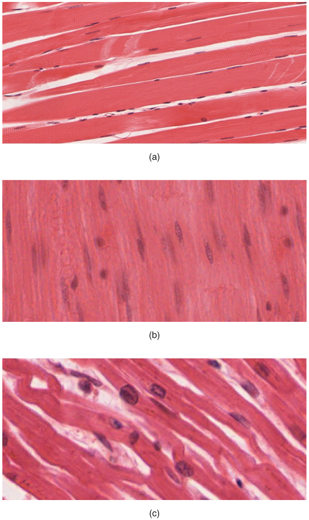
Skeletal Muscle Tissue
Skeletal muscle is muscle tissue that is attached to bones by tendons, which are bundles of collagen fibres. Whether you are moving your eyes or running a marathon, you are using skeletal muscles. Contractions of skeletal muscles are voluntary, or under conscious control of the central nervous system via the somatic nervous system. Skeletal muscle tissue is the most common type of muscle tissue in the human body. By weight, an average adult male is about 42% skeletal muscles, and the average adult female is about 36% skeletal muscles. Some of the major skeletal muscles in the human body are labeled in Figure 12.3.3 below.
Skeletal Muscle Pairs
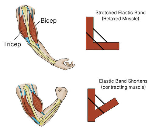
To move bones in opposite directions, skeletal muscles often consist of muscle pairs that work in opposition to one another, also called antagonistic muscle pairs. For example, when the biceps muscle (on the front of the upper arm) contracts, it can cause the elbow joint to flex or bend the arm, as shown in Figure 12.3.4. When the triceps muscle (on the back of the upper arm) contracts, it can cause the elbow to extend or straighten the arm. The biceps and triceps muscles, also shown in Figure 12.3.4, are an example of a muscle pair where the muscles work in opposition to each other.
Skeletal Muscle Structure
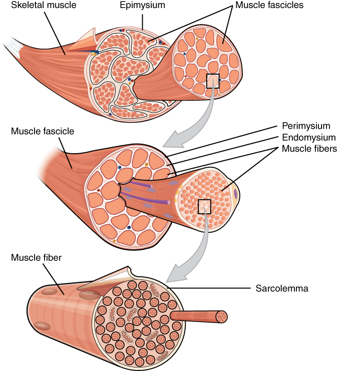
Each skeletal muscle consists of hundreds — or even thousands — of skeletal muscle fibres, which are long, string-like cells. As shown in Figure 12.3.5 below, skeletal muscle fibres are individually wrapped in connective tissue called endomysium. The skeletal muscle fibres are bundled together in units called muscle fascicles, which are surrounded by sheaths of connective tissue called perimysium. Each fascicle contains between ten and 100 (or even more!) skeletal muscle fibres. Fascicles, in turn, are bundled together to form individual skeletal muscles, which are wrapped in connective tissue called epimysium. The connective tissues in skeletal muscles have a variety of functions. They support and protect muscle fibres, allowing them to withstand the forces of contraction by distributing the forces applied to the muscle. They also provide pathways for nerves and blood vessels to reach the muscles. In addition, the epimysium anchors the muscles to tendons.
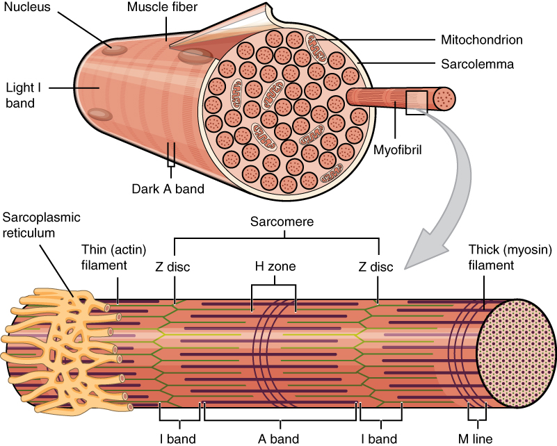
The same bundles-within-bundles structure is replicated within each muscle fibre. As shown in Figure 12.3.6, a muscle fibre consists of a bundle of myofibrils, which are themselves bundles of protein filaments. These protein filaments consist of thin filaments of the protein actin, which are anchored to structures called Z discs, and thick filaments of the protein myosin. The filaments are arranged together within a myofibril in repeating units called sarcomeres, which run from one Z disc to the next. The sarcomere is the basic functional unit of skeletal and cardiac muscles. It contracts as actin and myosin filaments slide over one another. Skeletal muscle tissue is said to be striated, because it appears striped. It has this appearance because of the regular, alternating A (dark) and I (light) bands of filaments arranged in sarcomeres inside the muscle fibres. Other components of a skeletal muscle fibre include multiple nuclei and mitochondria.
Slow- and Fast-Twitch Skeletal Muscle Fibres
Skeletal muscle fibres can be divided into two types, called slow-twitch (or type I) muscle fibres and fast-twitch (or type II) muscle fibres.
- Slow-twitch muscle fibres are dense with capillaries and rich in mitochondria and myoglobin, which is a protein that stores oxygen until needed for muscle activity. Relative to fast-twitch fibres, slow-twitch fibres can carry more oxygen and sustain aerobic (oxygen-using) activity. Slow-twitch fibres can contract for long periods of time, but not with very much force. They are relied upon primarily in endurance events, such as distance running or cycling.
- Fast-twitch muscle fibres contain fewer capillaries and mitochondria and less myoglobin. This type of muscle fibre can contract rapidly and powerfully, but it fatigues very quickly. Fast-twitch fibres can sustain only short, anaerobic (non-oxygen-using) bursts of activity. Relative to slow-twitch fibres, fast-twitch fibres contribute more to muscle strength and have a greater potential for increasing in mass. They are relied upon primarily in short, strenuous events, such as sprinting or weightlifting.
Proportions of fibre types vary considerably from muscle to muscle and from person to person. Individuals may be genetically predisposed to have a larger percentage of one type of muscle fibre than the other. Generally, an individual who has more slow-twitch fibres is better suited for activities requiring endurance, whereas an individual who has more fast-twitch fibres is better suited for activities requiring short bursts of power.
Smooth Muscle
Smooth muscle is muscle tissue in the walls of internal organs and other internal structures such as blood vessels. When smooth muscles contract, they help the organs and vessels carry out their functions. When smooth muscles in the stomach wall contract, for example, they squeeze the food inside the stomach, helping to mix and churn the food and break it into smaller pieces. This is an important part of digestion. Contractions of smooth muscles are involuntary, so they are not under conscious control. Instead, they are controlled by the autonomic nervous system, hormones, neurotransmitters, and other physiological factors.
Structure of Smooth Muscle
The cells that make up smooth muscle are generally called myocytes. Unlike the muscle fibres of striated muscle tissue, the myocytes of smooth muscle tissue do not have their filaments arranged in sarcomeres. Therefore, smooth tissue is not striated. However, the myocytes of smooth muscle do contain myofibrils, which in turn contain bundles of myosin and actin filaments. The filaments cause contractions when they slide over each other, as shown in Figure 12.3.7.

Functions of Smooth Muscle
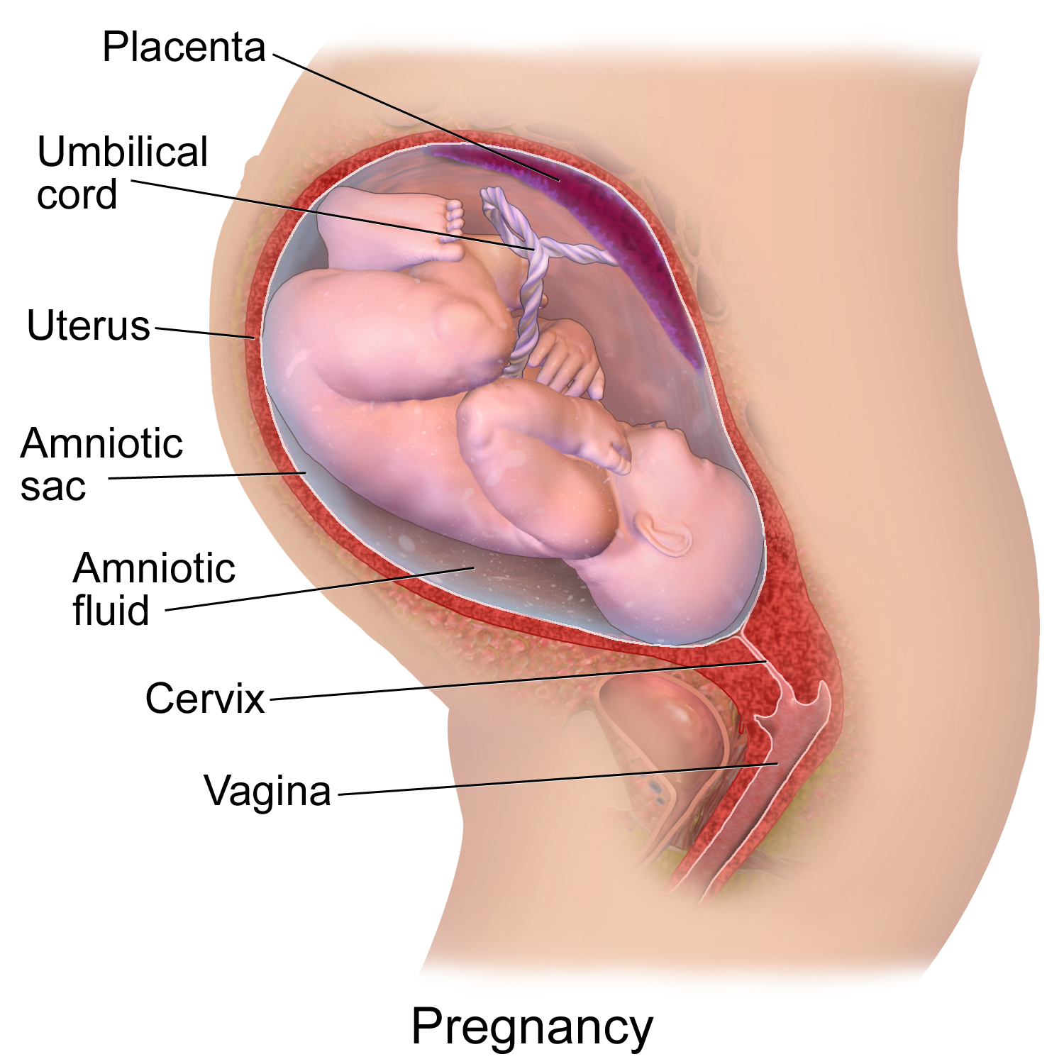
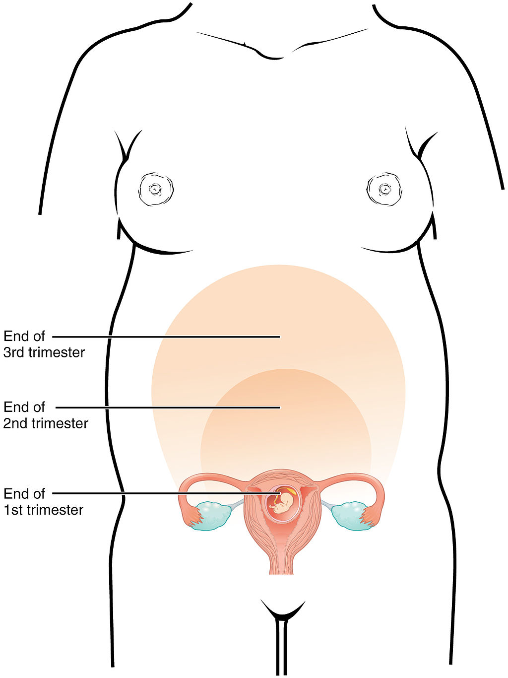
Unlike striated muscle, smooth muscle can sustain very long-term contractions. Smooth muscle can also stretch and still maintain its contractile function, which striated muscle cannot. The elasticity of smooth muscle is enhanced by an extracellular matrix secreted by myocytes. The matrix consists of elastin, collagen, and other stretchy fibres. The ability to stretch and still contract is an important attribute of smooth muscle in organs such as the stomach and uterus (see Figures 12.3.8 and 12.3.9), both of which must stretch considerably as they perform their normal functions.
 |
 |
The following list indicates where many smooth muscles are found, along with some of their specific functions.
- Walls of organs of the gastrointestinal tract (such as the esophagus, stomach, and intestines), moving food through the tract by peristalsis
- Walls of air passages of the respiratory tract (such as the bronchi), controlling the diameter of the passages and the volume of air that can pass through them
- Walls of organs of the male and female reproductive tracts; in the uterus, for example, pushing a baby out of the uterus and into the birth canal
- Walls of structures of the urinary system, including the urinary bladder, allowing the bladder to expand so it can hold more urine, and then contract as urine is released
- Walls of blood vessels, controlling the diameter of the vessels and thereby affecting blood flow and blood pressure
- Walls of lymphatic vessels, squeezing the fluid called lymph through the vessels
- Iris of the eyes, controlling the size of the pupils and thereby the amount of light entering the eyes
- Arrector pili in the skin, raising hairs in hair follicles in the dermis
Cardiac Muscle
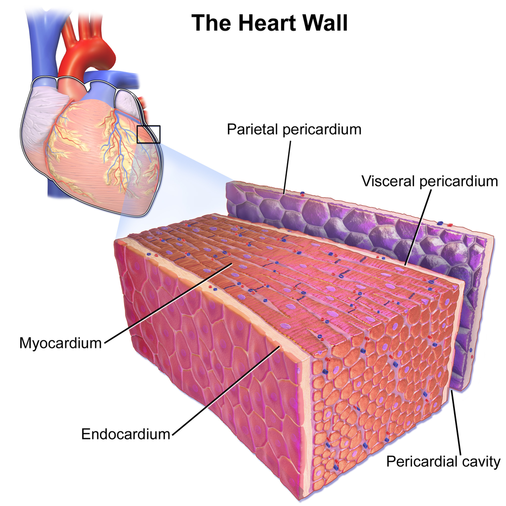
Cardiac muscle is found only in the wall of the heart. It is also called myocardium. As shown in Figure 12.3.10, myocardium is enclosed within connective tissues, including the endocardium on the inside of the heart and pericardium on the outside of the heart. When cardiac muscle contracts, the heart beats and pumps blood. Contractions of cardiac muscle are involuntary, like those of smooth muscles. They are controlled by electrical impulses from specialized cardiac muscle cells in an area of the heart muscle called the sinoatrial node.
Like skeletal muscle, cardiac muscle is striated because its filaments are arranged in sarcomeres inside the muscle fibres. However, in cardiac muscle, the myofibrils are branched at irregular angles rather than arranged in parallel rows (as they are in skeletal muscle). This explains why cardiac and skeletal muscle tissues look different from one another.
The cells of cardiac muscle tissue are arranged in interconnected networks. This arrangement allows rapid transmission of electrical impulses, which stimulate virtually simultaneous contractions of the cells. This enables the cells to coordinate contractions of the heart muscle.
The heart is the muscle that performs the greatest amount of physical work in the course of a lifetime. Although the power output of the heart is much less than the maximum power output of some other muscles in the human body, the heart does its work continuously over an entire lifetime without rest. Cardiac muscle contains a great many mitochondria, which produce ATP for energy and help the heart resist fatigue.
Feature: Human Biology in the News
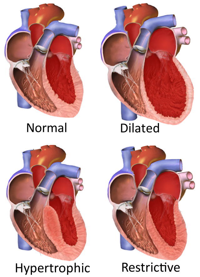
Cardiomyopathy is a disease in which the muscles of the heart are no longer able to effectively pump blood to the body — extreme forms of this disease can lead to heart failure. There are four main types of cardiomyopathy (also illustrated in Figure 12.3.11):
- Dilated (congestive) cardiomyopathy: the left ventricle (the chamber itself) of the heart becomes enlarged and can't pump blood our to the body. This is normally related to coronary artery disease and/or heart attack
- Hypertrophic cardiomyopathy: abnormal thickening of the muscular walls of the left ventricle make the chamber less able to work properly. This condition is more common in patients with a family history of the disease.
- Restrictive cardiomyopathy: the myocardium becomes abnormally rigid and inelastic and is unable to expand in between heartbeats to refill with blood. Restrictive cardiomyopathy typically affects older people.
- Arrhythmogenic right ventricular cardiomyopathy: the right ventricular muscle is replaced by adipose or scar tissue, reducing elasticity and interfering with normal heartbeat and rhythm. This disease is often caused by genetic mutations.
Cardiomyopathy is typically diagnosed with a physical exam supplemented by medical and family history, an angiogram, blood tests, chest x-rays and electrocardiograms. In some cases your doctor would also requisition a CT scan and/or genetic testing.
When treating cardiomyopathy, the goal is to reduce symptoms that affect everyday life. Certain medications can help regularize and slow heart rate, decrease chances of blood clots and cause vasodilation in the coronary arteries. If medication is not sufficient to manage symptoms, a pacemaker or even a heart transplant may be the best option. Lifestyle can also help manage the symptoms of cardiomyopathy — people living with this disease are encouraged to avoid drug and alcohol use, control high blood pressure, eat a healthy diet, get ample rest and exercise, as well as reduce stress levels.
12.3 Summary
- Muscle tissue is a soft tissue that makes up most of the tissues in the muscles of the human muscular system. It is the only type of tissue that has cells with the ability to contract.
- Skeletal muscle tissue is attached to bones by tendons. It allows voluntary body movements.
- Skeletal muscle is the most common type of muscle tissue in the human body. To move bones in opposite directions, skeletal muscles often consist of pairs of muscles that work in opposition to one another to move bones in different directions at joints.
- Skeletal muscle fibres are bundled together in units called muscle fascicles, which are bundled together to form individual skeletal muscles. Skeletal muscles also have connective tissue supporting and protecting the muscle tissue.
- Each skeletal muscle fibre consists of a bundle of myofibrils, which are bundles of protein filaments. The filaments are arranged in repeating units called sarcomeres, which are the basic functional units of skeletal muscles. Skeletal muscle tissue is striated because of the pattern of sarcomeres in its fibres.
- Skeletal muscle fibres can be divided into two types, called slow-twitch and fast-twitch fibres. Slow-twitch fibres are used mainly in aerobic endurance activities, such as long-distance running. Fast-twitch fibres are used mainly for non-aerobic, strenuous activities, such as sprinting. Proportions of the two types of fibres vary from muscle to muscle and person to person.
- Smooth muscle tissue is found in the walls of internal organs and vessels. When smooth muscles contract, they help the organs and vessels carry out their functions. Contractions of smooth muscles are involuntary and controlled by the autonomic nervous system, hormones, and other substances.
- Cells of smooth muscle tissue are not striated because they lack sarcomeres, but the cells contract in the same basic way as striated muscle cells. Unlike striated muscle, smooth muscle can sustain very long-term contractions and maintain its contractile function, even when stretched.
- Cardiac muscle tissue is found only in the wall of the heart. When cardiac muscle contracts, the heart beats and pumps blood. Contractions of cardiac muscle are involuntary, like those of smooth muscles. They are controlled by electrical impulses from specialized cardiac cells.
- Like skeletal muscle, cardiac muscle is striated because its filaments are arranged in sarcomeres inside the muscle fibres. However, the myofibrils are branched instead of arranged in parallel rows, making cardiac and skeletal muscle tissues look different from one another.
- The heart is the muscle that performs the greatest amount of physical work in the course of a lifetime. Its cells contain a great many mitochondria to produce ATP for energy and help the heart resist fatigue.
12.3 Review Questions
- What is muscle tissue?
-
-
- Where is skeletal muscle found, and what is its general function?
- Why do many skeletal muscles work in pairs?
- Describe the structure of a skeletal muscle.
- Relate muscle fibre structure to the functional units of muscles.
- Why is skeletal muscle tissue striated?
- Where is smooth muscle found? What controls the contraction of smooth muscle?
- Where is cardiac muscle found? What controls its contractions?
- The heart muscle is smaller and less powerful than some other muscles in the body. Why is the heart the muscle that performs the greatest amount of physical work in the course of a lifetime? How does the heart resist fatigue?
- Give one example of connective tissue that is found in muscles. Describe one of its functions.
12.3 Explore More
https://www.youtube.com/watch?v=3_PYnWVoUzM
What happens during a heart attack? - Krishna Sudhir, TED-Ed, 2017.
https://www.youtube.com/watch?v=bwOE1MEginA&feature=emb_logo
Three types of muscle | Circulatory system physiology | NCLEX-RN | KhanAcademyMedicine, 2012.
Attributions
Figure 12.3.1
Look by ali-yahya-155huuQwGvA [photo] by Ali Yahya on Unsplash is used under the Unsplash License (https://unsplash.com/license).
Figure 12.3.2
Skeletal_Smooth_Cardiac by OpenStax College on Wikimedia Commons is used under a CC BY 3.0 (https://creativecommons.org/licenses/by/3.0) license.
Figure 12.3.3
Anterior_and_Posterior_Views_of_Muscles by OpenStax on Wikimedia Commons is used under a CC BY 4.0 (https://creativecommons.org/licenses/by/4.0) license.
Figure 12.3.4
Antagonistic Muscle Pair by Laura Guerin at CK-12 Foundation on Wikimedia Commons is used under a CC BY-NC 3.0 (https://creativecommons.org/licenses/by-nc/3.0/) license.
Figure 12.3.5
Muscle_Fibes_(large) by OpenStax on Wikimedia Commons is used under a CC BY 4.0 (https://creativecommons.org/licenses/by/4.0) license.
Figure 12.3.6
Muscle_Fibers_(small) by OpenStax on Wikimedia Commons is used under a CC BY 4.0 (https://creativecommons.org/licenses/by/4.0) license.
Figure 12.3.7
Smooth_Muscle_Contraction by OpenStax on Wikimedia Commons is used under a CC BY 4.0 (https://creativecommons.org/licenses/by/4.0) license.
Figure 12.3.8
Blausen_0747_Pregnancy by BruceBlaus on Wikimedia Commons is used under a CC BY 3.0 (https://creativecommons.org/licenses/by/3.0) license.
Figure 12.3.9
Size_of_Uterus_Throughout_Pregnancy-02 by OpenStax College on Wikimedia Commons is used under a CC BY 3.0 (https://creativecommons.org/licenses/by/3.0) license.
Figure 12.3.10
1024px-Blausen_0470_HeartWall by BruceBlaus on Wikimedia Commons is used under a CC BY 3.0 (https://creativecommons.org/licenses/by/3.0) license.
Figure 12.3.11
Tipet_e_kardiomiopative by Npatchett at English Wikipedia on Wikimedia Commons is used under a CC BY-SA 3.0 (https://creativecommons.org/licenses/by-sa/3.0) license. (Work derived from Blausen 0165 Cardiomyopathy Dilated by BruceBlaus)
References
Betts, J. G., Young, K.A., Wise, J.A., Johnson, E., Poe, B., Kruse, D.H., Korol, O., Johnson, J.E., Womble, M., DeSaix, P. (2013, June 19). Figure 4.18 Muscle tissue [digital image]. In Anatomy and Physiology (Section 4.4). OpenStax. https://openstax.org/books/anatomy-and-physiology/pages/4-4-muscle-tissue-and-motion
Betts, J. G., Young, K.A., Wise, J.A., Johnson, E., Poe, B., Kruse, D.H., Korol, O., Johnson, J.E., Womble, M., DeSaix, P. (2013, June 19). Figure 28.18 Size of uterus throughout pregnancy [digital image]. In Anatomy and Physiology (Section 28.4). OpenStax. https://openstax.org/books/anatomy-and-physiology/pages/28-4-maternal-changes-during-pregnancy-labor-and-birth
Betts, J. G., Young, K.A., Wise, J.A., Johnson, E., Poe, B., Kruse, D.H., Korol, O., Johnson, J.E., Womble, M., DeSaix, P. (2016, May 18). Figure 10.3 The three connective tissue layers [digital image]. In Anatomy and Physiology (Section 10.2). OpenStax. https://openstax.org/books/anatomy-and-physiology/pages/10-2-skeletal-muscle
Betts, J. G., Young, K.A., Wise, J.A., Johnson, E., Poe, B., Kruse, D.H., Korol, O., Johnson, J.E., Womble, M., DeSaix, P. (2016, May 18). Figure 10.4 Muscle fiber [digital image]. In Anatomy and Physiology (Section 10.2). OpenStax. https://openstax.org/books/anatomy-and-physiology/pages/10-2-skeletal-muscle
Betts, J. G., Young, K.A., Wise, J.A., Johnson, E., Poe, B., Kruse, D.H., Korol, O., Johnson, J.E., Womble, M., DeSaix, P. (2016, May 18). Figure 10.24 Muscle contraction [digital image]. In Anatomy and Physiology (Section 10.8). OpenStax. https://openstax.org/books/anatomy-and-physiology/pages/10-8-smooth-muscle
Betts, J. G., Young, K.A., Wise, J.A., Johnson, E., Poe, B., Kruse, D.H., Korol, O., Johnson, J.E., Womble, M., DeSaix, P. (2016, May 18). Figure 11.5 Overview of the muscular system [digital image]. In Anatomy and Physiology (Section 11.2). OpenStax. https://openstax.org/books/anatomy-and-physiology/pages/11-2-naming-skeletal-muscles
Blausen.com staff. (2014). Medical gallery of Blausen Medical 2014. WikiJournal of Medicine 1 (2). DOI:10.15347/wjm/2014.010. ISSN 2002-4436.
Brainard, J/ CK-12 Foundation. (2012). Figure 5 Triceps and biceps muscles in the upper arm are opposing muscles. [digital image]. In CK-12 Biology (Section 21.3) [online Flexbook]. CK12.org. https://www.ck12.org/book/ck-12-biology/section/21.3/ (Last modified August 11, 2017.)
khanacademymedicine. (2012, October 19). Three types of muscle | Circulatory system physiology | NCLEX-RN | Khan Academy. YouTube.
TED-Ed. (2017, February 14). What happens during a heart attack? - Krishna Sudhir. YouTube. https://www.youtube.com/watch?v=3_PYnWVoUzM&feature=youtu.be
Created by: CK-12/Adapted by Christine Miller
Figure 6.7.1 Men from Maasai Mara, Kenya.
Built for Heat
These tall, slender men live near the equator in Kenya, East Africa — one of the hottest regions of the world. These and the other people of his tribal group, called the Maasai, are among the tallest, most linear people on the planet. Their body build is thought to be an adaptation to their climate, which is hot year-round.
Climate Extremes
Climate refers to the average weather conditions in a region over a long period of time. One of the main determinants of climate is temperature. Both hot and cold temperatures are serious environmental stresses on the human body.
In the cold, there is risk of hypothermia, which is a dangerous decrease in core body temperature. The normal temperature of the human body is 37 degrees C (98.6 degrees F). Hypothermia sets in when body temperature drops to 34.4 degrees C (94 degrees F). If body temperature falls below 29.4 degrees C (85 degrees F), it starts to cool very rapidly because the body’s temperature regulation mechanism starts to fail.
The opposite problem occurs in the heat, where the risk is hyperthermia, which is a dangerous increase in core body temperature. If human body temperature rises above about 40.6 degrees C (105 degrees F), hyperthermia may become life threatening. If a temperature this high persists more than a few days, it generally damages the brain and other internal organs, leading to death.
Human Adaptation to Heat and Cold
Humans are the most widespread species on the planet, and they have lived in extreme climates for tens of thousands of years. As a result, many human populations have had to cope with extreme temperatures for hundreds of generations, which has forced them to develop genetic adaptations to these climate extremes.
The size and proportions of the human body may play an important role in how well an individual is able to handle hot or cold temperatures. In general, people with a tall, slender build, like the Maasai man pictured in Figure 6.7.1, are well adapted to heat, whereas people with a short, stocky build (like the Indigenous North American Inuit pictured in Figure 6.7.2) are well adapted to cold. These relationships between body build and climate were first noticed in other animal species in the 1800s by biologists Carl Bergmann and Joel Allen. These scientists formulated what are now known as Bergmann’s and Allen’s rules.
Figure 6.7.2 Indigenous North American Inuit.
Bergmann’s Rule
Bergmann's rule states that within a broadly distributed taxonomic group, populations or species of larger size are found in colder environments, whereas populations or species of smaller size are found in warmer environments. Bergmann’s rule has been shown to generally apply to widespread species of mammals and birds, although there are also many exceptions to the rule.
What explains Bergmann’s rule? Larger animals have a lower surface area to volume ratio than smaller animals, which is illustrated in Table 6.7.1 for a simple shape, a cube. From the table, you can see how the surface area to volume ratio of a cube decreases dramatically as the size of the cube increases. Because heat is lost through the surface of the body, an animal with a smaller surface area to volume ratio radiates less body heat per unit of mass. The larger body mass also allows the animal to generate more heat. A larger animal has more cells, so it can produce more body heat as a byproduct of cellular metabolism. Both of these factors allow a larger animal to stay warmer in a cold climate.
Table 6.7.1
Relationship of Surface Area to Volume in Cubes of Different Sizes
| Relationship of Surface Area to Volume in Cubes of Different Sizes | |||
| Side of Cube (cm) | Surface Area of Cube (cm2) | Volume of Cube (cm3) | Surface Area:Volume Ratio |
| 2 | 24 | 8 | 3:1 |
| 4 | 96 | 64 | 3:2 |
| 6 | 216 | 216 | 3:3 |
| 12 | 864 | 1728 | 3:6 |
| 20 | 2400 | 8000 | 3:10 |
Warmer climates impose the opposite problem: body heat generated by metabolism needs to be dissipated quickly rather than stored within the body. Smaller animals have a higher surface area to volume ratio that maximizes heat loss through the surface of the body and helps cool the body. With less mass and fewer cells, smaller animals also generate less heat due to cellular metabolism.

Anthropologists have found that many human populations tend to follow Bergmann’s rule. For example, a study of 100 human populations in the 1950s found a strong negative correlation between mean body mass and average yearly temperature. In other words, higher body mass was generally found in colder places, and lower body mass was generally found in hotter places.
There are also exceptions to the rule, in part because we use cultural responses to temper environmental stresses so we do not need to change genetically or physiologically in order to cope. Humans, for example, use clothing and heated buildings to stay warm in cold climates, which tends to counter the effects of natural selection changing human body shape in cold climates.
Allen’s Rule
Allen’s rule is a corollary of Bergmann’s rule. It states that animals living in hotter climates generally have longer extremities (such as limbs, tails, snouts, and ears) than closely related animals living in colder climates. The explanation for Allen’s rule is similar to the rationale behind Bergmann’s rule. Longer extremities maximize an animal’s surface area, allowing greater heat loss through the surface of the body. Therefore, having long extremities is adaptive in hot climates where the main challenge is dissipating body heat.
Anthropologists have noted that, in populations that have lived in tropical regions for long periods of time, the limbs of people tend to be longer in proportion to overall body height. The Maasai man pictured in Figure 6.7.1 is a clear example. His exceptionally long limbs — like those of other members of his population — are optimally proportioned for the hot climate in Kenya. The shorter-limbed body proportions of the Inuit people (Figure 6.7.2) suit them well for their cold climate. Marked differences in limb length have also been observed in related populations that have lived for long periods of time at different altitudes. High altitudes have colder climates than lower altitudes and — consistent with Allen's rule —people tend to have shorter limbs at higher altitudes.
Other Human Responses to Heat
Humans exhibit several other responses to high temperatures that are generally considered either short-term physiological responses or examples of longer-term acclimatization.
Sweating and Humidity

Because humans are basically tropical animals, we generally have an easier time dealing with excessive heat than excessive cold. Evaporation of sweat is the main way we cool the body. The dancer in Figure 6.7.4 is sweating copiously while working out in a hot environment. Why does sweating cool us? When sweat evaporates from the skin, it requires heat. The heat comes from the surface of the body, resulting in evaporative cooling.
How well we can deal with high air temperatures depends in large part on the humidity of the air. We have a harder time losing excess body heat when the humidity is high because our sweat does not evaporate as well as it does when the humidity is low. Instead, the sweat stays on the skin, making us feel clammy and warmer than we would feel if the humidity were lower. If the air is dry, on the other hand, sweat evaporates readily, and we feel more comfortable. For this reason, we are able to tolerate higher temperatures when the humidity is low. This is the basis of the common aphorism, “It’s not the heat, but the humidity.”
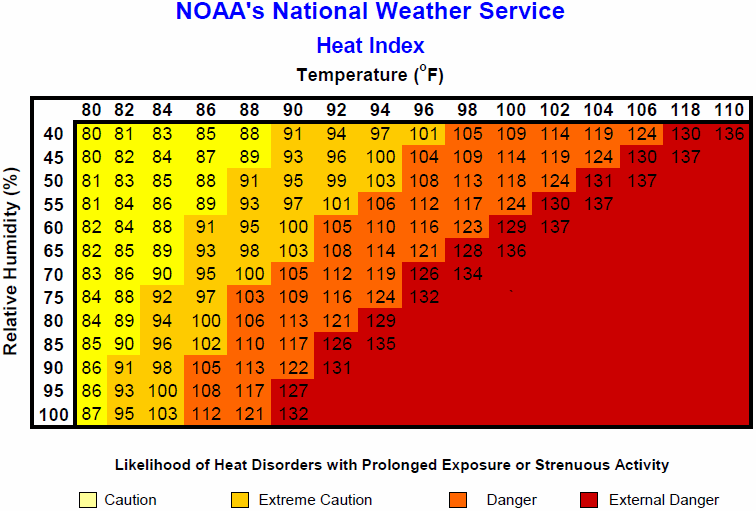
The heat index (HI) is a number that combines air temperature and relative humidity to indicate how hot the air feels due to the humidity. The heat index is also called "apparent temperature.” Figure 6.7.5 shows the heat index at different combinations of air temperature and relative humidity. As you can see, when the humidity is very high, even a 90-degree F (32 degrees C) temperature can be very dangerous.
Acclimatization to Heat
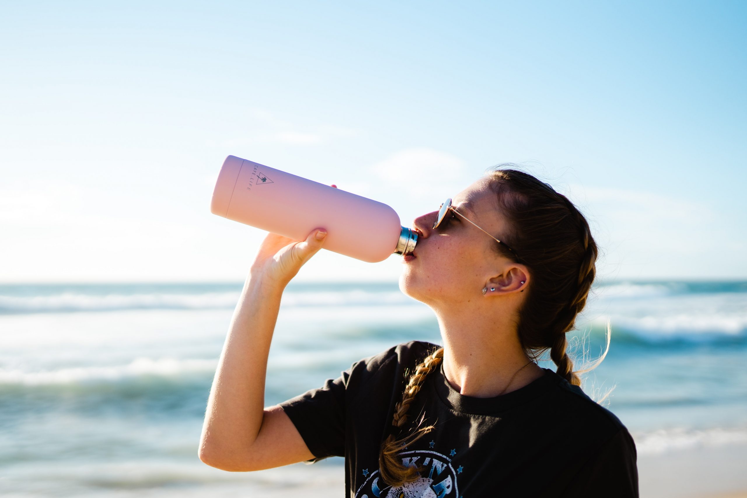
If humidity is low, evaporation of sweat can be an effective way to keep the body from overheating. However, the loss of water and salts in sweat can also be dangerous. In very hot conditions, an adult may lose up to four litres of sweat per hour and up to 14 litres per day. Such water losses may cause severe dehydration if the water is not replaced by drinking much more than usual. The loss of salts may also upset the normal salt balance in the body, which can be dangerous. Becoming acclimatized to heat by gradually increasing the exposure time to high temperatures — particularly while exercising or doing physical work — can reduce the risk of these effects.
It may take up to 14 days to attain maximum heat acclimatization. As the body becomes acclimatized, sweat output increases, and sweating begins sooner. The salt content of the sweat also declines, as does the output of urine. These and other physiological changes help the body lose heat through the evaporation of sweat, while maintaining the proper balance of salts and fluids in the body. There may also be increased blood flow to the body surface through the widening of blood vessels near the skin. This is called vasodilation. This brings more heat from the body core to the skin, and from there it may be radiated out into the environment.
Becoming acclimatized to heat allows one to safely perform more exercise or work in the heat. It also helps prevent heat-related illnesses by reducing strain on the body. Heat-related illnesses — from least to most serious — include heat cramps, heat exhaustion, and heat stroke.
- Heat cramps are muscle spasms caused by loss of water and salts. They often follow prolonged sweating brought on by over-exertion in hot weather.
- Heat exhaustion is a condition in which over-heating of the body causes dizziness, headache, profuse sweating, rapid heartbeat. and other symptoms. Without prompt treatment, heat exhaustion can lead to heat stroke.
- Heat stroke is potentially life threatening and a medical emergency. Heat stroke results from prolonged exposure to high temperatures, usually in combination with dehydration. It leads to failure of the body's temperature control system and is diagnosed when the core body temperature exceeds 105 degrees F (40 degrees C). Symptoms may include nausea, seizures, confusion, disorientation, and coma.
Acclimatization to heat, like other types of acclimatization, is a reversible process. Just as quickly as heat acclimatization occurs, the physiological changes fade away in the absence of heat exposure. The body returns to its baseline state within a week or two of no longer exercising or working at high temperatures.
Other Human Responses to Cold
Besides genetic difference in body build, there are two major ways the human body can respond to the cold. One way is by producing more body heat, and the other way is by conserving more body heat. An immediate response to cooling of the body is shivering. This is an involuntary and simultaneous contraction of many tiny muscles in the body. These muscle contractions generate a small amount of heat. Another early response to cold temperature is a narrowing of blood vessels near the skin. This is called vasoconstriction. This helps to shunt blood away from the body surface so more heat is held at the body core. The skin cools down and radiates less heat into the environment.
Hunting Response
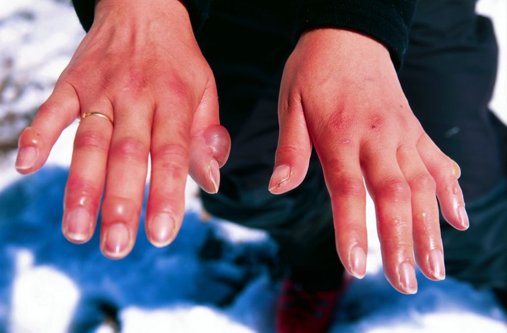
At temperatures below freezing, vasoconstriction can be dangerous if it lasts too long. The extremities become too cold because of lack of blood flow, and cold injury (such as frostbite) may occur. Frostbite is tissue destruction that occurs when tissue freezes. You can see a mild-to-moderate case of frostbite of the fingers in Figure 6.7.7. If frostbite is severe, it may lead to gangrene and amputation of the affected extremities.
The body counters the possibility of cold injury with a reaction called the hunting response. This is a process of alternating vasoconstriction and vasodilation in extremities exposed to cold. About five to ten minutes after the start of cold exposure, the blood vessels in the extremities suddenly dilate, which increases blood flow and subsequently the temperature of the extremities. This is soon followed by another phase of vasoconstriction, and then the process repeats.
The hunting response occurs in most people, but several factors may influence the strength of the response. People who live or work regularly in cold environments show an increased hunting response. Through acclimatization, however, tropical residents can develop an increased response, which is indistinguishable from that of arctic residents. Genetic factors may play a role in the hunting response, but this is uncertain because it is difficult to differentiate between adaptation and acclimatization.
Persistent Vasoconstriction
Where temperatures rarely fall below freezing but are repeatedly very chilly, the hunting response may not occur. Instead, vasoconstriction may persist to keep heat within the body at the expense of cooling the skin. As long as the temperature stays above freezing, cold injury (such as frostbite) will not occur. This type of response has been shown to occur in indigenous desert dwellers in southern Africa and Australia, where the temperature is hot during the day and very cold at night. People in these populations also tend to deposit fat around the organs in their chest and abdomen. The fat serves as insulation, protecting vital structures from the cold.
High-Fat Diet
Besides shivering, another way to increase body heat is to raise the basal metabolic rate. The basal metabolic rate (BMR) is the amount of energy that a person needs to keep the body functioning at rest. The higher the BMR, the more heat the body generates, even without exercise or physical labor. The BMR can be increased by consuming large quantities of high-calorie fatty foods. People living in very cold subarctic regions, including the Inuit, traditionally ate whale and seal blubber and other high-fat foods, which helped them maintain a high BMR and stay warm.
Figure 6.7.8 Whale and seal blubber (mainly on abundant ring seals) is an important part of the traditional Inuit diet.
Feature: Human Biology in the News
Too many news stories report young children being seriously injured or dying from heat stroke in hot vehicles. On average, 38 children die in hot vehicles each year from heat-related deaths after being trapped inside. Most often, this happens by accident, when a parent or caregiver unknowingly leaves a sleeping child in a car. In other cases, children get into cars on their own, and then cannot get out again.
A child’s thermoregulatory system is not as efficient as that of an adult, and a child’s body temperature may increase as much as five times faster. This makes children prime candidates for heat stroke. A motor vehicle is also easily heated by direct sun. The windows of the vehicle allow solar radiation to pass through and heat up objects inside. A dark-coloured dashboard or seat may quickly reach a temperature of more than 180 degrees F (82 degrees C)! These hot surfaces can just as quickly heat the adjacent air, rapidly increasing the temperature of the air trapped inside the vehicle.
Here are several simple tips that parents and caregivers can follow to prevent heat stroke tragedies:
- Never leave children alone in or around cars — not even for a minute.
- Always open the back door and check the back seat before leaving your vehicle to be sure no child has been left behind.
- Put something you will need, such as your cell phone or handbag, in the back seat so you will have to open the back door to retrieve it whenever you park the car.
- Keep a large stuffed animal in the child's car seat, and when the child is placed in the car seat, put the stuffed animal in the front passenger seat as a visual reminder that the child is in the back.
- Make sure you have a strict policy in place with everyone involved in the care of your child that you should always be called whenever your child does not show up at daycare or school as scheduled.
- Keep vehicles locked at all times, even in driveways and garages. Ask home visitors, child care providers, and neighbors to do the same.
- Keep car keys and remote vehicle openers out of reach of children.
- If a child is missing, immediately check the inside passenger compartments and trunks of all vehicles in the area. Check vehicles even if they are locked, because a child may lock a vehicle after entering and not be able to unlock it again to get out.
- If you see a child alone in a vehicle, call 911 immediately. If the child seems hot or sick, get them out of the vehicle as quickly as possible.
- Pay for gas at the pump and use drive-throughs at the bank, pharmacy, or wherever else they are available.
6.7 Summary
- Both hot and cold temperatures are serious environmental stresses on the human body. In the cold, there is risk of hypothermia, which is a dangerous decrease in core body temperature. In the heat, there is risk of hyperthermia, which is a dangerous increase in core body temperature.
- According to Bergmann’s rule, body size tends to be negatively correlated with temperature, because larger body size increases heat production and decreases heat loss. The opposite holds true for small body size. Bergmann’s rule applies to many human populations that are hot- or cold-adapted.
- According to Allen’s rule, the length of body extremities is positively correlated with temperature, because longer extremities are better at dissipating excess body heat. The opposite applies to shorter extremities. Allen’s rule applies to relative limb lengths in many human populations that have adapted to heat or cold.
- Sweating is the primary way that humans lose body heat. The evaporation of sweat from the skin cools the body. This only works well when the relative humidity is fairly low. At high relative humidity, sweat does not readily evaporate to cool us down. The heat index (HI) indicates how hot it feels due to the humidity.
- Gradually working longer and harder in the heat can bring about heat acclimatization, in which the body has improved responses to heat stress. For example, sweating starts earlier, sweat contains less salt, and vasodilation brings heat to the surface to help cool the body. Full acclimatization takes up to 14 days and reverses just as quickly when the heat stress is removed.
- The human body can respond to cold by producing more heat (by shivering or increasing the basal metabolic rate) or by conserving heat (by vasoconstriction at the body surface or a layer of fat-insulating internal organs).
- At temperatures below freezing, the hunting response occurs to prevent cold injury, such as frostbite. This is a process of alternating vasoconstriction and vasodilation in extremities that are exposed to dangerous cold. Where temperatures are repeatedly cold but rarely below freezing, the hunting response may not occur, and the skin may remain cold due to vasoconstriction alone.
6.7 Review Questions
- Compare and contrast hypothermia and hyperthermia.
- State Bergmann’s and Allen’s rules.
- How do the Maasai and Inuit match the predictions based on Bergmann’s and Allen’s rules?
- Explain how sweating cools the body.
- What is the heat index?
- Relate the heat index to evaporative cooling of the body.
- Identify three heat-related illnesses, from least to most serious.
- How does heat acclimatization occur?
- State two major ways the human body can respond to the cold, and give an example of each.
- Explain how and why the hunting response occurs.
- Define basal metabolic rate.
- How does a high-fat diet help prevent hypothermia?
- Explain why frostbite most commonly occurs in the extremities, such as the fingers and toes.
6.7 Explore More
https://www.youtube.com/watch?v=PpHM4DfPZQU
What happens when you get heat stroke? - Douglas J. Casa, TED-Ed, 2014.
https://youtu.be/_Ifq73REJiM
Hailstones' Inupiaq Traditions | Life Below Zero, National Geographic, 2014.
https://www.youtube.com/watch?v=1L7EI0vKVuU
How An Igloo Keeps You Warm, It's Okay To Be Smart, 2017.
https://www.youtube.com/watch?v=fctH_1NuqCQ&t=
Why do we sweat? - John Murnan, TED-Ed, 2018.
https://www.youtube.com/watch?time_continue=44&v=j3sY67aGFXY&feature=emb_logo
Wim Hof Method, Wim Hof, 2011.
Attributions
Figure 6.7.1
- Maasai warrior by Ninaras on Wikimedia Commons is used under a CC BY 4.0 (https://creativecommons.org/licenses/by/4.0/deed.en) license.
- Smiling man from Maasai Mara, Kenya by Sneha on Unsplash is used under the Unsplash License (https://unsplash.com/license).
Figure 6.7.2
- Inuit-Kleidung women by Ansgar Walk on Wikimedia Commons is used under a CC BY-SA 3.0 (https://creativecommons.org/licenses/by-sa/3.0/deed.lv) license.
- Kulusuk, Tunumiit Inuit couple by Arian Zwegers on Wikimedia Commons is used under a CC BY 2.0 (https://creativecommons.org/licenses/by/2.0/deed.en) license.
-
Inuit girls by Susan van Gelder on Flickr is used under a CC BY-NC-SA 2.0 (https://creativecommons.org/licenses/by-nc-sa/2.0/) license.
Figure 6.7.3
Bergmann’s_rule_-_Canis_lupus by Dhaval Vargiya at Yellowstone National Park on Wikimedia Commons is in the public domain (https://en.wikipedia.org/wiki/Public_domain).
Figure 6.7.4
Sweating [photo] by Avi Richards on Unsplash is used under the Unsplash License (https://unsplash.com/license).
Figure 6.7.5
Heat_Index by U.S. National Oceanic and Atmospheric Administration (NOAA) on Wikimedia Commons is in the public domain (https://en.wikipedia.org/wiki/Public_domain).
Figure 6.7.6
Thirsty [photo] by Dylan Alcock on Unsplash is used under the Unsplash License (https://unsplash.com/license).
Figure 6.7.7
Frostbitten_hands by Winky from Oxford, UK on Wikimedia Commons is used under a CC BY 2.0 (https://creativecommons.org/licenses/by/2.0/deed.en) license.
Figure 6.7.8
- Ringed seal preparation by Ansgar Walk on Wikimedia Commons is used under a CC BY-SA 2.5 (https://creativecommons.org/licenses/by-sa/2.5/deed.en) license.
- Butchering a narwhal by Spencer & Carole on Flickr is used under a CC BY-NC-SA 2.0 (https://creativecommons.org/licenses/by-nc-sa/2.0/) license.
- Inuit children playing while the family is on seal hunt, by GRID-Arendal on Flickr is used under a CC BY-NC-SA 2.0 (https://creativecommons.org/licenses/by-nc-sa/2.0/) license.
References
It's Okay To Be Smart. (2017, January 9). How an igloo keeps you warm. YouTube. https://www.youtube.com/watch?v=1L7EI0vKVuU&feature=youtu.be
National Geographic. (2014, April 7). Hailstones' Inupiaq traditions | Life below zero. YouTube. https://www.youtube.com/watch?v=_Ifq73REJiM&feature=youtu.be
TED-Ed. (2014, July 21). What happens when you get heat stroke? - Douglas J. Casa. YouTube. https://www.youtube.com/watch?v=PpHM4DfPZQU&feature=youtu.be
TED-Ed. (2018, May 15). Why do we sweat? - John Murnan. YouTube. https://www.youtube.com/watch?v=fctH_1NuqCQ&feature=youtu.be
Wim Hof. (2011, June 19). Wim Hof Method. YouTube. https://www.youtube.com/watch?v=j3sY67aGFXY&feature=youtu.be
Created by: CK-12/Adapted by Christine Miller

Got Lactase?
Do you remember the American “got milk?” slogan from the 1990s? It was used in advertisements for milk in which celebrities were pictured wearing milk “mustaches.” While the purpose of the “got milk?” ads was to sell more milk, there is no denying that drinking milk can actually be good for one’s health. Milk is naturally high in protein and minerals. It can also be low in fat or even fat-free if treated to remove the lipids that naturally occur in milk. However, before you reach for a tall, cold glass of milk, you might want to ask yourself another question: “got lactase?”
Adaptation to Lactose
Do you drink milk? Or do you avoid drinking milk and consuming milk products because they cause you discomfort? If the latter is the case, then you may have trouble digesting milk.
Milk, Lactose, and Lactase
Milk naturally contains more than just proteins and lipids — it also contains carbohydrates. Specifically, milk contains the sugar lactose, which is a disaccharide (two-sugar) compound that consists of one molecule each of galactose and glucose, as shown in the structural formula below (Figure 6.8.2). Lactose makes up between two and eight per cent of milk by weight. The exact amount varies both within and between species.

Lactose in milk must be broken down into its two component sugars to be absorbed by the small intestine. The enzyme lactase is needed for this process, as shown in Figure 6.8.3. Human infants are almost always born with the ability to synthesize lactase. This allows them to readily digest the lactose in their mother’s milk (or infant formula). In the majority of children, however, lactase synthesis begins to decline at about two years of age, and less and less lactase is produced throughout childhood.

Lactose Intolerance
Lactose intolerance is the inability of older children and adults to digest lactose in milk. People who are lactose intolerant may be able to drink small quantities of milk without any problems, but if they try to consume larger amounts, they are likely to suffer adverse effects. For example, they may have abdominal bloating and cramping, flatulence (gas), diarrhea, nausea, and vomiting. The symptoms may occur from 30 minutes to two hours after milk is consumed, and they're generally worse when the quantity of milk consumed is greater. The symptoms result from the small intestine's inability to digest and absorb lactose, so the lactose is passed on to the large intestine, where normal intestinal bacteria start breaking it down through the process of fermentation. This process releases gas and causes the other symptoms of lactose intolerance.
Lactose intolerance is actually the original and normal condition of the human species, as well as all other mammalian species. Early humans were hunter-gatherers that subsisted on wild plant and animal foods. The animal foods may have included meat and eggs, but did not include milk because animals had not been domesticated. Therefore, beyond the weaning period, milk was not available for people to drink in early human populations. It makes good biological sense to stop synthesizing an enzyme that the body does not need. After a young child is weaned, it is a waste of materials and energy to keep producing lactase when milk is no longer likely to be consumed.
Overall, an estimated 60 per cent of the world’s adult human population is thought to be lactose intolerant today. You can see the geographic distribution of modern human lactose intolerance on the map in Figure 6.8.4. Lactose intolerance (dark blue) approaches 100 per cent in populations throughout southern South America, southern Africa, and East and Southeast Asia.
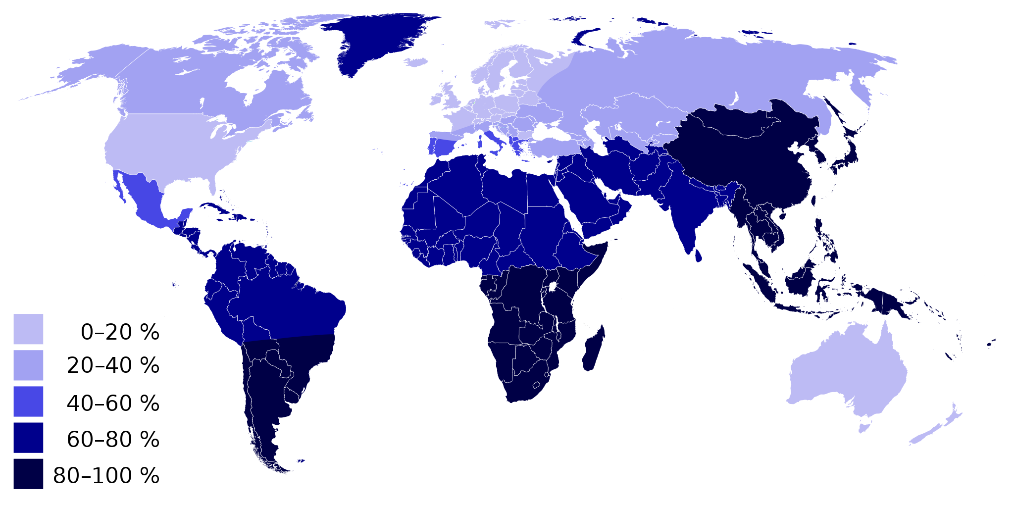
Lactose intolerance is not considered a medical problem, because its symptoms can be avoided by avoiding milk or milk products. Dietary control of lactose intolerance may be a matter of trial and error, however, because different people may be able to consume different quantities of milk or milk products before symptoms occur. If you are lactose intolerant, be aware that low-fat and fat-free milk may contain somewhat more lactose than full-fat milk because the former often have added milk solids that are relatively high in lactose.
Lactase Persistence
Lactase persistence is the opposite of lactose intolerance. People who are lactase persistent continue to produce the enzyme lactase beyond infancy and generally throughout life. As a consequence they are able to digest lactose and drink milk at older ages without adverse effects. The map in Figure 6.8.4 can also be read to show where lactase persistence occurs today. Populations with a low percentage of lactose intolerance (including most North Americans and Western and Northern Europeans) have high percentages of lactase-persistent people.
Lactase persistence is a uniquely human trait that is not found in any other mammalian species. Why did lactase persistence evolve in humans? When some human populations began domesticating and keeping herds of animals, animal milk became a potential source of food. Animals such as cows, sheep, goats, camels, and even reindeer (see Figure 6.8.5) can be kept for their milk. These animal milks also contain lactose, so natural selection would be strong for any individuals who kept producing lactase beyond infancy and could make use of this nutritious food. Eventually, the trait of lactase persistence would increase in frequency and come to be the predominant trait in dairying populations.
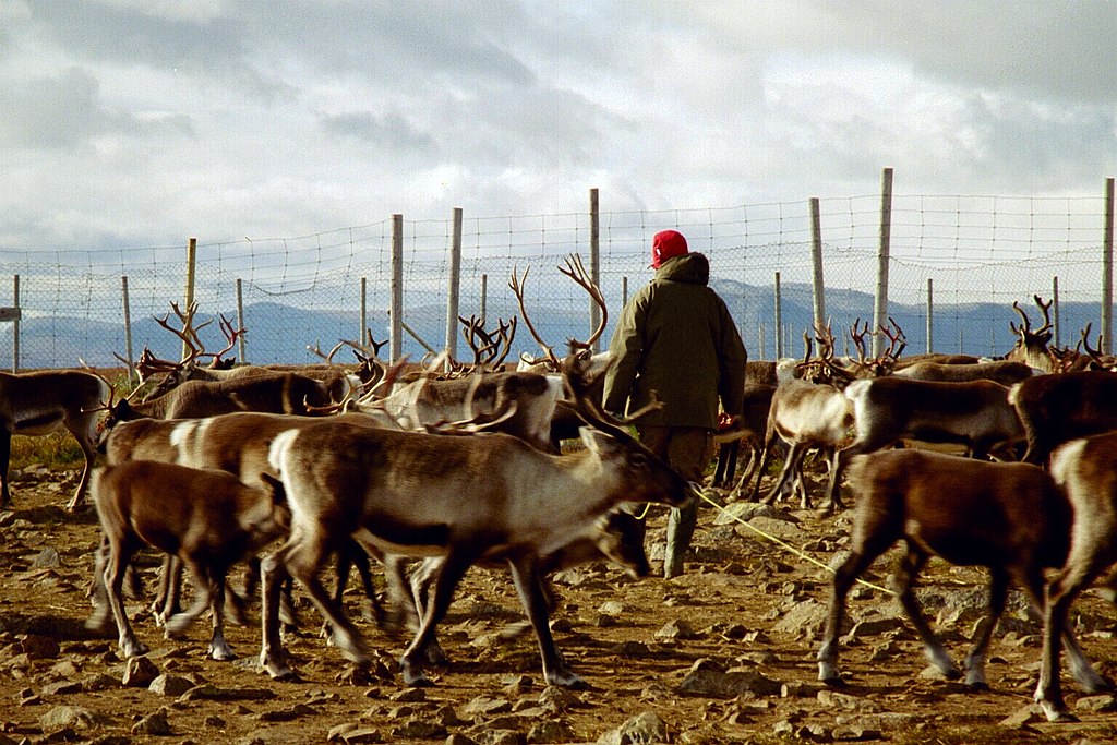
It is likely that lactase persistence occurs as a result of both genes and environment. Some people inherit genes that help them keep producing lactase after infancy. Geneticists think that several different mutations for lactase persistence arose independently in different populations within the last ten thousand years. Part of lactase persistence may be due to continued exposure to milk in the childhood and adulthood diet. In other words, a person may be genetically predisposed to synthesize lactase at older ages because of a mutation, but they may need the continued stimulation of milk drinking to keep producing lactase.
Thrifty Gene or Drifty Gene?
Besides variation in lactase persistence, human populations may vary in how efficiently they use calories in the foods they consume. People in some populations seem able to get by on quantities of food that would be inadequate for others, so they tend to gain weight easily. What explains these differences in people?
Thrifty Gene Hypothesis
In 1962, human geneticist James Neel proposed the thrifty gene hypothesis. According to this hypothesis, so-called “thrifty genes” evolved in some human populations because they allowed people to get by on fewer calories and store the rest as body fat when food was plentiful. According to Neel’s hypothesis, thrifty genes would have increased in frequency through natural selection, because they would help people survive during times of famine. People with the genes would be fatter and able to rely on their stored body fat for calories when food was scarce.
Such thrifty genes would have been advantageous in early human populations of hunter-gatherers if food scarcity was a recurrent stress. However, in modern times, when most people have access to enough food year-round, thrifty genes would no longer be advantageous. In fact, under conditions of plentiful food, having thrifty genes would predispose people to gain weight and develop obesity. They would also tend to develop the chronic diseases associated with obesity, particularly type II diabetes. Diabetes mellitus is a disease that occurs when there are problems with the pancreatic hormone insulin, which normally helps cells take up glucose from the blood and controls blood glucose levels. In type II diabetes, body cells become relatively resistant to insulin, leading to high blood glucose. This causes symptoms like excessive thirst and urination. Without treatment, diabetes can lead to serious consequences, such as blindness and kidney failure.
Neel proposed his thrifty gene hypothesis not on the basis of genetic evidence for thrifty genes, but as a possible answer to the mystery of why genes that seem to promote diabetes have not been naturally selected out of some populations. The mystery arose from observations that certain populations — such as South Pacific Islanders, sub-Saharan Africans, and southwestern Native Americans — developed high levels of obesity and diabetes after they abandoned traditional diets and adopted Western diets.
Assessing the Thrifty Gene Hypothesis
One of the assumptions underlying the thrifty gene hypothesis is that human populations that recently developed high rates of obesity and diabetes after Western contact had a long history of recurrent famine. Anthropological evidence contradicts this assumption for at least some of the populations in question. South Pacific Islanders, for example, have long lived in a “land of plenty,” with lush tropical forests year-round on islands surrounded by warm waters full of fish. Another assumption underlying the thrifty gene hypothesis is that hunter-gatherer people became significantly fatter during periods of plenty. Again, there is little or no evidence that hunter-gatherers traditionally deposited large fat stores when food was readily available.
Some geneticists have searched directly for so-called thrifty genes. Studies have revealed many genes with small effects associated with obesity or diabetes. However, these genes can explain only a few percentage points of the total population variation in obesity or diabetes.
The Drifty Gene and Other Hypotheses
Given the lack of evidence for the thrifty gene hypothesis, several researchers have suggested alternative hypotheses to explain population variation in obesity and diabetes. One hypothesis posits that susceptibility to obesity and diabetes may be a side effect of heat adaptation. According to this idea, some populations evolved lower metabolic rates as an adaptation to heat stress, because lower metabolic rates reduced the amount of heat that the body produced. The lower metabolic rates also predisposed people to gain excess weight and develop obesity and diabetes.
A thrifty phenotype hypothesis has also been proposed. This hypothesis suggests that individuals who have inadequate nutrition during fetal development might develop an insulin-resistant phenotype. The insulin-resistant phenotype would supposedly prepare these individuals for a life of famine, based on the environment within the womb. In a famine-free environment, however, the thrifty phenotype would lead to the development of diabetes.
The most recent alternative to the thrifty gene hypothesis is the drifty gene hypothesis, which was proposed by biologist John Speakman. He argues that genes protecting humans from obesity were under strong natural selection pressure for a very long period of time while human ancestors were subject to the risk of predation. According to this view, being able to outrun predators would have been an important factor selecting against fatness. When the risk of predation was lessened — perhaps as early as two million years ago — genes keeping fatness in check would no longer be selected for. Without selective pressure for these genes, their frequencies could change randomly due to genetic drift. In some populations, by chance, frequencies of the genes could decrease to relatively low levels, whereas in other populations the frequencies could be much higher.
Feature: Myth vs. Reality
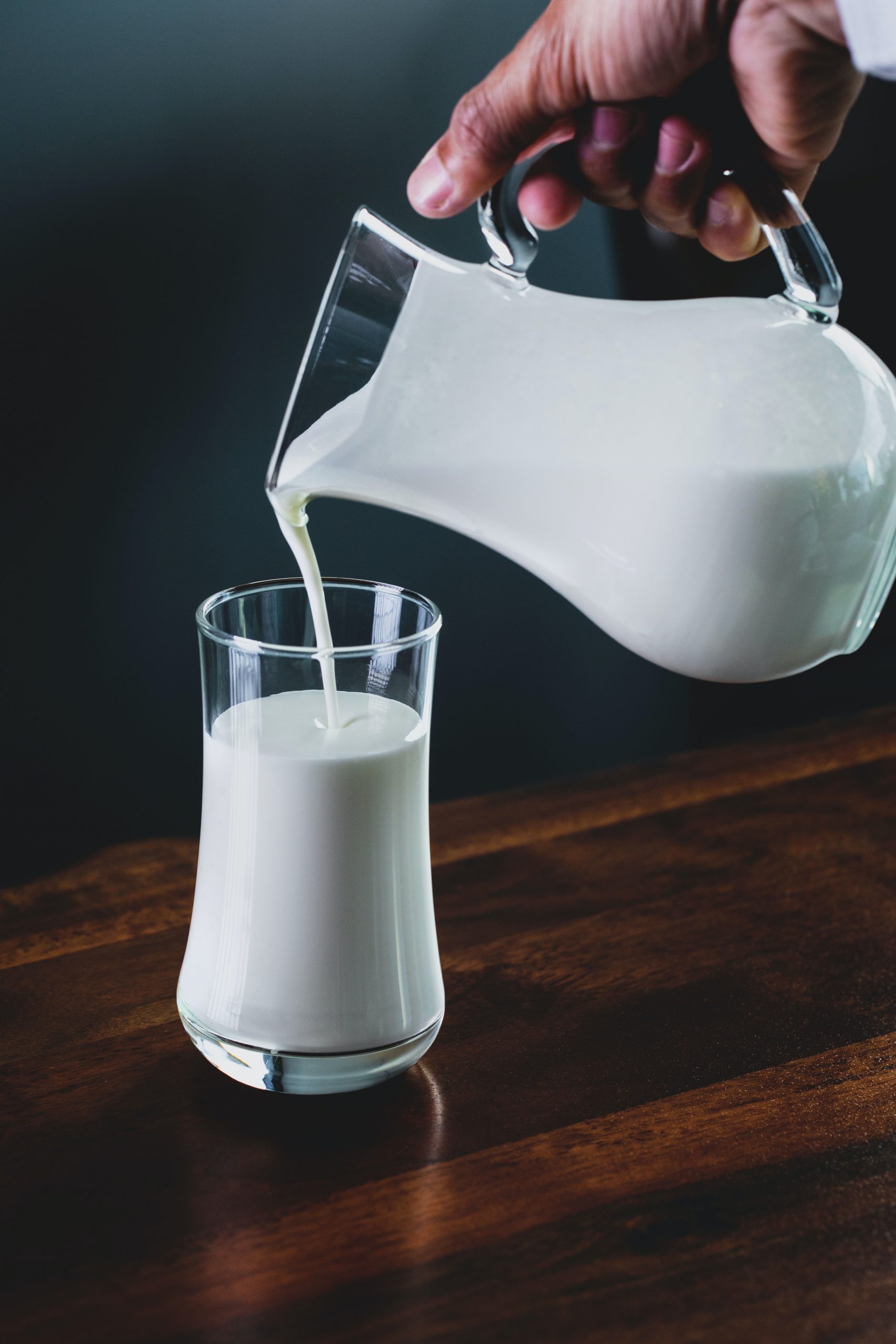
Myth: Lactose intolerance is an allergy to milk.
Reality: Lactose intolerance is not an allergy because it is not an immune system response. It is a sensitivity to milk caused by lactase deficiency so the sugar in milk cannot be digested. Milk allergy does exist, but it is a different condition that occurs in only about four per cent of people. It results when milk proteins (not milk sugar) trigger an immune reaction. How can you determine whether you have lactose intolerance or milk allergy? If you can drink lactose-free milk without symptoms, it is likely that you are lactose intolerant and not allergic to milk. However, if lactose-free milk also produces symptoms, it is likely that you have milk allergy. Note that it is possible to have both conditions.
Myth: If you are lactose intolerant, you will never be able to drink milk or consume other dairy products without suffering adverse physical symptoms.
Reality: Lactose intolerance does not mean that consuming milk and other dairy products is out of the question. Besides lactose-free milk, which is widely available, many dairy products have relatively low levels of lactose, so you may be able to consume at least small amounts of them without discomfort. You may be able to consume milk in the form of yogurt without any problems because the bacteria in yogurt produce lactase that breaks down the lactose. Greek yogurt may be your best bet, because it is lower in lactose to begin with. Aged cheeses also tend to have relatively low levels of lactose, because of the cheese-making process. Finally, by gradually adding milk or milk products to your diet, you may be able to increase your tolerance to lactose.
6.8 Summary
- Milk contains the sugar lactose, a disaccharide. Lactose must be broken down into its two component sugars to be absorbed by the small intestine, and the enzyme lactase is needed for this process.
- In about 60 per cent of people worldwide, the ability to synthesize lactase and digest lactose declines after the first two years of life. These people become lactose intolerant and cannot consume much milk without suffering symptoms of bloating, cramps, and diarrhea.
- In populations that herded milking animals for thousands of years, lactase persistence evolved. People who were able to synthesize lactase and digest lactose throughout life were strongly favored by natural selection. People — including many Europeans and European-Americans — who descended from these early herders generally still have lactase persistence.
- Human populations may vary in how efficiently they use calories in food. Some people (especially South Pacific Islanders, Native Americans, and sub-Saharan Africans) seem to be able to get by on fewer calories than would be adequate for others, so they tend to easily gain weight, become obese, and develop diseases such as diabetes.
- The thrifty gene hypothesis answers the question of how genes for this ability could have evolved. It proposes that “thrifty genes” were selected for because they allowed people to use calories efficiently and store body fat when food was plentiful so they had a reserve to use when food was scarce. Thrifty genes become detrimental and lead to obesity and diabetes when food is consistently plentiful.
- Several assumptions underlying the thrifty gene hypothesis have been called into question, and genetic research has been unable to actually identify thrifty genes. Alternate hypotheses to the thrifty gene hypothesis have been proposed, including the drifty gene hypothesis. The latter hypothesis explains variation in the tendency to become obese by genetic drift on neutral genes.
6.8 Review Questions
-
- Distinguish between the terms lactose and lactase.
- What is lactose intolerance, and what percentage of all people have it?
- Where and why did lactase persistence evolve?
- What is the thrifty gene hypothesis?
- How well is the thrifty gene hypothesis supported by evidence?
- Describe an alternative hypothesis to the thrifty gene hypothesis.
- Do you think that a lack of exposure to dairy products might affect a person’s lactase level? Why or why not?
- Describe an experiment you would want to do or data you would want to analyze that would help to test the thrifty phenotype hypothesis. Remember, you are studying people, so be sure it is ethical! Discuss possible confounding factors that you should control for, or that might affect the interpretation of your results.
- Explain the relationship between insulin, blood glucose, and type II diabetes.
6.8 Explore More
https://www.youtube.com/watch?v=G1NGzycaQV0&feature=emb_logo
Why Are People Lactose Intolerant?, Super Scienced, 2016.
https://www.youtube.com/watch?v=UMhLBPPtlrY
Peter Attia: What if we're wrong about diabetes?, TED, 2013.
https://www.youtube.com/watch?v=4O8k9qe8fjI
The Last Nomadic Reindeer Herders in the World, Great Big Story, 2018.
https://www.youtube.com/watch?v=XIYag5MWhPU
Experience a Traditional Whale Hunt in Northern Alaska | Short Film Showcase, National Geographic, 2018.
Attributions
Figure 6.8.1
IMG_4325 Milk Mustache licking 3 by Cedar Summit Farm on Flickr is used under a CC BY SA 2.0 (https://creativecommons.org/licenses/by-sa/2.0/) license.
Figure 6.8.2
Lactose Haworth by NEUROtiker on Wikimedia Commons is in the public domain (https://en.wikipedia.org/wiki/Public_domain).
Figure 6.8.3
Lactase by Boghog2 on Wikimedia Commons is released into the public domain (https://en.wikipedia.org/wiki/Public_domain).
Figure 6.8.4
Lactose Intolerance by Rainer Z ... on Wikimedia Commons is released into the public domain (https://en.wikipedia.org/wiki/Public_domain).
Figure 6.8.5
Reindeer_herding by Mats Andersson on Wikimedia Commons is used under a CC BY 2.0 (https://creativecommons.org/licenses/by/2.0/deed.en) license.
Figure 6.8.6
Milk Photo [photo] by Eiliv-Sonas Aceron on Unsplash is used under the Unsplash License (https://unsplash.com/license).
References
Great Big Story. (2018, November 29). The last nomadic reindeer herders in the world. YouTube. https://www.youtube.com/watch?v=4O8k9qe8fjI&feature=youtu.be
Super Scienced. (2016, February 26). Why are people lactose intolerant? YouTube. https://www.youtube.com/watch?v=G1NGzycaQV0&feature=youtu.be
National Geographic. (2018, November 27). Experience a traditional whale hunt in northern Alaska | Short film showcase. YouTube. https://www.youtube.com/watch?v=XIYag5MWhPU&feature=youtu.be
TED. (2013, June 25). Peter Attia: What if we're wrong about diabetes? YouTube. https://www.youtube.com/watch?v=UMhLBPPtlrY&feature=youtu.be
Wikipedia contributors. (2019, December 15). James V. Neel. In Wikipedia. https://en.wikipedia.org/w/index.php?title=James_V._Neel&oldid=930860629
Wikipedia contributors. (2020, June 9). John Speakman. In Wikipedia. https://en.wikipedia.org/w/index.php?title=John_Speakman&oldid=961610417
A complex organic chemical that provides energy to drive many processes in living cells, e.g. muscle contraction, nerve impulse propagation, and chemical synthesis. Found in all forms of life, ATP is often referred to as the "molecular unit of currency" of intracellular energy transfer.
The transfer of genetic variation from one population to another. If the rate of gene flow is high enough, then two populations are considered to have equivalent allele frequencies and therefore effectively be a single population.
Created by: CK-12/Adapted by Christine Miller

Bring on the S'mores!
This inviting camp fire can be used for both heat and light. Heat and light are two forms of energy that are released when a fuel like wood is burned. The cells of living things also get energy by "burning." They "burn" glucose in a process called cellular respiration.
What Is Cellular Respiration?
Cellular respiration is the process by which living cells break down glucose molecules and release energy. The process is similar to burning, although it doesn’t produce light or intense heat as a campfire does. This is because cellular respiration releases the energy in glucose slowly and in many small steps. It uses the energy released to form molecules of ATP, the energy-carrying molecules that cells use to power biochemical processes. In this way, cellular respiration is an example of energy coupling: glucose is broken down in an exothermic reaction, and then the energy from this reaction powers the endothermic reaction of the formation of ATP. Cellular respiration involves many chemical reactions, but they can all be summed up with this chemical equation:
C6H12O6 6O2 → 6CO2 6H2O Chemical Energy (in ATP)
In words, the equation shows that glucose (C6H12O6) and oxygen (O2) react to form carbon dioxide (CO2) and water (H2O), releasing energy in the process. Because oxygen is required for cellular respiration, it is an aerobic process.
Cellular respiration occurs in the cells of all living things, both autotrophs and heterotrophs. All of them burn glucose to form ATP. The reactions of cellular respiration can be grouped into three stages: glycolysis, the Krebs cycle (also called the citric acid cycle), and electron transport. Figure 4.10.2 gives an overview of these three stages, which are also described in detail below.
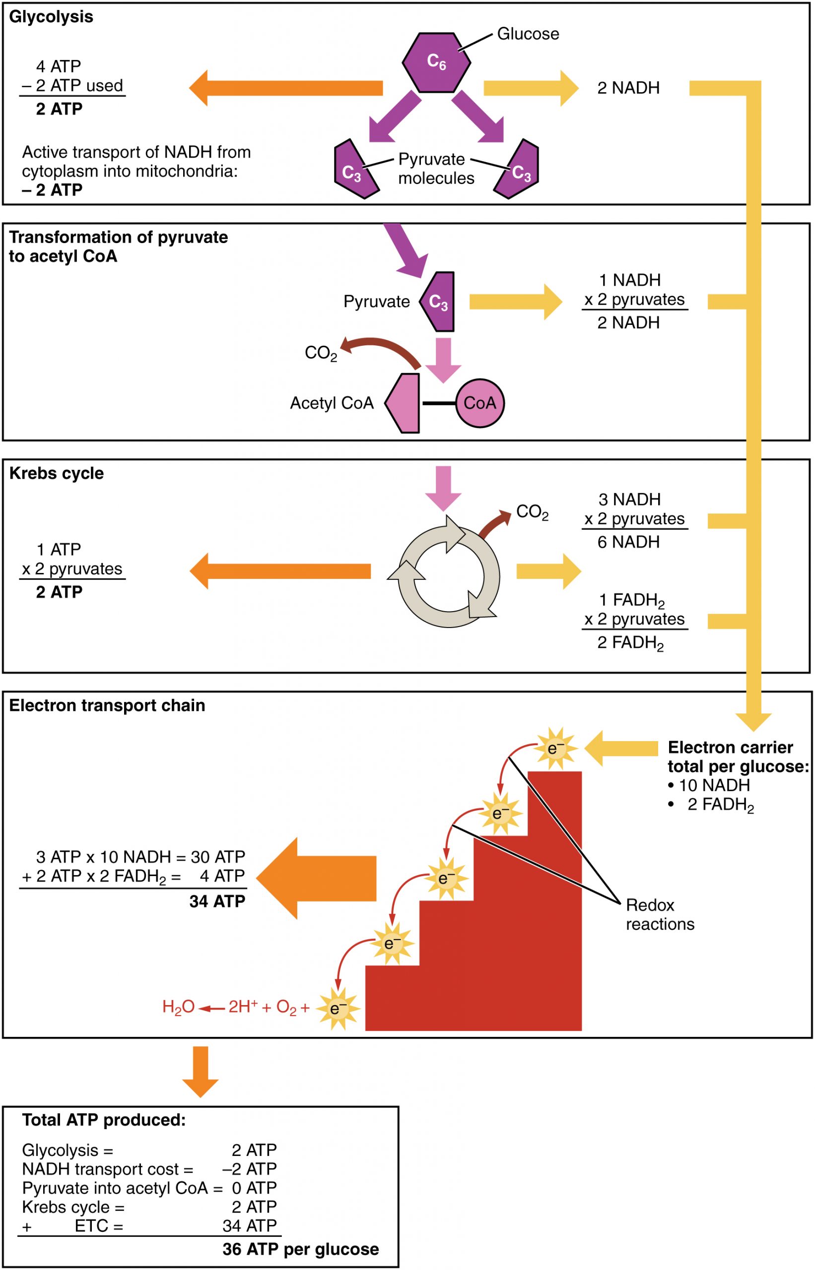
Cellular Respiration Stage I: Glycolysis
The first stage of cellular respiration is glycolysis, which happens in the cytosol of the cytoplasm.
Splitting Glucose
The word glycolysis literally means “glucose splitting,” which is exactly what happens in this stage. Enzymes split a molecule of glucose into two molecules of pyruvate (also known as pyruvic acid). This occurs in several steps, as summarized in the following diagram.
Results of Glycolysis
Energy is needed at the start of glycolysis to split the glucose molecule into two pyruvate molecules which go on to stage II of cellular respiration. The energy needed to split glucose is provided by two molecules of ATP; this is called the energy investment phase. As glycolysis proceeds, energy is released, and the energy is used to make four molecules of ATP; this is the energy harvesting phase. As a result, there is a net gain of two ATP molecules during glycolysis. During this stage, high-energy electrons are also transferred to molecules of NAD to produce two molecules of NADH, another energy-carrying molecule. NADH is used in stage III of cellular respiration to make more ATP.
Transition Reaction
Before pyruvate can enter the next stage of cellular respiration it needs to be modified slightly. The transition reaction is a very short reaction which converts the two molecules of pyruvate to two molecules of acetyl CoA, carbon dioxide, and two high energy electron pairs convert NAD to NADH. The carbon dioxide is released, the acetyl CoA moves to the mitochondria to enter the Kreb's Cycle (stage II), and the NADH carries the high energy electrons to the Electron Transport System (stage III).
Structure of the Mitochondrion
Before you read about the last two stages of cellular respiration, you need to know more about the mitochondrion, where these two stages take place. A diagram of a mitochondrion is shown in Figure 4.10.5.
The structure of a mitochondrion is defined by an inner and outer membrane. This structure plays an important role in aerobic respiration.
As you can see from the figure, a mitochondrion has an inner and outer membrane. The space between the inner and outer membrane is called the intermembrane space. The space enclosed by the inner membrane is called the matrix. The second stage of cellular respiration (the Krebs cycle) takes place in the matrix. The third stage (electron transport) happens on the inner membrane.
Cellular Respiration Stage II: The Krebs Cycle
Recall that glycolysis produces two molecules of pyruvate (pyruvic acid), which are then converted to acetyl CoA during the short transition reaction. These molecules enter the matrix of a mitochondrion, where they start the Krebs cycle (also known as the Citric Acid Cycle). The reason this stage is considered a cycle is because a molecule called oxaloacetate is present at both the beginning and end of this reaction and is used to break down the two molecules of acetyl CoA. The reactions that occur next are shown in Figure 4.10.6.
Steps of the Krebs Cycle
The Krebs cycle itself actually begins when acetyl-CoA combines with a four-carbon molecule called OAA (oxaloacetate) (see Figure 4.10.6). This produces citric acid, which has six carbon atoms. This is why the Krebs cycle is also called the citric acid cycle.
After citric acid forms, it goes through a series of reactions that release energy. The energy is captured in molecules of NADH, ATP, and FADH2, another energy-carrying coenzyme. Carbon dioxide is also released as a waste product of these reactions.
The final step of the Krebs cycle regenerates OAA, the molecule that began the Krebs cycle. This molecule is needed for the next turn through the cycle. Two turns are needed because glycolysis produces two pyruvic acid molecules when it splits glucose.
Results of the Glycolysis, Transition Reaction and Krebs Cycle
After glycolysis, transition reaction, and the Krebs cycle, the glucose molecule has been broken down completely. All six of its carbon atoms have combined with oxygen to form carbon dioxide. The energy from its chemical bonds has been stored in a total of 16 energy-carrier molecules. These molecules are:
- 4 ATP (2 from glycolysis, 2 from Krebs Cycle)
- 12 NADH (2 from glycolysis, 2 from transition reaction, and 8 from Krebs cycle)
- 2 FADH2 (both from the Krebs cycle)
The events of cellular respiration up to this point are exergonic reactions- they are releasing energy that had been stored in the bonds of the glucose molecule. This energy will be transferred to the third and final stage of cellular respiration: the Electron Transport System, which is an endergonic reaction. Using an exothermic reaction to power an endothermic reaction is known as energy coupling.
Cellular Respiration Stage III: Electron Transport Chain
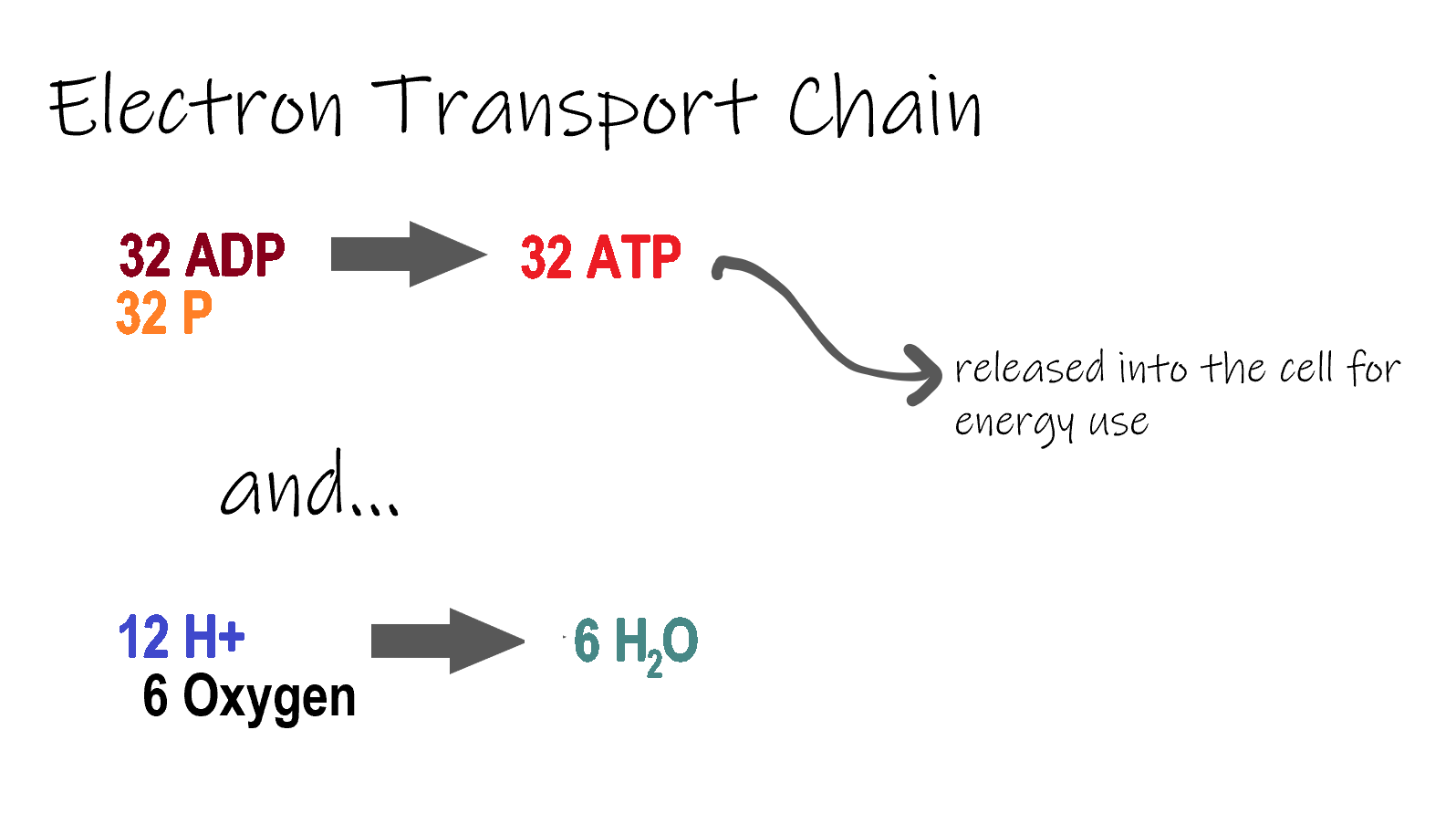
ETC, the final stage in cellular respiration produces 32 ATP. The Electron Transport Chain is the final stage of cellular respiration. In this stage, energy being transported by NADH and FADH2 is transferred to ATP. In addition, oxygen acts as the final proton acceptor for the hydrogens released from all the NADH and FADH2, forming water. Figure 4.10.8 shows the reactants and products of the ETC.
Transporting Electrons
The Electron transport chain is the third stage of cellular respiration and is illustrated in Figure 4.10.8. During this stage, high-energy electrons are released from NADH and FADH2, and they move along electron-transport chains on the inner membrane of the mitochondrion. An electron-transport chain is a series of molecules that transfer electrons from molecule to molecule by chemical reactions. Some of the energy from the electrons is used to pump hydrogen ions (H ) across the inner membrane, from the matrix into the intermembrane space. This ion transfer creates an electrochemical gradient that drives the synthesis of ATP.
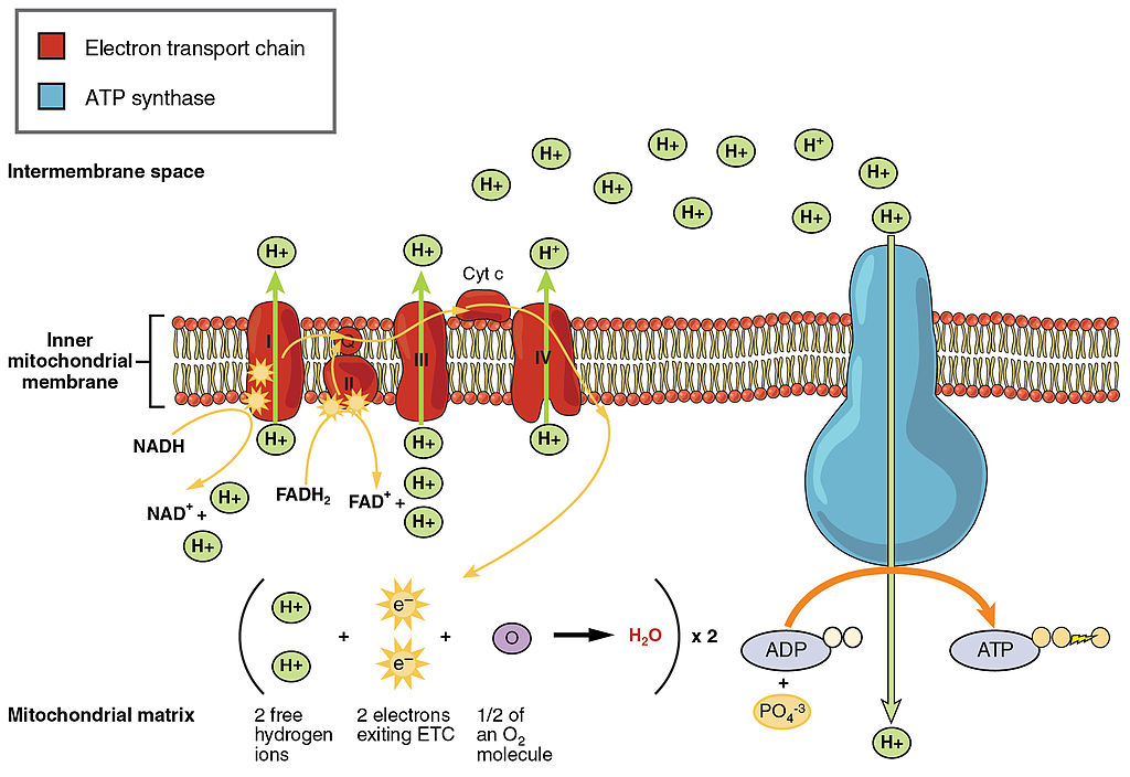
Making ATP
As shown in Figure 4.10.8, the pumping of hydrogen ions across the inner membrane creates a greater concentration of the ions in the intermembrane space than in the matrix. This gradient causes the ions to flow back across the membrane into the matrix, where their concentration is lower. ATP synthase acts as a channel protein, helping the hydrogen ions cross the membrane. It also acts as an enzyme, forming ATP from ADP and inorganic phosphate in a process called oxidative phosphorylation. After passing through the electron-transport chain, the “spent” electrons combine with oxygen to form water.
How Much ATP?
You have seen how the three stages of aerobic respiration use the energy in glucose to make ATP. How much ATP is produced in all three stages combined? Glycolysis produces two ATP molecules, and the Krebs cycle produces two more. Electron transport begins with several molecules of NADH and FADH2 from the Krebs cycle and transfers their energy into as many as 34 more ATP molecules. All told, then, up to 38 molecules of ATP can be produced from just one molecule of glucose in the process of cellular respiration.
4.10 Summary
- Cellular respiration is the aerobic process by which living cells break down glucose molecules, release energy, and form molecules of ATP. Generally speaking, this three-stage process involves glucose and oxygen reacting to form carbon dioxide and water.
- The first stage of cellular respiration, called glycolysis, takes place in the cytoplasm. In this step, enzymes split a molecule of glucose into two molecules of pyruvate, which releases energy that is transferred to ATP. Following glycolysis, a short reaction called the transition reaction converts the pyruvate into two molecules of acetyl CoA.
- The organelle called a mitochondrion is the site of the other two stages of cellular respiration. The mitochondrion has an inner and outer membrane separated by an intermembrane space, and the inner membrane encloses a space called the matrix.
- The second stage of cellular respiration, called the Krebs cycle, takes place in the matrix of a mitochondrion. During this stage, two turns through the cycle result in all of the carbon atoms from the two pyruvate molecules forming carbon dioxide and the energy from their chemical bonds being stored in a total of 16 energy-carrying molecules (including two from glycolysis and two from transition reaction).
- The third and final stage of cellular respiration, called electron transport, takes place on the inner membrane of the mitochondrion. Electrons are transported from molecule to molecule down an electron-transport chain. Some of the energy from the electrons is used to pump hydrogen ions across the membrane, creating an electrochemical gradient that drives the synthesis of many more molecules of ATP.
- In all three stages of cellular respiration combined, as many as 38 molecules of ATP are produced from just one molecule of glucose.
4.10 Review Questions
- What is the purpose of cellular respiration? Provide a concise summary of the process.
- State what happens during glycolysis.
- Describe the structure of a mitochondrion.
- What molecule is present at both the beginning and end of the Krebs cycle?
- What happens during the electron transport stage of cellular respiration?
- How many molecules of ATP can be produced from one molecule of glucose during all three stages of cellular respiration combined?
- Do plants undergo cellular respiration? Why or why not?
- Explain why the process of cellular respiration described in this section is considered aerobic.
- Name three energy-carrying molecules involved in cellular respiration.
-
- Which stage of aerobic cellular respiration produces the most ATP?
-
4.10 Explore More
https://www.youtube.com/watch?time_continue=2&v=00jbG_cfGuQ&feature=emb_logo
ATP & Respiration: Crash Course Biology #7, CrashCourse, 2012.
https://www.youtube.com/watch?v=4Eo7JtRA7lg&t=3s
Cellular Respiration and the Mighty Mitochondria, The Amoeba Sisters, 2014.
Attributions
Figure 4.10.1
Smores by Jessica Ruscello on Unsplash is used under the Unsplash License (https://unsplash.com/license).
Figure 4.10.2
Carbohydrate_Metabolism by OpenStax College on Wikimedia Commons is used under a CC BY 3.0 (https://creativecommons.org/licenses/by/3.0) license.
Figure 4.10.3
Glycolysis by Christine Miller is used under a CC BY 4.0 (https://creativecommons.org/licenses/by/4.0/) license.
Figure 4.10.4
Transition Reaction by Christine Miller is used under a CC BY 4.0 (https://creativecommons.org/licenses/by/4.0/) license.
Figure 4.10.5
Mitochondrion by Mariana Ruiz Villarreal [LadyofHats] on Wikimedia Commons is released into the public domain (https://en.wikipedia.org/wiki/Public_domain).
Figure 4.10.6
Krebs cycle by Christine Miller is used under a CC BY 4.0 (https://creativecommons.org/licenses/by/4.0/) license.
Figure 4.10.7
Electron Transport Chain (ETC) by Christine Miller is used under a CC BY 4.0 (https://creativecommons.org/licenses/by/4.0/) license.
Figure 4.10.8
The_Electron_Transport_Chain by OpenStax College on Wikimedia Commons is used under a CC BY 3.0 (https://creativecommons.org/licenses/by/3.0) license.
References
CrashCourse. (2012, March 12). ATP & Respiration: Crash Course Biology #7. YouTube. https://www.youtube.com/watch?time_continue=2&v=00jbG_cfGuQ&feature=emb_logo
Betts, J. G., Young, K.A., Wise, J.A., Johnson, E., Poe, B., Kruse, D.H., Korol, O., Johnson, J.E., Womble, M., DeSaix, P. (2013, April 25). Figure 24.8 Electron Transport Chain [digital image]. In Anatomy & Physiology, Connexions (Section ). OpenStax. https://openstax.org/books/anatomy-and-physiology/pages/24-2-carbohydrate-metabolism
Betts, J. G., Young, K.A., Wise, J.A., Johnson, E., Poe, B., Kruse, D.H., Korol, O., Johnson, J.E., Womble, M., DeSaix, P. (2013, April 25). Figure 24.9 Carbohydrate Metabolism [digital image]. In Anatomy & Physiology, Connexions (Section 24.2). OpenStax. https://openstax.org/books/anatomy-and-physiology/pages/24-2-carbohydrate-metabolism
The Amoeba Sisters. (2014, October 22). Cellular Respiration and the Mighty Mitochondria. YouTube. https://www.youtube.com/watch?v=4Eo7JtRA7lg&t=3s
Created by CK-12/Adapted by Christine Miller
As you read in the beginning of this chapter, new parents Samantha and Aki left their pediatrician’s office still unsure whether or not to vaccinate baby James. Dr. Rodriguez gave them a list of reputable sources where they could look up information about the safety of vaccines, including the Centers for Disease Control and Prevention (CDC). Samantha and Aki read that the consensus within the scientific community is that there is no link between vaccines and autism. They find a long list of studies published in peer-reviewed scientific journals that disprove any link. Additionally, some of the studies are “meta-analyses” that analyzed the findings from many individual studies. The new parents are reassured by the fact that many different researchers, using a large number of subjects in numerous well-controlled and well-reviewed studies, all came to the same conclusion.

Samantha also went back to the web page that originally scared her about the safety of vaccines. She found that the author was not a medical doctor or scientific researcher, but rather a self-proclaimed “child wellness expert.” He sold books and advertising on his site, some of which were related to claims of vaccine injury. She realized that he was both an unqualified and potentially biased source of information.
Samantha also realized that some of his arguments were based on correlations between autism and vaccines, but, as the saying goes, “correlation does not imply causation.” For instance, the recent rise in autism rates may have occurred during the same time period as an increase in the number of vaccines given in childhood, but Samantha could think of many other environmental and social factors that have also changed during this time period. There are just too many variables to come to the conclusion that vaccines, or anything else, are the cause of the rise in autism rates based on that type of argument alone. Also, she learned that the age of onset of autism symptoms happens to typically be around the time that the MMR vaccine is first given, so the apparent association in the timing may just be a coincidence.
Finally, Samantha came across news about a measles outbreak in Vancouver, British Columbia in the winter of 2019. Measles wasn’t just a disease of the past! She learned that measles and whooping cough, which had previously been rare thanks to widespread vaccinations, are now on the rise, and that people choosing not to vaccinate their children seems to be one of the contributing factors. She realized that it is important to vaccinate her baby against these diseases, not only to protect him from their potentially deadly effects, but also to protect others in the population.
In their reading, Samantha and Aki learn that scientists do not yet know the causes of autism, but they feels reassured by the abundance of data that disproves any link with vaccines. Both parents think that the potential benefits of protecting their baby’s health against deadly diseases outweighs any unsubstantiated claims about vaccines. They will be making an appointment to get baby James his shots soon.
Chapter 1 Summary
In this chapter, you learned about some of the same concepts that helped Samantha and Aki make an informed decision. Specifically:
- Science is a distinctive way of gaining knowledge about the natural world that is based on the use of evidence to logically test ideas. As such, science is a process, as well as a body of knowledge.
- A scientific theory, such as the germ theory of disease, is the highest level of explanation in science. A theory is a broad explanation for many phenomena that is widely accepted because it is supported by a great deal of evidence.
- The scientific investigation is the cornerstone of science as a process. A scientific investigation is a systematic approach to answering questions about the physical and natural world. An investigation may be observational or experimental.
- A scientific experiment is a type of scientific investigation in which the researcher manipulates variables under controlled conditions to test expected outcomes. Experiments are the gold standard for scientific investigations and can establish causation between variables.
- Nonexperimental scientific investigations such as observational studies and modeling may be undertaken when experiments are impractical, unethical, or impossible. Observational studies generally can establish correlation — but not causation — between variables.
- A pseudoscience, such as astrology, is a field that is presented as scientific but that does not adhere to scientific standards and methods. Other misuses of science include deliberate hoaxes, frauds, and fallacies made by researchers.
- Strict guidelines must be followed when using human subjects in scientific research. Among the most important protections is the requirement for informed consent.
Now that you know about the nature and process of science, you can apply these concepts in the next chapter to the study of human biology.
Chapter 1 Review
-
- Why does a good hypothesis have to be falsifiable?
- Name one scientific law.
- Name one scientific theory.
- Give an example of a scientific idea that was later discredited.
- A statistical measurement called a P-value is often used in science to determine whether or not a difference between two groups is actually significant or simply due to chance. A P-value of 0.03 means that there is a 3% chance that the difference is due to chance alone. Do you think a P-value of 0.03 would indicate that the difference is likely to be significant? Why or why not?
- Why is it important that scientists communicate their findings to others? How do they usually do this?
- What is a “control group” in science?
- In a scientific experiment, why is it important to only change one variable at a time?
- Which is the dependent variable – the variable that is manipulated or the variable that is being affected by the change?
- You see an ad for a “miracle supplement” called NQP3 that claims the supplement will reduce belly fat. They say it works by reducing the hormone cortisol and by providing your body with missing unspecified “nutrients”, but they do not cite any peer-reviewed clinical studies. They show photographs of three people who appear slimmer after taking the product. A board-certified plastic surgeon endorses the product on television. Answer the following questions about this product.
a. Do you think that because a doctor endorsed the product, it really works? Explain your answer.
b. What are two signs that these claims could actually be pseudoscience instead of true science?
c. Do you think the photographs are good evidence that the product works? Why or why not?
d. If you wanted to do a strong scientific study of whether this supplement does what it claims, what would you do? Be specific about the subjects, data collected, how you would control variables, and how you would analyze the data.
e. What are some ways that you would ensure that the subjects in your experiment in part d are treated ethically and according to human subjects protections regulations?
Attribution
Figure 1.8.1
[Photo of person sitting in front of personal computer] by Avel Chuklanov on Unsplash is used under the Unsplash License (https://unsplash.com/license).
Created by CK-12 Foundation/Adapted by Christine Miller
Figure 7.2.1 Complex machines.
A Fantastic Machine
These robots were created for research or to do complex tasks, but they look like they might be fun to play with too! They are all complex machines. Think about some other, more familiar machines, such as power drills, washing machines, and lawn mowers. Each machine consists of many parts, and each part does a specific job, yet all the parts work together to perform certain functions. Many people have compared the human body to a machine, albeit an extremely complex one. Like real machines, the human body also consists of many parts that work together to perform certain functions. In this case, these parts and functions keep the organism alive. The human body may be the most fantastic machine on Earth, as you will discover when you learn more about it in this concept.
What the Human Machine Can Do
Imagine a machine that has all of the following attributes:
- It can generate a “wind” of 166 km/hr (100 mi/hr).
- It can relay messages faster than 400 km/hr (249 mi/hr).
- It contains a pump that moves about a million barrels of fluid over its lifetime.
- It has a control center that contains billions of individual components.
- It can repair itself, if necessary.
- It may not wear out for up to a century or more.
This machine has all of these abilities, and yet it consists mainly of water. What is it? It is the human body.
Organization of the Human Body
The human body is a complicated, highly organized structure that consists of trillions of parts that function together to achieve all the functions needed to maintain life. The biology of the human body incorporates:
- The body’s structure, the study of which is called anatomy.
- The body’s functioning, the study of which is called physiology.
The organization of the human body can be seen as a hierarchy of increasing size and complexity, starting at the level of atoms and molecules, and ending at the level of the entire organism, which is an individual living thing. You can see the intervening levels of organization in Figure 7.2.2. Read about the levels in the sections that follow.
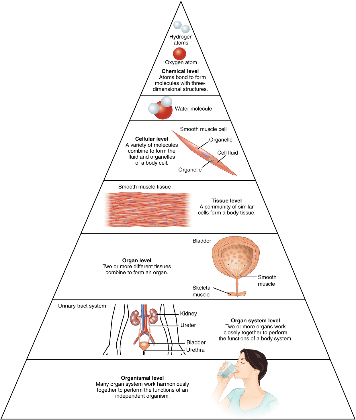
Cells
The basic units of structure and function of the human body — as in all living things — are cells. By the time the average person reaches adulthood, their body has an amazing 37 trillion of them! Each cell carries out basic life processes that allow the body to survive. In addition, most human cells are specialized in structure and function to carry out other specific roles. In fact, the human body may consist of as many as 200 different types of cells, each of which has a special job to do. Just a few of these different human cell types are pictured in Figure 7.2.3. These cells have obvious differences in structure that reflect their different functions. For example, nerve cells have long projections sticking out from the body of the cell. These projections help them carry electrical messages to other cells.

Tissues
The next level of organization in the human body is tissues. A tissue is a group of connected cells that have a similar function. There are four basic types of human tissues: epithelial, muscle, nervous, and connective tissues. These four tissue types (shown in Figure 7.2.4) make up all the organs of the human body.
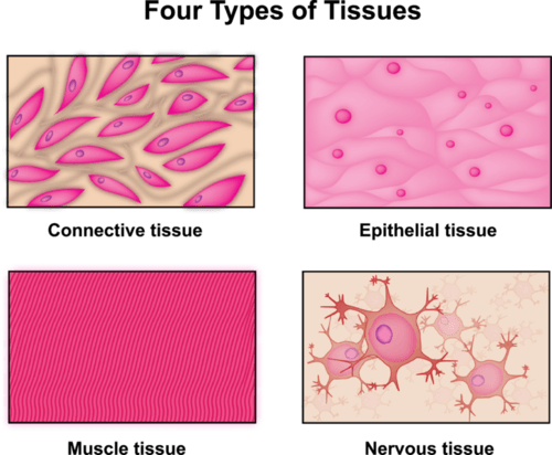
Organs and Organ Systems
Organs are the next level of organization of the human body. An organ is a structure that consists of two or more types of tissues that work together to do the same job. Examples of human organs include the heart, brain, lungs, skin, and kidneys. Human organs are organized into organ systems, which are shown in Figure 7.2.5. An organ system is a group of organs that work together to carry out a complex overall function. Each organ of the system does part of the larger job.
Figure 7.2.5 The Human Organ Systems. Some of the system names shown in this illustration differ from the terminology used in this book, but the systems are the same.
A Well-Oiled Machine
All of the organs and organ systems of the human body normally work together like a well-oiled machine, because they are closely regulated by the nervous and endocrine systems. The nervous system controls virtually all body activities, and the endocrine system secretes hormones that help to regulate these activities. Functioning together, the organ systems supply body cells with all the substances they need and eliminate their wastes. They also keep temperature, pH, and other conditions at just the right levels to support life.
7.2 Summary
- The human body is like an extremely complex machine. It consists of multiple parts that function together to maintain life. The biology of the human body incorporates the body’s structure (or anatomy) and the body’s functioning (or physiology).
- The organization of the human body is a hierarchy of increasing size and complexity, starting at the level of atoms and molecules, and ending at the level of the entire organism.
- Cells are the level of organization above atoms and molecules, and they are the basic units of structure and function of the human body. Each cell carries out basic life functions, as well as other specific roles. Variations in cell function are generally reflected in variations in cell structure.
- The next level of organization above cells is the tissue. A tissue is a group of connected cells that have a similar function. There are four basic types of human tissues: epithelial, muscle, nervous, and connective tissues. These four types of tissues make up all the organs of the human body.
- The next level of organization above tissues is the organ. An organ is a structure that consists of two or more types of tissues that work together to do the same job. Examples include the brain and heart.
- Human organs are organized into organ systems. An organ system is a group of organs that work together to carry out a complex overall function. For example, the skeletal system provides structure to the body and protects internal organs.
- All of the organs and organ systems of the body normally work together like a well-oiled machine, because they are closely regulated by the nervous and endocrine systems.
7.2 Review Questions
- How is the human body like a complex machine?
- Describe the difference between human anatomy and human physiology.
-
- Relate cell structure to cell function, and give examples of specific cell types in the human body.
- Define tissue, and identify the four types of tissues that make up the human body.
- What is an organ? Give three examples of organs in the human body.
- Define organ systems. Name five examples in the human body.
- How is the human body regulated so all of its organs and organ systems work together?
-
- Which organ system’s function is to provide structure to the body and protect internal organs?
- Give one example of how the respiratory and circulatory systems work together.
7.2 Explore More
https://www.youtube.com/watch?v=i-icXZ2tMRM
Rob Knight: How our microbes make us who we are, TED, 2015.
https://www.youtube.com/watch?time_continue=202&v=I43hq13MnYM&feature=emb_logo
Computers That Think Like Humans, Fw: Thinking, 2014.
Attributions
Figure 7.2.1
- White and brown human robot illustration by Franck V. on Unsplash is used under the Unsplash License (https://unsplash.com/license).
- Mighty Mouse, a Robotic Vehicle Range (RVR) by Science in HD on Unsplash is used under the Unsplash License (https://unsplash.com/license).
- Lauron4c 2009 FZI Karlsruhe from the FZI Research Center for Information Technology - Department IDS (Germany) on Wikimedia Commons is released for free use.
- NASA Mars Rover (artist's concept) by NASA/JPL/Cornell University, Maas Digital LLC (#PIA04413) on Wikimedia Commons is in the public domain (https://en.wikipedia.org/wiki/Public_domain).
Figure 7.2.2
101_Levels_of_Org_in_Body by OpenStax on Wikimedia Commons is used under a CC BY 4.0 (https://creativecommons.org/licenses/by/4.0/deed.en) licence.
Figure 7.2.3
Feature_Stem_Cell_new by OpenStax College on Wikimedia Commons is used under a CC BY 3.0 (https://creativecommons.org/licenses/by/3.0/deed.en) license.
Figure 7.2.4
Four types of tissues by CK-12 Foundation/ Zachary Wilson is used under a CC BY-NC 3.0 (https://creativecommons.org/licenses/by-nc/3.0/) license.
Figure 7.2.5
Organ Systems 1 by Connexions/ OpenStax on Wikimedia Commons is used under a CC BY 3.0 (https://creativecommons.org/licenses/by/3.0/deed.en) license.
References
Betts, J. G., Young, K.A., Wise, J.A., Johnson, E., Poe, B., Kruse, D.H., Korol, O., Johnson, J.E., Womble, M., DeSaix, P. (2013, April 25). Figure 1.3 Levels of Structural Organization of the Human Body [digital image]. In Anatomy and Physiology (Section 1.2). OpenStax. https://openstax.org/books/anatomy-and-physiology/pages/1-2-structural-organization-of-the-human-body
Betts, J. G., Young, K.A., Wise, J.A., Johnson, E., Poe, B., Kruse, D.H., Korol, O., Johnson, J.E., Womble, M., DeSaix, P. (2013, April 25). Figure 1.4 Organ Systems of the Human Body [digital image]. In Anatomy and Physiology (Section 1.2). OpenStax. https://openstax.org/books/anatomy-and-physiology/pages/1-2-structural-organization-of-the-human-body
Betts, J. G., Young, K.A., Wise, J.A., Johnson, E., Poe, B., Kruse, D.H., Korol, O., Johnson, J.E., Womble, M., DeSaix, P. (2013, April 25). Figure 3.36 Stem Cells [digital image]. In Anatomy and Physiology (Section 3.6). OpenStax. https://openstax.org/books/anatomy-and-physiology/pages/3-6-cellular-differentiation
Brainard, J/ CK-12 Foundation. (2016). Figure 4 The human body contains these four types of tissues [digital image]. In CK-12 College Human Biology (Section 9.12) [online Flexbook]. CK12.org. https://www.ck12.org/book/ck-12-college-human-biology/section/9.2/
Fw: Thinking. (2014, May 14). Computers that think like humans. YouTube. https://www.youtube.com/watch?v=I43hq13MnYM&feature=youtu.be
TED. (2015, Febuary 23). Rob Knight: How our microbes make us who we are. YouTube. https://www.youtube.com/watch?v=i-icXZ2tMRM&feature=youtu.be
A substance that takes part in and undergoes change during a chemical reaction.
Created by Christine Miller
Figure 7.4.1 Construction — It's important to have the right materials for the job.
The Right Material for the Job
Building a house is a big job and one that requires a lot of different materials for specific purposes. As you can see in Figure 7.4.1, many different types of materials are used to build a complete house, but each type of material fulfills certain functions. You wouldn't use insulation to cover your roof, and you wouldn't use lumber to wire your home. Just as a builder chooses the appropriate materials to build each aspect of a home (wires for electrical, lumber for framing, shingles for roofing), your body uses the right cells for each type of role. When many cells work together to perform a specific function, this is termed a tissue.
Tissues
Groups of connected cells form tissues. The cells in a tissue may all be the same type, or they may be of multiple types. In either case, the cells in the tissue work together to carry out a specific function, and they are always specialized to be able to carry out that function better than any other type of tissue. There are four main types of human tissues: connective, epithelial, muscle, and nervous tissues. We use tissues to build organs and organ systems. The 200 types of cells that the body can produce based on our single set of DNA can create all the types of tissue in the body.
Epithelial Tissue
Epithelial tissue is made up of cells that line inner and outer body surfaces, such as the skin and the inner surface of the digestive tract. Epithelial tissue that lines inner body surfaces and body openings is called mucous membrane. This type of epithelial tissue produces mucus, a slimy substance that coats mucous membranes and traps pathogens, particles, and debris. Epithelial tissue protects the body and its internal organs, secretes substances (such as hormones) in addition to mucus, and absorbs substances (such as nutrients).
The key identifying feature of epithelial tissue is that it contains a free surface and a basement membrane. The free surface is not attached to any other cells and is either open to the outside of the body, or is open to the inside of a hollow organ or body tube. The basement membrane anchors the epithelial tissue to underlying cells.
Epithelial tissue is identified and named by shape and layering. Epithelial cells exist in three main shapes: squamous, cuboidal, and columnar. These specifically shaped cells can, depending on function, be layered several different ways: simple, stratified, pseudostratified, and transitional.
Epithelial tissue forms coverings and linings and is responsible for a range of functions including diffusion, absorption, secretion and protection. The shape of an epithelial cell can maximize its ability to perform a certain function. The thinner an epithelial cell is, the easier it is for substances to move through it to carry out diffusion and/or absorption. The larger an epithelial cell is, the more room it has in its cytoplasm to be able to make products for secretion, and the more protection it can provide for underlying tissues. Their are three main shapes of epithelial cells: squamous (which is shaped like a pancake- flat and oval), cuboidal (cube shaped), and columnar (tall and rectangular).
Figure 7.4.2 The shape of epithelial tissues is important.
Epithelial tissue will also organize into different layerings depending on their function. For example, multiple layers of cells provide excellent protection, but would no longer be efficient for diffusion, whereas a single layer would work very well for diffusion, but no longer be as protective; a special type of layering called transitional is needed for organs that stretch, like your bladder. Your tissues exhibit the layering that makes them most efficient for the function they are supposed to perform. There are four main layerings found in epithelial tissue: simple (one layer of cells), stratified (many layers of cells), pseudostratified (appears stratified, but upon closer inspection is actually simple), and transitional (can stretch, going from many layers to fewer layers).
Figure 7.4.3 The layerings found in epithelial tissues is important.
See Table 7.4.1 for a summary of the different layering types and shapes epithelial cells can form and their related functions and locations.
Table 7.4.1
Summary of Epithelial Tissue Cells
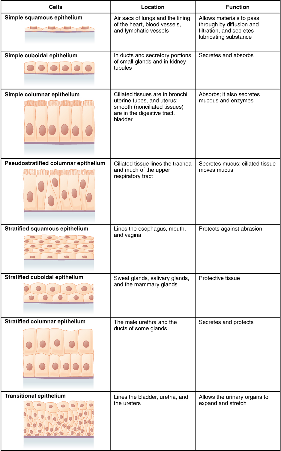
So far, we have identified epithelial tissue based on shape and layering. The representative diagrams we have seen so far are helpful for visualizing the tissue structures, but it is important to look at real examples of these cells. Since cells are too tiny to see with the naked eye, we rely on microscopes to help us study them. Histology is the study of the microscopic anatomy and cells and tissues. See Table 7.4.2 to see some examples of slides of epithelial tissues prepared for the purpose of histology.
Table 7.4.2
Epithelial Tissues and Histological Samples
| Epithelial Tissue Type | Tissue Diagram | Histological Sample |
| Stratified squamous
(from skin) |
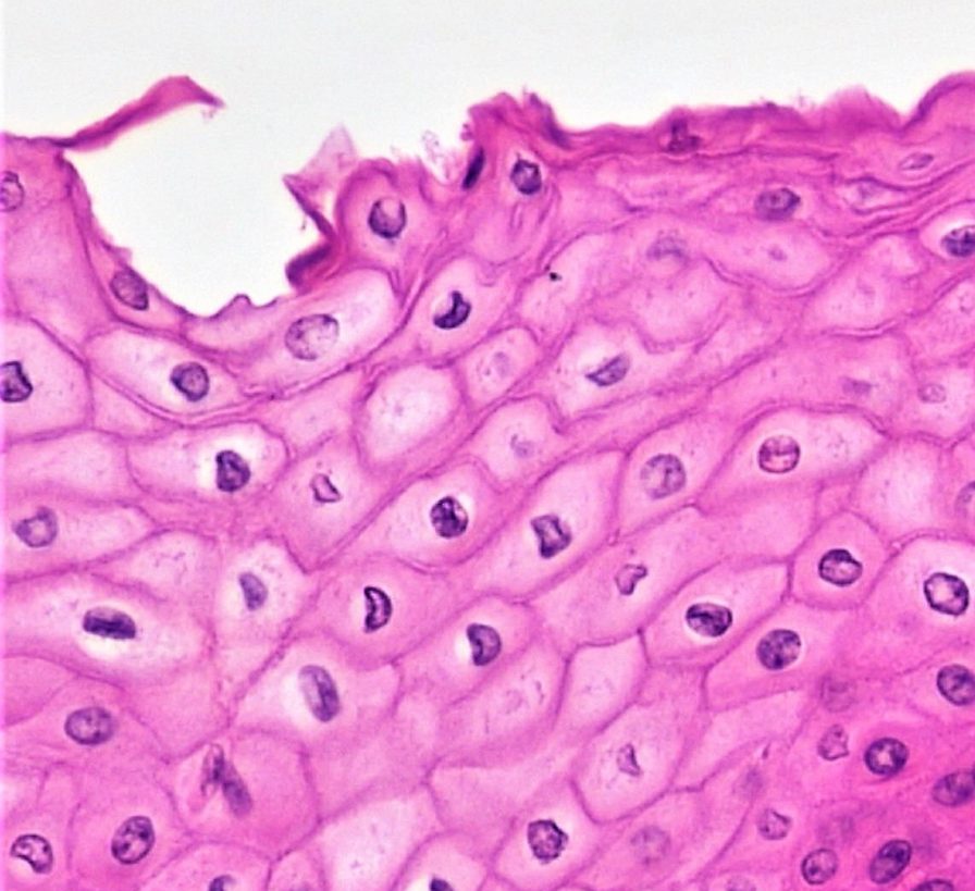 |
|
| Simple cuboidal
(from kidney tubules) |
 |
|
| Pseudostratified ciliated columnar
(from trachea) |
 |
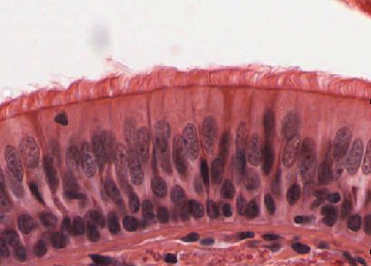 |
Connective Tissue
Bone and blood are examples of connective tissue. Connective tissue is very diverse. In general, it forms a framework and support structure for body tissues and organs. It's made up of living cells separated by non-living material, called extracellular matrix, which can be solid or liquid. The extracellular matrix of bone, for example, is a rigid mineral framework. The extracellular matrix of blood is liquid plasma.
The key identifying feature of connective tissue is that is is composed of a scattering of cells in a non-cellular matrix. There are three main categories of connective tissue, based on the nature of the matrix. They look very different from one another, which is a reflection of their different functions:
- Fibrous connective tissue: is characterized by a matrix which is flexible and is made of protein fibres including collagen, elastin and possibly reticular fibres. These tissues are found making up tendons, ligaments, and body membranes.
- Supportive connective tissue: is characterized by a solid matrix and is what is used to make bone and cartilage. These tissues are used for support and protection.
- Fluid connective tissue: is characterized by a fluid matrix and includes both blood and lymph.
Fibrous Connective Tissue
Fibrous connective tissue contains cells called fibroblasts. These cells produce fibres of collagen, elastin, or reticular fibre which makes up the matrix of this type of connective tissue. Based on how tightly packed these fibres are and how they are oriented changes the properties, and therefore the function of the fibrous connective tissue.
- Loose fibrous connective tissue: composed of a loose and disorganized weave of collagen and elastin fibres, creating a tissue that is thin and flexible, yet still tough. This tissue, which is also sometimes referred to as "areolar tissue", is found in membranes and surrounding blood vessels and most body organs. As you can see from the diagram in Figure 7.4.4, loose fibrous connective tissue fulfills the definition of connectives tissue since it is a scattering of cells (fibroblasts) in a non-cellular matrix (a mesh of collagen and elastin fibres). There are two types of specialized loose fibrous connective tissue: reticular and adipose. Adipose tissue stores fat and reticular tissue forms the spleen and lymph nodes.
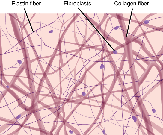
Figure 7.4.4 Diagram of loose fibrous connective tissue consists of a scattering of fibroblasts in a non-cellular matrix of loosely woven collagen and elastin fibres. 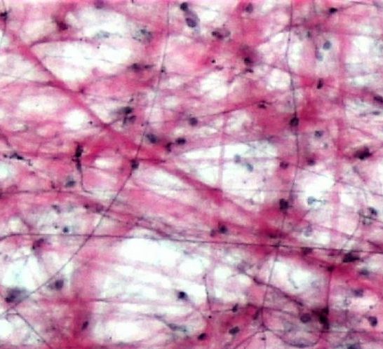
Figure 7.4.5 Microscopic view of loose fibrous connective tissue. - Dense Fibrous Connective Tissue: composed of a dense mat of parallel collagen fibres and a scattering of fibroblasts, creating a tissue that is very strong. Dense fibrous connective tissue forms tendons and ligaments, which connect bones to muscles and/or bones to neighbouring bones.
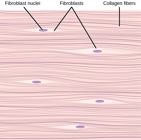
Figure 7.4.6 Dense fibrous connective tissue is composed of fibroblasts and a dense parallel packing of collagen fibres. 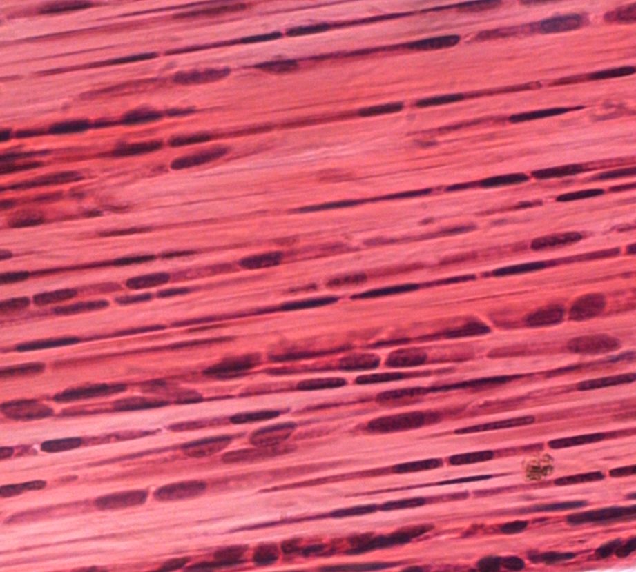
Figure 7.4.7 Microscopic view of dense fibrous connective tissue.
Supportive Connective Tissue
Supportive connective tissue exhibits the defining feature of connective tissue in that it is a scattering of cells in a non-cellular matrix; what sets it apart from other connective tissues is its solid matrix. In this tissue group, the matrix is solid- either bone or cartilage. While fibrous connective tissue contained cells called fibroblasts which produced fibres, supportive connective tissue contains cells that either create bone (osteocytes) or cells that create cartilage (chondrocytes).
Cartilage
Chondrocytes produce the cartilage matrix in which they reside. Cartilage is made up of protein fibres and chondrocytes in lacunae. This is tissue is strong yet flexible and is used many places in the body for protection and support. Cartilage is one of the few tissues that is not vascular (doesn't have a direct blood supply) meaning it relies on diffusion to obtain nutrients and gases; this is the cause of slow healing rates in injuries involving cartilage. There are three main types of cartilage:
- Hyaline cartilage: a smooth, strong and flexible tissue. Found at the ends of ribs and long bones, in the nose, and comprising the entire fetal skeleton.
- Fibrocartilage: a very strong tissue containing thick bundles of collagen. Found in joints that need cushioning from high impact (knees, jaw).
- Elastic cartilage: contains elastic fibres in addition to collagen, giving support with the benefit of elasticity. Found in earlobes and the epiglottis.
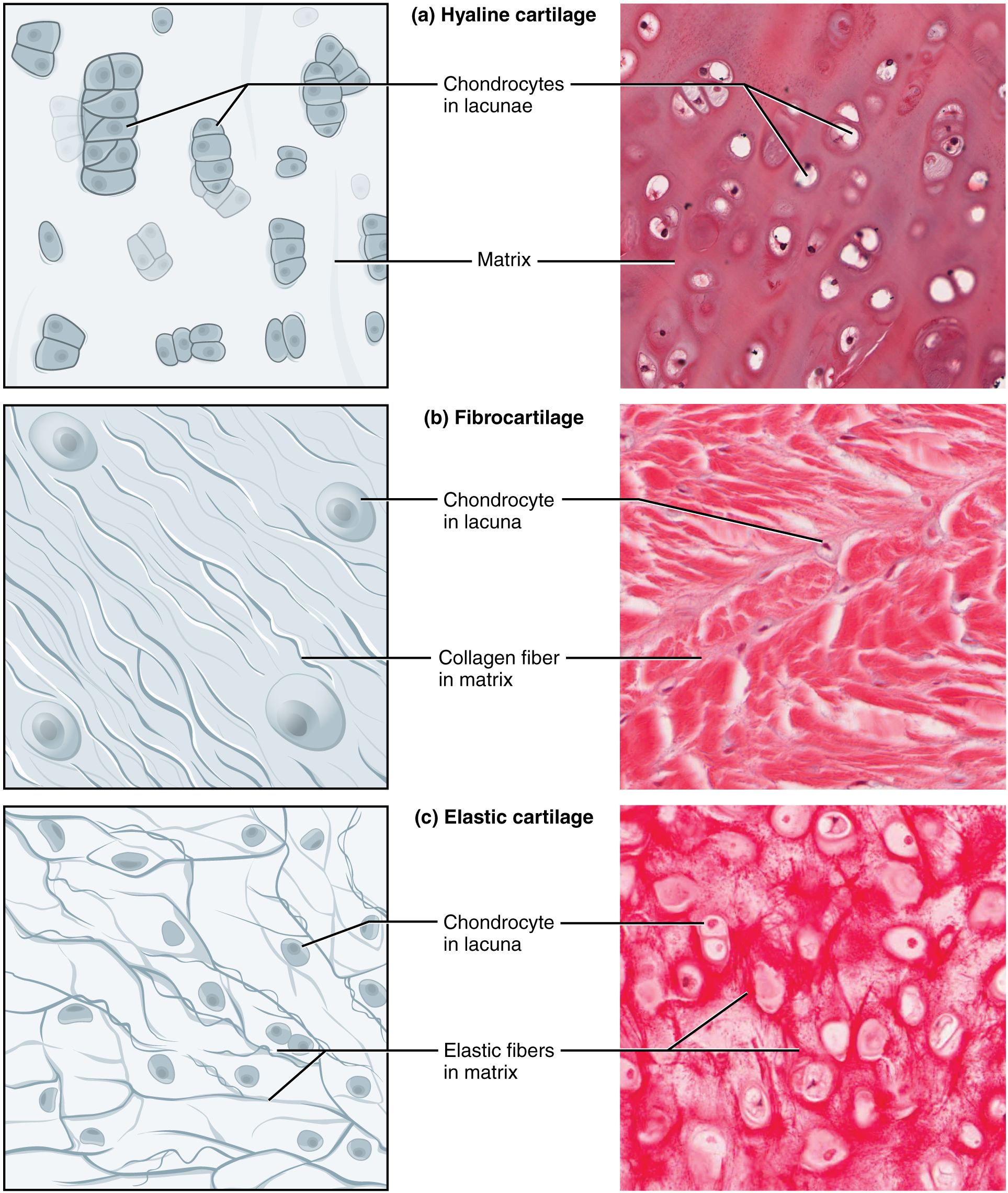
Figure 7.4.8 Three types of cartilage, each with distinct characteristics based on the nature of the matrix.
Bone
Osteocytes produce the bone matrix in which they reside. Since bone is very solid, these cells reside in small spaces called lacunae. This bone tissue is composed of collagen fibres embedded in calcium phosphate giving it strength without brittleness. There are two types of bone: compact and spongy.
- Compact bone: has a dense matrix organized into cylindrical units called osteons. Each osteon contains a central canal (sometimes called a Harversian Canal) which allows for space for blood vessels and nerves, as well as concentric rings of bone matrix and osteocytes in lacunae, as per the diagram here. Compact bone is found in long bones and forms a shell around spongy bone.
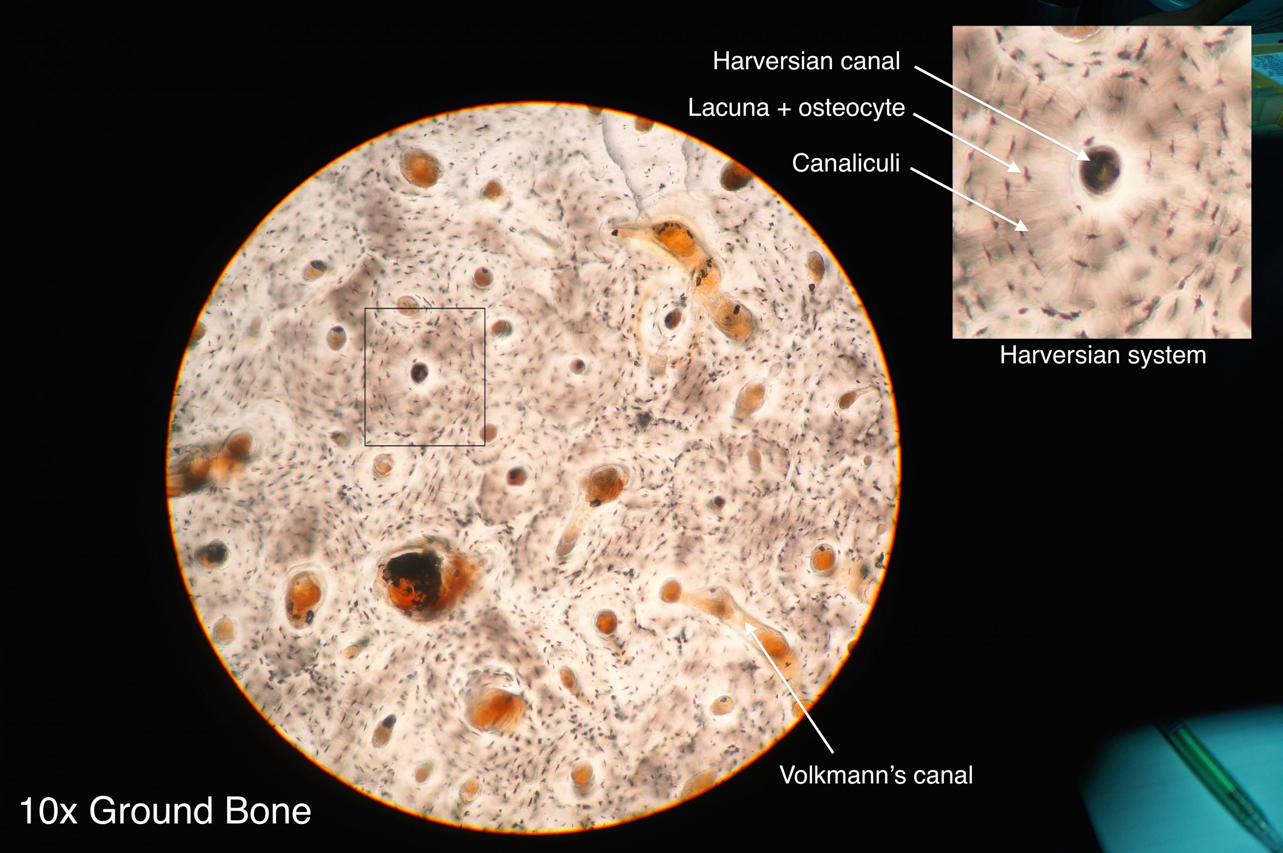
- Spongy bone: a very porous type of bone which most often contains bone marrow. It is found at the end of long bones, and makes up the majority of the ribs, shoulder blades and flat bones of the cranium.
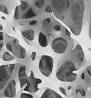
Fluid Connective Tissue
Fluid connective tissue has a matrix that is fluid; unlike the other two categories of connective tissue, the cells that reside in the matrix do not actually produce the matrix. Fibroblasts make the fibrous matrix, chondrocytes make the cartilaginous matrix, osteocytes make the bony matrix, yet blood cells do not make the fluid matrix of either lymph or plasma. This tissue still fits the definition of connective tissue in that it is still a scattering of cells in a non-cellular matrix.
There are two types of fluid connective tissue:
- Blood: blood contains three types of cells suspended in plasma, and is contained in the cardiovascular system.
- Eryththrocytes, more commonly called red blood cells, are present in high numbers (roughly 5 million cells per mL) and are responsible for delivering oxygen from to the lungs to all the other areas of the body. These cells are relatively small in size with a diameter of around 7 micrometres and live no longer than 120 days.
- Leukocytes, often referred to as white blood cells, are present in lower numbers (approximately 5 thousand cells per mL) are responsible for various immune functions. They are typically larger than erythrocytes, but can live much longer, particularly white blood cells responsible for long term immunity. The number of leukocytes in your blood can go up or down based on whether or not you are fighting an infection.
- Thrombocytes, also known as platelets, are very small cells responsible for blood clotting. Thrombocytes are not actually true cells, they are fragments of a much larger cell called a megakaryocyte.
- Lymph: contains a liquid matrix and white blood cells and is contained in the lymphatic system, which ultimately drains into the cardiovascular system.
Figure 7.4.11 A stained lymphocyte surrounded by red blood cells viewed using a light microscope.
Muscular Tissue
Muscular tissue is made up of cells that have the unique ability to contract- which is the defining feature of muscular tissue. There are three major types of muscle tissue, as pictured in Figure 7.4.12 skeletal, smooth, and cardiac muscle tissues.
Skeletal Muscle
Skeletal muscles are voluntary muscles, meaning that you exercise conscious control over them. Skeletal muscles are attached to bones by tendons, a type of connective tissue. When these muscles shorten to pull on the bones to which they are attached, they enable the body to move. When you are exercising, reading a book, or making dinner, you are using skeletal muscles to move your body to carry out these tasks.
Under the microscope, skeletal muscles are striated (or striped) in appearance, because of their internal structure which contains alternating protein fibres of actin and myosin. Skeletal muscle is described as multinucleated, meaning one "cell" has many nuclei. This is because in utero, individual cells destined to become skeletal muscle fused, forming muscle fibres in a process known as myogenesis. You will learn more about skeletal muscle and how it contracts in the Muscular System.
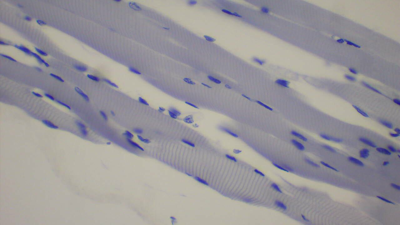
Smooth Muscle
Smooth muscles are nonstriated muscles- they still contain the muscle fibres actin and myosin, but not in the same alternating arrangement seen in skeletal muscle. Smooth muscle is found in the tubes of the body - in the walls of blood vessels and in the reproductive, gastrointestinal, and respiratory tracts. Smooth muscles are not under voluntary control meaning that they operate unconsciously, via the autonomic nervous system. Smooth muscles move substances through a wave of contraction which cascades down the length of a tube, a process termed peristalsis.
Watch the YouTube video "What is Peristalsis" by Mister Science to see peristalsis in action.
https://www.youtube.com/watch?v=kVjeNZA5pi4
What is Peristalsis, Mister Science, 2018.
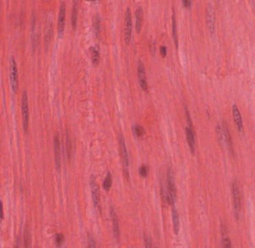
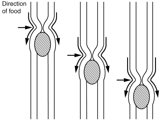
Cardiac Muscle
Cardiac muscles work involuntarily, meaning they are regulated by the autonomic nervous system. This is probably a good thing, since you wouldn't want to have to consciously concentrate on keeping your heart beating all the time! Cardiac muscle, which is found only in the heart, is mononucleated and striated (due to alternating bands of myosin and actin). Their contractions cause the heart to pump blood. In order to make sure entire sections of the heart contract in unison, cardiac muscle tissue contains special cell junctions called intercalated discs, which conduct the electrical signals used to "tell" the chambers of the heart when to contract.
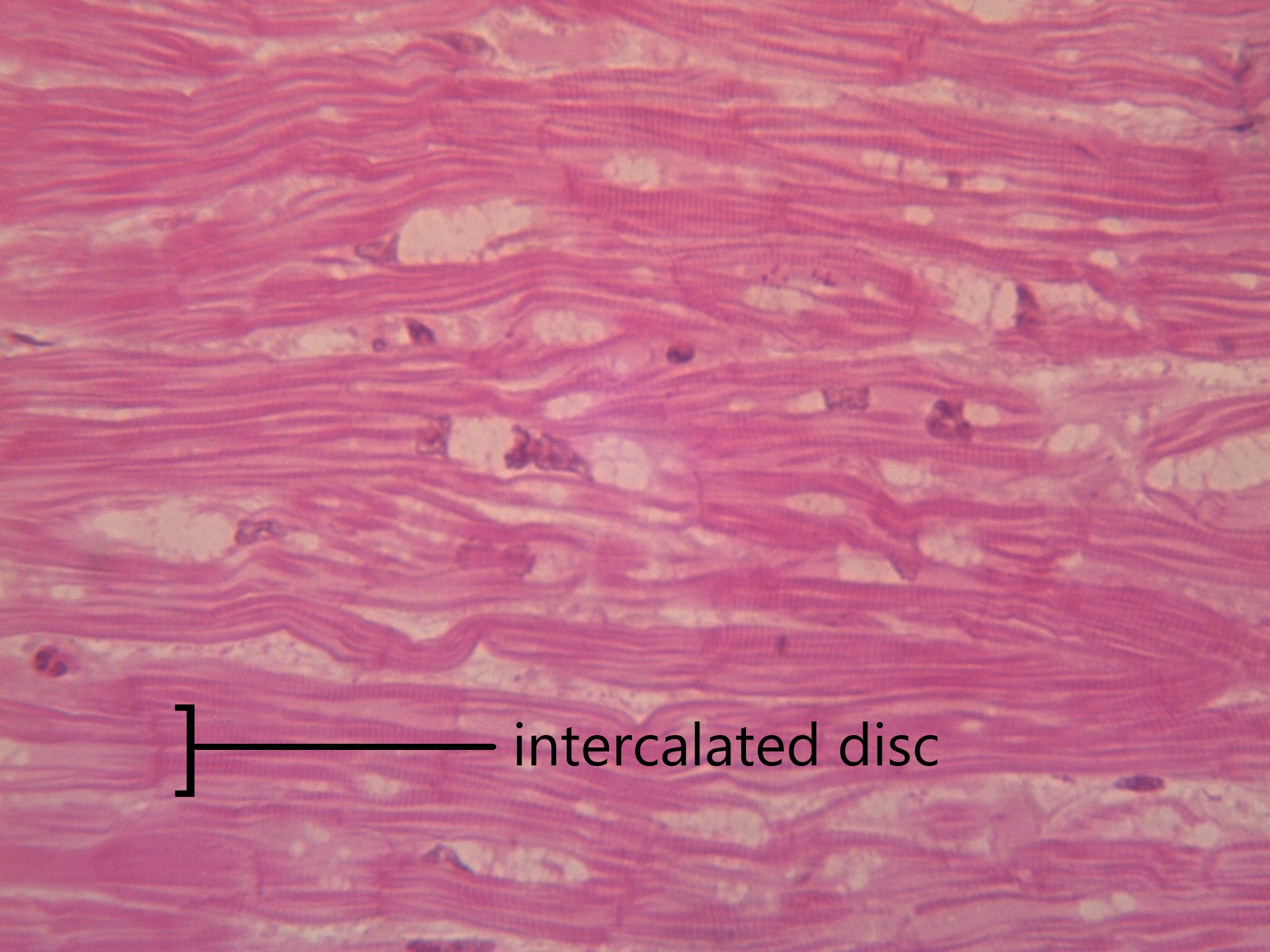
Nervous Tissue
Nervous tissue is made up of neurons and a group of cells called neuroglia (also known as glial cells). Nervous tissue makes up the central nervous system (mainly the brain and spinal cord) and peripheral nervous system (the network of nerves that runs throughout the rest of the body). The defining feature of nervous tissue is that it is specialized to be able to generate and conduct nerve impulses. This function is carried out by neurons, and the purpose of neuroglia is to support neurons.
A neuron has several parts to its structure:
- Dendrites which collect incoming nerve impulses
- A cell body, or soma, which contains the majority of the neuron's organelles, including the nucleus
- An axon, which carries nerve impulses away from the soma, to the next neuron in the chain
- A myelin sheath, which encases the axon and increases that rate at which nerve impulses can be conducted
- Axon terminals, which maintain physical contact with the dendrites of neighbouring neurons
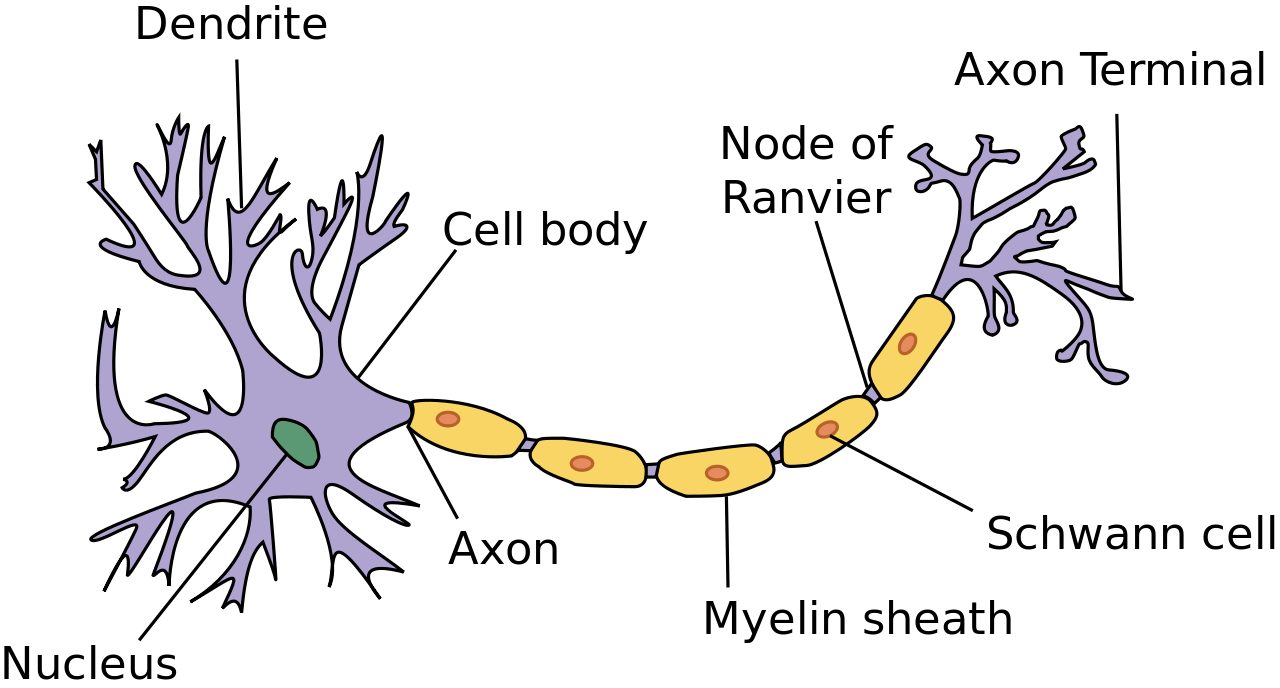
Neuroglia can be understood as support staff for the neuron. The neurons have such an important job, they need cells to bring them nutrients, take away cell waste, and build their mylein sheath. There are many types of neuroglia, which are categorized based on their function and/or their location in the nervous system. Neuroglia outnumber neurons by as much as 50 to 1, and are much smaller. See the diagram in 7.4.17 to compare the size and number of neurons and neuroglia.
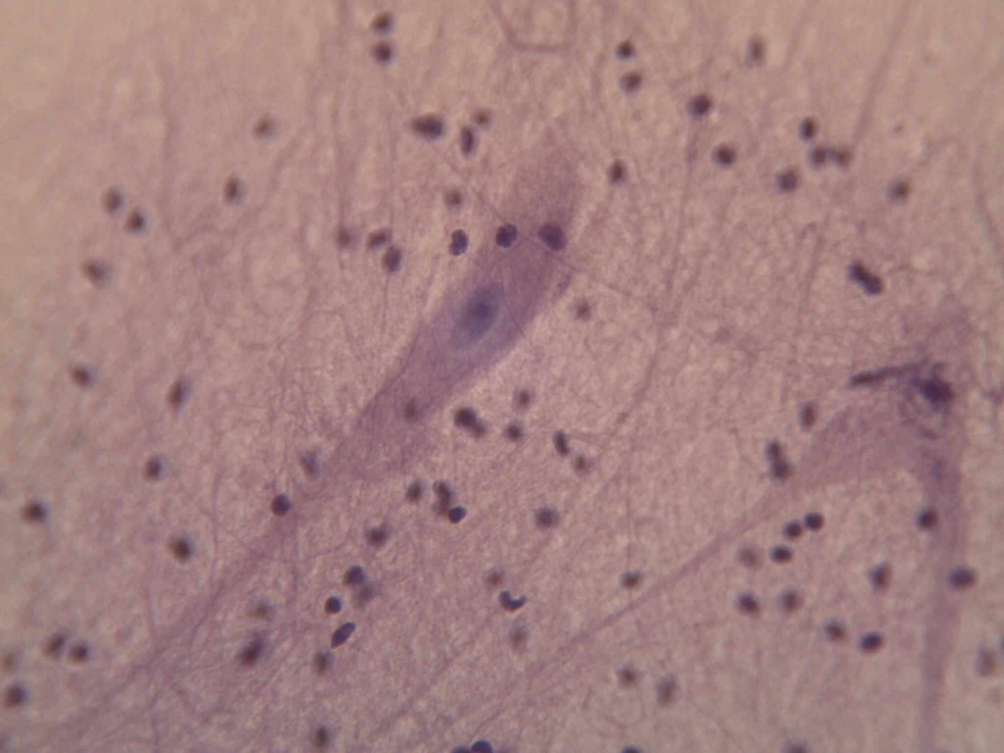
Try out this memory game to test your tissues knowledge:
7.4 Summary
- Tissues are made up of cells working together.
- There are four main types of tissues: epithelial, connective, muscular and nervous.
- Epithelial tissue makes up the linings and coverings of the body and is characterized by having a free surface and a basement membrane. Types of epithelial tissue are distinguished by shape of cell (squamous, cuboidal or columnar) and layering (simple, stratified, pseudostratified and transitional). Different epithelial tissues can carry out diffusion, secretion, absorption, and/or protection depending on their particular cell shape and layering.
- Connective tissue provides structure and support for the body and is characterized as a scattering of cells in a non-cellular matrix. There are three main categories of connective tissue, each characterized by a particular type of matrix:
- Fibrous connective tissue contains protein fibres. Both loose and dense fibrous connective tissue belong in this category.
- Supportive connective tissue contains a very solid matrix, and includes both bone and cartilage.
- Fluid connective tissue contains cells in a fluid matrix with the two types of blood and lymph.
- Muscular tissue's defining feature is that it is contractile. There are three types of muscular tissue: skeletal muscle which is found attached to the skeleton for voluntary movement, smooth muscle which moves substances through body tubes, and cardiac muscle which moves blood through the heart.
- Nervous tissue contains specialized cells called neurons which can conduct electrical impulses. Also found in nervous tissue are neuroglia, which support neurons by providing nutrients, removing wastes, and creating myelin sheath.
7.4 Review Questions
- Define the term tissue.
-
- If a part of the body needed a lining that was both protective, but still able to absorb nutrients, what would be the best type of epithelial tissue to use?
-
- Where do you find skeletal muscle? Smooth muscle? Cardiac muscle?
-
- What are some of the functions of neuroglia?
-
7.4 Explore More
https://www.youtube.com/watch?v=O0ZvbPak4ck
Types of Human Body Tissue, MoomooMath and Science, 2017.
https://www.youtube.com/watch?v=uHbn7wLN_3k
How to 3D print human tissue - Taneka Jones, TED-Ed, 2019.
https://www.youtube.com/watch?v=1Qfmkd6C8u8
How bones make blood - Melody Smith, TED-Ed, 2020.
Attributions
Figure 7.4.1
- Construction man kneeling in front of wall by Charles Deluvio on Unsplash is used under the Unsplash License (https://unsplash.com/license).
- Beige wooden frame by Charles Deluvio on Unsplash is used under the Unsplash License (https://unsplash.com/license).
- Tambour on green by Pierre Châtel-Innocention Unsplash is used under the Unsplash License (https://unsplash.com/license).
- Tags: Construction Studs Plumbing Wiring by JWahl on Pixabay is used under the Pixabay License (https://pixabay.com/es/service/license/).
Figure 7.4.2 and Figure 7.4.3
- Simple columnar epithelium tissue by Kamil Danak on Wikimedia Commons is used under a CC BY-SA 3.0 (https://creativecommons.org/licenses/by-sa/3.0/deed.en) license.
- Simple cuboidal epithelium by Kamil Danak on Wikimedia Commons is used under a CC BY-SA 3.0 (https://creativecommons.org/licenses/by-sa/3.0/deed.en) license.
- Simple squamous epithelium by Kamil Danak on Wikimedia Commons is used under a CC BY-SA 3.0 (https://creativecommons.org/licenses/by-sa/3.0/deed.en) license.
Figure 7.4.4
Loose fibrous connective tissue by CNX OpenStax. Biology. on Wikimedial Commons is used under a CC BY 4.0. (https://creativecommons.org/licenses/by/4.0) license.
Figure 7.4.5
Connective Tissue: Loose Aerolar by Berkshire Community College Bioscience Image Library on Flickr is used under a CC0 1.0 Universal public domain dedication (https://creativecommons.org/publicdomain/zero/1.0/) license.
Figure 7.4.6
Dense Fibrous Connective Tissue by by CNX OpenStax. Biology. on Wikimedial Commons is used under a CC BY 4.0. (https://creativecommons.org/licenses/by/4.0) license.
Figure 7.4.7
Dense_connective_tissue-400x by J Jana on Wikimedia Commons is used under a CC BY-SA 4.0 (https://creativecommons.org/licenses/by-sa/4.0/deed.en) license.
Figure 7.4.8
Types_of_Cartilage-new by OpenStax College on Wikipedia Commons is used under a CC BY 3.0 (https://creativecommons.org/licenses/by/3.0) license.
Figure 7.4.9
Compact_bone_histology_2014 by Athikhun.suw on Wikimedia Commons is used under a CC BY-SA 4.0 (https://creativecommons.org/licenses/by-sa/4.0/deed.en) license.
Figure 7.4.10
Bone_normal_and_degraded_micro_structure by Gtirouflet on Wikimedia Commons is used under a CC BY-SA 3.0 (https://creativecommons.org/licenses/by-sa/3.0/deed.en) license.
Figure 7.4.11
Lymphocyte2 by NicolasGrandjean on Wikimedia Commons is used under a CC BY-SA 3.0 (https://creativecommons.org/licenses/by-sa/3.0/deed.en) license. [No machine-readable author provided. NicolasGrandjean is assumed, based on copyright claims.]
Figure 7.4.12
Skeletal_muscle_横纹肌1 by 乌拉跨氪 on Wikimedia Commons is used under a CC BY-SA 4.0 (https://creativecommons.org/licenses/by-sa/4.0/deed.en) license.
Figure 7.4.13
Smooth_Muscle_new by OpenStax on Wikimedia Commons is used under a CC BY 4.0 (https://creativecommons.org/licenses/by/4.0/deed.en) license.
Figure 7.4.14
Peristalsis by OpenStax on Wikimedia Commons is used under a CC BY 3.0 (https://creativecommons.org/licenses/by/3.0/deed.en) license.
Figure 7.4.15
400x Cardiac Muscle by Jessy731 on Flickr is used and adapted by Christine Miller under a CC BY-NC 2.0 (https://creativecommons.org/licenses/by-nc/2.0/) license.
Figure 7.4.16
Neuron.svg by User:Dhp1080 on Wikimedia Commons is used under a CC BY-SA 3.0 (https://creativecommons.org/licenses/by-sa/3.0/deed.en) license.
Figure 7.4.17
400x Nervous Tissue by Jessy731 on Flickr is used under a CC BY-NC 2.0 (https://creativecommons.org/licenses/by-nc/2.0/) license.
Table 7.4.1
Summary of Epithelial Tissue Cells, by OpenStax College on Wikipedia Commons is used under a CC BY 3.0 (https://creativecommons.org/licenses/by/3.0) license.
Table 7.4.2
- Epithelial_Tissues_Stratified_Squamous_Epithelium_(40230842160) by
Berkshire Community College Bioscience Image Library on Wikimedia Commons is used under a CC0 1.0 Universal Public Domain Dedication (https://creativecommons.org/publicdomain/zero/1.0/) license. - Simple cuboidal epithelial tissue histology by Berkshire Community College on Flickr is used under a CC0 1.0 Universal Public Domain Dedication (https://creativecommons.org/publicdomain/zero/1.0/) license.
- Pseudostratified_Epithelium by OpenStax College on Wikimedia Commons is used under a CC BY 3.0 (https://creativecommons.org/licenses/by/3.0) license.
References
Betts, J. G., Young, K.A., Wise, J.A., Johnson, E., Poe, B., Kruse, D.H., Korol, O., Johnson, J.E., Womble, M., DeSaix, P. (2013, April 25). Figure 4.8 Summary of epithelial tissue cells [digital image]. In Anatomy and Physiology (Section 4.2). OpenStax. https://openstax.org/books/anatomy-and-physiology/pages/4-2-epithelial-tissue
Betts, J. G., Young, K.A., Wise, J.A., Johnson, E., Poe, B., Kruse, D.H., Korol, O., Johnson, J.E., Womble, M., DeSaix, P. (2013, April 25). Figure 4.16 Types of cartilage [digital image]. In Anatomy and Physiology (Section 4.3). OpenStax. https://openstax.org/books/anatomy-and-physiology/pages/4-3-connective-tissue-supports-and-protects
Betts, J. G., Young, K.A., Wise, J.A., Johnson, E., Poe, B., Kruse, D.H., Korol, O., Johnson, J.E., Womble, M., DeSaix, P. (2013, April 25). Figure 10.23 Smooth muscle [digital micrograph]. In Anatomy and Physiology (Section 10.8). OpenStax. https://openstax.org/books/anatomy-and-physiology/pages/10-8-smooth-muscle (Micrograph provided by the Regents of University of Michigan Medical School © 2012)
Betts, J. G., Young, K.A., Wise, J.A., Johnson, E., Poe, B., Kruse, D.H., Korol, O., Johnson, J.E., Womble, M., DeSaix, P. (2013, April 25). Figure 22.5 Pseudostratified ciliated columnar epithelium [digital micrograph]. In Anatomy and Physiology (Section 22.1). OpenStax. https://openstax.org/books/anatomy-and-physiology/pages/22-1-organs-and-structures-of-the-respiratory-system
Betts, J. G., Young, K.A., Wise, J.A., Johnson, E., Poe, B., Kruse, D.H., Korol, O., Johnson, J.E., Womble, M., DeSaix, P. (2013, April 25). Figure 23.5 Peristalsis [diagram]. In Anatomy and Physiology (Section 23.2). OpenStax. https://openstax.org/books/anatomy-and-physiology/pages/23-2-digestive-system-processes-and-regulation
Mister Science. (2018). What is peristalsis? YouTube. https://www.youtube.com/watch?v=kVjeNZA5pi4
MoomooMath and Science. (2017, May 18). Types of human body tissue. YouTube. https://www.youtube.com/watch?v=O0ZvbPak4ck&feature=youtu.be
Open Stax. (2016, May 27). Figure 6 Loose connective tissue [digital image]. In OpenStax Biology (Section 33.2). OpenStax CNX. https://cnx.org/contents/GFy_h8cu@10.53:-LfhWRES@4/Animal-Primary-Tissues
Open Stax. (2016, May 27). Figure 7 Fibrous connective tissue from the tendon [digital image]. In OpenStax Biology (Section 33.2). OpenStax CNX. https://cnx.org/contents/GFy_h8cu@10.53:-LfhWRES@4/Animal-Primary-Tissues
TED-Ed. (2019, October 17). How to 3D print human tissue - Taneka Jones. YouTube. https://www.youtube.com/watch?v=uHbn7wLN_3k&feature=youtu.be
TED-Ed. (2020, January 27). How bones make blood - Melody Smith. YouTube. https://www.youtube.com/watch?v=1Qfmkd6C8u8&feature=youtu.be
A group of diseases involving abnormal cell growth with the potential to invade or spread to other parts of the body.
Acquired Immunodeficiency Syndrome - a chronic, potentially life-threatening condition caused by the human immunodeficiency virus (HIV). By damaging your immune system, HIV interferes with your body's ability to fight infection and disease.
Created by CK-12 Foundation
Figure 7.7.1 Everyone on a baseball team has a special job.
Teamwork
Every player on a baseball team has a special job. In the Figure 7.7.1 collage, each player has their part of the infield or outfield covered in case the ball comes their way. Other players on the team cover different parts of the field, or they pitch or catch the ball. Playing baseball clearly requires teamwork. In that regard, the human body is like a baseball team. All of the organ systems of the human body must work together as a team to keep the body alive and well. Teamwork within the body begins with communication.
Communication Among Organ Systems
Communication among organ systems is vital if they are to work together as a team. They must be able to respond to each other and change their responses as needed to keep the body in balance. Communication among organ systems is controlled mainly by the autonomic nervous system and the endocrine system.
The autonomic nervous system is the part of the nervous system that controls involuntary functions. The autonomic nervous system, for example, controls heart rate, blood flow, and digestion. You don’t have to tell your heart to beat faster or to consciously squeeze muscles to push food through the digestive system. You don’t have to even think about these functions at all! The autonomic nervous system orchestrates all the signals needed to control them. It sends messages between parts of the nervous system, as well as between the nervous system and other organ systems via chemical messengers called neurotransmitters.
The endocrine system is the system of glands that secrete hormones directly into the bloodstream. Once in the blood, endocrine hormones circulate to cells everywhere in the body. The endocrine system itself is under control of the nervous system via a part of the brain called the hypothalamus. The hypothalamus secretes hormones that travel directly to cells of the pituitary gland, which is located beneath it. The pituitary gland is the master gland of the endocrine system. Most of its hormones either turn on or turn off other endocrine glands. For example, if the pituitary gland secretes thyroid-stimulating hormone, the hormone travels through the circulation to the thyroid gland, which is stimulated to secrete thyroid hormone. Thyroid hormone then travels to cells throughout the body, where it increases their metabolism.
Examples of Organ System Interactions
An increase in cellular metabolism requires more cellular respiration. Cellular respiration is a good example of organ system interactions, because it is a basic life process that occurs in all living cells.
Cellular Respiration
Cellular respiration is the intracellular process that breaks down glucose with oxygen to produce carbon dioxide and energy in the form of ATP molecules. It is the process by which cells obtain usable energy to power other cellular processes. Which organ systems are involved in cellular respiration? The glucose needed for cellular respiration comes from the digestive system via the cardiovascular system. The oxygen needed for cellular respiration comes from the respiratory system also via the cardiovascular system. The carbon dioxide produced in cellular respiration leaves the body by the opposite route. In short, cellular respiration requires — at a minimum — the digestive, cardiovascular, and respiratory systems.
Fight-or-Flight Response
The well-known fight-or-flight response is a good example of how the nervous and endocrine systems control other organ system responses. The fight-or-flight response begins when the nervous system perceives sudden danger, as shown in the Figure 7.7.2 diagram. The brain sends a message to the endocrine system (via the pituitary gland) for the adrenal glands to secrete the hormones cortisol and adrenaline. These hormones flood the circulation and affect other organ systems throughout the body, including the cardiovascular, urinary, sensory, and digestive systems. Specific responses include increased heart rate, bladder relaxation, tunnel vision, and a shunting of blood away from the digestive system and toward the muscles, brain, and other vital organs needed to fight or flee.
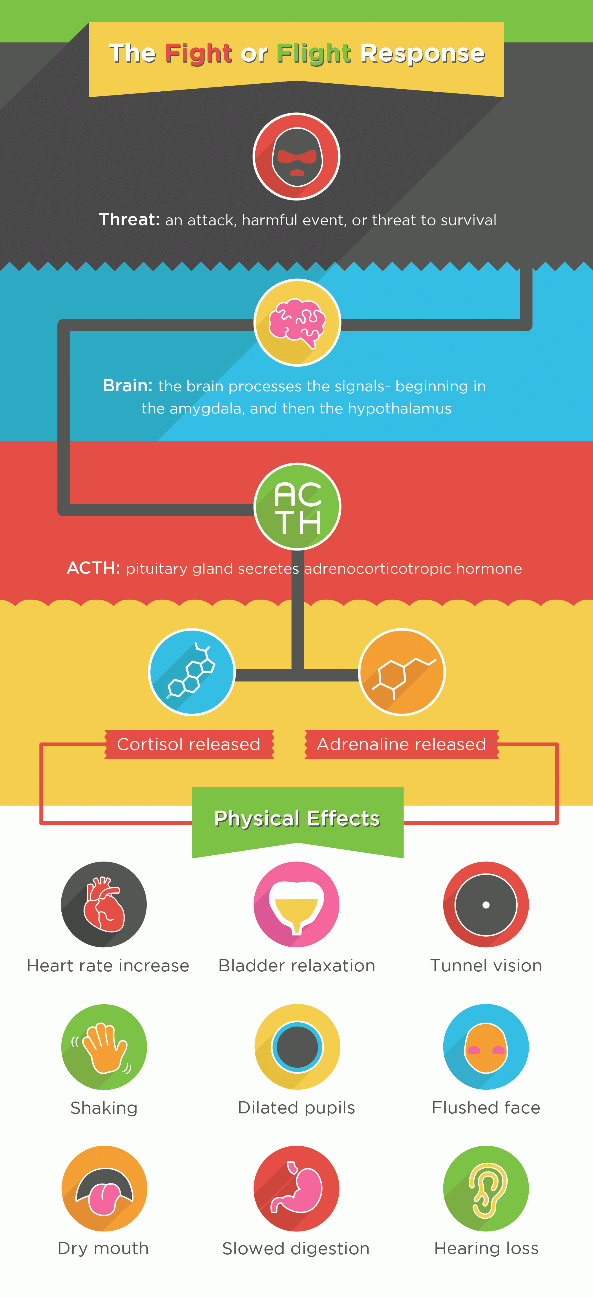
Playing Baseball
The people playing baseball in the opening collage (Figure 7.7.1) are using multiple organ systems in this voluntary activity. Their nervous systems are focused on observing and preparing to respond to the next play. Their other systems are being controlled by the autonomic nervous system. The players are using the muscular, skeletal, respiratory, and cardiovascular systems. Can you explain how each of these organ systems is involved in playing baseball?
Feature: Reliable Sources
Teamwork among organ systems allows the human organism to work like a finely tuned machine — at least, it does until one of the organ systems fails. When that happens, other organ systems interacting in the same overall process will also be affected. This is especially likely if the affected system plays a controlling role in the process. An example is type 1 diabetes. This disorder occurs when the pancreas does not secrete the endocrine hormone insulin. Insulin normally is secreted in response to an increasing level of glucose in the blood, and it brings the level of glucose back to normal by stimulating body cells to take up insulin from the blood.
Learn more about type 1 diabetes. Use several reliable Internet sources to answer the following questions:
- In type 1 diabetes, what causes the endocrine system to fail to produce insulin?
- If type 1 diabetes is not controlled, which organ systems are affected by high blood glucose levels? What are some of the specific effects?
- How can blood glucose levels be controlled in patients with type 1 diabetes?
7.7 Summary
- The human body's organ systems must work together to keep the body alive and functioning normally, which requires communication among systems. This communication is controlled by the autonomic nervous system and endocrine system. The autonomic nervous system controls involuntary body functions, such as heart rate and digestion. The endocrine system secretes hormones into the blood that travel to body cells and influence their activities.
- Cellular respiration is a good example of organ system interactions, because it is a basic life process that happens in all living cells. It is the intracellular process that breaks down glucose with oxygen to produce carbon dioxide and energy. Cellular respiration requires the interaction of the digestive, cardiovascular, and respiratory systems.
- The fight-or-flight response is a good example of how the nervous and endocrine systems control other organ system responses. It is triggered by a message from the brain to the endocrine system and prepares the body for flight or a fight. Many organ systems are stimulated to respond, including the cardiovascular, respiratory, and digestive systems.
- Playing baseball — or doing other voluntary physical activities — may involve the interaction of nervous, muscular, skeletal, respiratory, and cardiovascular systems.
7.7 Review Questions
- What is the autonomic nervous system?
- How do the autonomic nervous system and endocrine system communicate with other organ systems so the systems can interact?
- Explain how the brain communicates with the endocrine system.
- What is the role of the pituitary gland in the endocrine system?
- Identify the organ systems that play a role in cellular respiration.
- How does the hormone adrenaline prepare the body to fight or flee? What specific physiological changes does it bring about?
- Explain the role of the muscular system in digesting food.
- Describe how three different organ systems are involved when a player makes a particular play in baseball, such as catching a fly ball.
-
- What are two types of molecules that the body uses to communicate between organ systems?
- Explain why hormones can have such a wide variety of effects on the body.
7.7 Explore More
https://www.youtube.com/watch?time_continue=19&v=Ujr0UAbyPS4&feature=emb_logo
3D Medical Animation - Peristalsis in Large Intestine/Bowel ||
©Animated Biomedical Productions (ABP), 2013.
https://www.youtube.com/watch?v=FBnBTkcr6No&feature=emb_logo
Adrenaline: Fight or Flight Response, Henk van 't Klooster, 2013.
https://www.youtube.com/watch?v=m2GywoS77qc
Fight or Flight Response, Bozeman Science, 2012.
Attributions
Figure 7.7.1
- Baseball positions by Michael J on Wikimedia Commons is used under a CC BY-SA 4.0 (https://creativecommons.org/licenses/by-sa/4.0/deed.de) license.
- US Navy 040229-N-8629D-070 photo by US Navy's Photographer's Mate 2nd Class Brett A. Dawson on Wikimedia Commons is released into the public domain (https://en.wikipedia.org/wiki/Public_domain).
- David Ortiz batter's box by Albert Yau/ SecondPrint Productions on Flickr is used under a CC BY 2.0 (https://creativecommons.org/licenses/by/2.0/deed.es) license.
- Fenway-from Legend's Box by Jared Vincent on Wikipedia is used under a CC BY 2.0 (https://creativecommons.org/licenses/by/2.0/deed.en) license.
Figure 7.7.2
The_Fight_or_Flight_Response by Jvnkfood (original), converted to PNG and reduced to 8-bit by Pokéfan95 on Wikimedia Commons is used under a CC BY-SA 4.0 (https://creativecommons.org/licenses/by-sa/4.0/deed.en) license.
References
Animated Biomedical Productions. (2013, January 30). 3D Medical animation - Peristalsis in large intestine/bowel || ©ABP. YouTube. https://www.youtube.com/watch?v=Ujr0UAbyPS4&feature=youtu.be
Bozeman Science. (2012, January 9). Fight or flight response. YouTube. https://www.youtube.com/watch?v=m2GywoS77qc&feature=youtu.be
Henk van 't Klooster. (2013). Adrenaline: Fight or flight response. YouTube. https://www.youtube.com/watch?v=FBnBTkcr6No&t=4s
Mayo Clinic Staff. (n.d.). Type 1 diabetes. MayoClinic.org. https://www.mayoclinic.org/diseases-conditions/type-1-diabetes/symptoms-causes/syc-20353011
Wikipedia contributors. (2020, July 22). Thyroid-stimulating hormone. In Wikipedia. https://en.wikipedia.org/w/index.php?title=Thyroid-stimulating_hormone&oldid=968942540
Created by CK-12 Foundation/Adapted by Christine Miller
Steady as She Goes
This device (Figure 7.8.1) looks simple, but it controls a complex system that keeps a home at a steady temperature — it's a thermostat. The device shows the current temperature in the room, and also allows the occupant to set the thermostat to the desired temperature. A thermostat is a commonly cited model of how living systems — including the human body— maintain a steady state called homeostasis.
What Is Homeostasis?
Homeostasis is the condition in which a system (such as the human body) is maintained in a more or less steady state. It is the job of cells, tissues, organs, and organ systems throughout the body to maintain many different variables within narrow ranges compatible with life. Keeping a stable internal environment requires continually monitoring the internal environment and constantly making adjustments to keep things in balance.
Set Point and Normal Range
For any given variable, such as body temperature or blood glucose level, there is a particular set point that is the physiological optimum value. The set point for human body temperature, for example, is about 37 degrees C (98.6 degrees F). As the body works to maintain homeostasis for temperature or any other internal variable, the value typically fluctuates around the set point. Such fluctuations are normal, as long as they do not become too extreme. The spread of values within which such fluctuations are considered insignificant is called the normal range. In the case of body temperature, for example, the normal range for an adult is about 36.5 to 37.5 degrees C (97.7 to 99.5 degrees F).
A good analogy for set point, normal range, and maintenance of homeostasis is driving. When you are driving a vehicle on the road, you are supposed to drive in the centre of your lane — this is analogous to the set point. Sometimes, you are not driving in the exact centre of the lane, but you are still within your lines, so you are in the equivalent of the normal range. However, if you were to get too close to the centre line or the shoulder of the road, you would take action to correct your position. You'd move left if you were too close to the shoulder, or right if too close to the centre line — which is analogous to our next concept, negative feedback to maintain homeostasis.
Maintaining Homeostasis
Homeostasis is normally maintained in the human body by an extremely complex balancing act. Regardless of the variable being kept within its normal range, maintaining homeostasis requires at least four interacting components: stimulus, sensor, control centre, and effector.
- The stimulus is provided by the variable being regulated. Generally, the stimulus indicates that the value of the variable has moved away from the set point or has left the normal range.
- The sensor monitors the values of the variable and sends data on it to the control centre.
- The control centre matches the data with normal values. If the value is not at the set point or is outside the normal range, the control centre sends a signal to the effector.
- The effector is an organ, gland, muscle, or other structure that acts on the signal from the control centre to move the variable back toward the set point.
Each of these components is illustrated in Figure 7.8.2. The diagram on the left is a general model showing how the components interact to maintain homeostasis. The diagram on the right shows the example of body temperature. From the diagrams, you can see that maintaining homeostasis involves feedback, which is data that feeds back to control a response. Feedback may be negative (as in the example below) or positive. All the feedback mechanisms that maintain homeostasis use negative feedback. Biological examples of positive feedback are much less common.
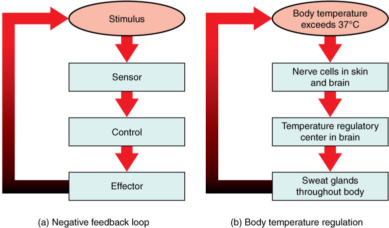
Negative Feedback
In a negative feedback loop, feedback serves to reduce an excessive response and keep a variable within the normal range. Two processes controlled by negative feedback are body temperature regulation and control of blood glucose.
Body Temperature
Body temperature regulation involves negative feedback, whether it lowers the temperature or raises it, as shown in Figure 7.8.3 and explained in the text that follows.
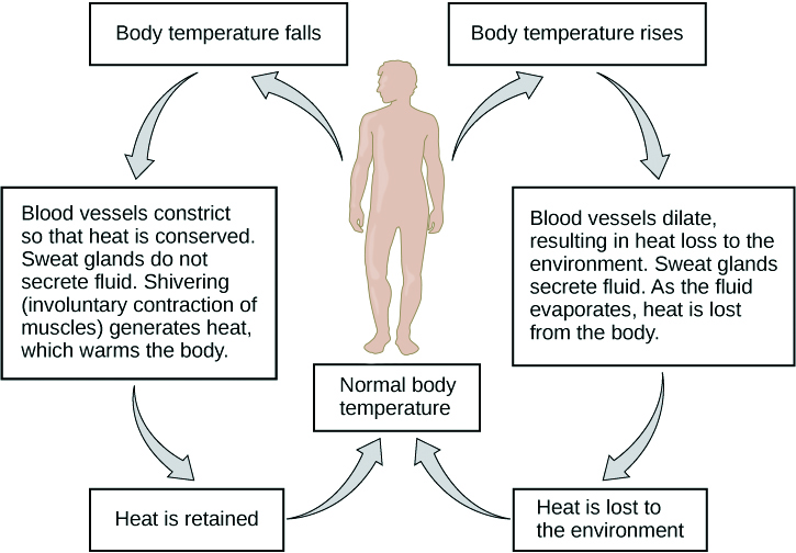
Cooling Down
The human body’s temperature regulatory centre is the hypothalamus in the brain. When the hypothalamus receives data from sensors in the skin and brain that body temperature is higher than the set point, it sets into motion the following responses:
- Blood vessels in the skin dilate (vasodilation) to allow more blood from the warm body core to flow close to the surface of the body, so heat can be radiated into the environment.
- As blood flow to the skin increases, sweat glands in the skin are activated to increase their output of sweat (diaphoresis). When the sweat evaporates from the skin surface into the surrounding air, it takes heat with it.
- Breathing becomes deeper, and the person may breathe through the mouth instead of the nasal passages. This increases heat loss from the lungs.
Heating Up
When the brain’s temperature regulatory centre receives data that body temperature is lower than the set point, it sets into motion the following responses:
- Blood vessels in the skin contract (vasoconstriction) to prevent blood from flowing close to the surface of the body, which reduces heat loss from the surface.
- As temperature falls lower, random signals to skeletal muscles are triggered, causing them to contract. This causes shivering, which generates a small amount of heat.
- The thyroid gland may be stimulated by the brain (via the pituitary gland) to secrete more thyroid hormone. This hormone increases metabolic activity and heat production in cells throughout the body.
- The adrenal glands may also be stimulated to secrete the hormone adrenaline. This hormone causes the breakdown of glycogen (the carbohydrate used for energy storage in animals) to glucose, which can be used as an energy source. This catabolic chemical process is exothermic, or heat producing.
Blood Glucose
In controlling the blood glucose level, certain endocrine cells in the pancreas (called alpha and beta cells) detect the level of glucose in the blood. They then respond appropriately to keep the level of blood glucose within the normal range.
- If the blood glucose level rises above the normal range, pancreatic beta cells release the hormone insulin into the bloodstream. Insulin signals cells to take up the excess glucose from the blood until the level of blood glucose decreases to the normal range.
- If the blood glucose level falls below the normal range, pancreatic alpha cells release the hormone glucagon into the bloodstream. Glucagon signals cells to break down stored glycogen to glucose and release the glucose into the blood until the level of blood glucose increases to the normal range.
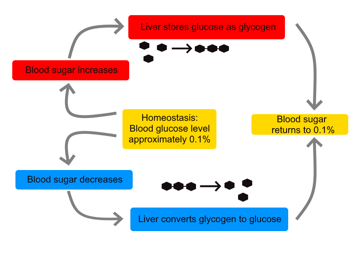
https://www.youtube.com/watch?v=Iz0Q9nTZCw4
Homeostasis and Negative/Positive Feedback, Amoeba Sisters, 2017.
Positive Feedback
In a positive feedback loop, feedback serves to intensify a response until an end point is reached. Examples of processes controlled by positive feedback in the human body include blood clotting and childbirth.
Blood Clotting
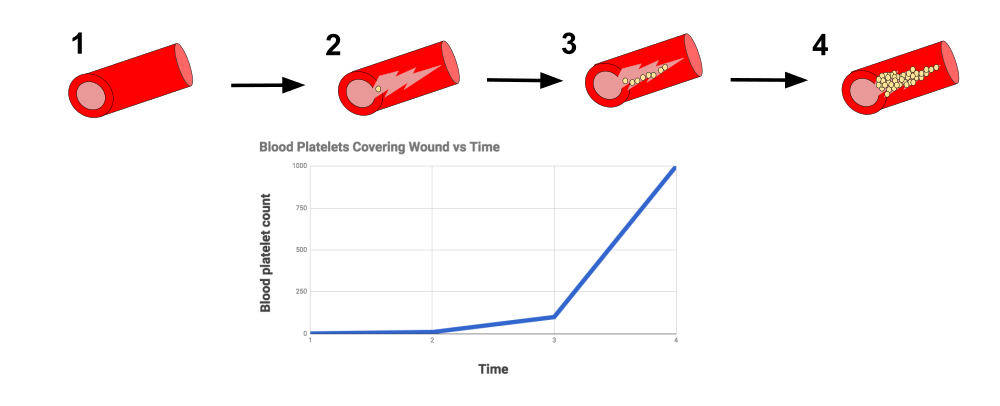
When a wound causes bleeding, the body responds with a positive feedback loop to clot the blood and stop blood loss. Substances released by the injured blood vessel wall begin the process of blood clotting. Platelets in the blood start to cling to the injured site and release chemicals that attract additional platelets. As the platelets continue to amass, more of the chemicals are released and more platelets are attracted to the site of the clot. The positive feedback accelerates the process of clotting until the clot is large enough to stop the bleeding.
Childbirth
Figure 7.8.6 shows the positive feedback loop that controls childbirth. The process normally begins when the head of the infant pushes against the cervix. This stimulates nerve impulses, which travel from the cervix to the hypothalamus in the brain. In response, the hypothalamus sends the hormone oxytocin to the pituitary gland, which secretes it into the bloodstream so it can be carried to the uterus. Oxytocin stimulates uterine contractions, which push the baby harder against the cervix. In response, the cervix starts to dilate in preparation for the passage of the baby. This cycle of positive feedback continues, with increasing levels of oxytocin, stronger uterine contractions, and wider dilation of the cervix until the baby is pushed through the birth canal and out of the body. At that point, the cervix is no longer stimulated to send nerve impulses to the brain, and the entire process stops.
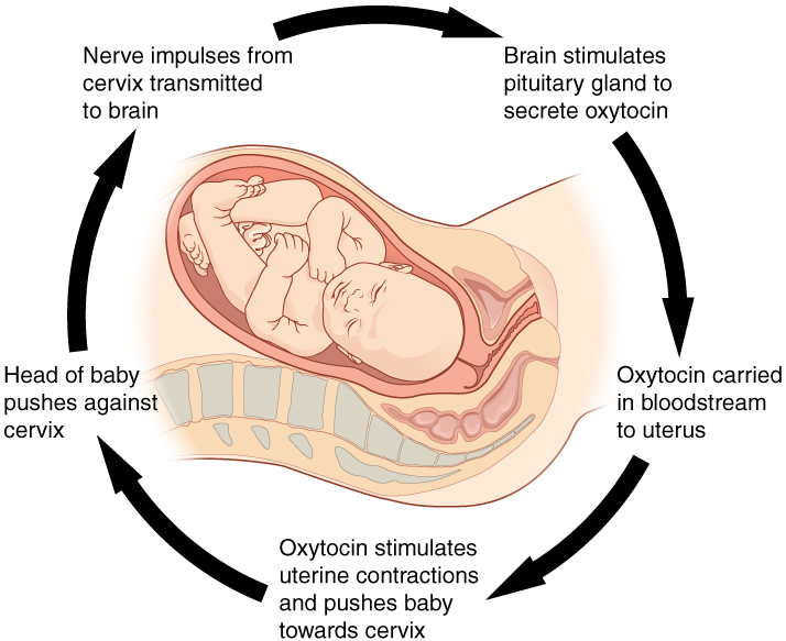
Normal childbirth is driven by a positive feedback loop. Positive feedback causes an increasing deviation from the normal state to a fixed end point, rather than a return to a normal set point as in homeostasis.
When Homeostasis Fails
Homeostatic mechanisms work continuously to maintain stable conditions in the human body. Sometimes, however, the mechanisms fail. When they do, homeostatic imbalance may result, in which cells may not get everything they need or toxic wastes may accumulate in the body. If homeostasis is not restored, the imbalance may lead to disease — or even death. Diabetes is an example of a disease caused by homeostatic imbalance. In the case of diabetes, blood glucose levels are no longer regulated and may be dangerously high. Medical intervention can help restore homeostasis and possibly prevent permanent damage to the organism.
Normal aging may bring about a reduction in the efficiency of the body’s control systems, which makes the body more susceptible to disease. Older people, for example, may have a harder time regulating their body temperature. This is one reason they are more likely than younger people to develop serious heat-induced illnesses, such as heat stroke.
Feature: My Human Body
Diabetes is diagnosed in people who have abnormally high levels of blood glucose after fasting for at least 12 hours. A fasting level of blood glucose below 100 is normal. A level between 100 and 125 places you in the pre-diabetes category, and a level higher than 125 results in a diagnosis of diabetes.
Of the two types of diabetes, type 2 diabetes is the most common, accounting for about 90 per cent of all cases of diabetes in the United States. Type 2 diabetes typically starts after the age of 40. However, because of the dramatic increase in recent decades in obesity in younger people, the age at which type 2 diabetes is diagnosed has fallen. Even children are now being diagnosed with type 2 diabetes. Today, about 3 million Canadians (8.1% of total population) are living with diabetes.
You may at some point have your blood glucose level tested during a routine medical exam. If your blood glucose level indicates that you have diabetes, it may come as a shock to you because you may not have any symptoms of the disease. You are not alone, because as many as one in four diabetics do not know they have the disease. Once the diagnosis of diabetes sinks in, you may be devastated by the news. Diabetes can lead to heart attacks, strokes, blindness, kidney failure, nerve damage, and loss of toes or feet. The risk of death in adults with diabetes is 50 per cent greater than it is in adults without diabetes, and diabetes is the seventh leading cause of death of adults. In addition, controlling diabetes usually requires frequent blood glucose testing, watching what and when you eat, and taking medications or even insulin injections. All of this may seem overwhelming.
The good news is that changing your lifestyle may stop the progression of type 2 diabetes or even reverse it. By adopting healthier habits, you may be able to keep your blood glucose level within the normal range without medications or insulin. Here’s how:
- Lose weight. Any weight loss is beneficial. Losing as little as seven per cent of your weight may be all that is needed to stop diabetes in its tracks. It is especially important to eliminate excess weight around your waist.
- Exercise regularly. You should try to exercise for at least 30 minutes, five days a week. This will not only lower your blood sugar and help your insulin work better, but it will also lower your blood pressure and improve your heart health. Another bonus of exercise is that it will help you lose weight by increasing your basal metabolic rate.
- Adopt a healthy diet. Decrease your consumption of refined carbohydrates, such as sweets and sugary drinks. Increase your intake of fibre-rich foods, such as fruits, vegetables, and whole grains. About one-quarter of each meal should consist of high-protein foods, such as fish, chicken, dairy products, legumes, or nuts.
- Control stress. Stress can increase your blood glucose and also raise your blood pressure and risk of heart disease. When you feel stressed out, do breathing exercises or take a brisk walk or jog. Try to replace stressful thoughts with more calming ones.
- Establish a support system. Enlist the help and support of loved ones, as well as medical professionals, such as a nutritionist and diabetes educator. Having a support system will help ensure that you are on the path to wellness, and that you can stick to your plan.
7.8 Summary
- Homeostasis is the condition in which a system (such as the human body) is maintained in a more or less steady state. It is the job of cells, tissues, organs, and organ systems throughout the body to maintain homeostasis.
- For any given variable, such as body temperature, there is a particular set point that is the physiological optimum value. The spread of values around the set point that is considered insignificant is called the normal range.
- Homeostasis is generally maintained by a negative feedback loop that includes a stimulus, sensor, control centre, and effector. Negative feedback serves to reduce an excessive response and to keep a variable within the normal range. Negative feedback loops control body temperature and the blood glucose level.
- Positive feedback loops are not common in biological systems. Positive feedback serves to intensify a response until an end point is reached. Positive feedback loops control blood clotting and childbirth.
- Sometimes homeostatic mechanisms fail, resulting in homeostatic imbalance. Diabetes is an example of a disease caused by homeostatic imbalance. Aging can bring about a reduction in the efficiency of the body’s control system, which makes the elderly more susceptible to disease.
7.8 Review Questions
-
-
- Compare and contrast negative and positive feedback loops.
- Explain how negative feedback controls body temperature.
- Give two examples of physiological processes controlled by positive feedback loops.
- During breastfeeding, the stimulus of the baby sucking on the nipple increases the amount of milk produced by the mother. The more sucking, the more milk is usually produced. Is this an example of negative or positive feedback? Explain your answer. What do you think might be the evolutionary benefit of the milk production regulation mechanism you described?
- Explain why homeostasis is regulated by negative feedback loops, rather than positive feedback loops.
- The level of a sex hormone, testosterone (T), is controlled by negative feedback. Another hormone, gonadotropin-releasing hormone (GnRH), is released by the hypothalamus of the brain, which triggers the pituitary gland to release luteinizing hormone (LH). LH stimulates the gonads to produce T. When there is too much T in the bloodstream, it feeds back on the hypothalamus, causing it to produce less GnRH. While this does not describe all the feedback loops involved in regulating T, answer the following questions about this particular feedback loop.
- What is the stimulus in this system? Explain your answer.
- What is the control centre in this system? Explain your answer.
- In this system, is the pituitary considered the stimulus, sensor, control centre, or effector? Explain your answer.
7.8 Explore More
https://www.youtube.com/watch?v=LSgEJSlk6W4
Homeostasis - What Is Homeostasis - What Is Set Point For Homeostasis - Homeostasis In The Human Body, Whats Up Dude, 2017.
https://www.youtube.com/watch?v=XMsJ-3qRVJM
Attributions
Figure 7.8.1
Nest_Thermostat by Amanitamano on Wikimedia Commons is used under a CC BY-SA 3.0 (https://creativecommons.org/licenses/by-sa/3.0/deed.en) license.
Figure 7.8.2
Negative_Feedback_Loops by OpenStax on Wikimedia Commons is used under a CC BY 4.0 (https://creativecommons.org/licenses/by/4.0/deed.en) license.
Figure 7.8.3
Body Temperature Homeostasis by OpenStax College, Biology is used under a CC BY 4.0 license.
Figure 7.8.4
Homeostasis_of_blood_sugar by Christinelmiller on Wikimedia Commons is used under a CC0 1.0 Universal Public Domain Dedication (https://creativecommons.org/publicdomain/zero/1.0/deed.en) license.
Figure 7.8.5
Positive_Feedback_Diagram_Blood_Clotting by Elliottuttle on Wikimedia Commons is used under a CC BY-SA 4.0 (https://creativecommons.org/licenses/by-sa/4.0) license.
Figure 7.8.6
Pregnancy-Positive_Feedback by OpenStax on Wikimedia Commons is used under a CC BY 4.0 (https://creativecommons.org/licenses/by/4.0/deed.en) license.
References
Amoeba Sisters. (2017, September 7). Homeostasis and negative/positive feedback. YouTube. https://www.youtube.com/watch?v=Iz0Q9nTZCw4&feature=youtu.be
Betts, J. G., Young, K.A., Wise, J.A., Johnson, E., Poe, B., Kruse, D.H., Korol, O., Johnson, J.E., Womble, M., DeSaix, P. (2013, April 25). Figure 1.10 Negative feedback loop [digital image/ diagram]. In Anatomy and Physiology (Section 1.5). OpenStax. https://openstax.org/books/anatomy-and-physiology/pages/1-5-homeostasis
Betts, J. G., Young, K.A., Wise, J.A., Johnson, E., Poe, B., Kruse, D.H., Korol, O., Johnson, J.E., Womble, M., DeSaix, P. (2013, April 25). Figure 1.11 Positive feedback loop normal childbirth is driven by a positive feedback loop [digital image/ diagram]. In Anatomy and Physiology (Section 1.5). OpenStax. https://openstax.org/books/anatomy-and-physiology/pages/1-5-homeostasis
Cognito. (2018, December 18). GCSE Biology - Homeostasis #38. YouTube. https://www.youtube.com/watch?v=XMsJ-3qRVJM&feature=youtu.be
Mayo Clinic Staff. (n.d.). Type 2 diabetes [online article]. MayoClinic.org. https://www.mayoclinic.org/diseases-conditions/type-2-diabetes/symptoms-causes/syc-20351193
OpenStax CNX. (2016, March 23). Figure 4 The body is able to regulate temperature in response to signals from the nervous system [digital image]. In OpenStax, Biology (Section 33.3). https://cnx.org/contents/GFy_h8cu@10.8:BP24ZReh@7/Homeostasis
Whats Up Dude. (2017, September 20). Homeostasis - What is homeostasis - What is set point for homeostasis - Homeostasis in the human body. YouTube. https://www.youtube.com/watch?v=LSgEJSlk6W4&feature=youtu.be
division of the peripheral nervous system that controls involuntary activities
A hormone is a signaling molecule produced by glands in multicellular organisms that target distant organs to regulate physiology and behavior.
Created by CK-12 Foundation/Adapted by Christine Miller
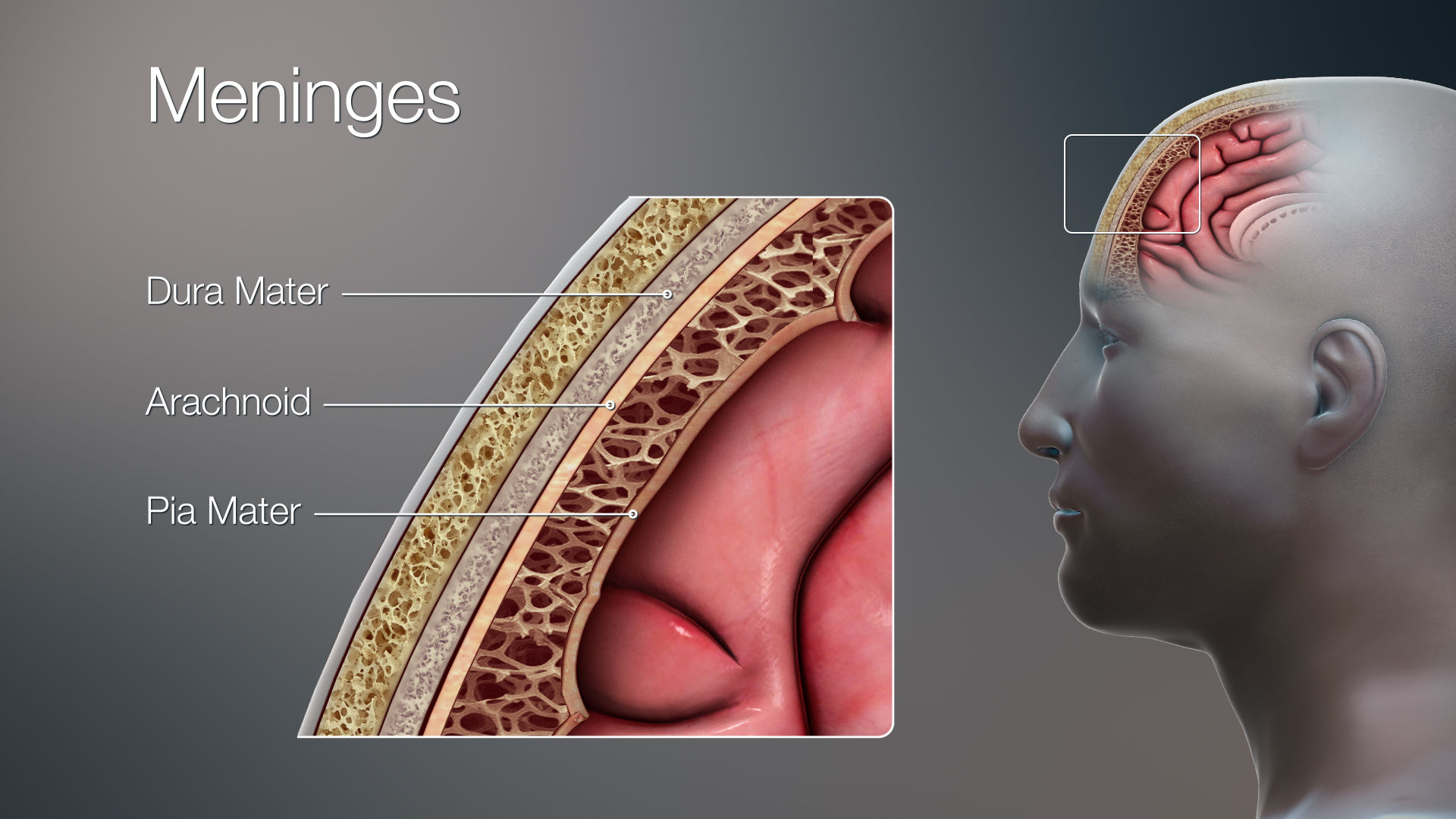
As you learned in this chapter, the human body consists of many complex systems that normally work together efficiently — like a well-oiled machine — to carry out life’s functions. For example, the image above (Figure 7.9.1) illustrates how the brain and spinal cord are protected by layers of membrane called meninges and fluid that flows between the meninges and in spaces called ventricles inside the brain. This fluid is called cerebrospinal fluid, and as you have learned, one of its important functions is to cushion and protect the brain and spinal cord, which make up most of the central nervous system (CNS). Additionally, cerebrospinal fluid circulates nutrients and removes waste products from the CNS. Cerebrospinal fluid is produced continually in the ventricles, circulates throughout the CNS, and is then reabsorbed by the bloodstream. If too much cerebrospinal fluid is produced, its flow is blocked, or not enough is reabsorbed, the system becomes out of balance and it can build up in the ventricles. This causes an enlargement of the ventricles called hydrocephalus that can put pressure on the brain, resulting in the types of neurological problems that former professional football player Jayson, described in the beginning of this chapter, is suffering from.
Recall that Jayson’s symptoms included loss of bladder control, memory loss, and difficulty walking. The cause of his symptoms was not immediately clear, although his doctors suspected that it related to the nervous system, since the nervous system acts as the control centre of the body, controlling and regulating many other organ systems. Jayson’s memory loss directly implicated the brain's involvement, since that is the site of thoughts and memory. The urinary system is also controlled in part by the nervous system, so the inability to hold urine appropriately can also be a sign of a neurological issue. Jayson’s trouble walking involved the muscular system, which works alongside the skeletal system to enable movement of the limbs. In turn, the contraction of muscles is regulated by the nervous system. You can see why a problem in the nervous system can cause a variety of different symptoms by affecting multiple organ systems in the human body.
To try to find the exact cause of Jayson’s symptoms, his doctors performed a lumbar puncture (or spinal tap), which is the removal of some cerebrospinal fluid through a needle inserted into the lower part of the spinal canal. They then analyzed Jayson’s cerebrospinal fluid for the presence of pathogens (such as bacteria) to determine whether an infection was the cause of his neurological symptoms. When no evidence of infection was found, they used an MRI to observe the structures of his brain. This is when they discovered his enlarged ventricles, which are a hallmark of hydrocephalus.
To treat Jayson’s hydrocephalus, a surgeon implanted a device called a shunt in his brain to remove the excess fluid. An illustration of a brain shunt is shown in Figure 9.7.2 . One side of the shunt consists of a small tube, called a catheter, which was inserted into Jayson’s ventricles. Excess cerebrospinal fluid is then drained through a one-way valve to the other end of the shunt, which was threaded under his skin to his abdominal cavity, where the fluid is released and can be reabsorbed by the bloodstream.
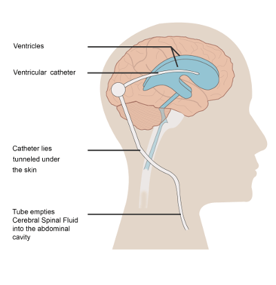
Implantation of a shunt is the most common way to treat hydrocephalus, and for some people, it can allow them to recover almost completely. However, there can be complications associated with a brain shunt. The shunt can have mechanical problems or cause an infection. Also, the rate of draining must be carefully monitored and adjusted to balance the rate of cerebrospinal fluid removal with the rate of its production. If it is drained too fast, it is called overdraining, and if it is drained too slowly, it is called underdraining. In the case of underdraining, the pressure on the brain and associated neurological symptoms will persist. In the case of overdraining, the ventricles can collapse, which can cause serious problems, such as the tearing of blood vessels and hemorrhaging. To avoid these problems, some shunts have an adjustable pressure valve, where the rate of draining can be adjusted by placing a special magnet over the scalp. You can see how the proper balance between cerebrospinal fluid production and removal is so critical – both in the causes of hydrocephalus and in its treatment.
In what other ways does your body regulate balance, or maintain a state of homeostasis? In this chapter you learned about the feedback loops that keep body temperature and blood glucose within normal ranges. Other important examples of homeostasis in the human body are the regulation of the pH in the blood and the balance of water in the body. You will learn more about homeostasis in different body systems in the coming chapters.
Thanks to Jayson’s shunt, his symptoms are starting to improve, but he has not fully recovered. Time may tell whether the removal of the excess cerebrospinal fluid from his ventricles will eventually allow him to recover normal functioning or whether permanent damage to his nervous system has already been done. The flow of cerebrospinal fluid might seem simple, but when it gets out of balance, it can easily wreak havoc on multiple organ systems because of the intricate interconnectedness of the systems within the human “machine."
To learn more about hydrocephalus and its treatment, watch this video from Boston Children's Hospital:
https://www.youtube.com/watch?v=bHD8zYImKqA
Hydrocephalus and its treatment | Boston Children’s Hospital, 2011.
Chapter 7 Summary
This chapter provided an overview of the organization and functioning of the human body. You learned that:
- The human body consists of multiple parts that function together to maintain life. The biology of the human body incorporates the body’s structure — or anatomy — and the body’s functioning, or physiology.
- The organization of the human body is a hierarchy of increasing size and complexity, starting at the level of atoms and molecules and ending at the level of the entire organism.
- Cells are the level of organization above atoms and molecules, and they are the basic units of structure and function of the human body. Each cell carries out basic life functions, as well as other specific roles. Cells of the human body show a lot of variation.
-
- Variations in cell function are generally reflected in variations in cell structure.
- Some cells are unattached to other cells and can move freely. Others are attached to each other and cannot move freely. Some cells can divide readily and form new cells, and others can divide only under exceptional circumstances. Many cells are specialized to produce and secrete particular substances.
- All the different cell types within an individual have the same genes. Cells can vary because different genes are expressed depending on the cell type.
- Many common types of human cells consist of several subtypes of cells, each of which has a special structure and function. For example, subtypes of bone cells include osteocytes, osteoblasts, osteogenic cells, and osteoclasts.
- A tissue is a group of connected cells that have a similar function. There are four basic types of human tissues that make up all the organs of the human body: epithelial, muscle, nervous, and connective tissues.
-
- Connective tissues, such as bone, tendons and blood, are made up of a scattering of living cells that are separated by non-living material, called extracellular matrix.
- Epithelial tissues, such as skin and mucous membranes, protect the body and its internal organs and secrete or absorb substances.
- Muscular tissues are made up of cells that have the unique ability to contract. They include skeletal, smooth, and cardiac muscle tissues.
- Nervous tissues are made up of neurons, which transmit messages, and neuroglia of various types, which play supporting roles.
- An organ is a structure that consists of two or more types of tissues that work together to do the same job. The brain and the heart are two examples.
-
- Many organs are composed of a major tissue that performs the organ’s main function, as well as other tissues that play supporting roles.
- The human body contains five organs that are considered vital for survival: the heart, brain, kidneys, liver, and lungs. If any of these five organs stops functioning, death of the organism is imminent without medical intervention.
- An organ system is a group of organs that work together to carry out a complex overall function. For example, the skeletal system provides structure to the body and protects internal organs.
-
- There are 11 major organ systems in the human organism. They are the integumentary, skeletal, muscular, nervous, endocrine, cardiovascular, lymphatic, respiratory, digestive, urinary, and reproductive systems. Only the reproductive system varies significantly between males and females.
- The human body is divided into a number of body cavities. A body cavity is a fluid-filled space in the body that holds and protects internal organs. The two largest human body cavities are the ventral cavity and dorsal cavity.
-
- The ventral cavity is at the anterior (or front) of the trunk. It is subdivided into the thoracic cavity, abdominal cavity and the pelvic cavity.
- The dorsal cavity is at the posterior (or back) of the body, and includes the head and the back of the trunk. It is subdivided into the cranial cavity and spinal cavity.
- Organ systems of the human body must work together to keep the body alive and functioning normally. This requires communication among organ systems. This is controlled by the autonomic nervous system and endocrine system. The autonomic nervous system controls involuntary body functions, such as heart rate and digestion. The endocrine system secretes hormones into the blood that travel to body cells and influence their activities.
-
- Cellular respiration is a good example of organ system interactions, because it is a basic life process that occurs in all living cells. It is the intracellular process that breaks down glucose with oxygen to produce carbon dioxide and energy. Cellular respiration requires the interaction of the digestive, cardiovascular, and respiratory systems.
- The fight-or-flight response is a good example of how the nervous and endocrine systems control other organ system responses. It is triggered by a message from the brain to the endocrine system and prepares the body for flight or a fight. Many organ systems are stimulated to respond, including the cardiovascular, respiratory, and digestive systems.
- Playing softball or doing other voluntary physical activities may involve the interaction of nervous, muscular, skeletal, respiratory, and cardiovascular systems.
- Homeostasis is the condition in which a system such as the human body is maintained in a more or less steady state. It is the job of cells, tissues, organs, and organ systems throughout the body to maintain homeostasis.
-
- For any given variable (such as body temperature), there is a particular set point that is the physiological optimum value. The spread of values around the set point that is considered insignificant is called the normal range.
- Homeostasis is generally maintained by a negative feedback loop that includes a stimulus, sensor, control centre, and effector. Negative feedback serves to reduce an excessive response and to keep a variable within the normal range. Negative feedback loops control body temperature and the blood glucose level.
- Sometimes homeostatic mechanisms fail, resulting in homeostatic imbalance. Diabetes is an example of a disease caused by homeostatic imbalance. Aging can bring about a reduction in the efficiency of the body’s control system, making the elderly more susceptible to disease.
- Positive feedback loops are not common in biological systems. Positive feedback serves to intensify a response until an end point is reached. Positive feedback loops control blood clotting and childbirth.
The severe and broad impact of hydrocephalus on the body’s systems highlights the importance of the nervous system and its role as the master control system of the body. In the next chapter, you will learn much more about the structures and functioning of this fascinating and important system.
Chapter 7 Review
-
- Compare and contrast tissues and organs.
-
-
- Which type of tissue lines the inner and outer surfaces of the body?
- What is a vital organ? What happens if a vital organ stops working?
- Name three organ systems that transport or remove wastes from the body.
- Name two types of tissue in the digestive system.
-
- Describe one way in which the integumentary and cardiovascular systems work together to regulate homeostasis in the human body.
-
- True or False: Body cavities are filled with air.
- In which organ system is the pituitary gland? Describe how the pituitary gland increases metabolism.
- When the level of thyroid hormone in the body gets too high, it acts on other cells to reduce production of more thyroid hormone. What type of feedback loop does this represent?
- Hypothetical organ A is the control centre in a feedback loop that helps maintain homeostasis. It secretes molecule A1 which reaches organ B, causing organ B to secrete molecule B1. B1 negatively feeds back onto organ A, reducing the production of A1 when the level of B1 gets too high.
- What is the stimulus in this feedback loop?
- If the level of B1 falls significantly below the set point, what do you think happens to the production of A1? Why?
- What is the effector in this feedback loop?
- If organs A and B are part of the endocrine system, what type of molecules do you think A1 and B1 are likely to be?
- What are the two main systems that allow various organ systems to communicate with each other?
- What are two functions of the hypothalamus?
Attributions
Figure 7.9.1
3D Medical Illustration Meninges Details by Scientific Animations on Wikimedia Commons is used under a CC BY-SA 4.0 (https://creativecommons.org/licenses/by-sa/4.0/deed.en) license.
Figure 7.9.2
Hydrocephalus with Shunt from CK-12 Foundation is used under a CC BY-NC 3.0 (https://creativecommons.org/licenses/by-nc/3.0/) license.
 ©CK-12 Foundation Licensed under
 ![]() • Terms of Use • Attribution
• Terms of Use • Attribution
References
Betts, J. G., Young, K.A., Wise, J.A., Johnson, E., Poe, B., Kruse, D.H., Korol, O., Johnson, J.E., Womble, M., DeSaix, P. (2013, April 25). Figure 1.3 Levels of structural organization of the human body [digital image]. In Anatomy and Physiology (Section 1.2). OpenStax. https://openstax.org/books/anatomy-and-physiology/pages/1-2-structural-organization-of-the-human-body
Betts, J. G., Young, K.A., Wise, J.A., Johnson, E., Poe, B., Kruse, D.H., Korol, O., Johnson, J.E., Womble, M., DeSaix, P. (2013, April 25). Figure 1.4 Organ systems of the human body [digital image]. In Anatomy and Physiology (Section 1.2). OpenStax. https://openstax.org/books/anatomy-and-physiology/pages/1-2-structural-organization-of-the-human-body
Betts, J. G., Young, K.A., Wise, J.A., Johnson, E., Poe, B., Kruse, D.H., Korol, O., Johnson, J.E., Womble, M., DeSaix, P. (2013, April 25). Figure 1.15 Dorsal and ventral body cavities [digital image]. In Anatomy and Physiology (Section 1.2). OpenStax. https://openstax.org/books/anatomy-and-physiology/pages/1-6-anatomical-terminology
Boston Children's Hospital. (2011, ). Hydrocephalus and its treatment | Boston Children’s Hospital. YouTube. https://www.youtube.com/watch?v=bHD8zYImKqA&feature=youtu.be
Brainard, J/ CK-12 Foundation. (2016). Figure 2 An illustration of a brain shunt [digital image]. In CK-12 College Human Biology (Section 9.8) [online Flexbook]. CK12.org. https://www.ck12.org/book/ck-12-college-human-biology/section/9.8/
File:Body cavities lateral view labeled.jpg. (2018, January 4). Wikimedia Commons. https://commons.wikimedia.org/w/index.php?title=File:Body_Cavities_Lateral_view_labeled.jpg&oldid=276851269. (Original image: Figure 1.15 Dorsal and ventral body cavities, from OpenStax, Anatomy and Physiology.)
File:Body cavities lateral view labeled.jpg. (2018, January 4). Wikimedia Commons. https://commons.wikimedia.org/w/index.php?title=File:Body_Cavities_Lateral_view_labeled.jpg&oldid=276851269. (Original image: OpenStax [Version 8.25 from the textbook OpenStax Anatomy and Physiology] adapted for Review questions by Christine Miller].
Created by CK-12 Foundation/ Adapted by Christine Miller

In the Blink of an Eye
As you drive into a parking lot, a boy on a skateboard suddenly flies in front of your car across your field of vision. You see the boy in the nick of time and react immediately. You slam on the brakes and steer sharply to the right — all in the blink of an eye. You avoid a collision, but just barely. You’re shaken up, but thankful that no one was hurt. How did you respond so quickly? Rapid responses like this are controlled by your nervous system.
Overview of the Nervous System
The nervous system, illustrated in the sketch below, is the human organ system that coordinates all of the body’s voluntary and involuntary actions, by transmitting electrical signals to and from different parts of the body. Specifically, the nervous system extracts information from the internal and external environments, using sensory receptors. Usually, it then sends signals encoding this information to the brain, which processes the information to determine an appropriate response. Finally, the brain sends signals to muscles, organs, or glands to bring about the response. In the example above, your eyes detected the boy, the information traveled to your brain, and your brain told your body to act so as to avoid a collision.
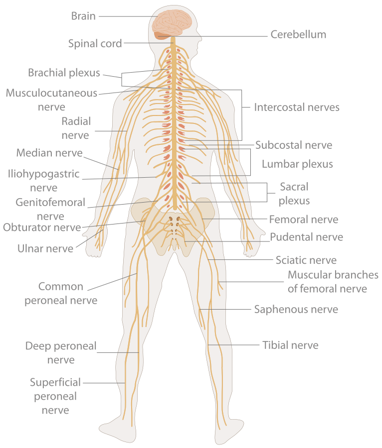
Signals of the Nervous System
The signals sent by the nervous system are electrical signals called nerve impulses, and they are transmitted by special nervous system cells called neurons (or nerve cells), like the one in Figure 8.2.3. Long projections (called axons) from neurons carry nerve impulses directly to specific target cells. A cell that receives nerve impulses from a neuron (typically a muscle or a gland) may be excited to perform a function, inhibited from carrying out an action, or otherwise controlled. In this way, the information transmitted by the nervous system is specific to particular cells and is transmitted very rapidly. In fact, the fastest nerve impulses travel at speeds greater than 100 metres per second! Compare this to the chemical messages carried by the hormones that are secreted into the blood by endocrine glands. These hormonal messages are “broadcast” to all the cells of the body, and they can travel only as quickly as the blood flows through the cardiovascular system.
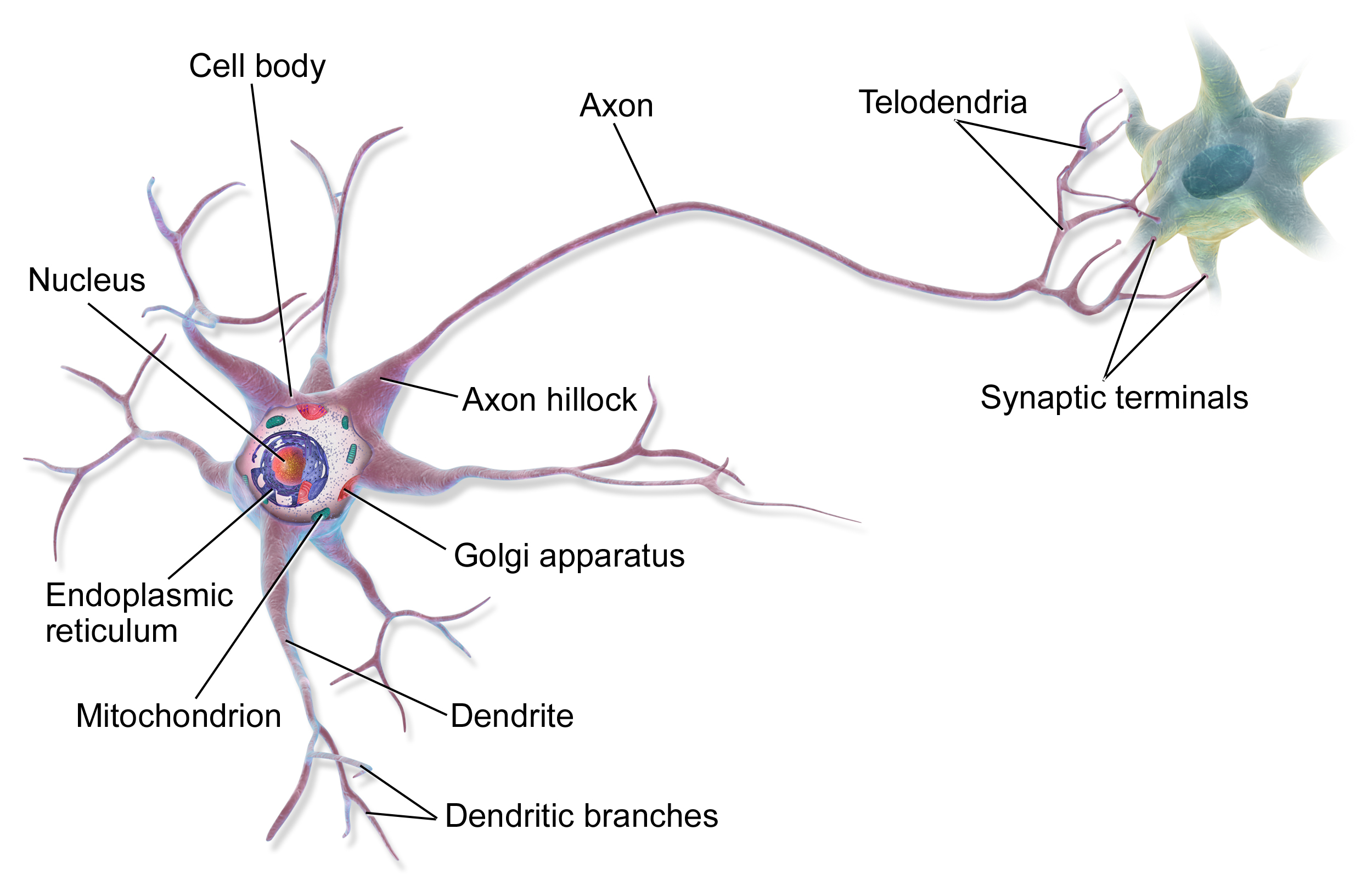
This simple model of a nerve cell shows part of its long axon which carries nerve impulses to other cells. The multiple shorter projections are called dendrites, and they receive nerve impulses from other cells.
Organization of the Nervous System
As you might predict, the human nervous system is very complex. It has multiple divisions, beginning with its two main parts, the central nervous system (CNS) and the peripheral nervous system (PNS), as shown in the diagram below (Figure 8.2.4). The CNS includes the brain and spinal cord, and the PNS consists mainly of nerves, which are bundles of axons from neurons. The nerves of the PNS connect the CNS to the rest of the body.
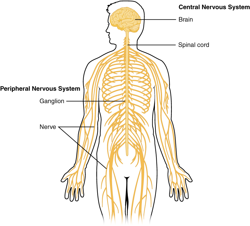
The PNS can be further subdivided into two divisions, known as the autonomic and somatic nervous systems (Figure 8.2.5). These divisions control different types of functions, and they often interact with the CNS to carry out these functions. The somatic nervous system controls activities that are under voluntary control, such as turning a steering wheel. The autonomic nervous system controls activities that are not under voluntary control, such as digesting a meal. The autonomic nervous system has three main divisions: the sympathetic division (which controls the fight-or-flight response during emergencies), the parasympathetic division (which controls the routine “housekeeping” functions of the body at other times), and the enteric division (which provides local control of the digestive system).
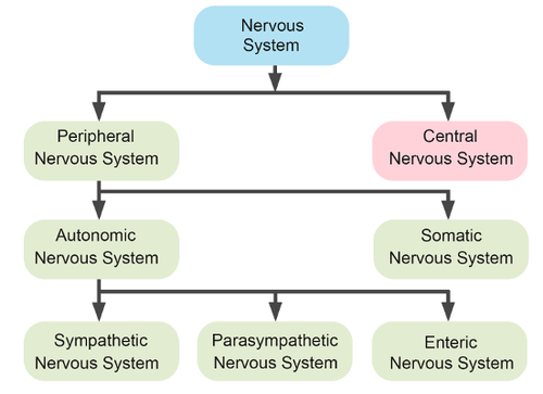
8.2 Summary
- The nervous system is the human organ system that coordinates all of the body’s voluntary and involuntary actions, by transmitting signals to and from different parts of the body.
- The nervous system has two major divisions, called the central nervous system (CNS) and the peripheral nervous system (PNS). The CNS includes the brain and spinal cord, and the PNS consists mainly of nerves that connect the CNS with the rest of the body.
- The PNS can be subdivided into two major divisions: the somatic nervous system and the autonomic nervous system.The somatic system controls activities that are under voluntary control. The autonomic system controls activities that are not under voluntary control. The autonomic nervous system is further divided into the sympathetic division (which controls the fight-or-flight response), the parasympathetic division (which controls most routine involuntary responses), and the enteric division (which provides local control of the digestive system).
- Electrical signals sent by the nervous system are called nerve impulses. They are transmitted by special cells called neurons. Nerve impulses can travel to specific target cells very rapidly.
8.2 Review Questions
- List the general steps through which the nervous system generates an appropriate response to information from the internal and external environments.
- What are neurons?
- Compare and contrast the central and peripheral nervous systems.
-
- Which major division of the peripheral nervous system allows you to walk to class? Which major division of the peripheral nervous system controls your heart rate?
- Identify the functions of the three main divisions of the autonomic nervous system.
-
- What is an axon, and what is its function?
- Define nerve impulses.
- Explain generally how the brain and spinal cord can interact with and control the rest of the body.
- How are nerves and neurons related?
- What type of information from the outside environment do you think is detected by sensory receptors in your ears?
8.2 Explore More
https://www.youtube.com/watch?v=qPix_X-9t7E
The Nervous System, Part 1: Crash Course A&P #8, CrashCourse, 2015.
https://www.youtube.com/watch?v=Nsxw5_Iz7mY&feature=emb_logo
Engineering the Human Nervous System: Megan Moynahan at TEDxBrussels,
TEDx Talks, 2013.
Attributions
Figure 8.2.1
Skateboard_1613 by Autoria propia on Wikimedia Commons is released into the public domain (https://en.wikipedia.org/wiki/Public_domain) (Derivative work of this file: SkateboardinDog.jpg)
Figure 8.2.2
Nervous_system_diagram.svg by The Emirr on Wikimedia Commons is used under a CC BY 3.0 (https://creativecommons.org/licenses/by/3.0/deed.en) license.
Figure 8.2.3
MultipolarNeuron by BruceBlaus on Wikimedia Commons is used under a CC BY 3.0 (https://creativecommons.org/licenses/by/3.0/deed.en) license.
Figure 8.2.4
Overview_of_Nervous_System by OpenStax on Wikimedia Commons is used under a CC BY 4.0 (https://creativecommons.org/licenses/by/4.0/deed.en) license.
Figure 8.2.5
Divisions of the Nervous System by CK-12 Foundation is used under the CC BY-NC 3.0 (https://creativecommons.org/licenses/by-nc/3.0/) license.
References
Betts, J. G., Young, K.A., Wise, J.A., Johnson, E., Poe, B., Kruse, D.H., Korol, O., Johnson, J.E., Womble, M., DeSaix, P. (2013, April 25). Figure 12.2 Central and peripheral nervous system [digital image]. In Anatomy and Physiology (Section 12.1). OpenStax. https://openstax.org/books/anatomy-and-physiology/pages/12-1-basic-structure-and-function-of-the-nervous-system
Brainard, J/ CK-12 Foundation. (2016). Figure 5 [digital image]. In CK-12 College Human Biology (Section 10.2) [online Flexbook]. CK12.org. https://www.ck12.org/book/ck-12-college-human-biology/section/10.2/
CrashCourse. (2015, February 23). The nervous system, Part 1: Crash Course A&P #8. YouTube. https://www.youtube.com/watch?v=qPix_X-9t7E&feature=youtu.be
TEDx Talks. (2013, November 3). Engineering the human nervous system: Megan Moynahan at TEDxBrussels. YouTube. https://www.youtube.com/watch?v=Nsxw5_Iz7mY&feature=youtu.be
A nervous system cell that provides support for neurons and helps them transmit nerve impulses.
Created by CK-12 Foundation/Adapted by Christine Miller
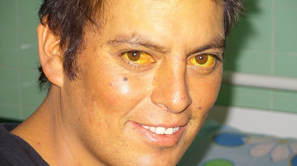
Jaundiced Eyes
Did you ever hear of a person looking at something or someone with a “jaundiced eye”? It means to take a negative view, such as envy, maliciousness, or ill will. The expression may be based on the antiquated idea that liver bile is associated with such negative emotions as these, as well as the fact that excessive liver bile causes jaundice, or yellowing of the eyes and skin. Jaundice is likely a sign of a liver disorder or blockage of the duct that carries bile away from the liver. Bile contains waste products, making the liver an organ of excretion. Bile has an important role in digestion, which makes the liver an accessory organ of digestion, too.
What Are Accessory Organs of Digestion?
Accessory organs of digestion are organs that secrete substances needed for the chemical digestion of food, but through which food does not actually pass as it is digested. Besides the liver, the major accessory organs of digestion are the gallbladder and pancreas. These organs secrete or store substances that are needed for digestion in the first part of the small intestine — the duodenum — where most chemical digestion takes place. You can see the three organs and their locations in Figure 15.6.2.
Liver
The liver is a vital organ located in the upper right part of the abdomen. It lies just below the diaphragm, to the right of the stomach. The liver plays an important role in digestion by secreting bile, but the liver has a wide range of additional functions unrelated to digestion. In fact, some estimates put the number of functions of the liver at about 500! A few of them are described below.
Structure of the Liver
The liver is a reddish brown, wedge-shaped structure. In adults, the liver normally weighs about 1.5 kg (about 3.3 lb). It is both the heaviest internal organ and the largest gland in the human body. The liver is divided into four lobes of unequal size and shape. Each lobe, in turn, is made up of lobules, which are the functional units of the liver. Each lobule consists of millions of liver cells, called hepatic cells (or hepatocytes). They are the basic metabolic cells that carry out the various functions of the liver.
As shown in Figure 15.6.3, the liver is connected to two large blood vessels: the hepatic artery and the portal vein. The hepatic artery carries oxygen-rich blood from the aorta, whereas the portal vein carries blood that is rich in digested nutrients from the GI tract and wastes filtered from the blood by the spleen. The blood vessels subdivide into smaller arteries and capillaries, which lead into the liver lobules. The nutrients from the GI tract are used to build many vital biochemical compounds, and the wastes from the spleen are degraded and excreted.
Functions of the Liver
The main digestive function of the liver is the production of bile. Bile is a yellowish alkaline liquid that consists of water, electrolytes, bile salts, and cholesterol, among other substances, many of which are waste products. Some of the components of bile are synthesized by hepatocytes. The rest are extracted from the blood.
As shown in Figure 15.6.4, bile is secreted into small ducts that join together to form larger ducts, with just one large duct carrying bile out of the liver. If bile is needed to digest a meal, it goes directly to the duodenum through the common bile duct. In the duodenum, the bile neutralizes acidic chyme from the stomach and emulsifies fat globules into smaller particles (called micelles) that are easier to digest chemically by the enzyme lipase. Bile also aids with the absorption of vitamin K. Bile that is secreted when digestion is not taking place goes to the gallbladder for storage until the next meal. In either case, the bile enters the duodenum through the common bile duct.
Besides its roles in digestion, the liver has many other vital functions:
- The liver synthesizes glycogen from glucose and stores the glycogen as required to help regulate blood sugar levels. It also breaks down the stored glycogen to glucose and releases it back into the blood as needed.
- The liver stores many substances in addition to glycogen, including vitamins A, D, B12, and K. It also stores the minerals iron and copper.
- The liver synthesizes numerous proteins and many of the amino acids needed to make them. These proteins have a wide range of functions. They include fibrinogen, which is needed for blood clotting; insulin-like growth factor (IGF-1), which is important for childhood growth; and albumen, which is the most abundant protein in blood serum and functions to transport fatty acids and steroid hormones in the blood.
- The liver synthesizes many important lipids, including cholesterol, triglycerides, and lipoproteins.
- The liver is responsible for the breakdown of many waste products and toxic substances. The wastes are excreted in bile or travel to the kidneys, which excrete them in urine.
The liver is clearly a vital organ that supports almost every other organ in the body. Because of its strategic location and diversity of functions, the liver is also prone to many diseases, some of which cause loss of liver function. There is currently no way to compensate for the absence of liver function in the long term, although liver dialysis techniques can be used in the short term. An artificial liver has not yet been developed, so liver transplantation may be the only option for people with liver failure.
Gallbladder
The gallbladder is a small, hollow, pouch-like organ that lies just under the right side of the liver (see Figure 15.6.5). It is about 8 cm (about 3 in) long and shaped like a tapered sac, with the open end continuous with the cystic duct. The gallbladder stores and concentrates bile from the liver until it is needed in the duodenum to help digest lipids. After the bile leaves the liver, it reaches the gallbladder through the cystic duct. At any given time, the gallbladder may store between 30 to 60 mL (1 to 2 oz) of bile. A hormone stimulated by the presence of fat in the duodenum signals the gallbladder to contract and force its contents back through the cystic duct and into the common bile duct to drain into the duodenum.
Pancreas
The pancreas is a glandular organ that is part of both the digestive system and the endocrine system. As shown in Figure 15.6.6, it is located in the abdomen behind the stomach, with the head of the pancreas surrounded by the duodenum of the small intestine. The pancreas is about 15 cm (almost 6 in) long, and it has two major ducts: the main pancreatic duct and the accessory pancreatic duct. Both of these ducts drain into the duodenum.
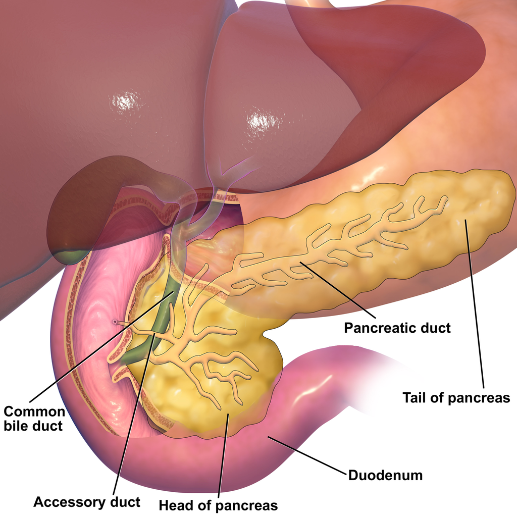
As an endocrine gland, the pancreas secretes several hormones, including insulin and glucagon, which circulate in the blood. The endocrine hormones are secreted by clusters of cells called pancreatic islets (or islets of Langerhans). As a digestive organ, the pancreas secretes many digestive enzymes and also bicarbonate, which helps neutralize acidic chyme after it enters the duodenum. The pancreas is stimulated to secrete its digestive substances when food in the stomach and duodenum triggers the release of endocrine hormones into the blood that reach the pancreas via the bloodstream. The pancreatic digestive enzymes are secreted by clusters of cells called acini, and they travel through the pancreatic ducts to the duodenum. In the duodenum, they help to chemically break down carbohydrates, proteins, lipids, and nucleic acids in chyme. The pancreatic digestive enzymes include:
- Amylase, which helps digest starch and other carbohydrates.
- Trypsin and chymotrypsin, which help digest proteins.
- Lipase, which helps digest lipids.
- Deoxyribonucleases and ribonucleases, which help digest nucleic acids.
15.6 Summary
- Accessory organs of digestion are organs that secrete substances needed for the chemical digestion of food, but through which food does not actually pass as it is digested. The accessory organs include the liver, gallbladder, and pancreas. These organs secrete or store substances that are carried to the duodenum of the small intestine as needed for digestion.
- The liver is a large organ in the abdomen that is divided into lobes and smaller lobules, which consist of metabolic cells called hepatic cells, or hepatocytes. The liver receives oxygen in blood from the aorta through the hepatic artery. It receives nutrients in blood from the GI tract and wastes in blood from the spleen through the portal vein.
- The main digestive function of the liver is the production of the alkaline liquid called bile. Bile is carried directly to the duodenum by the common bile duct or to the gallbladder first for storage. Bile neutralizes acidic chyme that enters the duodenum from the stomach, and also emulsifies fat globules into smaller particles (micelles) that are easier to digest chemically.
- Other vital functions of the liver include regulating blood sugar levels by storing excess sugar as glycogen, storing many vitamins and minerals, synthesizing numerous proteins and lipids, and breaking down waste products and toxic substances.
- The gallbladder is a small pouch-like organ near the liver. It stores and concentrates bile from the liver until it is needed in the duodenum to neutralize chyme and help digest lipids.
- The pancreas is a glandular organ that secretes both endocrine hormones and digestive enzymes. As an endocrine gland, the pancreas secretes insulin and glucagon to regulate blood sugar. As a digestive organ, the pancreas secretes digestive enzymes into the duodenum through ducts. Pancreatic digestive enzymes include amylase (starches) trypsin and chymotrypsin (proteins), lipase (lipids), and ribonucleases and deoxyribonucleases (RNA and DNA).
15.6 Review Questions
- Name three accessory organs of digestion. How do these organs differ from digestive organs that are part of the GI tract?
-
- Describe the liver and its blood supply.
- Explain the main digestive function of the liver and describe the components of bile and it's importance in the digestive process.
- What type of secretions does the pancreas release as part of each body system?
- List pancreatic enzymes that work in the duodenum, along with the substances they help digest.
- What are two substances produced by accessory organs of digestion that help neutralize chyme in the small intestine? Where are they produced?
- People who have their gallbladder removed sometimes have digestive problems after eating high-fat meals. Why do you think this happens?
- Which accessory organ of digestion synthesizes cholesterol?
15.6 Explore More
https://youtu.be/8dgoeYPoE-0
What does the pancreas do? - Emma Bryce, TED-Ed. 2015.
https://youtu.be/wbh3SjzydnQ
What does the liver do? - Emma Bryce, TED-Ed, 2014.
https://youtu.be/a0d1yvGcfzQ
Scar wars: Repairing the liver, nature video, 2018.
Attributions
Figure 15.6.1
Scleral_Icterus by Sheila J. Toro on Wikimedia Commons is used under a CC BY 4.0 (https://creativecommons.org/licenses/by/4.0) license.
Figure 15.6.2
Blausen_0428_Gallbladder-Liver-Pancreas_Location by BruceBlaus on Wikimedia Commons is used under a CC BY 3.0 (https://creativecommons.org/licenses/by/3.0) license.
Figure 15.6.3
Diagram_showing_the_two_lobes_of_the_liver_and_its_blood_supply_CRUK_376.svg by Cancer Research UK on Wikimedia Commons is used under a CC BY-SA 4.0 (https://creativecommons.org/licenses/by-sa/4.0) license.
Figure 15.6.4
Gallbladder by NIH Image Gallery on Flickr is used CC BY-NC 2.0 (https://creativecommons.org/licenses/by-nc/2.0/) license.
Figure 15.6.5
Gallbladder_(organ) (1) by BruceBlaus on Wikimedia Commons is used under a CC BY-SA 4.0 (https://creativecommons.org/licenses/by-sa/4.0) license. (See a full animation of this medical topic at blausen.com.)
Figure 15.6.6
Blausen_0698_PancreasAnatomy by BruceBlaus on Wikimedia Commons is used under a CC BY 3.0 (https://creativecommons.org/licenses/by/3.0) license.
References
Blausen.com Staff. (2014). Medical gallery of Blausen Medical 2014. WikiJournal of Medicine 1 (2). DOI:10.15347/wjm/2014.010. ISSN 2002-4436.
nature video. (2018, December 19). Scar wars: Repairing the liver. YouTube. https://www.youtube.com/watch?v=a0d1yvGcfzQ&feature=youtu.be
TED-Ed. (2014, November 25). What does the liver do? - Emma Bryce. YouTube. https://www.youtube.com/watch?v=wbh3SjzydnQ&feature=youtu.be
TED-Ed. (2015, February 19). What does the pancreas do? - Emma Bryce. YouTube. https://www.youtube.com/watch?v=8dgoeYPoE-0&feature=youtu.be
Created by CK-12 Foundation/Adapted by Christine Miller
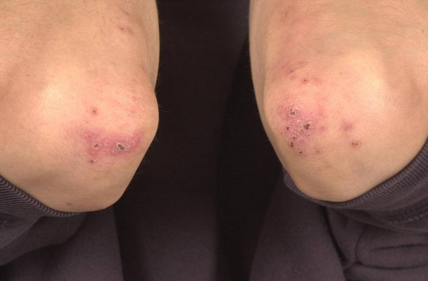
Crohn’s Rash
If you had a skin rash like the one shown in Figure 15.7.1, you probably wouldn’t assume that it was caused by a digestive system disease. However, that’s exactly why the individual in the picture has a rash. He has a gastrointestinal (GI) tract disorder called Crohn’s disease. This disease is one of a group of GI tract disorders that are known collectively as inflammatory bowel disease. Unlike other inflammatory bowel diseases, signs and symptoms of Crohn’s disease may not be confined to the GI tract.
Inflammatory Bowel Disease
Inflammatory bowel disease (IBD) is a collection of inflammatory conditions primarily affecting the intestines. The two principal inflammatory bowel diseases are Crohn’s disease and ulcerative colitis. Unlike Crohn’s disease — which may affect any part of the GI tract and the joints, as well as the skin — ulcerative colitis mainly affects just the colon and rectum. Both diseases occur when the body’s own immune system attacks the digestive system. Both diseases typically first appear in the late teens or early twenties, and occur equally in males and females. Approximately 270,000 Canadians are currently living with IBD, 7,000 of which are children. The annual cost of caring for these Canadians is estimated at $1.28 billion. The number of cases of IBD has been steadily increasing and it is expected that by 2030 the number of Canadians suffering from IBD will grow to 400,000.
Crohn’s Disease
Crohn’s disease is a type of inflammatory bowel disease that may affect any part of the GI tract from the mouth to the anus, among other body tissues. The most commonly affected region is the ileum, which is the final part of the small intestine. Signs and symptoms of Crohn’s disease typically include abdominal pain, diarrhea (with or without blood), fever, and weight loss. Malnutrition because of faulty absorption of nutrients may also occur. Potential complications of Crohn’s disease include obstructions and abscesses of the bowel. People with Crohn’s disease are also at slightly greater risk than the general population of developing bowel cancer. Although there is a slight reduction in life expectancy in people with Crohn’s disease, if the disease is well-managed, affected people can live full and productive lives. Approximately 135,000 Canadians are living with Crohn's disease.
Crohn’s disease is caused by a combination of genetic and environmental factors that lead to impairment of the generalized immune response (called innate immunity). The chronic inflammation of Crohn’s disease is thought to be the result of the immune system “trying” to compensate for the impairment. Dozens of genes are likely to be involved, only a few of which have been identified. Because of the genetic component, close relatives such as siblings of people with Crohn’s disease are many times more likely to develop the disease than people in the general population. Environmental factors that appear to increase the risk of the disease include smoking tobacco and eating a diet high in animal proteins. Crohn’s disease is typically diagnosed on the basis of a colonoscopy, which provides a direct visual examination of the inside of the colon and the ileum of the small intestine.
People with Crohn’s disease typically experience recurring periods of flare-ups followed by remission. There are no medications or surgical procedures that can cure Crohn’s disease, although medications such as anti-inflammatory or immune-suppressing drugs may alleviate symptoms during flare-ups and help maintain remission. Lifestyle changes, such as dietary modifications and smoking cessation, may also help control symptoms and reduce the likelihood of flare-ups. Surgery may be needed to resolve bowel obstructions, abscesses, or other complications of the disease.
Ulcerative Colitis
Ulcerative colitis is an inflammatory bowel disease that causes inflammation and ulcers (sores) in the colon and rectum. Unlike Crohn’s disease, other parts of the GI tract are rarely affected in ulcerative colitis. The primary symptoms of the disease are lower abdominal pain and bloody diarrhea. Weight loss, fever, and anemia may also be present. Symptoms typically occur intermittently with periods of no symptoms between flare-ups. People with ulcerative colitis have a considerably increased risk of colon cancer and should be screened for colon cancer more frequently than the general population. Ulcerative colitis, however, seems to primarily reduce the quality of life, and not the lifespan.
The exact cause of ulcerative colitis is not known. Theories about its cause involve immune system dysfunction, genetics, changes in normal gut bacteria, and lifestyle factors, such as a diet high in animal protein and the consumption of alcoholic beverages. Genetic involvement is suspected in part because ulcerative colitis tens to “run” in families. It is likely that multiple genes are involved. Diagnosis is typically made on the basis of colonoscopy and tissue biopsies.
Lifestyle changes, such as reducing the consumption of animal protein and alcohol, may improve symptoms of ulcerative colitis. A number of medications are also available to treat symptoms and help prolong remission. These include anti-inflammatory drugs and drugs that suppress the immune system. In cases of severe disease, removal of the colon and rectum may be required and can cure the disease.
Diverticulitis
Diverticulitis is a digestive disease in which tiny pouches in the wall of the large intestine become infected and inflamed. Symptoms typically include lower abdominal pain of sudden onset. There may also be fever, nausea, diarrhea or constipation, and blood in the stool. Having large intestine pouches called diverticula (see Figure 15.7.2) that are not inflamed is called diverticulosis. Diverticulosis is thought to be caused by a combination of genetic and environmental factors, and is more common in people who are obese. Infection and inflammation of the pouches (diverticulitis) occurs in about 10–25% of people with diverticulosis, and is more common at older ages. The infection is generally caused by bacteria.
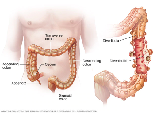
Diverticulitis can usually be diagnosed with a CT scan and can be monitored with a colonoscopy (as seen in Figure 15.7.3). Mild diverticulitis may be treated with oral antibiotics and a short-term liquid diet. For severe cases, intravenous antibiotics, hospitalization, and complete bowel rest (no nourishment via the mouth) may be recommended. Complications such as abscess formation or perforation of the colon require surgery.
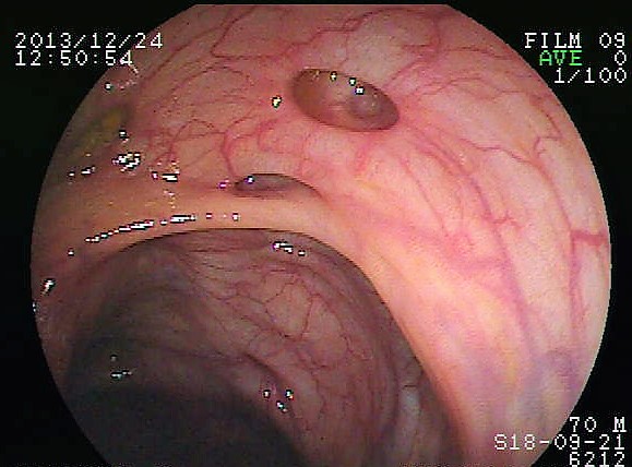
Peptic Ulcer
A peptic ulcer is a sore in the lining of the stomach or the duodenum (first part of the small intestine). If the ulcer occurs in the stomach, it is called a gastric ulcer. If it occurs in the duodenum, it is called a duodenal ulcer. The most common symptoms of peptic ulcers are upper abdominal pain that often occurs in the night and improves with eating. Other symptoms may include belching, vomiting, weight loss, and poor appetite. Many people with peptic ulcers, particularly older people, have no symptoms. Peptic ulcers are relatively common, with about ten per cent of people developing a peptic ulcer at some point in their life.
The most common cause of peptic ulcers is infection with the bacterium Helicobacter pylori, which may be transmitted by food, contaminated water, or human saliva (for example, by kissing or sharing eating utensils). Surprisingly, the bacterial cause of peptic ulcers was not discovered until the 1980s. The scientists who made the discovery are Australians Robin Warren and Barry J. Marshall. Although the two scientists eventually won a Nobel Prize for their discovery, their hypothesis was poorly received at first. To demonstrate the validity of their discovery, Marshall used himself in an experiment. He drank a culture of bacteria from a peptic ulcer patient and developed symptoms of peptic ulcer in a matter of days. His symptoms resolved on their own within a couple of weeks, but, at his wife's urging, he took antibiotics to kill any remaining bacteria. Marshall’s self-experiment was published in the Australian Medical Journal, and is among the most cited articles ever published in the journal. Figure 15.7.4 shows how H. pylori cause peptic ulcers.
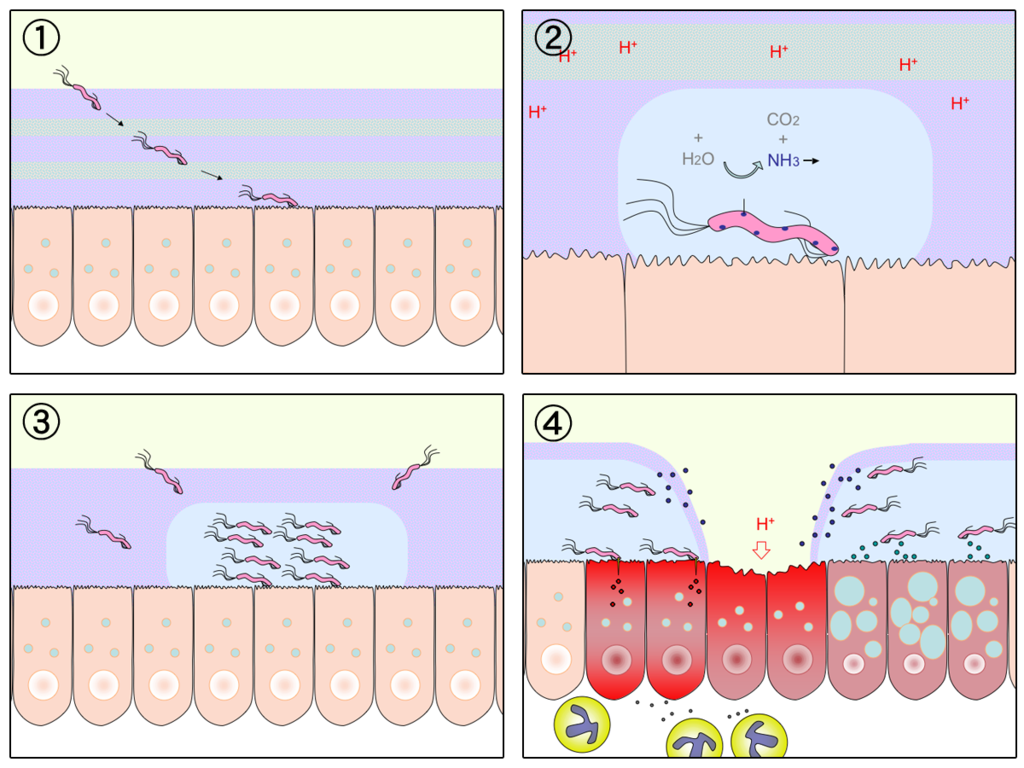
Another relatively common cause of peptic ulcers is chronic use of non-steroidal anti-inflammatory drugs (NSAIDs), such as aspirin or ibuprofen. Additional contributing factors may include tobacco smoking and stress, although these factors have not been demonstrated conclusively to cause peptic ulcers independent of H. pylori infection. Contrary to popular belief, diet does not appear to play a role in either causing or preventing peptic ulcers. Eating spicy foods and drinking coffee and alcohol were once thought to cause peptic ulcers. These lifestyle choices are no longer thought to have much (if any) of an effect on the development of peptic ulcers.
Peptic ulcers are typically diagnosed on the basis of symptoms or the presence of H. pylori in the GI tract. However, endoscopy (shown in Figure 15.7.5), which allows direct visualization of the stomach and duodenum with a camera, may be required for a definitive diagnosis. Peptic ulcers are usually treated with antibiotics to kill H. pylori, along with medications to temporarily decrease stomach acid and aid in healing. Unfortunately, H. pylori has developed resistance to commonly used antibiotics, so treatment is not always effective. If a peptic ulcer has penetrated so deep into the tissues that it causes a perforation of the wall of the stomach or duodenum, then emergency surgery is needed to repair the damage.
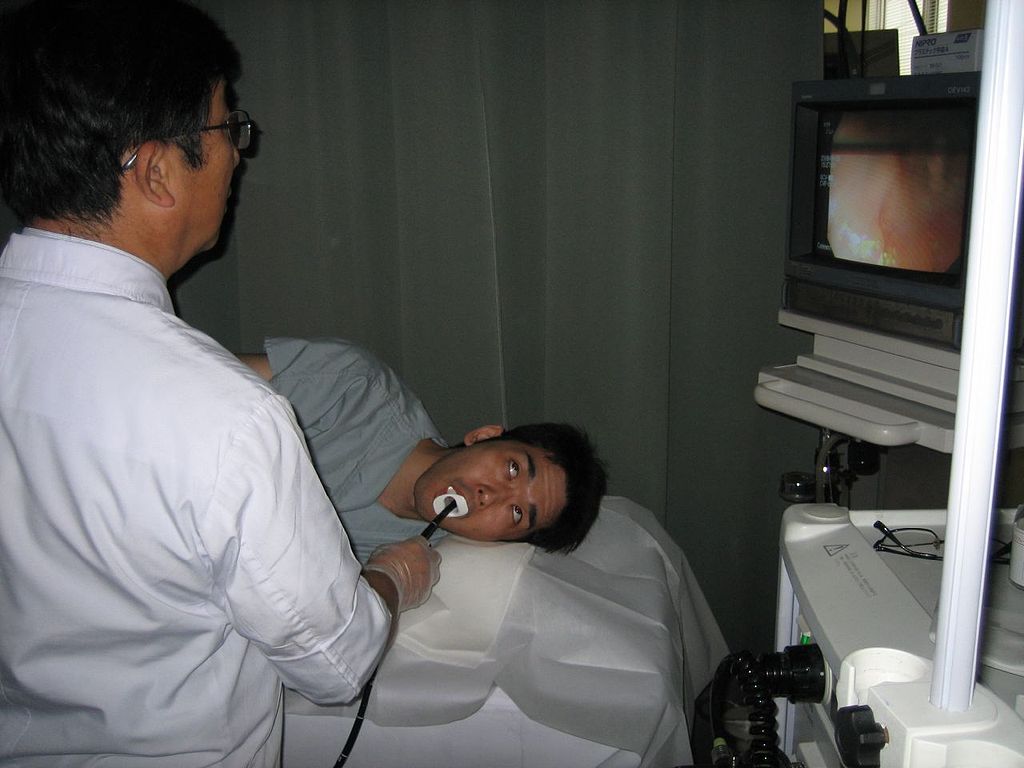
Gastroenteritis
Gastroenteritis, also known as infectious diarrhea or stomach flu, is an acute and usually self-limiting infection of the GI tract by pathogens. Symptoms typically include some combination of diarrhea, vomiting, and abdominal pain. Fever, lack of energy, and dehydration may also occur. The illness generally lasts less than two weeks, even without treatment, but in young children it is potentially deadly. Gastroenteritis is very common, especially in poorer nations. Worldwide, up to five billion cases occur each year, resulting in about 1.4 million deaths.
Commonly called “stomach flu,” gastroenteritis is unrelated to the influenza virus, although viruses are the most common cause of the disease (see Figure 15.7.6). In children, rotavirus is most often the cause which is why the British Columbia immunization schedule now includes a rotovirus vaccine. Norovirus is more likely to be the cause of gastroenteritis in adults. Besides viruses, other potential causes of gastroenteritis include fungi, bacteria (most often E. coli or Campylobacter jejuni), and protozoa(including Giardia lamblia, more commonly called Beaver Fever, described below). Transmission of pathogens may occur due to eating improperly prepared foods or foods left to stand at room temperature, drinking contaminated water, or having close contact with an infected individual.
Gastroenteritis is less common in adults than children, partly because adults have acquired immunity after repeated exposure to the most common infectious agents. Adults also tend to have better hygiene than children. If children have frequent repeated incidents of gastroenteritis, they may suffer from malnutrition, stunted growth, and developmental delays. Many cases of gastroenteritis in children can be avoided by giving them a rotavirus vaccine. Frequent and thorough handwashing can cut down on infections caused by other pathogens.
Treatment of gastroenteritis generally involves increasing fluid intake to replace fluids lost in vomiting or diarrhea. Oral rehydration solution, which is a combination of water, salts, and sugar, is often recommended. In severe cases, intravenous fluids may be needed. Antibiotics are not usually prescribed, because they are ineffective against viruses that cause most cases of gastroenteritis.
Giardiasis
Giardiasis, popularly known as beaver fever, is a type of gastroenteritis caused by a GI tract parasite, the single-celled protozoan Giardia lamblia (pictured in Figure 15.7.7). In addition to human beings, the parasite inhabits the digestive tract of a wide variety of domestic and wild animals, including cows, rodents, and sheep, as well as beavers (hence its popular name). Giardiasis is one of the most common parasitic infections in people the world over, with hundreds of millions of people infected worldwide each year.
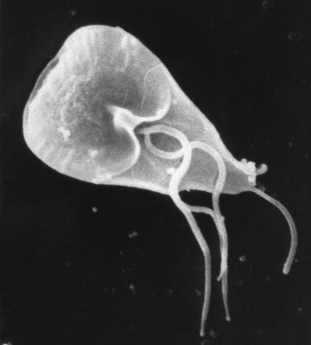
Transmission of G. lamblia is via a fecal-oral route (as in, you got feces in your food). Those at greatest risk include travelers to countries where giardiasis is common, people who work in child-care settings, backpackers and campers who drink untreated water from lakes or rivers, and people who have close contact with infected people or animals in other settings. In Canada, Giardia is the most commonly identified intestinal parasite and approximately 3,000 Canadians will contract the parasite annually.
Symptoms of giardiasis can vary widely. About one-third third of people with the infection have no symptoms, whereas others have severe diarrhea with poor absorption of nutrients. Problems with absorption occur because the parasites inhibit intestinal digestive enzyme production, cause detrimental changes in microvilli lining the small intestine, and kill off small intestinal epithelial cells. The illness can result in weakness, loss of appetite, stomach cramps, vomiting, and excessive gas. Without treatment, symptoms may continue for several weeks. Treatment with anti-parasitic medications may be needed if symptoms persist longer or are particularly severe.
15.7 Summary
- Inflammatory bowel disease is a collection of inflammatory conditions primarily affecting the intestines. The diseases involve the immune system attacking the GI tract, and they have multiple genetic and environmental causes. Typical symptoms include abdominal pain and diarrhea, which show a pattern of repeated flare-ups interrupted by periods of remission. Lifestyle changes and medications may control flare-ups and extend remission. Surgery is sometimes required.
- The two principal inflammatory bowel diseases are Crohn’s disease and ulcerative colitis. Crohn’s disease may affect any part of the GI tract from the mouth to the anus, among other body tissues. Ulcerative colitis affects the colon and/or rectum.
- Some people have little pouches, called diverticula, in the lining of their large intestine, a condition called diverticulosis. People with diverticulosis may develop diverticulitis, in which one or more of the diverticula become infected and inflamed. Diverticulitis is generally treated with antibiotics and bowel rest. Sometimes, surgery is required.
- A peptic ulcer is a sore in the lining of the stomach (gastric ulcer) or duodenum (duodenal ulcer). The most common cause is infection with the bacterium Helicobacter pylori. NSAIDs (such as aspirin) can also cause peptic ulcers, and some lifestyle factors may play contributing roles. Antibiotics and acid reducers are typically prescribed, and surgery is not often needed.
- Gastroenteritis, or infectious diarrhea, is an acute and usually self-limiting infection of the GI tract by pathogens, most often viruses. Symptoms typically include diarrhea, vomiting, and/or abdominal pain. Treatment includes replacing lost fluids. Antibiotics are not usually effective.
- Giardiasis is a type of gastroenteritis caused by infection of the GI tract with the protozoa parasite Giardia lamblia. It may cause malnutrition. Generally self-limiting, severe or long-lasting cases may require antibiotics.
15.7 Review Questions
-
-
- Compare and contrast Crohn’s disease and ulcerative colitis.
- How are diverticulosis and diverticulitis related?
- Identify the cause of giardiasis. Why may it cause malabsorption?
- Name three disorders of the GI tract that can be caused by bacteria.
- Name one disorder of the GI tract that can be helped by anti-inflammatory medications, and one that can be caused by chronic use of anti-inflammatory medications.
- Describe one reason why it can be dangerous to drink untreated water.
15.7 Explore More
https://youtu.be/H5zin8jKeT0
Who's at risk for colon cancer? - Amit H. Sachdev and Frank G. Gress, TED-Ed, 2018.
https://youtu.be/V_U6czbDHLE
The surprising cause of stomach ulcers - Rusha Modi, TED-Ed, 2017.
Attributions
Figure 15.7.1
BADAS_Crohn by Dayavathi Ashok and Patrick Kiely/ Journal of medical case reports on Wikimedia Commons is used under a CC BY 2.0 (https://creativecommons.org/licenses/by/2.0) license.
Figure 15.7.2
512px-Ds00070_an01934_im00887_divert_s_gif.webp by Lfreeman04 on Wikimedia Commons is used under a CC BY-SA 4.0 (https://creativecommons.org/licenses/by-sa/4.0) license.
Figure 15.7.3
Colon_diverticulum by melvil on Wikimedia Commons is used under a CC BY-SA 4.0 (https://creativecommons.org/licenses/by-sa/4.0) license.
Figure 15.7.4
H_pylori_ulcer_diagram by Y_tambe on Wikimedia Commons is used under a CC BY-SA 3.0 (http://creativecommons.org/licenses/by-sa/3.0/) license.
Figure 15.7.5
1024px-Endoscopy_training by Yuya Tamai on Wikimedia Commons is used under a CC BY 2.0 (https://creativecommons.org/licenses/by/2.0) license.
Figure 15.7.6
Gastroenteritis_viruses by Dr. Graham Beards [en:User:Graham Beards] at en.wikipedia on Wikimedia Commons is used under a CC BY 3.0 (https://creativecommons.org/licenses/by/3.0) license.
Figure 15.7.7
Giardia_lamblia_SEM_8698_lores by Janice Haney Carr from CDC/ Public Health Image Library (PHIL) ID# 8698 on Wikimedia Commons is in the public domain (https://en.wikipedia.org/wiki/public_domain).
References
Ashok, D., & Kiely, P. (2007). Bowel associated dermatosis - arthritis syndrome: a case report. Journal of medical case reports, 1, 81. https://doi.org/10.1186/1752-1947-1-81
Marshall, B. J., Armstrong, J. A., McGechie, D. B., & Glancy, R. J. (1985). Attempt to fulfil Koch's postulates for pyloric Campylobacter. The Medical Journal of Australia, 142(8), 436–439.
Marshall, B. J., McGechie, D. B., Rogers, P. A., & Glancy, R. J. (1985). Pyloric campylobacter infection and gastroduodenal disease. The Medical Journal of Australia, 142(8), 439–444.
TED-Ed. (2017, September 28). The surprising cause of stomach ulcers - Rusha Modi. YouTube. https://www.youtube.com/watch?v=V_U6czbDHLE&feature=youtu.be
TED-Ed. (2018, January 4). Who's at risk for colon cancer? - Amit H. Sachdev and Frank G. Gress. YouTube. https://www.youtube.com/watch?v=H5zin8jKeT0&feature=youtu.be

