11.8 Case Study Conclusion: A Pain in the Foot
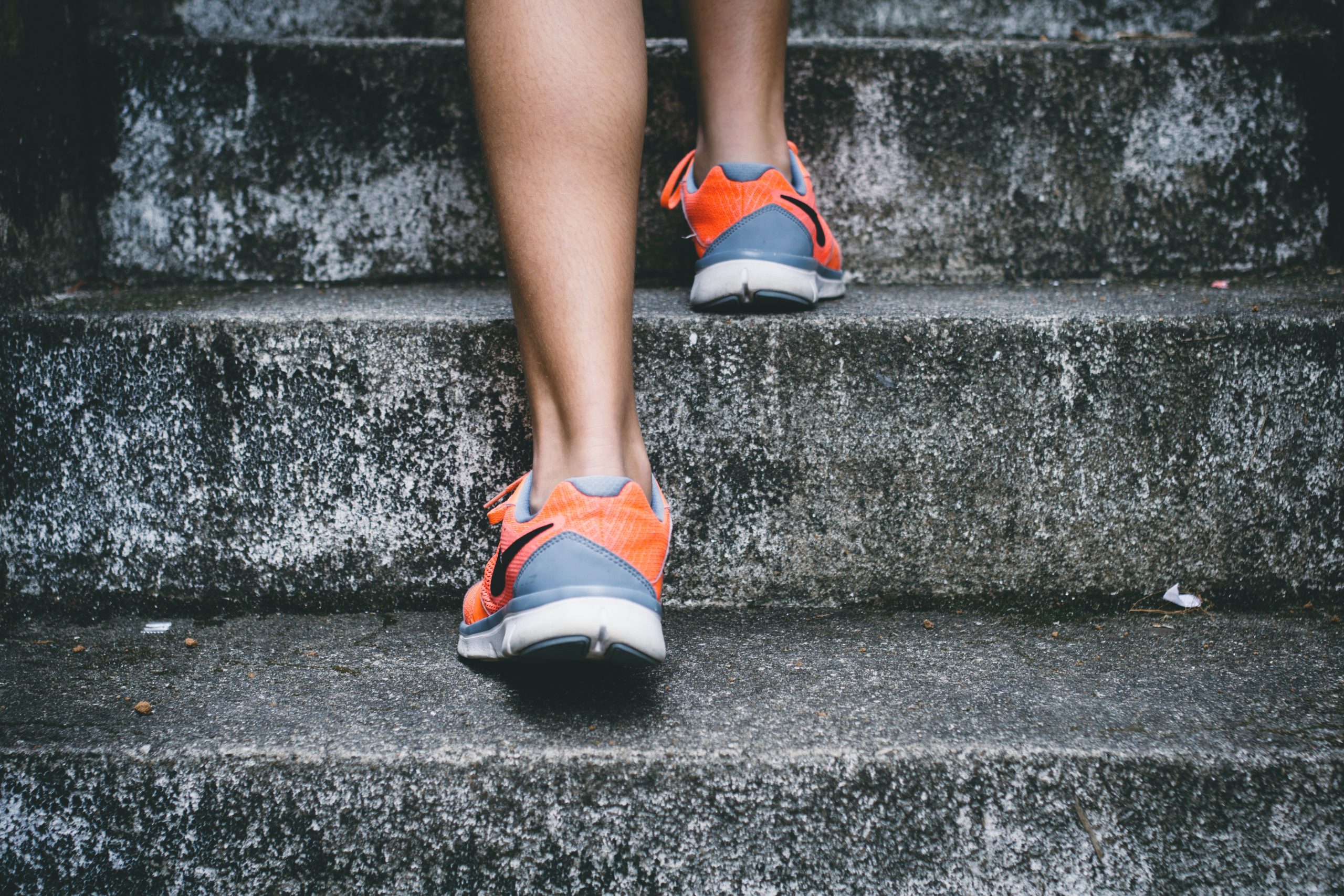
Case Study Conclusion: A Pain in the Foot
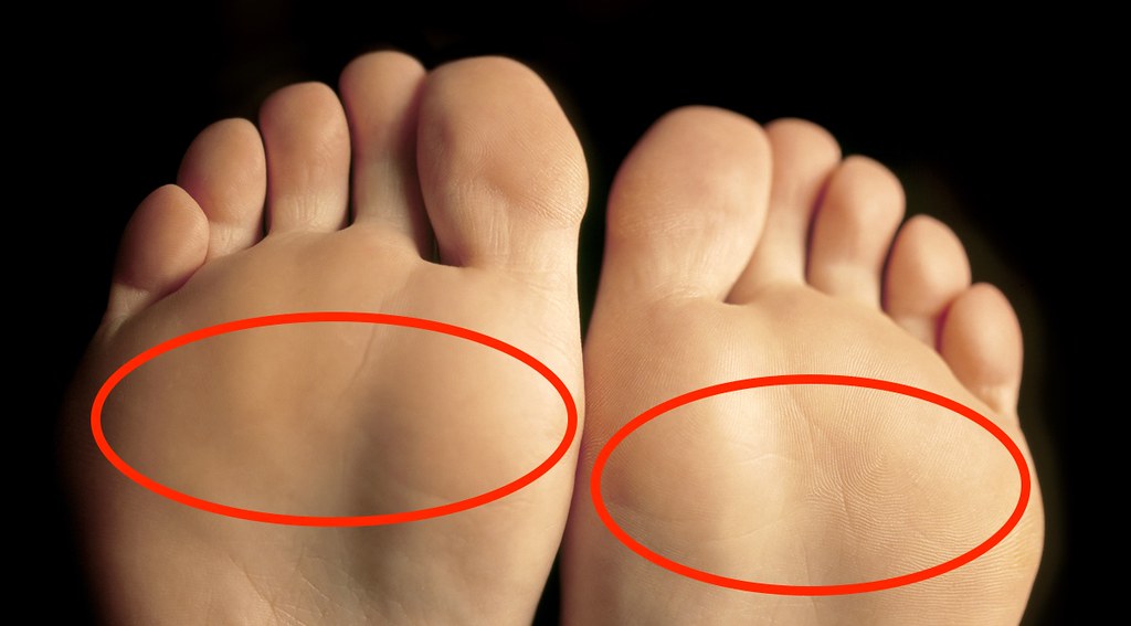
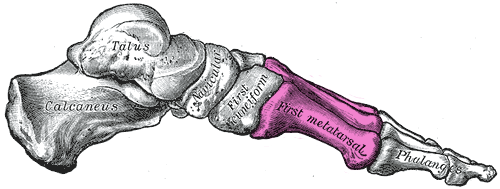
As Sophia discovered in the beginning of the chapter, wearing high heels can result in a condition called metatarsalgia. Metatarsalgia is named for the metatarsal bones, which are the five bones that run through the ball of the foot just behind the toes (highlighted in Figures 11.8.2 and 11.8.3). Wearing high heels causes excessive pressure on the ball of the foot, as described in the beginning of this chapter. Additionally, the toes are forced to pull upward in high heels, which moves the fleshy padding away from the ball of the foot and adds to the overall pressure placed on this region. Over time, this can cause inflammation and direct stress on the bones, resulting in the pain in the ball of the foot known as metatarsalgia. The pain occurs especially in weight-bearing positions, such as standing, walking, or running — which is what Sophia was experiencing. There may also be pain, numbness, or tingling in the toes associated with metatarsalgia.
 |
 |
Wearing high heels can also cause stress fractures in the feet, which are tiny breaks in bone that occur due to repeated mechanical stress. This is caused by the excessive pressure that high heels put on some of the bones of the feet. These fractures are somewhat similar to what occurs in osteoporosis when the bone mass decreases to the point where bones can fracture easily as a person goes about their daily activities. In both cases, a major noticeable injury is not necessary to create the tiny fractures. As you have learned, tiny fractures that accrue over time are the cause of dowager’s hump (or kyphosis), which is often seen in women with osteoporosis.
Don’t think you are immune to stress fractures just because you don’t wear high heels! This injury also commonly occurs in people who participate in sports involving repetitive striking of the foot on the ground, such as running, tennis, basketball, or gymnastics. They may be avoided by taking preventative measures. You should ramp up any increase in activity slowly, cross-train by engaging in a variety of different sports or activities, rest if you experience pain, and wear well-cushioned and supportive running shoes. It is important to know that your cardiovascular and muscular systems adapt to an increase in physical activity much more quickly than the skeletal system.
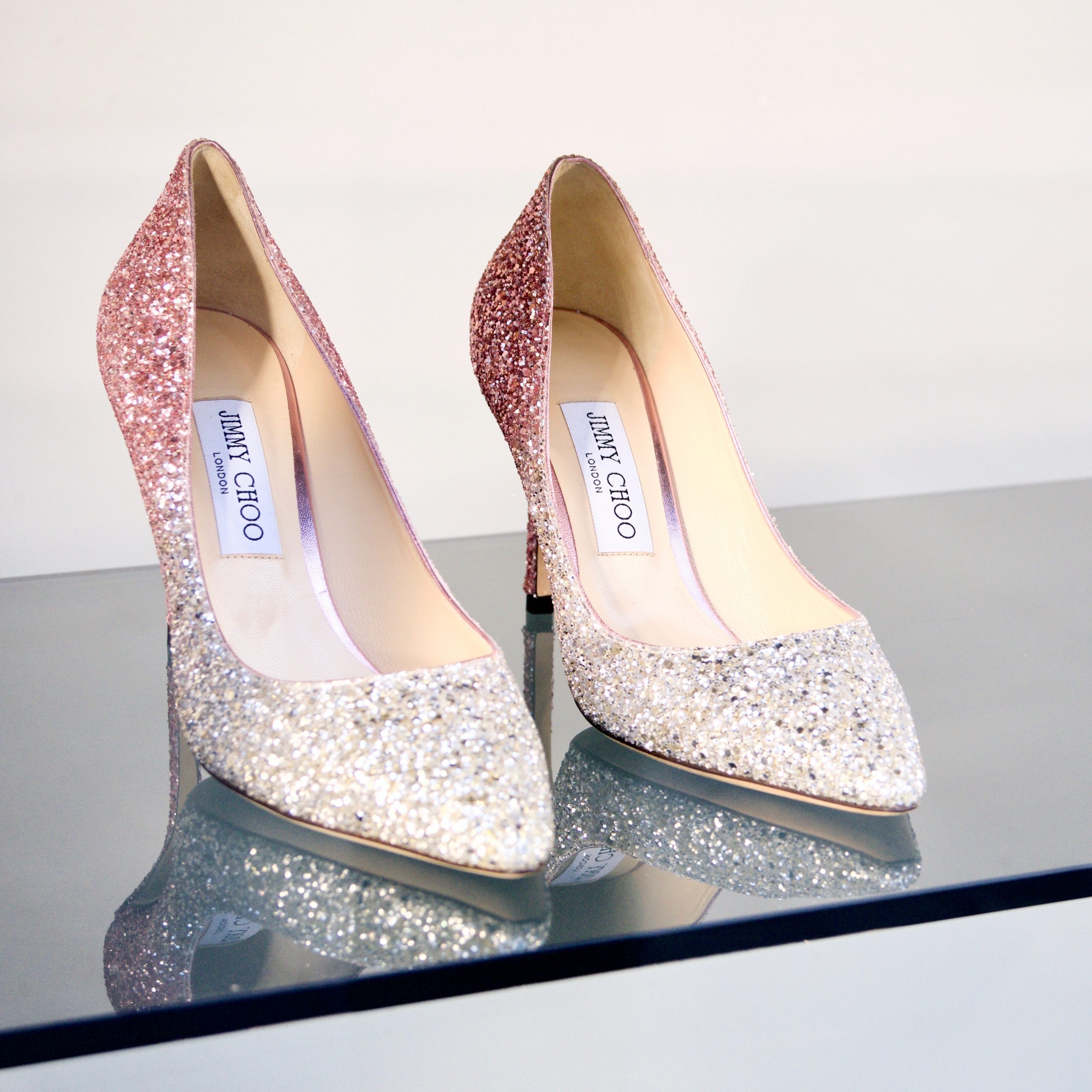
Sophia learned through her online research that wearing high heels can also lead to foot deformities, such as bunions and hammertoes. As you learned in an earlier chapter, a bunion is a protrusion on the side of the foot, most often at the base of the big toe. It can be caused by wearing shoes with a narrow, pointed toe box — a common shape for high heels (see Figure 11.8.4). The pressure of the shoes on the side of the foot causes an enlargement of bone or inflammation of other tissues in the region, which pushes the big toe toward the other toes.
Hammertoes are an abnormal bend in the middle joint of the second, third, or fourth toe (with the big toe being the first toe), causing the toe to be shaped similarly to a hammer. The narrow, pointed toe box of many high heels, combined with the way the toes are squished into the front of the shoe as a result of the height of the heel, can cause the toes to become deformed this way. Treatments for bunions and hammertoe include wearing shoes with a roomy toe box, padding or taping the toes, and toe exercises and stretches. If the bunion or hammertoe does not respond to these treatments, surgery may be necessary to correct the deformity.
Because the bones of the skeleton are connected and work together with other systems to support the body, wearing high heels can also cause physical problems in areas other than the feet. Wearing high heels shifts a person’s posture and alignment, and can put strain on tendons, muscles, and other joints in the body. Research published in 2014 from a team at Stanford University suggests that wearing high heels, particularly if the person is overweight or the heels are very high, may increase the risk of osteoarthritis (OA) in the knee, due to added stress on the knee joint as the person walks. As you have learned, OA results from the breakdown of cartilage and bone at the joint. Because it can only be treated to minimize symptoms — and not for a cure — OA could be an unfortunate long-term consequence of wearing high heels.
Sophia has decided that wearing high heels regularly is not worth the pain and potential long-term damage to her body. After consulting with her doctor, who confirmed she had metatarsalgia, she was able to successfully treat it with ice, rest, and wearing comfortable, supportive shoes instead of heels.
High heels are not the only kind of shoes that can cause problems. Flip-flops, worn-out sneakers, and shoes that are too tight can all cause foot issues. To prevent future problems from her shoe choices, Sophia is following guidelines recommended by medical experts. The guidelines include:
- Wearing shoes that fit well, have plenty of room in the toes, are supportive, and are comfortable right away. There should be no “break-in” period needed for shoes.
- Avoiding high heels, especially those with heels over two inches high, or those that have narrow, pointed toe boxes or very thin heels. The heels pictured in Figure 11.8.4 are an example of a type of shoe that should be avoided.
- If high heels must be worn, it should only be for a limited period of time.
As you have learned in this chapter, your skeletal system carries out a variety of important functions in your body, including physical support. But even though it is strong, your skeletal system can become damaged and deformed — even through such a seemingly innocuous act as wearing a certain type of shoe. Taking good care of your skeletal system is necessary to help it continue to take good care of the rest of you.
Chapter 11 Summary
In this chapter, you learned about the skeletal system. Specifically, you learned that:
- The skeletal system is the organ system that provides an internal framework for the human body. In adults, the skeletal system contains 206 bones.
- Bones are organs made of supportive connective tissues, mainly the tough protein collagen. Bones also contain blood vessels, nerves, and other tissues. Bones are hard and rigid, due to deposits of calcium and other mineral salts within their living tissues. Besides bones, the skeletal system includes cartilage and ligaments.
- The skeletal system has many different functions, including supporting the body and giving it shape, protecting internal organs, providing attachment surfaces for skeletal muscles, allowing body movements, producing blood cells, storing minerals, helping to maintain mineral homeostasis, and producing endocrine hormones.
- There is relatively little sexual dimorphism in the human skeleton, although the female skeleton tends to be smaller and less robust than the male skeleton. The greatest sex difference is in the pelvis, which is adapted for childbirth in females.
- The skeleton is traditionally divided into two major parts: the axial skeleton and the appendicular skeleton.
- The axial skeleton consists of a total of 80 bones. It includes the skull, vertebral column, and rib cage. It also includes the three tiny ossicles in the middle ear and the hyoid bone in the throat.
-
- The skull provides a bony framework for the head. It consists of 22 different bones: eight in the cranium, which encloses the brain, and 14 in the face, which includes the upper and lower jaw.
- The vertebral column is a flexible, S-shaped column of 33 vertebrae that connects the trunk with the skull and encloses the spinal cord. The vertebrae are divided into five regions: cervical, thoracic, lumbar, sacral, and coccygeal regions. The S shape of the vertebral column allows it to absorb shocks and distribute the weight of the body.
- The rib cage holds and protects the organs of the upper part of the trunk, including the heart and lungs. It includes the 12 thoracic vertebrae, the sternum, and 12 pairs of ribs.
- The appendicular skeleton consists of a total of 126 bones. It includes the bones of the four limbs, shoulder girdle, and pelvic girdle. The girdles attach the appendages to the axial skeleton.
-
- Each upper limb consists of 30 bones. There is one bone (called the humerus) in the upper arm, and two bones (called the ulna and radius) in the lower arm. The wrist contains eight carpal bones, the hand contains five metacarpals, and the fingers consist of 14 phalanges. The thumb is opposable to the palm and fingers of the same hand.
- Each lower limb also consists of 30 bones. There is one bone (called the femur) in the upper leg, and two bones (called the tibia and fibula) in the lower leg. The patella covers the knee joint. The ankle contains seven tarsal bones, and the foot contains five metatarsals. The tarsals and metatarsals form the heel and arch of the foot. The bones in the toes consist of 14 phalanges.
- The shoulder girdle attaches the upper limbs to the trunk of the body. It is connected to the axial skeleton only by muscles, allowing mobility of the upper limbs. Bones of the shoulder girdle include a right and left clavicle, and a right and left scapula.
- The pelvic girdle attaches the legs to the trunk of the body and supports the organs of the abdomen. It is connected to the axial skeleton by ligaments. The pelvic girdle consists of two halves that are fused together in adults. Each half consists of three bones: the ilium, pubis, and ischium.
- Bones are organs that consist mainly of bone (or osseous) tissue. Osseous tissue is a type of supportive connective tissue consisting of a collagen matrix that is mineralized with calcium and phosphorus crystals. The combination of flexible collagen and minerals makes bone hard, without making it brittle.
-
- There are two types of osseous tissues: compact bone tissue and spongy bone tissue. Compact bone tissue is smooth and dense. It forms the outer layer of bones. Spongy bone tissue is porous and light, and it is found inside many bones.
- Besides osseous tissues, bones also contain nerves, blood vessels, bone marrow, and periosteum.
- Bone tissue is composed of four different types of bone cells: osteoblasts, osteocytes, osteoclasts, and osteogenic cells. Osteoblasts form new collagen matrix and mineralize it, osteoclasts break down bone, osteocytes regulate the formation and breakdown of bone, and osteogenic cells divide and differentiate to form new osteoblasts. Bone is a very active tissue, constantly being remodeled by the work of osteoblasts and osteoclasts.
- There are six types of bones in the human body: long bones (such as the limb bones), short bones (such as the wrist bones), sesamoid bones (such as the patella), sutural bones in the skull, and irregular bones (such as the vertebrae).
- Early in the development of a human fetus, the skeleton is made almost entirely of cartilage. The relatively soft cartilage gradually turns into hard bone — a process that is called ossification. It begins at a primary ossification center in the middle of bone, and later also occurs at secondary ossification centers in the ends of bone. The bone can no longer grow in length after the areas of ossification meet and fuse at the time of skeletal maturity.
- Throughout life, bone is constantly being replaced in the process of bone remodeling. In this process, osteoclasts resorb bone and osteoblasts make new bone to replace it. Bone remodeling shapes the skeleton, repairs tiny flaws in bones, and helps maintain mineral homeostasis in the blood.
- Bone repair is the natural process in which a bone repairs itself following a bone fracture. This process may take several weeks. In the process, the periosteum produces cells that develop into osteoblasts, and the osteoblasts form new bone matrix to heal the fracture. Bone repair may be affected by diet, age, pre-existing bone disease, or other factors.
- Joints are locations at which bones of the skeleton connect with one another.
- Joints can be classified structurally or functionally, and there is significant overlap between the two types of classifications.
- The structural classification of joints depends on the type of tissue that binds the bones to each other at the joint. There are three types of joints in the structural classification: fibrous, cartilaginous, and synovial joints.
- The functional classification of joints is based on the type and degree of movement that they allow. There are three types of joints in the functional classification: immovable, partly movable, and movable joints.
- A number of disorders affect the skeletal system, including bone fractures and bone cancers. The two most common disorders of the skeletal system are osteoporosis and osteoarthritis.
- Osteoporosis is an age-related disorder in which bones lose mass, weaken, and break more easily than normal bones. The underlying mechanism in all cases of osteoporosis is an imbalance between bone formation and bone resorption in bone remodeling. Osteoporosis may also occur as a side effect of other disorders or certain medications.
-
- Osteoporosis is diagnosed by measuring a patient’s bone density and comparing it with the normal level of peak bone density. Fractures are the most dangerous aspect of osteoporosis. Osteoporosis is rarely fatal, but complications of fractures often are.
- Risk factors for osteoporosis include older age, female sex, European or Asian ancestry, family history of osteoporosis, short stature and small bones, smoking, alcohol consumption, lack of exercise, vitamin D deficiency, poor nutrition, and consumption of soft drinks.
- Osteoporosis is often treated with medications (such as bisphosphonates) that may slow or even reverse bone loss. Preventing osteoporosis includes eliminating any risk factors that can be controlled through changes of behavior, such as undertaking weight-bearing exercise.
- Osteoarthritis (OA) is a joint disease that results from the breakdown of joint cartilage and bone. The most common symptoms are joint pain and stiffness. OA is thought to be caused by mechanical stress on the joints with insufficient self-repair of cartilage, coupled with low-grade inflammation of the joints.
-
- Diagnosis of OA is typically made on the basis of signs and symptoms, such as joint deformities, pain, and stiffness. X-rays or other tests are sometimes used to either support the diagnosis or rule out other disorders. Age is the chief risk factor for OA. Other risk factors include joint injury, excess body weight, and a family history of OA.
- OA cannot be cured, but the symptoms can often be treated successfully. Treatments may include exercise, efforts to decrease stress on joints, pain medications, and surgery to replace affected hip or knee joints.
As you have learned in this chapter, one of the important functions of the skeletal system is to allow movement of the body. But it doesn’t do it alone. Movement is caused by the contraction of muscles, which pull on the bones, causing them to move. Read the next chapter to learn about this and other important functions of the muscular system.
Chapter 11 Review
-
-
- Why does the rib cage need to be flexible? Why can it be flexible?
- In general, what do “girdles” in the skeletal system do?
- Would swimming be more effective as an exercise for preventing osteoporosis or as a treatment for osteoarthritis? Explain your answer.
- Explain why some of the vertebrae become misshapen in the condition called dowager’s hump (or kyphosis).
- Explain why osteoarthritis often involves inflammation in the joints.
- Osteoporosis can involve excess bone resorption, as well as insufficient production of new bone tissue. What are the two main bone cell types that carry out these processes, respectively?
- Describe two roles that calcium in bones play in the body.
-
Attributions
Figure 11.8.1
Running Shoes by bruno-nascimento-PHIgYUGQPvU [photo] by Bruno Nascimento on Unsplash is used under the Unsplash License (https://unsplash.com/license).
Figure 11.8.2
Metatarsalgia/ Best Shoes for Metatarsalgia by Esther Max on Flickr is used under a CC BY 2.0 (https://creativecommons.org/licenses/by/2.0/) license.
Figure 11.8.3
Gray290_-_Mratatarsus (1) by Henry Vandyke Carter (1831-1897) (Revised by Warren H. Lewis, coloured by Was a bee) on Wikimedia Commons is in the public domain (https://en.wikipedia.org/wiki/Public_domain. (Bartleby.com: Gray’s Anatomy, Plate 290)
Figure 11.8.4
Heels by gavin-allanwood-ndpX28miBtE-unsplash by Photo by Gavin Allanwood on Unsplash is used under the Unsplash License (https://unsplash.com/license).
References
Mayo Clinic Staff. (n.d.). Hammertoe and mallet toe [online article]. MayoClinic.org. https://www.mayoclinic.org/diseases-conditions/hammertoe-and-mallet-toe/symptoms-causes/syc-20350839
VanDyke Carter, H. (1858). Illustration plate 290. In H. Gray, Anatomy of the Human Body. Lea & Febiger. Bartleby.com, 2000. www.bartleby.com/107/.
Created by: CK-12/Adapted by Christine Miller
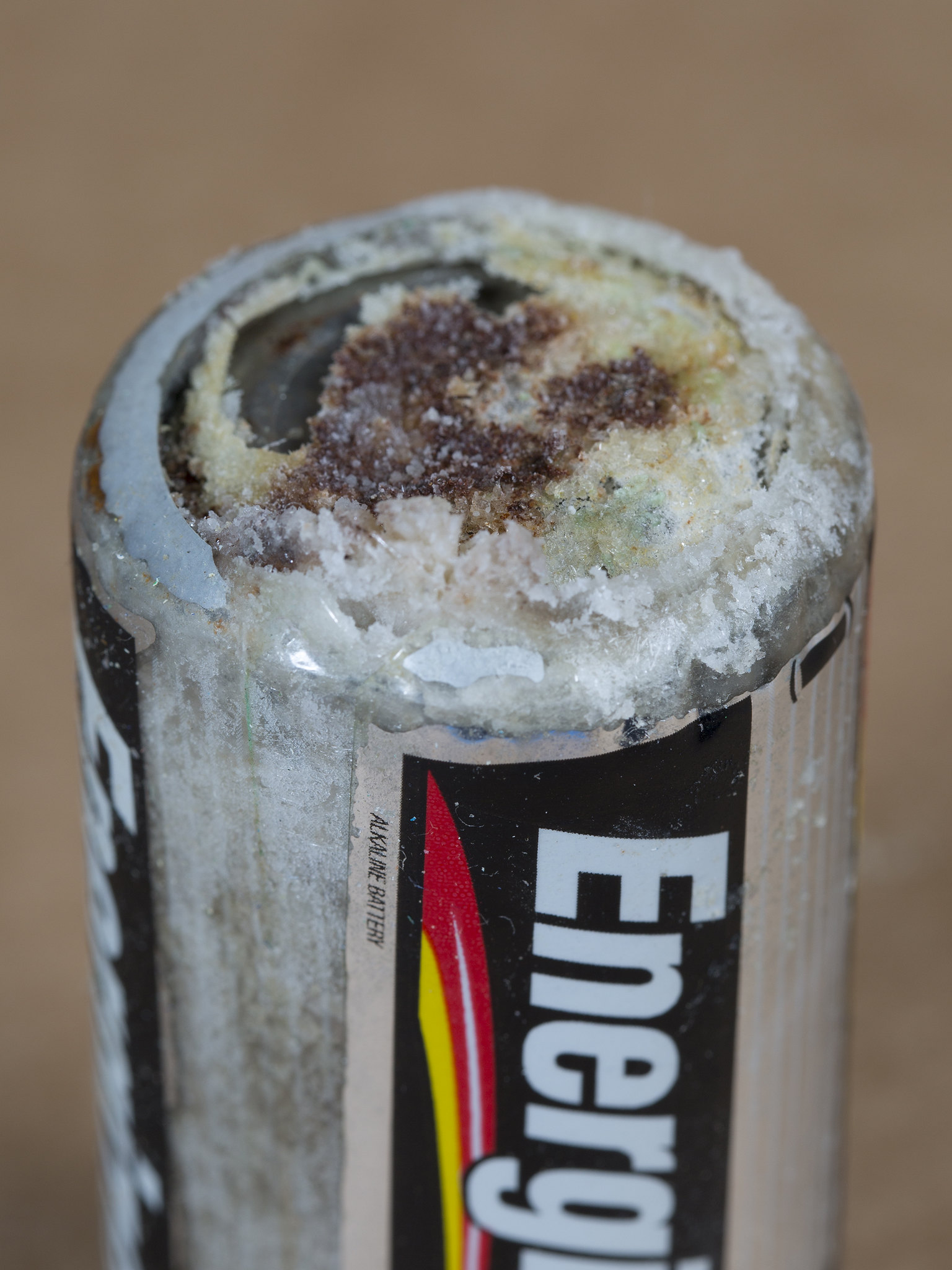
Danger! Acid!
You probably know that batteries contain dangerous chemicals, including strong acids. Strong acids can hurt you if they come into contact with your skin or eyes. Therefore, it may surprise you to learn that your life depends on acids. There are many acids inside your body, and some of them are as strong as battery acid. Acids are needed for digestion and some forms of energy production. Genes are made of nucleic acids, proteins of amino acids, and lipids of fatty acids.
Water and Solutions
Acids (such as battery acid) are solutions. A solution is a mixture of two or more substances that has the same composition throughout. Many solutions are a mixture of water and some other substance. Not all solutions are acids. Some are bases and some are neither acids nor bases. To understand acids and bases, you need to know more about pure water.
In pure water (such as distilled water), a tiny fraction of water molecules naturally breaks down to form ions. An ion is an electrically charged atom or molecule. The breakdown of water is represented by the chemical equation:
2 H2O → H3O+ + OH-
The products of this reaction are a hydronium ion (H3O+) and a hydroxide ion (OH-). The hydroxide ion, which has a negative charge, forms when a water molecule gives up a positively charged hydrogen ion (H+). The hydronium ion, which has a positive charge, forms when another water molecule accepts the hydrogen ion.
Acidity and pH
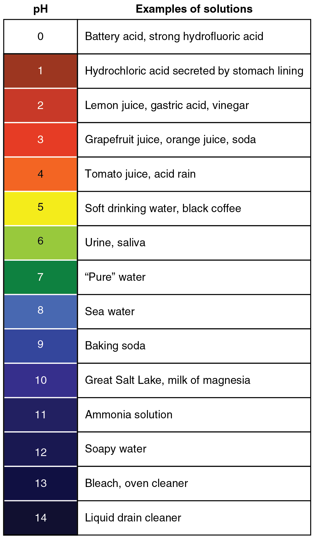
The concentration of hydronium ions in a solution is known as acidity. In pure water, the concentration of hydronium ions is very low; only about one in ten million water molecules naturally breaks down to form a hydronium ion. As a result, pure water is essentially neutral. Acidity is measured on a scale called pH, as shown in Figure 3.12.2. Pure water has a pH of 7, so the point of neutrality on the pH scale is 7.

This pH scale shows the acidity of many common substances. The lower the pH value, the more acidic a substance is.
Acids
If a solution has a higher concentration of hydronium ions than pure water, it has a pH lower than 7. A solution with a pH lower than 7 is called an acid. As the hydronium ion concentration increases, the pH value decreases. Therefore, the more acidic a solution is, the lower its pH value is.
Did you ever taste vinegar? Like other acids, it tastes sour. Stronger acids can be harmful to organisms. Even stomach acid would eat through the stomach if it were not lined with a layer of mucus. Strong acids can also damage materials, even hard materials such as glass.
Bases
If a solution has a lower concentration of hydronium ions than pure water, it has a pH higher than 7. A solution with a pH higher than 7 is called a base. Bases, such as baking soda, have a bitter taste. Like strong acids, strong bases can harm organisms and damage materials. For example, lye can burn the skin, and bleach can remove the colour from clothing.
Buffers
A buffer is a solution that can resist changes in pH. Buffers are able to maintain a certain pH by by absorbing any H+ or OH- ions added to the solution. Buffers are extremely important in biological systems in order to maintain a pH conducive to life. Bicarbonate is an example of a buffer which is used to maintain pH of the blood. In this buffering system, if blood becomes too acidic, carbonic acid will convert to carbon dioxide and water. If the blood becomes too basic, carbonic acid will convert to bicarbonate and H+ ions:
CO2 + H2O ↔ H2CO3 ↔ HCO3- + H+
Acids, Bases, and Enzymes
Many acids and bases in living things provide the pH that enzymes need. Enzymes are biological catalysts that must work effectively for biochemical reactions to occur. Most enzymes can do their job only at a certain level of acidity. Cells secrete acids and bases to maintain the proper pH for enzymes to do their work.
Every time you digest food, acids and bases are at work in your digestive system. Consider the enzyme pepsin, which helps break down proteins in the stomach. Pepsin needs an acidic environment to do its job. The stomach secretes a strong acid called hydrochloric acid that allows pepsin to work. When stomach contents enter the small intestine, the acid must be neutralized, because enzymes in the small intestine need a basic environment in order to work. An organ called the pancreas secretes a base named bicarbonate into the small intestine, and this base neutralizes the acid.
Feature: My Human Body
Do you ever have heartburn? The answer is probably "yes." More than 60 million Americans have heartburn at least once a month, and more than 15 million suffer from it on a daily basis. Knowing more about heartburn may help you prevent it or know when it's time to seek medical treatment.
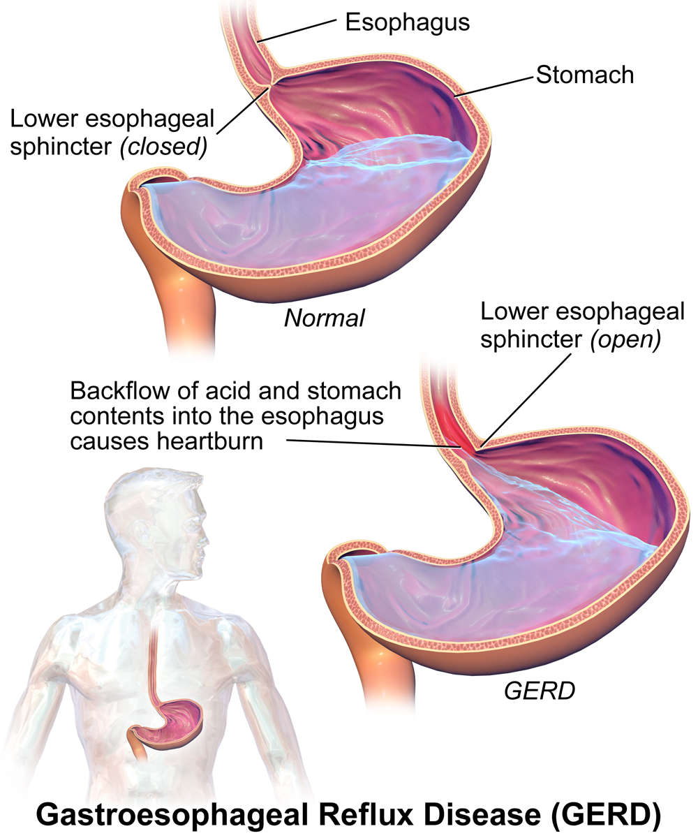
Heartburn doesn't have anything to do with the heart, but it does cause a burning sensation in the vicinity of the chest. Normally, the acid secreted into the stomach remains in the stomach where it is needed to allow pepsin to do its job of digesting proteins. A long tube called the esophagus carries food from the mouth to the stomach. A sphincter, or valve, between the esophagus and stomach opens to allow swallowed food to enter the stomach and then closes to prevent stomach contents from backflowing into the esophagus. If this sphincter is weak or relaxes inappropriately, stomach contents flow into the esophagus. Because stomach contents are usually acidic, this causes the burning sensation known as heartburn. People who are prone to heartburn and suffer from it often may be diagnosed with GERD, which stands for gastroesophageal reflux disease.
GERD — as well as occasional heartburn — often can be improved by dietary and other lifestyle changes that decrease the amount and acidity of reflux from the stomach into the esophagus.
- Some foods and beverages seem to contribute to GERD, so these should be avoided. Problematic foods include chocolate, fatty foods, peppermint, coffee, and alcoholic beverages.
- Decreasing portion size and eating the last meal of the day at least a couple of hours before bedtime may reduce the risk of reflux occurring.
- Smoking tends to weaken the lower esophageal sphincter, so quitting the habit may help control reflux.
- GERD is often associated with being overweight. Losing weight often brings improvement.
- Some people are helped by sleeping with the head of the bed elevated. This allows gravity to help control the backflow of acids into the esophagus from the stomach.
If you have frequent heartburn and lifestyle changes don't help, you may need medication to control the condition. Over-the-counter (OTC) antacids may be all that you need to control the occasional heartburn attack. OTC medications are usually bases that neutralize stomach acids. They may also create bubbles that help block stomach contents from entering the esophagus. For some people, OTC medications are not enough, and prescription medications are instead required for the control of GERD. These prescription medications generally work by inhibiting acid secretion in the stomach.
Be sure to see a doctor if you can't control your heartburn, or you have it often. Untreated GERD not only interferes with quality of life, it may also lead to more serious complications, ranging from esophageal bleeding to esophageal cancer.
3.12 Summary
- A solution is a mixture of two or more substances that has the same composition throughout. Many solutions consist of water and one or more dissolved substances.
- Acidity is a measure of the hydronium ion concentration in a solution. Pure water has a very low concentration and a pH of 7, which is the point of neutrality on the pH scale.
- Acids have a higher hydronium ion concentration than pure water and a pH lower than 7. Bases have a lower hydronium ion concentration than pure water and a pH higher than 7.
- Many acids and bases in living things are secreted to provide the proper pH for enzymes to work properly. Enzymes are the biological catalysts (like pepsin) needed to digest protein in the stomach. Pepsin requires an acidic environment.
3.12 Review Questions
-
- What is a solution?
- Define acidity.
- Explain how acidity is measured.
- Compare and contrast acids and bases.
- Hydrochloric acid is secreted by the stomach to provide an acidic environment for the enzyme pepsin. What is the pH of this acid? How strong of an acid is it compared with other acids?
- Define an ion. Identify the ions in the equation below, and explain what makes them ions:
- 2 H2O → H3O+ + OH-
- Explain why the pancreas secretes bicarbonate into the small intestine.
- Do you think pepsin would work in the small intestine? Why or why not?
- You may have mixed vinegar and baking soda and noticed that they bubble and react with each other. Explain why this happens. Explain also what happens to the pH of this solution after you mix the vinegar and baking soda.
- Pregnancy hormones can cause the lower esophageal sphincter to relax. What effect do you think this has on pregnant women? Explain your answer.
3.12 Explore More
https://www.youtube.com/watch?v=rIvEvwViJGk&feature=youtu.be
pH and Buffers by Bozeman Science, 2014.
https://www.youtube.com/watch?v=DupXDD87oHc&feature=youtu.be
The strengths and weaknesses of acids and bases - George Zaidan and Charles Morton, TED-Ed, 2013.
Attributions
Figure 3.12.1
Leaky battery by Carbon Arc on Flickr is used under a CC BY-NC-SA 2.0 (https://creativecommons.org/licenses/by-nc-sa/2.0/) license.
Figure 3.12.2
PH_Scale by Christinelmiller on Wikimedia Commons is used under a © CC0 1.0 (https://creativecommons.org/publicdomain/zero/1.0/) public domain dedication license.
Figure 3.12.3
Ph scale with examples by OpenStax College, on Wikimedia Commons, is used under a CC BY 3.0 (https://creativecommons.org/licenses/by/3.0) license.
Figure 3.12.4
GERD by BruceBlaus on Wikimedia Commons is used under a CC BY-SA 4.0 (https://creativecommons.org/licenses/by-sa/4.0) license.
References
Betts, J.G., Young, K.A., Wise, J.A., Johnson, E., Poe, B., Kruse, D.H., Korol, O., Johnson, J.E., Womble, M., DeSaix, P. (2013, April 25). Figure 26.15 The pH Scale [digital image]. In Anatomy and Physiology. OpenStax. https://openstax.org/books/anatomy-and-physiology/pages/26-4-acid-base-balance
Bozeman Science. (2014, February 22). pH and buffers. YouTube. https://www.youtube.com/watch?v=rIvEvwViJGk&feature=youtu.be
TED-Ed. (2013, October 24). The strengths and weaknesses of acids and bases - George Zaidan and Charles Morton. YouTube. https://www.youtube.com/watch?v=DupXDD87oHc&feature=youtu.be
Created by: CK-12/Adapted by Christine Miller
Express Yourself
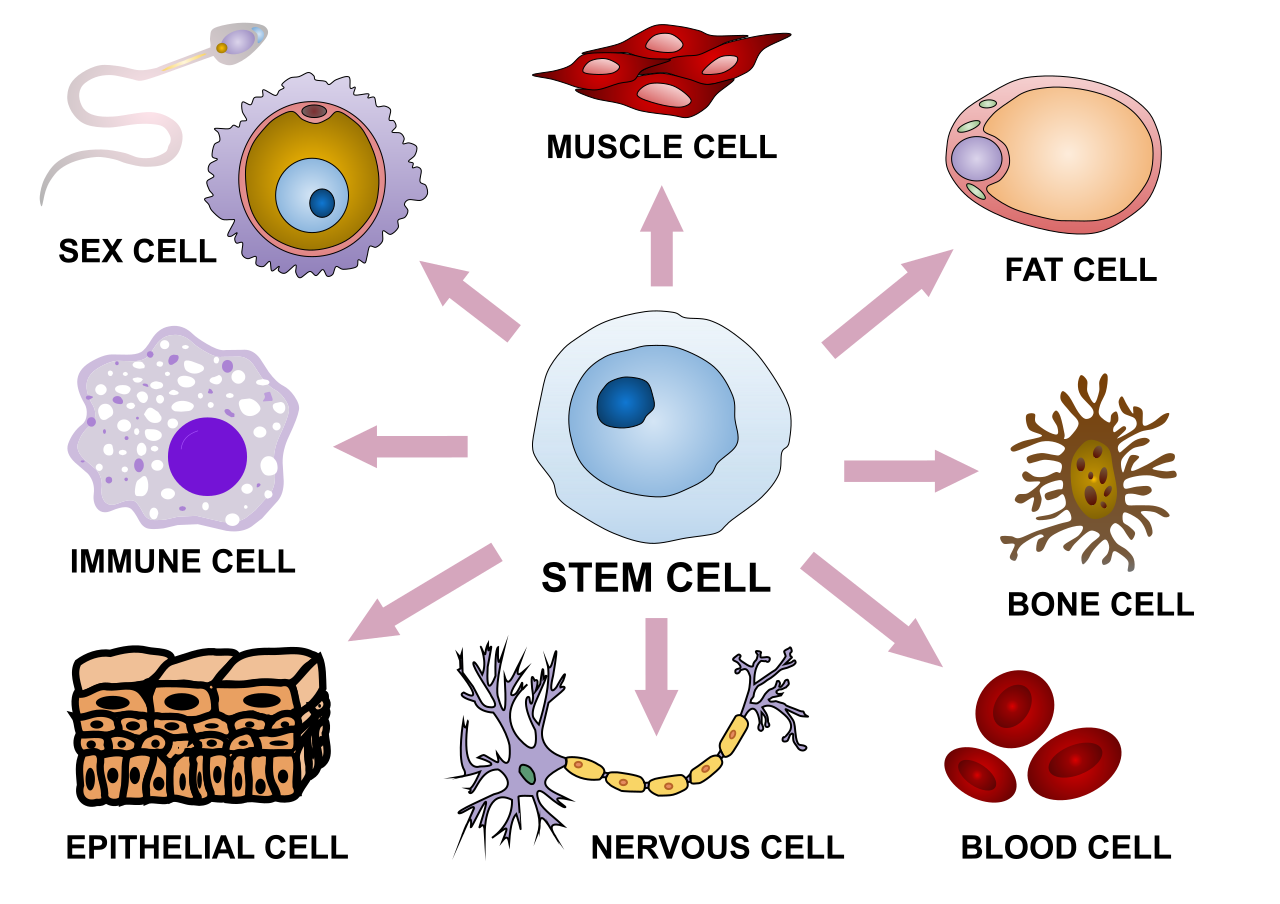
This sketch illustrates some of the variability in human cells. The shape and other characteristics that make each type of cell unique depend mainly on the specific proteins that particular cell type makes. Proteins are encoded in genes. All the cells in an organism have the same genes, so they all have genetic instructions for the same proteins. Obviously, different types of cells must use (or express) different genes to make different proteins.
What Is Gene Expression?
Using a gene to make a protein is called gene expression. It includes the synthesis of the protein by the processes of transcription of DNA into mRNA, and translation of mRNA into a protein. It may also include further processing of the protein after synthesis.
Gene expression is regulated to ensure that the correct proteins are made when and where they are needed. Regulation may occur at any point in the expression of a gene, from the start of the transcription phase of protein synthesis to the processing of a protein after synthesis occurs. The regulation of transcription is one of the most complicated parts of gene regulation in eukaryotic cells, and it is the focus of this concept.
Regulation of Transcription
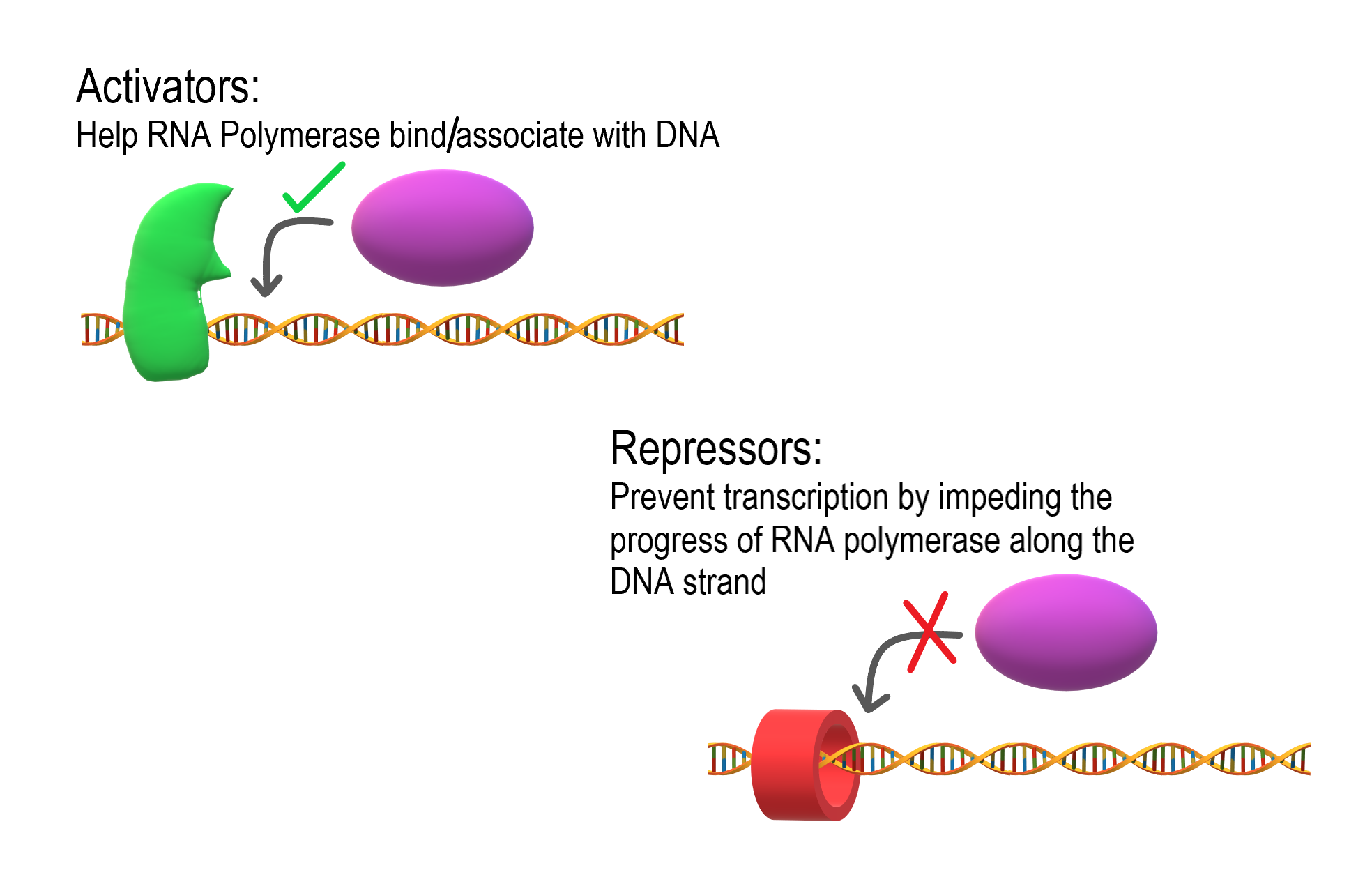
As shown in Figure 5.9.2, transcription is controlled by regulatory proteins. These proteins bind to regions of DNA, called regulatory elements, which are located near promoters. The promoter is the region of a gene where RNA polymerase binds to initiate transcription of the DNA to mRNA. After regulatory proteins bind to regulatory elements, the proteins can interact with RNA polymerase. Regulatory proteins are typically either activators or repressors. Activators are regulatory proteins that promote transcription by enhancing the interaction of RNA polymerase with the promoter. Repressors are regulatory proteins that prevent transcription by impeding the progress of RNA polymerase along the DNA strand, so the DNA cannot be transcribed to mRNA.
Enhancers
Although regulatory proteins and elements are typically the key players in the regulation of transcription, other factors may also be involved. Regulation of transcription may also involve enhancers. Enhancers are distant regions of DNA that can loop back to interact with a gene's promoter. They can also increase the likelihood that transcription of the gene will occur.Enhancers
The TATA Box
Different types of cells have unique patterns of regulatory elements that result in only the necessary genes being transcribed. That’s why a blood cell and nerve cell, for example, are so different from each other. Some regulatory elements, however, are common to virtually all genes, regardless of the cells in which they occur. An example is the TATA box, which is a regulatory element that is part of the promoter of almost every eukaryotic gene. A number of regulatory proteins bind to the TATA box, forming a multi-protein complex. It is only when all of the appropriate proteins are bound to the TATA box that RNA polymerase recognizes the complex and binds to the promoter so transcription can begin.

Regulation During Development
The regulation of gene expression is extremely important in an organism's early development. Regulatory proteins must "turn on" certain genes in particular cells at just the right time, so the individual develops normal organs and organ systems. Homeobox genes are important genes that regulate development.
Homeobox genes are a large group of similar genes that direct the formation of many body structures during the embryonic stage. In humans, there are an estimated 235 functional homeobox genes. They are present on every chromosome and generally grouped in clusters. Homeobox genes contain instructions for making chains of 60 amino acids, called homeodomains. Proteins containing homeodomains are transcription factors that bind to and control the activities of other genes. The homeodomain is the part of the protein that binds to the target gene and controls its expression.
Gene Expression and Cancer
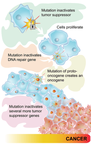
Some types of cancer occur because of mutations in the genes that control the cell cycle. Cancer-causing mutations most often occur in two types of regulatory genes: proto-oncogenes and tumor-suppressor genes. Both are shown in Figure 5.9.4.
- Proto-oncogenes are genes that normally help cells divide. When a proto-oncogene mutates to become an oncogene, it is continuously expressed, even when it is not supposed to be. This is like a car's accelerator pedal being stuck at full throttle. The car keeps racing at top speed. A cell, in this case, keeps dividing out of control, which can lead to cancer.
- Tumor suppressor genes are genes that normally slow down or stop cell division. When a mutation occurs in a tumor suppressor gene, it can no longer control cell division. This is like a car without brakes. The car can't be slowed or stopped. A cell, in this case, keeps dividing out of control, which can lead to cancer.
5.9 Summary
- Using a gene to make a protein is called gene expression. Gene expression is regulated to ensure that the correct proteins are made when and where they are needed. Regulation may occur at any stage of protein synthesis or processing.
- The regulation of transcription is controlled by regulatory proteins that bind to regions of DNA called regulatory elements, which are usually located near promoters. Most regulatory proteins are either activators that promote transcription, or repressors that impede transcription.
- A regulatory element common to almost all eukaryotic genes is the TATA box. A number of regulatory proteins must bind to the TATA box in the promoter before transcription can proceed.
- Regulation of gene expression is extremely important during an organism's early development. Homeobox genes — which encode for chains of amino acids called homeodomains — are important genes that regulate development.
- Some types of cancer occur because of mutations in the genes that control the cell cycle. Cancer-causing mutations most often occur in two types of regulatory genes: tumor-suppressor genes and proto-oncogenes.
5.9 Review Questions
- Define gene expression.
- Why must gene expression be regulated?
- Explain how regulatory proteins may activate or repress transcription.
-
- What is the TATA box, and how does it work?
- Describe homeobox genes and their role in an organism's development.
- Discuss the role of regulatory gene mutations in cancer.
- Explain the relationship between proto-oncogenes and oncogenes.
- If a newly fertilized egg contained a mutation in a homeobox gene, how do you think this would affect the developing embryo? Explain your answer.
- Compare and contrast enhancers and activators.
5.9 Explore More
https://www.youtube.com/watch?time_continue=3&v=vi-zWoobt_Q&feature=emb_logo
Regulated Transcription, ndsuvirtualcell, 2008.
https://www.youtube.com/watch?v=BmFEoCFDi-w
How do cancer cells behave differently from healthy ones? - George Zaidan,
TED-Ed, 2012.
https://www.youtube.com/watch?v=Z3B-AaqjyjE
What is leukemia? - Danilo Allegra and Dania Puggioni, 2015.
Attributions
Figure 5.9.1
Stem_cell_differentiation.svg by Haileyfournier on Wikimedia Commons is used under a CC BY-SA 4.0 (https://creativecommons.org/licenses/by-sa/4.0) license.
Figure 5.9.2
Activators and Repressors by Christine Miller is used under a CC BY-SA 4.0 (https://creativecommons.org/licenses/by-sa/4.0) license.
Figure 5.9.3
TATA_box_description by Luttysar on Wikimedia Commons is used under a CC BY-SA 4.0 (https://creativecommons.org/licenses/by-sa/4.0) license.
Figure 5.9.4
Pathways to cancer by CK-12 Foundation is used under a CC BY-NC 3.0 (http://creativecommons.org/licenses/by-nc/3.0/) license.
 ©CK-12 Foundation Licensed under
 ![]() • Terms of Use • Attribution
• Terms of Use • Attribution
References
Brainard, J/ CK-12 Foundation. (2012). Figure 3 Flow chart (series of mutations leading to cancer) [digital image]. In CK-12 College Human Biology (Section 5.8) [online Flexbook]. CK12.org. https://www.ck12.org/c/physical-science/concentration/?referrer=crossref
ndsuvirtualcell.(2008). Regulated transcription. YouTube. https://www.youtube.com/watch?v=vi-zWoobt_Q&feature=youtu.be
TED-Ed. (2012, December 5). How do cancer cells behave differently from healthy ones? - George Zaidan. YouTube. https://www.youtube.com/watch?v=BmFEoCFDi-w&feature=youtu.be
TED-Ed. (2015, April 15). What is leukemia? - Danilo Allegra and Dania Puggioni. YouTube. https://www.youtube.com/watch?v=Z3B-AaqjyjE&feature=youtu.be
Created by: CK-12/Adapted by Christine Miller
Bacteria Attack!
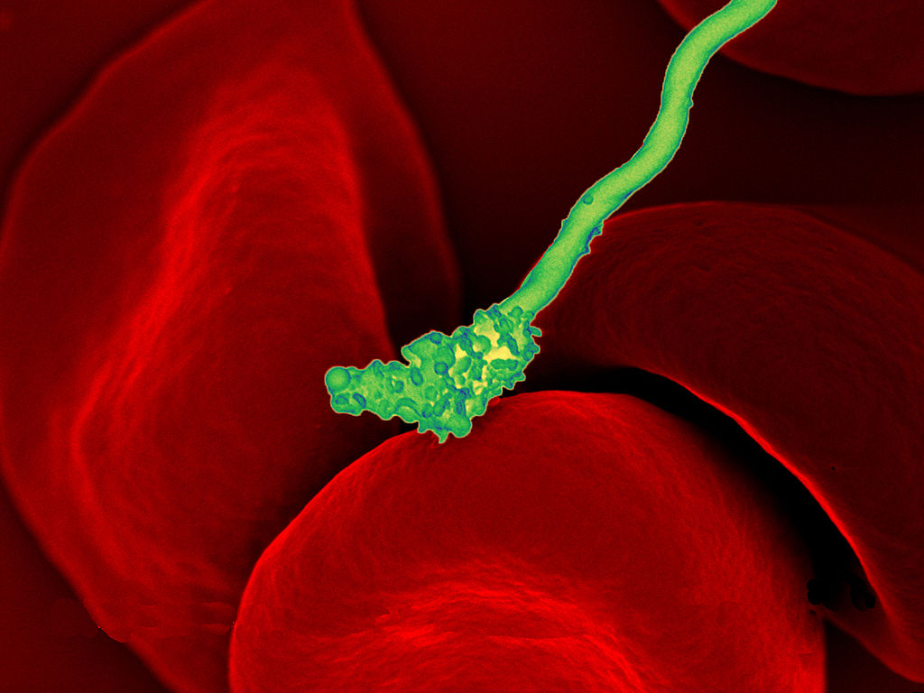
The colourful image in Figure 4.3.1 shows a bacterial cell (in green) attacking human red blood cells. The bacterium causes a disease called relapsing fever. The bacterial and human cells look very different in size and shape. Although all living cells have certain things in common — such as a plasma membrane and cytoplasm — different types of cells, even within the same organism, may have their own unique structures and functions. Cells with different functions generally have different shapes that suit them for their particular job. Cells vary not only in shape, but also in size, as this example shows. In most organisms, however, even the largest cells are no bigger than the period at the end of this sentence. Why are cells so small?
Explaining Cell Size
Most organisms, even very large ones, have microscopic cells. Why don't cells get bigger instead of remaining tiny and multiplying? Why aren't you one giant cell rolling around school? What limits cell size?
Once you know how a cell functions, the answers to these questions are clear. To carry out life processes, a cell must be able to quickly pass substances in and out of the cell. For example, it must be able to pass nutrients and oxygen into the cell and waste products out of the cell. Anything that enters or leaves a cell must cross its outer surface. The size of a cell is limited by its need to pass substances across that outer surface.
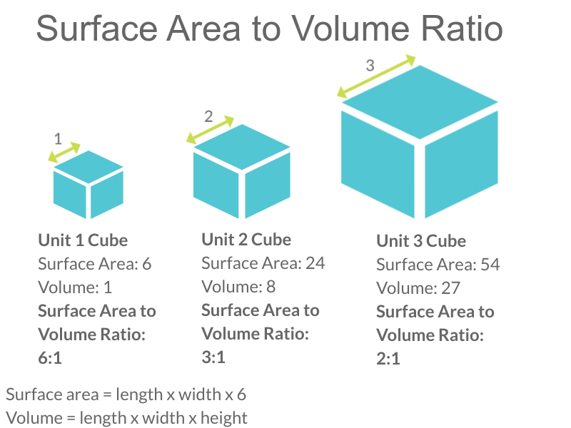
Look at the three cubes in Figure 4.3.2. A larger cube has less surface area relative to its volume than a smaller cube. This relationship also applies to cells — a larger cell has less surface area relative to its volume than a smaller cell. A cell with a larger volume also needs more nutrients and oxygen, and produces more waste. Because all of these substances must pass through the surface of the cell, a cell with a large volume will not have enough surface area to allow it to meet its needs. The larger the cell is, the smaller its ratio of surface area to volume, and the more difficult it will be for the cell to get rid of its waste and take in necessary substances. This is what limits the size of the cell.
Cell Form and Function
Cells with different functions often have varying shapes. The cells pictured below (Figure 4.3.3) are just a few examples of the many different shapes that human cells may have. Each type of cell has characteristics that help it do its job. The job of the nerve cell, for example, is to carry messages to other cells. The nerve cell has many long extensions that reach out in all directions, allowing it to pass messages to many other cells at once. Do you see the tail of each tiny sperm cell? Its tail helps a sperm cell "swim" through fluids in the female reproductive tract in order to reach an egg cell. The white blood cell has the job of destroying bacteria and other pathogens. It is a large cell that can engulf foreign invaders.
Figure 4.3.3 Human cells may have many different shapes that help them to do their jobs.
Cells With and Without a Nucleus
The nucleus is a basic cell structure present in many — but not all — living cells. The nucleus of a cell is a structure in the cytoplasm that is surrounded by a membrane (the nuclear membrane) and contains DNA. Based on whether or not they have a nucleus, there are two basic types of cells: prokaryotic cells and eukaryotic cells.
Prokaryotic Cells
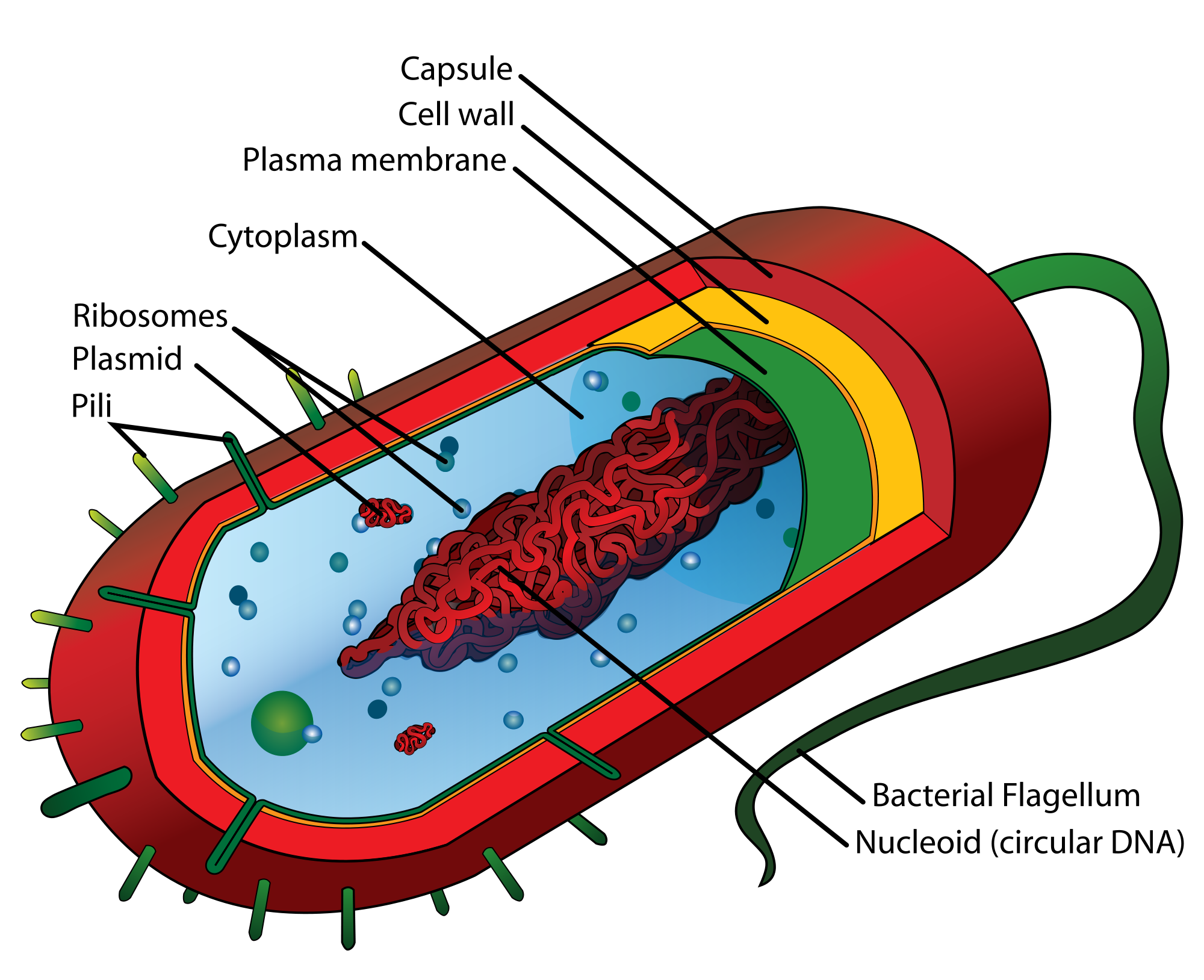
Prokaryotic cells are cells without a nucleus. The DNA in prokaryotic cells is in the cytoplasm, rather than enclosed within a nuclear membrane. In addition, these cells are typically smaller than eukaryotic cells and contain fewer organelles. Prokaryotic cells are found in single-celled organisms, such as the bacterium represented by the model in Figure 4.3.3. Organisms with prokaryotic cells are called prokaryotes. They were the first type of organisms to evolve, and they are still the most common organisms today.
Eukaryotic Cells
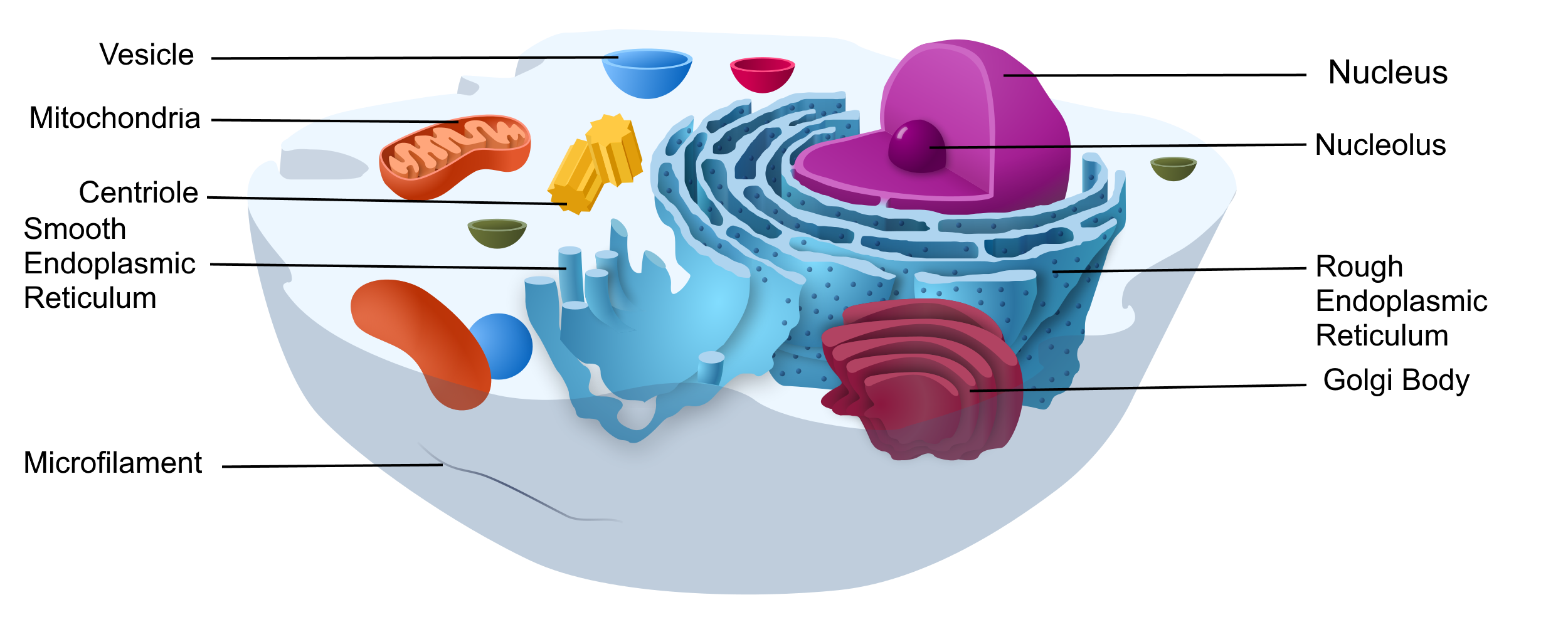
Eukaryotic cells are cells that contain a nucleus. A typical eukaryotic cell is represented by the model in Figure 4.3.4. Eukaryotic cells are usually larger than prokaryotic cells. They are found in some single-celled and all multicellular organisms. Organisms with eukaryotic cells are called eukaryotes, and they range from fungi to humans.
Besides a nucleus, eukaryotic cells also contain other organelles. An organelle is a structure within the cytoplasm that performs a specific job in the cell. Organelles called mitochondria, for example, provide energy to the cell, and organelles called vesicles store substances in the cell. Organelles allow eukaryotic cells to carry out more functions than prokaryotic cells can.
Interestingly, scientists think that mitochondria were once free-living prokaryotes that infected (or were engulfed by) larger cells. The two organisms developed a symbiotic relationship that was beneficial to both of them, resulting in the smaller prokaryote becoming an organelle within the larger cell. This is called endosymbiotic theory, and it is supported by a lot of evidence, including the fact that mitochondria have their own DNA separate from the DNA in the nucleus of the eukaryotic cell. Endosymbiotic theory will be described in more detail in later sections, and it's also discussed in the video below.
https://www.youtube.com/watch?v=FGnS-Xk0ZqU
Endosymbiotic Theory, Amoeba Sisters, 2017.
4.3 Summary
- Cells must be very small so they have a large enough surface area-to-volume ratio to maintain normal cell processes.
- Cells with different functions often have different shapes.
- Prokaryotic cells do not have a nucleus. Eukaryotic cells do have a nucleus, along with other organelles.
4.3 Review Questions
- Explain why most cells are very small.
- Discuss variations in the form and function of cells.
-
-
- Do human cells have organelles? Explain your answer.
- Which are usually larger – prokaryotic or eukaryotic cells? What do you think this means for their relative ability to take in needed substances and release wastes? Discuss your answer.
- DNA in eukaryotes is enclosed within the _______ ________.
- Name three different types of cells in humans.
- Which organelle provides energy in eukaryotic cells?
- What is a function of a vesicle in a cell?
4.3 Explore More
https://www.youtube.com/watch?time_continue=1&v=9i7kAt97XYU&feature=emb_logo
How we think complex cells evolved - Adam Jacobson, TED-Ed, 2015.
https://www.youtube.com/watch?v=Pxujitlv8wc
Prokaryotic vs. Eukaryotic Cells (updated), Amoeba Sisters, 2018.
Attributions
Figure 4.3.1
Borrelia_hermsii_Bacteria_(13758011613) by NAID on Wikimedia Commons is released into the public domain (https://en.wikipedia.org/wiki/Public_domain).
Figure 4.3.2
Cell Size by Christine Miller is released into the Public Domain (https://creativecommons.org/publicdomain/mark/1.0/).
Figure 4.3.3
- Chondrocyte. BioTek-Wikipedia-Image by BioTek Instruments, Inc. on Wikimedia Commons is used under a CC BY-SA 3.0 (https://creativecommons.org/licenses/by-sa/3.0/deed.en) license.
- Neutrophil with anthrax copy by Volker Brinkmann from PLOS Pathogens on Wikimedia Commons is used under a CC BY 2.5 (https://creativecommons.org/licenses/by/2.5/deed.en) license.
- PLoSBiol4.e126.Fig6fNeuron by Lee, et al. from PLOS Biology on Wikimedia Commons is used under a CC BY 2.5 (https://creativecommons.org/licenses/by/2.5/deed.en) license.
- Sperm (265 33) human by Doc. RNDr. Josef Reischig, CSc. on Wikimedia Commons is used under a CC BY-SA 3.0 (https://creativecommons.org/licenses/by-sa/3.0) license.
Figure 4.3.4
Model of a prokaryotic cell: bacterium by Mariana Ruiz Villarreal [LadyofHats] on Wikimedia Commons is released into the public domain (https://en.wikipedia.org/wiki/Public_domain).
Figure 4.3.5
Animal Cell adapted by Christine Miller is used under a CC0 1.0 (https://creativecommons.org/publicdomain/zero/1.0/deed.en) public domain dedication license. (Original image, Animal Cell Unannotated, is by Kelvin Song on Wikimedia Commons.)
References
Amoeba Sisters. (2017, May 3). Endosymbiotic theory. YouTube. https://www.youtube.com/watch?v=FGnS-Xk0ZqU&feature=youtu.be
Amoeba Sisters. (2018, July 30). Prokaryotic vs. eukaryotic cells (updated). YouTube. https://www.youtube.com/watch?v=Pxujitlv8wc&feature=youtu.be
Brinkmann, V. (November 2005). Neutrophil engulfing Bacillus anthracis. PLoS Pathogens 1 (3): Cover page [digital image]. DOI:10.1371. https://journals.plos.org/plospathogens/issue?id=10.1371/issue.ppat.v01.i03
Lee, W.C.A., Huang, H., Feng, G., Sanes, J.R., Brown, E.N., et al. (2005, December 27) Figure 6f, slightly altered (plus scalebar, minus letter "f".) [digital image]. Dynamic Remodeling of Dendritic Arbors in GABAergic Interneurons of Adult Visual Cortex. PLoS Biology, 4(2), e29. doi:10.1371/journal.pbio.0040029. https://journals.plos.org/plosbiology/article?id=10.1371/journal.pbio.0040029
TED-Ed. (2015, February 17). How we think complex cells evolved - Adam Jacobson. https://www.youtube.com/watch?v=9i7kAt97XYU&feature=youtu.be
A rigid organ that constitutes part of the vertebrate skeleton in animals.
Created by: CK-12/Adapted by Christine Miller
Is It Magic?
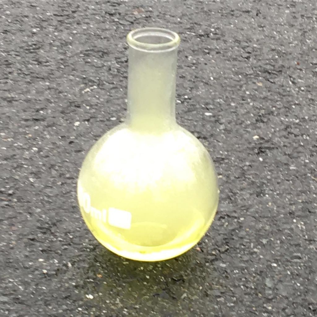
The harmless-looking bottle in Figure 3.8.1 contains a greenish-yellow, poisonous gas. The gas is chlorine, which is also used as bleach and to keep the water in pools and hot tubs free of germs. Chlorine can kill just about anything. Would you breathe in chlorine gas or drink liquid chlorine? Of course not, but you often eat a compound containing chlorine. You probably eat this chlorine compound just about every day. Can you guess what it is? It's table salt.
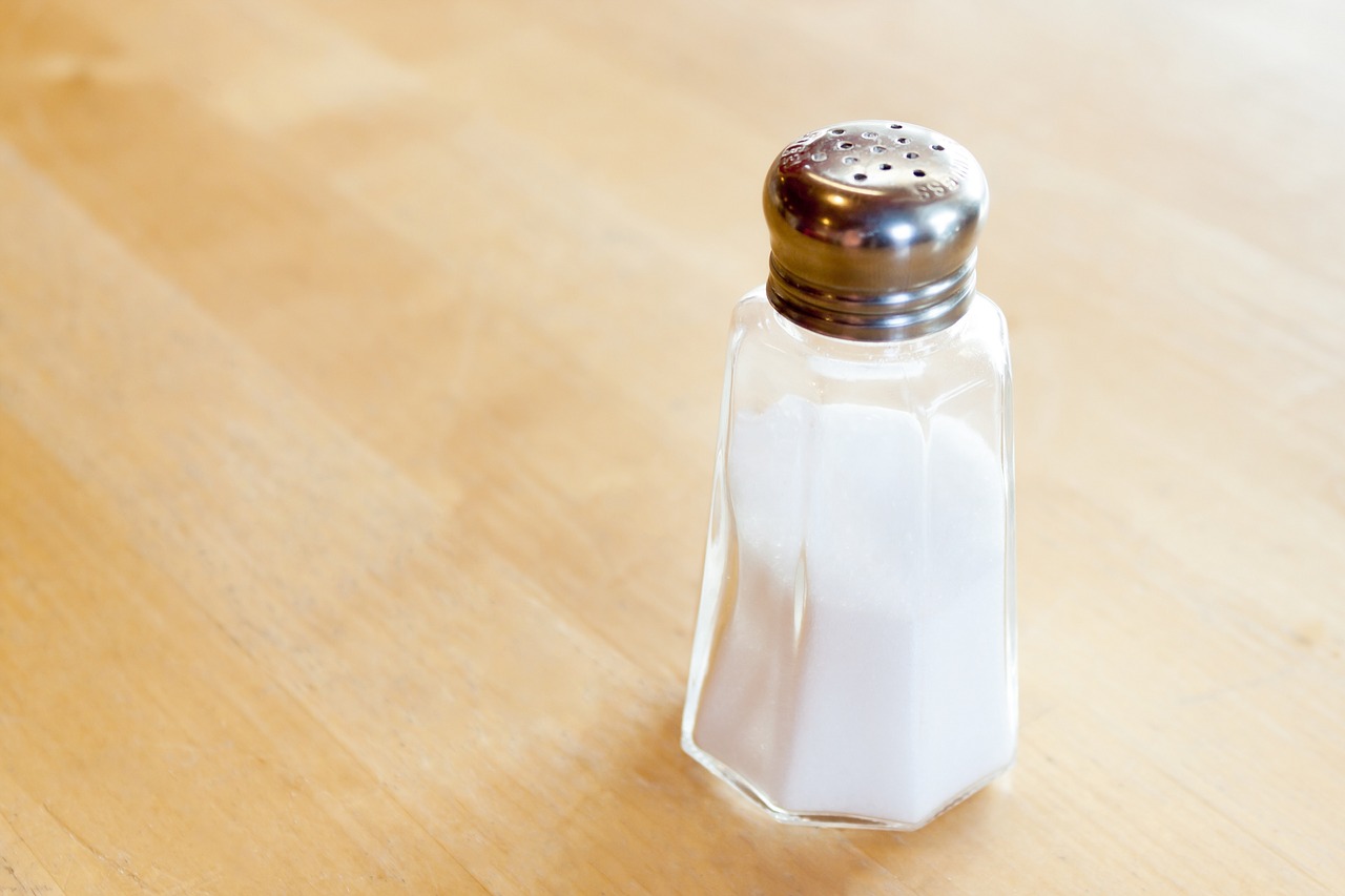
Table salt is actually sodium chloride (NaCl), which forms when chlorine and sodium (Na) combine in certain proportions. How does the toxic green chemical chlorine change into the harmless white compound we know as table salt? It isn't magic — it's chemistry, and it happens in a chemical reaction.
What Is a Chemical Reaction?
A chemical reaction is a process that changes some chemical substances into others. A substance that starts a chemical reaction is called a reactant, and a substance that forms as a result of a chemical reaction is called a product. During the reaction, the reactants are used up to create the products.
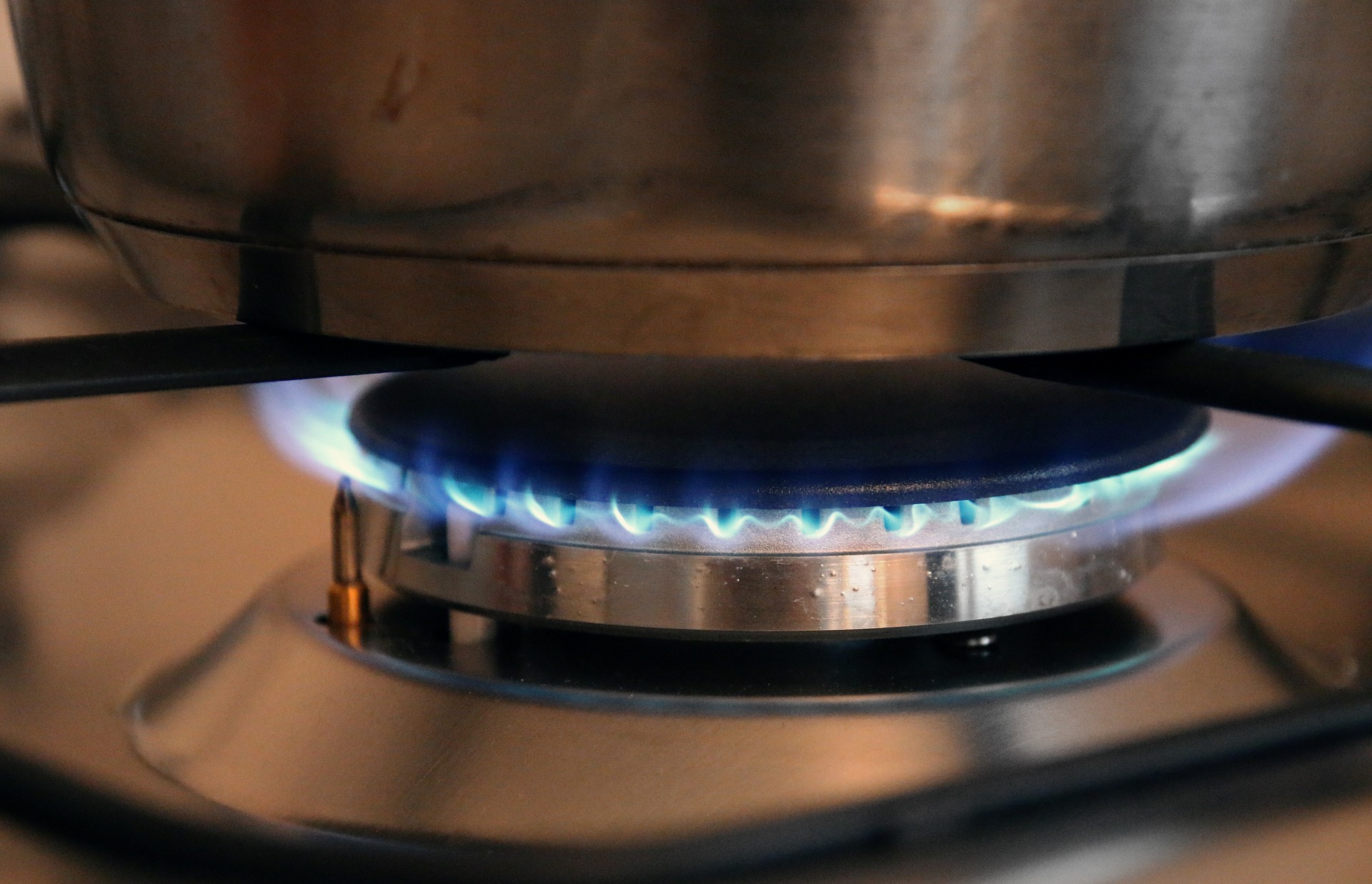
The burning of methane gas, as shown in the picture below, is a chemical reaction. In this reaction, the reactants are methane (CH4) and oxygen (O2), and the products are carbon dioxide (CO2) and water (H2O). As this example shows, a chemical reaction involves the breaking and forming of chemical bonds, which are forces that hold together the atoms of a molecule. When methane burns, for example, bonds break within the methane and oxygen molecules, and new bonds form in the molecules of carbon dioxide and water.
Chemical Equations
Chemical reactions can be represented by chemical equations. A chemical equation is a symbolic way of showing what happens during a chemical reaction. The burning of methane, for example, can be represented by the chemical equation:
CH4 + 2O2 → CO2 + 2H2O
The arrow in a chemical equation separates the reactants from the products, and shows the direction in which the reaction proceeds. If the reaction could occur in the opposite direction as well, two arrows pointing in opposite directions would be used. The number 2 in front of O2 and H2O, called the coefficient, shows that two oxygen molecules and two water molecules are involved in the reaction. If just one molecule is involved, no number is placed in front of the chemical symbol. Note the subscript of 2 for the oxygen (O) and hydrogen (H) atoms in the oxygen and water molecules, respectively. That tells you that each oxygen molecule is made up of two oxygen atoms. If there is no subscript, then there is a single atom. Thus, one water molecule is made up of two hydrogen atoms and one oxygen atom. In order for this chemical reaction to take place, one methane molecule reacts with two oxygen molecules to form one carbon dioxide molecule and two water molecules.
Conservation of Mass
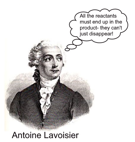
In a chemical reaction, the quantity of each element does not change. There is the same amount of each element in the products as there was in the reactants. Mass is always conserved. According to the law of conservation of mass — which was first demonstrated convincingly by French chemist Antoine Lavoisier in 1785 — mass is neither created nor destroyed during a chemical reaction. Therefore, during a chemical reaction, the total mass of products is equal to the total mass of reactants. The conservation of mass is reflected in a reaction's chemical equation. The same number of atoms of each element appears on each side of the arrow. In the chemical equation above, there are four hydrogen atoms on each side of the arrow. Can you find all four of them on each side of the equation?
Chemical vs. Physical Changes
Many processes that happen all around us on a daily basis involve chemical reactions. Not every change, however, is a chemical change. Some changes are simply physical and do not involve chemical reactions. Physical changes include change in size of pieces and change in state. If you break an eggshell and pour out the egg into a pan, its chemical makeup and properties do not change. This is just a physical change. No chemical reactions have occurred, and no chemical bonds have broken or formed. Other examples of physical changes are cutting paper into smaller pieces and letting an ice cube melt. What if you put the egg in the pan over a hot flame? The egg turns to a rubbery solid and changes colour. The properties of the egg have changed because its chemical makeup has changed. Cooking the egg is a chemical change that involves chemical reactions.
Other common examples of chemical changes include a cake baking, metal rusting, and a candle burning. More practice is below.
Figure 3.8.5 Chemical changes often involve chemical reactions as well.
3.8 Summary
- A chemical reaction is a process that changes some chemical substances into others. A substance that starts a chemical reaction is called a reactant, and a substance that forms during a chemical reaction is called a product. During the chemical reaction, bonds break in reactants and new bonds form in products.
- Chemical reactions can be represented by chemical equations. According to the law of conservation of mass, mass is always conserved in a chemical reaction, so a chemical equation must be balanced, with the same number of atoms of each type of element in the products as in the reactants.
- Many chemical reactions — such as iron rusting and organic matter rotting — occur all around us each day, but not all changes are chemical processes. Some changes — like ice melting or paper being torn into smaller pieces — are physical processes that do not involve chemical reactions and the formation of new substances.
3.8 Review Questions
- What is a chemical reaction?
- Define the reactants and products in a chemical reaction.
- List three examples of common changes that involve chemical reactions.
- Define a chemical bond.
- What is a chemical equation? Give an example.
- What does it mean for a chemical equation to be balanced? Why must a chemical equation be balanced?
- Our cells use glucose (C6H12O6) to obtain energy in a chemical reaction called cellular respiration. In this reaction, six oxygen molecules (O2) react with one glucose molecule. Answer the following questions about this reaction:
- How many oxygen atoms are in one molecule of glucose?
- Write out what the reactant side of this equation would look like.
- In total, how many oxygen atoms are in the reactants? Explain how you calculated your answer.
- In total, how many oxygen atoms are in the products? Is it possible to answer this question without knowing what the products are? Why or why not?
- Answer the following questions about the following equation: CH4+ 2O2 → CO2 + 2H2O
- Can carbon dioxide (CO2)transform into methane (CH4) and oxygen (O2) in this reaction? Why or why not?
- How many molecules of carbon dioxide (CO2) are produced in this reaction?
- Is the evaporation of liquid water into water vapor a chemical reaction? Why or why not?
- Why do bonds break in the reactants during a chemical reaction?
3.8 Explore More
https://www.youtube.com/watch?v=2S6e11NBwiw&feature=emb_logo
The law of conservation of mass - Todd Ramsey, TED-Ed, 2015.
https://www.youtube.com/watch?v=37pir0ej_SE
Chemical Changes: Crash Course Kids #19.2, by Crash Course Kids, 2015.
Attributions
Figure 3.8.1
Chlorine_gas_in_high_concentration by Larenmclane on Wikimedia Commons, is used under a CC BY-SA 4.0 (https://creativecommons.org/licenses/by-sa/4.0/deed.en) license.
Figure 3.8.2
Tags: Salt Salt Shaker Spices Kitchen Spice Component; salt-4160306_1280 by katie175 from Pixabay is used under the Pixabay License (https://pixabay.com/de/service/license/).
Figure 3.8.3
Tags: Gas Flame Gas Stove Italy Gas Cook Kitchen by moerschy from Pixabay is used under the Pixabay License (https://pixabay.com/de/service/license/).
Figure 3.8.4
Antoine_lavoisier by unknown on Wikimedia Commons has been adapted by Christine Miller. The orginal work, believed to be from http://www.schuster-ingolstadt.de/Chemie.htm has been released into the public domain (https://en.wikipedia.org/wiki/Public_domain).
Figure 3.8.5
- Ice cream melting by Aron Visuals on Unsplash is used under the Unsplash License (https://unsplash.com/license).
- Kombucha [photo] by Klara Avsenik on Unsplash is used under the Unsplash License (https://unsplash.com/license).
- Grated cheese by Steve Buissinne on PublicDomainPictures is used under the CC0 1.0 Universal Public Domain Dedication license (https://creativecommons.org/publicdomain/zero/1.0/).
References
Crash Course Kids. (2015, July 16). Chemical changes: Crash Course Kids #19.2. YouTube. https://www.youtube.com/watch?v=37pir0ej_SE
TED-Ed. (2015, February 26 ). The law of conservation of mass - Todd Ramsey. YouTube. https://www.youtube.com/watch?v=2S6e11NBwiw&feature=emb_logo
Wikipedia contributors. (2020, June 15). Antoine Lavoisier. Wikipedia. https://en.wikipedia.org/w/index.php?title=Antoine_Lavoisier&oldid=962631283
The ability of an organism to maintain constant internal conditions despite external changes.
Created by: CK-12/Adapted by: Christine Miller
Ouch!
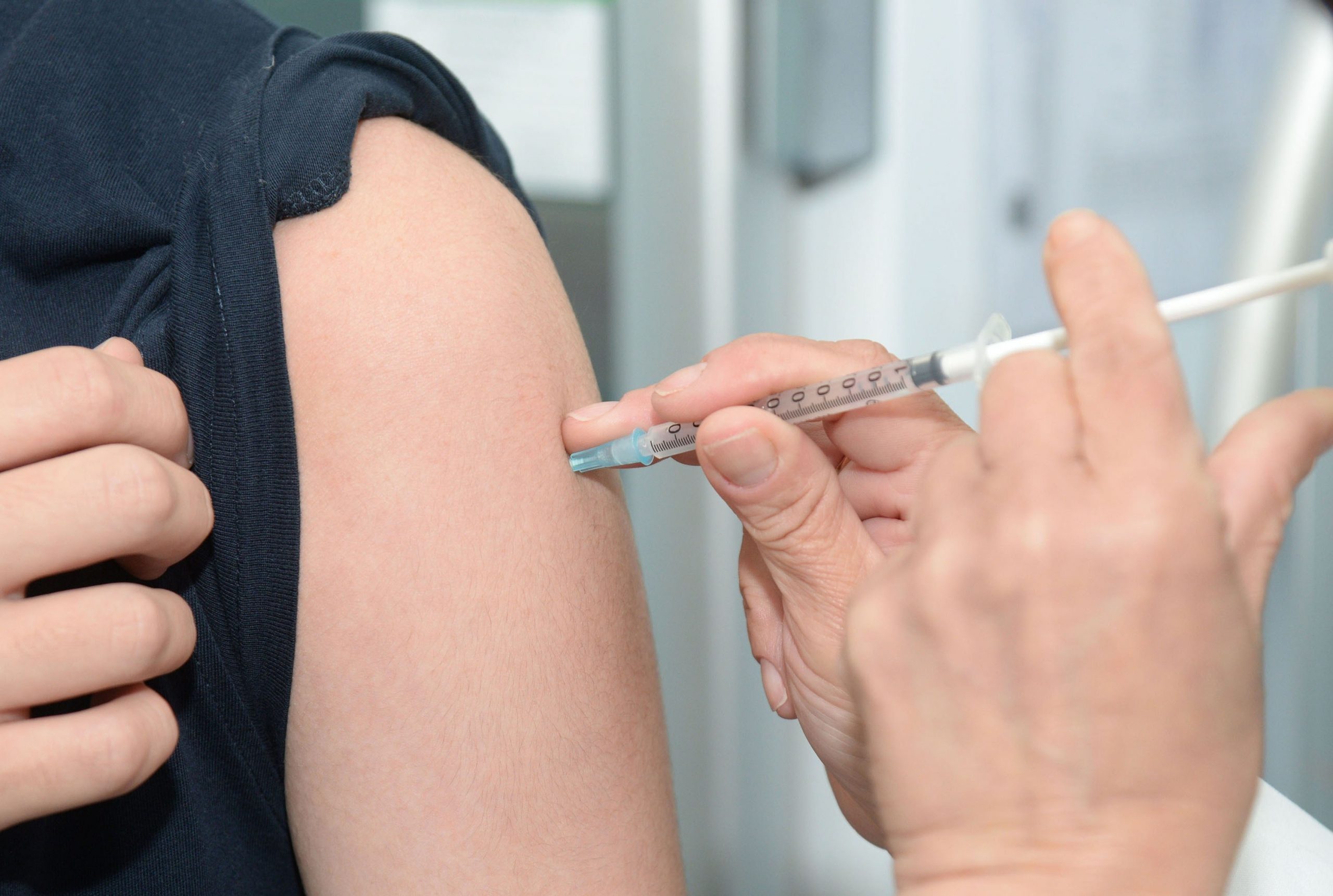
The person in Figure 1.2.1 is getting a flu vaccine. You probably know that getting a vaccine can hurt — but it's usually worth it. A vaccine contains dead or altered forms of germs that normally cause a disease, such as flu or measles. The germs in vaccines have been inactivated or weakened so they can no longer cause illness, but are still "noticed" by the immune system.
They stimulate the immune system to produce chemicals that can kill the actual germs if they enter the body, thus preventing future disease. How was such an ingenious way to prevent disease discovered? The short answer is more than two centuries of science.
Science as Process
You may think of science as a large and detailed body of knowledge, but science is also the process by which this knowledge is obtained. Science uses experimentation, evidence, and logic to continuously test ideas. Over time and through repeated experimentation and testing, scientific knowledge advances.
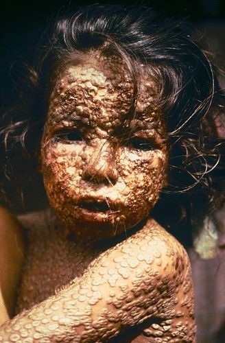
We've been accumulating knowledge of vaccines for more than two centuries. The discovery of the first vaccine, as well as the process of vaccination, dates back to 1796. An English doctor named Edward Jenner observed that people who became infected with cowpox did not get sick from smallpox, a similar but much more severe disease (Figure 1.2.2). Jenner decided to transmit cowpox to a young boy to see if it would protect him from smallpox. He gave the boy cowpox by scratching liquid from cowpox sores into the boy's skin. Then, six weeks later, he scratched liquid from smallpox sores into the boy's skin. As Jenner predicted, the boy did not get sick from smallpox. Jenner had discovered the first vaccine, although additional testing was needed to show that it really was effective.
Almost a century passed before the next vaccine was discovered, a vaccine for cholera in 1879. Around the same time, French chemist Louis Pasteur found evidence that many human diseases are caused by germs, which earned him the title of "father of germ theory." Since Pasteur's time, vaccines have been discovered for scores of additional diseases caused by germs, and scientists are currently researching vaccines for many others.
Benefits of Science
Medical advances such as the discovery of vaccines are one of the most important benefits of science, but science and scientific knowledge are also crucial for most other human endeavors. Science is needed to design safe cars, predict storms, control global warming, develop new technologies of many kinds, help couples have children, and put humans on the moon. Clearly, the diversity of applications of scientific knowledge is vast!
1.2 Summary
- Science is a large and detailed body of knowledge. It is also the process by which this knowledge is obtained.
- Science uses experimentation, evidence, and logic to continuously test ideas. Over time and through repeated experimentation and testing, scientific knowledge advances.
- Medical advances such as the development of vaccines are one of the most important benefits of science, but science and scientific knowledge are also crucial for most other human endeavors.
1.2 Review Questions
- Explain why science is considered both a process and a body of knowledge.
- State three specific examples of human endeavors that are based on scientific knowledge.
- How does science influence your daily life?
- Jenner used a young boy as a research subject in his smallpox vaccine research. Today, scientists must follow strict guidelines when using human subjects in their research. What unique concerns do you think might arise when human beings are used as research subjects?
- What gave Jenner the idea to develop a vaccine for smallpox?
- Why do you think almost a century passed between the development of the first vaccine (for smallpox) and the development of the next vaccine (for cholera)
1.2 Explore More
https://www.youtube.com/watch?time_continue=1&v=yqUFy-t4MlQ&feature=emb_logohttps://www.ted.com/talks/uri_alon_why_science_demands_a_leap_into_the_unknown?language=en
How we conquered the deadly smallpox virus - Simona Zompi, TED-Ed, 2013.
Attributions
Figure 1.2.1
Vacina, centro de vacinação, by Hyttalo Souza on Unsplash, used under the Unsplash License (https://unsplash.com/license).
Figure 1.2.2
Child with Smallpox/ID#3265, by CDC/ James Hicks, from the Centre for Disease Control and Prevention, is in the public domain (https://en.wikipedia.org/wiki/Public_domain).
References
TED-Ed. (2013, October 28). How we conquered the deadly smallpox virus - Simona Zompi. YouTube. https://www.youtube.com/watch?v=yqUFy-t4MlQ&feature=youtu.be
Wikipedia contributors. (2020, August 9). Edward Jenner. In Wikipedia. https://en.wikipedia.org/w/index.php?title=Edward_Jenner&oldid=971970576
Wikipedia contributors. (2020, August 5). Louis Pasteur. In Wikipedia. https://en.wikipedia.org/w/index.php?title=Louis_Pasteur&oldid=971330056
Created by: CK-12/Adapted by: Christine Miller
What Is Pseudoscience?
Pseudoscience is a claim, belief, or practice that is presented as scientific but does not adhere to the standards and methods of science. True science is based on repeated evidence-gathering and testing of falsifiable hypotheses. Pseudoscience does not adhere to these criteria. In addition to phrenology, some other examples of pseudoscience include astrology, extrasensory perception (ESP), reflexology, reincarnation, and Scientology,
Characteristics of Pseudoscience
Whether a field is actually science or just pseudoscience is not always clear. However, pseudoscience generally exhibits certain common characteristics. Indicators of pseudoscience include:
- The use of vague, exaggerated, or untestable claims: Many claims made by pseudoscience cannot be tested with evidence. As a result, they cannot be falsified, even if they are not true.
- An over-reliance on confirmation rather than refutation: Any incident that appears to justify a pseudoscience claim is treated as proof of the claim. Claims are assumed true until proven otherwise, and the burden of disproof is placed on skeptics of the claim.
- A lack of openness to testing by other experts: Practitioners of pseudoscience avoid subjecting their ideas to peer review. They may refuse to share their data and justify the need for secrecy with claims of proprietary or privacy.
- An absence of progress in advancing knowledge: In pseudoscience, ideas are not subjected to repeated testing followed by rejection or refinement, as hypotheses are in true science. Ideas in pseudoscience may remain unchanged for hundreds — or even thousands — of years. In fact, the older an idea is, the more it tends to be trusted in pseudoscience.
- Personalization of issues: Proponents of pseudoscience adopt beliefs that have little or no rational basis, so they may try to confirm their beliefs by treating critics as enemies. Instead of arguing to support their own beliefs, they attack the motives and character of their critics.
- The use of misleading language: Followers of pseudoscience may use scientific-sounding terms to make their ideas sound more convincing. For example, they may use the formal name dihydrogen monoxide to refer to plain old water.
Persistence of Pseudoscience
Despite failing to meet scientific standards, many pseudosciences survive. Some pseudosciences remain very popular with large numbers of believers. A good example is astrology.
Astrology is the study of the movements and relative positions of celestial objects as a means for divining information about human affairs and terrestrial events. Many ancient cultures attached importance to astronomical events, and some developed elaborate systems for predicting terrestrial events from celestial observations. Throughout most of its history in the West, astrology was considered a scholarly tradition and was common in academic circles. With the advent of modern Western science, astrology was called into question. It was challenged on both theoretical and experimental grounds, and it was eventually shown to have no scientific validity or explanatory power.
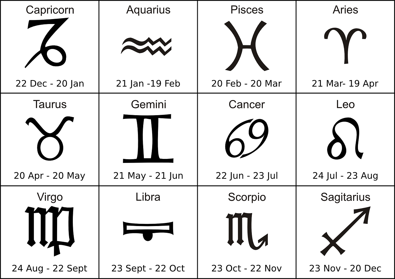
Today, astrology is considered a pseudoscience, yet it continues to have many devotees. Most people know their astrological sign, and many people are familiar with the personality traits supposedly associated with their sign. Astrological readings and horoscopes are readily available online and in print media, and a lot of people read them, even if only occasionally. About a third of all adult Americans actually believe that astrology is scientific. Studies suggest that the persistent popularity of pseudosciences such as astrology reflects a high level of scientific illiteracy. It seems that many Americans do not have an accurate understanding of scientific principles and methodology. They are not convinced by scientific arguments against their beliefs.
Dangers of Pseudoscience
Belief in astrology is unlikely to cause a person harm, but belief in some other pseudosciences might — especially in health care-related areas. Treatments that seem scientific but are not may be ineffective, expensive, and even dangerous to patients. Seeking out pseudoscientific treatments may also delay or preclude patients from seeking scientifically-based medical treatments that have been tested and found safe and effective. In short, irrational health care may not be harmless.
Scientific Hoaxes, Frauds, and Fallacies
Pseudoscience is not the only way that science may be misused. Scientific hoaxes, frauds, and fallacies may misdirect the pursuit of science, put patients at risk, or mislead and confuse the public. An example of each of these misuses of science and its negative effects is described below.
The Piltdown Hoax
![Image by By James Howard McGregor [Public domain], via Wikimedia Commons A side profile view of an artists rendition of what the Piltdown Man may have looked like, had he been real.](https://pressbooks.ccconline.org/acchumanbio/wp-content/uploads/sites/152/2023/10/Piltdown-Man-1.jpg)
Piltdown Man (see picture left) was a paleontological hoax in which bone fragments were presented as the fossilized remains of a previously unknown early human. These fragments consisted of parts of a skull and jawbone, reported to have been found in 1908 in a gravel pit at Piltdown, East Sussex, England. The significance of the specimen remained the subject of controversy until it was exposed in 1953 as a hoax. It eventually came to light that the specimen consisted of the lower jawbone of an orangutan deliberately combined with skull bones of a modern human. The Piltdown hoax is perhaps the most infamous paleontological hoax ever perpetrated, both for its impact on the direction of research on human evolution and for the length of time between its "discovery" and its full exposure as a forgery.
![Photo by Anrie [CC BY-SA 3.0 (https://creativecommons.org/licenses/by-sa/3.0)], from Wikimedia Commons A replica of the infamous Piltdown skull. The skull is encased in a glass sphere. The replica shows portions of the skull which were bone in white, and the portions of the skull which were inferred in black.](https://pressbooks.ccconline.org/acchumanbio/wp-content/uploads/sites/152/2023/10/Sterkfontein_Piltdown_man-1.jpg)
In 1912, the head of the geological department at the British Museum proposed that Piltdown man represented an evolutionary missing link between apes and humans. With its human-like cranium and ape-like jaw, it seemed to support the idea then prevailing in England that human evolution began with the brain. The Piltdown specimen led scientists down a blind alley in the belief that the human brain increased in size before the jaw underwent size reductions to become more like the modern human jaw. This belief confused and misdirected the study of human evolution for decades, and actual fossils of early humans were ignored because they didn't support the accepted paradigm.
The Vaccine-Autism Fraud
You may have heard that certain vaccines put the health of young children at risk. This persistent idea is not supported by scientific evidence or accepted by the vast majority of experts in the field. It stems largely from an elaborate medical research fraud that was reported in a 1998 article published in the respected British medical journal, The Lancet. The main author of the article was a British physician named Andrew Wakefield. In the article, Wakefield and his colleagues described case histories of 12 children, most of whom were reported to have developed autism soon after the administration of the MMR (measles, mumps, rubella) vaccine.
Several subsequent peer-reviewed studies failed to show any association between the MMR vaccine and autism. It also later emerged that Wakefield had received research funding from a group of people who were suing vaccine manufacturers. In 2004, ten of Wakefield's 12 coauthors formally retracted the conclusions in their paper. In 2010, editors of The Lancetretracted the entire paper. That same year, Wakefield was charged with deliberate falsification of research and barred from practicing medicine in the United Kingdom. Unfortunately, by then, the damage had already been done. Parents afraid that their children would develop autism had refrained from having them vaccinated. British MMR vaccination rates fell from nearly 100 per cent to 80 per cent in the years following the study. The consensus of medical experts today is that Wakefield's fraud put hundreds of thousands of children at risk because of the lower vaccination rates and also diverted research efforts and funding away from finding the true cause of autism.
Correlation-Causation Fallacy
Many statistical tests used in scientific research calculate correlations between variables. Correlation refers to how closely related two data sets are, which may be a useful starting point for further investigation. Correlation, however, is also one of the most misused types of evidence, primarily because of the logical fallacy that correlation implies causation. In reality, just because two variables are correlated does not necessarily mean that either variable causes the other.
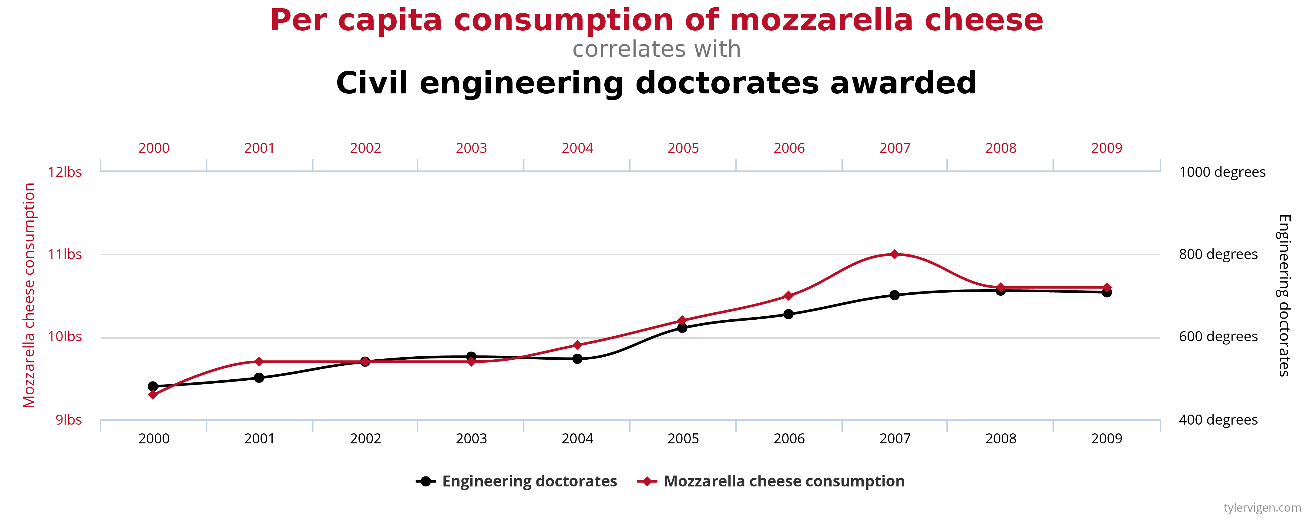
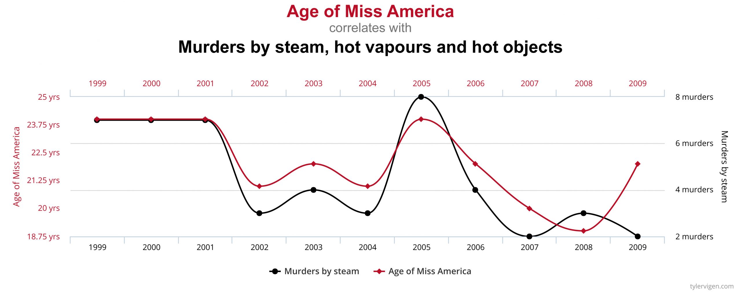
A few simple examples, illustrated by the graphs below, can be used to demonstrate the correlation-causation fallacy. Assume a study found that both per capita consumption of mozzarella cheese and the number of Civil Engineering doctorates awarded are correlated; that is, rates of both events increase together. If correlation really did imply causation, then you could conclude from the second example that the increase in age of Miss America causes an increase in murders of a specific type or vice versa.
An actual example of the correlation-causation fallacy occurred during the latter half of the 20th century. Numerous studies showed that women taking hormone replacement therapy (HRT) to treat menopausal symptoms also had a lower-than-average incidence of coronary heart disease (CHD). This correlation was misinterpreted as evidence that HRT protects women against CHD. Subsequent studies that controlled other factors related to CHD disproved this presumed causal connection. The studies found that women taking HRT were more likely to come from higher socio-economic groups, with better-than-average diets and exercise regimens. Rather than HRT causing lower CHD incidence, these studies concluded that HRT and lower CHD were both effects of higher socio-economic status and related lifestyle factors.
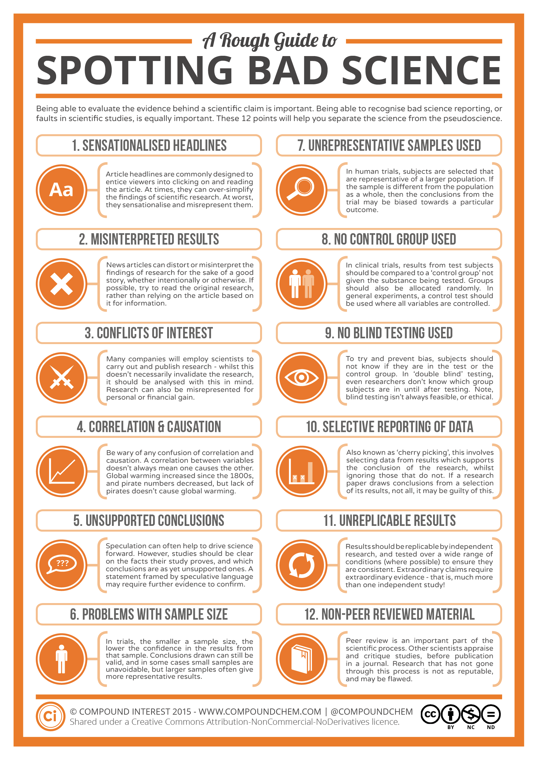
Check out this “Rough Guide to Spotting Bad Science” infographic from Compound Interest:
1.7 Summary
- Pseudoscience is a claim, belief, or practice that is presented as scientific, but does not adhere to scientific standards and methods.
- Indicators of pseudoscience include untestable claims, lack of openness to testing by experts, absence of progress in advancing knowledge, and attacks on the motives and character of critics.
- Some pseudosciences, including astrology, remain popular. This suggests that many people do not possess the scientific literacy needed to distinguish pseudoscience from true science, or to be convinced by scientific arguments against them.
- Belief in a pseudoscience such as astrology is unlikely to cause harm, but belief in pseudoscientific medical treatments may be harmful.
- In addition to pseudoscience, other examples of the misuse of science include scientific hoaxes (such as the Piltdown hoax), scientific frauds (such as the MMR vaccine-autism fraud), and scientific fallacies (such as the correlation-causation fallacy).
1.7 Review Questions
- Define pseudoscience. Give three examples.
- What are some indicators that a claim, belief, or practice might be pseudoscience rather than true science?
- Astrology was once considered a science, and it was common in academic circles. Why did its status change from a science to a pseudoscience?
- What are possible reasons that some pseudosciences remain popular even after they have been shown to have no scientific validity or explanatory power?
- List three other ways besides pseudoscience that science can be misused, and identify an example of each.
- Explain how misuses of science may waste money and effort. How can they potentially cause harm to the public?
- Many claims made by pseudoscience cannot be tested with evidence. From a scientific perspective, why is it important that claims be testable?
- What do you think is the difference between pseudoscience and belief?
- If you see a website that claims that an herbal supplement causes weight loss and they use a lot of scientific terms to explain how it works, can you be assured that the drug is scientifically proven to work? If not, what are some steps you can take to determine whether or not the drug does in fact work?
- Why do you think it was problematic that Andrew Wakefield received funding from a group of people who were suing vaccine manufacturers?
- What do you think it says about the 1998 Wakefield paper that ten of the 12 coauthors formally retracted their conclusions?
1.7 Explore More
https://www.youtube.com/watch?v=E91bGT9BjYk
How to spot a misleading graph - Lea Gaslowitz, TED-Ed, 2017.
https://www.youtube.com/watch?v=sxYrzzy3cq8
How statistics can be misleading - Mark Liddell, TED-Ed, 2016.
Attributions
Figure 1.7.1
Zodiac Signs Cancer Aquarius Aries Gemini Leo from Max Pixel, is used under a CC0 1.0 Universal Public Domain Dedication license (https://creativecommons.org/publicdomain/zero/1.0/deed.en).
Figure 1.7.2
Piltdown Man - McGregor model, by James Howard McGregor on Wikimedia Commons is in the public domain (https://en.wikipedia.org/wiki/Public_domain).
Figure 1.7.3
Sterkfontein Piltdown man, by Anrie on Wikimedia Commons is used under a CC BY-SA 3.0 (https://creativecommons.org/licenses/by-sa/3.0) license.
Figure 1.7.4
Spurious Correlations (Causation Fallacy) - Consumption of mozzarella cheese and awarded Doctorates by Tyler Vigen on Tylervigen.com is used under a CC BY 4.0 (https://creativecommons.org/licenses/by/4.0/) license.
Figure 1.7.5
Spurious Correlations (Causation Fallacy) - Miss America and Murder, by Tyler Vigen, is used under a CC BY 4.0 (https://creativecommons.org/licenses/by/4.0/) license.
Figure 1.7.6
A rough guide to spotting bad science, by Compound Interest, is used under a CC BY-NC-ND 2.0 (https://creativecommons.org/licenses/by-nc-nd/2.0/ca/) license
References
TED-Ed. (2017, July 6). How to spot a misleading graph - Lea Gaslowitz. YouTube. https://www.youtube.com/watch?v=E91bGT9BjYk&feature=youtu.be
Wakefield, A.J., Murch, S.H., Anthony, A., Linnell, J., Casson, D.M., Malik, M., et al. (1998). Ileal-lymphoid-nodular hyperplasia, non-specific colitis, and pervasive developmental disorder in children. Lancet, 351: 637–41.
Wikipedia contributors. (2020, June 18). Andrew Wakefield. Wikipedia. https://en.wikipedia.org/w/index.php?title=Andrew_Wakefield&oldid=963243135
Created by: CK-12/Adapted by Christine Miller
After reading this chapter, you should be able to see numerous connections between chemistry, human life, and health. In Joseph’s situation, chemistry is involved in the reasons why his father has diabetes, why his personal risk of getting diabetes is high, and why the dietary changes he is considering could be effective.
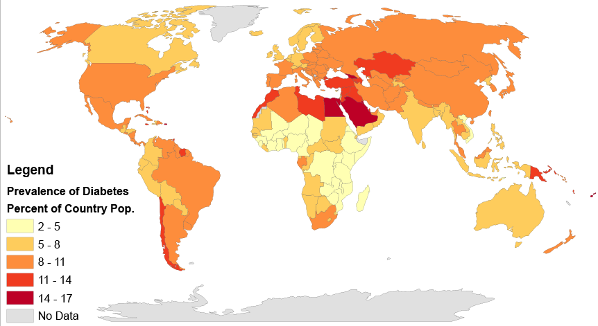
Type 2 diabetes affects populations worldwide and is caused primarily by a lack of response in the body to the hormone insulin, which causes problems in the regulation of blood sugar, or glucose. Insulin is a peptide hormone, and as you have learned, peptides are chains of amino acids. Therefore, insulin is in the class of biochemical compounds called proteins. Joseph is at increased risk of diabetes partly because there is a genetic component to the disease. DNA, which is a type of chemical compound called a nucleic acid, is passed down from parents to their offspring, and carries the instructions for the production of proteins in units called genes. If there is a problem in a gene (or genes) that contributes to the development of a disease, such as type 2 diabetes, this can get passed down to the offspring and may raise that child’s risk of getting the disease.
But genetics is only part of the reason why Joseph is at an increased risk of diabetes. Obesity itself is a risk factor, and one that can be shared in families due to shared lifestyle factors (such as poor diet and lack of exercise), as well as genetics. Consumption of too many refined carbohydrates (like white bread and soda) may also contribute to obesity and the development of diabetes. As you probably now know, these simple carbohydrates are more easily and quickly broken down in the digestive system into glucose than larger complex carbohydrate molecules, such as those found in vegetables and whole grains. This can lead to dramatic spikes in blood sugar levels, which is particularly problematic for people with diabetes because they have trouble maintaining their blood sugar at a safe level. You can understand why Joseph’s father limits his consumption of refined carbohydrates, and in fact, some scientific studies have shown that avoiding refined carbohydrates may actually help reduce the risk of getting diabetes in the first place.
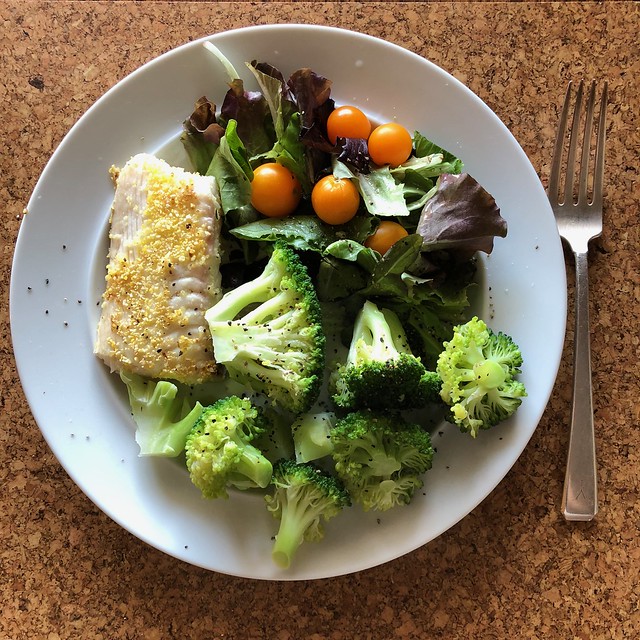
Joseph’s friend recommended eating a low fat, high carbohydrate diet to lose weight, but you can see that the type of carbohydrate — simple or complex — is an important consideration. Eating a large amount of white bread and rice may not help Joseph reduce his risk of diabetes, but a healthy diet that helps him lose weight may lower his risk of diabetes, since obesity itself is a factor. Which specific diet will work best to help him lose weight probably depends on a variety of factors, including his biology, lifestyle, and food preferences. Joseph should consult with his doctor about his diet and exercise plan, so that his specific situation can be taken into account and monitored by a medical professional.
Drinking enough water is usually good advice for everyone, especially if it replaces sugary drinks like soda. You now know that water is important for many of the chemical reactions that take place in the body. But you can have too much of a good thing — as in the case of marathon runners who can make themselves sick from drinking too much water! As you can see, proper balance, or homeostasis, is very important to the health of living organisms.
Finally, you probably now realize that “chemicals” do not have to be scary, toxic substances. All matter consists of chemicals, including water, your body, and healthy fresh fruits and vegetables, like the ones pictured in Figure 3.12.2. When people advocate “clean eating” and avoiding “chemicals” in food, they are usually referring to avoiding synthetic — or man-made — chemical additives, such as preservatives. This can be a healthy way to eat because it involves eating a variety of whole, fresh, unprocessed foods. But there is no reason to be scared of chemicals in general — they are simply molecules and how they react depends on what they are, what other molecules are present, and the environmental conditions surrounding them.
Chapter 3 Summary
By now, you should have a good understanding of the basics of the chemistry of life. Specifically, you have learned:
- All matter consists of chemical substances. A chemical substance has a definite and consistent composition and may be either an element or a compound.
- An element is a pure substance that cannot be broken down into other types of substances.
- An atom is the smallest particle of an element that still has the properties of that element. Atoms, in turn, are composed of subatomic particles, including negative electrons, positive protons, and neutral neutrons. The number of protons in an atom determines the element it represents.
- Atoms have equal numbers of electrons and protons, so they have no charge. Ions are atoms that have lost or gained electrons, so they have either a positive or negative charge. Atoms with the same number of protons but different numbers of neutrons are called isotopes.
- There are almost 120 known elements. The majority of elements are metals. A smaller number are nonmetals, including carbon, hydrogen, and oxygen.
- A compound is a substance that consists of two or more elements in a unique composition. The smallest particle of a compound is called a molecule. Chemical bonds hold together the atoms of molecules. Compounds can form only in chemical reactions, and they can break down only in other chemical reactions.
- Biochemical compounds are carbon-based compounds found in living things. They make up cells and other structures of organisms and carry out life processes. Most biochemical compounds are large molecules called polymers that consist of many repeating units of smaller molecules called monomers.
- There are millions of different biochemical compounds, but all of them fall into four major classes: carbohydrates, lipids, proteins, and nucleic acids.
- Carbohydrates are the most common class of biochemical compounds. They provide cells with energy, store energy, and make up organic structures, such as the cell walls of plants. The basic building block of carbohydrates is the monosaccharide.
- Sugars are short-chain carbohydrates that supply us with energy. Simple sugars, such as glucose, consist of just one monosaccharide. Some sugars, such as sucrose (or table sugar) consist of two monosaccharides and are called disaccharides.
- Complex carbohydrates, or polysaccharides, consist of hundreds or even thousands of monosaccharides. They include starch, glycogen, cellulose, and chitin.
- Starch is made by plants to store energy and is readily broken down into its component sugars during digestion.
- Glycogen is made by animals and fungi to store energy and plays a critical part in the homeostasis of blood glucose levels in humans.
- Cellulose is the most common biochemical compound in living things. It forms the cell walls of plants and certain algae. Humans cannot digest cellulose, but it makes up most of the crucial dietary fibre in the human diet.
- Chitin makes up organic structures, such as the cell walls of fungi and the exoskeletons of insects and other arthropods.
- Lipids include fats and oils. They store energy, form cell membranes, and carry messages.
- Lipid molecules consist mainly of repeating units called fatty acids. Fatty acids may be saturated or unsaturated, depending on the proportion of hydrogen atoms they contain. Animals store fat as saturated fatty acids, while plants store fat as unsaturated fatty acids.
- Types of lipids include triglycerides, phospholipids, and steroids.
- Triglycerides contain glycerol (an alcohol) in addition to fatty acids. Humans and other animals store fat as triglycerides in fat cells.
- Phospholipids contain phosphate and glycerol in addition to fatty acids. They are the main component of cell membranes in all living things.
- Steroids are lipids with a four-ring structure. Some steroids, such as cholesterol, are important components of cell membranes. Many other steroids are hormones.
- In living things, proteins include enzymes, antibodies, and numerous other important compounds. They help cells keep their shape, make up muscles, speed up chemical reactions, and carry messages and materials (among other functions).
- Proteins are made up of small monomer molecules called amino acids.
- Long chains of amino acids form polypeptides. The sequence of amino acids in polypeptides makes up the primary structure of proteins. Secondary structure refers to configurations such as helices and sheets within polypeptide chains. Tertiary structure is a protein's overall three-dimensional shape, which controls the molecule's basic function. A quaternary structure forms if multiple protein molecules join together and function as a complex.
- The chief characteristic that allows proteins' diverse functions is their ability to bind specifically and tightly with other molecules.
- Nucleic acids include DNA and RNA. They encode instructions for making proteins, helping make proteins, and passing the encoded instructions from parents to offspring.
- Nucleic acids are built of monomers called nucleotides, which bind together in long chains to form polynucleotides. DNA consists of two polynucleotides, and RNA consists of one polynucleotide.
- Each nucleotide consists of a sugar molecule, phosphate group, and nitrogen base. Sugars and phosphate groups of adjacent nucleotides bind together to form the "backbone" of the polynucleotide. Bonds between complementary bases hold together the two polynucleotide chains of DNA and cause it to take on its characteristic double helix shape.
- DNA makes up genes, and the sequence of nitrogen bases in DNA makes up the genetic code for the synthesis of proteins. RNA helps synthesize proteins in cells. The genetic code in DNA is also passed from parents to offspring during reproduction, explaining how inherited characteristics are passed from one generation to the next.
- A chemical reaction is a process that changes some chemical substances into others. A substance that starts a chemical reaction is called a reactant, and a substance that forms in a chemical reaction is called a product. During the chemical reaction, bonds break in reactants and new bonds form in products.
- Chemical reactions can be represented by chemical equations. According to the law of conservation of mass, mass is always conserved in a chemical reaction, so a chemical equation must be balanced, with the same number of atoms of each type of element in the products as in the reactants.
- Many chemical reactions occur all around us each day, such as iron rusting and organic matter rotting, but not all changes are chemical processes. Some changes, such as ice melting or paper being torn into smaller pieces, are physical processes that do not involve chemical reactions and the formation of new substances.
- All chemical reactions involve energy, and they require activation energy to begin. Exothermic reactions release energy. Endothermic reactions absorb energy.
- Biochemical reactions are chemical reactions that take place inside living things. The sum of all the biochemical reactions in an organism is called metabolism. Metabolism includes catabolic reactions (exothermic reactions) and anabolic reactions (endothermic reactions).
- Most biochemical reactions require a biological catalyst called an enzyme to speed up the reaction by reducing the amount of activation energy needed for the reaction to begin. Most enzymes are proteins that affect just one specific substance, called the enzyme's substrate.
- Virtually all living things on Earth require liquid water. Only a tiny per cent of Earth's water is fresh liquid water. Water exists as a liquid over a wide range of temperatures, and it dissolves many substances. These properties depend on water's polarity, which causes water molecules to "stick" together through weak bonds called hydrogen bonds.
- The human body is about 70 per cent water (outside of fat). Organisms need water to dissolve many substances and for most biochemical processes, including photosynthesis and cellular respiration.
- A solution is a mixture of two or more substances that has the same composition throughout. Many solutions consist of water and one or more dissolved substances.
- Acidity is a measure of the hydronium ion concentration in a solution. Pure water has a very low concentration and a pH of 7, which is the point of neutrality on the pH scale. Acids have a higher hydronium ion concentration than pure water and a pH lower than 7. Bases have a lower hydronium ion concentration than pure water and a pH higher than 7.
- Many acids and bases in living things are secreted to provide the proper pH for enzymes to work properly.
Now you understand the chemistry of the molecules that make up living things. In the next chapter, you will learn how these molecules make up the basic unit of structure and function in living organisms — cells — and you will be able to understand some of the crucial chemical reactions that occur within cells.
Chapter 3 Review
-
- The chemical formula for the complex carbohydrate glycogen is C24H42O21.
- What are the elements in glycogen?
- How many atoms are in one molecule of glycogen?
- Is glycogen an ion? Why or why not?
- Is glycogen a monosaccharide or a polysaccharide? Besides memorizing this fact, how would you know this based on the information in the question?
- What is the function of glycogen in the human body?
- What is the difference between an ion and a polar molecule? Give an example of each in your explanation.
- Define monomer and polymer.
-
- What is the difference between a protein and a polypeptide?
-
- People with diabetes have trouble controlling the level of glucose in their bloodstream. Knowing this, why do you think it is often recommended that people with diabetes limit their consumption of carbohydrates?
- Identify each of the following reactions as endothermic or exothermic.
- cellular respiration
- photosynthesis
- catabolic reactions
- anabolic reactions
- Pepsin is an enzyme in the stomach that helps us digest protein. Answer the following questions about pepsin:
- What is the substrate for pepsin?
- How does pepsin work to speed up protein digestion?
- Given what you know about the structure of proteins, what do you think are some of the products of the reaction that pepsin catalyzes?
- The stomach is normally acidic. What do you think would happen to the activity of pepsin and protein digestion if the pH is raised significantly?
Attributions
Figure 3.13.1
Prevalence_of_Diabetes_by_Percent_of_Country_Population_(2014)_Gradient_Map by Walter Scott Wilkens [Wwilken2], University of Illinois - Urbana Champaign Department of Geography and GIScience, on Wikimedia Commons, is used under a CC BY-SA 4.0 (https://creativecommons.org/licenses/by-sa/4.0) license.
Figure 3.13.2
Healthy plate by Melinda Young Stuart on Flickr is used under a CC BY-NC-ND 2.0 (https://creativecommons.org/licenses/by-nc-nd/2.0/) license.
Created by: CK-12/Adapted by Christine Miller
What Are You Made of?

Your entire body is made of cells and cells are made of molecules.If you look at your hand, what do you see? Of course, you see skin, which consists of cells. But what are skin cells made of? Like all living cells, they are made of matter. In fact, all things are made of matter. Matter is anything that takes up space and has mass. Matter, in turn, is made up of chemical substances. A chemical substance is matter that has a definite composition that is consistent throughout. A chemical substance may be either an element or a compound.
Elements and Atoms
An element is a pure substance. It cannot be broken down into other types of substances. Each element is made up of just one type of atom.
Structure of an Atom
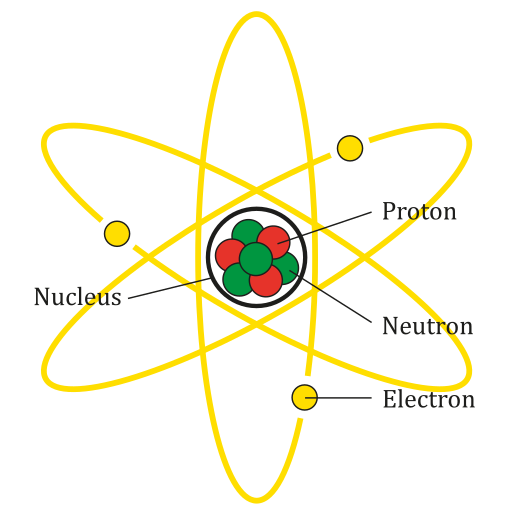
An atom is the smallest particle of an element that still has the properties of that element. Every substance is composed of atoms. Atoms are extremely small, typically about a ten-billionth of a metre in diametre. However, atoms do not have well-defined boundaries, as suggested by the atomic model shown below.
Every atom is composed of a central area — called the nucleus — and one or more subatomic particles called electrons, which move around the nucleus. The nucleus also consists of subatomic particles. It contains one or more protons and typically a similar number of neutrons. The number of protons in the nucleus determines the type of element an atom represents. An atom of hydrogen, for example, contains just one proton. Atoms of the same element may have different numbers of neutrons in the nucleus. Atoms of the same element with the same number of protons — but different numbers of neutrons — are called isotopes.
Protons have a positive electric charge and neutrons have no electric charge. Virtually all of an atom's mass is in the protons and neutrons in the nucleus. Electrons surrounding the nucleus have almost no mass, as well as a negative electric charge. If the number of protons and electrons in an atom are equal, then an atom is electrically neutral, because the positive and negative charges cancel each other out. If an atom has more or fewer electrons than protons, then it has an overall negative or positive charge, respectively, and it is called an ion.
The negatively-charged electrons of an atom are attracted to the positively-charged protons in the nucleus by a force called electromagnetic force, for which opposite charges attract. Electromagnetic force between protons in the nucleus causes these subatomic particles to repel each other, because they have the same charge. However, the protons and neutrons in the nucleus are attracted to each other by a different force, called nuclear force, which is usually stronger than the electromagnetic force. Nuclear force repels the positively-charged protons from each other.
Periodic Table of the Elements
There are almost 120 known elements. As you can see in the Periodic Table of the Elements shown below, the majority of elements are metals. Examples of metals are iron (Fe) and copper (Cu). Metals are shiny and good conductors of electricity and heat. Nonmetal elements are far fewer in number. They include hydrogen (H) and oxygen (O). They lack the properties of metals.
The periodic table of the elements arranges elements in groups based on their properties. The element most important to life is carbon (C). Find carbon in the table. What type of element is it: metal or nonmetal?
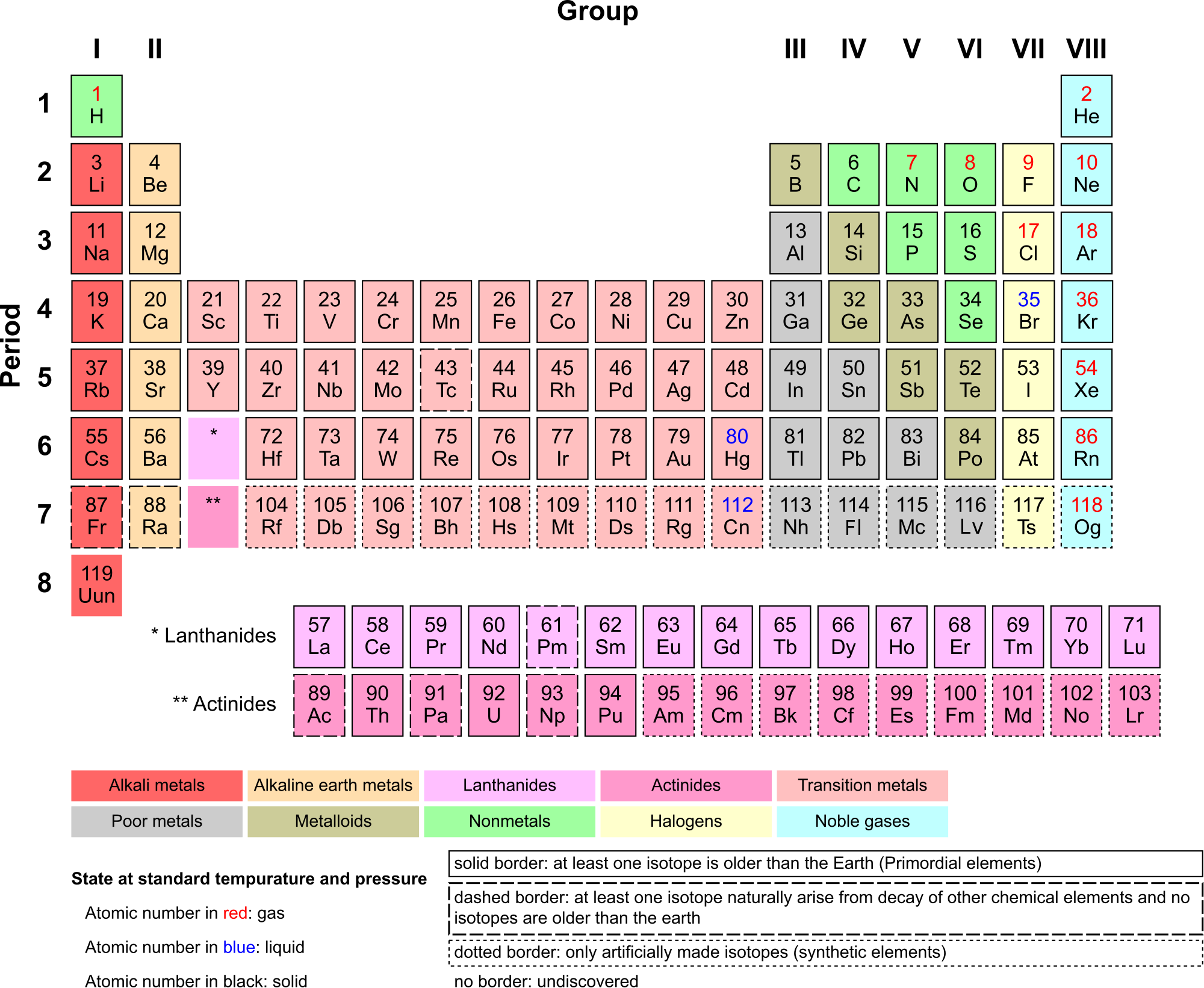
Compounds and Molecules
A compound is a unique substance that consists of two or more elements combined in fixed proportions. This means that the composition of a compound is always the same. The smallest particle of most compounds in living things is called a molecule.
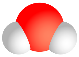
Consider water as an example. A molecule of water always contains one atom of oxygen and two atoms of hydrogen. The composition of water is expressed by the chemical formula H2O. A model of a water molecule is shown in Figure 3.2.4.
What causes the atoms of a water molecule to “stick” together? The answer is chemical bonds. A chemical bond is a force that holds together the atoms of molecules. Bonds in molecules involve the sharing of electrons among atoms. New chemical bonds form when substances react with one another. A chemical reaction is a process that changes some chemical substances into others. A chemical reaction is needed to form a compound, and another chemical reaction is needed to separate the substances in that compound.
3.2 Summary
- All matter consists of chemical substances. A chemical substance has a definite composition which is consistent throughout. A chemical substance may be either an element or a compound.
- An element is a pure substance that cannot be broken down into other types of substances.
- An atom is the smallest particle of an element that still has the properties of that element. Atoms, in turn, are composed of subatomic particles, including negative electrons, positive protons, and neutral neutrons. The number of protons in an atom determines the element it represents.
- Atoms have equal numbers of electrons and protons, so they have no charge. Ions are atoms that have lost or gained electrons, and as a result have either a positive or negative charge. Atoms with the same number of protons — but different numbers of neutrons — are called isotopes.
- There are almost 120 known elements. The majority of elements are metals. A smaller number are nonmetals. The latter include carbon, hydrogen, and oxygen.
- A compound is a substance that consists of two or more elements in a unique composition. The smallest particle of a compound is called a molecule. Chemical bonds hold together the atoms of molecules. Compounds can form only in chemical reactions, and they can break down only in other chemical reactions.
3.2 Review Questions
-
- What is an element? Give three examples.
- Define compound. Explain how compounds form.
- Compare and contrast atoms and molecules.
- The compound called water can be broken down into its constituent elements by applying an electric current to it. What ratio of elements is produced in this process?
- Relate ions and isotopes to elements and atoms.
- What is the most important element to life?
- Iron oxide is often known as rust — the reddish substance you might find on corroded metal. The chemical formula for this type of iron oxide is Fe2O3. Answer the following questions about iron oxide and briefly explain each answer.
- Is iron oxide an element or a compound?
- Would one particle of iron oxide be considered a molecule or an atom?
- Describe the relative proportion of atoms in iron oxide.
- What causes the Fe and O to stick together in iron oxide?
- Is iron oxide made of metal atoms, metalloid atoms, nonmetal atoms, or a combination of any of these?
- 14C is an isotope of carbon used in the radiocarbon dating of organic material. The most common isotope of carbon is 12C. Do you think 14C and 12C have different numbers of neutrons or protons? Explain your answer.
- Explain why ions have a positive or negative charge.
- Name the three subatomic particles described in this section.
3.2 Explore More
https://www.youtube.com/watch?v=yQP4UJhNn0I&feature=emb_logo
Just how small is an atom? TED-Ed, 2012
Attributions
Figure 3.2.1
Man Sitting, by Gregory Culmer, on Unsplash, is used under the Unsplash license (https://unsplash.com/license).
Figure 3.2.2
Lithium Atom diagram, by AG Caesar, is used under a CC BY-SA 4.0 (https://creativecommons.org/licenses/by-sa/4.0/deed.en)
Figure 3.2.3
Periodic Table Armtuk3, by Armtuk, is used under a CC BY-SA 3.0 (https://creativecommons.org/licenses/by-sa/3.0/) license.
Figure 3.2.4
Water molecule, by Sakurambo, is released into the public domain (https://en.wikipedia.org/wiki/Public_domain).
References
TED-Ed. (2012, April 16). Just how small is an atom. YouTube. https://www.youtube.com/watch?v=yQP4UJhNn0I&feature=youtu.be
Created by: CK-12/Adapted by Christine Miller
Figure 5.14.1 Collage of Diverse Faces.
This collage shows some of the variation in human skin colour, which can range from very light to very dark, with every possible gradation in between. As you might expect, the skin color trait has a more complex genetic basis than just one gene with two alleles, which is the type of simple trait that Mendel studied in pea plants. Like skin color, many other human traits have more complicated modes of inheritance than Mendelian traits. Such modes of inheritance are called non-Mendelian inheritance, and they include inheritance of multiple allele traits, traits with codominance or incomplete dominance, and polygenic traits, among others. All of these modes are described below.
Multiple Allele Traits
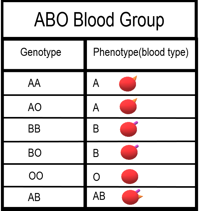
The majority of human genes are thought to have more than two normal versions, or alleles. Traits controlled by a single gene with more than two alleles are called multiple allele traits. An example is ABO blood type. Your blood type refers to which of certain proteins called antigens are found on your red blood cells. There are three common alleles for this trait, which are represented by the letters A, B, and O.
As shown in the table there are six possible ABO genotypes, because the three alleles, taken two at a time, result in six possible combinations. The A and B alleles are dominant to the O allele. As a result, both AA and AO genotypes have the same phenotype, with the A antigen in their blood (type A blood). Similarly, both BB and BO genotypes have the same phenotype, with the B antigen in their blood (type B blood). No antigen is associated with the O allele, so people with the OO genotype have no antigens for ABO blood type in their blood (type O blood).
Codominance
Look at the genotype AB in the ABO blood group table. Alleles A and B for ABO blood type are neither dominant nor recessive to one another. Instead, they are codominant. Codominance occurs when two alleles for a gene are expressed equally in the phenotype of heterozygotes. In the case of ABO blood type, AB heterozygotes have a unique phenotype, with both A and B antigens in their blood (type AB blood).
Incomplete Dominance
Another relationship that may occur between alleles for the same gene is incomplete dominance. This occurs when the dominant allele is not completely dominant. In this case, an intermediate phenotype results in heterozygotes who inherit both alleles. Generally, this happens when the two alleles for a given gene both produce proteins, but one protein is not functional. As a result, the heterozygote individual produces only half the amount of normal protein as is produced by an individual who is homozygous for the normal allele.
An example of incomplete dominance in humans is Tay Sachs disease. The normal allele for the gene in this case produces an enzyme that is responsible for breaking down lipids. A defective allele for the gene results in the production of a nonfunctional enzyme. Heterozygotes who have one normal and one defective allele produce half as much functional enzyme as the normal homozygote, and this is enough for normal development. Homozygotes who have only defective allele, however, produce only nonfunctional enzyme. This leads to the accumulation of lipids in the brain starting in utero, which causes significant brain damage. Most individuals with Tay Sachs disease die at a young age, typically by the age of five years.
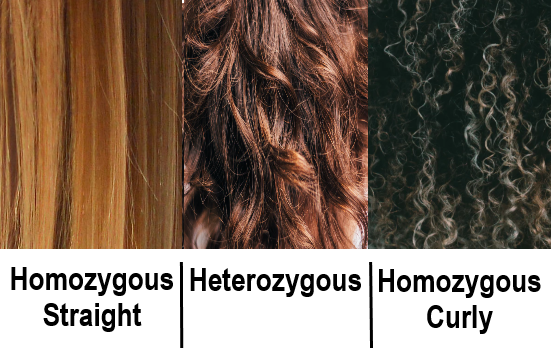
Another good example of incomplete dominance in humans is hair type. There are genes for straight and curly hair, and if an individual is heterozygous, they will typically have the phenotype of wavy hair.
Polygenic Traits
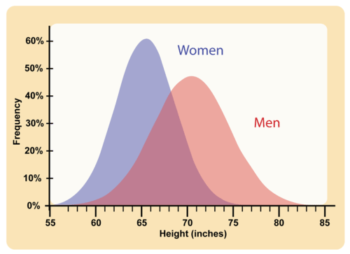
Many human traits are controlled by more than one gene. These traits are called polygenic traits. The alleles of each gene have a minor additive effect on the phenotype. There are many possible combinations of alleles, especially if each gene has multiple alleles. Therefore, a whole continuum of phenotypes is possible.
An example of a human polygenic trait is adult height. Several genes, each with more than one allele, contribute to this trait, so there are many possible adult heights. One adult’s height might be 1.655 m (5.430 feet), and another adult’s height might be 1.656 m (5.433 feet). Adult height ranges from less than 5 feet to more than 6 feet, with males, on average, being somewhat taller than females. The majority of people fall near the middle of the range of heights for their sex, as shown in Figure 5.14.4.
Environmental Effects on Phenotype
Many traits are affected by the environment, as well as by genes. This may be especially true for polygenic traits. Adult height, for example, might be negatively impacted by poor diet or childhood illness. Skin color is another polygenic trait. There is a wide range of skin colors in people worldwide. In addition to differences in genes, differences in exposure to ultraviolet (UV) light cause some variation. As shown in Figure 5.14.5, exposure to UV light darkens the skin.
Pleiotropy
Some genes affect more than one phenotypic trait. This is called pleiotropy. There are numerous examples of pleiotropy in humans. They generally involve important proteins that are needed for the normal development or functioning of more than one organ system. An example of pleiotropy in humans occurs with the gene that codes for the main protein in collagen, a substance that helps form bones. This protein is also important in the ears and eyes. Mutations in the gene result in problems not only in bones, but also in these sensory organs, which is how the gene's pleiotropic effects were discovered.
Another example of pleiotropy occurs with sickle cell anemia. This recessive genetic disorder occurs when there is a mutation in the gene that normally encodes the red blood cell protein called hemoglobin. People with the disorder have two alleles for sickle cell hemoglobin, so named for the sickle shape (pictured in Figure 5.14.6) that their red blood cells take on under certain conditions (like physical exertion). The sickle-shaped red blood cells clog small blood vessels, causing multiple phenotypic effects, including stunting of physical growth, certain bone deformities, kidney failure, and strokes.
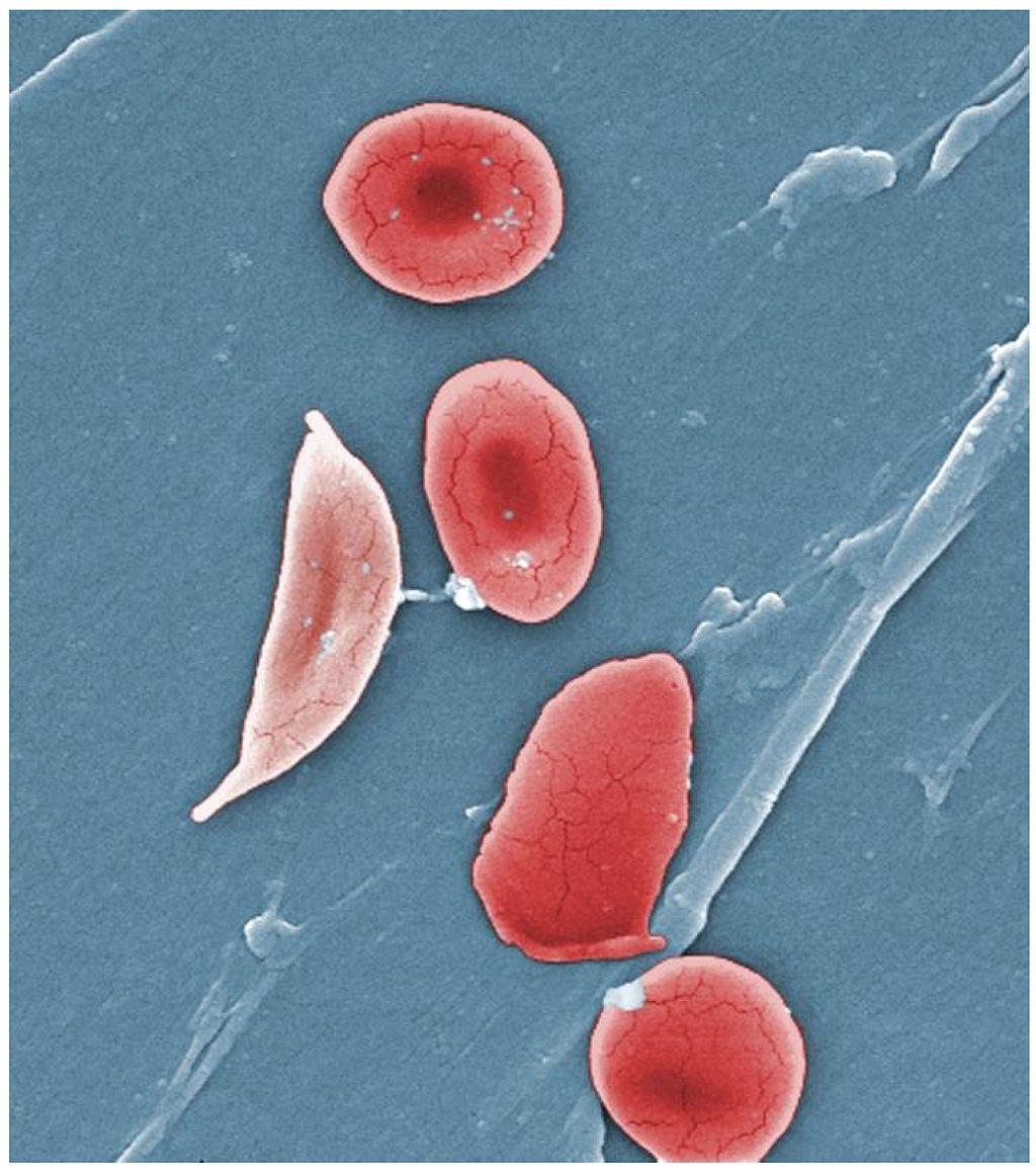
Epistasis
Some genes affect the expression of other genes. This is called epistasis. Epistasis is similar to dominance, except that it occurs between different genes, rather than between different alleles for the same gene.
Albinism is an example of epistasis. A person with albinism has virtually no pigment in the skin. The condition occurs due to an entirely different gene than the genes that encode skin color. Albinism occurs because a protein called tyrosinase, which is needed for the production of normal skin pigment, is not produced, due to a gene mutation. If an individual has the albinism mutation, he or she will not have any skin pigment, regardless of the skin color genes that were inherited.
Feature: My Human Body
Do you know your ABO blood type? In an emergency, knowing this valuable piece of information could possibly save your life. If you ever need a blood transfusion, it is vital that you receive blood that matches your own blood type. Why? If the blood transfused into your body contains an antigen that your own blood does not contain, antibodies in your blood plasma (the liquid part of your blood) will recognize the antigen as foreign to your body and cause a reaction called agglutination. In this reaction, the transfused red blood cells will clump together. The agglutination reaction is serious and potentially fatal.
Knowing the antigens and antibodies present in each of the ABO blood types will help you understand which type(s) of blood you can safely receive if you ever need a transfusion. This information is shown in Figure 5.14.7 for all of the ABO blood types. If you have blood type A, this means that your red blood cells have the A antigen and that your blood plasma contains anti-B antibodies. If you were to receive a transfusion of type B or type AB blood, both of which have the B antigen, your anti-B antibodies would attack the transfused red blood cells, causing agglutination.
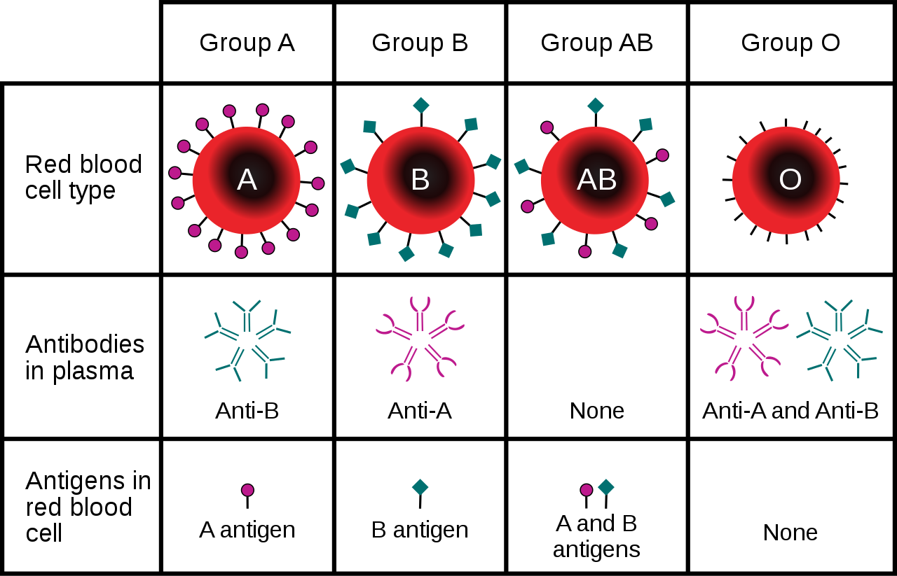
You may have heard that people with blood type O are called "universal donors," and that people with blood type AB are called universal recipients. People with type O blood have neither A nor B antigens in their blood, so if their blood is transfused into someone with a different ABO blood type, it causes no immune reaction, meaning they can donate blood to anyone. On the other hand, people with type AB blood have no anti-A or anti-B antibodies in their blood, so they can receive a transfusion of blood from anyone. Which blood type(s) can safely receive a transfusion of type AB blood, and which blood type(s) can be safely received by those with type O blood?
5.14 Summary
- Non-Mendelian inheritance refers to the inheritance of traits that have a more complex genetic basis than one gene with two alleles and complete dominance.
- Multiple allele traits are controlled by a single gene with more than two alleles. An example of a human multiple allele trait is ABO blood type, for which there are three common alleles: A, B, and O.
- Codominance occurs when two alleles for a gene are expressed equally in the phenotype of heterozygotes. A human example of codominance also occurs in the ABO blood type, in which the A and B alleles are codominant.
- Incomplete dominance is the case in which the dominant allele for a gene is not completely dominant to a recessive allele for the gene, so an intermediate phenotype occurs in heterozygotes who inherit both alleles. A human example of incomplete dominance is Tay Sachs disease, in which heterozygotes produce half as much functional enzyme as normal homozygotes.
- Polygenic traits are controlled by more than one gene, each of which has a minor additive effect on the phenotype. This results in a whole continuum of phenotypes. Examples of human polygenic traits include skin color and adult height.
- Many traits are affected by the environment, as well as by genes. This may be especially true for polygenic traits. Skin color, for example, may be affected by exposure to UV light, and adult stature may be affected by diet or childhood disease.
- Pleiotropy refers to the situation in which a gene affects more than one phenotypic trait. A human example of pleiotropy occurs with sickle cell anemia. People who inherit two recessive alleles for this disorder have abnormal red blood cells and may exhibit multiple other phenotypic effects, such as stunting of physical growth, kidney failure, and strokes.
- Epistasis is the situation in which one gene affects the expression of other genes. An example of epistasis is albinism, in which the albinism mutation negates the expression of skin color genes.
5.14 Review Questions
- What is non-Mendelian inheritance?
-
- Explain why the human ABO blood group is an example of a multiple allele trait with codominance.
- What is incomplete dominance? Give an example of this type of non-Mendelian inheritance in humans.
- Explain the genetic basis of human skin color.
- How can the human trait of adult height be influenced by the environment?
- Define pleiotropy, and give a human example.
- Compare and contrast epistasis and dominance.
- What is the difference between pleiotropy and epistasis?
5.14 Explore More
https://www.youtube.com/watch?time_continue=1&v=YJHGfbW55l0&feature=emb_logo
Incomplete Dominance, Codominance, Polygenic Traits, and Epistasis!,
Amoeba Sisters, 2015.
https://www.youtube.com/watch?v=-4vsio8TZrU&feature=emb_logo
Non-Mendelian Genetics, Teacher's Pet, 2015.
Attributes
Figure 5.14.1
- Woman's Face from Iran by Omid Armin on Unsplash is used under the Unsplash License (https://unsplash.com/license).
- Woman Wearing Black Coat by Anastasiya Pavlova on Unsplash is used under the Unsplash License (https://unsplash.com/license).
- Dark haired man, Queretaro, México by Leonel Hernandez Arteaga on Unsplash is used under the Unsplash License (https://unsplash.com/license). <not found on Unsplash>
- Man in White V-Neck T-Shirt (self) by Joseph Gonzalez on Unsplash is used under the Unsplash License (https://unsplash.com/license).
- Natural Redhead in Brazil by Gabriel Silvério on Unsplash is used under the Unsplash License (https://unsplash.com/license).
- Dark-Skinned Woman with Large White Rose by Oladimeji Oduns on Unsplash is used under the Unsplash License (https://unsplash.com/license).
Figure 5.14.2
ABO Blood Types Per Genotype by Christine Miller is released into the public domain (https://en.wikipedia.org/wiki/Public_domain).
Figure 5.14.3
Three Phenotypes of Hair Based on Inheritance/ Incomplete Dominance Hair by Christine Miller is released into the public domain (https://en.wikipedia.org/wiki/Public_domain).
Figure 5.14.4
Average height /Human Adult Height by CK-12 Foundation is used under a CC BY 3.0 (https://creativecommons.org/licenses/by-nc/3.0/) license.
 ©CK-12 Foundation Licensed under
 ![]() • Terms of Use • Attribution
• Terms of Use • Attribution
Figure 5.14.5
Tan lines by katiebordner on Flickr is used under a CC BY 2.0 (https://creativecommons.org/licenses/by/2.0/) license.
Figure 5.14.6
Sickle cell anemia by OpenStax College on Wikimedia Commons is used under a CC BY 3.0 (https://creativecommons.org/licenses/by/3.0) ©
Figure 5.14.7
ABO_blood_type.svg by InvictaHOG on Wikimedia Commons is in the public domain (https://en.wikipedia.org/wiki/Public_domain).
References
Amoeba Sisters. (2015, May 25). Incomplete dominance, codominance, polygenic traits, and epistasis! YouTube. https://www.youtube.com/watch?v=YJHGfbW55l0
Betts, J. G., Young, K.A., Wise, J.A., Johnson, E., Poe, B., Kruse, D.H., Korol, O., Johnson, J.E., Womble, M., DeSaix, P. (2013, April 25). Figure 18.9 Sickle cells [digital image]. In Anatomy and Physiology. OpenStax. https://openstax.org/books/anatomy-and-physiology/pages/18-3-erythrocytes
Brainard, J/ CK-12 Foundation. (2016). Figure 2 Human adult height [digital image]. In CK-12 College Human Biology (Section 5.13) [online Flexbook]. CK12.org. https://www.ck12.org/book/ck-12-college-human-biology/section/5.13/
Mayo Clinic Staff. (n.d.). Tay-Sachs disease [online article]. MayoClinic.org. https://www.mayoclinic.org/diseases-conditions/tay-sachs-disease/symptoms-causes/syc-20378190
Mayo Clinic Staff. (n.d.). Sickle cell anemia [online article]. MayoClinic.org. https://www.mayoclinic.org/diseases-conditions/sickle-cell-anemia/symptoms-causes/syc-20355876
Teacher's Pet. (2015, January 25). Non-mendelian genetics. YouTube. https://www.youtube.com/watch?v=-4vsio8TZrU
Created by: CK-12/Adapted by: Christine Miller
Defining Science
Science is a distinctive way of gaining knowledge about the natural world that starts with a question and then tries to answer the question using evidence and logic. It is an exciting exploration of all the whys and hows that any curious person might ask about the world. You can be part of that exploration! Besides your curiosity, all you need is a basic understanding of how scientists think and how science is done. In this concept, you'll learn how to think like a scientist.
Thinking Like a Scientist
Thinking like a scientist rests on certain underlying assumptions. Scientists assume that:
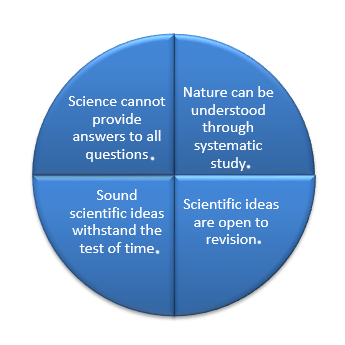
Nature Is Understandable
Scientists think of nature as a single system controlled by natural laws. By discovering natural laws, scientists strive to increase their understanding of the natural world. Laws of nature are expressed as scientific laws. A scientific law is a statement that describes what always happens under certain conditions in nature.
Scientific Ideas Are Open to Change
Science is both a process and body of knowledge. Scientific knowledge is generated through systematic processes, such as observation and experimentation. Scientists are always testing and revising their ideas, and as new observations are made, existing ideas may be challenged. Ideas may be replaced with new ideas that better fit the facts, but more often, existing ideas are simply revised. Through many new discoveries over time, scientists gradually build an increasingly accurate and detailed understanding of the natural world.
Scientific Knowledge May Be Long Lasting
Many scientific ideas have stood the test of time. About 200 years ago, the scientist John Dalton proposed atomic theory — the theory that all matter is made of tiny particles called atoms. This theory is still valid today. During the two centuries since the theory was first proposed, scientists have learned a lot more about atoms and the even smaller particles that compose them. Nonetheless, the idea that all matter consists of atoms remains valid. There are many other examples of basic scientific ideas that have been tested repeatedly and proven sound. You will learn about many of them as you study human biology.
Not All Questions Can be Answered by Science
Science rests on evidence and logic, and evidence comes from observations. Therefore, science deals only with things that can be observed. An observation is anything that is detected through human senses or with instruments or measuring devices that extend human senses. Things that cannot be observed or measured by current means — such as supernatural beings or events — are outside the bounds of science. Consider these two questions about life on Earth:
- Did life on Earth evolve over time?
- How did life on Earth originate?
The first question can be answered by science on the basis of scientific evidence (such as fossils and logical arguments). The second question could be a matter of belief, but no evidence can be gathered to support or refute it. Therefore, it is outside the realm of science.
1.3 Summary
- Science is a distinctive way of gaining knowledge about the natural world that tries to answer questions using evidence and logic.
- Scientists assume that nature can be understood through systematic study.
- Scientific ideas are open to revision.
- Sound scientific ideas withstand the test of time.
- Science cannot provide answers to all of our questions.
1.3 Review Questions
- Define science.
- What is the general goal of science?
-
- Identify four basic assumptions that scientists make when they study the natural world.
- Do observations in science have to be made by the naked eye? Can you think of a way in which scientists might be able to make observations about something they cannot directly see?
- If something cannot be observed, can it be tested scientifically? Explain your reasoning.
- Scientific knowledge builds upon itself. Give an example of a scientific idea from the reading where the initial idea developed further as science advanced.
- Discuss this statement: “Scientific ideas are always changing, so they can't be trusted.” Do you think this is true?
- Why do you think that scientific knowledge expands as technology becomes more advanced?
1.3 Explore More
https://youtu.be/3nAETHZTObk
Nature of Science with the Ameoba Sisters, 2019.
References
Amoeba Sisters. (2019, Jun 6). Nature of science with Ameoba Sisters. YouTube. https://www.youtube.com/watch?v=3nAETHZTObk
Wikipedia contributors. (2020, July 25). John Dalton. In Wikipedia. https://en.wikipedia.org/w/index.php?title=John_Dalton&oldid=969425891
Created by CK-12 Foundation/Adapted by Christine Miller

Art in a Cup
Who knew that a cup of coffee could also be a work of art? A talented barista can make coffee look as good as it tastes. If you are a coffee drinker, you probably know that coffee can also affect your mental state. It can make you more alert, and it may improve your concentration. That’s because the caffeine in coffee is a psychoactive drug. In fact, caffeine is the most widely consumed psychoactive substance in the world. In North America, for example, 90 per cent of adults consume caffeine daily.
What Are Psychoactive Drugs?
Psychoactive drugs are substances that change the function of the brain and result in alterations of mood, thinking, perception, and/or behavior. Psychoactive drugs may be used for many purposes, including therapeutic, ritual, or recreational purposes. Besides caffeine, other examples of psychoactive drugs include cocaine, LSD, alcohol, tobacco, codeine, and morphine. Psychoactive drugs may be legal prescription medications (codeine and morphine), legal nonprescription drugs (alcohol and tobacco), or illegal drugs (cocaine and LSD).
Cannabis (or marijuana) is also a psychoactive drug that while illegal in many countries is legal for use in Canada by individuals over the age of 19 years. Legal prescription medications (such as opioids) are also used illegally by increasingly large numbers of people. Some legal drugs, such as alcohol and nicotine, are readily available almost everywhere, as illustrated by the images below.
Figure 8.8.2 These psychoactive drugs are legal and accessible almost anywhere.
Classes of Psychoactive Drugs
Psychoactive drugs are divided into different classes based on their pharmacological effects. Several classes are listed below, along with examples of commonly used drugs in each class.
- Stimulants are drugs that stimulate the brain and increase alertness and wakefulness. Examples of stimulants include caffeine, nicotine, cocaine, and amphetamines (such as Adderall).
- Depressants are drugs that calm the brain, reduce anxious feelings, and induce sleepiness. Examples of depressants include ethanol (in alcoholic beverages) and opioids, such as codeine and heroin.
- Anxiolytics are drugs that have a tranquilizing effect and inhibit anxiety. Examples of anxiolytic drugs include benzodiazepines (such as diazepam/Valium), barbiturates (such as phenobarbital), opioids, and antidepressant drugs (such as sertraline/Zoloft).
- Euphoriants are drugs that bring about a state of euphoria, or intense feelings of well-being and happiness. Examples of euphoriants include the so-called "club drug" MDMA (ecstasy), amphetamines, ethanol, and opioids (such as morphine).
- Hallucinogens are drugs that can cause hallucinations and other perceptual anomalies. They also cause subjective changes in thoughts, emotions, and consciousness. Examples of hallucinogens include LSD, mescaline, nitrous oxide, and psilocybin.
- Empathogens are drugs that produce feelings of empathy, or sympathy with other people. Examples of empathogens include amphetamines and MDMA.
Many psychoactive drugs have multiple effects, so they may be placed in more than one class. One example is MDMA, pictured below, which may act both as a euphoriant and as an empathogen. In some people, MDMA may also have stimulant or hallucinogenic effects. As of 2016, MDMA had no accepted medical uses, but it was undergoing testing for use in the treatment of post-traumatic stress disorder and certain other types of anxiety disorders.
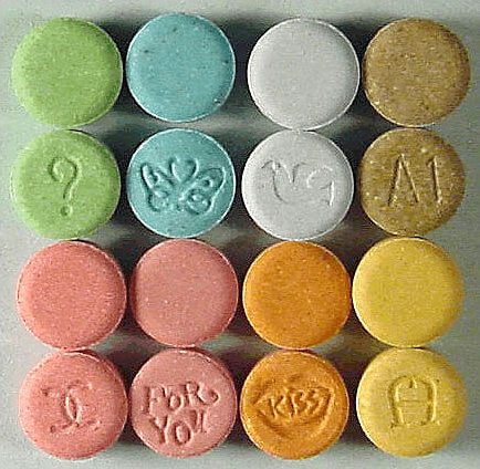
Mechanisms of Action
Psychoactive drugs generally produce their effects by affecting brain chemistry, which in turn may cause changes in a person’s mood, thinking, perception, and behavior. Each drug tends to have a specific action on one or more neurotransmitters or neurotransmitter receptors in the brain. Generally, they act as either agonists or antagonists.
- Agonists are drugs that increase the activity of particular neurotransmitters. They might act by promoting the synthesis of the neurotransmitters, reducing their reuptake from synapses, or mimicking their action by binding to receptors for the neurotransmitters.
- Antagonists are drugs that decrease the activity of particular neurotransmitters. They might act by interfering with the synthesis of the neurotransmitters or by blocking their receptors so the neurotransmitters cannot bind to them.
Consider the example of the neurotransmitter GABA. This is one of the most common neurotransmitters in the brain, and it normally has an inhibitory effect on cells. GABA agonists — which increase its activity — include ethanol, barbiturates, and benzodiazepines, among other psychoactive drugs. All of these drugs work by promoting the activity of GABA receptors in the brain.
Uses of Psychoactive Drugs
You may have been prescribed psychoactive drugs by your doctor. For example, your doctor may have prescribed you an opioid drug, such as codeine for pain (most likely in the form of Tylenol with added codeine). Chances are you also use nonprescription psychoactive drugs (like caffeine) for mental alertness. These are just two of the many possible uses of psychoactive drugs.
Medical Uses
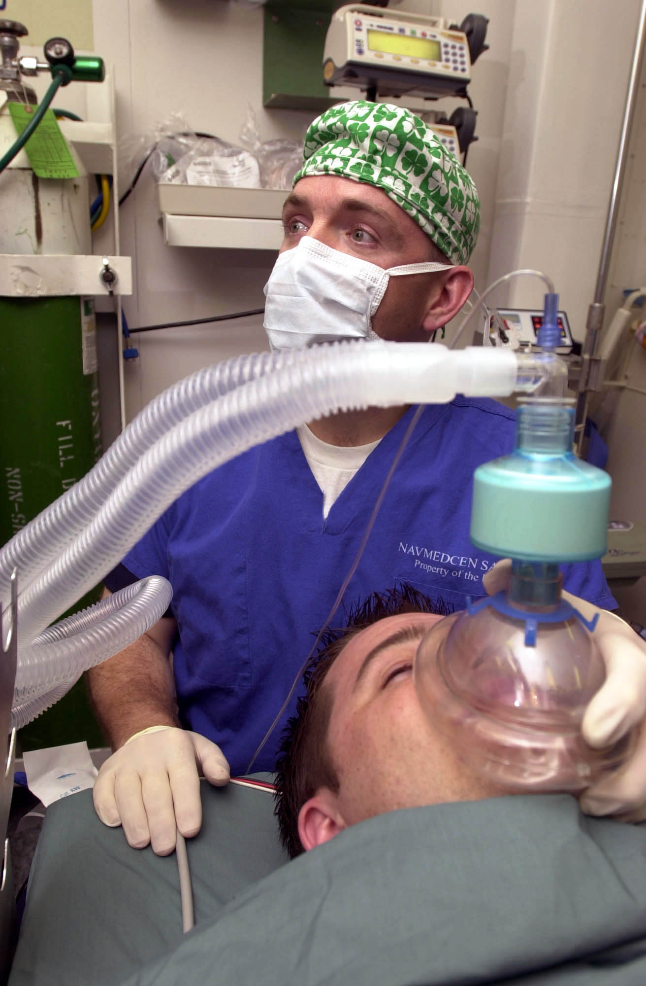
General anesthesia is one use of psychoactive drugs in medicine. With general anesthesia, pain is blocked and unconsciousness is induced. General anesthetics are most often used during surgical procedures and may be administered in gaseous form, as in Figure 8.8.4. General anesthetics include the drugs halothane and ketamine. Other psychoactive drugs are used to manage pain without affecting consciousness. They may be prescribed either for acute pain in cases of trauma (such as broken bones) or for chronic pain caused by arthritis, cancer, or fibromyalgia. Most often, the drugs used for pain control are opioids, such as morphine and codeine.
Many psychiatric disorders are also managed with psychoactive drugs. Antidepressants like sertraline, for example, are used to treat depression, anxiety, and eating disorders. Anxiety disorders may also be treated with anxiolytics, such as buspirone and diazepam. Stimulants (such as amphetamines) are used to treat attention deficit disorder. Antipsychotics (such as clozapine and risperidone) — as well as mood stabilizers, such as lithium — are used to treat schizophrenia and bipolar disorder.
Ritual Uses

Certain psychoactive drugs, particularly hallucinogens, have been used for ritual purposes since prehistoric times. For example, Native Americans have used the mescaline-containing peyote cactus (pictured in Figure 8.8.5) for religious ceremonies for as long as 5,700 years. In prehistoric Europe, the mushroom Amanita muscaria, which contains a hallucinogenic drug called muscimol, was used for similar purposes. Various other psychoactive drugs — including jimsonweed, psilocybin mushrooms, and cannabis — have also been used for millennia, by various peoples, for ritual purposes.
Recreational Uses
The recreational use of psychoactive drugs generally has the purpose of altering one’s consciousness and creating a feeling of euphoria commonly called a “high.” Some of the drugs used most commonly for recreational purposes are cannabis, ethanol (alcohol), opioids, and stimulants (such as nicotine). Hallucinogens are also used recreationally, primarily for the alterations they cause in thinking and perception.
Some investigators have suggested that the urge to alter one’s state of consciousness is a universal human drive, similar to the drive to satiate thirst, hunger, or sexual desire. They think that this instinct is even present in children, who may attain an altered state by repetitive motions, such as spinning or swinging. Some nonhuman animals also exhibit a drive to experience altered states. They may consume fermented berries or fruit and become intoxicated. The way cats respond to catnip (see Figure 8.8.6) is another example.

Addiction, Dependence, and Rehabilitation
Psychoactive substances often bring about subjective changes that the user may find pleasant (euphoria) or advantageous (increased alertness). These changes are rewarding and positively reinforcing, so they have the potential for misuse, addiction, and dependence. Addiction refers to the compulsive use of a drug, despite negative consequences that such use may entail. Sustained use of an addictive drug may produce dependence on the drug. Dependence may be physical and/or psychological. It occurs when cessation of drug use produces withdrawal symptoms. Physical dependence produces physical withdrawal symptoms, which may include tremors, pain, seizures, or insomnia. Psychological dependence produces psychological withdrawal symptoms, such as anxiety, depression, paranoia, or hallucinations.
Rehabilitation for drug dependence and addiction typically involves psychotherapy, which may include both individual and group therapy. Organizations such as Alcoholics Anonymous (AA) and Narcotics Anonymous (NA) may also be helpful for people trying to recover from addiction. These groups are self-described as international mutual aid fellowships, and their primary purpose is to help addicts achieve and maintain sobriety. In some cases, rehabilitation is aided by the temporary use of psychoactive substances that reduce cravings and withdrawal symptoms without creating addiction themselves. The drug methadone, for example, is commonly used to treat heroin addiction.
Feature: Human Biology in the News
In North America, a lot of media attention is currently given to a rising tide of opioid addiction and overdose deaths. Opioids are drugs derived from the opium poppy or synthetic versions of such drugs. They include the illegal drug heroin, as well as prescription painkillers such as codeine, morphine, hydrocodone, oxycodone, and fentanyl. In 2016, fentanyl received wide media attention when it was announced that an accidental fentanyl overdose was responsible for the death of music icon Prince. Fentanyl is an extremely strong and dangerous drug, said to be 50 to 100 times stronger than morphine, making risk of overdose death from fentanyl very high.
The dramatic increase in opioid addiction and overdose deaths has been called an opioid epidemic. It is considered to be the worst drug crisis in Canadian history. Consider the following facts:
- In 2016, there were almost 2,500 opioid-related deaths in Canada — almost 7 per day.
- The number of prescriptions written for opioids quadrupled between 1999 and 2010. If you have been prescribed codeine, fentanyl, morphine, oxycodone, hydromorphone or medical heroin, then you have been prescribed an opiate.
- There are many long-term health effects of using opioids, which include:
- Increased tolerance to the drug.
- Liver damage.
- Substance use disorder or addiction.
Doctors, public health professionals, and politicians have all called for new policies, funding, programs, and laws to address the opioid epidemic. Changes that have already been made include a shift from criminalizing to medicalizing the problem, more treatment programs, and more widespread distribution and use of the opioid-overdose antidote naloxone (Narcan). Opioids can slow or stop a person's breathing, which is what usually causes overdose deaths. Naloxone helps the person wake up and keeps them breathing until emergency medical treatment can be provided.
What, if anything, will work to stop the opioid epidemic in Canada and the United States? Keep watching the news to find out.
8.8 Summary
- Psychoactive drugs are substances that change the function of the brain and result in alterations of mood, thinking, perception, and behavior. They include prescription medications (such as opioid painkillers), legal substances (such as nicotine and alcohol), and illegal drugs (such as LSD and heroin).
- Psychoactive drugs are divided into different classes according to their pharmacological effects. They include stimulants, depressants, anxiolytics, euphoriants, hallucinogens, and empathogens. Many psychoactive drugs have multiple effects, so they may be placed in more than one class.
- Psychoactive drugs generally produce their effects by affecting brain chemistry. Generally, they act either as agonists — which enhance the activity of particular neurotransmitters — or as antagonists, which decrease the activity of particular neurotransmitters.
- Psychoactive drugs are used for various purposes, including medical, ritual, and recreational purposes.
- Misuse of psychoactive drugs may lead to addiction, which is the compulsive use of a drug despite the negative consequences such use may entail. Sustained use of an addictive drug may produce physical or psychological dependence on the drug. Rehabilitation typically involves psychotherapy, and sometimes the temporary use of other psychoactive drugs.
8.8 Review Questions
- What are psychoactive drugs?
- Identify six classes of psychoactive drugs, along with an example of a drug in each class.
- Compare and contrast psychoactive drugs that are agonists and psychoactive drugs that are antagonists.
- Describe two medical uses of psychoactive drugs.
- Give an example of a ritual use of a psychoactive drug.
- Generally speaking, why do people use psychoactive drugs recreationally?
- Define addiction.
- Identify possible withdrawal symptoms associated with physical dependence on a psychoactive drug.
- Why might a person with a heroin addiction be prescribed the psychoactive drug methadone?
- The prescription drug Prozac inhibits the reuptake of the neurotransmitter serotonin, causing more serotonin to be present in the synapse. Prozac can elevate mood, which is why it is sometimes used to treat depression. Answer the following questions about Prozac:
- Is Prozac an agonist or an antagonist for serotonin? Explain your answer.
- Is Prozac a psychoactive drug? Explain your answer.
- Name three classes of psychoactive drugs that include opioids.
- True or False: All psychoactive drugs are either illegal or available by prescription only.
- True or False: Anxiolytics might be prescribed by a physician.
- Name two drugs that activate receptors for the neurotransmitter GABA. Why do you think these drugs generally have a depressant effect?
8.8 Explore More
https://www.youtube.com/watch?v=foLf5Bi9qXs
How does caffeine keep us awake? - Hanan Qasim, TED-Ed, 2017.
https://www.youtube.com/watch?v=8qK0hxuXOC8
How do drugs affect the brain? - Sara Garofalo, TED-Ed, 2017.
https://www.youtube.com/watch?v=Nlcr1jd_Tok
Is marijuana bad for your brain? - Anees Bahji, TED-Ed, 2019.
Attributions
Figure 8.8.1
Cappucino Art by drew-coffman-tZKwLRO904E [photo] by Drew Coffman on Unsplash is used under the Unsplash License (https://unsplash.com/license).
Figure 8.8.2
- 3804, Saint-Laurent, Montreal - Cannabis Culture shop by Exile on Ontario St (Montreal, Canada) on Wikimedia Commons is used under a CC BY SA 2.0 (https://creativecommons.org/licenses/by-sa/2.0/deed.en) license.
- Drive Through Cigarette Store by Cosmo Spacely on Flickr is used under CC BY-NC-SA 2.0 (https://creativecommons.org/licenses/by-nc-sa/2.0/) license.
- Franklin-Nicollet Liquors by Max Sparber on Flickr is used under a CC BY 2.0 (https://creativecommons.org/licenses/by/2.0/deed.en) license.
Figure 8.8.3
Ecstasy_monogram by Drug Enforcement Administration on Wikimedia Commons is in the public domain (https://en.wikipedia.org/wiki/Public_domain).
Figure 8.8.4
US Navy 030513-N-1577S-001 Lt. Cmdr. Joe Casey, Ship's Anesthetist, trains on anesthetic procedures with Hospital Corpsman 3rd Class Eric Wichman aboard USS Nimitz (CVN 68) by U.S. Navy photo by Photographer’s Mate Airman Timothy F. Sosais on Wikimedia Commons is in the public domain (https://en.wikipedia.org/wiki/Public_domain).
Figure 8.8.5
Peyote Lophophora_williamsii_pm by Peter A. Mansfeld on Wikimedia Commons is used under a CC BY 3.0 (https://creativecommons.org/licenses/by/3.0/deed.en) license.
Figure 8.8.6
Cat under effects of catnip/Self Indulgence by Katieb50 on Flickr is used under a CC BY 2.0 (https://creativecommons.org/licenses/by/2.0/deed.en) license.
Alcoholics Anonymous World Services, Inc. (n.d.). Regional correspondent U.S. and Canada [website]. https://www.aa.org/pages/en_US/regional-correspondent-us-and-canada
Belzak, L., & Halverson, J. (2018). The opioid crisis in Canada: a national perspective. La crise des opioïdes au Canada : une perspective nationale. Health promotion and chronic disease prevention in Canada : research, policy and practice, 38(6), 224–233. https://doi.org/10.24095/hpcdp.38.6.02
British Columbia Regional Service Committee of Narcotics Anonymous. (n.d.). Welcome to the B.C. region of N.A. [website]. https://www.bcrna.ca/
Centers for Disease Control and Prevention (CDC). (2011 November 4). Vital signs: overdoses of prescription opioid pain relievers—United States, 1999–2008. Morbidity and Mortality Weekly Report (MMWR),60(43):1487-1492. https://www.cdc.gov/mmwr/preview/mmwrhtml/mm6043a4.htm
TED-Ed. (2017, June 29). How do drugs affect the brain? - Sara Garofalo. YouTube. https://www.youtube.com/watch?v=8qK0hxuXOC8&feature=youtu.be
TED-Ed. (2017, July 17). How does caffeine keep us awake? - Hanan Qasim. YouTube. https://www.youtube.com/watch?v=foLf5Bi9qXs&feature=youtu.be
TED-Ed. (2019, December 2). Is marijuana bad for your brain? - Anees Bahji. YouTube. https://www.youtube.com/watch?v=Nlcr1jd_Tok&feature=youtu.be
Created by CK-12 Foundation/Adapted by Christine Miller
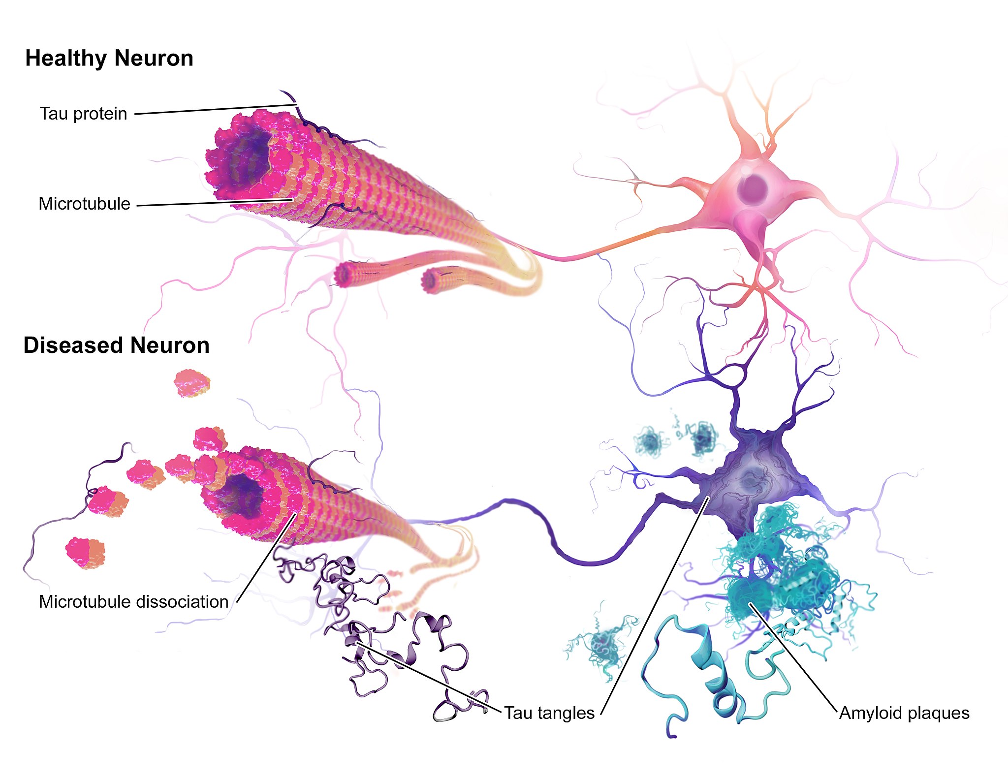
Case Study Conclusion: Fading Memory
The illustration above (Figure 8.9.1) shows some of the molecular and cellular changes that occur in Alzheimer’s disease (AD). Rosa was diagnosed with AD at the beginning of this chapter after experiencing memory problems and other changes in her cognitive functioning, mood, and personality. These abnormal changes in the brain include the development of amyloid plaques between brain cells and neurofibrillary tangles inside of neurons. These hallmark characteristics of AD are associated with the loss of synapses between neurons, and ultimately the death of neurons.
After reading this chapter, you should have a good appreciation for the importance of keeping neurons alive and communicating with each other at synapses. The nervous system coordinates all of the body’s voluntary and involuntary activities. It interprets information from the outside world through sensory systems, and makes appropriate responses through the motor system, through communication between the PNS and CNS. The brain directs the rest of the nervous system and controls everything from basic vital functions (such as heart rate and breathing) to high-level functions (such as problem solving and abstract thought). The nervous system can perform these important functions by generating action potentials in neurons in response to stimulation and sending messages between cells at synapses, typically using chemical neurotransmitter molecules. When neurons are not functioning properly, lose their synapses, or die, they cannot carry out the signaling essential for the proper functioning of the nervous system.
AD is a progressive neurodegenerative disease, meaning that the damage to the brain becomes more extensive as time goes on. The picture in Figure 8.9.2 illustrates how the damage progresses from before AD is diagnosed (preclinical AD), to mild and moderate AD, to severe AD.
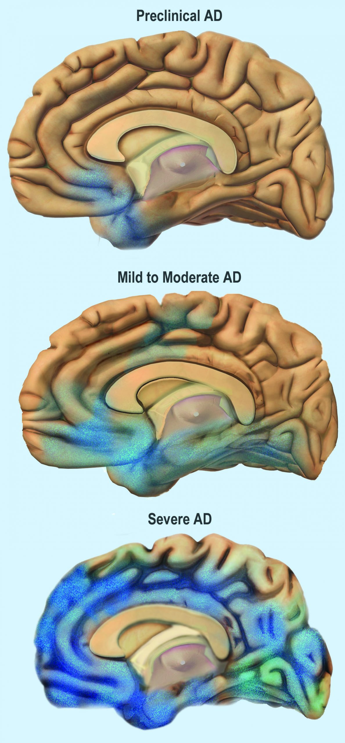
You can see that the damage starts in a relatively small location toward the bottom of the brain. One of the earliest brain areas to be affected by AD is the hippocampus. As you have learned, the hippocampus is important for learning and memory, which explains why many of Rosa’s symptoms of mild AD involve deficits in memory, such as trouble remembering where she placed objects, recent conversations, and appointments.
As AD progresses, more of the brain is affected, including areas involved in emotional regulation, social behavior, planning, language, spatial navigation, and higher-level thought. Rosa is beginning to show signs of problems in these areas, including irritability, lashing out at family members, getting lost in her neighborhood, problems finding the right words, putting objects in unusual locations, and difficulty in managing her finances. You can see that as AD progresses, damage spreads further across the cerebrum, which you now know controls conscious functions like reasoning, language, and interpretation of sensory stimuli. You can also see how the frontal lobe — which controls executive functions such as planning, self-control, and abstract thought — becomes increasingly damaged.
Increasing damage to the brain causes corresponding deficits in functioning. In moderate AD, patients have increased memory, language, and cognitive deficits, compared to mild AD. They may not recognize their own family members, and may wander and get lost, engage in inappropriate behaviors, become easily agitated, and have trouble carrying out daily activities such as dressing. In severe AD, much of the brain is affected. Patients usually cannot recognize family members or communicate, and they are often fully dependent on others for their care. They begin to lose the ability to control their basic functions, such as bladder control, bowel control, and proper swallowing. Eventually, AD causes death, usually as a result of this loss of basic functions.
For now, Rosa only has mild AD and is still able to function relatively well with care from her family. The medication her doctor gave her has helped improve some of her symptoms. It is a cholinesterase inhibitor, which blocks an enzyme that normally degrades the neurotransmitter acetylcholine. With more of the neurotransmitter available, more of it can bind to neurotransmitter receptors on postsynaptic cells. Therefore, this drug acts as an agonist for acetylcholine, which enhances communication between neurons in Rosa’s brain. This increase in neuronal communication can help restore some of the functions lost in early Alzheimer’s disease and may slow the progression of symptoms.
But medication such as this is only a short-term measure, and does not halt the progression of the underlying disease. Ideally, the damaged or dead neurons would be replaced by new, functioning neurons. Why does this not happen automatically in the body? As you have learned, neurogenesis is very limited in adult humans, so once neurons in the brain die, they are not normally replaced to any significant extent. Scientists, however, are studying the ways in which neurogenesis might be increased in cases of disease or injury to the brain. They are also investigating the possibility of using stem cell transplants to replace damaged or dead neurons with new neurons. But this research is in very early stages and is not currently a treatment for AD.
One promising area of research is in the development of methods to allow earlier detection and treatment of AD, given that the changes in the brain may actually start ten to 20 years before diagnosis of AD. A radiolabeled chemical called Pittsburgh Compound B (PiB) binds to amyloid plaques in the brain, and in the future, it may be used in conjunction with brain imaging techniques to detect early signs of AD. Scientists are also looking for biomarkers in bodily fluids (such as blood and cerebrospinal fluid) that might indicate the presence of AD before symptoms appear. Finally, researchers are also investigating possible early and subtle symptoms (such as changes in how people move or a loss of smell) to see whether they can be used to identify people who will go on to develop AD. This research is in the early stages, but the hope is that patients can be identified earlier, allowing for earlier and more effective treatment, as well as more planning time for families.
Scientists are also still trying to fully understand the causes of AD, which affects more than five million Americans. Some genetic mutations have been identified as contributors, but environmental factors also appear to be important. With more research into the causes and mechanisms of AD, hopefully a cure can be found, and people like Rosa can live a longer and better life.
Chapter 8 Summary
In this chapter, you learned about the human nervous system. Specifically, you learned that:
- The nervous system is the organ system that coordinates all of the body’s voluntary and involuntary actions by transmitting signals to and from different parts of the body. It has two major divisions: the central nervous system (CNS) and the peripheral nervous system (PNS).
- The CNS includes the brain and spinal cord.
- The PNS consists mainly of nerves that connect the CNS with the rest of the body. It has two major divisions: the somatic nervous system and the autonomic nervous system. These divisions control different types of functions, and often interact with the CNS to carry out these functions. The somatic system controls activities that are under voluntary control. The autonomic system controls activities that are involuntary.
- The autonomic nervous system is further divided into the sympathetic division (which controls the fight-or-flight response), the parasympathetic division (which controls most routine involuntary responses), and the enteric division (which provides local control for digestive processes).
- Signals sent by the nervous system are electrical signals called nerve impulses. They are transmitted by special, electrically excitable cells called neurons, which are one of two major types of cells in the nervous system.
- Neuroglia are the other major type of nervous system cells. There are many types of glial cells, and they have many specific functions. In general, neuroglia function to support, protect, and nourish neurons.
- The main parts of a neuron include the cell body, dendrites, and axon. The cell body contains the nucleus. Dendrites receive nerve impulses from other cells, and the axon transmits nerve impulses to other cells at axon terminals. A synapse is a complex membrane junction at the end of an axon terminal that transmits signals to another cell.
- Axons are often wrapped in an electrically-insulating myelin sheath, which is produced by oligodendrocytes or schwann cells, both of which are types of neuroglia. Electrical impulses called action potentials occur at gaps in the myelin sheath, called nodes of Ranvier, which speeds the conduction of nerve impulses down the axon.
- Neurogenesis, or the formation of new neurons by cell division, may occur in a mature human brain — but only to a limited extent.
- The nervous tissue in the brain and spinal cord consists of gray matter — which contains mainly unmyelinated cell bodies and dendrites of neurons — and white matter, which contains mainly myelinated axons of neurons. Nerves of the peripheral nervous system consist of long bundles of myelinated axons that extend throughout the body.
- There are hundreds of types of neurons in the human nervous system, but many can be classified on the basis of the direction in which they carry nerve impulses. Sensory neurons carry nerve impulses away from the body and toward the central nervous system, motor neurons carry them away from the central nervous system and toward the body, and interneurons often carry them between sensory and motor neurons.
- A nerve impulse is an electrical phenomenon that occurs because of a difference in electrical charge across the plasma membrane of a neuron.
- The sodium-potassium pump maintains an electrical gradient across the plasma membrane of a neuron when it is not actively transmitting a nerve impulse. This gradient is called the resting potential of the neuron.
- An action potential is a sudden reversal of the electrical gradient across the plasma membrane of a resting neuron. It begins when the neuron receives a chemical signal from another cell or some other type of stimulus. The action potential travels rapidly down the neuron’s axon as an electric current.
- A nerve impulse is transmitted to another cell at either an electrical or a chemical synapse. At a chemical synapse, neurotransmitter chemicals are released from the presynaptic cell into the synaptic cleft between cells. The chemicals travel across the cleft to the postsynaptic cell and bind to receptors embedded in its membrane.
- There are many different types of neurotransmitters. Their effects on the postsynaptic cell generally depend on the type of receptor they bind to. The effects may be excitatory, inhibitory, or modulatory in more complex ways. Both physical and mental disorders may occur if there are problems with neurotransmitters or their receptors.
- The CNS includes the brain and spinal cord. It is physically protected by bones, meninges, and cerebrospinal fluid. It is chemically protected by the blood-brain barrier.
- The brain is the control center of the nervous system and of the entire organism. The brain uses a relatively large proportion of the body’s energy, primarily in the form of glucose.
-
- The brain is divided into three major parts, each with different functions: the forebrain, the midbrain and the hindbrain.
- The forebrain includes the cerebrum, the thalamus, the hypothalamus, the hippocampus and the amygdala. The cerebrum is further divided into left and right hemispheres. Each hemisphere has four lobes: frontal, parietal, temporal, and occipital. Each lobe is associated with specific senses or other functions. The cerebrum has a thin outer layer called the cerebral cortex. Its many folds give it a large surface area. This is where most information processing takes place.
- The thalamus, hypothalamus, hippocampus and amygdala are all part of the limbic system which helps regulate memories, coordination and attention
- The brain is divided into three major parts, each with different functions: the forebrain, the midbrain and the hindbrain.
- The spinal cord is a tubular bundle of nervous tissues that extends from the head down the middle of the back to the pelvis. It functions mainly to connect the brain with the PNS. It also controls certain rapid responses called reflexes without input from the brain.
- A spinal cord injury may lead to paralysis (loss of sensation and movement) of the body below the level of the injury, because nerve impulses can no longer travel up and down the spinal cord beyond that point.
- The PNS consists of all the nervous tissue that lies outside of the CNS. Its main function is to connect the CNS to the rest of the organism.
- The tissues that make up the PNS are nerves and ganglia. Nerves are bundles of axons and ganglia are groups of cell bodies. Nerves are classified as sensory, motor, or a mix of the two.
- The PNS is not as well protected physically or chemically as the CNS, so it is more prone to injury and disease. PNS problems include injury from diabetes, shingles, and heavy metal poisoning. Two disorders of the PNS are Guillain-Barre syndrome and Charcot-Marie-Tooth disease.
- The human body has two major types of senses: special senses and general senses. Special senses have specialized sense organs and include vision (eyes), hearing (ears), balance (ears), taste (tongue), and smell (nasal passages). General senses are all associated with touch and lack special sense organs. Touch receptors are found throughout the body but particularly in the skin.
- All senses depend on sensory receptor cells to detect sensory stimuli and transform them into nerve impulses. Types of sensory receptors include mechanoreceptors (mechanical forces), thermoreceptors (temperature), nociceptors (pain), photoreceptors (light), and chemoreceptors (chemicals).
- Touch includes the ability to sense pressure, vibration, temperature, pain, and other tactile stimuli. The skin includes several different types of touch receptor cells.
- Vision is the ability to sense light and see. The eye is the special sensory organ that collects and focuses light, forms images, and changes them to nerve impulses. Optic nerves send information from the eyes to the brain, which processes the visual information and “tells” us what we are seeing.
- Common vision problems include myopia (nearsightedness), hyperopia (farsightedness), and presbyopia (age-related decline in close vision).
- Hearing is the ability to sense sound waves, and the ear is the organ that senses sound. It changes sound waves to vibrations that trigger nerve impulses, which travel to the brain through the auditory nerve. The brain processes the information and “tells” us what we are hearing.
- The ear is also the organ responsible for the sense of balance, which is the ability to sense and maintain an appropriate body position. The ears send impulses on head position to the brain, which sends messages to skeletal muscle via the peripheral nervous system. The muscles respond by contracting to maintain balance.
- Taste and smell are both abilities to sense chemicals. Taste receptors in taste buds on the tongue sense chemicals in food, and olfactory receptors in the nasal passages sense chemicals in the air. The sense of smell contributes significantly to the sense of taste.
- Psychoactive drugs are substances that change the function of the brain and result in alterations of mood, thinking, perception, and behavior. They include prescription medications (such as opioid painkillers), legal substances (such as nicotine and alcohol), and illegal drugs (such as LSD and heroin).
- Psychoactive drugs are divided into different classes according to their pharmacological effects. They include stimulants, depressants, anxiolytics, euphoriants, hallucinogens, and empathogens. Many psychoactive drugs have multiple effects, so they may be placed in more than one class.
- Psychoactive drugs generally produce their effects by affecting brain chemistry. Generally, they act either as agonists, which enhance the activity of particular neurotransmitters, or as antagonists, which decrease the activity of particular neurotransmitters.
- Psychoactive drugs are used for medical, ritual, and recreational purposes.
- Misuse of psychoactive drugs may lead to addiction, which is the compulsive use of a drug, despite its negative consequences. Sustained use of an addictive drug may produce physical or psychological dependence on the drug. Rehabilitation typically involves psychotherapy, and sometimes the temporary use of other psychoactive drugs.
In addition to the nervous system, there is another system of the body that is important for coordinating and regulating many different functions – the endocrine system. You will learn about the endocrine system in the next chapter.
Chapter 8 Review
- Imagine that you decide to make a movement. To carry out this decision, a neuron in the cerebral cortex of your brain (neuron A) fires a nerve impulse that is sent to a neuron in your spinal cord (neuron B). Neuron B then sends the signal to a muscle cell, causing it to contract, resulting in movement. Answer the following questions about this pathway.
- Which part of the brain is neuron A located in — the cerebellum, cerebrum, or brain stem? Explain how you know.
- The cell body of neuron A is located in a lobe of the brain that is involved in abstract thought, problem solving, and planning. Which lobe is this?
- Part of neuron A travels all the way down to the spinal cord to meet neuron B. Which part of neuron A travels to the spinal cord?
- Neuron A forms a chemical synapse with neuron B in the spinal cord. How is the signal from neuron A transmitted to neuron B?
- Is neuron A in the central nervous system (CNS) or peripheral nervous system (PNS)?
- The axon of neuron B travels in a nerve to a skeletal muscle cell. Is the nerve part of the CNS or PNS? Is this an afferent nerve or an efferent nerve?
- What part of the PNS is involved in this pathway — the autonomic nervous system or the somatic nervous system? Explain your answer.
- What are the differences between a neurotransmitter receptor and a sensory receptor?
-
- If a person has a stroke and then has trouble using language correctly, which hemisphere of their brain was most likely damaged? Explain your answer.
- Electrical gradients are responsible for the resting potential and action potential in neurons. Answer the following questions about the electrical characteristics of neurons.
- Define an electrical gradient, in the context of a cell.
- What is responsible for maintaining the electrical gradient that results in the resting potential?
- Compare and contrast the resting potential and the action potential.
- Where along a myelinated axon does the action potential occur? Why does it happen here?
- What does it mean that the action potential is “all-or-none?”
- Compare and contrast Schwann cells and oligodendrocytes.
- For the senses of smell and hearing, name their respective sensory receptor cells, what type of receptor cells they are, and what stimuli they detect.
- Nicotine is a psychoactive drug that binds to and activates a receptor for the neurotransmitter acetylcholine. Is nicotine an agonist or an antagonist for acetylcholine? Explain your answer.
Attributions
Figure 8.9.1
Alzheimers_Disease by BruceBlaus on Wikimedia Commons is used under a CC BY-SA 4.0 (https://creativecommons.org/licenses/by-sa/4.0/deed.en) license.
Figure 8.9.2
Alzheimer’s Disease stagess by NIH Image Gallery on Flickr is in the public domain (https://en.wikipedia.org/wiki/Public_domain).
Created by CK-12 Foundation/Adapted by Christine Miller
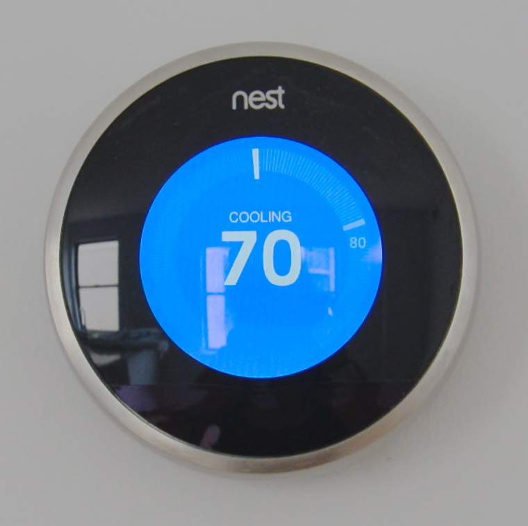
Steady as She Goes
This device (Figure 7.8.1) looks simple, but it controls a complex system that keeps a home at a steady temperature — it's a thermostat. The device shows the current temperature in the room, and also allows the occupant to set the thermostat to the desired temperature. A thermostat is a commonly cited model of how living systems — including the human body— maintain a steady state called homeostasis.
What Is Homeostasis?
Homeostasis is the condition in which a system (such as the human body) is maintained in a more or less steady state. It is the job of cells, tissues, organs, and organ systems throughout the body to maintain many different variables within narrow ranges compatible with life. Keeping a stable internal environment requires continually monitoring the internal environment and constantly making adjustments to keep things in balance.
Set Point and Normal Range
For any given variable, such as body temperature or blood glucose level, there is a particular set point that is the physiological optimum value. The set point for human body temperature, for example, is about 37 degrees C (98.6 degrees F). As the body works to maintain homeostasis for temperature or any other internal variable, the value typically fluctuates around the set point. Such fluctuations are normal, as long as they do not become too extreme. The spread of values within which such fluctuations are considered insignificant is called the normal range. In the case of body temperature, for example, the normal range for an adult is about 36.5 to 37.5 degrees C (97.7 to 99.5 degrees F).
A good analogy for set point, normal range, and maintenance of homeostasis is driving. When you are driving a vehicle on the road, you are supposed to drive in the centre of your lane — this is analogous to the set point. Sometimes, you are not driving in the exact centre of the lane, but you are still within your lines, so you are in the equivalent of the normal range. However, if you were to get too close to the centre line or the shoulder of the road, you would take action to correct your position. You'd move left if you were too close to the shoulder, or right if too close to the centre line — which is analogous to our next concept, negative feedback to maintain homeostasis.
Maintaining Homeostasis
Homeostasis is normally maintained in the human body by an extremely complex balancing act. Regardless of the variable being kept within its normal range, maintaining homeostasis requires at least four interacting components: stimulus, sensor, control centre, and effector.
- The stimulus is provided by the variable being regulated. Generally, the stimulus indicates that the value of the variable has moved away from the set point or has left the normal range.
- The sensor monitors the values of the variable and sends data on it to the control centre.
- The control centre matches the data with normal values. If the value is not at the set point or is outside the normal range, the control centre sends a signal to the effector.
- The effector is an organ, gland, muscle, or other structure that acts on the signal from the control centre to move the variable back toward the set point.
Each of these components is illustrated in Figure 7.8.2. The diagram on the left is a general model showing how the components interact to maintain homeostasis. The diagram on the right shows the example of body temperature. From the diagrams, you can see that maintaining homeostasis involves feedback, which is data that feeds back to control a response. Feedback may be negative (as in the example below) or positive. All the feedback mechanisms that maintain homeostasis use negative feedback. Biological examples of positive feedback are much less common.
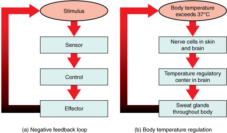
Negative Feedback
In a negative feedback loop, feedback serves to reduce an excessive response and keep a variable within the normal range. Two processes controlled by negative feedback are body temperature regulation and control of blood glucose.
Body Temperature
Body temperature regulation involves negative feedback, whether it lowers the temperature or raises it, as shown in Figure 7.8.3 and explained in the text that follows.
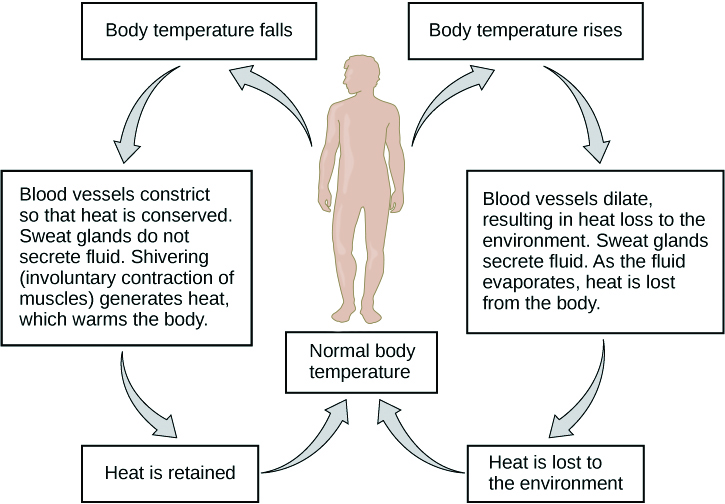
Cooling Down
The human body’s temperature regulatory centre is the hypothalamus in the brain. When the hypothalamus receives data from sensors in the skin and brain that body temperature is higher than the set point, it sets into motion the following responses:
- Blood vessels in the skin dilate (vasodilation) to allow more blood from the warm body core to flow close to the surface of the body, so heat can be radiated into the environment.
- As blood flow to the skin increases, sweat glands in the skin are activated to increase their output of sweat (diaphoresis). When the sweat evaporates from the skin surface into the surrounding air, it takes heat with it.
- Breathing becomes deeper, and the person may breathe through the mouth instead of the nasal passages. This increases heat loss from the lungs.
Heating Up
When the brain’s temperature regulatory centre receives data that body temperature is lower than the set point, it sets into motion the following responses:
- Blood vessels in the skin contract (vasoconstriction) to prevent blood from flowing close to the surface of the body, which reduces heat loss from the surface.
- As temperature falls lower, random signals to skeletal muscles are triggered, causing them to contract. This causes shivering, which generates a small amount of heat.
- The thyroid gland may be stimulated by the brain (via the pituitary gland) to secrete more thyroid hormone. This hormone increases metabolic activity and heat production in cells throughout the body.
- The adrenal glands may also be stimulated to secrete the hormone adrenaline. This hormone causes the breakdown of glycogen (the carbohydrate used for energy storage in animals) to glucose, which can be used as an energy source. This catabolic chemical process is exothermic, or heat producing.
Blood Glucose
In controlling the blood glucose level, certain endocrine cells in the pancreas (called alpha and beta cells) detect the level of glucose in the blood. They then respond appropriately to keep the level of blood glucose within the normal range.
- If the blood glucose level rises above the normal range, pancreatic beta cells release the hormone insulin into the bloodstream. Insulin signals cells to take up the excess glucose from the blood until the level of blood glucose decreases to the normal range.
- If the blood glucose level falls below the normal range, pancreatic alpha cells release the hormone glucagon into the bloodstream. Glucagon signals cells to break down stored glycogen to glucose and release the glucose into the blood until the level of blood glucose increases to the normal range.
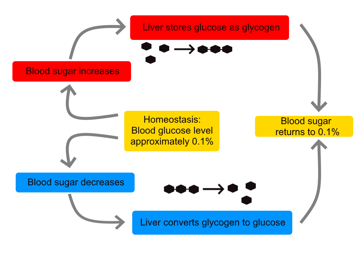
https://www.youtube.com/watch?v=Iz0Q9nTZCw4
Homeostasis and Negative/Positive Feedback, Amoeba Sisters, 2017.
Positive Feedback
In a positive feedback loop, feedback serves to intensify a response until an end point is reached. Examples of processes controlled by positive feedback in the human body include blood clotting and childbirth.
Blood Clotting
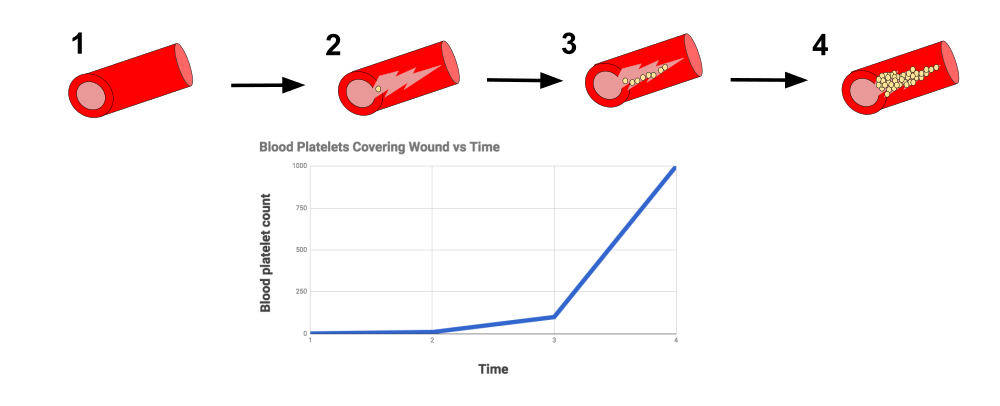
When a wound causes bleeding, the body responds with a positive feedback loop to clot the blood and stop blood loss. Substances released by the injured blood vessel wall begin the process of blood clotting. Platelets in the blood start to cling to the injured site and release chemicals that attract additional platelets. As the platelets continue to amass, more of the chemicals are released and more platelets are attracted to the site of the clot. The positive feedback accelerates the process of clotting until the clot is large enough to stop the bleeding.
Childbirth
Figure 7.8.6 shows the positive feedback loop that controls childbirth. The process normally begins when the head of the infant pushes against the cervix. This stimulates nerve impulses, which travel from the cervix to the hypothalamus in the brain. In response, the hypothalamus sends the hormone oxytocin to the pituitary gland, which secretes it into the bloodstream so it can be carried to the uterus. Oxytocin stimulates uterine contractions, which push the baby harder against the cervix. In response, the cervix starts to dilate in preparation for the passage of the baby. This cycle of positive feedback continues, with increasing levels of oxytocin, stronger uterine contractions, and wider dilation of the cervix until the baby is pushed through the birth canal and out of the body. At that point, the cervix is no longer stimulated to send nerve impulses to the brain, and the entire process stops.
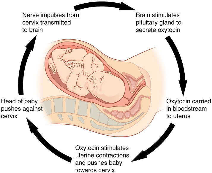
Normal childbirth is driven by a positive feedback loop. Positive feedback causes an increasing deviation from the normal state to a fixed end point, rather than a return to a normal set point as in homeostasis.
When Homeostasis Fails
Homeostatic mechanisms work continuously to maintain stable conditions in the human body. Sometimes, however, the mechanisms fail. When they do, homeostatic imbalance may result, in which cells may not get everything they need or toxic wastes may accumulate in the body. If homeostasis is not restored, the imbalance may lead to disease — or even death. Diabetes is an example of a disease caused by homeostatic imbalance. In the case of diabetes, blood glucose levels are no longer regulated and may be dangerously high. Medical intervention can help restore homeostasis and possibly prevent permanent damage to the organism.
Normal aging may bring about a reduction in the efficiency of the body’s control systems, which makes the body more susceptible to disease. Older people, for example, may have a harder time regulating their body temperature. This is one reason they are more likely than younger people to develop serious heat-induced illnesses, such as heat stroke.
Feature: My Human Body
Diabetes is diagnosed in people who have abnormally high levels of blood glucose after fasting for at least 12 hours. A fasting level of blood glucose below 100 is normal. A level between 100 and 125 places you in the pre-diabetes category, and a level higher than 125 results in a diagnosis of diabetes.
Of the two types of diabetes, type 2 diabetes is the most common, accounting for about 90 per cent of all cases of diabetes in the United States. Type 2 diabetes typically starts after the age of 40. However, because of the dramatic increase in recent decades in obesity in younger people, the age at which type 2 diabetes is diagnosed has fallen. Even children are now being diagnosed with type 2 diabetes. Today, about 3 million Canadians (8.1% of total population) are living with diabetes.
You may at some point have your blood glucose level tested during a routine medical exam. If your blood glucose level indicates that you have diabetes, it may come as a shock to you because you may not have any symptoms of the disease. You are not alone, because as many as one in four diabetics do not know they have the disease. Once the diagnosis of diabetes sinks in, you may be devastated by the news. Diabetes can lead to heart attacks, strokes, blindness, kidney failure, nerve damage, and loss of toes or feet. The risk of death in adults with diabetes is 50 per cent greater than it is in adults without diabetes, and diabetes is the seventh leading cause of death of adults. In addition, controlling diabetes usually requires frequent blood glucose testing, watching what and when you eat, and taking medications or even insulin injections. All of this may seem overwhelming.
The good news is that changing your lifestyle may stop the progression of type 2 diabetes or even reverse it. By adopting healthier habits, you may be able to keep your blood glucose level within the normal range without medications or insulin. Here’s how:
- Lose weight. Any weight loss is beneficial. Losing as little as seven per cent of your weight may be all that is needed to stop diabetes in its tracks. It is especially important to eliminate excess weight around your waist.
- Exercise regularly. You should try to exercise for at least 30 minutes, five days a week. This will not only lower your blood sugar and help your insulin work better, but it will also lower your blood pressure and improve your heart health. Another bonus of exercise is that it will help you lose weight by increasing your basal metabolic rate.
- Adopt a healthy diet. Decrease your consumption of refined carbohydrates, such as sweets and sugary drinks. Increase your intake of fibre-rich foods, such as fruits, vegetables, and whole grains. About one-quarter of each meal should consist of high-protein foods, such as fish, chicken, dairy products, legumes, or nuts.
- Control stress. Stress can increase your blood glucose and also raise your blood pressure and risk of heart disease. When you feel stressed out, do breathing exercises or take a brisk walk or jog. Try to replace stressful thoughts with more calming ones.
- Establish a support system. Enlist the help and support of loved ones, as well as medical professionals, such as a nutritionist and diabetes educator. Having a support system will help ensure that you are on the path to wellness, and that you can stick to your plan.
7.8 Summary
- Homeostasis is the condition in which a system (such as the human body) is maintained in a more or less steady state. It is the job of cells, tissues, organs, and organ systems throughout the body to maintain homeostasis.
- For any given variable, such as body temperature, there is a particular set point that is the physiological optimum value. The spread of values around the set point that is considered insignificant is called the normal range.
- Homeostasis is generally maintained by a negative feedback loop that includes a stimulus, sensor, control centre, and effector. Negative feedback serves to reduce an excessive response and to keep a variable within the normal range. Negative feedback loops control body temperature and the blood glucose level.
- Positive feedback loops are not common in biological systems. Positive feedback serves to intensify a response until an end point is reached. Positive feedback loops control blood clotting and childbirth.
- Sometimes homeostatic mechanisms fail, resulting in homeostatic imbalance. Diabetes is an example of a disease caused by homeostatic imbalance. Aging can bring about a reduction in the efficiency of the body’s control system, which makes the elderly more susceptible to disease.
7.8 Review Questions
-
-
- Compare and contrast negative and positive feedback loops.
- Explain how negative feedback controls body temperature.
- Give two examples of physiological processes controlled by positive feedback loops.
- During breastfeeding, the stimulus of the baby sucking on the nipple increases the amount of milk produced by the mother. The more sucking, the more milk is usually produced. Is this an example of negative or positive feedback? Explain your answer. What do you think might be the evolutionary benefit of the milk production regulation mechanism you described?
- Explain why homeostasis is regulated by negative feedback loops, rather than positive feedback loops.
- The level of a sex hormone, testosterone (T), is controlled by negative feedback. Another hormone, gonadotropin-releasing hormone (GnRH), is released by the hypothalamus of the brain, which triggers the pituitary gland to release luteinizing hormone (LH). LH stimulates the gonads to produce T. When there is too much T in the bloodstream, it feeds back on the hypothalamus, causing it to produce less GnRH. While this does not describe all the feedback loops involved in regulating T, answer the following questions about this particular feedback loop.
- What is the stimulus in this system? Explain your answer.
- What is the control centre in this system? Explain your answer.
- In this system, is the pituitary considered the stimulus, sensor, control centre, or effector? Explain your answer.
7.8 Explore More
https://www.youtube.com/watch?v=LSgEJSlk6W4
Homeostasis - What Is Homeostasis - What Is Set Point For Homeostasis - Homeostasis In The Human Body, Whats Up Dude, 2017.
https://www.youtube.com/watch?v=XMsJ-3qRVJM
Attributions
Figure 7.8.1
Nest_Thermostat by Amanitamano on Wikimedia Commons is used under a CC BY-SA 3.0 (https://creativecommons.org/licenses/by-sa/3.0/deed.en) license.
Figure 7.8.2
Negative_Feedback_Loops by OpenStax on Wikimedia Commons is used under a CC BY 4.0 (https://creativecommons.org/licenses/by/4.0/deed.en) license.
Figure 7.8.3
Body Temperature Homeostasis by OpenStax College, Biology is used under a CC BY 4.0 license.
Figure 7.8.4
Homeostasis_of_blood_sugar by Christinelmiller on Wikimedia Commons is used under a CC0 1.0 Universal Public Domain Dedication (https://creativecommons.org/publicdomain/zero/1.0/deed.en) license.
Figure 7.8.5
Positive_Feedback_Diagram_Blood_Clotting by Elliottuttle on Wikimedia Commons is used under a CC BY-SA 4.0 (https://creativecommons.org/licenses/by-sa/4.0) license.
Figure 7.8.6
Pregnancy-Positive_Feedback by OpenStax on Wikimedia Commons is used under a CC BY 4.0 (https://creativecommons.org/licenses/by/4.0/deed.en) license.
References
Amoeba Sisters. (2017, September 7). Homeostasis and negative/positive feedback. YouTube. https://www.youtube.com/watch?v=Iz0Q9nTZCw4&feature=youtu.be
Betts, J. G., Young, K.A., Wise, J.A., Johnson, E., Poe, B., Kruse, D.H., Korol, O., Johnson, J.E., Womble, M., DeSaix, P. (2013, April 25). Figure 1.10 Negative feedback loop [digital image/ diagram]. In Anatomy and Physiology (Section 1.5). OpenStax. https://openstax.org/books/anatomy-and-physiology/pages/1-5-homeostasis
Betts, J. G., Young, K.A., Wise, J.A., Johnson, E., Poe, B., Kruse, D.H., Korol, O., Johnson, J.E., Womble, M., DeSaix, P. (2013, April 25). Figure 1.11 Positive feedback loop normal childbirth is driven by a positive feedback loop [digital image/ diagram]. In Anatomy and Physiology (Section 1.5). OpenStax. https://openstax.org/books/anatomy-and-physiology/pages/1-5-homeostasis
Cognito. (2018, December 18). GCSE Biology - Homeostasis #38. YouTube. https://www.youtube.com/watch?v=XMsJ-3qRVJM&feature=youtu.be
Mayo Clinic Staff. (n.d.). Type 2 diabetes [online article]. MayoClinic.org. https://www.mayoclinic.org/diseases-conditions/type-2-diabetes/symptoms-causes/syc-20351193
OpenStax CNX. (2016, March 23). Figure 4 The body is able to regulate temperature in response to signals from the nervous system [digital image]. In OpenStax, Biology (Section 33.3). https://cnx.org/contents/GFy_h8cu@10.8:BP24ZReh@7/Homeostasis
Whats Up Dude. (2017, September 20). Homeostasis - What is homeostasis - What is set point for homeostasis - Homeostasis in the human body. YouTube. https://www.youtube.com/watch?v=LSgEJSlk6W4&feature=youtu.be
Created by CK-12 Foundation/Adapted by Christine Miller
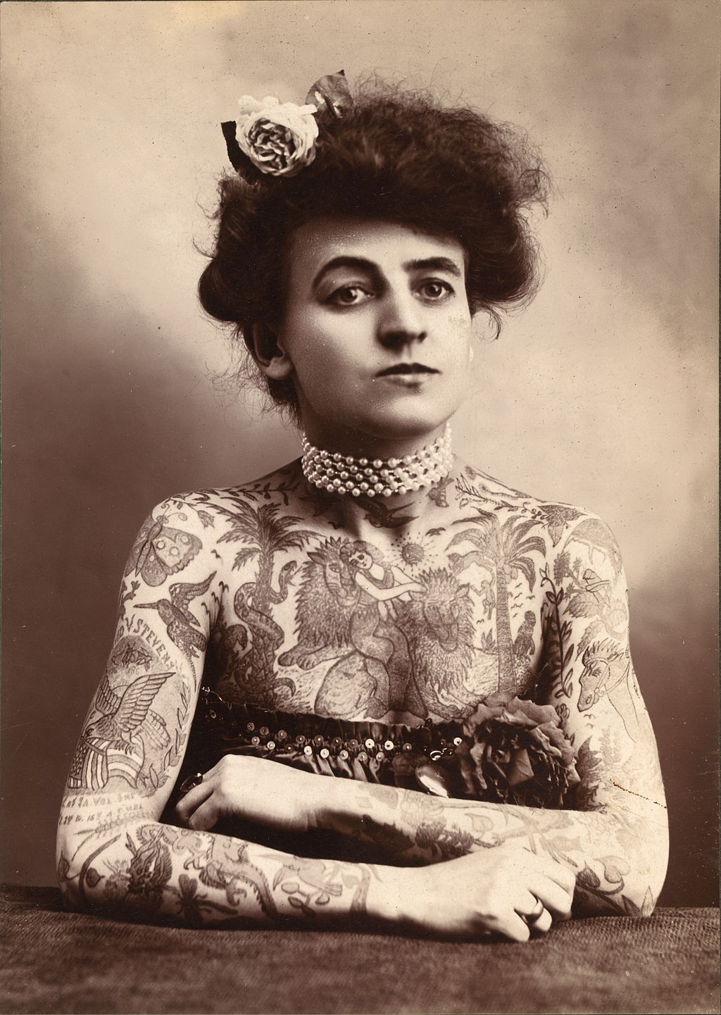
Art for All Eras
Pictured in Figure 10.2.1, is Maud Stevens Wagner, a tattoo artist from 1907. Tattoos are not just a late 20th and early 21st century trend. They have been popular in many eras and cultures. Tattoos literally illustrate the biggest organ of the human body: the skin. The skin is very thin, but it covers a large area — about 2 m2 in adults. The skin is the major organ in the integumentary system.
What Is the Integumentary System?
In addition to the skin, the integumentary system includes the hair and nails, which are organs that grow out of the skin. Because the organs of the integumentary system are mostly external to the body, you may think of them as little more than accessories, like clothing or jewelry, but they serve vital physiological functions. They provide a protective covering for the body, sense the environment, and help the body maintain homeostasis.
The Skin
The skin is remarkable not only because it is the body’s largest organ: the average square inch of skin has 20 blood vessels, 650 sweat glands, and more than 1,000 nerve endings. Incredibly, it also has 60,000 pigment-producing cells. All of these structures are packed into a stack of cells that is just 2 mm thick. Although the skin is thin, it consists of two distinct layers: the epidermis and dermis, as shown in the diagram (Figure 10.2.2).
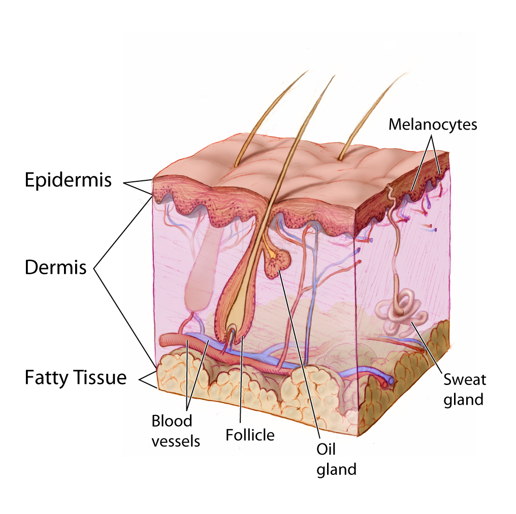
Outer Layer of Skin
The outer layer of skin is the epidermis. This layer is thinner than the inner layer (the dermis). The epidermis consists mainly of epithelial cells, called keratinocytes, which produce the tough, fibrous protein keratin. The innermost cells of the epidermis are stem cells that divide continuously to form new cells. The newly formed cells move up through the epidermis toward the skin surface, while producing more and more keratin. The cells become filled with keratin and die by the time they reach the surface, where they form a protective, waterproof layer. As the dead cells are shed from the surface of the skin, they are replaced by other cells that move up from below. The epidermis also contains melanocytes, the cells that produce the brown pigment melanin, which gives skin most of its colour. Although the epidermis contains some sensory receptor cells — called Merkel cells — it contains no nerves, blood vessels, or other structures.
Inner Layer of Skin
The dermis is the inner, thicker layer of skin. It consists mainly of tough connective tissue, and is attached to the epidermis by collagen fibres. The dermis contains many structures (as shown in Figure 10.2.2), including blood vessels, sweat glands, and hair follicles, which are structures where hairs originate. In addition, the dermis contains many sensory receptors, nerves, and oil glands.
Functions of the Skin
The skin has multiple roles in the body. Many of these roles are related to homeostasis. The skin’s main functions are preventing water loss from the body and serving as a barrier to the entry of microorganisms. Another function of the skin is synthesizing vitamin D, which occurs when the skin is exposed to ultraviolet (UV) light. Melanin in the epidermis blocks some of the UV light and protects the dermis from its damaging effects.
Another important function of the skin is helping to regulate body temperature. When the body is too warm, for example, the skin lowers body temperature by producing sweat, which cools the body when it evaporates. The skin also increases the amount of blood flowing near the body surface through vasodilation (widening of blood vessels), bringing heat from the body core to radiate out into the environment. The sweaty hair and flushed skin of the young man pictured in Figure 10.2.3 reflect these skin responses to overheating.

Hair
Hair is a fibre found only in mammals. It consists mainly of keratin-producing keratinocytes. Each hair grows out of a follicle in the dermis. By the time the hair reaches the surface, it consists mainly of dead cells filled with keratin. Hair serves several homeostatic functions. Head hair is important in preventing heat loss from the head and protecting its skin from UV radiation. Hairs in the nose trap dust particles and microorganisms in the air, and prevent them from reaching the lungs. Hair all over the body provides sensory input when objects brush against it, or when it sways in moving air. Eyelashes and eyebrows (see Figure 10.2.4) protect the eyes from water, dirt, and other irritants.
Nails
Fingernails and toenails consist of dead keratinocytes filled with keratin. The keratin makes them hard but flexible, which is important for the functions they serve. Nails prevent injury by forming protective plates over the ends of the fingers and toes. They also enhance sensation by acting as a counterforce to the sensitive fingertips when objects are handled. In addition, the fingernails can be used as tools.
Interactions with Other Organ Systems
The skin and other parts of the integumentary system work with other organ systems to maintain homeostasis.
- The skin works with the immune system to defend the body from pathogens by serving as a physical barrier to microorganisms.
- Vitamin D is needed by the digestive system to absorb calcium from food. By synthesizing vitamin D, the skin works with the digestive system to ensure that calcium can be absorbed.
- To control body temperature, the skin works with the cardiovascular system to either lose body heat, or to conserve it through vasodilation or vasoconstriction.
- To detect certain sensations from the outside world, the nervous system depends on nerve receptors in the skin.
10.2 Summary
- The integumentary system consists of the skin, hair, and nails. Functions of the integumentary system include providing a protective covering for the body, sensing the environment, and helping the body maintain homeostasis.
- The skin consists of two distinct layers: a thinner outer layer called the epidermis, and a thicker inner layer called the dermis.
- The epidermis consists mainly of epithelial cells called keratinocytes, which produce keratin. New keratinocytes form at the bottom of the epidermis. They become filled with keratin and die as they move upward toward the surface of the skin, where they form a protective, waterproof layer.
- The dermis consists mainly of tough connective tissues and many structures, including blood vessels, sensory receptors, nerves, hair follicles, and oil and sweat glands.
- The skin’s main functions are preventing water loss from the body, serving as a barrier to the entry of microorganisms, synthesizing vitamin D, blocking UV light, and helping to regulate body temperature.
- Hair consists mainly of dead keratinocytes and grows out of follicles in the dermis. Hair helps prevent heat loss from the head, and protects its skin from UV light. Hair in the nose filters incoming air, and the eyelashes and eyebrows keep harmful substances out of the eyes. Hair all over the body provides tactile sensory input.
- Like hair, nails also consist mainly of dead keratinocytes. They help protect the ends of the fingers and toes, enhance the sense of touch in the fingertips, and may be used as tools.
10.2 Review Questions
- Name the organs of the integumentary system.
- Compare and contrast the epidermis and dermis.
- Identify functions of the skin.
-
- What is the composition of hair?
- Describe three physiological roles played by hair.
- What do nails consist of?
- List two functions of nails.
- In terms of composition, what do the outermost surface of the skin, the nails, and hair have in common?
- Identify two types of cells found in the epidermis of the skin. Describe their functions.
- Which structure and layer of skin does hair grow out of?
- Identify three main functions of the integumentary system. Give an example of each.
- What are two ways in which the integumentary system protects the body against UV radiation?
10.2 Explore More
https://www.youtube.com/watch?v=OxPlCkTKhzY
The science of skin - Emma Bryce, TED-Ed, 2018.
https://www.youtube.com/watch?v=ZSJITdsTze0&feature=emb_logo
Why do we have to wear sunscreen? - Kevin P. Boyd, TED-Ed, 2013.
https://www.youtube.com/watch?time_continue=1&v=Lfhot7tQcWs&feature=emb_logo
Scarification | National Geographic, 2008.
Attributions
Figure 10.2.1
Maud_Stevens_Wagner -The Plaza Gallery, Los Angeles, 1907 from the Library of Congress on Wikimedia Commons is in the public domain (https://en.wikipedia.org/wiki/public_domain).
Figure 10.2.2
Anatomy_The_Skin_-_NCI_Visuals_Online by Don Bliss (artist) from National Cancer Institute, on Wikimedia Commons is in the public domain (https://en.wikipedia.org/wiki/public_domain).
Figure 10.2.3
shashank-shekhar-Db1J_qp_ctc [photo] by Shashank Shekhar on Unsplash is used under the Unsplash License (https://unsplash.com/license).
Figure 10.2.4
Eyelashes by aryan-dhiman-93NBu0zG_H4 [photo] by Aryan Dhiman on Unsplash is used under the Unsplash License (https://unsplash.com/license).
Reference
National Geographic. (2008). Scarification | National Geographic. YouTube. https://www.youtube.com/watch?v=Lfhot7tQcWs&t=1s
TED-Ed. (2018, March 12). The science of skin - Emma Bryce. YouTube. https://www.youtube.com/watch?v=OxPlCkTKhzY&feature=youtu.be
TED-Ed. (2013, August 6). Why do we have to wear sunscreen? - Kevin P. Boyd. YouTube. https://www.youtube.com/watch?v=ZSJITdsTze0&feature=youtu.be
Created by CK-12 Foundation/Adapted by Christine Miller
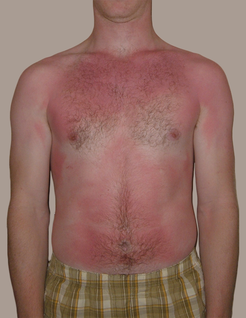
Feel the Burn
The person in Figure 10.3.1 is no doubt feeling the burn — sunburn, that is. Sunburn occurs when the outer layer of the skin is damaged by UV light from the sun or tanning lamps. Some people deliberately allow UV light to burn their skin, because after the redness subsides, they are left with a tan. A tan may look healthy, but it is actually a sign of skin damage. People who experience one or more serious sunburns are significantly more likely to develop skin cancer. Natural pigment molecules in the skin help protect it from UV light damage. These pigment molecules are found in the layer of the skin called the epidermis.
What is the Epidermis?
The epidermis is the outer of the two main layers of the skin. The inner layer is the dermis. It averages about 0.10 mm thick, and is much thinner than the dermis. The epidermis is thinnest on the eyelids (0.05 mm) and thickest on the palms of the hands and soles of the feet (1.50 mm). The epidermis covers almost the entire body surface. It is continuous with — but structurally distinct from — the mucous membranes that line the mouth, anus, urethra, and vagina.
Structure of the Epidermis
There are no blood vessels and very few nerve cells in the epidermis. Without blood to bring epidermal cells oxygen and nutrients, the cells must absorb oxygen directly from the air and obtain nutrients via diffusion of fluids from the dermis below. However, as thin as it is, the epidermis still has a complex structure. It has a variety of cell types and multiple layers.
Cells of the Epidermis
There are several different types of cells in the epidermis. All of the cells are necessary for the important functions of the epidermis.
- The epidermis consists mainly of stacks of keratin-producing epithelial cells called keratinocytes. These cells make up at least 90 per cent of the epidermis. Near the top of the epidermis, these cells are also called squamous cells.
- Another eight per cent of epidermal cells are melanocytes. These cells produce the pigment melanin that protects the dermis from UV light.
- About one per cent of epidermal cells are Langerhans cells. These are immune system cells that detect and fight pathogens entering the skin.
- Less than one per cent of epidermal cells are Merkel cells, which respond to light touch and connect to nerve endings in the dermis.
Layers of the Epidermis
The epidermis in most parts of the body consists of four distinct layers. A fifth layer occurs in the palms of the hands and soles of the feet, where the epidermis is thicker than in the rest of the body. The layers of the epidermis are shown in Figure 10.3.2, and described in the following text.
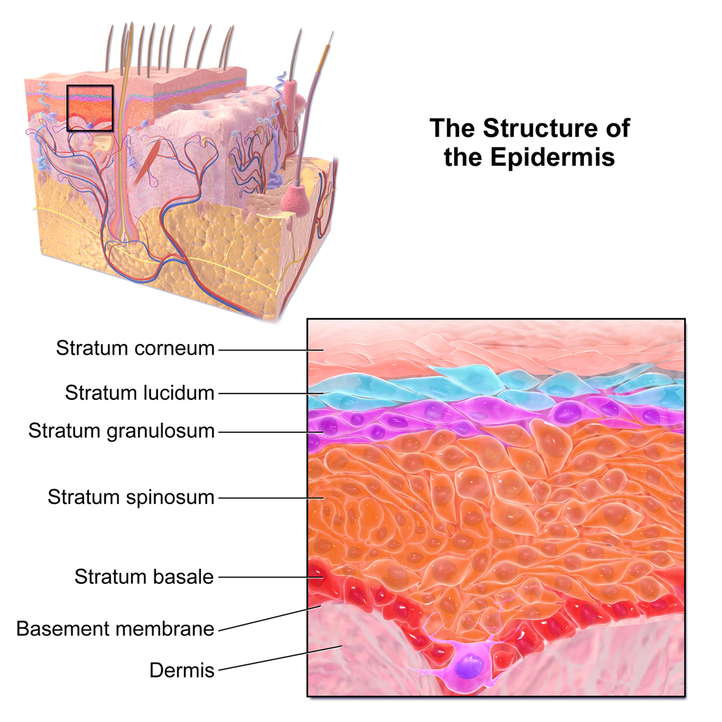
Stratum Basale
The stratum basale is the innermost (or deepest) layer of the epidermis. It is separated from the dermis by a membrane called the basement membrane. The stratum basale contains stem cells — called basal cells — which divide to form all the keratinocytes of the epidermis. When keratinocytes first form, they are cube-shaped and contain almost no keratin. As more keratinocytes are produced, previously formed cells are pushed up through the stratum basale. Melanocytes and Merkel cells are also found in the stratum basale. The Merkel cells are especially numerous in touch-sensitive areas, such as the fingertips and lips.
Stratum Spinosum
Just above the stratum basale is the stratum spinosum. This is the thickest of the four epidermal layers. The keratinocytes in this layer have begun to accumulate keratin, and they have become tougher and flatter. Spiny cellular projections form between the keratinocytes and hold them together. In addition to keratinocytes, the stratum spinosum contains the immunologically active Langerhans cells.
Stratum Granulosum
The next layer above the stratum spinosum is the stratum granulosum. In this layer, keratinocytes have become nearly filled with keratin, giving their cytoplasm a granular appearance. Lipids are released by keratinocytes in this layer to form a lipid barrier in the epidermis. Cells in this layer have also started to die, because they are becoming too far removed from blood vessels in the dermis to receive nutrients. Each dying cell digests its own nucleus and organelles, leaving behind only a tough, keratin-filled shell.
Stratum Lucidum
Only on the palms of the hands and soles of the feet, the next layer above the stratum granulosum is the stratum lucidum. This is a layer consisting of stacks of translucent, dead keratinocytes that provide extra protection to the underlying layers.
Stratum Corneum
The uppermost layer of the epidermis everywhere on the body is the stratum corneum. This layer is made of flat, hard, tightly packed dead keratinocytes that form a waterproof keratin barrier to protect the underlying layers of the epidermis. Dead cells from this layer are constantly shed from the surface of the body. The shed cells are continually replaced by cells moving up from lower layers of the epidermis. It takes a period of about 48 days for newly formed keratinocytes in the stratum basale to make their way to the top of the stratum corneum to replace shed cells.
Functions of the Epidermis
The epidermis has several crucial functions in the body. These functions include protection, water retention, and vitamin D synthesis.
Protective Functions
The epidermis provides protection to underlying tissues from physical damage, pathogens, and UV light.
Protection from Physical Damage
Most of the physical protection of the epidermis is provided by its tough outer layer, the stratum corneum. Because of this layer, minor scrapes and scratches generally do not cause significant damage to the skin or underlying tissues. Sharp objects and rough surfaces have difficulty penetrating or removing the tough, dead, keratin-filled cells of the stratum corneum. If cells in this layer are pierced or scraped off, they are quickly replaced by new cells moving up to the surface from lower skin layers.
Protection from Pathogens
When pathogens such as viruses and bacteria try to enter the body, it is virtually impossible for them to enter through intact epidermal layers. Generally, pathogens can enter the skin only if the epidermis has been breached, for example by a cut, puncture, or scrape (like the one pictured in Figure 10.3.3). That’s why it is important to clean and cover even a minor wound in the epidermis. This helps ensure that pathogens do not use the wound to enter the body. Protection from pathogens is also provided by conditions at or near the skin surface. These include relatively high acidity (pH of about 5.0), low amounts of water, the presence of antimicrobial substances produced by epidermal cells, and competition with non-pathogenic microorganisms that normally live on the epidermis.
Protection from UV Light
UV light that penetrates the epidermis can damage epidermal cells. In particular, it can cause mutations in DNA that lead to the development of skin cancer, in which epidermal cells grow out of control. UV light can also destroy vitamin B9 (in forms such as folate or folic acid), which is needed for good health and successful reproduction. In a person with light skin, just an hour of exposure to intense sunlight can reduce the body’s vitamin B9 level by 50 per cent.
Melanocytes in the stratum basale of the epidermis contain small organelles called melanosomes, which produce, store, and transport the dark brown pigment melanin. As melanosomes become full of melanin, they move into thin extensions of the melanocytes. From there, the melanosomes are transferred to keratinocytes in the epidermis, where they absorb UV light that strikes the skin. This prevents the light from penetrating deeper into the skin, where it can cause damage. The more melanin there is in the skin, the more UV light can be absorbed.
Water Retention
Skin's ability to hold water and not lose it to the surrounding environment is due mainly to the stratum corneum. Lipids arranged in an organized way among the cells of the stratum corneum form a barrier to water loss from the epidermis. This is critical for maintaining healthy skin and preserving proper water balance in the body.
Although the skin is impermeable to water, it is not impermeable to all substances. Instead, the skin is selectively permeable, allowing certain fat-soluble substances to pass through the epidermis. The selective permeability of the epidermis is both a benefit and a risk.
- Selective permeability allows certain medications to enter the bloodstream through the capillaries in the dermis. This is the basis of medications that are delivered using topical ointments, or patches (see Figure 10.3.4) that are applied to the skin. These include steroid hormones, such as estrogen (for hormone replacement therapy), scopolamine (for motion sickness), nitroglycerin (for heart problems), and nicotine (for people trying to quit smoking).
- Selective permeability of the epidermis also allows certain harmful substances to enter the body through the skin. Examples include the heavy metal lead, as well as many pesticides.
Vitamin D Synthesis
Vitamin D is a nutrient that is needed in the human body for the absorption of calcium from food. Molecules of a lipid compound named 7-dehydrocholesterol are precursors of vitamin D. These molecules are present in the stratum basale and stratum spinosum layers of the epidermis. When UV light strikes the molecules, it changes them to vitamin D3. In the kidneys, vitamin D3 is converted to calcitriol, which is the form of vitamin D that is active in the body.
What Gives Skin Its Colour?
Melanin in the epidermis is the main substance that determines the colour of human skin. It explains most of the variation in skin colour in people around the world. Two other substances also contribute to skin colour, however, especially in light-skinned people: carotene and hemoglobin.
- The pigment carotene is present in the epidermis and gives skin a yellowish tint, especially in skin with low levels of melanin.
- Hemoglobin is a red pigment found in red blood cells. It is visible through skin as a pinkish tint, mainly in skin with low levels of melanin. The pink colour is most visible when capillaries in the underlying dermis dilate, allowing greater blood flow near the surface.
Hear what Bill Nye has to say about the subject of skin colour in the video here.
Bacteria on Skin
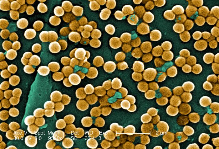
The surface of the human skin normally provides a home to countless numbers of bacteria. Just one square inch of skin normally has an average of about 50 million bacteria. These generally harmless bacteria represent roughly one thousand bacterial species (including the one in Figure 10.3.5) from 19 different bacterial phyla. Typical variations in the moistness and oiliness of the skin produce a variety of rich and diverse habitats for these microorganisms. For example, the skin in the armpits is warm and moist and often hairy, whereas the skin on the forearms is smooth and dry. These two areas of the human body are as diverse to microorganisms as rainforests and deserts are to larger organisms. The density of bacterial populations on the skin depends largely on the region of the skin and its ecological characteristics. For example, oily surfaces, such as the face, may contain over 500 million bacteria per square inch. Despite the huge number of individual microorganisms living on the skin, their total volume is only about the size of a pea.
In general, the normal microorganisms living on the skin keep one another in check, and thereby play an important role in keeping the skin healthy. If the balance of microorganisms is disturbed, however, there may be an overgrowth of certain species, and this may result in an infection. For example, when a patient is prescribed antibiotics, it may kill off normal bacteria and allow an overgrowth of single-celled yeast. Even if skin is disinfected, no amount of cleaning can remove all of the microorganisms it contains. Disinfected areas are also quickly recolonized by bacteria residing in deeper areas (such as hair follicles) and in adjacent areas of the skin.
Feature: Myth vs. Reality
Because of the negative health effects of excessive UV light exposure, it is important to know the facts about protecting the skin from UV light.
Myth |
Reality |
| "Sunblock and sunscreen are just different names for the same type of product. They both work the same way and are equally effective." | Sunscreens and sunblocks are different types of products that protect the skin from UV light in different ways. They are not equally effective. Sunblocks are opaque, so they do not let light pass through. They prevent most of the rays of UV light from penetrating to the skin surface. Sunblocks are generally stronger and more effective than sunscreens. Sunblocks also do not need to be reapplied as often as sunscreens. Sunscreens, in contrast, are transparent once they are applied the skin. Although they can prevent most UV light from penetrating the skin when first applied, the active ingredients in sunscreens tend to break down when exposed to UV light. Sunscreens, therefore, must be reapplied often to remain effective. |
| "The skin needs to be protected from UV light only on sunny days. When the sky is cloudy, UV light cannot penetrate to the ground and harm the skin." | Even on cloudy days, a significant amount of UV radiation penetrates the atmosphere to strike Earth’s surface. Therefore, using sunscreens or sunblocks to protect exposed skin is important even when there are clouds in the sky. |
| "People who have dark skin, such as African Americans, do not need to worry about skin damage from UV light." | No matter what colour skin you have, your skin can be damaged by too much exposure to UV light. Therefore, even dark-skinned people should use sunscreens or sunblocks to protect exposed skin from UV light. |
| "Sunscreens with an SPF (sun protection factor) of 15 are adequate to fully protect the skin from UV light." | Most dermatologists recommend using sunscreens with an SPF of at least 35 for adequate protection from UV light. They also recommend applying sunscreens at least 20 minutes before sun exposure and reapplying sunscreens often, especially if you are sweating or spending time in the water. |
| "Using tanning beds is safer than tanning outside in natural sunlight." | The light in tanning beds is UV light, and it can do the same damage to the skin as the natural UV light in sunlight. This is evidenced by the fact that people who regularly use tanning beds have significantly higher rates of skin cancer than people who do not. It is also the reason that the use of tanning beds is prohibited in many places in people who are under the age of 18, just as youth are prohibited from using harmful substances, such as tobacco and alcohol. |
10.3 Summary
- The epidermis is the outer of the two main layers of the skin. It is very thin, but has a complex structure.
- Cell types in the epidermis include keratinocytes that produce keratin and make up 90 per cent of epidermal cells, melanocytes that produce melanin, Langerhans cells that fight pathogens in the skin, and Merkel cells that respond to light touch.
- The epidermis in most parts of the body consists of four distinct layers. A fifth layer occurs only in the epidermis of the palms of the hands and soles of the feet.
- The innermost layer of the epidermis is the stratum basale, which contains stem cells that divide to form new keratinocytes. The next layer is the stratum spinosum, which is the thickest layer and contains Langerhans cells and spiny keratinocytes. This is followed by the stratum granulosum, in which keratinocytes are filling with keratin and starting to die. The stratum lucidum is next, but only on the palms and soles. It consists of translucent dead keratinocytes. The outermost layer is the stratum corneum, which consists of flat, dead, tightly packed keratinocytes that form a tough, waterproof barrier for the rest of the epidermis.
- Functions of the epidermis include protecting underlying tissues from physical damage and pathogens. Melanin in the epidermis absorbs and protects underlying tissues from UV light. The epidermis also prevents loss of water from the body and synthesizes vitamin D.
- Melanin is the main pigment that determines the colour of human skin. The pigments carotene and hemoglobin, however, also contribute to skin colour, especially in skin with low levels of melanin.
- The surface of healthy skin normally is covered by vast numbers of bacteria representing about one thousand species from 19 phyla. Different areas of the body provide diverse habitats for skin microorganisms. Usually, microorganisms on the skin keep each other in check unless their balance is disturbed.
10.3 Review Questions
- What is the epidermis?
- Identify the types of cells in the epidermis.
- Describe the layers of the epidermis.
-
- State one function of each of the four epidermal layers found all over the body.
- Explain three ways the epidermis protects the body.
- What makes the skin waterproof?
- Why is the selective permeability of the epidermis both a benefit and a risk?
- How is vitamin D synthesized in the epidermis?
- Identify three pigments that impart colour to skin.
- Describe bacteria that normally reside on the skin, and explain why they do not usually cause infections.
- Explain why the keratinocytes at the surface of the epidermis are dead, while keratinocytes located deeper in the epidermis are still alive.
- Which layer of the epidermis contains keratinocytes that have begun to die?
-
- Explain why our skin is not permanently damaged if we rub off some of the surface layer by using a rough washcloth.
10.3 Explore More
https://www.youtube.com/watch?v=27lMmdmy-b8
Jonathan Eisen: Meet your microbes, TED, 2015.
https://www.youtube.com/watch?v=9AcQXnOscQ8
Why Do We Blush?, SciShow, 2014.
https://www.youtube.com/watch?v=_r4c2NT4naQ
The science of skin colour - Angela Koine Flynn, TED-Ed, 2016.
Attributions
Figure 10.3.1
Sunburn by QuinnHK at English Wikipedia on Wikimedia Commons is released into the public domain (https://en.wikipedia.org/wiki/Public_domain).
Figure 10.3.2
Blausen_0353_Epidermis by BruceBlaus on Wikimedia Commons is used under a CC BY 3.0 (https://creativecommons.org/licenses/by/3.0) license.
Figure 10.3.3
Isaac's scraped knee close-up by Alpha on Flickr is used under a CC BY-NC-SA 2.0 (https://creativecommons.org/licenses/by-nc-sa/2.0/) license.
Figure 10.3.4
Nicoderm by RegBarc on Wikimedia Commons is used under a CC BY-SA 3.0 (http://creativecommons.org/licenses/by-sa/3.0/) license. (No machine-readable author provided for original.)
Figure 10.3.5
Staphylococcus aureus bacteria, MRSA by Microbe World on Flickr is used under a CC BY-NC-SA 2.0 (https://creativecommons.org/licenses/by-nc-sa/2.0/) license.
References
Blausen.com staff. (2014). Medical gallery of Blausen Medical 2014. WikiJournal of Medicine 1 (2). DOI:10.15347/wjm/2014.010. ISSN 2002-4436.
Jeff Bone 'n' Pookie. (2020, July 19). Bill Nye the science guy explains we have different skin color. Youtube. https://www.youtube.com/watch?v=zOkj5jgC4sM&feature=youtu.be
SciShow. (2014, July 15). Why do we blush? YouTube. https://www.youtube.com/watch?v=9AcQXnOscQ8
TED. (2015, July 17). Jonathan Eisen: Meet your microbes. YouTube. https://www.youtube.com/watch?v=27lMmdmy-b8
TED-Ed. (2016, February 16). The science of skin color - Angela Koine Flynn. YouTube. https://youtu.be/_r4c2NT4naQ
Created by CK-12 Foundation/Adapted by Christine Miller
Goose Bumps
No doubt you’ve experienced the tiny, hair-raising skin bumps called goose bumps, like those you see in Figure 10.4.1. They happen when you feel chilly. Do you know what causes goose bumps, or why they pop up when you are cold? The answers to these questions involve the layer of skin known as the dermis.
What is the Dermis?
The dermis is the inner of the two major layers that make up the skin, the outer layer being the epidermis. The dermis consists mainly of connective tissues. It also contains most skin structures, such as glands and blood vessels. The dermis is anchored to the tissues below it by flexible collagen bundles that permit most areas of the skin to move freely over subcutaneous (“below the skin”) tissues. Functions of the dermis include cushioning subcutaneous tissues, regulating body temperature, sensing the environment, and excreting wastes.
Anatomy of the Dermis
The basic anatomy of the dermis is a matrix, or sort of scaffolding, composed of connective tissues. These tissues include collagen fibres — which provide toughness — and elastin fibres, which provide elasticity. Surrounding these fibres, the matrix also includes a gel-like substance made of proteins. The tissues of the matrix give the dermis both strength and flexibility.
The dermis is divided into two layers: the papillary layer and the reticular layer. Both layers are shown in Figure 10.4.2 below and described in the text that follows.
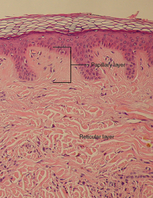
Papillary Layer
The papillary layer is the upper layer of the dermis, just below the basement membrane that connects the dermis to the epidermis above it. The papillary layer is the thinner of the two dermal layers. It is composed mainly of loosely arranged collagen fibres. The papillary layer is named for its fingerlike projections — or papillae — that extend upward into the epidermis. The papillae contain capillaries and sensory touch receptors.
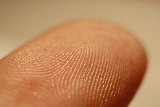
The papillae give the dermis a bumpy surface that interlocks with the epidermis above it, strengthening the connection between the two layers of skin. On the palms and soles, the papillae create epidermal ridges. Epidermal ridges on the fingers are commonly called fingerprints (see Figure 10.4.3). Fingerprints are genetically determined, so no two people (other than identical twins) have exactly the same fingerprint pattern. Therefore, fingerprints can be used as a means of identification, for example, at crime scenes. Fingerprints were much more commonly used forensically before DNA analysis was introduced for this purpose.
Reticular Layer
The reticular layer is the lower layer of the dermis, located below the papillary layer. It is the thicker of the two dermal layers. It is composed of densely woven collagen and elastin fibres. These protein fibres give the dermis its properties of strength and elasticity. This layer of the dermis cushions subcutaneous tissues of the body from stress and strain. The reticular layer of the dermis also contains most of the structures in the dermis, such as glands and hair follicles.
Structures in the Dermis
Both papillary and reticular layers of the dermis contain numerous sensory receptors, which make the skin the body’s primary sensory organ for the sense of touch. Both dermal layers also contain blood vessels. They provide nutrients to remove wastes from dermal cells, as well as cells in the lowest layer of the epidermis, the stratum basale. The circulatory components of the dermis are shown in Figure 10.4.4 below.
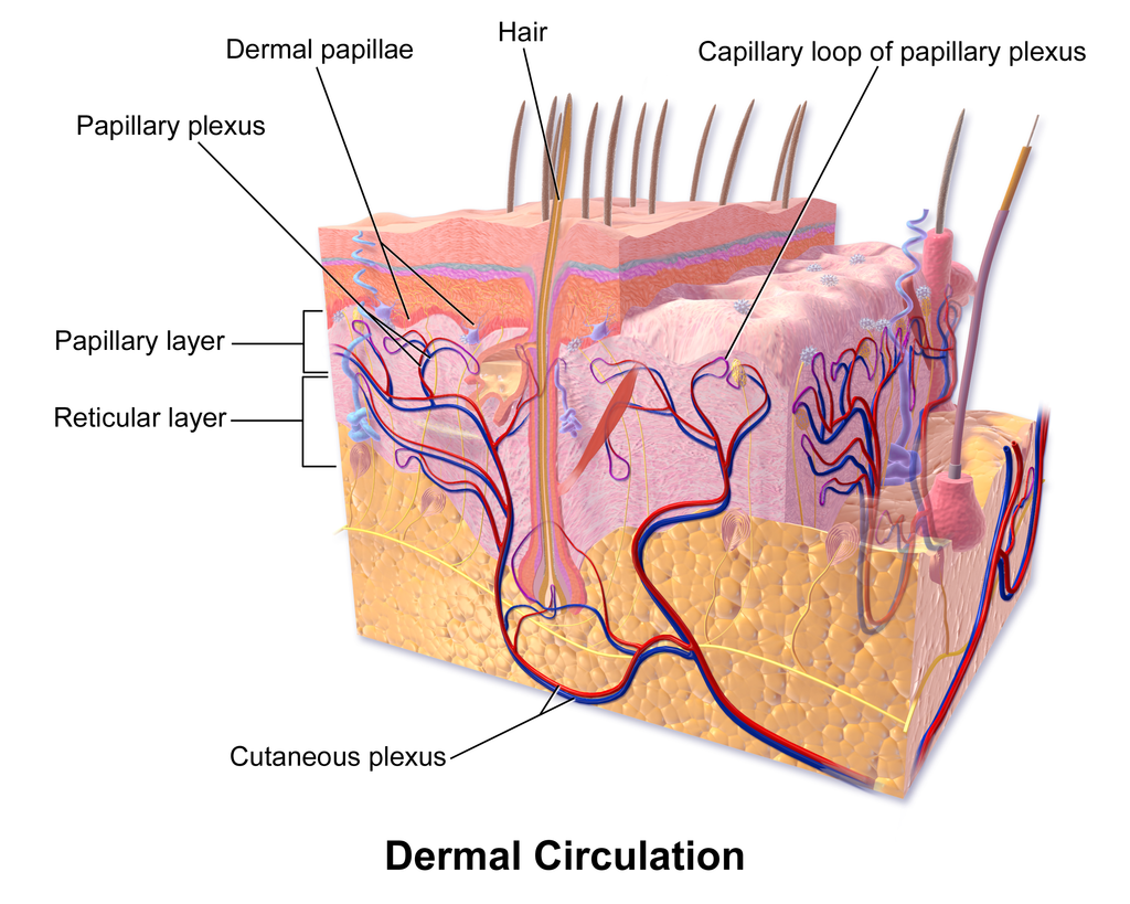
Glands
Glands in the reticular layer of the dermis include sweat glands and sebaceous (oil) glands. Both are exocrine glands, which are glands that release their secretions through ducts to nearby body surfaces. The diagram in Figure 10.4.5 shows these glands, as well as several other structures in the dermis.
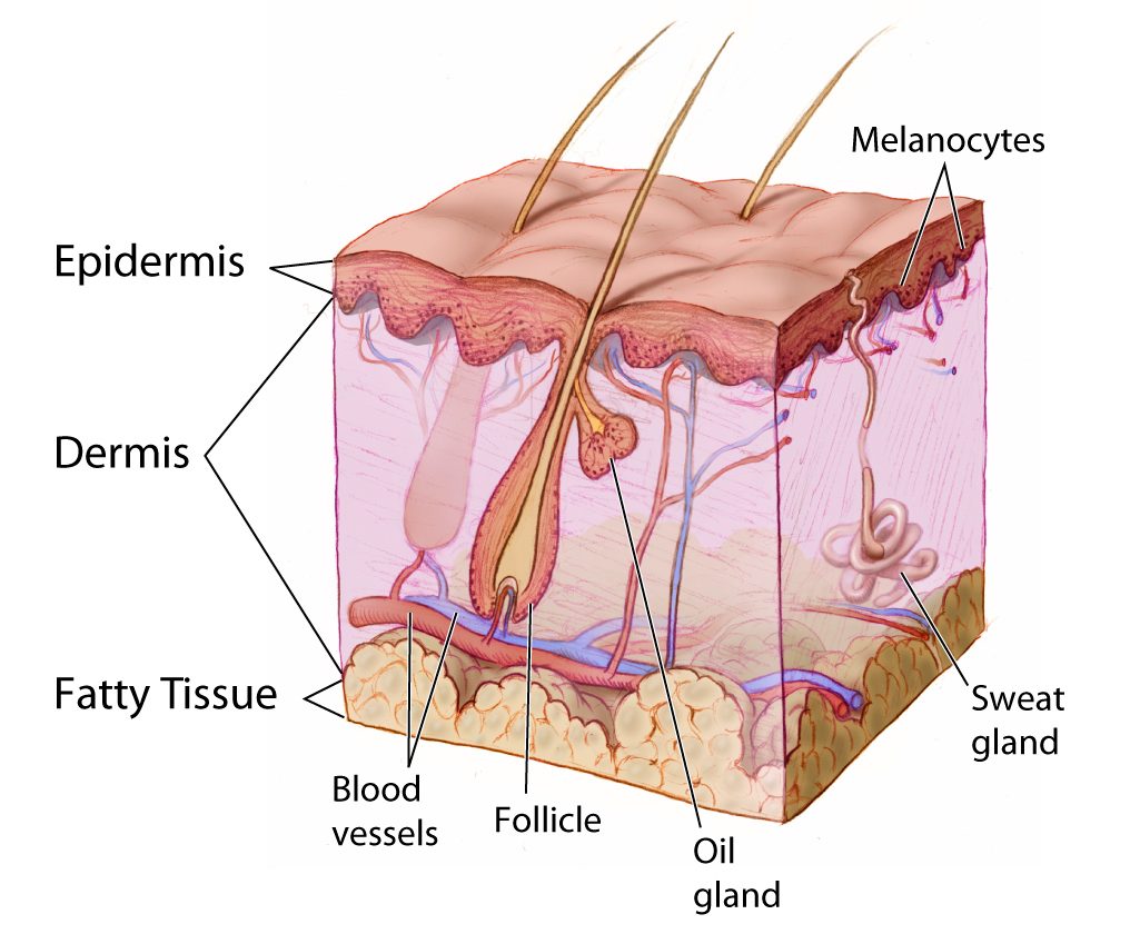
Sweat Glands
Sweat glands produce the fluid called sweat, which contains mainly water and salts. The glands have ducts that carry the sweat to hair follicles, or to the surface of the skin. There are two different types of sweat glands: eccrine glands and apocrine glands.
- Eccrine sweat glands occur in skin all over the body. Their ducts empty through tiny openings called pores onto the skin surface. These sweat glands are involved in temperature regulation.
- Apocrine sweat glands are larger than eccrine glands, and occur only in the skin of the armpits and groin. The ducts of apocrine glands empty into hair follicles, and then the sweat travels along hairs to reach the surface. Apocrine glands are inactive until puberty, at which point they start producing an oily sweat that is consumed by bacteria living on the skin. The digestion of apocrine sweat by bacteria causes body odor.
Sebaceous Glands
Sebaceous glands are exocrine glands that produce a thick, fatty substance called sebum. Sebum is secreted into hair follicles and makes its way to the skin surface along hairs. It waterproofs the hair and skin, and helps prevent them from drying out. Sebum also has antibacterial properties, so it inhibits the growth of microorganisms on the skin. Sebaceous glands are found in every part of the skin — except for the palms of the hands and soles of the feet, where hair does not grow.
Hair Follicles
Hair follicles are the structures where hairs originate (see the diagram above). Hairs grow out of follicles, pass through the epidermis, and exit at the surface of the skin. Associated with each hair follicle is a sebaceous gland, which secretes sebum that coats and waterproofs the hair. Each follicle also has a bed of capillaries, a nerve ending, and a tiny muscle called an arrector pili.
Functions of the Dermis
The main functions of the dermis are regulating body temperature, enabling the sense of touch, and eliminating wastes from the body.
Temperature Regulation
Several structures in the reticular layer of the dermis are involved in regulating body temperature. For example, when body temperature rises, the hypothalamus of the brain sends nerve signals to sweat glands, causing them to release sweat. An adult can sweat up to four litres an hour. As the sweat evaporates from the surface of the body, it uses energy in the form of body heat, thus cooling the body. The hypothalamus also causes dilation of blood vessels in the dermis when body temperature rises. This allows more blood to flow through the skin, bringing body heat to the surface, where it can radiate into the environment.
When the body is too cool, sweat glands stop producing sweat, and blood vessels in the skin constrict, thus conserving body heat. The arrector pili muscles also contract, moving hair follicles and lifting hair shafts. This results in more air being trapped under the hairs to insulate the surface of the skin. These contractions of arrector pili muscles are the cause of goose bumps.
Sensing the Environment
Sensory receptors in the dermis are mainly responsible for the body’s tactile senses. The receptors detect such tactile stimuli as warm or cold temperature, shape, texture, pressure, vibration, and pain. They send nerve impulses to the brain, which interprets and responds to the sensory information. Sensory receptors in the dermis can be classified on the basis of the type of touch stimulus they sense. Mechanoreceptors sense mechanical forces such as pressure, roughness, vibration, and stretching. Thermoreceptors sense variations in temperature that are above or below body temperature. Nociceptors sense painful stimuli. Figure 10.4.6 shows several specific kinds of tactile receptors in the dermis. Each kind of receptor senses one or more types of touch stimuli.
- Free nerve endings sense pain and temperature variations.
- Merkel cells sense light touch, shapes, and textures.
- Meissner’s corpuscles sense light touch.
- Pacinian corpuscles sense pressure and vibration.
- Ruffini corpuscles sense stretching and sustained pressure.
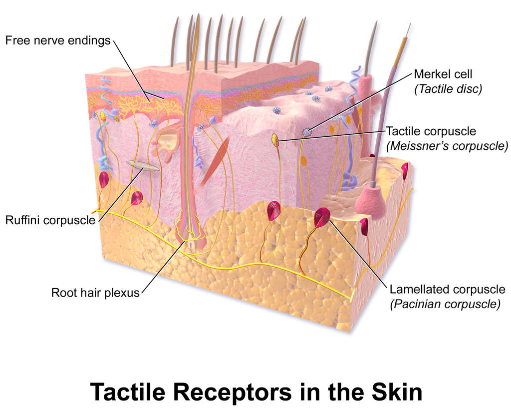
Excreting Wastes
The sweat released by eccrine sweat glands is one way the body excretes waste products. Sweat contains excess water, salts (electrolytes), and other waste products that the body must get rid of to maintain homeostasis. The most common electrolytes in sweat are sodium and chloride. Potassium, calcium, and magnesium electrolytes may be excreted in sweat, as well. When these electrolytes reach high levels in the blood, more are excreted in sweat. This helps to bring their blood levels back into balance. Besides electrolytes, sweat contains small amounts of waste products from metabolism, including ammonia and urea. Sweat may also contain alcohol in someone who has been drinking alcoholic beverages.
Feature: My Human Body
Acne is the most common skin disorder in the Canada. At least 20% of Canadians have acne at any given time and it affects approximately 90% of adolescents (as in Figure 10.4.7). Although acne occurs most commonly in teens and young adults, but it can occur at any age. Even newborn babies can get acne.
The main sign of acne is the appearance of pimples (pustules) on the skin, like those in the photo above. Other signs of acne may include whiteheads, blackheads, nodules, and other lesions. Besides the face, acne can appear on the back, chest, neck, shoulders, upper arms, and buttocks. Acne can permanently scar the skin, especially if it isn’t treated appropriately. Besides its physical effects on the skin, acne can also lead to low self-esteem and depression.
Acne is caused by clogged, sebum-filled pores that provide a perfect environment for the growth of bacteria. The bacteria cause infection, and the immune system responds with inflammation. Inflammation, in turn, causes swelling and redness, and may be associated with the formation of pus. If the inflammation goes deep into the skin, it may form an acne nodule.
Mild acne often responds well to treatment with over-the-counter (OTC) products containing benzoyl peroxide or salicylic acid. Treatment with these products may take a month or two to clear up the acne. Once the skin clears, treatment generally needs to continue for some time to prevent future breakouts.
If acne fails to respond to OTC products, nodules develop, or acne is affecting self-esteem, a visit to a dermatologist is in order. A dermatologist can determine which treatment is best for a given patient. A dermatologist can also prescribe prescription medications (which are likely to be more effective than OTC products) and provide other medical treatments, such as laser light therapies or chemical peels.
What can you do to maintain healthy skin and prevent or reduce acne? Dermatologists recommend the following tips:
- Wash affected or acne-prone skin (such as the face) twice a day, and after sweating.
- Use your fingertips to apply a gentle, non-abrasive cleanser. Avoid scrubbing, which can make acne worse.
- Use only alcohol-free products and avoid any products that irritate the skin, such as harsh astringents or exfoliants.
- Rinse with lukewarm water, and avoid using very hot or cold water.
- Shampoo your hair regularly.
- Do not pick, pop, or squeeze acne. If you do, it will take longer to heal and is more likely to scar.
- Keep your hands off your face. Avoid touching your skin throughout the day.
- Stay out of the sun and tanning beds. Some acne medications make your skin very sensitive to UV light.
10.4 Summary
- The dermis is the inner and thicker of the two major layers that make up the skin. It consists mainly of a matrix of connective tissues that provide strength and stretch. It also contains almost all skin structures, including sensory receptors and blood vessels.
- The dermis has two layers. The upper papillary layer has papillae extending upward into the epidermis and loose connective tissues. The lower reticular layer has denser connective tissues and structures, such as glands and hair follicles. Glands in the dermis include eccrine and apocrine sweat glands and sebaceous glands. Hair follicles are structures where hairs originate.
- Functions of the dermis include cushioning subcutaneous tissues, regulating body temperature, sensing the environment, and excreting wastes. The dense connective tissues of the dermis provide cushioning. The dermis regulates body temperature mainly by sweating and by vasodilation or vasoconstriction. The many tactile sensory receptors in the dermis make it the main organ for the sense of touch. Wastes excreted in sweat include excess water, electrolytes, and certain metabolic wastes.
10.4 Review Questions
- What is the dermis?
- Describe the basic anatomy of the dermis.
- Compare and contrast the papillary and reticular layers of the dermis.
- What causes epidermal ridges, and why can they be used to identify individuals?
- Name the two types of sweat glands in the dermis, and explain how they differ.
- What is the function of sebaceous glands?
- Describe the structures associated with hair follicles.
- Explain how the dermis helps regulate body temperature.
- Identify three specific kinds of tactile receptors in the dermis, along with the type of stimuli they sense.
- How does the dermis excrete wastes? What waste products does it excrete?
- What are subcutaneous tissues? Which layer of the dermis provides cushioning for subcutaneous tissues? Why does this layer provide most of the cushioning, instead of the other layer?
- For each of the functions listed below, describe which structure within the dermis carries it out.
- Brings nutrients to and removes wastes from dermal and lower epidermal cells
- Causes hairs to move
- Detects painful stimuli on the skin
10.4 Explore More
https://www.youtube.com/watch?v=FX-FwK0IIrE
How do you get rid of acne? SciShow, 2016.
https://www.youtube.com/watch?v=VcHQWMAClhQ&feature=emb_logo
When You Can't Scratch Away An Itch, Seeker, 2013.
Attributions
Figure 10.4.1
Goose_bumps by EverJean on Wikimedia Commons is used under a CC BY 2.0 (https://creativecommons.org/licenses/by/2.0) license.
Figure 10.4.2
Layers_of_the_Dermis by OpenStax College on Wikimedia Commons is used under a CC BY 3.0 (https://creativecommons.org/licenses/by/3.0) license.
Figure 10.4.3
Fingerprint_detail_on_male_finger_in_Třebíč,_Třebíč_District by Frettie on Wikimedia Commons is used under a CC BY 3.0 (https://creativecommons.org/licenses/by/3.0) license.
Figure 10.4.4
Blausen_0802_Skin_Dermal Circulation by BruceBlaus on Wikimedia commons is used under a CC BY 3.0 (https://creativecommons.org/licenses/by/3.0) license.
Figure 10.4.5
Anatomy_The_Skin_-_NCI_Visuals_Online by Don Bliss (artist) / National Cancer Institute (National Institutes of Health, with the ID 4604) is in the public domain (https://en.wikipedia.org/wiki/public_domain).
Figure 10.4.6
Blausen_0809_Skin_TactileReceptors by BruceBlaus on Wikimedia commons is used under a CC BY 3.0 (https://creativecommons.org/licenses/by/3.0) license.
Figure 10.4.7
Akne-jugend by Ellywa on Wikimedia Commons is released into the public domain (https://en.wikipedia.org/wiki/public_domain). (No machine-readable author provided. Ellywa assumed, based on copyright claims).
References
Betts, J. G., Young, K.A., Wise, J.A., Johnson, E., Poe, B., Kruse, D.H., Korol, O., Johnson, J.E., Womble, M., DeSaix, P. (2013, June 19). Figure 5.7 Layers of the dermis [digital image]. In Anatomy and Physiology (Section 5.1 Layers of the skin). OpenStax. https://openstax.org/books/anatomy-and-physiology/pages/5-1-layers-of-the-skin
Blausen.com staff. (2014). Medical gallery of Blausen Medical 2014. WikiJournal of Medicine 1 (2). DOI:10.15347/wjm/2014.010. ISSN 2002-4436.
SciShow. (2016, October 26). How do you get rid of acne? YouTube. https://www.youtube.com/watch?v=FX-FwK0IIrE
Seeker. (2013, October 26). When you can't scratch away an itch. YouTube. https://www.youtube.com/watch?v=VcHQWMAClhQ&feature=emb_logo
Created by CK-12 Foundation/Adapted by Christine Miller

Fashion Statement
This colourful hairstyle makes quite a fashion statement. Many people spend a lot of time and money on their hair, even if they don’t have an exceptional hairstyle like this one. Besides its display value, hair actually has important physiological functions.
What is Hair?
Hair is a filament that grows from a hair follicle in the dermis of the skin. It consists mainly of tightly packed, keratin-filled cells called keratinocytes. The human body is covered with hair follicles, with the exception of a few areas, including the mucous membranes, lips, palms of the hands, and soles of the feet.
Structure of Hair
The part of the hair located within the follicle is called the hair root. The root is the only living part of the hair. The part of the hair that is visible above the surface of the skin is the hair shaft. The shaft of the hair has no biochemical activity and is considered dead.
Follicle and Root
Hair growth begins inside a follicle (see Figure 10.5.2 below). Each hair follicle contains stem cells that can keep dividing, which allows hair to grow. The stem cells can also regrow a new hair after one falls out. Another structure associated with a hair follicle is a sebaceous gland that produces oily sebum. The sebum lubricates and helps to waterproof the hair. A tiny arrector pili muscle is also attached to the follicle. When it contracts, the follicle moves, and the hair in the follicle stands up.
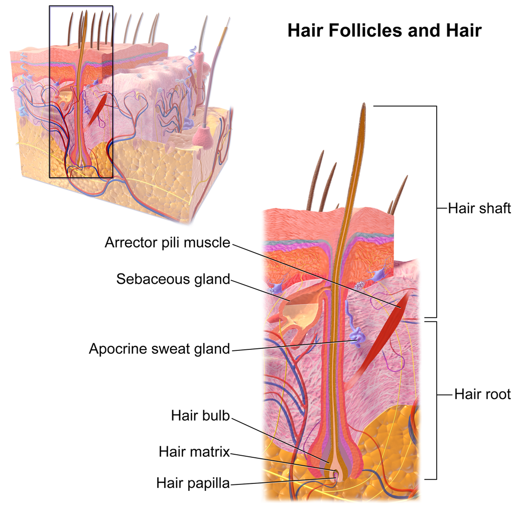
Shaft
The hair shaft is a hard filament that may grow very long. Hair normally grows in length by about half an inch a month. In cross-section, a hair shaft can be divided into three zones, called the cuticle, cortex, and medulla.
- The cuticle (or outer coat) is the outermost zone of the hair shaft. It consists of several layers of flat, thin keratinocytes that overlap one another like shingles on a roof. This arrangement helps the cuticle repel water. The cuticle is also covered with a layer of lipids, just one molecule thick, which increases its ability to repel water. This is the zone of the hair shaft that is visible to the eye.
- The cortex is the middle zone of the hair shaft, and it is also the widest part. The cortex is highly structured and organized, consisting of keratin bundles in rod-like structures. These structures give hair its mechanical strength. The cortex also contains melanin, which gives hair its colour.
- The medulla is the innermost zone of the hair shaft. This is a small, disorganized, and more open area at the center of the hair shaft. The medulla is not always present. When it is present, it contains highly pigmented cells full of keratin.
Characteristics of Hair
Two visible characteristics of hair are its colour and texture. In adult males, the extent of balding is another visible characteristic. All three characteristics are genetically controlled.
Hair Colour
All natural hair colours are the result of melanin, which is produced in hair follicles and packed into granules in the hair. Two forms of melanin are found in human hair: eumelanin and pheomelanin. Eumelanin is the dominant pigment in brown hair and black hair, and pheomelanin is the dominant pigment in red hair. Blond hair results when you have only a small amount of melanin in the hair. Gray and white hair occur when melanin production slows down, and eventually stops.
Figure 10.5.3 Variation in hair colouration. Which types of melanin are present for each hair colour shown?
Hair Texture
Hair exists in a variety of textures. The main aspects of hair texture are the curl pattern, thickness, and consistency.
- The shape of the hair follicle determines the shape of the hair shaft. The shape of the hair shaft, in turn, determines the curl pattern of the hair. Round hair shafts produce straight hair. Hair shafts that are oval or have other shapes produce wavy or curly hair .
- The size of the hair follicle determines the thickness of hair. Thicker hair has greater volume than thinner hair.
- The consistency of hair is determined by the hair follicle volume and the condition of the hair shaft. The consistency of hair is generally classified as fine, medium, or coarse. Fine hair has the smallest circumference, and coarse hair has the largest circumference. Medium hair falls in between these two extremes. Coarse hair also has a more open cuticle than thin or medium hair does, which causes it to be more porous.

Figure 10.5.4 Curly hair has a differently shaped shaft than straight hair.
Functions of Hair
In humans, one function of head hair is to provide insulation and help the head retain heat. Head hair also protects the skin on the head from damage by UV light.
The function of hair in other locations on the body is debated. One idea is that body hair helps keep us warm in cold weather. When the body is too cold, arrector pili muscles contract and cause hairs to stand up (shown in Figure 10.5.5), trapping a layer of warm air above the epidermis. However, this is more effective in mammals that have thick hair or fur than it is in relatively hairless human beings.
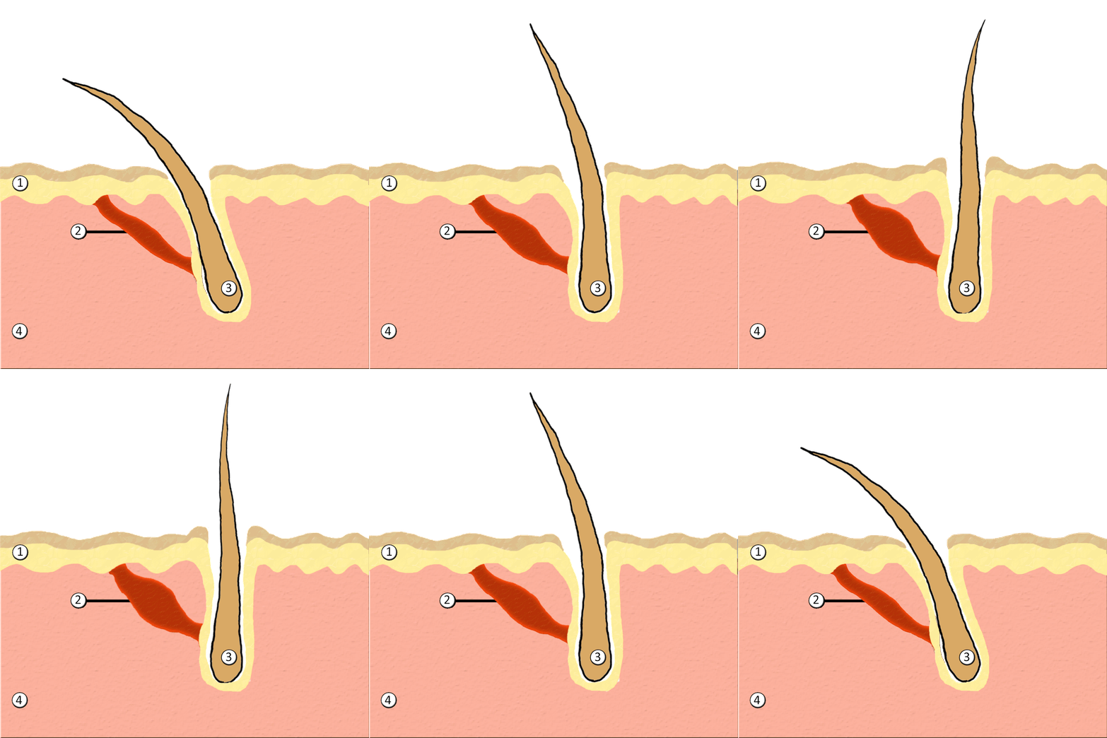
Human hair has an important sensory function, as well. Sensory receptors in the hair follicles can sense when the hair moves, whether it moves because of a breeze, or because of the touch of a physical object. The receptors may also provide sensory awareness of the presence of parasites on the skin.

Some hairs, such as the eyelashes, are especially sensitive to the presence of potentially harmful matter. The eyelashes grow at the edge of the eyelid and can sense when dirt, dust, or another potentially harmful object is too close to the eye. The eye reflexively closes as a result of this sensation. The eyebrows also provide some protection to the eyes. They protect the eyes from dirt, sweat, and rain. In addition, the eyebrows play a key role in nonverbal communication (see Figure 10.5.6). They help express emotions such as sadness, anger, surprise, and excitement.
Hair in Human Evolution
Among mammals, humans are nearly unique in having undergone significant loss of body hair during their evolution. Humans are also unlike most other mammals in having curly hair as one variation in hair texture. Even non-human primates (see Figure 10.5.7) all have straight hair. This suggests that curly hair evolved at some point during human evolution.
Loss of Body Hair
One hypothesis for the loss of body hair in the human lineage is that it would have facilitated cooling of the body by the evaporation of sweat. Humans also evolved far more sweat glands than other mammals, which is consistent with this hypothesis, because sweat evaporates more quickly from less hairy skin. Another hypothesis for human hair loss is that it would have led to fewer parasites on the skin. This might have been especially important when humans started living together in larger, more crowded social groups.
These hypotheses may explain why we lost body hair, but they can’t explain why we didn’t also lose head hair and hair in the pubic region and armpits. It is possible that head hair was retained because it protected the scalp from UV light. As our bipedal ancestors walked on the open savannas of equatorial Africa, the skin on the head would have been an area exposed to the most direct rays of sunlight in an upright hominid. Pubic and armpit hair may have been retained because they served as signs of sexual maturity, which would have been important for successful mating and reproduction.
Evolution of Curly Hair
Greater protection from UV light has also been posited as a possible selective agent favoring the evolution of curly hair. Researchers have found that straight hair allows more light to pass into the body through the hair shaft via the follicle than does curly hair. In this way, human hair is like a fibre optic cable. It allows light to pass through easily when it is straight, but it impedes the passage of light when it is kinked or coiled. This is indirect evidence that UV light may have been a selective agent leading to the evolution of curly hair.
Social and Cultural Significance of Hair
Hair has great social significance for human beings. Body hair is an indicator of biological sex, because hair distribution is sexually dimorphic. Adult males are generally hairier than adult females, and facial hair in particular is a notable secondary male sex characteristic. Hair may also be an indicator of age. White hair is a sign of older age in both males and females, and male pattern baldness is a sign of older age in males. In addition, hair colour and texture can be a sign of ethnic ancestry.
Hair also has great cultural significance. Hairstyle and colour may be an indicator of social group membership and for better or worse can be associated with specific stereotypes. Head shaving has been used in many times and places as a punishment, especially for women. On the other hand, in some cultures, cutting off one’s hair symbolizes liberation from one’s past. In other cultures, it is a sign of mourning. There are also many religious-based practices involving hair. For example, the majority of Muslim women hide their hair with a headscarf. Sikh men grow their hair long and cover it with a turban. Amish men (like the one pictured in Figure 10.5.8) grow facial hair only after they marry — but just a beard, and not a mustache.

Unfortunately, sometimes hairstyle, colour and characteristics are used to apply stereotypes, particularly with respect to women. "Blonde jokes" are a good example of how negative stereotypes are maintained despite having no actual truth behind them. Many stereotypes related to hair are hidden, even from persons perpetrating the stereotype. Often a hairstyle is judged by another as having ties to gender, sexuality, worldview and/or socioeconomic status; even when these inferences are woefully inaccurate. It is important to be aware of our own biases and determine if these biases are appropriate - take a look at the collage in Figure 10.5.9. What are your initial reactions? Are these reactions founded in fact? Do you harbor an unfair bias?
Figure 10.5.9 What are your biases? Are they fair?
10.5 Summary
- Hair is a filament that grows from a hair follicle in the dermis of the skin. It consists mainly of tightly packed, keratin-filled cells called keratinocytes. The human body is almost completely covered with hair follicles.
- The part of a hair that is within the follicle is the hair root. This is the only living part of a hair. The part of a hair that is visible above the skin surface is the hair shaft. It consists of dead cells.
- Hair growth begins inside a follicle when stem cells within the follicle divide to produce new keratinocytes. An individual hair may grow to be very long.
- A hair shaft has three zones: the outermost zone called the cuticle; the middle zone called the cortex; and the innermost zone called the medulla.
- Genetically controlled, visible characteristics of hair include hair colour, hair texture, and the extent of balding in adult males. Melanin (eumelanin and/or pheomelanin) is the pigment that gives hair its colour. Aspects of hair texture include curl pattern, thickness, and consistency.
- Functions of head hair include providing insulation and protecting skin on the head from UV light. Hair everywhere on the body has an important sensory function. Hair in eyelashes and eyebrows protects the eyes from dust, dirt, sweat, and other potentially harmful substances. The eyebrows also play a role in nonverbal communication.
- Among mammals, humans are nearly unique in having undergone significant loss of body hair during their evolution, probably because sweat evaporates more quickly from less hairy skin. Curly hair also is thought to have evolved at some point during human evolution, perhaps because it provided better protection from UV light.
- Hair has social significance for human beings, because it is an indicator of biological sex, age, and ethnic ancestry. Human hair also has cultural significance. Hairstyle may be an indicator of social group membership, for example.
10.5 Review Questions
-
- Compare and contrast the hair root and hair shaft.
- Describe hair follicles.
-
-
- Explain variation in human hair colour.
- What factors determine the texture of hair?
- Describe two functions of human hair.
- What hypotheses have been proposed for the loss of body hair during human evolution?
- Discuss the social and cultural significance of human hair.
- Describe one way in which hair can be used as a method of communication in humans.
- Explain why waxing or tweezing body hair, which typically removes hair down to the root, generally keeps the skin hair-free for a longer period of time than shaving, which cuts hair off at the surface of the skin.
10.5 Explore More
https://www.youtube.com/watch?v=8diYLhl8bWU
Why do some people go bald? - Sarthak Sinha, TED-Ed, 2015.
https://www.youtube.com/watch?v=kNw8V_Fkw28
Hair Love | Oscar®-Winning Short Film (Full) | Sony Pictures Animation, 2019.
https://www.youtube.com/watch?v=hDW5e3NR1Cw
Why do we care about hair | Naomi Abigail | TEDxBaDinh, TEDx Talks, 2015.
Attributions
Figure 10.5.1
Hair by jessica-dabrowski-TETR8YLSqt4 [photo] by Jessica Dabrowski on Unsplash is used under the Unsplash License (https://unsplash.com/license).
Figure 10.5.2
Blausen_0438_HairFollicleAnatomy_02 by BruceBlaus on Wikimedia Commons is used under a CC BY 3.0 (https://creativecommons.org/licenses/by/3.0) license.
Figure 10.5.3
- Standing tall by Ilaya Raja on Unsplash is used under the Unsplash License (https://unsplash.com/license).
- Blond-haired woman smiling by Carlos Lindner on Unsplash is used under the Unsplash License (https://unsplash.com/license).
- Smith Mountain Lake redhead by Chris Ross Harris on Unsplash is used under the Unsplash License (https://unsplash.com/license).
- Through the look of experience by Laura Margarita Cedeño Peralta on Unsplash is used under the Unsplash License (https://unsplash.com/license).
Figure 10.5.4
Curly hair by chris-benson-clvEami9RN4 [photo] by Chris Benson on Unsplash is used under the Unsplash License (https://unsplash.com/license).
Figure 10.5.5
1024px-PilioerectionAnimation by AnthonyCaccese on Wikimedia Commons is used under a CC BY-SA 4.0 (https://creativecommons.org/licenses/by-sa/4.0/deed.en) license.
Figure 10.5.6
Pout by alexander-dummer-Em8I8Z_DwA4 [photo] by Alexander Dummer on Unsplash is used under the Unsplash License (https://unsplash.com/license).
Figure 10.5.7
Cotton_top_tamarin_monkey._(12046035746) by Bernard Spragg. NZ, from Christchurch, New Zealand on Wikimedia Commons is used under a CC0 1.0 Universal
Public Domain Dedication license (https://creativecommons.org/publicdomain/zero/1.0/deed.en).
Figure 10.5.8
Amish hairstyle by CK-12 Foundation is used under a CC BY-NC 3.0 (https://creativecommons.org/licenses/by-nc/3.0/) license.
 ©CK-12 Foundation Licensed under
 ![]() • Terms of Use • Attribution
• Terms of Use • Attribution
Figure 10.5.9
- Rainbow Hair Bubble Man by Behrouz Jafarnezhad on Unsplash is used under the Unsplash License (https://unsplash.com/license).
- Pink hair in Atlanta, United States by Tammie Allen on Unsplash is used under the Unsplash License (https://unsplash.com/license).
- Magdalena 2 by Valerie Elash on Unsplash is used under the Unsplash License (https://unsplash.com/license).
- Perfect Style by Daria Volkova on Unsplash is used under the Unsplash License (https://unsplash.com/license)
- Stay Classy by Fayiz Musthafa on Unsplash is used under the Unsplash License (https://unsplash.com/license)
- Take your time by Jan Tinneberg on Unsplash is used under the Unsplash License (https://unsplash.com/license)
References
Blausen.com staff. (2014). Medical gallery of Blausen Medical 2014. WikiJournal of Medicine 1 (2). DOI:10.15347/wjm/2014.010. ISSN 2002-4436.
Brainard, J/ CK-12 Foundation. (2016). Figure 7 This style of facial hair is adopted by most Amish men after they marry [digital image]. In CK-12 College Human Biology (Section 12.5) [online Flexbook]. CK12.org. https://www.ck12.org/book/ck-12-college-human-biology/section/12.5/
Sony Pictures Animation. (2019, December 5). Hair love | Oscar®-winning short film (Full) | Sony Pictures Animation. YouTube. https://www.youtube.com/watch?v=kNw8V_Fkw28
TED-Ed. (2015, August 25). Why do some people go bald? – Sarthak Sinha. YouTube. https://www.youtube.com/watch?v=8diYLhl8bWU
TEDx Talks. (2015, February 4). Why do we care about hair | Naomi Abigail | TEDxBaDinh. YouTube. https://www.youtube.com/watch?v=hDW5e3NR1Cw
Created by CK-12 Foundation/Adapted by Christine Miller
Nail Art
Painting nails with coloured polish for aesthetic reasons is nothing new. In fact, there is evidence of this practice dating back to at least 3000 BCE. Today, painting and otherwise decorating the nails is big business, with annual revenues in the billions of dollars in North America alone! With all the attention (and money) given to nails as decorative objects, it’s easy to forget that they also have important biological functions.
What Are Nails?
Nails are accessory organs of the skin. They are made of sheets of dead keratinocytes and are found on the far (or distal) ends of the fingers and toes. The keratin in nails makes them hard, but flexible. Nails serve a number of purposes, including protecting the digits, enhancing sensations, and acting as tools.
Nail Anatomy
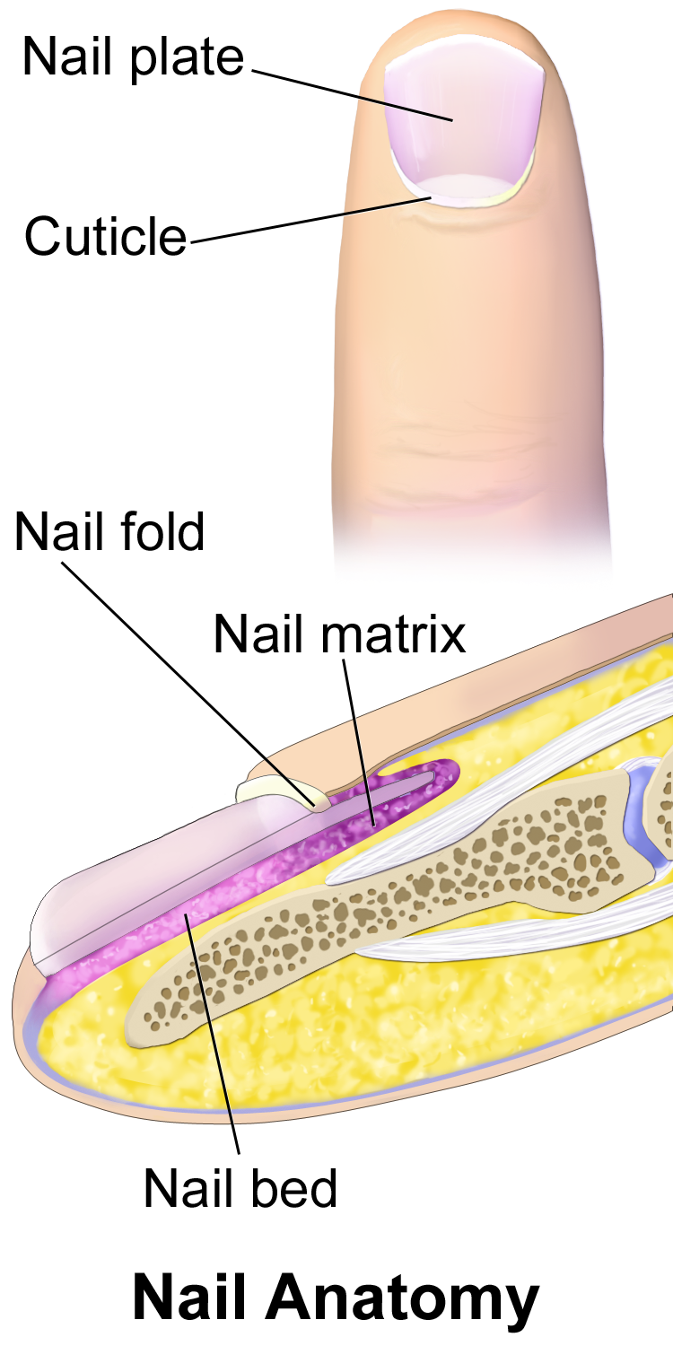
A nail has three main parts: the root, plate, and free margin. Other structures around or under the nail include the nail bed, cuticle, and nail fold. These structures are shown in Figure 10.6.2.
- The nail root is the portion of the nail found under the surface of the skin at the near (or proximal) end of the nail. It is where the nail begins.
- The nail plate (or body) is the portion of the nail that is external to the skin. It is the visible part of the nail.
- The free margin is the portion of the nail that protrudes beyond the distal end of the finger or toe. This is the part that is cut or filed to keep the nail trimmed.
- The nail bed is the area of skin under the nail plate. It is pink in colour, due to the presence of capillaries in the dermis.
- The cuticle is a layer of dead epithelial cells that overlaps and covers the edge of the nail plate. It helps to seal the edges of the nail to prevent infection of the underlying tissues.
- The nail fold is a groove in the skin in which the side edges of the nail plate are embedded.
Nail Growth
Nails grow from a deep layer of living epidermal tissue, known as the nail matrix, at the proximal end of the nail (see the bottom of the diagram in Figure 10.6.2). The nail matrix surrounds the nail root. It contains stem cells that divide to form keratinocytes, which are cells that produce keratin and make up the nail.
Formation of the Nail Root and Nail Plate
The keratinocytes produced by the nail matrix accumulate to form tough, hard, translucent sheets of dead cells filled with keratin. The sheets make up the nail root, which slowly grows out of the skin and becomes the nail plate when it reaches the skin surface. As the nail grows longer, the cells of the nail root and nail plate are pushed toward the distal end of the finger or toe by new cells being formed in the nail matrix. The upper epidermal cells of the nail bed also move along with the nail plate as it grows toward the tip of the digit.
The proximal end of the nail plate near the root has a whitish crescent shape called the lunula. This is where a small amount of the nail matrix is visible through the nail plate. The lunula is most pronounced in the nails of the thumbs, and may not be visible in the nails of the little fingers.
Rate of Nail Growth
Nails grow at an average rate of 3 mm a month. Fingernails, however, grow up to four times as fast as toenails. If a fingernail is lost, it takes between three and six months to regrow completely, whereas a toenail takes between 12 and 18 months to regrow. The actual rate of growth of an individual’s nails depends on many factors, including age, sex, season, diet, exercise level, and genes. If protected from breaking, nails can sometimes grow to be very long. The Chinese doctor in the photo below (Figure 10.6.3) has very long nails on two fingers of his left hand. This picture was taken in 1920 in China, where having long nails was a sign of aristocracy since it implied that one was wealthy enough to not have to do physical labour.

Functions of Nails
Both fingernails and toenails protect the soft tissues of the fingers and toes from injury. Fingernails also serve to enhance sensation and precise movements of the fingertips through the counter-pressure exerted on the pulp of the fingers by the nails. In addition, fingernails can function as several different types of tools. For example, they enable a fine precision grip like tweezers, and can also be used for cutting and scraping.
Nails and Health
Healthcare providers, particularly EMTs, often examine the fingernail beds as a quick and easy indicator of oxygen saturation of the blood, or the amount of blood reaching the extremities. If the nail beds are bluish or purple, it is generally a sign of low oxygen saturation. To see if blood flow to the extremities is adequate, a blanch test may be done. In this test, a fingernail is briefly depressed to turn the nail bed white by forcing the blood out of its capillaries. When the pressure is released, the pink colour of the nail bed should return within a second or two if there is normal blood flow. If the return to a pink colour is delayed, then it can be an indicator of low blood volume, due to dehydration or shock.
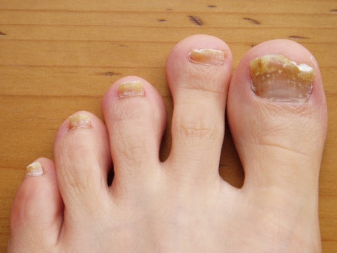
How the visible portion of the nails appears can be used as an indicator of recent health status. In fact, nails have been used as diagnostic tools for hundreds — if not thousands — of years. Nail abnormalities, such as deep grooves, brittleness, discolouration, or unusually thin or thick nails, may indicate various illnesses, nutrient deficiencies, drug reactions, or other health problems.
Nails — especially toenails — are common sites of fungal infections (shown in Figure 10.6.4), causing nails to become thickened and yellowish in colour. Toenails are more often infected than fingernails because they are often confined in shoes, which creates a dark, warm, moist environment where fungi can thrive. Toes also tend to have less blood flow than fingers, making it harder for the immune system to detect and stop infections in toenails.
Although nails are harder and tougher than skin, they are also more permeable. Harmful substances may be absorbed through the nails and cause health problems. Some of the substances that can pass through the nails include the herbicide Paraquat, fungicidal agents such as miconazole (e.g., Monistat), and sodium hypochlorite, which is an ingredient in common household bleach. Care should be taken to protect the nails from such substances when handling or immersing the hands in them by wearing latex or rubber gloves.
Feature: Reliable Sources
Do you get regular manicures or pedicures from a nail technician? If so, there is a chance that you are putting your health at risk. Nail tools that are not properly disinfected between clients may transmit infections from one person to another. Cutting the cuticles with scissors may create breaks in the skin that let infective agents enter the body. Products such as acrylics, adhesives, and UV gels that are applied to the nails may be harmful, especially if they penetrate the nails and enter the skin.
Use the Internet to find several reliable sources that address the health risks of professional manicures or pedicures. Try to find answers to the following questions:
- What training and certification are required for professional nail technicians?
- What licenses and inspections are required for nail salons?
- What hygienic practices should be followed in nail salons to reduce the risk of infections being transmitted to clients?
- Which professional nail products are potentially harmful to the human body and which are safer?
- How likely is it to have an adverse health consequence when you get a professional manicure or pedicure?
- What steps can you take to ensure that a professional manicure or pedicure is safe?
10.6 Summary
- Nails are accessory organs of the skin, consisting of sheets of dead, keratin-filled keratinocytes. The keratin in nails makes them hard, but flexible.
- A nail has three main parts: the nail root (which is under the epidermis), the nail plate (which is the visible part of the nail), and the free margin (which is the distal edge of the nail). Other structures under or around a nail include the nail bed, cuticle, and nail fold.
- A nail grows from a deep layer of living epidermal tissues — called the nail matrix — at the proximal end of the nail. Stem cells in the nail matrix keep dividing to allow nail growth, forming first the nail root and then the nail plate as the nail continues to grow longer and emerges from the epidermis.
- Fingernails grow faster than toenails. Actual rates of growth depend on many factors, such as age, sex, and season.
- Functions of nails include protecting the digits, enhancing sensations and precise movements of the fingertips, and acting as tools.
- The colour of the nail bed can be used to quickly assess oxygen and blood flow in a patient. How the nail plate grows out can reflect recent health problems, such as illness or nutrient deficiency.
- Nails — and especially toenails — are prone to fungus infections. Nails are more permeable than skin and can absorb several harmful substances, such as herbicides.
10.6 Review Questions
- What are nails?
-
- Explain why most of the nail plate looks pink.
- Describe a lunula.
- Explain how a nail grows.
- Identify three functions of nails.
- Give several examples of how nails are related to health.
- What is the cuticle of the nail composed of? What is the function of the cuticle? Why is it a bad idea to cut the cuticle during a manicure?
- Is the nail plate composed of living or dead cells?
10.6 Explore More
https://www.youtube.com/watch?v=G35kPhbUZdg
Longest Fingernails - Guinness World Records 60th Anniversary,
Guinness World Records, 2014.
https://www.youtube.com/watch?v=aTSVHwzkYI4&feature=emb_logo
5 Things Your Nails Can Say About Your Health, SciShow, 2015.
https://www.youtube.com/watch?v=7w2gCBL1MCg
Claws vs. Nails - Matthew Borths, TED-Ed, 2019.
Attributions
Figure 10.6.1
Nails by allison-christine-vPrqHSLdF28 [photo] by allison christine on Unsplash is used under the Unsplash License (https://unsplash.com/license).
Figure 10.6.2
Blausen_0406_FingerNailAnatomy by BruceBlaus on Wikimedia Commons is used under a CC BY 3.0 (https://creativecommons.org/licenses/by/3.0) license.
Figure 10.6.3
Chinese_doctor_with_long_finger_nails_(an_aristocrat),_ca.1920_(CHS-249) by Pierce, C.C. (Charles C.), 1861-1946 from the USC Digital Library on Wikimedia Commons is in the public domain (https://en.wikipedia.org/wiki/public_domain).
Figure 10.6.4
Toenail fungus Nagelpilz-3 by Pepsyrock on Wikimedia Commons is released into the public domain (https://en.wikipedia.org/wiki/public_domain).
Figure 10.6.5
OLYMPUS DIGITAL CAMERA by Stoive at the English language Wikipedia, on Wikimedia Commons is used under a CC BY-SA 3.0 (http://creativecommons.org/licenses/by-sa/3.0/) license.
References
Blausen.com staff. (2014). Medical gallery of Blausen Medical 2014. WikiJournal of Medicine 1 (2). DOI:10.15347/wjm/2014.010. ISSN 2002-4436.
Guiness World Records. (2014, December 8). Longest fingernails - Guinness World Records 60th Anniversary. YouTube. https://www.youtube.com/watch?v=G35kPhbUZdg
SciShow. (2015, September 14). 5 things your nails can say about your health. YouTube. https://www.youtube.com/watch?v=aTSVHwzkYI4
TED-Ed. (2019, October 29). Claws vs. nails - Matthew Borths. YouTube. https://www.youtube.com/watch?v=7w2gCBL1MCg
Created by CK-12 Foundation/Adapted by Christine Miller

Roasted Bone Marrow
Do you recognize the food item in the top left of Figure 11.4.1? It’s roasted bone marrow, still inside the bones, and it is considered a delicacy in some cuisines. Marrow is a type of tissue found inside many animal bones, including our own. It’s a soft tissue that, in adults, may be mostly fat. You’ll learn more about bone marrow and other tissues that make up bones when you read this section.
What Are Bones?
Bones are organs that consist primarily of bone tissue, also called osseous tissue. Bone tissue is a type of connective tissue consisting mainly of a collagen matrix that is mineralized with calcium and phosphorus crystals. The combination of flexible collagen and hard mineral crystals makes bone tissue hard, without making it brittle.
Bone Anatomy
There are several different types of tissues in bones, including two types of osseous tissues. Osseous tissues, in turn, consist of several different types of bone cells.
Types of Osseous Tissue
The two different types of osseous tissue are compact bone tissue (also called hard or cortical bone) and spongy bone tissue (also called cancellous or trabecular bone). Both are shown in the diagrams of a typical bone in Figures 11.4.2 and 11.4.3.
Flat bones are typically enveloped by compact bone, with a center of spongy bone.
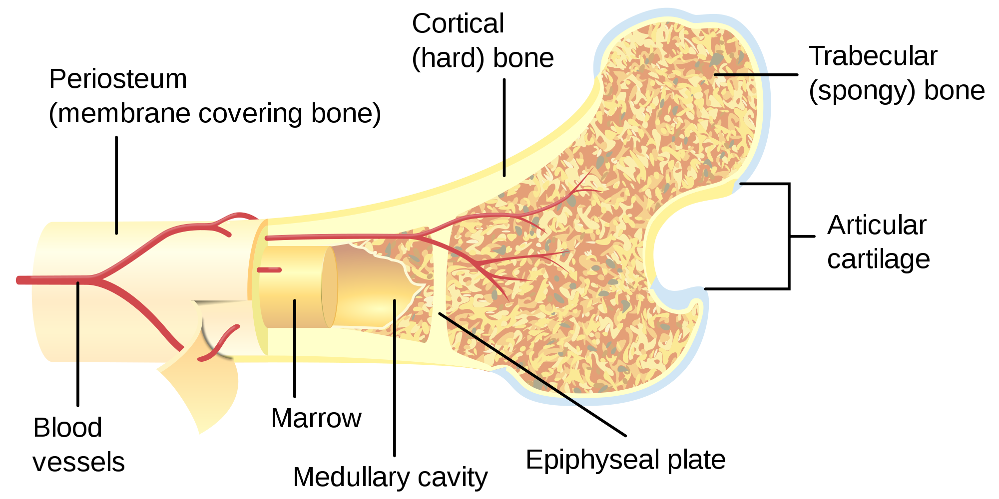 |
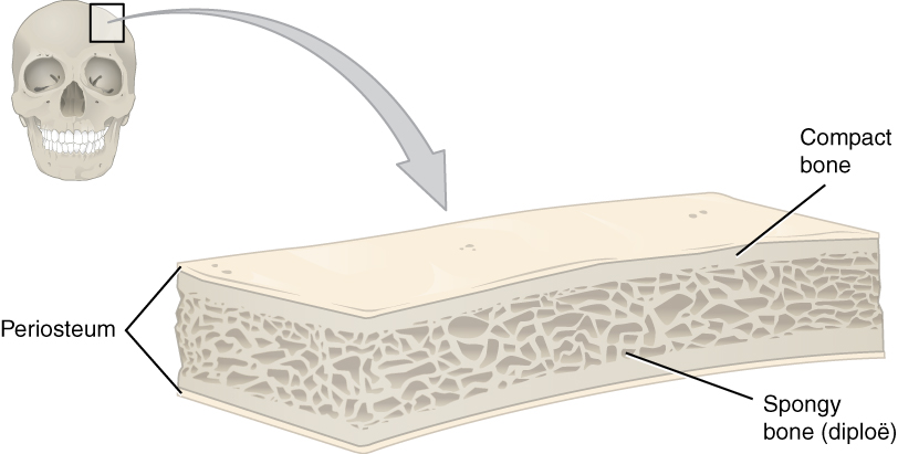 |
Compact bone tissue forms the extremely hard outside layer of bones. Compact bone tissue gives bone its smooth, dense, solid appearance. It accounts for about 80% of the total bone mass of the adult skeleton. Spongy bone tissue fills part or all of the interior of many bones. As its name suggests, spongy bone is porous like a sponge, containing an irregular network of spaces, as shown in Figures 11.4.4 and 11.4.5. This makes spongy bone much less dense than compact bone. Spongy bone has a greater surface area than compact bone, but makes up only 20% of bone mass.
 |
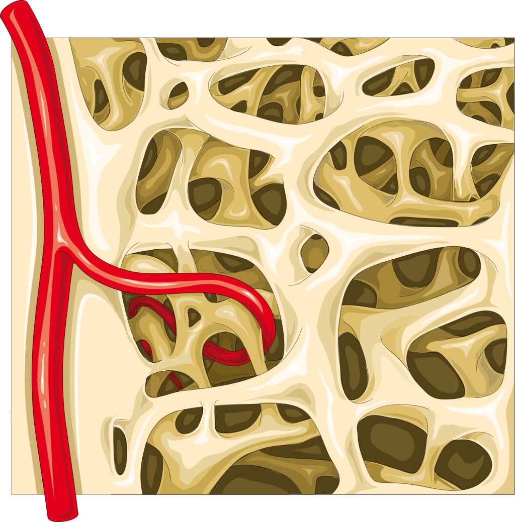 |
Both compact and spongy bone tissues have the same types of cells, but they differ in how the cells are arranged. The cells in compact bone are arranged in multiple microscopic columns, whereas the cells in spongy bone are arranged in a looser, more open network. These cellular differences explain why compact and spongy bone tissues have such different structures.
Other Tissues in Bones
Besides compact and spongy bone tissues, bones contain several other tissues, including blood vessels and nerves. In addition, bones contain bone marrow and periosteum.
- Bone marrow is a soft connective tissue found inside a cavity, called the marrow cavity. There are two types of marrow in adults — yellow bone marrow (which consists mostly of fat) and red bone marrow. All marrow is red in newborns, but by adulthood, much of the red marrow has changed to yellow marrow. In adults, red marrow is found mainly in the femur, ribs, vertebrae, and pelvic bones. Only red bone marrow contains hematopoietic stem cells that give rise to red blood cells, white blood cells, and platelets in the process of hematopoiesis.
- Periosteum is a tough, fibrous membrane that covers the outer surface of bones. It provides a protective covering for compact bone tissue. It is also the source of new bone cells.
Bone Cells
As shown in Figure 11.4.6, bone tissues are composed of four different types of bone cells: osteoblasts, osteocytes, osteoclasts, and osteogenic cells.
- Osteoblasts are bone cells with a single nucleus that make and mineralize bone matrix. They make a protein mixture that is composed primarily of collagen and creates the organic part of the matrix. They also release calcium and phosphate ions that form mineral crystals within the matrix. In addition, they produce hormones that play a role in the mineralization of the matrix.
- Osteocytes are mainly inactive bone cells that form from osteoblasts that have become entrapped within their own bone matrix. Osteocytes help regulate the formation and breakdown of bone tissue. They have multiple cell projections that are thought to be involved in communication with other bone cells.
- Osteoclasts are bone cells with multiple nuclei that resorb bone tissue and break down bone. They dissolve the minerals in bone and release them into the blood.
- Osteogenic cells are undifferentiated stem cells. They are the only bone cells that can divide. When they do, they differentiate and develop into osteoblasts.
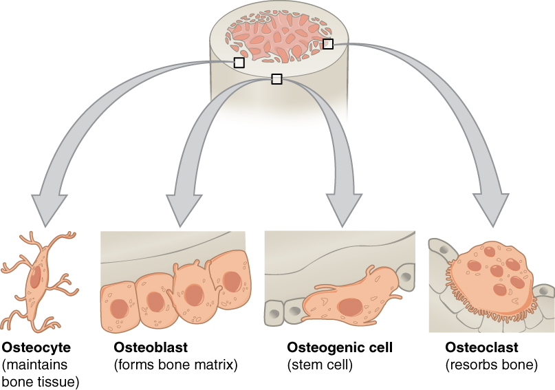
Bone is very active tissue. It is constantly remodeled by the work of osteoblasts and osteoclasts. Osteoblasts continuously make new bone, and osteoclasts keep breaking down bone. This allows for minor repair of bones, as well as homeostasis of mineral ions in the blood.
Types of Bones
There are six types of bones in the human body, categorized based on their shape or location: long, short, flat, sesamoid, sutural, and irregular bones. You can see an example of each type of bone in Figure 11.4.7.
- Long bones are characterized by a shaft that is much longer than it is wide, and by a rounded head at each end of the shaft. Long bones are made mostly of compact bone, with lesser amounts of spongy bone and marrow. Most bones of the limbs, including those of the fingers and toes, are long bones.
- Short bones are roughly cube-shaped and have only a thin layer of compact bone surrounding a spongy bone interior. The bones of the wrists and ankles are short bones.
- Flat bones are thin and generally curved, with two parallel layers of compact bone sandwiching a layer of spongy bone. Most of the bones of the skull are flat bones, as is the sternum (breast bone).
- Sesamoid bones are embedded in tendons, the connective tissues that bind muscles to bones. Sesamoid bones hold tendons farther away from joints, so the angle of the tendons is increased, thus increasing the leverage of muscles. The patella (knee cap) is an example of a sesamoid bone.
- Sutural bones are very small bones located between the major bones of the skull, within the joints (sutures) between the larger bones. They are not always present.
- Irregular bones are those that do not fit into any of the above categories. They generally consist of thin layers of compact bone surrounding a spongy bone interior. Their shapes are irregular and complicated. Examples of irregular bones include the vertebrae and the bones of the pelvis.
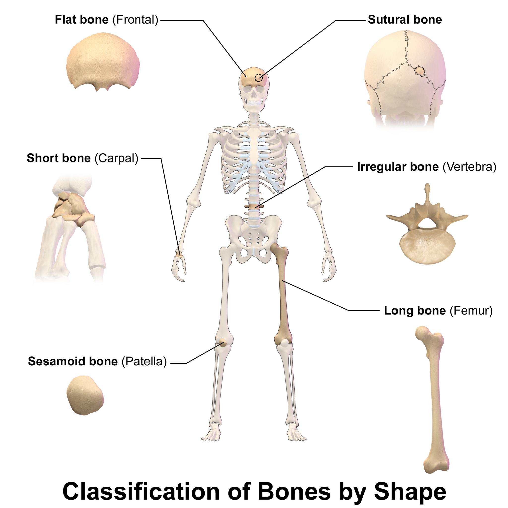
Feature: Reliable Sources
Diseased or damaged bone marrow can be replaced by donated bone marrow cells, which help treat and often cure many life-threatening conditions, including leukemia, lymphoma, sickle cell anemia, and thalassemia. If a bone marrow transplant is successful, the new bone marrow will start making healthy blood cells and improve the patient’s condition.
Learn more about bone marrow donation, and consider whether you might want to do it yourself. Find reliable sources to answer the following questions:
- How does one become a potential bone marrow donor?
- Who can and who cannot donate bone marrow?
- How is a bone marrow donation made?
- What risks are there in donating bone marrow?
11.4 Summary
- Bones are organs that consist mainly of bone tissue (or osseous tissue). Osseous tissue is a type of connective tissue consisting of a collagen matrix that is mineralized with calcium and phosphorus crystals. The combination of flexible collagen and minerals makes bone hard, without making it brittle.
- There are two types of osseous tissues: compact bone tissue and spongy bone tissue. Compact bone tissue is smooth and dense. It forms the outer layer of bones. Spongy bone tissue is porous and light, and it is found inside many bones.
- Besides osseous tissues, bones also contain nerves, blood vessels, bone marrow, and periosteum.
- Bone tissue is composed of four different types of bone cells: osteoblasts, osteocytes, osteoclasts, and osteogenic cells. Osteoblasts form new collagen matrix and mineralize it, osteoclasts break down bone, osteocytes regulate the formation and breakdown of bone, and osteogenic cells divide and differentiate to form new osteoblasts. Bone is a very active tissue, constantly being remodeled by the work of osteoblasts and osteoclasts.
- There are six types of bones in the human body: long bones (such as the limb bones), short bones (such as the wrist bones), sesamoid bones (such as the patella), sutural bones in the skull, and irregular bones (such as the vertebrae).
11.4 Review Questions
- Describe osseous tissue.
- Why are bones hard, but not brittle?
- Compare and contrast the compact and spongy bone.
- What non-osseous tissues are found in bones?
- List four types of bone cells and their functions.
- Identify six types of bones. Give an example of each type.
-
- Compare and contrast yellow bone marrow and red bone marrow.
- Which type of bone cell divides to produce new bone cells? Where is this cell type located?
- Where do osteoblasts and osteocytes come from? How are they related to each other?
- Which type of bone is embedded in tendons?
11.4 Explore More
https://www.youtube.com/watch?v=rDGqkMHPDqE&feature=emb_logo
The Skeletal System: Crash Course A&P #19, CrashCourse, 2015.
https://www.youtube.com/watch?v=0dV1Bwe2v6c&feature=emb_logo
Bone Remodeling and Modeling, Amgen, 2012.
https://www.youtube.com/watch?v=1Qfmkd6C8u8
How bones make blood - Melody Smith, TED-Ed, 2020.
Attributions
Figure 11.4.1
Bone_marrow_grilled_on_the_barbecue,_sliced_young_raw_garlic,_salted_leek_flowers_from_last_year,_lovage,_and_kale_(19098148350) by City Foodsters on Wikimedia Commons is used under a CC BY 2.0 (https://creativecommons.org/licenses/by/2.0) license.
Figure 11.4.2
Bone_cross-section.svg by Pbroks13 on Wikimedia Commons is used under a CC BY 3.0 (https://creativecommons.org/licenses/by/3.0) license.
Figure 11.4.3
Anatomy_of_a_Flat_Bone by OpenStax College on Wikimedia Commons is used under a CC BY 3.0 (https://creativecommons.org/licenses/by/3.0) license.
Figure 11.4.4
the-detail-of-the-bones-the-structure-of-the-bones-spongy-bone-tramčina-close-up-structure on pxfuel are used according to the pxfuel Terms of Use.
Figure 11.4.5
Spongy_bone_-_Trabecular_bone_2_–_Smart-Servier by Laboratoires Servier/ Smart Servier website on Wikimedia Commons is used under a CC BY-SA 3.0 (https://creativecommons.org/licenses/by-sa/3.0) license.
Figure 11.4.6
Bone_cells by OpenStax College on Wikimedia Commons is used under a CC BY 3.0 (https://creativecommons.org/licenses/by/3.0) license.
Figure 11.4.7
Blausen_0229_ClassificationofBones by BruceBlaus on Wikimedia Commons is used under a CC BY 3.0 (https://creativecommons.org/licenses/by/3.0/deed.en) license.
References
Amgen. (2012, January 19). Bone remodeling and modeling. YouTube. https://www.youtube.com/watch?v=0dV1Bwe2v6c
Betts, J. G., Young, K.A., Wise, J.A., Johnson, E., Poe, B., Kruse, D.H., Korol, O., Johnson, J.E., Womble, M., DeSaix, P. (2013, June 19). Figure 6.9 Anatomy of a flat bone [digital image]. In Anatomy and Physiology (Section 6.3). OpenStax. https://openstax.org/books/anatomy-and-physiology/pages/6-3-bone-structure
Betts, J. G., Young, K.A., Wise, J.A., Johnson, E., Poe, B., Kruse, D.H., Korol, O., Johnson, J.E., Womble, M., DeSaix, P. (2013, June 19). Figure 6.11 Bone cells [digital image]. In Anatomy and Physiology (Section 6.3). OpenStax. https://openstax.org/books/anatomy-and-physiology/pages/6-3-bone-structure
CK-12 Foundation. (n.d.). Communication: Identifies means of communication between animals. ck12.org. https://www.ck12.org/c/life-science/communication/
CrashCourse. (2015, May 18). The skeletal system: Crash Course A&P #19. YouTube. https://www.youtube.com/watch?v=rDGqkMHPDqE
TED-Ed. (2020, January 27). How bones make blood - Melody Smith. YouTube. https://www.youtube.com/watch?v=1Qfmkd6C8u8
Created by CK-12 Foundation/Adapted by Christine Miller
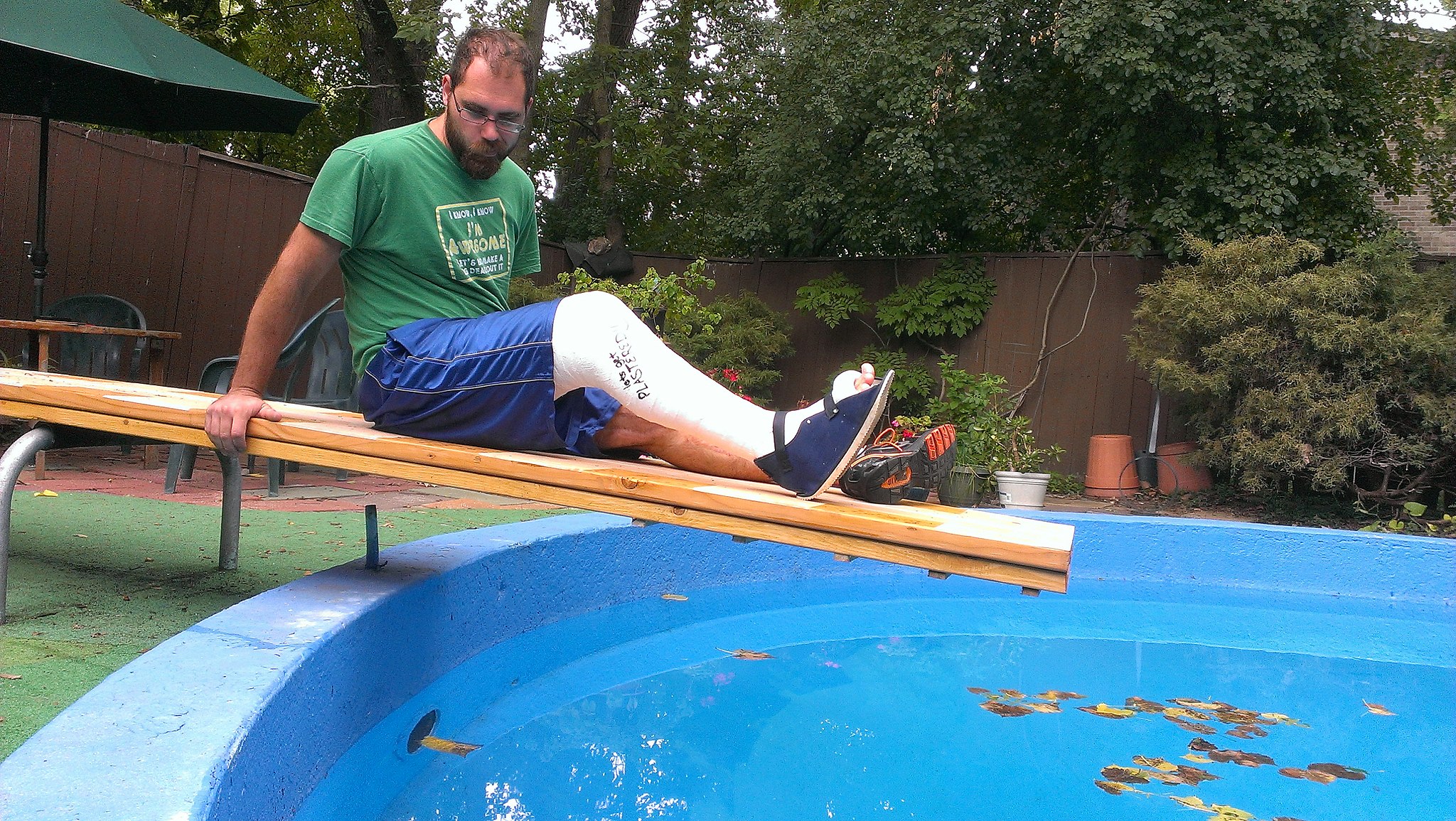
Break A Leg
Did you ever break a leg or other bone, like the man looking longingly at the water in this swimming pool (Figure 11.5.1)? Having a broken bone can really restrict your activity. Bones are very hard, but they will break (or fracture) if enough force is applied to them. Fortunately, bones are highly active organs that can repair themselves if they break. Bones can also remodel themselves and grow. You’ll learn how bones can do all of these things in this section.
Bone Growth
Early in the development of a human fetus, the skeleton is made almost entirely of cartilage. The relatively soft cartilage gradually turns into hard bone through ossification. Ossification is a process in which bone tissue is created from cartilage. The steps in which bones of the skeleton form from cartilage are illustrated in 11.5.2. The steps are as follows:
- Cartilage “model” of bone forms. This model continues to grow as ossification takes place.
- Ossification begins at a primary ossification center in the middle of bone.
- Ossification then starts to occur at secondary ossification centers at the ends of bone.
- The medullary cavity forms. This cavity will contain red bone marrow.
- Areas of ossification meet at epiphyseal plates, and articular cartilage forms. Bone growth ends.
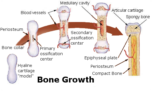
The ossification of cartilage in the human skeleton is a process that lasts throughout childhood in some bones.
Primary and Secondary Ossification Centers
When bone forms from cartilage, ossification begins with a point in the cartilage called the primary ossification center. This generally appears during fetal development, although a few short bones begin their primary ossification after birth. Ossification happens toward both ends of the bone from the primary ossification center, and — in the case of long bones — it eventually forms the shaft of the bone.
Secondary ossification centers form after birth. Ossification from secondary centers eventually forms the ends of the bones. The shaft and ends of the bone are separated by a growing zone of cartilage until the individual reaches skeletal maturity.
Skeletal Maturity
Throughout childhood, the cartilage remaining in the skeleton keeps growing, and allows for bones to grow in size. Once all of the cartilage has been replaced by bone, and fusion has taken place at the epiphyseal plates, bones can no longer keep growing in length. At this point, skeletal maturity has been reached. It generally takes place by age 18 to 25.
The use of anabolic steroids by teens can speed up the process of skeletal maturity, resulting in a shorter period of cartilage growth before fusion takes place. This means that teens who use steroids are likely to end up shorter as adults than they would otherwise have been.
Bone Remodeling
Even after skeletal maturity has been attained, bone is constantly being resorbed and replaced with new bone in a process called bone remodeling. In this lifelong process, mature bone tissue is continually turned over, with about ten per cent of the skeletal mass of an adult being remodeled each year. Bone remodeling is carried out through the work of osteoclasts — which are bone cells that resorb bone and dissolve its minerals — and osteoblasts, which are bone cells that make new bone matrix.
Bone remodeling serves several functions. It shapes the bones of the skeleton as a child grows, and it repairs tiny flaws in bone that result from everyday movements. Remodeling also makes bones thicker at points where muscles place the most stress on them. In addition, remodeling helps regulate mineral homeostasis, because it either releases mineral from bones into the blood or absorbs mineral from the blood into bones. Figure 11.5.3 shows how osteoclasts in bones are involved in calcium regulation.
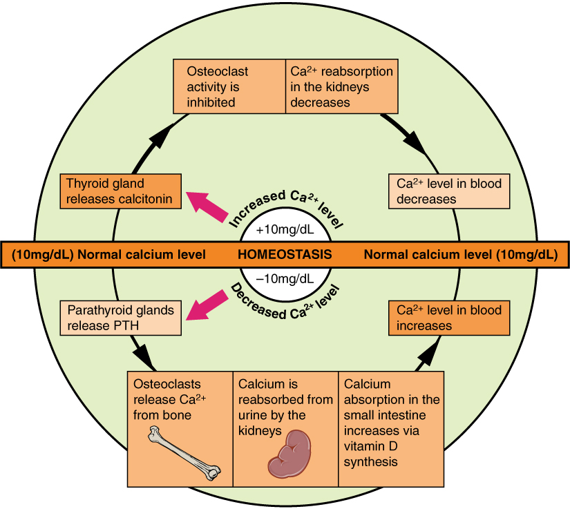
The action of osteoblasts and osteoclasts in bone remodeling and calcium homeostasis is controlled by a number of enzymes, hormones, and other substances that either promote or inhibit the activity of the cells. In this way, these substances control the rate at which bone is made, destroyed, and changed in shape. For example, the rate at which osteoclasts resorb bone and release calcium into the blood is promoted by parathyroid hormone (PTH) and inhibited by calcitonin, which is produced by the thyroid gland (see the diagram in Figure 11.5.3). The rate at which osteoblasts create new bone is stimulated by growth hormone, which is produced by the anterior lobe of the pituitary gland. Thyroid hormone and sex hormones (estrogens and androgens) also stimulate osteoblasts to create new bone.
Bone Repair
Bone repair (or healing) is the process in which a bone repairs itself following a bone fracture. You can see an X-ray of a bone fracture in Figure 11.5.4. In this fracture, the humerus in the upper arm has been completely broken through its shaft. Before this fracture heals, a physician must push the displaced bone parts back into their correct positions. Then, the bone must be stabilized — with a cast and/or pins surgically inserted into the bone, for example (as shown in Figure 11.5.5) — until the bone’s natural healing process is complete. This process may take several weeks.
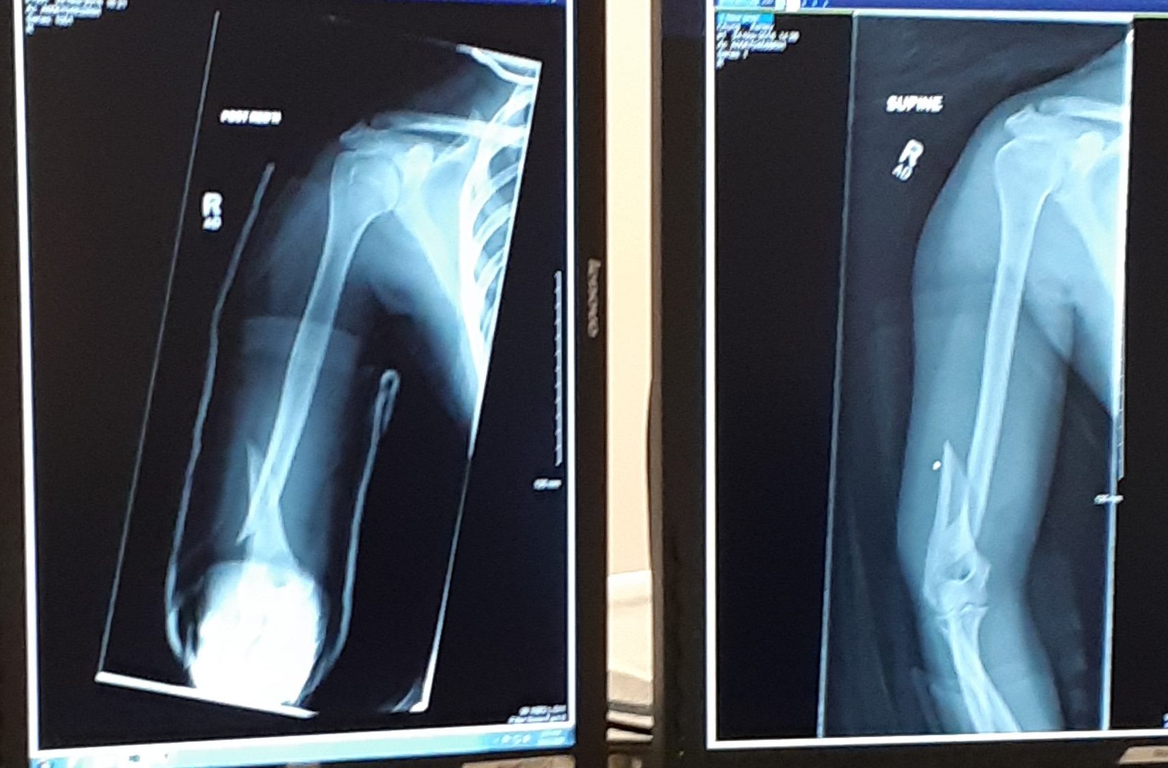 |
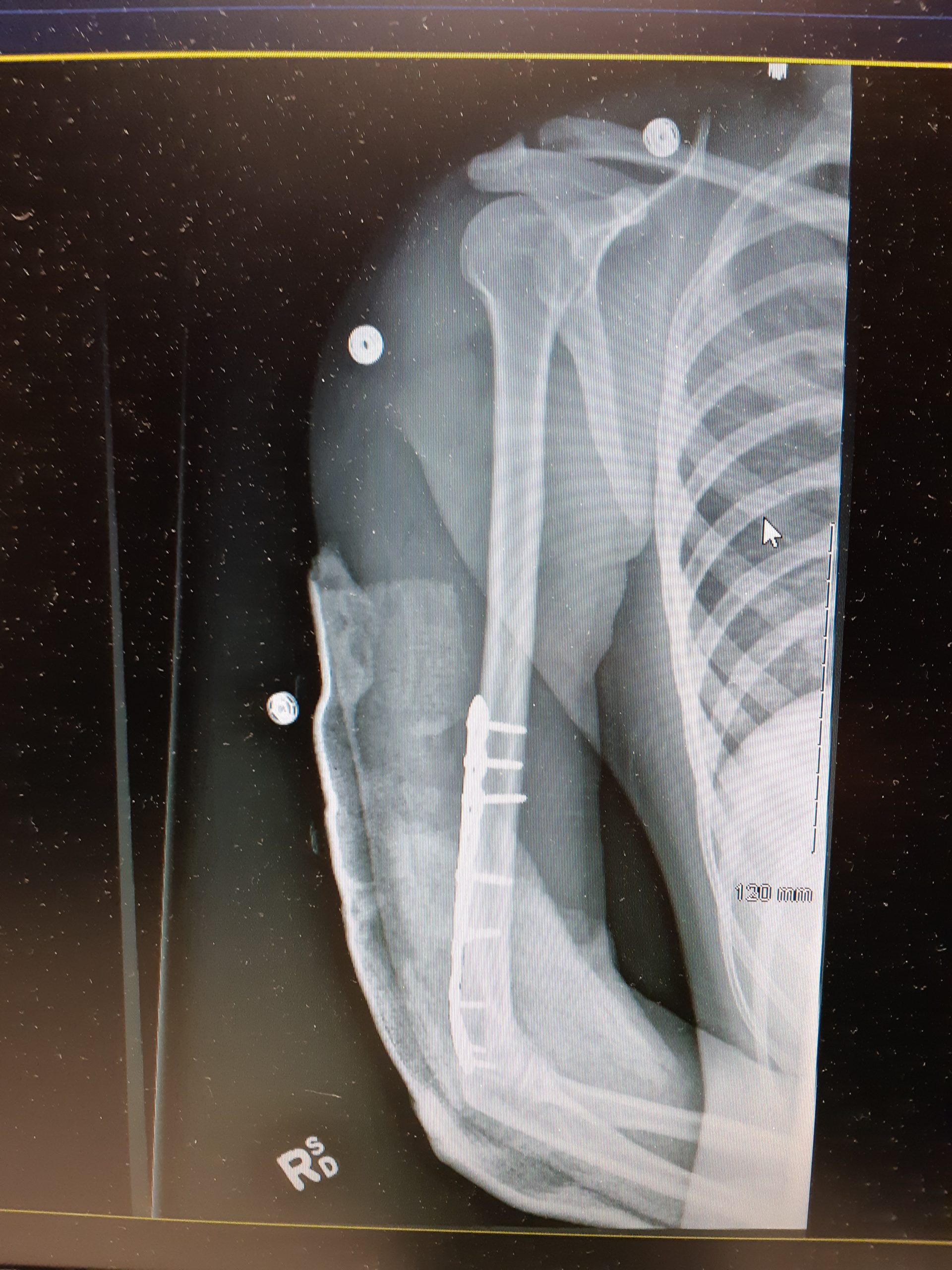 |
Although bone repair is a natural physiological process, it may be promoted or inhibited by several factors. Fracture repair is more likely to be successful with adequate nutrient intake. Age, bone type, drug therapy, and pre-existing bone disease are additional factors that may affect healing. Bones that are weakened by disease (such as osteoporosis or bone cancer) are not only likely to heal more slowly, but are also more likely to fracture in the first place.
Feature: Myth vs. Reality
Bone fractures are fairly common, and there are many myths about them. Knowing the facts is important, because fractures generally require emergency medical treatment.
| Myth | Reality |
|---|---|
| "A bone fracture is a milder injury than a broken bone." | A bone fracture is the same thing as a broken bone. |
| "If you still have full range of motion in a limb, then it must not be fractured." | Even if a bone is fractured, the muscles and tendons attached to it may still be able to move the bone normally. This is especially likely if the bone is cracked — but not broken — into two pieces. Even if a bone is broken all the way through, range of motion may not be affected if the bones on either side of the fracture remain properly aligned. |
| "A fracture always produces a bruise." | Many — but not all — fractures produce a bruise. If a fracture does produce a bruise, it may take several hours (or even a day or more!) for the bruise to appear. |
| "Fractures are so painful that you will immediately know if you break a bone." | Ligament sprains and muscle strains are also very painful, sometimes more painful than fractures. Additionally, every person has a different pain tolerance. People with a high pain tolerance may continue using a broken bone in spite of the pain. |
| "You can tell when a bone is fractured because there will be very localized pain over the break." | A broken bone is often accompanied by injuries to surrounding muscles or ligaments. As a result, the pain may extend far beyond the location of the fracture. The pain may be greater directly over the fracture, but the intensity of the pain may make it difficult to pinpoint exactly where the pain originates. |
11.5 Summary
- Bone is very active tissue. Its cells are constantly forming and resorbing bone matrix.
- Early in the development of a human fetus, the skeleton is made almost entirely of cartilage. The relatively soft cartilage gradually turns into hard bone. This is called ossification. It begins at a primary ossification center in the middle of bone, and later also occurs at secondary ossification centers in the ends of bone. The bone can no longer grow in length after the areas of ossification meet and fuse at the time of skeletal maturity.
- Throughout life, bone is constantly being replaced in the process of bone remodeling. In this process, osteoclasts resorb bone, and osteoblasts make new bone to replace it. Bone remodeling shapes the skeleton, repairs tiny flaws in bones, and helps maintain mineral homeostasis in the blood.
- Bone repair is the natural process in which a bone repairs itself following a bone fracture. This process may take several weeks. In the process, periosteum produces cells that develop into osteoblasts, and the osteoblasts form new bone matrix to heal the fracture. Bone repair may be affected by diet, age, pre-existing bone disease, or other factors.
11.5 Review Questions
- Outline how bone develops starting early in the fetal stage, and through the age of skeletal maturity.
- Describe the process of bone remodeling. When does it occur?
- What purposes does bone remodeling serve?
- Define bone repair. How long does this process take?
- Explain how bone repair occurs.
- Identify factors that may affect bone repair.
-
- If there is a large region between the primary and secondary ossification centers in a bone, is the person young or old? Explain your answer.
- If bones can repair themselves, why are casts and pins sometimes necessary in the process?
- When calcium levels are low, which type of bone cell causes the release of calcium to the bloodstream?
- Which tissue and bone cell type are primarily involved in bone repair after a fracture?
- Describe one way in which hormones are involved in bone remodeling.
11.5 Explore More
https://www.youtube.com/watch?v=yJoQj5-TIvE
How to grow a bone - Nina Tandon, TED-Ed, 2015.
https://www.youtube.com/watch?v=-P6LsendHxU&feature=emb_logo
Healing Process of Bone Fracture, Aldo Fransiskus Marsetio, 2015.
https://www.youtube.com/watch?v=RC2w_9DcY38
The Skeleton From Fetal to Adult, Samantha Espolt, 2012.
Attributions
Figure 11.5.1
First_plaster_long_leg_cast…._-_9383569051 by 4x4king10 on Wikimedia Commons is used under a CC BY 2.0 (https://creativecommons.org/licenses/by/2.0) license.
Figure 11.5.2
Bone_growth by Chaldor (derivative work) on Wikimedia Commons is in the public domain (https://en.wikipedia.org/wiki/en:Public_domain). (Original, Illu_bone_growth.jpg is by Fuelbottle)
Figure 11.5.3
Calcium_Homeostasis by OpenStax on Wikimedia commons is used under a CC BY 3.0 (https://creativecommons.org/licenses/by/3.0/deed.en) license.
Figure 11.5.4
Broken Arm by Ashley Chung is used with permission.
Figure 11.5.5
Broken Arm with plate and pins by Ashley Chung is used with permission.
References
Aldo Fransiskus Marsetio. (2015, ). Healing process of bone fracture. YouTube. https://www.youtube.com/watch?v=-P6LsendHxU
Betts, J. G., Young, K.A., Wise, J.A., Johnson, E., Poe, B., Kruse, D.H., Korol, O., Johnson, J.E., Womble, M., DeSaix, P. (2013, June 19). Figure 6.24 Pathways in calcium homeostasis [digital image]. In Anatomy and Physiology (Section 6.7). OpenStax. https://openstax.org/books/anatomy-and-physiology/pages/6-7-calcium-homeostasis-interactions-of-the-skeletal-system-and-other-organ-systems
Samantha Espolt. (2012, ). The skeleton from fetal to adult. YouTube. https://www.youtube.com/watch?v=RC2w_9DcY38&t=3s
TED-Ed. (2015, ). How to grow a bone - Nina Tandon. YouTube. https://www.youtube.com/watch?v=yJoQj5-TIvE
Created by CK-12 Foundation/Adapted by Christine Miller
Case Study Conclusion: A Pain in the Foot
As Sophia discovered in the beginning of the chapter, wearing high heels can result in a condition called metatarsalgia. Metatarsalgia is named for the metatarsal bones, which are the five bones that run through the ball of the foot just behind the toes (highlighted in Figures 11.8.2 and 11.8.3). Wearing high heels causes excessive pressure on the ball of the foot, as described in the beginning of this chapter. Additionally, the toes are forced to pull upward in high heels, which moves the fleshy padding away from the ball of the foot and adds to the overall pressure placed on this region. Over time, this can cause inflammation and direct stress on the bones, resulting in the pain in the ball of the foot known as metatarsalgia. The pain occurs especially in weight-bearing positions, such as standing, walking, or running — which is what Sophia was experiencing. There may also be pain, numbness, or tingling in the toes associated with metatarsalgia.
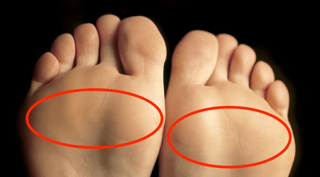 |
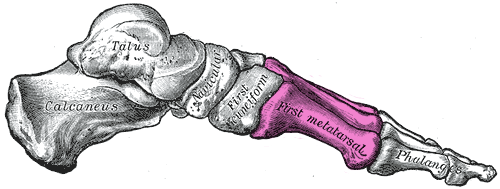 |
Wearing high heels can also cause stress fractures in the feet, which are tiny breaks in bone that occur due to repeated mechanical stress. This is caused by the excessive pressure that high heels put on some of the bones of the feet. These fractures are somewhat similar to what occurs in osteoporosis when the bone mass decreases to the point where bones can fracture easily as a person goes about their daily activities. In both cases, a major noticeable injury is not necessary to create the tiny fractures. As you have learned, tiny fractures that accrue over time are the cause of dowager’s hump (or kyphosis), which is often seen in women with osteoporosis.
Don’t think you are immune to stress fractures just because you don’t wear high heels! This injury also commonly occurs in people who participate in sports involving repetitive striking of the foot on the ground, such as running, tennis, basketball, or gymnastics. They may be avoided by taking preventative measures. You should ramp up any increase in activity slowly, cross-train by engaging in a variety of different sports or activities, rest if you experience pain, and wear well-cushioned and supportive running shoes. It is important to know that your cardiovascular and muscular systems adapt to an increase in physical activity much more quickly than the skeletal system.
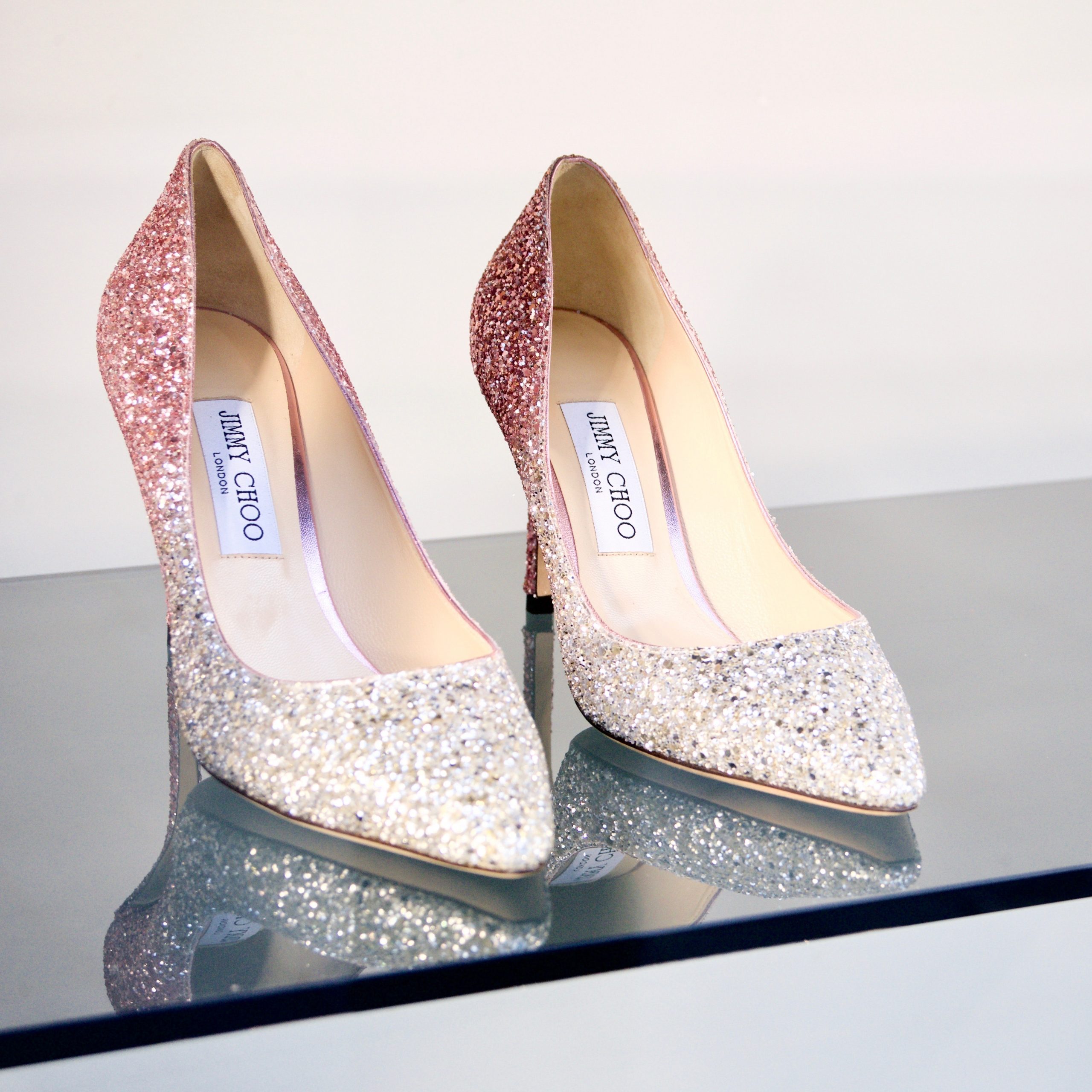
Sophia learned through her online research that wearing high heels can also lead to foot deformities, such as bunions and hammertoes. As you learned in an earlier chapter, a bunion is a protrusion on the side of the foot, most often at the base of the big toe. It can be caused by wearing shoes with a narrow, pointed toe box — a common shape for high heels (see Figure 11.8.4). The pressure of the shoes on the side of the foot causes an enlargement of bone or inflammation of other tissues in the region, which pushes the big toe toward the other toes.
Hammertoes are an abnormal bend in the middle joint of the second, third, or fourth toe (with the big toe being the first toe), causing the toe to be shaped similarly to a hammer. The narrow, pointed toe box of many high heels, combined with the way the toes are squished into the front of the shoe as a result of the height of the heel, can cause the toes to become deformed this way. Treatments for bunions and hammertoe include wearing shoes with a roomy toe box, padding or taping the toes, and toe exercises and stretches. If the bunion or hammertoe does not respond to these treatments, surgery may be necessary to correct the deformity.
Because the bones of the skeleton are connected and work together with other systems to support the body, wearing high heels can also cause physical problems in areas other than the feet. Wearing high heels shifts a person’s posture and alignment, and can put strain on tendons, muscles, and other joints in the body. Research published in 2014 from a team at Stanford University suggests that wearing high heels, particularly if the person is overweight or the heels are very high, may increase the risk of osteoarthritis (OA) in the knee, due to added stress on the knee joint as the person walks. As you have learned, OA results from the breakdown of cartilage and bone at the joint. Because it can only be treated to minimize symptoms — and not for a cure — OA could be an unfortunate long-term consequence of wearing high heels.
Sophia has decided that wearing high heels regularly is not worth the pain and potential long-term damage to her body. After consulting with her doctor, who confirmed she had metatarsalgia, she was able to successfully treat it with ice, rest, and wearing comfortable, supportive shoes instead of heels.
High heels are not the only kind of shoes that can cause problems. Flip-flops, worn-out sneakers, and shoes that are too tight can all cause foot issues. To prevent future problems from her shoe choices, Sophia is following guidelines recommended by medical experts. The guidelines include:
- Wearing shoes that fit well, have plenty of room in the toes, are supportive, and are comfortable right away. There should be no “break-in” period needed for shoes.
- Avoiding high heels, especially those with heels over two inches high, or those that have narrow, pointed toe boxes or very thin heels. The heels pictured in Figure 11.8.4 are an example of a type of shoe that should be avoided.
- If high heels must be worn, it should only be for a limited period of time.
As you have learned in this chapter, your skeletal system carries out a variety of important functions in your body, including physical support. But even though it is strong, your skeletal system can become damaged and deformed — even through such a seemingly innocuous act as wearing a certain type of shoe. Taking good care of your skeletal system is necessary to help it continue to take good care of the rest of you.
Chapter 11 Summary
In this chapter, you learned about the skeletal system. Specifically, you learned that:
- The skeletal system is the organ system that provides an internal framework for the human body. In adults, the skeletal system contains 206 bones.
- Bones are organs made of supportive connective tissues, mainly the tough protein collagen. Bones also contain blood vessels, nerves, and other tissues. Bones are hard and rigid, due to deposits of calcium and other mineral salts within their living tissues. Besides bones, the skeletal system includes cartilage and ligaments.
- The skeletal system has many different functions, including supporting the body and giving it shape, protecting internal organs, providing attachment surfaces for skeletal muscles, allowing body movements, producing blood cells, storing minerals, helping to maintain mineral homeostasis, and producing endocrine hormones.
- There is relatively little sexual dimorphism in the human skeleton, although the female skeleton tends to be smaller and less robust than the male skeleton. The greatest sex difference is in the pelvis, which is adapted for childbirth in females.
- The skeleton is traditionally divided into two major parts: the axial skeleton and the appendicular skeleton.
- The axial skeleton consists of a total of 80 bones. It includes the skull, vertebral column, and rib cage. It also includes the three tiny ossicles in the middle ear and the hyoid bone in the throat.
-
- The skull provides a bony framework for the head. It consists of 22 different bones: eight in the cranium, which encloses the brain, and 14 in the face, which includes the upper and lower jaw.
- The vertebral column is a flexible, S-shaped column of 33 vertebrae that connects the trunk with the skull and encloses the spinal cord. The vertebrae are divided into five regions: cervical, thoracic, lumbar, sacral, and coccygeal regions. The S shape of the vertebral column allows it to absorb shocks and distribute the weight of the body.
- The rib cage holds and protects the organs of the upper part of the trunk, including the heart and lungs. It includes the 12 thoracic vertebrae, the sternum, and 12 pairs of ribs.
- The appendicular skeleton consists of a total of 126 bones. It includes the bones of the four limbs, shoulder girdle, and pelvic girdle. The girdles attach the appendages to the axial skeleton.
-
- Each upper limb consists of 30 bones. There is one bone (called the humerus) in the upper arm, and two bones (called the ulna and radius) in the lower arm. The wrist contains eight carpal bones, the hand contains five metacarpals, and the fingers consist of 14 phalanges. The thumb is opposable to the palm and fingers of the same hand.
- Each lower limb also consists of 30 bones. There is one bone (called the femur) in the upper leg, and two bones (called the tibia and fibula) in the lower leg. The patella covers the knee joint. The ankle contains seven tarsal bones, and the foot contains five metatarsals. The tarsals and metatarsals form the heel and arch of the foot. The bones in the toes consist of 14 phalanges.
- The shoulder girdle attaches the upper limbs to the trunk of the body. It is connected to the axial skeleton only by muscles, allowing mobility of the upper limbs. Bones of the shoulder girdle include a right and left clavicle, and a right and left scapula.
- The pelvic girdle attaches the legs to the trunk of the body and supports the organs of the abdomen. It is connected to the axial skeleton by ligaments. The pelvic girdle consists of two halves that are fused together in adults. Each half consists of three bones: the ilium, pubis, and ischium.
- Bones are organs that consist mainly of bone (or osseous) tissue. Osseous tissue is a type of supportive connective tissue consisting of a collagen matrix that is mineralized with calcium and phosphorus crystals. The combination of flexible collagen and minerals makes bone hard, without making it brittle.
-
- There are two types of osseous tissues: compact bone tissue and spongy bone tissue. Compact bone tissue is smooth and dense. It forms the outer layer of bones. Spongy bone tissue is porous and light, and it is found inside many bones.
- Besides osseous tissues, bones also contain nerves, blood vessels, bone marrow, and periosteum.
- Bone tissue is composed of four different types of bone cells: osteoblasts, osteocytes, osteoclasts, and osteogenic cells. Osteoblasts form new collagen matrix and mineralize it, osteoclasts break down bone, osteocytes regulate the formation and breakdown of bone, and osteogenic cells divide and differentiate to form new osteoblasts. Bone is a very active tissue, constantly being remodeled by the work of osteoblasts and osteoclasts.
- There are six types of bones in the human body: long bones (such as the limb bones), short bones (such as the wrist bones), sesamoid bones (such as the patella), sutural bones in the skull, and irregular bones (such as the vertebrae).
- Early in the development of a human fetus, the skeleton is made almost entirely of cartilage. The relatively soft cartilage gradually turns into hard bone — a process that is called ossification. It begins at a primary ossification center in the middle of bone, and later also occurs at secondary ossification centers in the ends of bone. The bone can no longer grow in length after the areas of ossification meet and fuse at the time of skeletal maturity.
- Throughout life, bone is constantly being replaced in the process of bone remodeling. In this process, osteoclasts resorb bone and osteoblasts make new bone to replace it. Bone remodeling shapes the skeleton, repairs tiny flaws in bones, and helps maintain mineral homeostasis in the blood.
- Bone repair is the natural process in which a bone repairs itself following a bone fracture. This process may take several weeks. In the process, the periosteum produces cells that develop into osteoblasts, and the osteoblasts form new bone matrix to heal the fracture. Bone repair may be affected by diet, age, pre-existing bone disease, or other factors.
- Joints are locations at which bones of the skeleton connect with one another.
- Joints can be classified structurally or functionally, and there is significant overlap between the two types of classifications.
- The structural classification of joints depends on the type of tissue that binds the bones to each other at the joint. There are three types of joints in the structural classification: fibrous, cartilaginous, and synovial joints.
- The functional classification of joints is based on the type and degree of movement that they allow. There are three types of joints in the functional classification: immovable, partly movable, and movable joints.
- A number of disorders affect the skeletal system, including bone fractures and bone cancers. The two most common disorders of the skeletal system are osteoporosis and osteoarthritis.
- Osteoporosis is an age-related disorder in which bones lose mass, weaken, and break more easily than normal bones. The underlying mechanism in all cases of osteoporosis is an imbalance between bone formation and bone resorption in bone remodeling. Osteoporosis may also occur as a side effect of other disorders or certain medications.
-
- Osteoporosis is diagnosed by measuring a patient’s bone density and comparing it with the normal level of peak bone density. Fractures are the most dangerous aspect of osteoporosis. Osteoporosis is rarely fatal, but complications of fractures often are.
- Risk factors for osteoporosis include older age, female sex, European or Asian ancestry, family history of osteoporosis, short stature and small bones, smoking, alcohol consumption, lack of exercise, vitamin D deficiency, poor nutrition, and consumption of soft drinks.
- Osteoporosis is often treated with medications (such as bisphosphonates) that may slow or even reverse bone loss. Preventing osteoporosis includes eliminating any risk factors that can be controlled through changes of behavior, such as undertaking weight-bearing exercise.
- Osteoarthritis (OA) is a joint disease that results from the breakdown of joint cartilage and bone. The most common symptoms are joint pain and stiffness. OA is thought to be caused by mechanical stress on the joints with insufficient self-repair of cartilage, coupled with low-grade inflammation of the joints.
-
- Diagnosis of OA is typically made on the basis of signs and symptoms, such as joint deformities, pain, and stiffness. X-rays or other tests are sometimes used to either support the diagnosis or rule out other disorders. Age is the chief risk factor for OA. Other risk factors include joint injury, excess body weight, and a family history of OA.
- OA cannot be cured, but the symptoms can often be treated successfully. Treatments may include exercise, efforts to decrease stress on joints, pain medications, and surgery to replace affected hip or knee joints.
As you have learned in this chapter, one of the important functions of the skeletal system is to allow movement of the body. But it doesn’t do it alone. Movement is caused by the contraction of muscles, which pull on the bones, causing them to move. Read the next chapter to learn about this and other important functions of the muscular system.
Chapter 11 Review
-
-
- Why does the rib cage need to be flexible? Why can it be flexible?
- In general, what do “girdles” in the skeletal system do?
- Would swimming be more effective as an exercise for preventing osteoporosis or as a treatment for osteoarthritis? Explain your answer.
- Explain why some of the vertebrae become misshapen in the condition called dowager’s hump (or kyphosis).
- Explain why osteoarthritis often involves inflammation in the joints.
- Osteoporosis can involve excess bone resorption, as well as insufficient production of new bone tissue. What are the two main bone cell types that carry out these processes, respectively?
- Describe two roles that calcium in bones play in the body.
-
Attributions
Figure 11.8.1
Running Shoes by bruno-nascimento-PHIgYUGQPvU [photo] by Bruno Nascimento on Unsplash is used under the Unsplash License (https://unsplash.com/license).
Figure 11.8.2
Metatarsalgia/ Best Shoes for Metatarsalgia by Esther Max on Flickr is used under a CC BY 2.0 (https://creativecommons.org/licenses/by/2.0/) license.
Figure 11.8.3
Gray290_-_Mratatarsus (1) by Henry Vandyke Carter (1831-1897) (Revised by Warren H. Lewis, coloured by Was a bee) on Wikimedia Commons is in the public domain (https://en.wikipedia.org/wiki/Public_domain. (Bartleby.com: Gray’s Anatomy, Plate 290)
Figure 11.8.4
Heels by gavin-allanwood-ndpX28miBtE-unsplash by Photo by Gavin Allanwood on Unsplash is used under the Unsplash License (https://unsplash.com/license).
References
Mayo Clinic Staff. (n.d.). Hammertoe and mallet toe [online article]. MayoClinic.org. https://www.mayoclinic.org/diseases-conditions/hammertoe-and-mallet-toe/symptoms-causes/syc-20350839
VanDyke Carter, H. (1858). Illustration plate 290. In H. Gray, Anatomy of the Human Body. Lea & Febiger. Bartleby.com, 2000. www.bartleby.com/107/.
Created by CK-12 Foundation/Adapted by Christine Miller

Case Study: Needing to Relax
This dog (Figure 12.1.1) is expressing his interest in something — perhaps a piece of food — by using the neck muscles to tilt its head in an adorable fashion. Humans also sometimes tilt their heads to express interest. But imagine how disturbing and painful it would be if your neck tilted involuntarily, without you being able to control it! Forty-three year old Edward unfortunately knows just how debilitating this can be.
Edward has a rare condition called cervical dystonia, which is also called spasmodic torticollis. In this condition, the muscles in the neck contract involuntarily, often causing the person’s head to twist to one side. Figure 12.1.2 shows one type of abnormal head positioning that can be caused by cervical dystonia. The muscles may contract in a sustained fashion, holding the head and neck in one position, or they may spasm repeatedly, causing jerky movements of the head and neck.
Cervical dystonia is painful and can significantly interfere with a person’s ability to carry out their usual daily activities. In Edward’s case, he can no longer drive a car, because his uncontrollable head and neck movements and abnormal head positioning prevent him from navigating the road safely. He also has severe neck and shoulder pain much of the time.
Although it can be caused by an injury, there is no known cause of cervical dystonia — and there is also no cure. Fortunately for Edward, and others who suffer from cervical dystonia, there is a treatment that can significantly reduce symptoms in many people. You may be surprised to learn that this treatment is the same substance which, when injected into the face, is used for cosmetic purposes to reduce wrinkles!
The substance is botulinum toxin, one preparation of which may be familiar to you by its brand name — Botox. It is a neurotoxin produced by the bacterium Clostridium botulinum, and can cause a life-threatening illness called botulism. However, when injected in very small amounts by a skilled medical professional, botulinum toxins have some safe and effective uses. In addition to cervical dystonia, botulinum toxins can be used to treat other disorders involving the muscular system, such as strabismus (misalignment of the eyes); eye twitches; excessive muscle contraction due to neurological conditions like cerebral palsy; and even overactive bladder.
Botulinum toxin has its effect on the muscular system by inhibiting muscle contractions. When used to treat wrinkles, it relaxes the muscles of the face, lessening the appearance of wrinkles. When used to treat cervical dystonia and other disorders involving excessive muscle contraction, it reduces the abnormal contractions.
In this chapter, you will learn about the muscles of the body, how they contract to produce movements and carry out their functions, and some disorders that affect the muscular system. At the end of the chapter, you will find out if botulinum toxin helped relieve Edward’s cervical dystonia, and how this toxin works to inhibit muscle contraction.
Chapter Overview: Muscular System
In this chapter, you will learn about the muscular system, which carries out both voluntary body movements and involuntary contractions of internal organs and structures. Specifically, you will learn about:
- The different types of muscle tissue — skeletal, cardiac, and smooth muscle — and their different characteristics and functions.
- How muscle cells are specialized to contract and cause voluntary and involuntary movements.
- The ways in which muscle contraction is controlled.
- How skeletal muscles can grow or shrink, causing changes in strength.
- The structure and organization of skeletal muscles, including the different types of muscle fibres, and how actin and myosin filaments move across each other — according to the sliding filament theory — to cause muscle contraction.
- Cardiac muscle tissue in the heart that contracts to pump blood through the body.
- Smooth muscle tissue that makes up internal organs and structures, such as the digestive system, blood vessels, and uterus.
- The physical and mental health benefits of aerobic and anaerobic exercise, such as running and weight lifting.
- How individuals vary in their response to exercise.
- Disorders of the muscular system, including musculoskeletal disorders (such as strains and carpal tunnel syndrome) and neuromuscular disorders (such as muscular dystrophy, myasthenia gravis, and Parkinson’s disease).
As you read the chapter, think about the following questions:
- How is the contraction of skeletal muscles controlled?
- Botulinum toxin works on the cellular and molecular level to inhibit muscle contraction. Based on what you learn about how muscle contraction works, can you think of some ways it could potentially be inhibited?
- What is one disorder involving a lack of sufficient muscle contraction? Why does it occur?
Attributions
Figure 12.1.1
Whiskey’s 2nd Birthday by Kelly Hunter on Flickr is used under a CC BY 2.0 (https://creativecommons.org/licenses/by/2.0/) license.
Figure 12.1.2
1024px-Dystonia2010 by James Heilman, MD on Wikimedia Commons is used under a CC BY-SA 3.0 (https://creativecommons.org/licenses/by-sa/3.0) license.
References
Botulism [online article]. (2018, January 10). World Health Organization (WHO). https://www.who.int/news-room/fact-sheets/detail/botulism
Mayo Clinic Staff. (n.d.) Cervical dystonia [online article]. MayoClinic.org. https://www.mayoclinic.org/diseases-conditions/cervical-dystonia/symptoms-causes/syc-20354123
Created by CK-12 Foundation/Adapted by Christine Miller

Marvelous Muscles
Does the word muscle make you think of the well-developed muscles of a weightlifter, like the woman in Figure 12.2.1? Her name is Natalia Zabolotnaya, and she’s a Russian Olympian. The muscles that are used to lift weights are easy to feel and see, but they aren’t the only muscles in the human body. Many muscles are deep within the body, where they form the walls of internal organs and other structures. You can flex your biceps at will, but you can’t control internal muscles like these. It’s a good thing that these internal muscles work without any conscious effort on your part, because movement of these muscles is essential for survival. Muscles are the organs of the muscular system.
What Is the Muscular System?
The muscular system consists of all the muscles of the body. The largest percentage of muscles in the muscular system consists of skeletal muscles, which are attached to bones and enable voluntary body movements (shown in Figure 12.2.2). There are almost 650 skeletal muscles in the human body, many of them shown in Figure 12.2.2. Besides skeletal muscles, the muscular system also includes cardiac muscle, which makes up the walls of the heart, and smooth muscles, which control movement in other internal organs and structures.
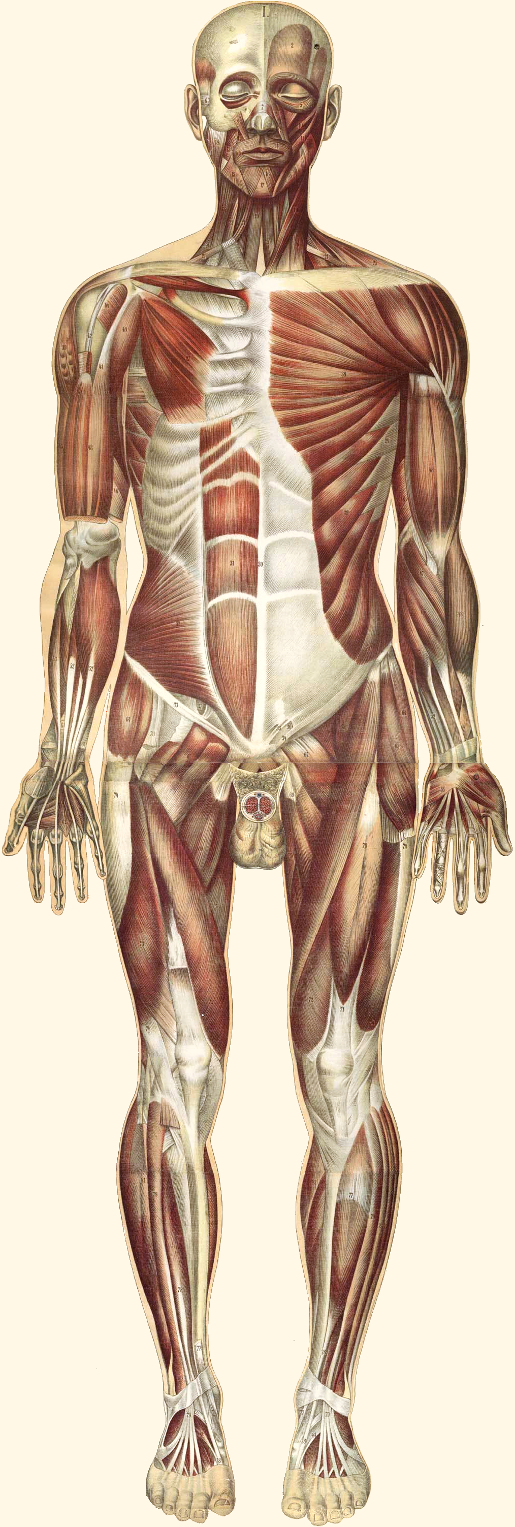
Muscle Structure and Function
Muscles are organs composed mainly of muscle cells, which are also called muscle fibres (mainly in skeletal and cardiac muscle) or myocytes (mainly in smooth muscle). Muscle cells are long, thin cells that are specialized for the function of contracting. They contain protein filaments that slide over one another using energy in ATP. The sliding filaments increase the tension in — or shorten the length of — muscle cells, causing a contraction. Muscle contractions are responsible for virtually all the movements of the body, both inside and out.
Skeletal muscles are attached to bones of the skeleton. When these muscles contract, they move the body. They allow us to use our limbs in a variety of ways, from walking to turning cartwheels. Skeletal muscles also maintain posture and help us to keep balance.
Smooth muscles in the walls of blood vessels contract to cause vasoconstriction, which may help conserve body heat. Relaxation of these muscles causes vasodilation, which may help the body lose heat. In the organs of the digestive system, smooth muscles squeeze food through the gastrointestinal tract by contracting in sequence to form a wave of muscle contractions called peristalsis. Think of squirting toothpaste through a tube by applying pressure in sequence from the bottom of the tube to the top, and you have a good idea of how food is moved by muscles through the digestive system. Peristalsis of smooth muscles also moves urine through the urinary tract.
Cardiac muscle tissue is found only in the walls of the heart. When cardiac muscle contracts, it makes the heart beat. The pumping action of the beating heart keeps blood flowing through the cardiovascular system.
Muscle Hypertrophy and Atrophy
Muscles can grow larger, or hypertrophy. This generally occurs through increased use, although hormonal or other influences can also play a role. The increase in testosterone that occurs in males during puberty, for example, causes a significant increase in muscle size. Physical exercise that involves weight bearing or resistance training can increase the size of skeletal muscles in virtually everyone. Exercises (such as running) that increase the heart rate may also increase the size and strength of cardiac muscle. The size of muscle, in turn, is the main determinant of muscle strength, which may be measured by the amount of force a muscle can exert.
Muscles can also grow smaller, or atrophy, which can occur through lack of physical activity or from starvation. People who are immobilized for any length of time — for example, because of a broken bone or surgery — lose muscle mass relatively quickly. People in concentration or famine camps may be so malnourished that they lose much of their muscle mass, becoming almost literally just “skin and bones.” Astronauts on the International Space Station may also lose significant muscle mass because of weightlessness in space (see Figure 12.2.3).
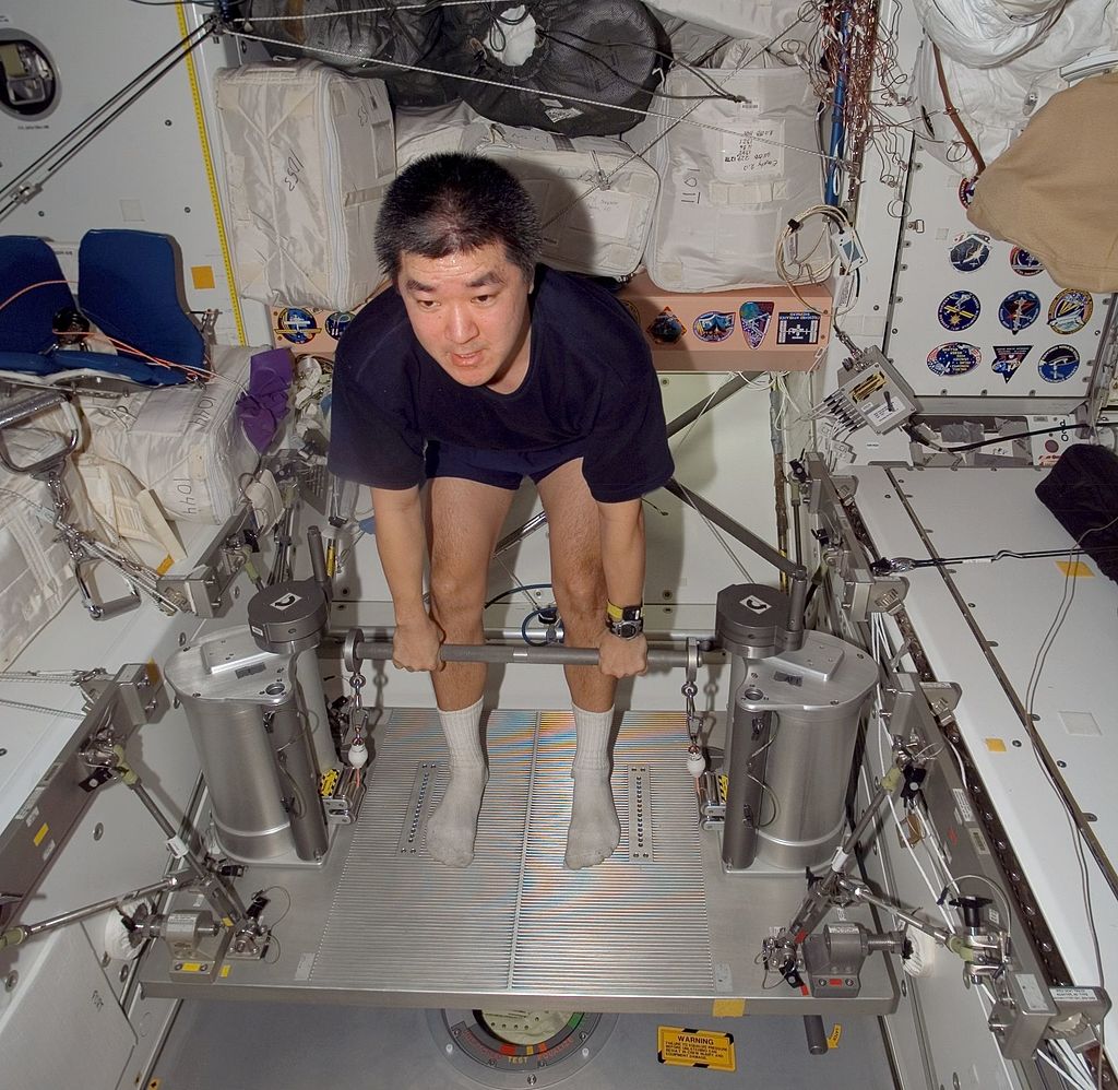
Many diseases, including cancer and AIDS, are often associated with muscle atrophy. Atrophy of muscles also happens with age. As people grow older, there is a gradual decrease in the ability to maintain skeletal muscle mass, known as sarcopenia. The exact cause of sarcopenia is not known, but one possible cause is a decrease in sensitivity to growth factors that are needed to maintain muscle mass. Because muscle size determines strength, muscle atrophy causes a corresponding decline in muscle strength.
In both hypertrophy and atrophy, the number of muscle fibres does not change. What changes is the size of the muscle fibres. When muscles hypertrophy, the individual fibres become wider. When muscles atrophy, the fibres become narrower.
Interactions with Other Body Systems
Muscles cannot contract on their own. Skeletal muscles need stimulation from motor neurons in order to contract. The point where a motor neuron attaches to a muscle is called a neuromuscular junction. Let’s say you decide to raise your hand in class. Your brain sends electrical messages through motor neurons to your arm and shoulder. The motor neurons, in turn, stimulate muscle fibres in your arm and shoulder to contract, causing your arm to rise.
Involuntary contractions of smooth and cardiac muscles are also controlled by electrical impulses, but in the case of these muscles, the impulses come from the autonomic nervous system (smooth muscle) or specialized cells in the heart (cardiac muscle). Hormones and some other factors also influence involuntary contractions of cardiac and smooth muscles. For example, the fight-or-flight hormone adrenaline increases the rate at which cardiac muscle contracts, thereby speeding up the heartbeat.
Muscles cannot move the body on their own. They need the skeletal system to act upon. The two systems together are often referred to as the musculoskeletal system. Skeletal muscles are attached to the skeleton by tough connective tissues called tendons. Many skeletal muscles are attached to the ends of bones that meet at a joint. The muscles span the joint and connect the bones. When the muscles contract, they pull on the bones, causing them to move. The skeletal system provides a system of levers that allow body movement. The muscular system provides the force that moves the levers.
12.2 Summary
- The muscular system consists of all the muscles of the body. There are three types of muscle: skeletal muscle (which is attached to bones and enables voluntary body movements), cardiac muscle (which makes up the walls of the heart and makes it beat), and smooth muscle (which is found in the walls of internal organs and other internal structures and controls their movements).
- Muscles are organs composed mainly of muscle cells, which may also be called muscle fibres or myocytes. Muscle cells are specialized for the function of contracting, which occurs when protein filaments inside the cells slide over one another using energy in ATP.
- Muscles can grow larger, or hypertrophy. This generally occurs through increased use (physical exercise), although hormonal or other influences can also play a role. Muscles can also grow smaller, or atrophy. This may occur through lack of use, starvation, certain diseases, or aging. In both hypertrophy and atrophy, the size — but not the number — of muscle fibres changes. The size of muscles is the main determinant of muscle strength.
- Skeletal muscles need the stimulus of motor neurons to contract, and to move the body, they need the skeletal system to act upon. Involuntary contractions of cardiac and smooth muscles are controlled by special cells in the heart, nerves of the autonomic nervous system, hormones, or other factors.
12.2 Review Questions
- What is the muscular system?
- Describe muscle cells and their function.
- Identify three types of muscle tissue and where each type is found.
- Define muscle hypertrophy and muscle atrophy.
- What are some possible causes of muscle hypertrophy?
- Give three reasons that muscle atrophy may occur.
- How do muscles change when they increase or decrease in size?
- How do changes in muscle size affect strength?
- Explain why astronauts can easily lose muscle mass in space.
- Describe how the terms muscle cells, muscle fibres, and myocytes relate to each other.
-
- Name two systems in the body that work together with the muscular system to carry out movements.
- Describe one way in which the muscular system is involved in regulating body temperature.
12.2 Explore More
https://www.youtube.com/watch?v=VVL-8zr2hk4
How your muscular system works - Emma Bryce, TED-Ed, 2017.
https://www.youtube.com/watch?v=Ujr0UAbyPS4&feature=emb_logo
3D Medical Animation - Peristalsis in Large Intestine/Bowel || ABP ©, AnimatedBiomedical, 2013.
https://www.youtube.com/watch?v=LkXwfTsqQgQ&feature=emb_logo
Muscle matters: Dr Brendan Egan at TEDxUCD, TEDx Talks, 2014.
Attributions
Figure 12.2.1
Natalia_Zabolotnaya_2012b by Simon Q on Wikimedia Commons is used under a CC BY 2.0 (https://creativecommons.org/licenses/by/2.0/deed.en) license.
Figure 12.2.2
Bougle_whole2_retouched by Bouglé, Julien from the National LIbrary of Medicine (NLM) on Wikimedia Commons is in the public domain (https://en.wikipedia.org/wiki/Public_domain).
Figure 12.2.3
Daniel_Tani_iss016e027910 by NASA/ International Space Station Imagery on Wikimedia Commons is in the public domain (https://en.wikipedia.org/wiki/Public_domain).
References
AnimatedBiomedical. (2013, January 30). 3D Medical animation - Peristalsis in large intestine/bowel || ABP ©. YouTube. https://www.youtube.com/watch?v=Ujr0UAbyPS4&feature=youtu.be
Bouglé, J. (1899). Le corps humain en grandeur naturelle : planches coloriées et superposées, avec texte explicatif. J. B. Baillière et fils. In Historical Anatomies on the Web. http://www.nlm.nih.gov/exhibition/historicalanatomies/bougle_home.html
TED-Ed. (2017, October 26). How your muscular system works - Emma Bryce. YouTube. https://www.youtube.com/watch?v=VVL-8zr2hk4&feature=youtu.be
TEDx Talks. (2014, June 27). Muscle matters: Dr Brendan Egan at TEDxUCD. YouTube. https://www.youtube.com/watch?v=LkXwfTsqQgQ&feature=youtu.be
Wikipedia contributors. (2020, June 15). Natalya Zabolotnaya. In Wikipedia. https://en.wikipedia.org/w/index.php?title=Natalya_Zabolotnaya&oldid=962630409
A peptide hormone, produced by alpha cells of the pancreas. It works to raise the concentration of glucose and fatty acids in the bloodstream, and is considered to be the main catabolic hormone of the body. It is also used as a medication to treat a number of health conditions.
Created by CK-12 Foundation/Adapted by Christine Miller
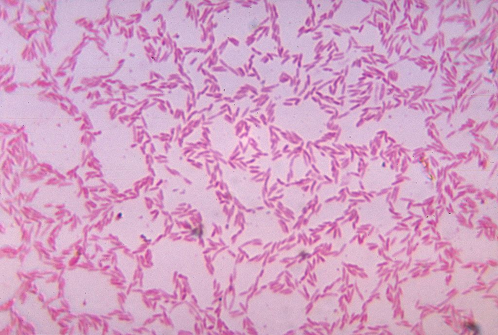
What Is It?
Figure 15.5.1 shows some of the cells of what has been called “the last human organ to be discovered.” This “organ” weighs about 200 grams (about 7 oz) and consists of a hundred trillion cells, yet scientists are only now beginning to learn everything it does and how it varies among individuals. What is it? It’s the mass of bacteria that live in our lower gastrointestinal tract.
Organs of the Lower Gastrointestinal Tract
Most of the bacteria that normally live in the lower gastrointestinal (GI) tract live in the large intestine. They have important and mutually beneficial relationships with the human organism. We provide them with a great place to live, and they provide us with many benefits, some of which you can read about below. Besides the large intestine and its complement of helpful bacteria, the lower GI tract also includes the small intestine. The latter is arguably the most important organ of the digestive system. It is where most chemical digestion and virtually all absorption of nutrients take place.
Small Intestine
The small intestine (also called the small bowel or gut) is the part of the GI tract between the stomach and large intestine. Its average length in adults is 4.6 m in females and 6.9 m in males. It is approximately 2.5 to 3.0 cm in diameter. It is called “small” because it is much smaller in diameter than the large intestine. The internal surface area of the small intestine totals an average of about 30 m2. Structurally and functionally, the small intestine can be divided into three parts, called the duodenum, jejunum, and ileum, as shown in Figure 15.5.2 and described below.
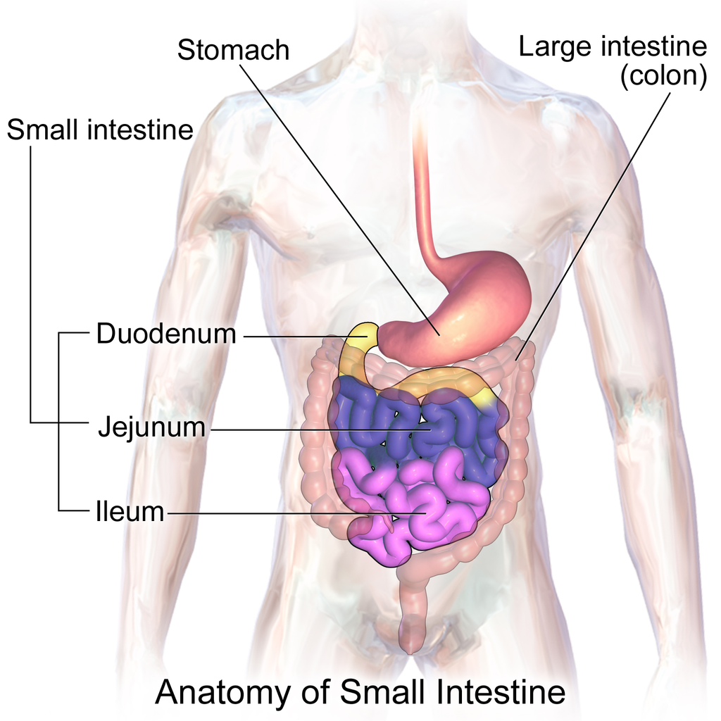
The mucosa lining the small intestine is highly enfolded and covered with the finger-like projections called villi. In fact, each square inch (about 6.5 cm2) of mucosa contains around 20 thousand villi. The individual cells on the surface of the villi also have many finger-like projections — the microvilli, shown in Figure 15.5.3. There are thought to be well over 17 billion microvilli per square centimetre of intestinal mucosa! All of these folds, villi, and microvilli greatly increase the surface area for chyme to come into contact with digestive enzymes, which coat the microvilli, as well as forming a tremendous surface area for the absorption of nutrients. Inside each of the villi is a network of tiny blood and lymph vessels that receive the absorbed nutrients and carry them away in the blood or lymph circulation. The wrinkles and projections in the intestinal mucosa also slow down the passage of chyme so there is more time for digestion and absorption to take place.
Duodenum
The duodenum is the first part of the small intestine, directly connected to the stomach. It is also the shortest part of the small intestine, averaging only about 25 cm (almost 10 in) in length in adults. Its main function is chemical digestion, and it is where most of the chemical digestion in the entire GI tract takes place.
The duodenum receives partially digested, semi-liquid chyme from the stomach. It receives digestive enzymes and alkaline bicarbonate from the pancreas through the pancreatic duct, and it receives bile from the liver via the gallbladder through the common bile duct (see Figure 15.5.4). In addition, the lining of the duodenum secretes digestive enzymes and contains glands — called Brunner’s glands — that secrete mucous and bicarbonate. The bicarbonate from the pancreas and Brunner’s glands — along with bile from the liver — neutralize the highly acidic chyme after it enters the duodenum from the stomach. This is necessary because the digestive enzymes in the duodenum require a nearly neutral environment in order to work. The three major classes of compounds that undergo chemical digestion in the duodenum are carbohydrates, proteins, and lipids.
Digestion of Carbohydrates in the Duodenum
Complex carbohydrates (such as starches) are broken down by the digestive enzyme amylase from the pancreas to short-chain molecules consisting of just a few saccharides (that is, simple sugars). Disaccharides, including sucrose and lactose, are broken down to simple sugars by duodenal enzymes. Sucrase breaks down sucrose, and lactase (if present) breaks down lactose. Some carbohydrates are not digested in the duodenum, and they ultimately pass undigested to the large intestine, where they may be digested by intestinal bacteria.
Digestion of Proteins in the Duodenum
In the duodenum, the pancreatic enzymes trypsin and chymotrypsin cleave proteins into peptides. Then, these smaller molecules are broken down into amino acids. Their digestion is catalyzed by pancreatic enzymes called peptidases.
Digestion of Lipids in the Duodenum
Pancreatic lipase breaks down triglycerides into fatty acids and glycerol. Lipase works with the help of bile secreted by the liver and stored in the gallbladder. Bile salts attach to triglycerides to help them emulsify, or form smaller particles (called micelles) that can disperse through the watery contents of the duodenum. This increases access to the molecules by pancreatic lipase.
Jejunum
The jejunum is the middle part of the small intestine, connecting the duodenum and the ileum. The jejunum is about 2.5 m (about 8 ft) long. Its main function is absorption of the products of digestion, including sugars, amino acids, and fatty acids. Absorption occurs by simple diffusion (water and fatty acids), facilitated diffusion (the simple sugar fructose), or active transport (amino acids, small peptides, water-soluble vitamins, and most glucose). All nutrients are absorbed into the blood, except for fatty acids and fat-soluble vitamins, which are absorbed into the lymph. Although most nutrients are absorbed in the jejunum, there are a few exceptions:
- Iron is absorbed in the duodenum.
- Vitamin B12 and bile salts are absorbed in the ileum.
- Water and lipids are absorbed throughout the small intestine, including the duodenum and ileum in addition to the jejunum.
Ileum
The ileum is the third and final part of the small intestine, directly connected at its distal end to the large intestine. The ileum is about 3 m (almost 10 ft) long. Some cells in the lining of the ileum secrete enzymes that catalyze the final stages of digestion of any undigested protein and carbohydrate molecules. However, the main function of the ileum is to absorb vitamin B12 and bile salts. It also absorbs any other remaining nutrients that were not absorbed in the jejunum. All substances in chyme that remain undigested or unabsorbed by the time they reach the distal end of the ileum pass into the large intestine.
Large Intestine
The large intestine — also called the large bowel — is the last organ of the GI tract. In adults, it averages about 1.5 m (about 5ft) in length. It is shorter than the small intestine, but at least twice as wide, averaging about 6.5 cm (about 2.5 in) in diameter. Water is absorbed from chyme as it passes through the large intestine, turning the chyme into solid feces. Feces is stored in the large intestine until it leaves the body during defecation.
Parts of the Large Intestine
Like the small intestine, the large intestine can be divided into several parts, as shown in Figure 15.5.5. The large intestine begins at the end of the small intestine, where a valve separates the small and large intestines and regulates the movement of chyme into the large intestine. Most of the large intestine is called the colon. The first part of the colon, where chyme enters from the small intestine, is called the cecum. From the cecum, the colon continues upward as the ascending colon, travels across the upper abdomen as the transverse colon, and then continues downward as the descending colon. It then becomes a V-shaped region called the sigmoid colon, which is attached to the rectum. The rectum stores feces until elimination occurs. It transitions to the final part of the large intestine, called the anus, which has an opening to the outside of the body for feces to pass through.
The diagram below (Figure 15.5.6) shows a projection from the cecum of the colon that is known as the appendix. The function of the appendix is somewhat uncertain, but it does not seem to be involved in digestion or absorption. It may play a role in immunity, and in the fetus, it seems to have an endocrine function, releasing hormones needed for homeostasis. Some biologists speculate that the appendix may also store a sample of the colon’s normal bacteria. If so, it may be able to repopulate the colon with the bacteria if illness or antibiotic medications deplete these microorganisms. Appendicitis, or infection and inflammation of the appendix, is a fairly common medical problem, typically resolved by surgical removal of the appendix (appendectomy). People who have had their appendix surgically removed do not seem to suffer any ill effects, so the organ is considered dispensable.
Functions of the Large Intestine
The removal of water from chyme to form feces starts in the ascending colon and continues throughout much of the length of the organ. Salts (such as sodium) are also removed from food wastes in the large intestine before the wastes are eliminated from the body. This allows salts — as well as water — to be recycled in the body.
The large intestine is also the site where huge numbers of beneficial bacteria ferment many unabsorbed materials in food waste. The bacterial breakdown of undigested polysaccharides produces nitrogen, carbon dioxide, methane, and other gases responsible for intestinal gas, or flatulence. These bacteria are particularly prevalent in the descending colon. Some of the bacteria also produce vitamins that are absorbed from the colon. The vitamins include vitamins B1 (thiamine), B2 (riboflavin), B7 (biotin), B12, and K. Another role of bacteria in the colon is an immune function. The bacteria may stimulate the immune system to produce antibodies that are effective against similar, but pathogenic, bacteria, thereby preventing infections. Still other roles played by bacteria in the large intestine include breaking down toxins before they can poison the body, producing substances that help prevent colon cancer, and inhibiting the growth of harmful bacteria.
Feature: My Human Body
Serotonin is a neurotransmitter with a wide variety of functions in the body. Sometimes called the "happy chemical," it is used in the central nervous system to stabilize mood by contributing to a feeling of well being and happiness. While this "happy chemical" function is important, serotonin is also important for critical brain functions, including support of learning, memory, and reward structures and regulating sleep. Since serotonin's main target is brain function, you may be surprised to learn that 90% of the human body's supply of this neurotransmitter is located in the gastrointestinal tract, where it regulates intestinal movements.
The enteric nervous system (ENS), a division of the autonomic nervous system, is a mesh-like system of neurons that control GI function. Interestingly, the ENS can act independently from both the sympathetic/parasympathetic nervous systems and the brain/spinal cord, which is why some scientists refer to the ENS as the "second brain". The ENS consists of over 500 million neurons (five times as many as in the spinal cord!) and lines the GI tract from the esophagus all the way to the anus. Serotonin is made in and secreted by the gut (small and large intestine) by enterochromaffin cells (also known as Kulchitsky cells) residing in the inner lining of the lower GI tract. While the main function of serotonin in the gut is to regulate digestion, it is also suspected to play a role in brain function — meaning that your gut may actually be partly dictating your mood! Another piece of evidence for the gut-dictating-your-mood theory is the nature vagus nerve, a fibrous visceral nerve, in which 90% of the fibres are dedicated to sending information too the brain, and only 10% of the fibres receiving information from the brain. Some treatments for depression actually involved electrical stimulation of the vagus nerve!
In a 2015 study by researchers at Caltech, it was shown that gut bacteria promote serotonin production by enterochromaffin cells. The study involved measuring the levels of serotonin levels in mice with normal gut bacteria, and then comparing that to the levels of serotonin in a population of mice with no gut bacteria. The germ-free mice were found to produce only 40% of the serotonin that the mice with normal gut flora were producing. Once the germ-free mice were re-colonized with normal gut flora, their production of serotonin returned to normal. This lead researchers to the conclusion that enterochromaffin cells depend on an interaction with gut bacterial flora to be able to produce necessary levels of serotonin.
15.5 Summary
- The lower GI tract includes the small intestine and large intestine. The small intestine is where most chemical digestion and virtually all absorption of nutrients occur. The large intestine contains huge numbers of beneficial bacteria and removes water and salts from food waste before it is eliminated.
- The small intestine consists of three parts: the duodenum, jejunum, and ileum. All three parts of the small intestine are lined with mucosa that is highly enfolded and covered with villi and microvilli, giving the small intestine a huge surface area for digestion and absorption.
- The duodenum secretes digestive enzymes, and also receives bile from the liver via the gallbladder and digestive enzymes and bicarbonate from the pancreas. These digestive substances neutralize acidic chyme and allow for the chemical digestion of carbohydrates, proteins, lipids, and nucleic acids in the duodenum.
- The jejunum carries out most of the absorption of nutrients in the small intestine, including the absorption of simple sugars, amino acids, fatty acids, and many vitamins.
- The ileum carries out any remaining digestion and absorption of nutrients, but its main function is to absorb vitamin B12 and bile salts.
- The large intestine consists of the colon (which, in turn, includes the cecum, ascending colon, transverse colon, descending colon, and sigmoid colon), rectum, and anus. The vestigial organ called the appendix is attached to the cecum of the colon.
- The main function of the large intestine is to remove water and salts from chyme for recycling within the body and eliminating the remaining solid feces from the body through the anus. The large intestine is also the site where trillions of bacteria help digest certain compounds, produce vitamins, stimulate the immune system, and break down toxins, among other important functions.
15.5 Review Questions
-
- How is the mucosa of the small intestine specialized for digestion and absorption?
- What digestive substances are secreted into the duodenum? What compounds in food do they help digest?
- What is the main function of the jejunum?
- What roles does the ileum play?
- How do beneficial bacteria in the large intestine help the human organism?
- When diarrhea occurs, feces leaves the body in a more liquid state than normal. What part of the digestive system do you think is involved in diarrhea? Explain your answer
- What causes intestinal gas, or flatulence?
15.5 Explore More
https://youtu.be/GTvnjaUU6Xk
Why do we pass gas? - Purna Kashyap, TED-Ed, 2014.
https://youtu.be/0IVO50DuMCs
What causes constipation? - Heba Shaheed, TED-Ed, 2018.
Attributions
Figure 15.5.1
1024px-Bacteroides_biacutis_01 by CDC/Dr. V.R. Dowell, Jr. Public Health Image Library (PHIL) ID#3087) on Wikimedia Commons is in the public domain (https://en.wikipedia.org/wiki/public_domain).
Figure 15.5.2
Blausen_0817_SmallIntestine_Anatomy by BruceBlaus on Wikimedia Commons is used under a CC BY 3.0 (https://creativecommons.org/licenses/by/3.0) license.
Figure 15.5.3
Human_jejunum_microvilli_1_-_TEM by Louisa Howard, Katherine Connollly / E. M. Facility, Dartmouth, on Wikimedia Commons is in the public domain (https://en.wikipedia.org/wiki/public_domain).
Figure 15.5.4
512px-2422_Accessory_Organs by OpenStax College on Wikimedia Commons is used under a CC BY 3.0 (https://creativecommons.org/licenses/by/3.0) license.
Figure 15.5.5
1024px-Blausen_0603_LargeIntestine_Anatomy by BruceBlaus on Wikimedia Commons is used under a CC BY 3.0 (https://creativecommons.org/licenses/by/3.0) license.
Figure 15.5.6
512px-Ds00070_an01934_im00887_divert_s_gif.webp (1) by Lfreeman04 on Wikimedia Commons is used under a CC BY-SA 4.0 (https://creativecommons.org/licenses/by-sa/4.0) license.
References
Betts, J. G., Young, K.A., Wise, J.A., Johnson, E., Poe, B., Kruse, D.H., Korol, O., Johnson, J.E., Womble, M., DeSaix, P. (2013, June 19). Figure 23.24 Accessory organs [digital image]. In Anatomy and Physiology (Section 23.6). OpenStax. https://openstax.org/books/anatomy-and-physiology/pages/23-6-accessory-organs-in-digestion-the-liver-pancreas-and-gallbladder
Blausen.com Staff. (2014). Medical gallery of Blausen Medical 2014. WikiJournal of Medicine 1 (2). DOI:10.15347/wjm/2014.010. ISSN 2002-4436. /
TED-Ed. (2018, May 7). What causes constipation? - Heba Shaheed. YouTube. https://www.youtube.com/watch?v=0IVO50DuMCs&feature=youtu.be
TED-Ed. (2014, September 8). Why do we pass gas? - Purna Kashyap. YouTube. https://www.youtube.com/watch?v=GTvnjaUU6Xk&feature=youtu.be
Yano, J. M., Yu, K., Donaldson, G. P., Shastri, G. G., Ann, P., Ma, L., Nagler, C. R., Ismagilov, R. F., Mazmanian, S. K., & Hsiao, E. Y. (2015). Indigenous bacteria from the gut microbiota regulate host serotonin biosynthesis. Cell, 161(2), 264–276. https://doi.org/10.1016/j.cell.2015.02.047
Created by: Christine Miller
Definition
In order to truly understand the concept of Traditional Ecological Knowledge (TEK), it is important to gather as many definitions as possible- this gives us an accurate breadth of the term with all its nuances. Click through the images below to read several descriptions of TEK.
Value
People who have lived in a community for generations are often the first to notice any signs of environmental change. Information about a particular region's climate and ecology is retained when the people from the region take on location as part of their cultural identity across generations. This traditional knowledge is passed from generation to generation through story telling and mentorship.
TEK shares some similarities with what is termed "Western Science". Both recognize that knowledge is always growing and changing and that observations are critical to recognizing patterns and causalities in nature. In addition, both TEK and Western Science recognize interdependence in biological systems and the need to treat ecology as a complex system. TEK differs in some ways from Western Science: knowledge is passed on orally, partly through metaphor and story, and this learned knowledge is embedded into daily living. TEK also differs from Western Science in that TEK is tied in to morality, spirituality and individual identity, making it more than just knowledge; it is sacred knowledge.
Examples

People who are indigenous to the province of British Columbia have been managing natural resources in this area for time immemorial. Numerous examples of sustainable harvesting methods can be found across the province, but our example, harvesting and management of the Avalanche Lily, comes from the Secwepemc peoples of the interior of British Columbia. Many thanks to Nancy Turner, Marianne Boelscher Ignace and Ronald Ignace for their documentation of these practices in their paper: Traditional Ecological Knowledge and Wisdom of Aboriginal Peoples in British Columbia
The Avalanche Lily is a yellow-flowered member of the lily family native to Western North America. This flower grows from an edible bulb which ranges in size from 3-5 centimetres. The Secwepemc people have harvested these bulbs as an important food source for generations. Oral transmission of knowledge allowed the Secwepemc people to use thousands of years of accumulated data around growing cycles, seasons and management practices to harvest these plants effectively while maintaining a healthy population of lilies for future use. Very intentional conservation strategies were/are practiced when harvesting the bulbs:
- Bulbs were harvested according to elevation in order to collect bulbs in the right stage of maturity and to spread out harvesting and yield over several months. Bulbs picked early in the season were too soft, and bulbs picked late were too watery.
- Only the largest bulbs were selected by choosing stems with multiple fruiting bodies.
- As bulbs were harvested, smaller bulbs were replanted, seeds were scattered and the soil was tilled by the harvesters.
- Areas which had been extensively harvested were often left for several years to allow the lily population time to recover.
The two videos below show how knowledge of a particular ecosystem is handed down through the traditions of mentorship and storytelling:
Modern science, native knowledge, by The Nature Conservancy, 2015

First Stories - Nganawendaanan Nde'ing (I Keep Them in My Heart), by Shannon Letande, 2006
TEK is Part of Place
The Traditional Ecological Knowledge held by Indigenous communities often includes very location specific knowledge. There are many diverse groups of First Peoples in British Columbia, each with expert knowledge about the ecology of their specific ancestral regions. The link to the map from the Native Land Digital website shows some of the traditional boundaries of the Indigenous people in British Columbia. Click on the areas to see where First Nations communities are located.
1.6 Summary
- Traditional Ecological Knowledge (TEK) is an important and valuable body of knowledge
- People groups who have lived in an area over generations pass down TEK through storytelling and mentorship
- TEK and Western Science share certain characteristics, including use of observations, and identification of patterns and causalities in nature
- TEK and Western Science differ in that TEK is passed down through oral storytelling and is deeply rooted in morality, spirituality and individual identity
1.6 Review Questions
Type your exercises here.
- Define Traditional Ecological Knowledge.
- How is TEK passed down through generations?
- How does TEK differ from Western Science?
- What are some ways in which TEK can inform resource management?
- What are some of the ramifications of loss of TEK? How can TEK be maintained?
1.6 Explore More
https://www.youtube.com/watch?time_continue=2&v=pHNlel72eQc&feature=emb_logo
TEDxTC - Winona LaDuke - Seeds of Our Ancestors, Seeds of Life, by TEDx-TC, 2012
Attributions
Definitions of Traditional Ecological Knowledge
- Kamloops, BC, Canada by Louis Paulin on Unsplash is used under the Unsplash License (https://unsplash.com/license).
- The Totem Poles of Capilano, Vancouver by Udayaditya Barua on Unsplash is used under the Unsplash License (https://unsplash.com/license).
- Alouette Lake by Chloe Evans on Unsplash is used under the Unsplash License (https://unsplash.com/license).
Figure 1.6.1
Erythronium grandiflorum 5077, by Walter Siegmund on Wikimedia Commons is used under a CC BY-SA 3.0 (https://creativecommons.org/licenses/by-sa/3.0) license.
References
Herkes, J. (n.d.). Continuing studies: Traditional ecological knowledge. University of Northern British Columbia. Retrieved from https://www.unbc.ca/continuing-studies/courses/traditional-ecological-knowledge
(1993). Traditional ecological knowledge: Concepts and cases (p. vi). Canadian Museum of Nature, Ottawa, Ontario, Canada.
Letande, S. (2006). First Stories - Nganawendaanan Nde'ing (I keep them in my heart). https://www.nfb.ca/film/first_stories_nganawendaanan_ndeing/
Minerals Management Service (n.d.). What is Traditional Knowledge [online]. Government of Alaska.https://web.archive.org/web/20030328053734/http://www.mms.gov/alaska/native/tradknow/tk_mms2.htm
TEDx-TC. (2012, March 4). TEDxTC - Winona LaDuke - Seeds of our ancestors, seeds of life. https://www.youtube.com/watch?v=pHNlel72eQc&feature=youtu.be
The Nature Conservancy. (2015, February 25). Modern science, native knowledge. YouTube. https://www.youtube.com/watch?v=1QRpnHoGivk
Turner, N., Ignace, M., & Ignace, R. (2000, October 1). Traditional ecological knowledge and wisdom of Aboriginal peoples in British Columbia. Ecological Applications, 10(5), 1275-1287. doi:10.2307/2641283
Wakefield, A.J. (1999, September 11). MMR vaccination and autism. Lancet, 354(9182), 949-950. https://www.thelancet.com/journals/lancet/article/PIIS0140-6736(05)75696-8/fulltext doi:https://doi.org/10.1016/S0140-6736(05)75696-8
A nervous system cell that provides support for neurons and helps them transmit nerve impulses.
Created by CK-12 Foundation/Adapted by Christine Miller
Case Study Conclusion: Defending Your Defenses
These people are participating in a bike ride to raise funds for leukemia and lymphoma research (Figure 17.7.1). Leukemia and lymphoma are blood cancers. In 2020, approximately 6,900 Canadians will be diagnosed with leukemia and 3,000 will die from this cancer. Lymphoma is the most common type of blood cancer. As a lymphoma patient, Hakeem, who you learned about in the beginning of this chapter, may eventually benefit from research funded by a bike ride like this one.
What type of blood cell is affected in lymphoma? As the name implies, lymphoma is a cancer that affects lymphocytes, which are a type of leukocyte. As you have learned in this chapter, there are different types of lymphocytes, including the B and T cells of the adaptive immune system. Different types of lymphoma affect different types of lymphocytes in different ways. It is important to correctly identify the type of lymphoma, so that patients can be treated appropriately.
You may recall that one of Hakeem’s symptoms was a swollen lymph node, and he was diagnosed with lymphoma after a biopsy of that lymph node. Swollen lymph nodes are a common symptom of lymphoma. As you have learned, lymph nodes are distributed throughout the body along lymphatic vessels, as part of the lymphatic system. The lymph nodes filter lymph and store lymphocytes. Therefore, they play an important role in fighting infections. Because of this, they will often swell in response to an infection. In Hakeem’s case, the swelling and other symptoms did not improve after several weeks and a course of antibiotics, which caused Dr. Hayes to suspect lymphoma instead. The biopsy showed that Hakeem did indeed have cancerous lymphocytes in his lymph nodes.
But which type of lymphocytes were affected? Lymphoma most commonly affects B or T lymphocytes. The two major types of lymphoma are called Hodgkin (HL) or non-Hodgkin lymphoma (NHL). NHL is more common than HL. In 2020, the Canadian Cancer Society estimates 10,400 Canadians will be diagnosed with non-Hodgkin lymphoma, whereas 1,000 will be diagnosed with Hodgkin lymphoma. While HL is one distinct type of lymphoma, NHL has about 60 different subtypes, depending on which specific cells are affected and how.
Hakeem was diagnosed with a type of NHL called diffuse large B-cell lymphoma (DLBCL) — the most common type of NHL. This type of lymphoma affects B cells and causes them to appear large under the microscope. In addition to Hakeem’s symptoms of fatigue, swollen lymph nodes, loss of appetite, and weight loss, common symptoms of this type of lymphoma include fever and night sweats. It is an aggressive and fast-growing type of lymphoma that is fatal if not treated. The good news is that with early detection and proper treatment, about 70% of patients with DLBCL can be cured.
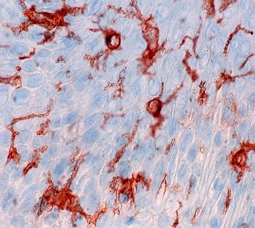
How do physicians determine the specific type of lymphoma? Tissue obtained from a biopsy can be examined under a microscope to observe physical changes (such as abnormal cell size or shape) that are characteristic of a particular subtype of lymphoma. Additionally, tests can be performed on the tissue to determine which cell-surface antigens are present. Recall that antigens are molecules that bind to specific antibodies. Antibodies can be produced in the laboratory and labeled with compounds that can be identified by their colour under a microscope. When these antibodies are applied to a tissue sample, this colour will appear wherever the antigen is present, because it binds to the antibody. This technique was used in the photomicrograph in Figure 17.7.2 to identify the presence of a cell-surface antigen (shown as reddish-brown) in a sample of skin cells. This technique, called immunohistochemistry, is also commonly used to identify antigens in tissue samples from lymphoma patients.
Why would identifying cell-surface antigens be important in diagnosing and treating lymphoma? As you have learned, the immune system uses antigens present on the surface of cells or pathogens to distinguish between self and non-self, and to launch adaptive immune responses. Cells that become cancerous often change their cell-surface antigens. This is one way that the immune system can identify and destroy them. Also, different cell types in the body can sometimes be identified by the presence of specific cell-surface antigens. Knowing the types of cell-surface antigens present in a tissue sample can help physicians identify which cells are cancerous, and possibly the specific subtype of cancer. Knowing this information can be helpful in choosing more tailored and effective treatments.
One treatment for NHL is, in fact, the use of medications made from antibodies that bind to cell-surface antigens present on cells affected by the specific subtype of NHL. This is called immunotherapy. These drugs can directly bind to and kill the cancerous cells. For patients with DLBCL like Hakeem, immunotherapy is often used in conjunction with chemotherapy and radiation as a course of treatment. Although Hakeem has a difficult road ahead, he and his medical team are optimistic that — given the high success rate when DLBCL is caught and treated early — he may be cured. More research into how the immune system functions may lead to even better treatments for lymphoma — and other types of cancers — in the future.
Chapter 17 Summary
In this chapter, you learned about the immune system. Specifically, you learned that:
- Any agent that can cause disease is called a pathogen. Most human pathogens are microorganisms, such as bacteria and viruses. The immune system is the body system that defends the human host from pathogens and cancerous cells.
- The innate immune system is a subset of the immune system that provides very quick, but non-specific responses to pathogens. It includes multiple types of barriers to pathogens, leukocytes that phagocytize pathogens, and several other general responses.
- The adaptive immune system is a subset of the immune system that provides specific responses tailored to particular pathogens. It takes longer to put into effect, but it may lead to immunity to the pathogens.
- Both innate and adaptive immune responses depend on the ability of the immune system to distinguish between self and non-self molecules. Most body cells have major histocompatibility complex (MHC) proteins that identify them as self. Pathogens, infected cells, and tumor cells have non-self proteins called antigens that the immune system recognizes as foreign.
- Antigens are proteins that bind to specific receptors on immune system cells and elicit an adaptive immune response. Some immune cells (B cells) respond to foreign antigens by producing antibodies that bind with antigens and target pathogens for destruction.
- An important role of the immune system is tumor surveillance. Killer T cells of the adaptive immune system find and destroy tumor cells, which they can identify from their abnormal antigens.
- The neuroimmune system that protects the central nervous system is thought to be distinct from the peripheral immune system that protects the rest of the human body. The blood-brain and blood-spinal cord barriers are one type of protection of the neuroimmune system. Neuroglia also play a role in this system, for example, by carrying out phagocytosis.
- The lymphatic system is a human organ system that is a vital part of the adaptive immune system. It consists of several organs and a system of vessels that transport or filter the fluid called lymph. The main immune function of the lymphatic system is to produce, mature, harbor, and circulate white blood cells called lymphocytes, which are the main cells in the adaptive immune system, and are circulated in lymph.
-
- The return of lymph to the bloodstream is one of the functions of the lymphatic system. Lymph flows from tissue spaces, where it leaks out of blood vessels, to major veins in the upper chest. It is then returned to the cardiovascular system. Lymph is similar in composition to blood plasma. Its main cellular components are lymphocytes.
- Lymphatic vessels called lacteals are found in villi that line the small intestine. Lacteals absorb fatty acids from the digestion of lipids in the digestive system. The fatty acids are then transported through the network of lymphatic vessels to the bloodstream.
- Lymphocytes, which include B cells and T cells, are the subset of leukocytes involved in adaptive immune responses. They may create a lasting memory of and immunity to specific pathogens.
- All lymphocytes are produced in bone marrow and then go through a process of maturation, in which they “learn” to distinguish self from non-self. B cells mature in the bone marrow, and T cells mature in the thymus. Both the bone marrow and thymus are considered primary lymphatic organs.
- Secondary lymphatic organs include the tonsils, spleen, and lymph nodes. There are four pairs of tonsils that encircle the throat. The spleen filters blood, as well as lymph. There are hundreds of lymph nodes located in clusters along the lymphatic vessels. All of these secondary organs filter lymph and store lymphocytes, so they are sites where pathogens encounter and activate lymphocytes and initiate adaptive immune responses.
- Unlike the adaptive immune system, the innate immune system does not confer immunity. The innate immune system includes surface barriers, inflammation, the complement system, and a variety of cellular responses.
-
- The body’s first line of defense consists of three different types of barriers that keep most pathogens out of body tissues. The types of barriers are mechanical, chemical, and biological barriers.
-
-
- Mechanical barriers — which include the skin, mucous membranes, and fluids (such as tears and urine) — physically block pathogens from entering the body.
- Chemical barriers — such as enzymes in sweat, saliva, and semen — kill pathogens on body surfaces.
- Biological barriers are harmless bacteria that use up food and space so pathogenic bacteria cannot colonize the body.
- If pathogens breach the protective barriers, inflammation occurs. This creates a physical barrier against the spread of infection and repairs tissue damage. Inflammation is triggered by chemicals (such as cytokines and histamines), and it causes swelling, redness, and warmth.
- The complement system is a complex biochemical mechanism that helps antibodies kill pathogens. Once activated, the complement system consists of more than two dozen proteins that lead to disruption of the cell membrane of pathogens and bursting of the cells.
- Cellular responses of the innate immune system involve various types of leukocytes (white blood cells). For example, neutrophils, macrophages, and dendritic cells phagocytize pathogens. Basophils and mast cells release chemicals that trigger inflammation. Natural killer cells destroy cancerous or virus-infected cells, and eosinophils kill parasites.
- Many pathogens have evolved mechanisms that help them evade the innate immune system. For example, some pathogens form a protective capsule around themselves, and some mimic host cells so the immune system does not recognize them as foreign.
-
- The main cells of the adaptive immune system are lymphocytes. There are two major types of lymphocytes: T cells and B cells. Both types must be activated by foreign antigens to become functioning immune cells.
-
- Most activated T cells become either killer T cells or helper T cells. Killer T cells destroy cells that are infected with pathogens or are cancerous. Helper T cells manage immune responses by releasing cytokines that control other types of leukocytes.
- Activated B cells form plasma cells that secrete antibodies, which bind to specific antigens on pathogens or infected cells. The antibody-antigen complexes generally lead to the destruction of the cells, for example, by attracting phagocytes or triggering the complement system.
- After an adaptive immune response occurs, long-lasting memory B cells and memory T cells may remain to confer immunity to the specific pathogen that caused the adaptive immune response. These memory cells are ready to activate an immediate response if they are exposed to the same antigen again in the future.
- Immunity may be active or passive.
-
- Active immunity occurs when the immune system has been presented with antigens that elicit an adaptive immune response. This may occur naturally as the result of an infection, or artificially as the result of immunization. Active immunity may last for years or even for life.
- Passive immunity occurs without an adaptive immune response by the transfer of antibodies or activated T cells. This may occur naturally between a mother and her fetus or her nursing infant, or it may occur artificially by injection. Passive immunity lasts only as long as the antibodies or activated T cells remain alive in the body, generally just weeks or months.
- Many pathogens have evolved mechanisms to evade the adaptive immune system. For example, human immunodeficiency virus (HIV) evades the adaptive immune system by frequently changing its antigens and by forming its outer envelope from the host’s cell membrane.
- An allergy is a disorder in which the immune system makes an inflammatory response to a harmless antigen. Any antigen that causes allergies is called an allergen. Common allergens include pollen, dust mites, mold, specific foods (such as peanuts), insect stings, and certain medications (such as aspirin).
-
- The prevalence of allergies has been increasing for decades, especially in developed countries, where they are much more common than in developing countries. The hygiene hypothesis posits that this has occurred because humans evolved to cope with more pathogens than we now typically face in our relatively sterile environments in developed countries. As a result, the immune system “keeps busy” by attacking harmless antigens.
- Allergies occur when B cells are first activated to produce large amounts of antibodies to an otherwise harmless allergen, and the antibodies attach to mast cells. On subsequent exposures to the allergen, the mast cells immediately release cytokines and histamines that cause inflammation.
- Mild allergy symptoms are frequently treated with antihistamines that counter histamines and reduce allergy symptoms. A severe systemic allergic reaction, called anaphylaxis, is a medical emergency that is usually treated with injections of epinephrine. Immunotherapy for allergies involves injecting increasing amounts of allergens to desensitize the immune system to them.
- Autoimmune diseases occur when the immune system fails to recognize the body’s own molecules as self and attacks them, causing damage to tissues and organs. A family history of autoimmunity and female gender are risk factors for autoimmune diseases.
-
- In some autoimmune diseases, such as type I diabetes, the immune system attacks and damages specific body cells. In other autoimmune diseases, such as systemic lupus erythematosus, many different tissues and organs may be attacked and injured. Autoimmune diseases generally cannot be cured, but their symptoms can often be managed with drugs or other treatments.
- Immunodeficiency occurs when the immune system is not working properly, generally because one or more of its components are inactive. As a result, the immune system is unable to fight off pathogens or cancers that a normal immune system would be able to resist.
-
- Primary immunodeficiency is present at birth and caused by rare genetic diseases. An example is severe combined immunodeficiency. Secondary immunodeficiency occurs because of some event or exposure experienced after birth. Possible causes include substance abuse, obesity, and malnutrition, among others.
- The most common cause of immunodeficiency in the world today is human immunodeficiency virus (HIV), which infects and destroys helper T cells. HIV is transmitted through mucous membranes or body fluids. The virus may eventually lead to such low levels of helper T cells that opportunistic infections occur. When this happens, the patient is diagnosed with acquired immunodeficiency syndrome (AIDS). Medications can control the multiplication of HIV in the human body, but it can't eliminate the virus completely.
Up to this point, this book has covered body systems that carry out processes within individuals, such as the digestive, muscular, and immune systems. Read the next chapter to learn about the body system that allows humans to produce new individuals — the reproductive system.
Chapter 17 Review
-
- Compare and contrast a pathogen and an allergen.
- Describe three ways in which pathogens can enter the body.
- The complement system involves the activation of several proteins to kill pathogens. Why do you think this is considered part of the innate immune system, instead of the adaptive immune system?
- Why are innate immune responses generally faster than adaptive immune responses?
- Explain how an autoimmune disease could be triggered by a pathogen.
- What is an opportunistic infection? Name two diseases or conditions that could result in opportunistic infections. Explain your answer.
- Which cell type in the immune system can be considered an “antibody factory?"
- Besides foreign pathogens, what is one thing that the immune system protects the body against?
- What cell type in the immune system is infected and killed by HIV?
- Name two types of cells that produce cytokines in the immune system. What are two functions of cytokines in the immune system?
- Many pathogens evade the immune system by altering their outer surface in some way. Based on what you know about the functioning of the immune system, why is this often a successful approach?
- What is “missing self?" How does this condition arise?
17.7 Explore More
https://youtu.be/Z3B-AaqjyjE
What is leukemia? - Danilo Allegra and Dania Puggioni, TED-Ed, 2015.
Attributions
Figure 17.7.1
Cycling to Beat Blood Cancer by Blood Cancer UK (Formerly Bloodwise) on Flickr is used under a CC BY-NC-ND 2.0 (https://creativecommons.org/licenses/by-nc-nd/2.0/) license.
Figure 17.7.2
antigen stain by Ed Uthman from Houston, TX, USA on Wikimedia Commons is used under a CC BY 2.0 (https://creativecommons.org/licenses/by/2.0) license.
References
Hodgkin lymphoma statistics [online article]. (2020). Canadian Cancer Society. https://www.cancer.ca:443/en/cancer-information/cancer-type/hodgkin-lymphoma/statistics/?region=on
Non-Hodgkin lymphoma statistics [online article]. (2020). Canadian Cancer Society. https://www.cancer.ca:443/en/cancer-information/cancer-type/non-hodgkin-lymphoma/statistics/?region=on
TED-Ed. (2015, April 30). What is leukemia? - Danilo Allegra and Dania Puggioni. YouTube. https://www.youtube.com/watch?v=Z3B-AaqjyjE&feature=youtu.be
Created by CK-12 Foundation/Adapted by Christine Miller
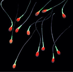
Colourful Sperm
This false-colour image (Figure 18.4.1) shows real human sperm. The tiny gametes are obviously greatly magnified in the picture, because they are actually the smallest of all human cells. In fact, human sperm cells are small, even when compared with sperm cells of other animals. Mice sperm are about twice the length of human sperm! Human sperm may be small in size, but in a normal, healthy man, huge numbers of them are usually released during each ejaculation. There may be hundreds of millions of sperm cells in a single teaspoon of semen. Producing sperm is one of the major functions of the male reproductive system.
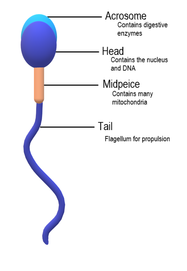
Sperm Anatomy
A mature sperm cell has several structures that help it reach and penetrate an egg. These are labeled in the drawing of a sperm shown in Figure 18.4.2.
- The head is the part of the sperm that contains the nucleus — and not much else. The nucleus, in turn, contains tightly coiled DNA that is the male parent’s contribution to the genetic makeup of a zygote (if one forms). Each sperm is a haploid cell, containing half the chromosomal complement of a normal, diploid body cell.
- The front of the head is an area called the acrosome. The acrosome contains enzymes that help the sperm penetrate an ovum (if it reaches one).
- The midpiece is the part of the sperm between the head and the flagellum. The midpiece is packed with mitochondria that produce the energy needed to move the flagellum.
- The flagellum (also called the tail) can rotate like a propeller, allowing the sperm to “swim” through the female reproductive tract to reach an ovum if one is present.
Spermatogenesis
The process of producing sperm is known as spermatogenesis. Spermatogenesis normally starts when a male reaches puberty, and it usually continues uninterrupted until death, although a decrease in sperm production generally occurs at older ages. A young, healthy male may produce hundreds of millions of sperm a day! Only about half of these, however, are likely to become viable, mature sperm.
Where Sperm Are Produced
Spermatogenesis occurs in the seminiferous tubules in the testes. Spermatogenesis requires high concentrations of testosterone. Testosterone is secreted by Leydig cells, which are adjacent to the seminiferous tubules in the testes.
Sperm production in the seminiferous tubules is very sensitive to temperature. This may be the most important reason the testes are located outside the body in the scrotum. The temperature inside the scrotum is generally about 2 degrees Celsius cooler than core body temperature. This lower temperature is optimal for spermatogenesis. The scrotum regulates its internal temperature as needed by contractions of the smooth muscles lining the scrotum. When the temperature inside the scrotum becomes too low, the scrotal muscles contract. The contraction of the muscles pulls the scrotum higher against the body, where the temperature is warmer. The opposite occurs when the temperature inside the scrotum becomes too high.
Events of Spermatogenesis
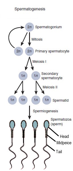
Figure 18.4.3 summarizes the main cellular events that occur in the process of spermatogenesis. The process begins with a diploid stem cell called a spermatogonium (plural, spermatogonia), and involves several cell divisions. The entire process takes at least ten weeks to complete, including maturation in the epididymis.
- A spermatogonium undergoes mitosis to produce two diploid cells called primary spermatocytes. One of the primary spermatocytes goes on to produce sperm. The other replenishes the reserve of spermatogonia.
- The primary spermatocyte undergoes meiosis I to produce two haploid daughter cells called secondary spermatocytes.
- The secondary spermatocytes rapidly undergo meiosis II to produce a total of four haploid daughter cells called spermatids.
- The spermatids begin to form a tail, and their DNA becomes highly condensed. Unnecessary cytoplasm and organelles are removed from the cells, and they form a head, midpiece, and flagellum. The resulting cells are sperm (spermatozoa).
As shown in Figure 18.4.4, the events of spermatogenesis begin near the wall of the seminiferous tubule — where spermatogonia are located — and continue inward toward the lumen of the tubule. Sertoli cells extend from the wall of the seminiferous tubule inward toward the lumen, so they are in contact with developing sperm at all stages of spermatogenesis. Sertoli cells play several roles in spermatogenesis:
- They secrete endocrine hormones that help regulate spermatogenesis.
- They secrete substances that initiate meiosis.
- They concentrate testosterone (from Leydig cells), which is needed at high levels to maintain spermatogenesis.
- They phagocytize the extra cytoplasm that is shed from developing sperm cells.
- They secrete testicular fluid that helps carry sperm into the epididymis.
- They maintain a blood-testis barrier, so immune system cells cannot reach and attack the sperm.
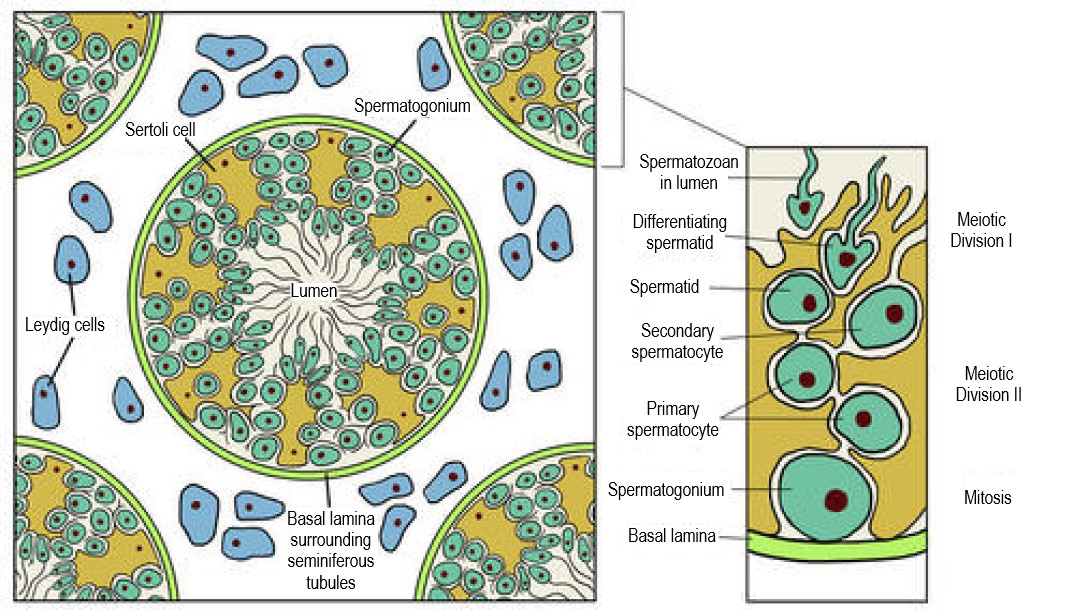
Maturation in the Epididymis
Although the sperm produced in the testes have tails, they are not yet motile (able to “swim”). The non-motile sperm are transported to the epididymis in testicular fluid that is secreted by Sertoli cells with the help of peristaltic contractions. In the epididymis, the sperm gain motility, so they are capable of swimming up the female genital tract and reaching an ovum. The mature sperm are stored in the epididymis until ejaculation occurs.
Ejaculation
Sperm are released from the body during ejaculation, which typically occurs during orgasm. Hundreds of millions of mature sperm — contained within a small amount of thick, whitish fluid called semen — are propelled from the penis during a normal ejaculation.
How Ejaculation Occurs
Ejaculation occurs when peristalsis of the muscle layers of the vas deferens and other accessory structures propel sperm from the epididymes, where mature sperm are stored. The muscle contractions force the sperm through the vas deferens and the ejaculatory ducts, and then out of the penis through the urethra. Due to the peristaltic action of the muscles, the ejaculation occurs in a series of spurts.
The Role of Semen
As sperm travel through the ejaculatory ducts during ejaculation, they mix with secretions from the seminal vesicles, prostate gland, and bulbourethral glands to form semen (see Figure 18.4.5 ). The average amount of semen per ejaculate is about 3.7 mL, which is a little less than a teaspoonful. Most of this volume of semen consists of glandular secretions, with the hundreds of millions of sperm cells actually contributing relatively little to the total volume.
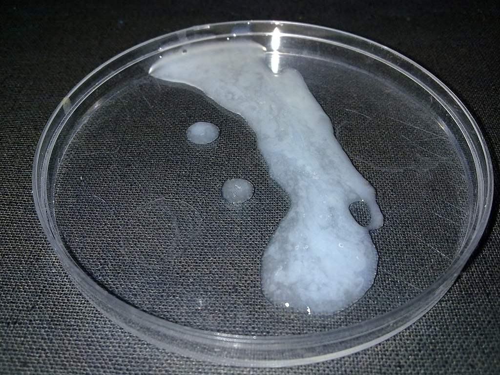
The secretions in semen are important for the survival and motility of sperm. They provide a medium through which sperm can swim. They also include sperm-sustaining substances, such as high concentrations of the sugar fructose, which is the main source of energy for sperm. In addition, semen contains many alkaline substances that help neutralize the acidic environment in the female vagina. This protects the DNA in sperm from being denatured by acid, and prolongs the life of sperm in the female reproductive tract.
Erection
Besides providing a way for sperm to leave the body, the main role of the penis in reproduction is intromission, or depositing sperm in the vagina of the female reproductive tract. Intromission depends on the ability of the penis to become stiff and erect, a state referred to as an erection. The human penis, unlike that of most other mammals, contains no erectile bone. Instead, in order to reach its erect state, it relies entirely on engorgement with blood of its columns of spongy tissue. During sexual arousal, the arteries that supply blood to the penis dilate, allowing more blood to fill the spongy tissue. The now-engorged spongy tissue presses against and constricts the veins that carry blood away from the penis. As a result, more blood enters than leaves the penis, until a constant erectile size is achieved.
In addition to sperm, the penis also transports urine out of the body. These two functions cannot occur simultaneously. During an erection, the sphincters that prevent urine from leaving the bladder are controlled by centres in the brain so they cannot relax and allow urine to enter the urethra.
Testosterone Production
The final major function of the male reproductive system is the production of the male sex hormone testosterone. In mature males, this occurs mainly in the testes. Testosterone production is under the control of luteinizing hormone (LH) from the pituitary gland. LH stimulates Leydig cells in the testes to secrete testosterone.
Testosterone is important for male sexual development at puberty. It stimulates maturation of the male reproductive organs, as well as the development of secondary male sex characteristics (such as facial hair). Testosterone is also needed in mature males for normal spermatogenesis to be maintained in the testes. Follicle stimulating hormone (FSH) from the pituitary gland is also needed for spermatogenesis to occur, in part because it helps Sertoli cells in the testes concentrate testosterone to high enough levels to maintain sperm production. Testosterone is also needed for proper functioning of the prostate gland. In addition, testosterone plays a role in erection, allowing sperm to be deposited within the female reproductive tract.
Feature: My Human Body

If you’re a man and you use a laptop computer on your lap for long periods of time, you may be decreasing your fertility. The reason? A laptop computer generates considerable heat, and its proximity to the scrotum during typical use results in a significant rise in temperature inside the scrotum. Spermatogenesis is very sensitive to high temperatures, so it may be adversely affected by laptop computer use. If you want to avoid the potentially fertility-depressing effect of laptop computer use, you might want to consider using your laptop computer on a table or other surface rather than on your lap — at least when you log on for long computer sessions. Other activities that raise scrotal temperature and have the potential to reduce spermatogenesis including soaking in hot tubs, wearing tight clothing, and biking. Although the effects of short-term scrotal heating on fertility seem to be temporary, years of such heat exposure may cause irreversible effects on sperm production.
18.4 Summary
- Parts of a mature sperm include the head, acrosome, midpiece, and flagellum. The process of producing sperm is called spermatogenesis. This normally starts during puberty, and continues uninterrupted until death.
- Spermatogenesis occurs in the seminiferous tubules in the testes, and requires high concentrations of testosterone. Sertoli cells in the testes play many roles in spermatogenesis, including concentrating testosterone under the influence of follicle stimulating hormone from the pituitary gland.
- Spermatogenesis begins with a diploid stem cell called a spermatogonium, which undergoes mitosis to produce a primary spermatocyte. The primary spermatocyte undergoes meiosis I to produce haploid secondary spermatocytes, and these cells in turn undergo meiosis II to produce spermatids. After the spermatids grow a tail and undergo other changes, they become sperm.
- Before sperm are able to “swim,” they must mature in the epididymis. The mature sperm are then stored in the epididymis until ejaculation occurs.
- Ejaculation is the process in which semen is propelled by peristalsis in the vas deferens and ejaculatory ducts from the urethra in the penis. Semen is a whitish fluid that contains sperm and secretions from the seminal vesicles, prostate gland, and bulbourethral glands. These alkaline secretions are important for sperm survival and motility.
- Besides ejaculating sperm, another reproductive role of the penis is intromission, which is depositing sperm in the female vagina. This requires the penis to become stiff and erect, a state referred to as an erection. Erection usually occurs with sexual arousal as the columns of spongy tissue inside the penis become engorged with blood.
- Leydig cells in the testes secrete testosterone under the control of luteinizing hormone (LH) from the pituitary gland. Testosterone is needed for male sexual development at puberty, and to maintain normal spermatogenesis after puberty. It also plays a role in prostate function and penis's ability to become erect.
18.4 Review Questions
-
- Compare and contrast the terms: erection, ejaculation, and intromission.
- Describe semen and its components.
- Explain how erection occurs.
18.4 Explore More
https://youtu.be/gNHSTa0Yct4
How You're Destroying Your Sperm! Seeker, 2014.
https://youtu.be/krSMZDsjLuU
Human Physiology - Reproduction: Spermatogenesis, Janux, 2015.
Attributions
Figure 18.4.1
Sperm-20051108 by Gilberto Santa Rosa from Rio de Janeiro, Brazil on Wikimedia Commons is used under a CC BY 2.0 (https://creativecommons.org/licenses/by/2.0) license.
Figure 18.4.2
Sperm Anatomy by Christinelmiller on Wikimedia Commons is used under a CC BY-SA 4.0 (https://creativecommons.org/licenses/by-sa/4.0) license.
Figure 18.4.3
Spermatogenesis by OpenStax College is used and adapted by Christine Miller under a CC BY 3.0 (https://creativecommons.org/licenses/by/3.0) license.
Figure 18.4.4
Testis-cross-section by CK-12 Foundation is used under a CC BY-NC 3.0 (https://creativecommons.org/licenses/by-nc/3.0/) license.
Figure 18.4.5
Human_semen_in_a_petri_dish by Digitalkil on Wikimedia Commons is in the public domain (https://en.wikipedia.org/wiki/en:public_domain).
Figure 18.4.6
Laptop by logan-weaver-b76PEyeIptQ-unsplash [photo] by LOGAN WEAVER on Unsplash is used under the Unsplash License (https://unsplash.com/license).
References
Betts, J. G., Young, K.A., Wise, J.A., Johnson, E., Poe, B., Kruse, D.H., Korol, O., Johnson, J.E., Womble, M., DeSaix, P. (2013, June 19). Figure 27.5 Spermatogenesis [digital image]. In Anatomy and Physiology (Section 27.1). OpenStax. https://openstax.org/books/anatomy-and-physiology/pages/27-1-anatomy-and-physiology-of-the-male-reproductive-system
Brainard, J/ CK-12 Foundation. (2016). Figure 4 Cross-section of a testis and seminiferous tubules [digital image]. In CK-12 College Human Biology (Section 20.4) [online Flexbook]. CK12.org. https://www.ck12.org/book/ck-12-college-human-biology/section/20.4/
Janux. (2015, January 10). Human physiology - Reproduction: spermatogenesis. YouTube. https://www.youtube.com/watch?v=krSMZDsjLuU&feature=youtu.be
Seeker. (2014, June 16). How you're destroying your sperm! YouTube. https://www.youtube.com/watch?v=gNHSTa0Yct4&feature=youtu.be
Created by CK-12 Foundation/Adapted by Christine Miller

Fertility Symbol
The geometric design on the ancient stone carving in Figure 18.6.1 represents a powerful symbol of female fertility: the vagina. The symbol is called yoni in Hindu, and it reflects the value placed by Hindu culture on the ability of females to give birth. The vagina is one of several organs in the female reproductive system.
Female Reproductive Organs
The female reproductive system is made up of internal and external organs that function to produce haploid female gametes called ova (or oocytes), secrete female sex hormones (such as estrogen), and carry and give birth to a fetus. The internal female reproductive organs include the vagina, uterus, oviducts, and ovaries. The external organs — collectively called the vulva — include the clitoris and labia.
The vagina is an elastic, muscular canal leading from its opening in the vulva to the neck of the uterus, called the cervix. It is about 7.5 cm (about 3 in) long at the front, and about 9 cm (3.5 in) long at the back. The vagina accommodates the penis and is the site where sperm are usually ejaculated during sexual intercourse. In the context of pregnancy and natural (vaginal) childbirth, the vagina is referred to as the birth canal. In addition, it channels the flow of menstrual blood from the uterus.
Structure of the Vagina
Muscles and ligaments support the vagina within the pelvic cavity. The vagina itself is made up of several layers of fibrous and muscular tissues and lined with mucous membranes. Folds in the mucosa provide the vagina with extra surface area so it can stretch in both length and width during intercourse or childbirth. The elasticity of the vagina and the extra mucosa allow it to stretch to many times its normal diameter in order to deliver a baby.
Bacteria and pH in the Vagina
A healthy vagina is home to many symbiotic bacteria that help prevent pathogens (such as yeast) from colonizing the vagina. The pH in the vagina is normally between 3.8 and 4.5, and this acidity also helps keep pathogenic microorganisms from colonizing it. The vagina constantly sheds its epithelium, so it is considered self-cleaning. As a consequence, there is no need for douching to clean it. Physicians actually discourage the practice, as it may upset the normal bacterial and pH balance in the vagina, although washing the vulva with a mild soap is good practice.
Uterus
The uterus (commonly called the womb) is a pear-shaped, muscular organ that is about 7.6 cm (about 3 in) long. It is located above the vagina and behind the bladder in the centre of the pelvis. The position of the uterus in the pelvis is stabilized by several ligaments and bands of supportive tissue. The uterus is where a fetus develops during gestation, and the organ provides mechanical protection and support for the developing offspring. Contractions of the muscular wall of the uterus are responsible for pushing the fetus out of the uterus during childbirth.
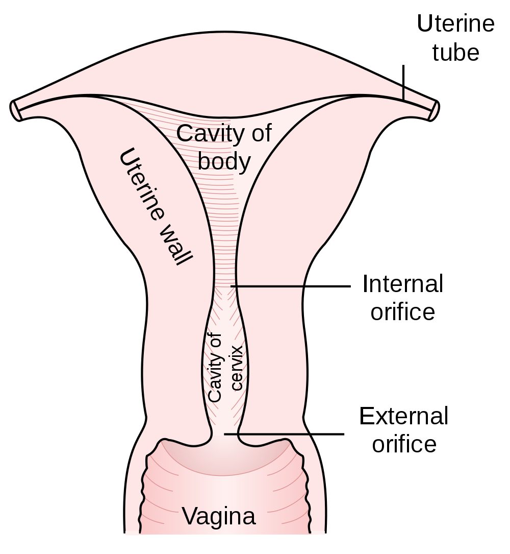
Parts of the Uterus
As shown in Figure 18.6.2, the lower end of the uterus forms the cervix, which is also called the neck of the uterus. The cervix is about 2.5 cm (almost 1 in) long and protrudes downward into the vagina. A small canal runs the length of the cervix, connecting the uterine cavity with the lumen of the vagina. This allows semen deposited in the vagina to enter the uterus, and a baby to pass from the uterus into the vagina during birth. Glands in the cervix secrete mucus that varies in water content and thickness, so it can function either as a barrier to keep microorganisms out of the uterus during pregnancy, or as a transport medium to help sperm enter the uterus around the time of ovulation. The rest of the uterus above the cervix is called the body of the uterus. The upper end of the uterus is connected with the two oviducts.
Tissues of the Uterus
As indicated in Figure 18.6.3, the uterus consists of three tissue layers, called the endometrium, myometrium, and perimetrium.
- The endometrium is the innermost tissue layer of the uterus. It consists of epithelial tissue, including mucous membranes. This layer thickens during each menstrual cycle and, unless an egg is fertilized, sloughs off during the following menstrual period. If an ovum is fertilized, the thickened endometrium is maintained by hormones and provides nourishment to the embryo. As gestation progresses, the endometrium develops into the maternal portion of the placenta.
- The placenta is a temporary organ that consists of a mass of maternal and fetal blood vessels through which the mother’s and fetus’s blood exchange substances.
- The myometrium is the middle layer of the uterus. It consists mostly of a thick layer of smooth muscle tissue. Powerful contractions of the smooth muscle allow the uterus to contract and expel an infant during childbirth.
- The perimetrium is the outermost layer of the uterus. It covers the outer surface of the uterus. This layer actually consists of two layers of epithelium that secrete fluid into the space between them. The fluid allows for small movements of the uterus within the pelvis, without causing it to rub against other organs.
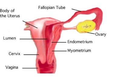
Oviducts
The oviducts (often referred to as Fallopian tubes) are two thin tubes that lie between the ovaries and the uterus. The oviducts are not attached to the ovaries, but their broad upper ends — called infundibula — lie very close to the ovaries. The infundibula also have fringe-like extensions called fimbriae that move in a waving motion to help guide eggs from the ovaries into the oviducts. The lower ends of the oviducts are attached to the upper part of the body of the uterus on either side of the body. They open into the uterus.
The oviducts are made up of multiple tissue layers. The innermost layer consists of mucosal epithelium. The epithelium is covered with cilia, which can move in a sweeping motion to help ova move through the tube from the ovary to the uterus. In between the ciliated cells of the epithelium are cells that secrete a fluid called tubular fluid. This fluid contains nutrients for sperm, ova, and zygotes. The secretions in tubular fluid also remove certain molecules from the plasma membrane of sperm so they are better able to penetrate an egg. Other layers of the oviducts consist of connective tissue and smooth muscle. Contractions of the smooth muscle allow peristalsis to help move eggs through the tubes.
Ovaries
Like the testes in males, the ovaries in females are gonads that produce gametes and secrete sex hormones. The gametes produced by the ovaries are called ova, or oocytes. The main sex hormone secreted by the ovaries is estrogen. The position of the paired ovaries relative to the other reproductive system organs is shown in Figure 18.6.4. Each ovary lies along one side of the uterus and is about 4 cm (a little more than 1.5 in) long. Fibrous ligaments attach one end of each ovary to its nearby oviduct and the other and to its side of the uterus. These ligaments keep the ovaries in place within the pelvis.
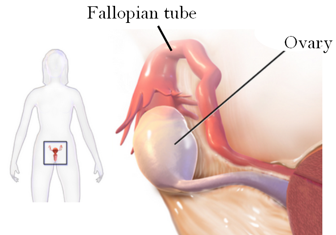
Ovarian Follicles
The ovary consists of two main layers, called the ovarian medulla (the inner layer) and the ovarian cortex (the outer layer). The ovary also contains blood and lymphatic vessels. The ovarian cortex consists primarily of the functional units of the ovaries, which are called ovarian follicles. The follicles are nests of epithelial cells, within each of which is an ovum. The photomicrograph in Figure 18.6.5 shows an ovarian follicle and the developing ovum inside it. If an ovum and follicle complete maturation, the follicle ruptures and the ovum is released from the ovary. This event is called ovulation.
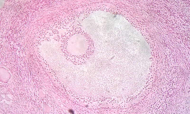
Ova in the Ovaries
Whereas the male testes produce sperm continuously after puberty, the female ovary already contains all the ova it will ever produce by the time a female is born. At birth, a baby girl’s ovaries contain at least a million eggs, each of which is contained within a follicle. Only about 500 of these eggs will eventually mature and be ovulated. This process starts at puberty and typically continues at monthly intervals until menopause occurs around age 52. The remaining eggs never mature, and their number declines as the woman ages. By menopause, a woman’s reserve of eggs is nearly depleted, and ovulation no longer occurs.
Vulva
The vulva is a general term for all of the external female reproductive organs. The vulva includes the clitoris, labia, and external openings for the urethra and vagina.
Labia
The labia (singular, labium) refer to the “lips” of the vulva, which are folds of tissue that contain and protect the other, more delicate structures of the vulva (as shown in Figure 18.6.6). There are two pairs of labia: the outer and larger labia majora, and the inner and smaller labia minora. The labia minora contain numerous sebaceous glands. These glands release secretions that help lubricate the labia and vulvar area.
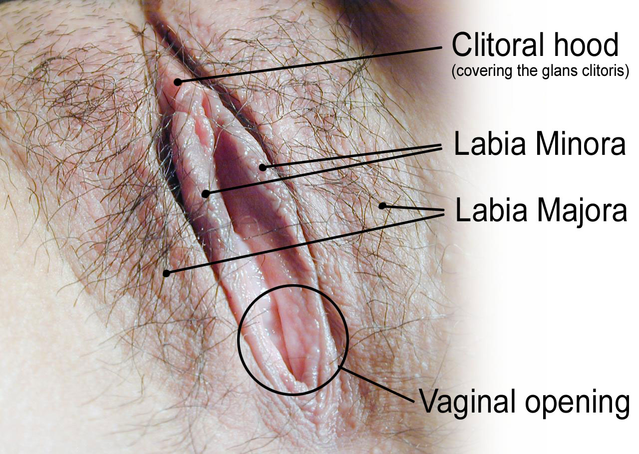
Clitoris
The clitoris, is located at the front of the vulva where the labia minora meet. The visible portion of the clitoris is called the glans clitoris. It is roughly the size and shape of a pea. It is highly sensitive, because it contains many nerve endings. A hood of tissue called the clitoral hood (shown in Figure 18.6.5 above), or prepuce, normally covers and protects the clitoris. The clitoris is the homologue to the male penis, and they both contain spongy tissue. Stimulation of the glans clitoris during sexual activity generally results in sexual arousal in females, and may lead to orgasm. The glans clitoris is the only part of the overall clitoris visible externally, but this spongy tissue extends down either side of the openings to the urethra and vagina, as seen in Figure 18.6.7.
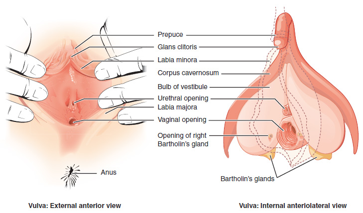
Other Vulvar Structures
The area between the two labia minora is called the vestibule of the vulva. Both the urethra and vagina have openings to the outside of the body in the vestibule. As you can see in Figure 18.6.7 above, the urethral opening (or meatus) is located just in front of, and is much smaller than, the vaginal opening. Both openings are protected by the labia. Two glands — called Bartholin’s glands — open on either side of the vaginal opening. These glands secrete mucus and a vaginal and vulvar lubricant.
Breasts
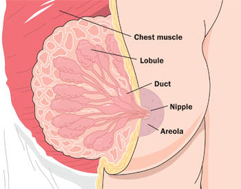
The breasts are not directly involved in reproduction, but because they contain mammary glands, they can provide nourishment to an infant after birth. The breasts overlay major muscles in the chest from which they project outward in a conical shape. The two main types of tissues in the breast are adipose (fat) tissue and glandular tissue that produces milk. As shown in Figure 18.6.8, each mature breast contains many lobules, where milk is produced and stored during pregnancy. During breastfeeding (or lactation), the milk drains into ducts and sacs, which in turn converge at the nipple. Milk exits the breast through the nipple in response to the suckling action of an infant and is regulated by a positive feedback loop. The nipple is surrounded by a more darkly pigmented area called the areola. The areola contains glands that secrete an oily fluid, which lubricates and protects the nipple during breastfeeding.
18.6 Summary
- The female reproductive system is made up of internal and external organs that function to produce haploid female gametes called ova, secrete female sex hormones (such as estrogen), and carry and give birth to a fetus.
- The vagina is an elastic, muscular canal that can accommodate the penis. It is also where sperm are usually ejaculated during sexual intercourse. The vagina is the birth canal, and it channels the flow of menstrual blood from the uterus. A healthy vagina has a balance of symbiotic bacteria and an acidic pH.
- The uterus is a muscular organ above the vagina where a fetus develops. Its muscular walls contract to push out the fetus during childbirth. The cervix is the neck of the uterus that extends down into the vagina. It contains a canal connecting the vagina and uterus for sperm, or for an infant to pass through. The innermost layer of the uterus — the endometrium — thickens each month in preparation for an embryo, but is shed in the following menstrual period if fertilization does not occur.
- The oviducts extend from the uterus to the ovaries. Waving fimbriae at the ovary ends of the oviducts guide ovulated eggs into the tubes where fertilization may occur as the ova travel to the uterus. Cilia and peristalsis help ova move through the tubes. Tubular fluid helps nourish sperm as they swim up the tubes toward ova.
- The ovaries are gonads that produce ova and secrete sex hormones, including estrogen. Nests of cells called follicles in the ovarian cortex are the functional units of ovaries. Each follicle surrounds an immature ovum. At birth, a baby girl’s ovaries contain at least a million ova, and they will not produce any more during her lifetime. During a woman’s reproductive years, one ova typically matures and is ovulated each month.
- The vulva is a general term for external female reproductive organs. The vulva includes the clitoris, two pairs of labia, and openings for the urethra and vagina. Secretions from mucosal glands near the vaginal opening lubricate the vulva.
- The breasts are not technically reproductive organs, but their mammary glands produce milk to feed an infant after birth. Milk drains through ducts and sacs, and out through the nipple when a baby sucks during breastfeeding.
18.6 Review Questions
-
- State the general functions of the female reproductive system.
- Describe the vagina and its reproductive functions.
- Outline the structure and basic functions of the uterus.
- What is the endometrium? How does it change during the monthly cycle?
- Why are breasts included in discussions of reproduction, if they are not organs of the female reproductive system?
- What is the function of the folds in the mucous membrane lining of the vagina?
- What are two ways in which the female reproductive system protects itself from pathogens?
18.6 Explore More
https://youtu.be/SkB4gG8ke7Q
The uncomplicated truth about women's sexuality | Sarah Barmak, TED, 2019.
https://youtu.be/9rs2gNchQig
Human Physiology - Functional Anatomy of the Female Reproductive System, Janux, 2015.
Attributes
Figure 18.6.1
1024px-Cattien_stone_yoni by Binh Giang on Wikimedia Commons is in the public domain (https://en.wikipedia.org/wiki/en:public_domain).
Figure 18.6.2
1000px-Gray1167.svg by Henry Vandyke Carter (1831-1897) on Wikimedia Commons is in the public domain (https://en.wikipedia.org/wiki/public_domain). (Bartleby.com: Gray’s Anatomy, Plate 1167).
Figure 18.6.3
Uterine_anatomy. from Uterine Stem cells by The Stem Cell Research Community, StemBook on Wikimedia Commons is used under a CC BY 3.0 (https://creativecommons.org/licenses/by/3.0) license.
Figure 18.6.4
Sites_of_tubo_ovarian_abscess by Bfpage on Wikimedia Commons is used under a CC BY-SA 4.0 (https://creativecommons.org/licenses/by-sa/4.0) license.
Figure 18.6.5
Ovarian_follicle by TiagoLubiana on Wikimedia Commons is used under a CC BY 4.0 (https://creativecommons.org/licenses/by-sa/4.0) license.
Figure 18.6.6
HumanVulva-NewText-PhiloViv by Amphis (edited) on Wikimedia Commons is used under a CC BY-SA 3.0 (https://creativecommons.org/licenses/by-sa/3.0/deed.en) license. (Original en:Image:HumanVulva-NoText-PhiloVivero.jpg by en:user:PhiloVivero)
Figure 18.6.7
Vulva by OpenStax College on Wikimedia Commons is used under a CC BY 3.0 (https://creativecommons.org/licenses/by/3.0) license.
Figure 18.6.8
Breast-Diagram by Women's Health (NCI/ NIH) on Wikimedia Commons is in the public domain (https://en.wikipedia.org/wiki/en:Public_domain).
References
Betts, J. G., Young, K.A., Wise, J.A., Johnson, E., Poe, B., Kruse, D.H., Korol, O., Johnson, J.E., Womble, M., DeSaix, P. (2013, June 19). Figure 27.10 The vulva [digital image]. In Anatomy and Physiology (Section 27.2). OpenStax. https://openstax.org/books/anatomy-and-physiology/pages/27-2-anatomy-and-physiology-of-the-female-reproductive-system
Janux. (2015, January 10). Human physiology - Functional anatomy of the female reproductive system. YouTube. https://www.youtube.com/watch?v=9rs2gNchQig&feature=youtu.be
TED. (2019, March 22). The uncomplicated truth about women's sexuality | Sarah Barmak. YouTube. https://www.youtube.com/watch?v=SkB4gG8ke7Q&feature=youtu.be
Teixeira, J., Rueda, B.R., and Pru, J.K. (September 30, 2008). Figure 1 Uterine anatomy. In Uterine Stem Cells (StemBook, ed.). The Stem Cell Research Community, StemBook, doi/10.3824/stembook.1.16.1, http://www.stembook.org
Created by CK-12 Foundation/Adapted by Christine Miller
Taboo Topic
The banner in Figure 18.8.1 was carried in a 2014 march in Uganda as part of the celebration of Menstrual Hygiene Day. Menstrual Hygiene Day is an awareness day on May 28 of each year that aims to raise awareness worldwide about menstruation and menstrual hygiene. Maintaining good menstrual hygiene is difficult in developing countries like Uganda because of taboos on discussing menstruation and lack of availability of menstrual hygiene products. Poor menstrual hygiene, in turn, can lead to embarrassment, degradation, and reproductive health problems in females. May 28 was chosen as Menstrual Hygiene Day because of its symbolism. May is the fifth month of the year, and most women average five days of menstrual bleeding each month. The 28th day was chosen because the menstrual cycle averages about 28 days.
What Is the Menstrual Cycle?
The menstrual cycle refers to natural changes that occur in the female reproductive system each month during the reproductive years. The cycle is necessary for the production of ova and the preparation of the uterus for pregnancy. It involves changes in both the ovaries and the uterus, and is controlled by pituitary and ovarian hormones. Day 1 of the cycle is the first day of the menstrual period, when bleeding from the uterus begins as the built-up endometrium lining the uterus is shed. The endometrium builds up again during the remainder of the cycle, only to be shed again during the beginning of the next cycle if pregnancy does not occur. In the ovaries, the menstrual cycle includes the development of a follicle, ovulation of a secondary oocyte, and then degeneration of the follicle if pregnancy does not occur. Both uterine and ovarian changes during the menstrual cycle are generally divided into three phases, although the phases are not the same in the two organs.
Menarche and Menopause
The female reproductive years are delineated by the start and stop of the menstrual cycle. The first menstrual period usually occurs around 12 or 13 years of age, an event that is known as menarche. There is considerable variation among individuals in the age at menarche. It may occasionally occur as early as eight years of age or as late as 16 years of age and still be considered normal. The average age is generally later in the developing world, and earlier in the developed world. This variation is thought to be largely attributable to nutritional differences.
The cessation of menstrual cycles at the end of a woman’s reproductive years is termed menopause. The average age of menopause is 52 years, but it may occur normally at any age between about 45 and 55 years of age. The age of menopause varies due to a variety of biological and environmental factors. It may occur earlier as a result of certain illnesses or medical treatments.
Variation in the Menstrual Cycle
The length of the menstrual cycle — as well as its phases — may vary considerably, not only among different women, but also from month to month for a given woman. The average length of time between the first day of one menstrual period and the first day of the next menstrual period is 28 days, but it may range from 21 days to 45 days. Cycles are considered regular when a woman’s longest and shortest cycles differ by less than eight days. The menstrual period itself is usually about five days long, but it may vary in length from about two days to seven days.
Ovarian Cycle
The events of the menstrual cycle that take place in the ovaries make up the ovarian cycle. It consists of changes that occur in the follicles of one of the ovaries. The ovarian cycle is divided into the following three phases: follicular phase, ovulation, and luteal phase. These phases are illustrated in Figure 18.8.2.
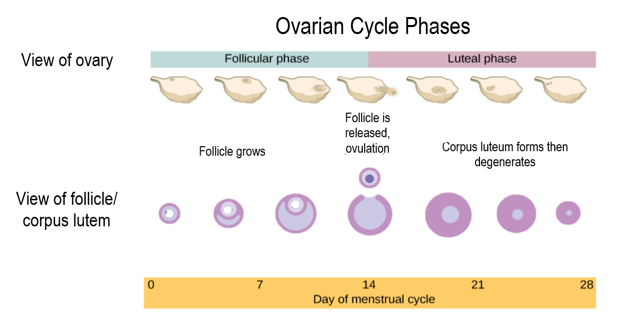
Follicular Phase
The follicular phase is the first phase of the ovarian cycle. It generally lasts about 12 to 14 days for a 28-day menstrual cycle. During this phase, several ovarian follicles are stimulated to begin maturing, but usually only one — called the Graafian follicle — matures completely so it is ready to release an egg. The other maturing follicles stop growing and disintegrate. Follicular development occurs because of a rise in the blood level of follicle stimulating hormone (FSH), which is secreted by the pituitary gland. The maturing follicle releases estrogen, the level of which rises throughout the follicular phase. You can see these and other changes in hormone levels that occur during the menstrual cycle in the following chart.
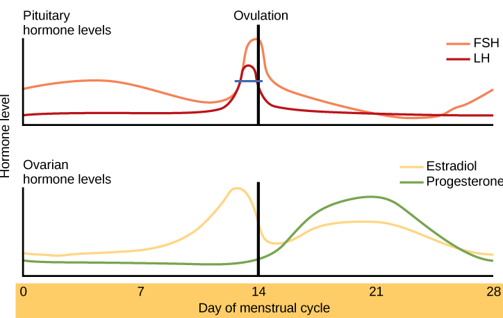
Ovulation
Ovulation is the second phase of the ovarian cycle. It usually occurs around day 14 of a 28-day menstrual cycle. During this phase, the Graafian follicle ruptures and releases its ovum. Ovulation is stimulated by a sudden rise in the blood level of luteinizing hormone (LH) from the pituitary gland. This is called the LH surge. You can see the LH surge in the top hormone graph in Figure 18.8.3. The LH surge generally starts around day 12 of the cycle and lasts for a day or two. The surge in LH is triggered by a continued rise in estrogen from the maturing follicle in the ovary. During the follicular phase, the rising estrogen level actually suppresses LH secretion by the pituitary gland. However, by the time the follicular phase is nearing its end, the level of estrogen reaches a threshold level above which this effect is reversed, and estrogen stimulates the release of a large amount of LH. The surge in LH matures the ovum and weakens the wall of the follicle, causing the fully developed follicle to release its secondary oocyte.
Luteal Phase
The luteal phase is the third and final phase of the ovarian cycle. It typically lasts about 14 days in a 28-day menstrual cycle. At the beginning of the luteal phase, FSH and LH cause the Graafian follicle that ovulated the egg to transform into a structure called a corpus luteum. The corpus luteum secretes progesterone, which in turn suppresses FSH and LH production by the pituitary gland and stimulates the continued buildup of the endometrium in the uterus. How this phase ends depends on whether or not the ovum has been fertilized.
- If fertilization has not occurred, the falling levels of FSH and LH during the luteal phase cause the corpus luteum to atrophy, so its production of progesterone declines. Without a high level of progesterone to maintain it, the endometrium starts to break down. By the end of the luteal phase, the endometrium can no longer be maintained, and the next menstrual cycle begins with the shedding of the endometrium (menses).
- If fertilization has occurred so a zygote forms and then divides to become a blastocyst, the outer layer of the blastocyst produces a hormone called human chorionic gonadotropin (HCG). This hormone is very similar to LH and preserves the corpus luteum. The corpus luteum can then continue to secrete progesterone to maintain the new pregnancy.
Uterine Cycle
The events of the menstrual cycle that take place in the uterus make up the uterine cycle. This cycle consists of changes that occur mainly in the endometrium, which is the layer of tissue that lines the uterus. The uterine cycle is divided into the following three phases: menstruation, proliferative phase, and secretory phase. These phases are illustrated in Figure 18.8.4.
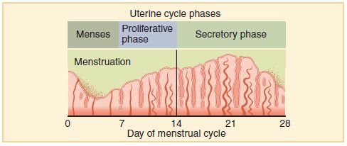
Menstruation
Menstruation (also called menstrual period or menses) is the first phase of the uterine cycle. It occurs if fertilization has not taken place during the preceding menstrual cycle. During menstruation, the endometrium of the uterus, which has built up during the preceding cycle, degenerates and is shed from the uterus, flowing through an opening in the cervix, and out through the external opening of the vagina. The average loss of blood during menstruation is about 35 mL (about 1 oz or 2 tablespoons). The flow of blood is often accompanied by uterine cramps, which may be severe in some women.
Proliferative Phase
The proliferative phase is the second phase of the uterine cycle. During this phase, estrogen secreted by cells of the maturing ovarian follicle causes the lining of the uterus to grow, or proliferate. Estrogen also stimulates the cervix of the uterus to secrete larger amounts of thinner mucus that can help sperm swim through the cervix and into the uterus, making fertilization more likely.
Secretory Phase
The secretory phase is the third and final phase of the uterine cycle. During this phase, progesterone produced by the corpus luteum in the ovary stimulates further changes in the endometrium so it is more receptive to implantation of a blastocyst. For example, progesterone increases blood flow to the uterus and promotes uterine secretions. It also decreases the contractility of smooth muscle tissue in the uterine wall.
Bringing it All Together
It is important to note that the pituitary gland, the ovaries and the uterus are all responsible for parts of the ovarian and uterine cycles. The pituitary hormones, LH and FSH affect the ovarian cycle and its hormones. The ovarian hormones, estrogen and progesterone affect the uterine cycle and also feedback on the pituitary gland. Look at Figure 18.8.5 and look at what is happening on different days of the cycle in each of the sets of hormones, the ovarian cycle and the uterine cycle.
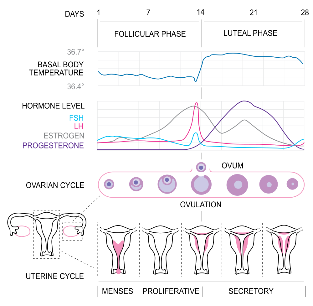
18.8 Summary
- The menstrual cycle refers to natural changes that occur in the female reproductive system each month during the reproductive years, except when a woman is pregnant. The cycle is necessary for the production of ova and the preparation of the uterus for pregnancy. It involves changes in both the ovaries and uterus, and is controlled by pituitary gland hormones (FSH and LH) and ovarian hormones (estrogen and progesterone).
- The female reproductive period is delineated by menarche, or the first menstrual period, which usually occurs around age 12 or 13; and by menopause, or the cessation of menstrual periods, which typically occurs around age 52. A typical menstrual cycle averages 28 days in length but may vary normally from 21 to 45 days. The average menstrual period is five days long, but may vary normally from two to seven days. These variations in the menstrual cycle may occur both between women and within individual women from month to month.
- The events of the menstrual cycle that take place in the ovaries make up the ovarian cycle. It includes the follicular phase (when a follicle and its ovum mature due to rising levels of FSH), ovulation (when the ovum is released from the ovary due to a rise in estrogen and a surge in LH), and the luteal phase (when the follicle is transformed into a structure called a corpus luteum that secretes progesterone). In a 28-day menstrual cycle, the follicular and luteal phases typically average about two weeks in length, with ovulation generally occurring around day 14 of the cycle.
- The events of the menstrual cycle that take place in the uterus make up the uterine cycle. It includes menstruation, which generally occurs on days 1 to 5 of the cycle and involves shedding of endometrial tissue that built up during the preceding cycle; the proliferative phase, during which the endometrium builds up again until ovulation occurs; and the secretory phase, which follows ovulation and during which the endometrium secretes substances and undergoes other changes that prepare it to receive an embryo.
18.8 Review Questions
-
- What is the menstrual cycle? Why is the menstrual cycle necessary in order for pregnancy to occur?
- What organs are involved in the menstrual cycle?
- Identify the two major events that mark the beginning and end of the reproductive period in females. When do these events typically occur?
- Discuss the average length of the menstrual cycle and menstruation, as well as variations that are considered normal.
- If the LH surge did not occur in a menstrual cycle, what do you think would happen? Explain your answer.
- Give one reason why FSH and LH levels drop in the luteal phase of the menstrual cycle.
18.8 Explore More
https://youtu.be/cjbgZwgdY7Q
Why do women have periods? TED-Ed, 2015.
https://youtu.be/5B3Abpv0ysM
Girl's Rite of Passage | National Geographic, 2007.
Attributions
Figure 18.8.1
WaterforPeople_Uganda by WaterforPeople_Uganda on Wikimedia Commons is used under a CC BY 2.0 (https://creativecommons.org/licenses/by/2.0) license.
Figure 18.8.2
Ovarian Cycle by CNX OpenStax on Wikimedia Commons is used and adapted under a CC BY 4.0 (https://creativecommons.org/licenses/by/4.0) license.
Figure 18.8.3
Figure_43_04_04 by CNX OpenStax on Wikimedia Commons is used under a CC BY 4.0 (https://creativecommons.org/licenses/by/4.0) license. (Original: modification of work by Mikael Häggström)
Figure 18.8.4
Ovarian and menstrual cycle by OpenStax College on Wikimedia Commons is used under a CC BY 3.0 (https://creativecommons.org/licenses/by/3.0) license.
Figure 18.8.5
1000px-MenstrualCycle2_en.svg by Isometrik on Wikimedia Common is used under a CC BY-SA 3.0 (https://creativecommons.org/licenses/by-sa/3.0) license.
References
Betts, J. G., Young, K.A., Wise, J.A., Johnson, E., Poe, B., Kruse, D.H., Korol, O., Johnson, J.E., Womble, M., DeSaix, P. (2013, June 19). Figure 27.15 Hormone levels in ovarian and menstrual cycles [digital image]. In Anatomy and Physiology (Section 27.2). OpenStax. https://openstax.org/books/anatomy-and-physiology/pages/27-2-anatomy-and-physiology-of-the-female-reproductive-system
National Geographic. (2007, May 31). Girl's rite of passage | National Geographic. YouTube. https://www.youtube.com/watch?v=5B3Abpv0ysM&feature=youtu.be
OpenStax. (2016, May 27) Figure 4 Rising and falling hormone levels result in progression of the ovarian and menstrual cycles [digital image]. In Open Stax, Biology (Section 43.4). OpenStax CNX. https://cnx.org/contents/GFy_h8cu@10.53:Ha3dnFEx@6/Hormonal-Control-of-Human-Reproduction
TED-Ed. (2015, October 19). Why do women have periods? YouTube. https://www.youtube.com/watch?v=cjbgZwgdY7Q&feature=youtu.be
A synovial joint in which an oval-shaped process of one bone fits into a roughly elliptical cavity of the other, allowing movement in two planes.
Created by CK-12 Foundation/Adapted by Christine Miller
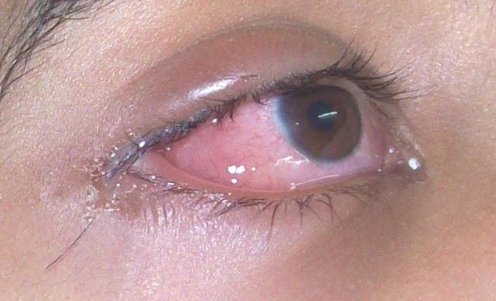
Allergy Eyes
Eyes that are red, watery, and itchy are typical of an allergic reaction known as allergic rhinitis. Commonly called hay fever, allergic rhinitis is an immune system reaction, typically to the pollen of certain plants. Your immune system usually protects you from pathogens and keeps you well. However, like any other body system, the immune system itself can develop problems. Sometimes, it responds to harmless foreign substances as though they were pathogens. This is the basis of allergies like hay fever.
Allergies
An allergy is a disorder in which the immune system makes an inflammatory response to a harmless antigen. It occurs when the immune system is hypersensitive to an antigen in the environment that causes little or no response in most people. Allergies are strongly familial. Allergic parents are more likely to have allergic children, and those children’s allergies are likely to be more severe, which is evidence that there is a heritable tendency to develop allergies. Allergies are more common in children than adults, because many children outgrow their allergies by adulthood.
Allergens
Any antigen that causes an allergy is called an allergen. Common allergens are plant pollens, dust mites, mold, specific foods (such as peanuts or shellfish), insect stings, and certain common medications (such as aspirin and penicillin). Allergens may be inhaled or ingested, or they may come into contact with the skin or eyes. Symptoms vary depending on the type of exposure, and the severity of the immune system response. Some of the most common causes of allergies are shown in Figure 17.6.2: latex, pollen, dust mites, pet dander, insect stings and various foods. Inhaling pollen may cause symptoms of allergic rhinitis, such as sneezing and red itchy eyes. Insect stings may cause an itchy rash. This type of allergy is called contact dermatitis.
Figure 17.6.2 Common allergens include latex, pollen, dust mites, pet dander, insect stings, and foods.
Prevalence of Allergies
There has been a significant increase in the prevalence of allergies over the past several decades, especially in the rich nations of the world, where allergies are now very common disorders. In the developed countries, about 20% of people have or have had hay fever, another 20% have had contact dermatitis, and about 6% have food allergies. In the poorer nations of the world, on the other hand, allergies of all types are much less common.
One explanation for the rise in allergies in the developed world is the hygiene hypothesis. According to this hypothesis, people in developed countries live in relatively sterile environments because of hygienic practices and sanitation systems. As a result, people in these countries are exposed to fewer pathogens than their immune system evolved to cope with. To compensate, their immune system “keeps busy” by attacking harmless antigens in allergic responses.
How Allergies Occur
The diagram in Figure 17.6.3 shows how an allergic reaction occurs. At the first exposure to an allergen, B cells are activated to form plasma cells that produce large amounts of antibodies to the allergen. These antibodies attach to leukocytes called mast cells. Subsequently, every time the person encounters the allergen again, the mast cells are already primed and ready to deal with it. The primed mast cells immediately release cytokines and histamines, which in turn cause inflammation and recruitment of leukocytes, among other responses. These responses are responsible for the signs and symptoms of allergies.
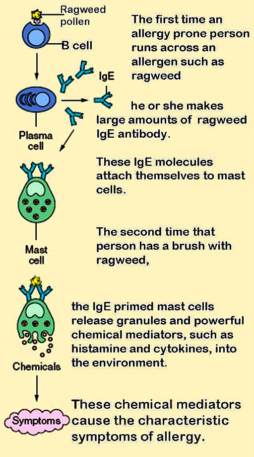
Treating Allergies
The symptoms of allergies can range from mild to life-threatening. Mild allergy symptoms are often treated with antihistamines. These are drugs that reduce or eliminate the effects of the histamines that produce allergy symptoms.
Treating Anaphylaxis
The most severe allergic reaction is a systemic reaction called anaphylaxis. This is a life-threatening response caused by a massive release of histamines. Many of the signs and symptoms of anaphylaxis are shown in Figure 17.6.4. Some of them include a drop in blood pressure, changes in heart rate, shortness of breath, and swelling of the tongue and throat, which may threaten the patient with suffocation unless emergency treatment is given. People who have had anaphylactic reactions may carry an epinephrine autoinjector (widely known by its brand name EpiPen®) so they can inject themselves with epinephrine if they start to experience an anaphylactic response. The epinephrine helps control the immune reaction until medical care can be provided. Epinephrine constricts blood vessels to increase blood pressure, relaxes smooth muscles in the lungs to reduce wheezing and improve breathing, modulates heart rate, and works to reduce swelling that may otherwise block the airways.
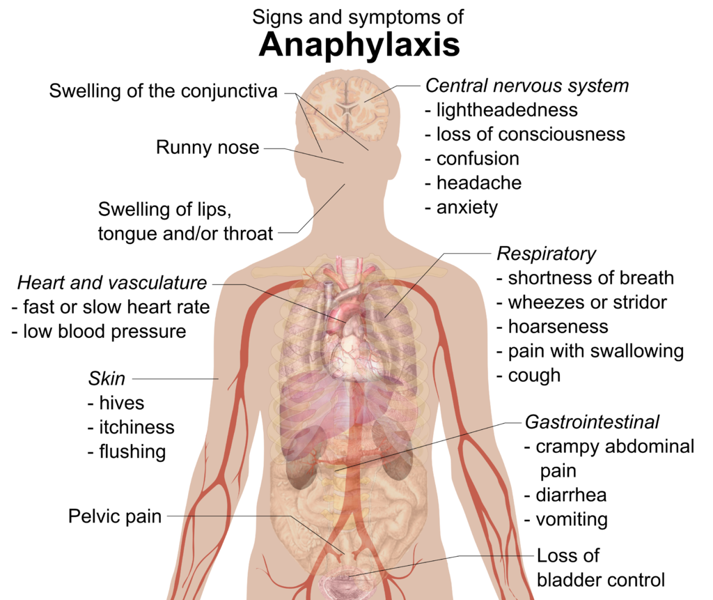
Immunotherapy for Allergies
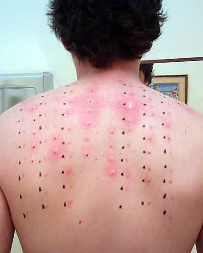
Another way to treat allergies is called immunotherapy, commonly called “allergy shots.” This approach may actually cure specific allergies, at least for several years if not permanently. It may be particularly beneficial for allergens that are difficult or impossible to avoid (such as pollen). First, however, patients must be tested to identify the specific allergens that are causing their allergies. As shown in Figure 17.6.5, this may involve scratching tiny amounts of common allergens into the skin, and then observing whether there is a localized reaction to any of them. Each allergen is applied in a different numbered location on the skin, so if there is a reaction — such as redness or swelling — the responsible allergens can be identified. Then, through periodic injections (usually weekly or monthly), patients are gradually exposed to larger and larger amounts of the allergens. Over time, generally from months to years, the immune system becomes desensitized to the allergens. This method of treating allergies is often effective for allergies to pollen or insect stings, but its usefulness for allergies to food is unclear.
Autoimmune Diseases
Autoimmune diseases occur when the immune system fails to recognize the body’s own molecules as self. As a result, instead of ignoring the body’s healthy cells, it attacks them, causing damage to tissues and altering organ growth and function. Most often, B cells are at fault for autoimmune responses. They are generally the cells that lose tolerance for self. Why does this occur? Some autoimmune diseases are thought to be caused by exposure to pathogens that have antigens similar to the body’s own molecules. After this exposure, the immune system responds to body cells as though they were pathogens, as well.
Certain individuals are genetically susceptible to developing autoimmune diseases. These individuals are also more likely to develop more than one such disease. Gender is a risk factor for autoimmunity — females are much more likely than males to develop autoimmune diseases. This is likely due, in part, to gender differences in sex hormones.
At a population level, autoimmune diseases are less common where infectious diseases are more common. The hygiene hypothesis has been proposed to explain the inverse relationship between infectious and autoimmune diseases, as well as the prevalence of allergies. According to the hypothesis, without infectious diseases to “keep it busy,” the immune system may attack the body’s own cells instead.
Common Autoimmune Diseases
An estimated 15 million or more people worldwide have one or more autoimmune diseases. Two of the most common autoimmune diseases are type I diabetes and multiple sclerosis. In terms of the specific body cells that are attacked by the immune system, both are localized diseases. In the case of type I diabetes, the immune system attacks and destroys insulin-secreting islet cells in the pancreas. In the case of multiple sclerosis, the immune system attacks and destroys the myelin sheaths that normally insulate the axons of neurons and allow rapid transmission of nerve impulses.
Some relatively common autoimmune diseases are systemic — or body-wide — diseases. They include rheumatoid arthritis and systemic lupus erythematosus (SLE). In these diseases, the immune system may attack and injure many tissues and organs. For example, as you can see in Figure 17.6.6, symptoms of SLE may involve the muscular, skeletal, integumentary, respiratory, and cardiovascular systems.
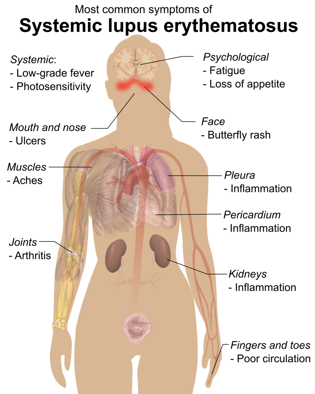
Treatment for Autoimmune Diseases
None of these common autoimmune diseases can be cured, although all of them have treatments that may help relieve symptoms and prevent some of the long-term damage they may cause. Traditional treatments for autoimmune diseases include immunosuppressive drugs to block the immune response, as well as anti-inflammatory drugs to quell inflammation. Hormone replacement may be another option. Type I diabetes, for example, is treated with injections of the hormone insulin, because islet cells in the pancreas can no longer secrete it.
Immunodeficiency
Immunodeficiency occurs when the immune system is not working properly, generally because one or more components of the immune system are inactive. As a result, the immune system may be unable to fight off pathogens or cancers that a normal immune system would be able to resist. Immunodeficiency may occur for a variety of reasons.
Causes of Immunodeficiency
Dozens of rare genetic diseases can result in a defective immune system. This type of immunodeficiency is called primary immunodeficiency. One is born with one of these diseases, rather than acquiring it after birth. Probably the best known of these primary immunodeficiency diseases is severe combined immunodeficiency (SCID). It is also known as “bubble boy disease,” because people with this disorder are extremely vulnerable to infectious diseases, and some of them have become well known for living inside a bubble that provides a sterile environment. SCID is most often caused by an X-linked recessive mutation that interferes with normal B cell and T cell production.
Other types of immunodeficiency are not present at birth, but are acquired due to experiences or exposures that occur after birth. Acquired immunodeficiency is called secondary immunodeficiency because it is secondary to some other event or exposure. Secondary immunodeficiency may occur for a number of different reasons:
- Some pathogens attack and destroy immune system cells. An example is the virus known as HIV, which attacks and destroys T cells.
- The immune system naturally becomes less effective as people get older. This age-related decline — called immunosenescence — generally begins around the age of 50 and worsens with increasing age. Immunosenescence is the reason older people are generally more susceptible to disease than younger people.
- The immune system may be damaged by another disorder, such as obesity, alcoholism, or the abuse of other drugs.
- In developing countries, malnutrition is the most common cause of immune system damage and immunodeficiency. Inadequate protein intake is especially damaging to the immune system. It can lead to impaired complement system activity, phagocyte malfunction, and lower-than-normal production of antibodies and cytokines.
- Certain medications can suppress the immune system. This is the intended effect of immunosuppressant drugs given to people with transplanted organs so they do not reject them. In many cases, however, immunosuppression is an unwanted side effect of drugs used to treat other disorders.
Focus on HIV
Human immunodeficiency virus (HIV) is the most common cause of immunodeficiency in the world today. HIV infections of human hosts are a relatively recent phenomenon. Scientists think that the virus originally infected monkeys, but then jumped to human populations. most likely from a bite, probably sometime during the early to mid-1900s. This most likely occurred in West Africa, but the virus soon spread around the world. HIV was first identified by medical researchers in 1981. Since then, HIV has killed almost 40 million people worldwide, and its economic toll has also been enormous. The hardest hit countries are in Africa, where the virus has infected human populations the longest, and medications to control the virus are least available. In 2016, over 63,000 Canadians were living with HIV.
HIV Transmission
HIV is transmitted through direct contact of mucous membranes or body fluids such as blood, semen, or breast milk. As shown in Figure 17.6.7, transmission of the virus can occur through sexual contact or the use of contaminated hypodermic needles. It can also be transmitted from an infected mother’s blood during late pregnancy or childbirth, or through breast milk after birth. In the past, HIV was also transmitted occasionally through blood transfusions. Because donated blood is now screened for HIV, the virus is no longer transmitted this way.
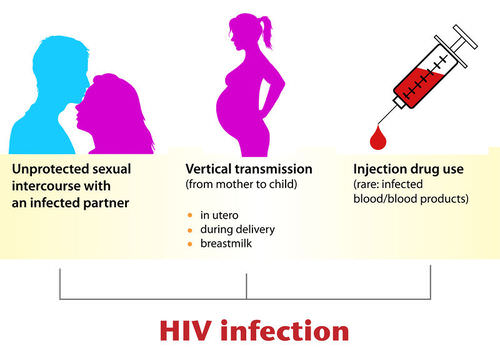
HIV and the Immune System
HIV infects and destroys helper T cells, the type of lymphocytes that regulate the immune response. This process is illustrated in the diagram in Figure 17.6.8. The virus injects its own DNA into a helper T cell and uses the T cell’s “machinery” to make copies of itself. In the process, the helper T cell is destroyed, and the virus copies go on to infect other helper T cells. HIV is able to evade the immune system and keep destroying helper T cells by mutating frequently so its surface antigens keep changing, and by using the host cell’s membrane to hide its own antigens.
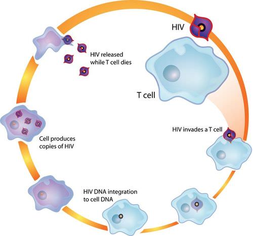
Acquired immunodeficiency syndrome (AIDS) may result from years of damage to the immune system by HIV. It occurs when helper T cells fall to a very low level and opportunistic diseases occur. Opportunistic diseases are infections and tumors that are rare, except in people with a damaged immune system. The diseases take advantage of the “opportunity” presented by people whose immune system cannot fight back. Opportunistic diseases are usually the direct cause of death for people with AIDS.
Treating HIV/AIDS
For patients who have access to HIV medications, infection with the virus is no longer the death sentence that it once was. By 1995, combinations of drugs called “highly active antiretroviral therapy” were developed. For some patients, these drugs can reduce the amount of virus they are carrying to undetectable levels. However, some level of virus always hides in the body’s immune cells, and it will multiply again if a patient stops taking the medications. Researchers are trying to develop drugs to kill these hidden viruses, as well. If their efforts are successful, it could end AIDS.
Feature: Human Biology in the News
EpiPens® and their sole manufacturer (pharmaceutical company Mylan) were featured in headlines in 2016, but not for a good reason. The media outburst was triggered by a drastic price hike in EpiPens® — and Mylan’s apparent greed.
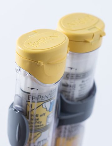
EpiPens® are auto-injectable syringes preloaded with a measured dose of epinephrine, a drug that can rapidly stop a life-threatening anaphylactic response to an allergen. Using the device is easy and does not require any special training. The injector just needs to be jammed against the thigh, which can be done through clothing or on bare skin. Each year, doctors write millions of prescriptions for EpiPens®. Many people with severe allergies always carry two of the devices with them, just in case they experience anaphylaxis, although most of them never need to use them. Other people with severe allergies have literally had their lives saved multiple times by EpiPens® when they had anaphylactic reactions. Even when the devices haven’t been used, they must be replaced each year due to expiration of the epinephrine.
You might think that EpiPens® would be relatively inexpensive, given their life-saving potential. As recently as 2009, a two-pack of EpiPens® cost about $100. However, in just seven years, the cost of the same two-pack of EpiPens® skyrocketed by an incredible 400%! By 2016, the cost was $600 or more. Mylan apparently raised the price for the sole purpose of increasing profits. The company also raised prices significantly on many other drugs. The price hike in EpiPens® alone was certainly profitable. In 2015, the sale of EpiPens® earned Mylan $1 billion. Mylan’s CEO took home almost $19 million the same year, which was an increase of more than 600% over her prior salary.
News coverage of the price hike in EpiPens® began in the summer of 2016 after a price increase in May of that year. Both private citizens and elected officials expressed outrage over the price increase, especially when coupled with the gluttonous profits of the company and its CEO. By late August, Mylan responded to the backlash by offering discount coupons for EpiPens®. A few days later, the company promised to introduce a cheaper, generic version of the device. Analysts quickly determined that selling a generic version would allow Mylan to make more money on the product than reducing the price of the name-brand device, which they still declined to do. By September of 2016, Mylan was being investigated for antitrust violations related to sales of EpiPens® to public schools in New York City.
The Mylan/EpiPen® story may still be making the news. But whatever its outcome, the story has already added fuel to public and private debates about important ethical issues — issues such as the excessive costs of life-saving drugs and the huge profits of big pharma. What is the most recent news on EpiPens® and Mylan? If you are interested, you can check the headlines online to find out. What are your views on the ethical issues they raise?
17.6 Summary
- An allergy is a disorder in which the immune system makes an inflammatory response to a harmless antigen. Any antigen that causes allergies is called an allergen. Common allergens include pollen, dust mites, mold, specific foods (such as peanuts), insect stings, and certain medications (such as aspirin).
- The prevalence of allergies has been increasing for decades, especially in developed countries, where they are much more common than in developing countries. The hygiene hypothesis posits that this has occurred because humans evolved to cope with more pathogens than we now typically face in our relatively sterile environments in developed countries. As a result, the immune system “keeps busy” by attacking harmless antigens.
- Allergies occur when B cells are first activated to produce large amounts of antibodies to an otherwise harmless allergen, and the antibodies attach to mast cells. On subsequent exposures to the allergen, the mast cells immediately release cytokines and histamines that cause inflammation.
- Mild allergy symptoms are frequently treated with antihistamines that counter histamines and reduce allergy symptoms. A severe systemic allergic reaction, called anaphylaxis, is a medical emergency that is usually treated with injections of epinephrine. Immunotherapy for allergies involves injecting increasing amounts of allergens to desensitize the immune system to them.
- Autoimmune diseases occur when the immune system fails to recognize the body’s own molecules as self and attacks them, causing damage to tissues and organs. A family history of autoimmunity and female gender are risk factors for autoimmune diseases.
- In some autoimmune diseases, such as type I diabetes, the immune system attacks and damages specific body cells. In other autoimmune diseases, such as systemic lupus erythematosus, many different tissues and organs may be attacked and injured. Autoimmune diseases generally cannot be cured, but their symptoms can often be managed with drugs or other treatments.
- Immunodeficiency occurs when the immune system is not working properly, generally because one or more of its components are inactive. As a result, the immune system is unable to fight off pathogens or cancers that a normal immune system would be able to resist.
- Primary immunodeficiency is present at birth and caused by rare genetic diseases. An example is severe combined immunodeficiency. Secondary immunodeficiency occurs because of some event or exposure experienced after birth. Possible causes include aging, certain medications, infections with pathogens, and other disorders, such as obesity or malnutrition.
- The most common cause of immunodeficiency in the world today is human immunodeficiency virus (HIV), which infects and destroys helper T cells. HIV is transmitted through mucous membranes or body fluids. The virus may eventually lead to such low levels of helper T cells that opportunistic infections occur. When this happens, the patient is diagnosed with acquired immunodeficiency syndrome (AIDS). Medications can control the multiplication of HIV in the human body — but they don't eliminate the virus completely.
17.6 Review Questions
-
- How does immunotherapy for allergies work?
- What are autoimmune diseases?
- Identify two risk factors for autoimmune diseases.
- Autoimmune diseases may be specific to particular tissues, or they may be systemic. Give an example of each type of autoimmune disease.
- What is immunodeficiency? Compare and contrast primary and secondary immunodeficiency. Give an example of each.
- What is the most common cause of immunodeficiency in the world today? How does this affect the immune system?
- Distinguish between HIV and AIDS.
17.6 Explore More
https://youtu.be/-q7Fz7NIMWM
Why do people have seasonal allergies? - Eleanor Nelsen, TED-Ed, 2016.
https://youtu.be/0TipTogQT3E
Why it’s so hard to cure HIV/AIDS - Janet Iwasa, TED-Ed, 2015.
https://youtu.be/pJa6KVLwl9U
The Boy in the Bubble | Retro Report | The New York Times, 2015.
https://youtu.be/Mjr9h_QmdeM
Why Are Peanut Allergies Becoming So Common? Seeker, 2014.
https://youtu.be/RiMSmDBvgto
What Are Tonsil Stones? | Gross Science, 2015.
Attributions
Figure 17.6.1
Oedema by Championswimmer on Wikimedia Commons is in the public domain (https://en.wikipedia.org/wiki/en:public_domain).
Figure 17.6.2
- Medical (latex) gloves from pngimg.com is used under a CC BY-NC 4.0 (https://creativecommons.org/licenses/by-nc/4.0/) license.
- House dust mites (5247996458) by Gilles San Martin from Namur, Belgium on Wikimedia Commons is used under a CC BY-SA 2.0 (https://creativecommons.org/licenses/by-sa/2.0/deed.en) license.
- Honey bee macro by Karunakar Rayker on Flickr is used under a CC BY 2.0 (https://creativecommons.org/licenses/by/2.0/) license.
- Peanuts by Karolina Grabowska on Pexels is used under a CC0 1.0 Universal Public Domain Dedication license (https://creativecommons.org/publicdomain/zero/1.0/).
- Photo of dog and grey cat by ERC4N51 on pxhere is used under a CC0 1.0 Universal Public Domain Dedication license (https://creativecommons.org/publicdomain/zero/1.0/).
- Tags: Pollen Allergy Spring by Castagnari53 on Pixabay, is used under the Pixabay License (https://pixabay.com/service/license/).
Figure 17.6.3
512px-Mast_cells by National Institute of Allergy and Infectious Diseases (U.S.) & National Cancer Institute (p.29) is in the public domain (https://en.wikipedia.org/wiki/en:public_domain).
Figure 17.6.4
Signs_and_symptoms_of_anaphylaxis by Mikael Häggström on Wikimedia Commons is used under a CC0 1.0 Universal Public Domain Dedication license (https://creativecommons.org/publicdomain/zero/1.0/).
Figure 17.6.5
Allergy Tests by Dan Pupius on Flickr is used under a CC BY-NC-SA 2.0 (https://creativecommons.org/licenses/by-nc-sa/2.0/) license.
Figure 17.6.6
1024px-Symptoms_of_SLE by Mikael Häggström on Wikimedia Commons is used under a CC0 1.0 Universal Public Domain Dedication license (https://creativecommons.org/publicdomain/zero/1.0/).
Figure 17.6.7
HIV transmission by CK-12 Foundation is used under a CC BY NC 3.0 (https://creativecommons.org/licenses/by-nc/3.0/) license.
Figure 17.6.8
HIV life cycle by CK-12 Foundation is used under a CC BY NC 3.0 (https://creativecommons.org/licenses/by-nc/3.0/) license.
 ©CK-12 Foundation Licensed under
 ![]() • Terms of Use • Attribution
• Terms of Use • Attribution
Figure 17.6.9
Epipen by Stock Catalog on flickr by Stock Catalog on Flickr is used under a CC BY 2.0 (https://creativecommons.org/licenses/by/2.0/) license.
References
Brainard, J/ CK-12 Foundation. (2016). Figure 8 HIV may be transmitted in all of the ways shown here [digital image]. In CK-12 College Human Biology (Section 19.6) [online Flexbook]. CK12.org. https://www.ck12.org/book/ck-12-human-biology/section/19.6/
Brainard, J/ CK-12 Foundation. (2016). Figure 9 This diagram shows how HIV infects and destroys helper T cells [digital image]. In CK-12 College Human Biology (Section 19.6) [online Flexbook]. CK12.org. https://www.ck12.org/book/ck-12-human-biology/section/19.6/
CBS News. (2016, August 16). Rising cost of potentially life-saving EpiPen puts pinch on families [online article]. CBS Interactive Inc. https://www.cbsnews.com/news/allergy-medication-epipen-epinephrine-rising-costs-impact-on-families/
Gross Science. (2015, June 29). What are tonsil stones? | Gross Science. YouTube. https://www.youtube.com/watch?v=RiMSmDBvgto&feature=youtu.be
Häggström, M. (2014). Medical gallery of Mikael Häggström 2014. WikiJournal of Medicine 1 (2). DOI:10.15347/wjm/2014.008. ISSN 2002-4436
National Institute of Allergy and Infectious Diseases (NIAID). (n.d.). Severe combined immunodeficiency (SCID) [online article]. National Institute of Health (NIH). https://www.niaid.nih.gov/diseases-conditions/severe-combined-immunodeficiency-scid
National Institute of Allergy and Infectious Diseases (U.S.) & National Cancer Institute (U.S.). (2003, September). Understanding the immune system and how it works [NIH Publication No. 03-5423]. Scholar Works - Indiana University. https://scholarworks.iupui.edu/handle/1805/748
[The] New York Times. (2015, December 15). The boy in the bubble | Retro Report | The New York Times. YouTube. https://www.youtube.com/watch?v=pJa6KVLwl9U&feature=youtu.be
Seeker. (2014, October 3). Why are peanut allergies becoming so common? YouTube. https://www.youtube.com/watch?v=Mjr9h_QmdeM&feature=youtu.be
Summary: Estimates of HIV incidence, prevalence, and Canada's progress on meeting the 90-90-90 HIV targets 2016. (2018, July). Public Health Agency of Canada. https://www.canada.ca/content/dam/phac-aspc/documents/services/publications/diseases-conditions/summary-estimates-hiv-incidence-prevalence-canadas-progress-90-90-90/pub-eng.pdf
Swetlitz, I., Silverman, E. (2016, August 25). Mylan may have violated antitrust law in its EpiPen sales to schools, legal experts say [online article]. STATNews.com. https://www.statnews.com/2016/08/25/mylan-antitrust-epipen-schools/
TED-Ed. (2016, May 26). Why do people have seasonal allergies? - Eleanor Nelsen. YouTube. https://www.youtube.com/watch?v=-q7Fz7NIMWM&feature=youtu.be
TED-Ed. (2015, March 16). Why it’s so hard to cure HIV/AIDS - Janet Iwasa, https://www.youtube.com/watch?v=0TipTogQT3E&feature=youtu.be
Created by CK-12 Foundation/Adapted by Christine Miller
Figure 18.10.1 Families all have something in common.
Family Portrait
What do all these families (Figure 18.10.1) have in common? They were born! Every person on this planet was conceived, carried in utero and then born. While families come in all shapes, sizes and styles, we all came into existence in the same way. Virtually all human societies past and present — value having children. Indeed, for many people, parenthood is an important life goal. Unfortunately, some people are unable to achieve that goal because of infertility.
What Is Infertility?
Infertility is the inability of a sexually mature adult to reproduce by natural means. For scientific and medical purposes, infertility is generally defined as the failure to achieve a successful pregnancy after at least one year of regular, unprotected sexual intercourse. Infertility may be primary or secondary. Primary infertility applies to cases in which an individual has never achieved a successful pregnancy. Secondary infertility applies to cases in which an individual has had at least one successful pregnancy, but fails to achieve another after trying for at least a year. Infertility is a common problem. The government of Canada reported that in 2019, 16% of Canadian couples experience infertility, a number which has doubled since the 1980s. If you look around at the couples you know, that means that almost 1 in 6 of them are having issues with fertility.
Causes of Infertility
Pregnancy is the result of a multi-step process. In order for a normal pregnancy to occur, a woman must release an ovum from one of her ovaries, the ovum must go through an oviduct, a man’s sperm must fertilize the ovum as it passes through the oviduct, and then the resulting zygote must implant in the uterus. If there is a problem with any of these steps, infertility can result.
A couple’s infertility may be due to a problem with either the male or the female partner. As shown in the circle graph below (Figure 18.10.2), about 40% of infertility cases are due to female infertility, and about 30% are due to male infertility. The remaining 30% of cases are due to a combination of male and female problems or unknown causes.
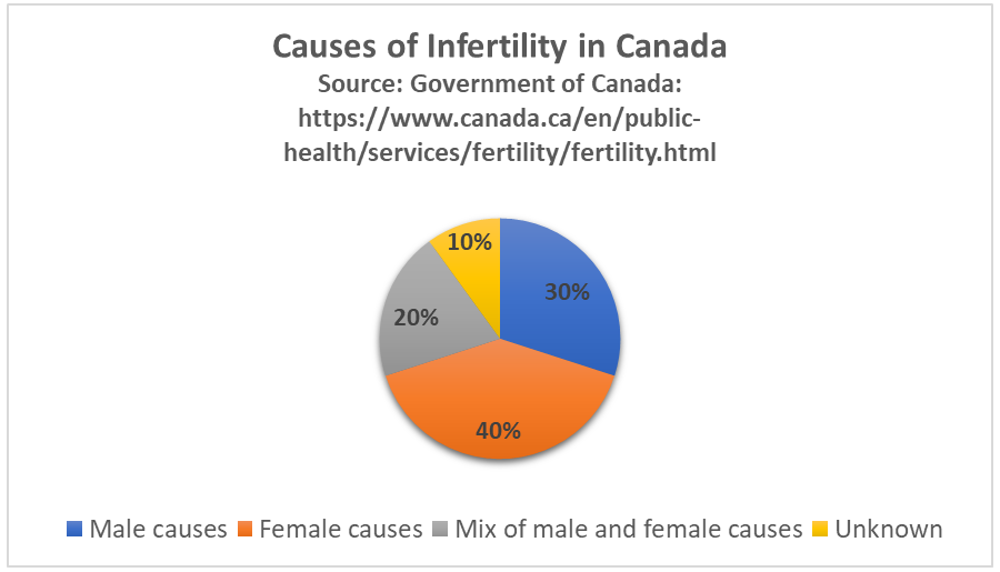
Causes of Male Infertility
Male infertility occurs when there are no, or too few, sperm, or when the sperm are not healthy and motile and cannot travel through the female reproductive tract to fertilize an egg. A common cause of inadequate numbers or motility of sperm is varicocele, which is enlargement of blood vessels in the scrotum. This may raise the temperature of the testes and adversely affect sperm production. In other cases, there is no problem with the sperm, but there is a blockage in the male reproductive tract that prevents the sperm from being ejaculated.
Factors that increase a man’s risk of infertility include heavy alcohol use, drug abuse, cigarette smoking, exposure to environmental toxins (such as pesticides or lead), certain medications, serious diseases (such as kidney disease), and radiation or chemotherapy for cancer. Another risk factor is advancing age. Male fertility normally peaks in the mid-twenties and gradually declines after about age 40, although it may never actually drop to zero.
Causes of Female Infertility
Female infertility generally occurs due to one of two problems: failure to produce viable ova by the ovaries, or structural problems in the oviducts or uterus. The most common cause of female infertility is a problem with ovulation. Without ovulation, there are no ova to be fertilized. Anovulatory cycles (menstrual cycles in which ovulation does not occur) may be associated with no or irregular menstrual periods, but even regular menstrual periods may be anovulatory for a variety of reasons. The most common cause of anovulatory cycles is polycystic ovary syndrome (PCOS), which causes hormone imbalances that can interfere with normal ovulation. Another relatively common cause of anovulation is primary ovarian insufficiency. In this condition, the ovaries stop working normally and producing viable eggs at a relatively early age, generally before the age of 40.
Structural problems with the oviducts or uterus are less common causes of female infertility. The oviducts may be blocked as a result of endometriosis. Another possible cause is pelvic inflammatory disease, which occurs when sexually transmitted infections spread to the oviducts or other female reproductive organs (see Figure 18.10.3). The infection may lead to scarring and blockage of the oviducts. If an ovum is produced and the oviducts are functioning — and a woman has a condition such as uterine fibroids — implantation in the uterus may not be possible. Uterine fibroids are non-cancerous clumps of tissue and muscle that form on the walls of the uterus.
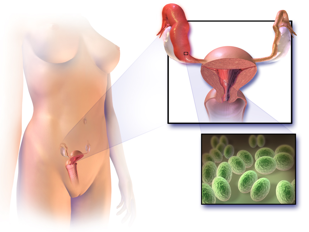
Factors that increase a woman’s risk of infertility include tobacco smoking, excessive use of alcohol, stress, poor diet, strenuous athletic training, and being overweight or underweight. Advanced age is even more problematic for females than males. Female fertility normally peaks in the mid-twenties, and continuously declines after age 30 and until menopause around the age of 52, after which the ovary no longer releases eggs. About 1/3 of couples in which the woman is over age 35 have fertility problems. In older women, more cycles are likely to be anovulatory, and the eggs may not be as healthy.
Diagnosing Causes of Infertility
Diagnosing the cause(s) of a couple’s infertility often requires testing both the man and the woman for potential problems. In men, the semen is likely to be examined for the number, shape, and motility of sperm. If problems are found with sperm, further studies are likely to be done, such as medical imaging to look for structural problems with the testes or ducts.
In women, the first step is most often determining whether ovulation is occurring. This can be done at home by carefully monitoring body temperature (it rises slightly around the time of ovulation) or using a home ovulation test kit, which is available over the counter at most drugstores. Whether or not ovulation is occurring can also be detected with blood tests or ultrasound imaging of the ovaries. If ovulation is occurring normally, then the next step may be an X-ray of the oviducts and uterus to see if there are any blockages or other structural problems. Another approach to examining the female reproductive tract for potential problems is laparoscopy. In this surgical procedure, a tiny camera is inserted into the woman’s abdomen through a small incision. This allows the doctor to directly inspect the reproductive organs.
Treating Infertility
Infertility often can be treated successfully. The type of treatment depends on the cause of infertility.
Treating Male Infertility
Medical problems that interfere with sperm production may be treated with medications or other interventions that may lead to the resumption of normal sperm production. If, for example, an infection is interfering with sperm production, then antibiotics that clear up the infection may resolve the problem. If there is a blockage in the male reproductive tract that prevents the ejaculation of sperm, surgery may be able to remove the blockage. Alternatively, the man’s sperm may be removed from his body and then used for artificial insemination of his partner. In this procedure, the sperm are injected into the woman’s reproductive tract.
Treating Female Infertility
In females, it may be possible to correct blocked Fallopian tubes or uterine fibroids with surgery. Ovulation problems, on the other hand, are usually treated with hormones that act either on the pituitary gland or on the ovaries. Hormonal treatments that stimulate ovulation often result in more than one egg being ovulated at a time, thus increasing the chances of a woman having twins, triplets, or even higher multiple births. Multiple fetuses are at greater risk of being born too early or having health and developmental problems. The mother is also at greater risk of complications arising during pregnancy. Therefore, the possibility of multiple fetuses should be weighed in making a decision about this type of infertility treatment.
Assisted Reproductive Technology
Some cases of infertility are treated with assisted reproductive technology (ART). This is a collection of medical procedures in which ova are removed from the woman’s body and sperm are taken from the man’s body to be manipulated in ways that increase the chances of fertilization occurring. The eggs and sperm may be injected into one of the woman’s oviducts for fertilization to take place in vivo (in the body). More commonly, however, the eggs and sperm are mixed together outside the body so fertilization takes place in vitro (in a test tube or dish in a lab). The latter approach is illustrated in Figure 18.10.4. With in vitro fertilization, the fertilized eggs may be allowed to develop into embryos before being placed in the woman’s uterus.
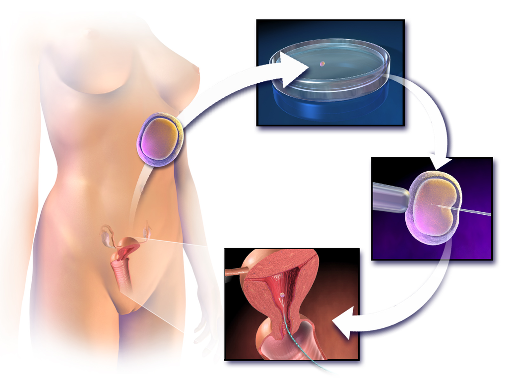
ART has about a 40% chance of leading to a live birth in women under the age of 35, but only about a 20%t chance of success in women over the age of 35. Some studies have found a higher-than-average risk of birth defects in children produced by ART procedures, but this may be due to the generally higher ages of the parent — not the technologies used.
Other Approaches
Other approaches for certain causes of infertility include the use of a surrogate mother, a gestational carrier, or sperm donation.
- A surrogate mother is a woman who agrees to become pregnant using the man’s sperm and her own egg. The child, who will be the biological offspring of the surrogate and the male partner, is given up at birth for adoption by the couple. Surrogacy might be selected by women with no eggs or unhealthy eggs. A woman who carries a mutant gene for a serious genetic disorder might choose this option to ensure that the defective gene is not passed on to the offspring.
- A gestational carrier is a woman who agrees to receive a transplanted embryo from a couple and carry it to term. The child, who will be the biological offspring of the couple, is given to the parents at birth. A gestational carrier might be used by women who have normal ovulation but no uterus, or who cannot safely carry a fetus to term because of a serious health problem (such as kidney disease or cancer).
- Sperm donation is the use of sperm from a fertile man (generally through artificial insemination) for cases in which the male partner in a couple is infertile, or in which a woman seeks to become pregnant without a male partner. A lesbian couple may use donated sperm to enable one of them to become pregnant and have a child. Sperm can be obtained from a sperm bank, which buys and stores sperm for artificial insemination, or a male friend or other individual may donate sperm to a specific woman.
Social and Ethical Issues Relating to Infertility
For people who have a strong desire for children of their own, infertility may lead to deep disappointment and depression. Individuals who are infertile may even feel biologically inadequate. Partners in infertile couples may argue and feel resentment toward each other, and married couples may get divorced because of infertility. Infertility treatments — especially ART procedures — are generally time-consuming and expensive. The high cost of the treatments can put them out of financial reach of many couples.
Ethical Concerns
Some people question whether the allocation of medical resources to infertility treatments is justified, and whether the resources could be better used in other ways. The status of embryos that are created in vitro and then not used for a pregnancy is another source of debate. Some people oppose their destruction on religious grounds, and couples may sometimes argue about what should be done with their extra embryos. Ethical issues are also raised by procedures that increase the chances of multiple births, because of the medical and developmental risks associated with multiple births.
Infertility in Developing Countries
Infertility is an under-appreciated problem in the poorer nations of the world, because of assumptions about overpopulation problems and high birth rates in developing countries. In fact, infertility is at least as great a problem in developing as in developed countries. High rates of health problems and inadequate health care in the poorer nations increase the risk of infertility. At the same time, infertility treatments are usually not available — or are far too expensive — for the vast majority of people who may need them. In addition, in many developing countries, the production of children is highly valued. Children may be needed for family income generation and economic security of the elderly. It is not uncommon for infertility to lead to social stigmatization, psychological problems, and abandonment by spouses.
18.10 Summary
- Infertility is the inability of a sexually mature adult to reproduce by natural means. It is defined scientifically and medically as the failure to achieve a successful pregnancy after at least one year of regular, unprotected sexual intercourse.
- About 40% of infertility in couples is due to female infertility, and another 30% is due to male infertility. In the remaining cases, a couple’s infertility is due to problems in both partners, or to unknown causes.
- Male infertility occurs when there are no, or too few, healthy, motile sperm. This may be caused by problems with spermatogenesis, or by blockage of the male reproductive tract that prevents sperm from being ejaculated. Risk factors for male infertility include heavy alcohol use, smoking, certain medications, and advancing age, to name just a few.
- Female infertility occurs due to failure to produce viable ova by the ovaries, or structural problems in the oviducts or uterus. Polycystic ovary syndrome (PCOS) is the most common cause of failure to produce viable ova. Endometriosis and uterine fibroids are possible causes of structural problems in the oviducts and uterus. Risk factors for female infertility include smoking, stress, poor diet, and older age, among others.
- Diagnosing the cause(s) of a couple’s infertility generally requires testing both the man and the woman for potential problems. For men, semen is likely to be examined for adequate numbers of healthy, motile sperm. For women, signs of ovulation are monitored, for example, with an ovulation test kit or ultrasound of the ovaries. For both partners, the reproductive tract may be medically imaged to look for blockages or other abnormalities.
- Treatments for infertility depend on the cause. For example, if a medical problem is interfering with sperm production, medication may resolve the underlying problem so sperm production is restored. Blockages in either the male or the female reproductive tract can often be treated surgically. If there are problems with ovulation, hormonal treatments may stimulate ovulation.
- Some cases of infertility are treated with assisted reproductive technology (ART). This is a collection of medical procedures in which ova and sperm are taken from the couple and manipulated in a lab to increase the chances of fertilization occurring and an embryo forming. Other approaches for certain causes of infertility include the use of a surrogate mother, gestational carrier, or sperm donation.
- Infertility can negatively impact a couple socially and psychologically, and it may be a major cause of marital friction or even divorce. Infertility treatments may raise ethical issues relating to the costs of the procedures and the status of embryos that are created in vitro, but not used for pregnancy. Infertility is an under-appreciated problem in developing countries, where birth rates are high and children have high economic — as well as social — value. In these countries, poor health care is likely to lead to more problems with infertility and fewer options for treatment.
18.10 Review Questions
- What is infertility? How is infertility defined scientifically and medically?
- What percentage of infertility in couples is due to male infertility? What percentage is due to female infertility?
- Identify causes of and risk factors for male infertility.
- Identify causes of and risk factors for female infertility.
- How are causes of infertility in couples diagnosed?
- How is infertility treated?
- Discuss some of the social and ethical issues associated with infertility or its treatment.
- Why is infertility an under-appreciated problem in developing countries?
- Describe two similarities between causes of male and female infertility.
- Explain the difference between males and females in terms of how age affects fertility.
-
- Do you think that taking medication to stimulate ovulation is likely to improve fertility in cases where infertility is due to endometriosis? Explain your answer.
18.10 Explore More
https://youtu.be/P27waC05Hdk
How in vitro fertilization (IVF) works - Nassim Assefi and Brian A. Levine, TED-Ed, 2015
https://youtu.be/6BBmMtVfZ4Y
A journey through infertility -- over terror's edge | Camille Preston | TEDxBeaconStreet, TEDx Talks, 2014.
https://youtu.be/iqA8uAjvEdM
Smoking Marijuana May Lower Sperm Count by 33%, David Pakman Show, 2015.
https://youtu.be/V6-v4eF9dyA
ivf embryo developing over 5 days by fertility Dr Raewyn Teirney, Fertility Specialist Sydney, 2014.
https://youtu.be/4Khn_z9FPmU
Homosexuality: It's about survival - not sex | James O'Keefe | TEDxTallaght, 2016.
Attributions
Figure 18.10.1
- Gay Pride Parade NYC 2013 - Happy Family by Bob Jagendorf on Flickr is used under a CC BY-NC 2.0 (https://creativecommons.org/licenses/by-nc/2.0/) license.
- #beaches #summer #family #blue #water by Jove Duero on Unsplash is used under the Unsplash License (https://unsplash.com/license).
- Photograph of five men near outdoor by Dollar Gill on Unsplash is used under the Unsplash License (https://unsplash.com/license).
- Família by Laercio Cavalcanti on Unsplash is used under the Unsplash License (https://unsplash.com/license).
- Happiness 🙂 by Ashwini Chaudhary on Unsplash is used under the Unsplash License (https://unsplash.com/license).
Figure 18.10.2
Causes of infertility in Canada by Christine Miller is in the Public Domain (https://creativecommons.org/publicdomain/mark/1.0/).
Figure 18.10.3
1024px-Blausen_0719_PelvicInflammatoryDisease by BruceBlaus on Wikimedia Commons is used under a CC BY 3.0 (https://creativecommons.org/licenses/by/3.0) license.
Figure 18.10.4
1024px-Blausen_0060_AssistedReproductiveTechnology by BruceBlaus on Wikimedia Commons is used under a CC BY 3.0 (https://creativecommons.org/licenses/by/3.0) license.
References
Blausen.com Staff. (2014). Medical gallery of Blausen Medical 2014. WikiJournal of Medicine 1 (2). DOI:10.15347/wjm/2014.010. ISSN 2002-4436.
David Pakman Show. (2015, September 1). Smoking marijuana may lower sperm count by 33%. YouTube. https://www.youtube.com/watch?v=iqA8uAjvEdM
Fertility Specialist Sydney. (2014, April 11). ivf embryo developing over 5 days by fertility Dr Raewyn Teirney. YouTube. https://www.youtube.com/watch?v=V6-v4eF9dyA&t=5s
Public Health Agency of Canada. (2019, May 28). Fertility. Government of Canada. https://www.canada.ca/en/public-health/services/fertility/fertility.html
TED-Ed. (2015, May 7). How in vitro fertilization (IVF) works - Nassim Assefi and Brian A. Levine. YouTube. https://www.youtube.com/watch?v=P27waC05Hdk&t=4s
TEDx Talks. (2014, June 26). A journey through infertility -- over terror's edge | Camille Preston | TEDxBeaconStreet. YouTube. https://www.youtube.com/watch?v=6BBmMtVfZ4Y&t=2s
TEDx Talks. (2016, November 15). Homosexuality: It's about survival - not sex | James O'Keefe | TEDxTallaght. YouTube. https://www.youtube.com/watch?v=4Khn_z9FPmU&t=1s

