11.5 Bone Growth, Remodeling, and Repair
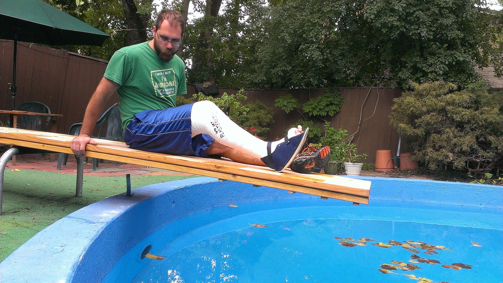
Break A Leg
Did you ever break a leg or other bone, like the man looking longingly at the water in this swimming pool (Figure 11.5.1)? Having a broken bone can really restrict your activity. Bones are very hard, but they will break (or fracture) if enough force is applied to them. Fortunately, bones are highly active organs that can repair themselves if they break. Bones can also remodel themselves and grow. You’ll learn how bones can do all of these things in this section.
Bone Growth
Early in the development of a human fetus, the skeleton is made almost entirely of cartilage. The relatively soft cartilage gradually turns into hard bone through ossification. Ossification is a process in which bone tissue is created from cartilage. The steps in which bones of the skeleton form from cartilage are illustrated in 11.5.2. The steps are as follows:
- Cartilage “model” of bone forms. This model continues to grow as ossification takes place.
- Ossification begins at a primary ossification center in the middle of bone.
- Ossification then starts to occur at secondary ossification centers at the ends of bone.
- The medullary cavity forms. This cavity will contain red bone marrow.
- Areas of ossification meet at epiphyseal plates, and articular cartilage forms. Bone growth ends.
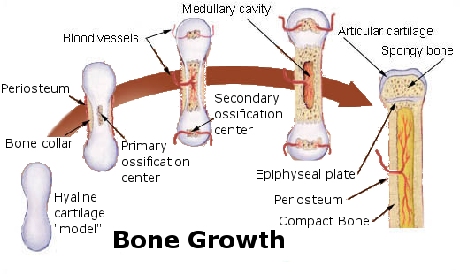
The ossification of cartilage in the human skeleton is a process that lasts throughout childhood in some bones.
Primary and Secondary Ossification Centers
When bone forms from cartilage, ossification begins with a point in the cartilage called the primary ossification center. This generally appears during fetal development, although a few short bones begin their primary ossification after birth. Ossification happens toward both ends of the bone from the primary ossification center, and — in the case of long bones — it eventually forms the shaft of the bone.
Secondary ossification centers form after birth. Ossification from secondary centers eventually forms the ends of the bones. The shaft and ends of the bone are separated by a growing zone of cartilage until the individual reaches skeletal maturity.
Skeletal Maturity
Throughout childhood, the cartilage remaining in the skeleton keeps growing, and allows for bones to grow in size. Once all of the cartilage has been replaced by bone, and fusion has taken place at the epiphyseal plates, bones can no longer keep growing in length. At this point, skeletal maturity has been reached. It generally takes place by age 18 to 25.
The use of anabolic steroids by teens can speed up the process of skeletal maturity, resulting in a shorter period of cartilage growth before fusion takes place. This means that teens who use steroids are likely to end up shorter as adults than they would otherwise have been.
Bone Remodeling
Even after skeletal maturity has been attained, bone is constantly being resorbed and replaced with new bone in a process called bone remodeling. In this lifelong process, mature bone tissue is continually turned over, with about ten per cent of the skeletal mass of an adult being remodeled each year. Bone remodeling is carried out through the work of osteoclasts — which are bone cells that resorb bone and dissolve its minerals — and osteoblasts, which are bone cells that make new bone matrix.
Bone remodeling serves several functions. It shapes the bones of the skeleton as a child grows, and it repairs tiny flaws in bone that result from everyday movements. Remodeling also makes bones thicker at points where muscles place the most stress on them. In addition, remodeling helps regulate mineral homeostasis, because it either releases mineral from bones into the blood or absorbs mineral from the blood into bones. Figure 11.5.3 shows how osteoclasts in bones are involved in calcium regulation.
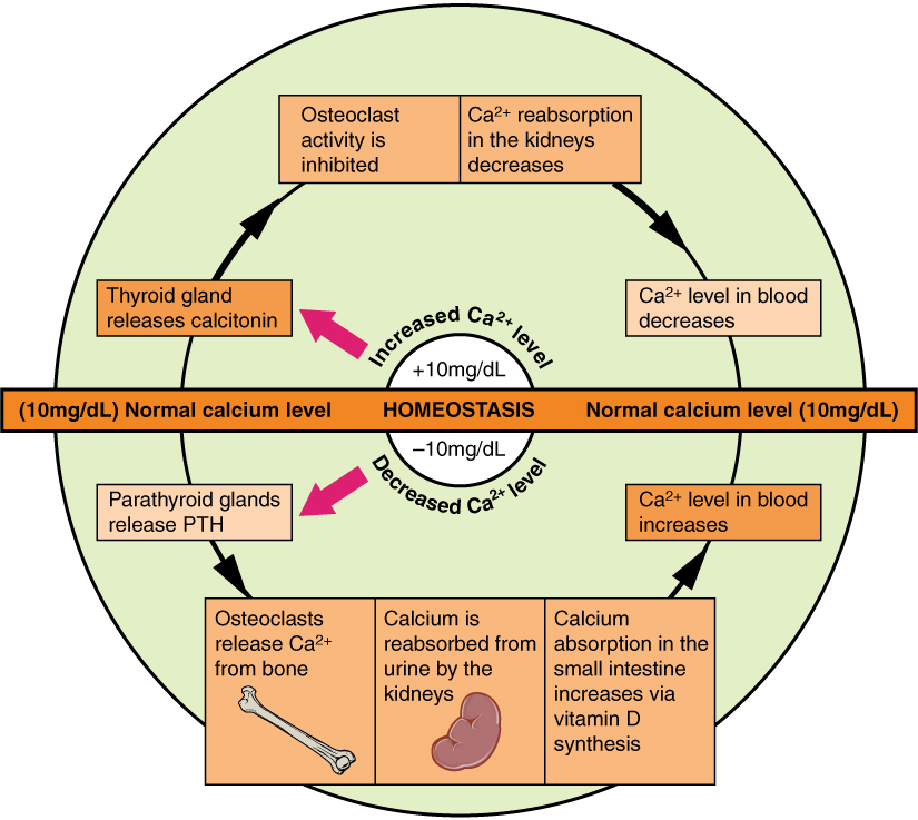
The action of osteoblasts and osteoclasts in bone remodeling and calcium homeostasis is controlled by a number of enzymes, hormones, and other substances that either promote or inhibit the activity of the cells. In this way, these substances control the rate at which bone is made, destroyed, and changed in shape. For example, the rate at which osteoclasts resorb bone and release calcium into the blood is promoted by parathyroid hormone (PTH) and inhibited by calcitonin, which is produced by the thyroid gland (see the diagram in Figure 11.5.3). The rate at which osteoblasts create new bone is stimulated by growth hormone, which is produced by the anterior lobe of the pituitary gland. Thyroid hormone and sex hormones (estrogens and androgens) also stimulate osteoblasts to create new bone.
Bone Repair
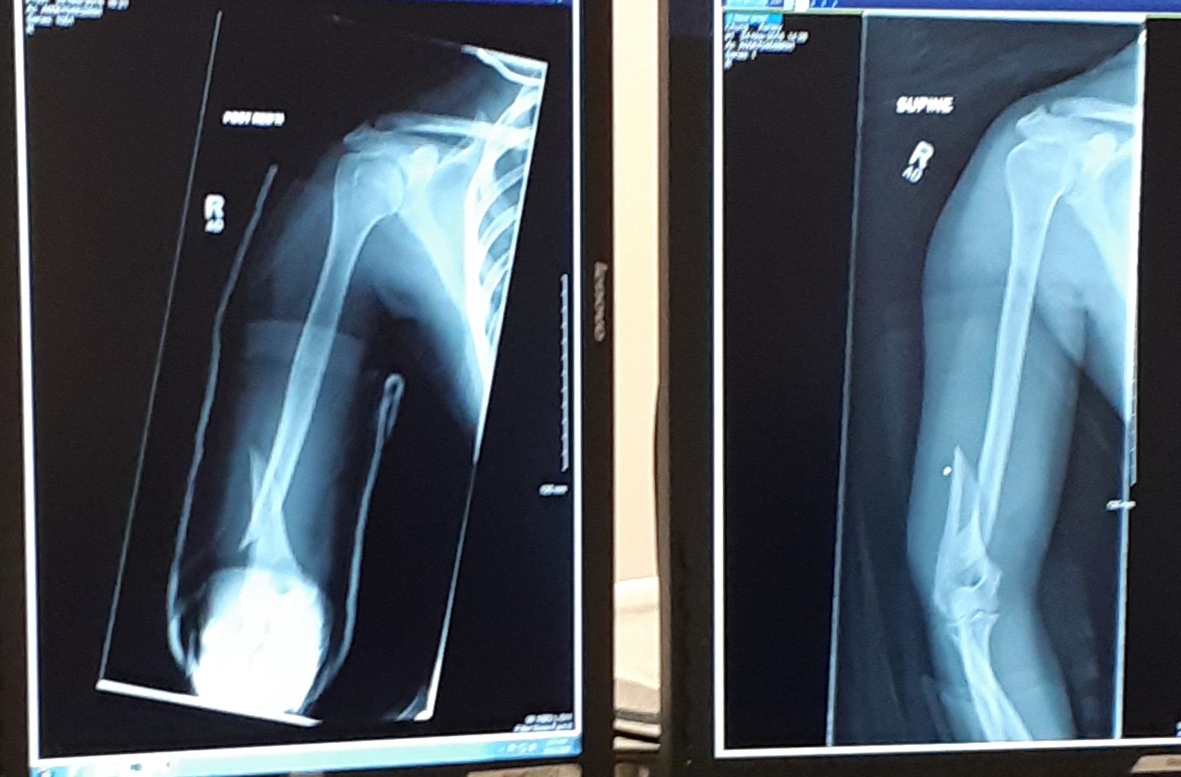
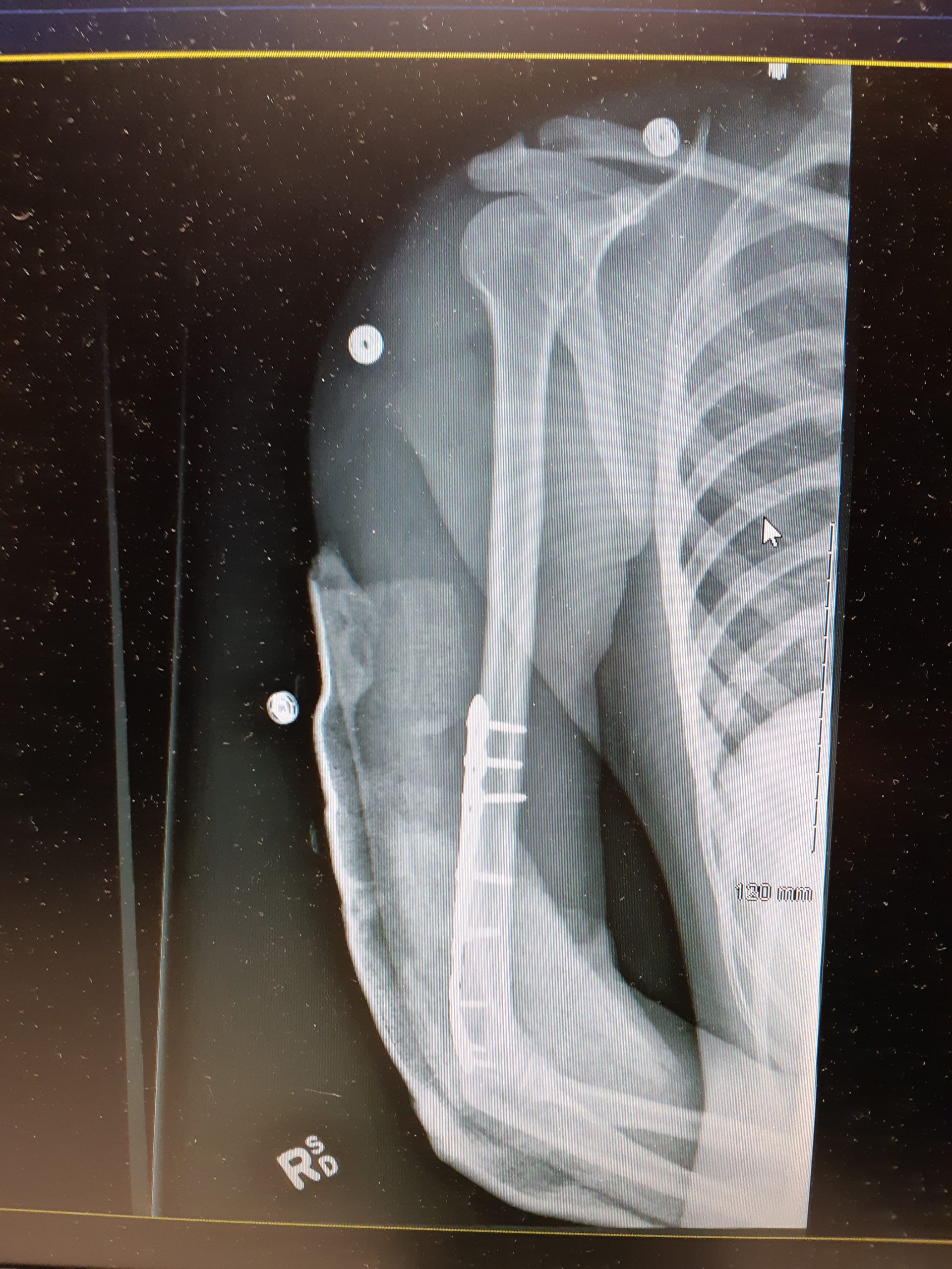
Bone repair (or healing) is the process in which a bone repairs itself following a bone fracture. You can see an X-ray of a bone fracture in Figure 11.5.4. In this fracture, the humerus in the upper arm has been completely broken through its shaft. Before this fracture heals, a physician must push the displaced bone parts back into their correct positions. Then, the bone must be stabilized — with a cast and/or pins surgically inserted into the bone, for example (as shown in Figure 11.5.5) — until the bone’s natural healing process is complete. This process may take several weeks.
 |
 |
Although bone repair is a natural physiological process, it may be promoted or inhibited by several factors. Fracture repair is more likely to be successful with adequate nutrient intake. Age, bone type, drug therapy, and pre-existing bone disease are additional factors that may affect healing. Bones that are weakened by disease (such as osteoporosis or bone cancer) are not only likely to heal more slowly, but are also more likely to fracture in the first place.
Feature: Myth vs. Reality
Bone fractures are fairly common, and there are many myths about them. Knowing the facts is important, because fractures generally require emergency medical treatment.
| Myth | Reality |
|---|---|
| “A bone fracture is a milder injury than a broken bone.” | A bone fracture is the same thing as a broken bone. |
| “If you still have full range of motion in a limb, then it must not be fractured.” | Even if a bone is fractured, the muscles and tendons attached to it may still be able to move the bone normally. This is especially likely if the bone is cracked — but not broken — into two pieces. Even if a bone is broken all the way through, range of motion may not be affected if the bones on either side of the fracture remain properly aligned. |
| “A fracture always produces a bruise.” | Many — but not all — fractures produce a bruise. If a fracture does produce a bruise, it may take several hours (or even a day or more!) for the bruise to appear. |
| “Fractures are so painful that you will immediately know if you break a bone.” | Ligament sprains and muscle strains are also very painful, sometimes more painful than fractures. Additionally, every person has a different pain tolerance. People with a high pain tolerance may continue using a broken bone in spite of the pain. |
| “You can tell when a bone is fractured because there will be very localized pain over the break.” | A broken bone is often accompanied by injuries to surrounding muscles or ligaments. As a result, the pain may extend far beyond the location of the fracture. The pain may be greater directly over the fracture, but the intensity of the pain may make it difficult to pinpoint exactly where the pain originates. |
11.5 Summary
- Bone is very active tissue. Its cells are constantly forming and resorbing bone matrix.
- Early in the development of a human fetus, the skeleton is made almost entirely of cartilage. The relatively soft cartilage gradually turns into hard bone. This is called ossification. It begins at a primary ossification center in the middle of bone, and later also occurs at secondary ossification centers in the ends of bone. The bone can no longer grow in length after the areas of ossification meet and fuse at the time of skeletal maturity.
- Throughout life, bone is constantly being replaced in the process of bone remodeling. In this process, osteoclasts resorb bone, and osteoblasts make new bone to replace it. Bone remodeling shapes the skeleton, repairs tiny flaws in bones, and helps maintain mineral homeostasis in the blood.
- Bone repair is the natural process in which a bone repairs itself following a bone fracture. This process may take several weeks. In the process, periosteum produces cells that develop into osteoblasts, and the osteoblasts form new bone matrix to heal the fracture. Bone repair may be affected by diet, age, pre-existing bone disease, or other factors.
11.5 Review Questions
- Outline how bone develops starting early in the fetal stage, and through the age of skeletal maturity.
- Describe the process of bone remodeling. When does it occur?
- What purposes does bone remodeling serve?
- Define bone repair. How long does this process take?
- Explain how bone repair occurs.
- Identify factors that may affect bone repair.
-
- If there is a large region between the primary and secondary ossification centers in a bone, is the person young or old? Explain your answer.
- If bones can repair themselves, why are casts and pins sometimes necessary in the process?
- When calcium levels are low, which type of bone cell causes the release of calcium to the bloodstream?
- Which tissue and bone cell type are primarily involved in bone repair after a fracture?
- Describe one way in which hormones are involved in bone remodeling.
11.5 Explore More
How to grow a bone – Nina Tandon, TED-Ed, 2015.
Healing Process of Bone Fracture, Aldo Fransiskus Marsetio, 2015.
The Skeleton From Fetal to Adult, Samantha Espolt, 2012.
Attributions
Figure 11.5.1
First_plaster_long_leg_cast…._-_9383569051 by 4x4king10 on Wikimedia Commons is used under a CC BY 2.0 (https://creativecommons.org/licenses/by/2.0) license.
Figure 11.5.2
Bone_growth by Chaldor (derivative work) on Wikimedia Commons is in the public domain (https://en.wikipedia.org/wiki/en:Public_domain). (Original, Illu_bone_growth.jpg is by Fuelbottle)
Figure 11.5.3
Calcium_Homeostasis by OpenStax on Wikimedia commons is used under a CC BY 3.0 (https://creativecommons.org/licenses/by/3.0/deed.en) license.
Figure 11.5.4
Broken Arm by Ashley Chung is used with permission.
Figure 11.5.5
Broken Arm with plate and pins by Ashley Chung is used with permission.
References
Aldo Fransiskus Marsetio. (2015, ). Healing process of bone fracture. YouTube. https://www.youtube.com/watch?v=-P6LsendHxU
Betts, J. G., Young, K.A., Wise, J.A., Johnson, E., Poe, B., Kruse, D.H., Korol, O., Johnson, J.E., Womble, M., DeSaix, P. (2013, June 19). Figure 6.24 Pathways in calcium homeostasis [digital image]. In Anatomy and Physiology (Section 6.7). OpenStax. https://openstax.org/books/anatomy-and-physiology/pages/6-7-calcium-homeostasis-interactions-of-the-skeletal-system-and-other-organ-systems
Samantha Espolt. (2012, ). The skeleton from fetal to adult. YouTube. https://www.youtube.com/watch?v=RC2w_9DcY38&t=3s
TED-Ed. (2015, ). How to grow a bone – Nina Tandon. YouTube. https://www.youtube.com/watch?v=yJoQj5-TIvE
Created by CK-12 Foundation/Adapted by Christine Miller
What Is It?
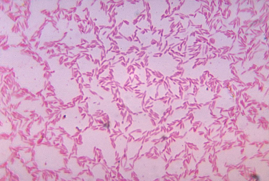
Figure 15.5.1 shows some of the cells of what has been called “the last human organ to be discovered.” This “organ” weighs about 200 grams (about 7 oz) and consists of a hundred trillion cells, yet scientists are only now beginning to learn everything it does and how it varies among individuals. What is it? It’s the mass of bacteria that live in our lower gastrointestinal tract.
Organs of the Lower Gastrointestinal Tract
Most of the bacteria that normally live in the lower gastrointestinal (GI) tract live in the large intestine. They have important and mutually beneficial relationships with the human organism. We provide them with a great place to live, and they provide us with many benefits, some of which you can read about below. Besides the large intestine and its complement of helpful bacteria, the lower GI tract also includes the small intestine. The latter is arguably the most important organ of the digestive system. It is where most chemical digestion and virtually all absorption of nutrients take place.
Small Intestine
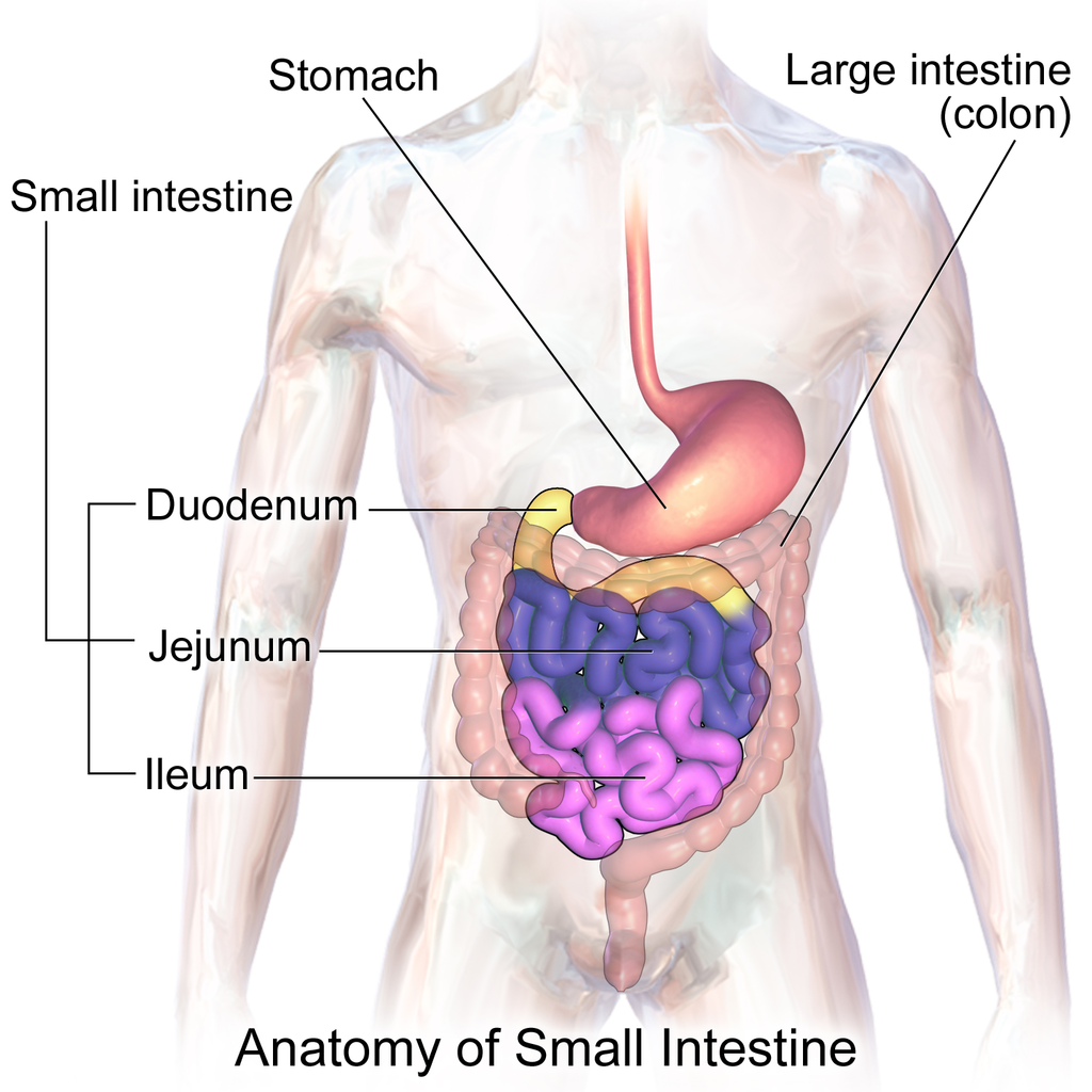
The small intestine (also called the small bowel or gut) is the part of the GI tract between the stomach and large intestine. Its average length in adults is 4.6 m in females and 6.9 m in males. It is approximately 2.5 to 3.0 cm in diameter. It is called “small” because it is much smaller in diameter than the large intestine. The internal surface area of the small intestine totals an average of about 30 m2. Structurally and functionally, the small intestine can be divided into three parts, called the duodenum, jejunum, and ileum, as shown in Figure 15.5.2 and described below.
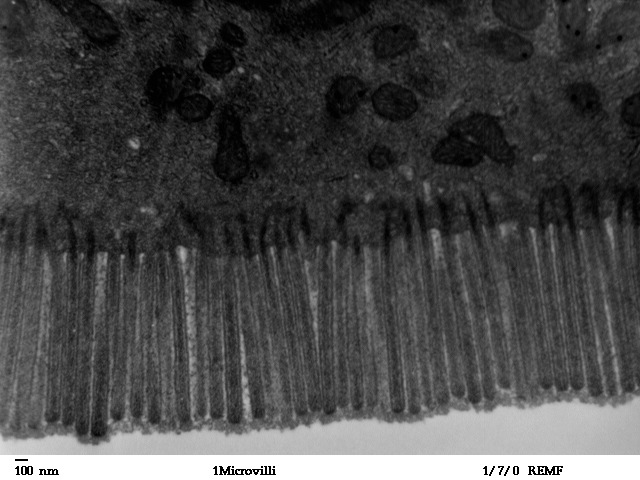
The mucosa lining the small intestine is highly enfolded and covered with the finger-like projections called villi. In fact, each square inch (about 6.5 cm2) of mucosa contains around 20 thousand villi. The individual cells on the surface of the villi also have many finger-like projections — the microvilli, shown in Figure 15.5.3. There are thought to be well over 17 billion microvilli per square centimetre of intestinal mucosa! All of these folds, villi, and microvilli greatly increase the surface area for chyme to come into contact with digestive enzymes, which coat the microvilli, as well as forming a tremendous surface area for the absorption of nutrients. Inside each of the villi is a network of tiny blood and lymph vessels that receive the absorbed nutrients and carry them away in the blood or lymph circulation. The wrinkles and projections in the intestinal mucosa also slow down the passage of chyme so there is more time for digestion and absorption to take place.
Duodenum
The duodenum is the first part of the small intestine, directly connected to the stomach. It is also the shortest part of the small intestine, averaging only about 25 cm (almost 10 in) in length in adults. Its main function is chemical digestion, and it is where most of the chemical digestion in the entire GI tract takes place.
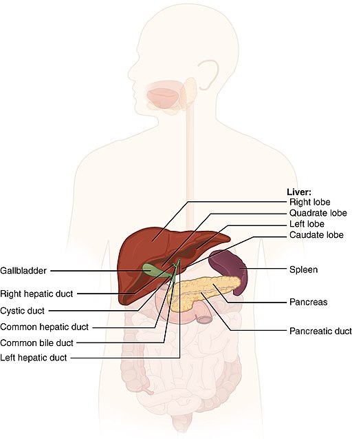
The duodenum receives partially digested, semi-liquid chyme from the stomach. It receives digestive enzymes and alkaline bicarbonate from the pancreas through the pancreatic duct, and it receives bile from the liver via the gallbladder through the common bile duct (see Figure 15.5.4). In addition, the lining of the duodenum secretes digestive enzymes and contains glands — called Brunner’s glands — that secrete mucous and bicarbonate. The bicarbonate from the pancreas and Brunner’s glands — along with bile from the liver — neutralize the highly acidic chyme after it enters the duodenum from the stomach. This is necessary because the digestive enzymes in the duodenum require a nearly neutral environment in order to work. The three major classes of compounds that undergo chemical digestion in the duodenum are carbohydrates, proteins, and lipids.
Digestion of Carbohydrates in the Duodenum
Complex carbohydrates (such as starches) are broken down by the digestive enzyme amylase from the pancreas to short-chain molecules consisting of just a few saccharides (that is, simple sugars). Disaccharides, including sucrose and lactose, are broken down to simple sugars by duodenal enzymes. Sucrase breaks down sucrose, and lactase (if present) breaks down lactose. Some carbohydrates are not digested in the duodenum, and they ultimately pass undigested to the large intestine, where they may be digested by intestinal bacteria.
Digestion of Proteins in the Duodenum
In the duodenum, the pancreatic enzymes trypsin and chymotrypsin cleave proteins into peptides. Then, these smaller molecules are broken down into amino acids. Their digestion is catalyzed by pancreatic enzymes called peptidases.
Digestion of Lipids in the Duodenum
Pancreatic lipase breaks down triglycerides into fatty acids and glycerol. Lipase works with the help of bile secreted by the liver and stored in the gallbladder. Bile salts attach to triglycerides to help them emulsify, or form smaller particles (called micelles) that can disperse through the watery contents of the duodenum. This increases access to the molecules by pancreatic lipase.
Jejunum
The jejunum is the middle part of the small intestine, connecting the duodenum and the ileum. The jejunum is about 2.5 m (about 8 ft) long. Its main function is absorption of the products of digestion, including sugars, amino acids, and fatty acids. Absorption occurs by simple diffusion (water and fatty acids), facilitated diffusion (the simple sugar fructose), or active transport (amino acids, small peptides, water-soluble vitamins, and most glucose). All nutrients are absorbed into the blood, except for fatty acids and fat-soluble vitamins, which are absorbed into the lymph. Although most nutrients are absorbed in the jejunum, there are a few exceptions:
- Iron is absorbed in the duodenum.
- Vitamin B12 and bile salts are absorbed in the ileum.
- Water and lipids are absorbed throughout the small intestine, including the duodenum and ileum in addition to the jejunum.
Ileum
The ileum is the third and final part of the small intestine, directly connected at its distal end to the large intestine. The ileum is about 3 m (almost 10 ft) long. Some cells in the lining of the ileum secrete enzymes that catalyze the final stages of digestion of any undigested protein and carbohydrate molecules. However, the main function of the ileum is to absorb vitamin B12 and bile salts. It also absorbs any other remaining nutrients that were not absorbed in the jejunum. All substances in chyme that remain undigested or unabsorbed by the time they reach the distal end of the ileum pass into the large intestine.
Large Intestine
The large intestine — also called the large bowel — is the last organ of the GI tract. In adults, it averages about 1.5 m (about 5ft) in length. It is shorter than the small intestine, but at least twice as wide, averaging about 6.5 cm (about 2.5 in) in diameter. Water is absorbed from chyme as it passes through the large intestine, turning the chyme into solid feces. Feces is stored in the large intestine until it leaves the body during defecation.
Parts of the Large Intestine
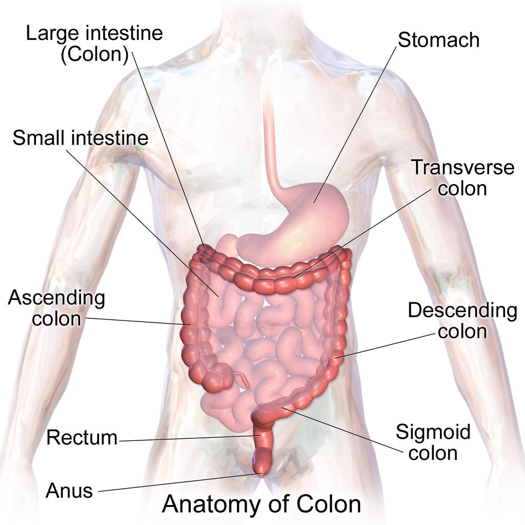
Like the small intestine, the large intestine can be divided into several parts, as shown in Figure 15.5.5. The large intestine begins at the end of the small intestine, where a valve separates the small and large intestines and regulates the movement of chyme into the large intestine. Most of the large intestine is called the colon. The first part of the colon, where chyme enters from the small intestine, is called the cecum. From the cecum, the colon continues upward as the ascending colon, travels across the upper abdomen as the transverse colon, and then continues downward as the descending colon. It then becomes a V-shaped region called the sigmoid colon, which is attached to the rectum. The rectum stores feces until elimination occurs. It transitions to the final part of the large intestine, called the anus, which has an opening to the outside of the body for feces to pass through.
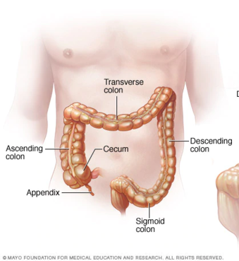
The diagram below (Figure 15.5.6) shows a projection from the cecum of the colon that is known as the appendix. The function of the appendix is somewhat uncertain, but it does not seem to be involved in digestion or absorption. It may play a role in immunity, and in the fetus, it seems to have an endocrine function, releasing hormones needed for homeostasis. Some biologists speculate that the appendix may also store a sample of the colon’s normal bacteria. If so, it may be able to repopulate the colon with the bacteria if illness or antibiotic medications deplete these microorganisms. Appendicitis, or infection and inflammation of the appendix, is a fairly common medical problem, typically resolved by surgical removal of the appendix (appendectomy). People who have had their appendix surgically removed do not seem to suffer any ill effects, so the organ is considered dispensable.
Functions of the Large Intestine
The removal of water from chyme to form feces starts in the ascending colon and continues throughout much of the length of the organ. Salts (such as sodium) are also removed from food wastes in the large intestine before the wastes are eliminated from the body. This allows salts — as well as water — to be recycled in the body.
The large intestine is also the site where huge numbers of beneficial bacteria ferment many unabsorbed materials in food waste. The bacterial breakdown of undigested polysaccharides produces nitrogen, carbon dioxide, methane, and other gases responsible for intestinal gas, or flatulence. These bacteria are particularly prevalent in the descending colon. Some of the bacteria also produce vitamins that are absorbed from the colon. The vitamins include vitamins B1 (thiamine), B2 (riboflavin), B7 (biotin), B12, and K. Another role of bacteria in the colon is an immune function. The bacteria may stimulate the immune system to produce antibodies that are effective against similar, but pathogenic, bacteria, thereby preventing infections. Still other roles played by bacteria in the large intestine include breaking down toxins before they can poison the body, producing substances that help prevent colon cancer, and inhibiting the growth of harmful bacteria.
Feature: My Human Body
Serotonin is a neurotransmitter with a wide variety of functions in the body. Sometimes called the "happy chemical," it is used in the central nervous system to stabilize mood by contributing to a feeling of well being and happiness. While this "happy chemical" function is important, serotonin is also important for critical brain functions, including support of learning, memory, and reward structures and regulating sleep. Since serotonin's main target is brain function, you may be surprised to learn that 90% of the human body's supply of this neurotransmitter is located in the gastrointestinal tract, where it regulates intestinal movements.
The enteric nervous system (ENS), a division of the autonomic nervous system, is a mesh-like system of neurons that control GI function. Interestingly, the ENS can act independently from both the sympathetic/parasympathetic nervous systems and the brain/spinal cord, which is why some scientists refer to the ENS as the "second brain". The ENS consists of over 500 million neurons (five times as many as in the spinal cord!) and lines the GI tract from the esophagus all the way to the anus. Serotonin is made in and secreted by the gut (small and large intestine) by enterochromaffin cells (also known as Kulchitsky cells) residing in the inner lining of the lower GI tract. While the main function of serotonin in the gut is to regulate digestion, it is also suspected to play a role in brain function — meaning that your gut may actually be partly dictating your mood! Another piece of evidence for the gut-dictating-your-mood theory is the nature vagus nerve, a fibrous visceral nerve, in which 90% of the fibres are dedicated to sending information too the brain, and only 10% of the fibres receiving information from the brain. Some treatments for depression actually involved electrical stimulation of the vagus nerve!
In a 2015 study by researchers at Caltech, it was shown that gut bacteria promote serotonin production by enterochromaffin cells. The study involved measuring the levels of serotonin levels in mice with normal gut bacteria, and then comparing that to the levels of serotonin in a population of mice with no gut bacteria. The germ-free mice were found to produce only 40% of the serotonin that the mice with normal gut flora were producing. Once the germ-free mice were re-colonized with normal gut flora, their production of serotonin returned to normal. This lead researchers to the conclusion that enterochromaffin cells depend on an interaction with gut bacterial flora to be able to produce necessary levels of serotonin.
15.5 Summary
- The lower GI tract includes the small intestine and large intestine. The small intestine is where most chemical digestion and virtually all absorption of nutrients occur. The large intestine contains huge numbers of beneficial bacteria and removes water and salts from food waste before it is eliminated.
- The small intestine consists of three parts: the duodenum, jejunum, and ileum. All three parts of the small intestine are lined with mucosa that is highly enfolded and covered with villi and microvilli, giving the small intestine a huge surface area for digestion and absorption.
- The duodenum secretes digestive enzymes, and also receives bile from the liver via the gallbladder and digestive enzymes and bicarbonate from the pancreas. These digestive substances neutralize acidic chyme and allow for the chemical digestion of carbohydrates, proteins, lipids, and nucleic acids in the duodenum.
- The jejunum carries out most of the absorption of nutrients in the small intestine, including the absorption of simple sugars, amino acids, fatty acids, and many vitamins.
- The ileum carries out any remaining digestion and absorption of nutrients, but its main function is to absorb vitamin B12 and bile salts.
- The large intestine consists of the colon (which, in turn, includes the cecum, ascending colon, transverse colon, descending colon, and sigmoid colon), rectum, and anus. The vestigial organ called the appendix is attached to the cecum of the colon.
- The main function of the large intestine is to remove water and salts from chyme for recycling within the body and eliminating the remaining solid feces from the body through the anus. The large intestine is also the site where trillions of bacteria help digest certain compounds, produce vitamins, stimulate the immune system, and break down toxins, among other important functions.
15.5 Review Questions
-
- How is the mucosa of the small intestine specialized for digestion and absorption?
- What digestive substances are secreted into the duodenum? What compounds in food do they help digest?
- What is the main function of the jejunum?
- What roles does the ileum play?
- How do beneficial bacteria in the large intestine help the human organism?
- When diarrhea occurs, feces leaves the body in a more liquid state than normal. What part of the digestive system do you think is involved in diarrhea? Explain your answer
- What causes intestinal gas, or flatulence?
15.5 Explore More
https://youtu.be/GTvnjaUU6Xk
Why do we pass gas? - Purna Kashyap, TED-Ed, 2014.
https://youtu.be/0IVO50DuMCs
What causes constipation? - Heba Shaheed, TED-Ed, 2018.
Attributions
Figure 15.5.1
1024px-Bacteroides_biacutis_01 by CDC/Dr. V.R. Dowell, Jr. Public Health Image Library (PHIL) ID#3087) on Wikimedia Commons is in the public domain (https://en.wikipedia.org/wiki/public_domain).
Figure 15.5.2
Blausen_0817_SmallIntestine_Anatomy by BruceBlaus on Wikimedia Commons is used under a CC BY 3.0 (https://creativecommons.org/licenses/by/3.0) license.
Figure 15.5.3
Human_jejunum_microvilli_1_-_TEM by Louisa Howard, Katherine Connollly / E. M. Facility, Dartmouth, on Wikimedia Commons is in the public domain (https://en.wikipedia.org/wiki/public_domain).
Figure 15.5.4
512px-2422_Accessory_Organs by OpenStax College on Wikimedia Commons is used under a CC BY 3.0 (https://creativecommons.org/licenses/by/3.0) license.
Figure 15.5.5
1024px-Blausen_0603_LargeIntestine_Anatomy by BruceBlaus on Wikimedia Commons is used under a CC BY 3.0 (https://creativecommons.org/licenses/by/3.0) license.
Figure 15.5.6
512px-Ds00070_an01934_im00887_divert_s_gif.webp (1) by Lfreeman04 on Wikimedia Commons is used under a CC BY-SA 4.0 (https://creativecommons.org/licenses/by-sa/4.0) license.
References
Betts, J. G., Young, K.A., Wise, J.A., Johnson, E., Poe, B., Kruse, D.H., Korol, O., Johnson, J.E., Womble, M., DeSaix, P. (2013, June 19). Figure 23.24 Accessory organs [digital image]. In Anatomy and Physiology (Section 23.6). OpenStax. https://openstax.org/books/anatomy-and-physiology/pages/23-6-accessory-organs-in-digestion-the-liver-pancreas-and-gallbladder
Blausen.com Staff. (2014). Medical gallery of Blausen Medical 2014. WikiJournal of Medicine 1 (2). DOI:10.15347/wjm/2014.010. ISSN 2002-4436. /
TED-Ed. (2018, May 7). What causes constipation? - Heba Shaheed. YouTube. https://www.youtube.com/watch?v=0IVO50DuMCs&feature=youtu.be
TED-Ed. (2014, September 8). Why do we pass gas? - Purna Kashyap. YouTube. https://www.youtube.com/watch?v=GTvnjaUU6Xk&feature=youtu.be
Yano, J. M., Yu, K., Donaldson, G. P., Shastri, G. G., Ann, P., Ma, L., Nagler, C. R., Ismagilov, R. F., Mazmanian, S. K., & Hsiao, E. Y. (2015). Indigenous bacteria from the gut microbiota regulate host serotonin biosynthesis. Cell, 161(2), 264–276. https://doi.org/10.1016/j.cell.2015.02.047
Created by CK-12 Foundation/Adapted by Christine Miller
Crohn’s Rash
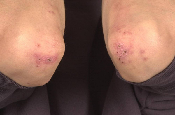
If you had a skin rash like the one shown in Figure 15.7.1, you probably wouldn’t assume that it was caused by a digestive system disease. However, that’s exactly why the individual in the picture has a rash. He has a gastrointestinal (GI) tract disorder called Crohn’s disease. This disease is one of a group of GI tract disorders that are known collectively as inflammatory bowel disease. Unlike other inflammatory bowel diseases, signs and symptoms of Crohn’s disease may not be confined to the GI tract.
Inflammatory Bowel Disease
Inflammatory bowel disease (IBD) is a collection of inflammatory conditions primarily affecting the intestines. The two principal inflammatory bowel diseases are Crohn’s disease and ulcerative colitis. Unlike Crohn’s disease — which may affect any part of the GI tract and the joints, as well as the skin — ulcerative colitis mainly affects just the colon and rectum. Both diseases occur when the body’s own immune system attacks the digestive system. Both diseases typically first appear in the late teens or early twenties, and occur equally in males and females. Approximately 270,000 Canadians are currently living with IBD, 7,000 of which are children. The annual cost of caring for these Canadians is estimated at $1.28 billion. The number of cases of IBD has been steadily increasing and it is expected that by 2030 the number of Canadians suffering from IBD will grow to 400,000.
Crohn’s Disease
Crohn’s disease is a type of inflammatory bowel disease that may affect any part of the GI tract from the mouth to the anus, among other body tissues. The most commonly affected region is the ileum, which is the final part of the small intestine. Signs and symptoms of Crohn’s disease typically include abdominal pain, diarrhea (with or without blood), fever, and weight loss. Malnutrition because of faulty absorption of nutrients may also occur. Potential complications of Crohn’s disease include obstructions and abscesses of the bowel. People with Crohn’s disease are also at slightly greater risk than the general population of developing bowel cancer. Although there is a slight reduction in life expectancy in people with Crohn’s disease, if the disease is well-managed, affected people can live full and productive lives. Approximately 135,000 Canadians are living with Crohn's disease.
Crohn’s disease is caused by a combination of genetic and environmental factors that lead to impairment of the generalized immune response (called innate immunity). The chronic inflammation of Crohn’s disease is thought to be the result of the immune system “trying” to compensate for the impairment. Dozens of genes are likely to be involved, only a few of which have been identified. Because of the genetic component, close relatives such as siblings of people with Crohn’s disease are many times more likely to develop the disease than people in the general population. Environmental factors that appear to increase the risk of the disease include smoking tobacco and eating a diet high in animal proteins. Crohn’s disease is typically diagnosed on the basis of a colonoscopy, which provides a direct visual examination of the inside of the colon and the ileum of the small intestine.
People with Crohn’s disease typically experience recurring periods of flare-ups followed by remission. There are no medications or surgical procedures that can cure Crohn’s disease, although medications such as anti-inflammatory or immune-suppressing drugs may alleviate symptoms during flare-ups and help maintain remission. Lifestyle changes, such as dietary modifications and smoking cessation, may also help control symptoms and reduce the likelihood of flare-ups. Surgery may be needed to resolve bowel obstructions, abscesses, or other complications of the disease.
Ulcerative Colitis
Ulcerative colitis is an inflammatory bowel disease that causes inflammation and ulcers (sores) in the colon and rectum. Unlike Crohn’s disease, other parts of the GI tract are rarely affected in ulcerative colitis. The primary symptoms of the disease are lower abdominal pain and bloody diarrhea. Weight loss, fever, and anemia may also be present. Symptoms typically occur intermittently with periods of no symptoms between flare-ups. People with ulcerative colitis have a considerably increased risk of colon cancer and should be screened for colon cancer more frequently than the general population. Ulcerative colitis, however, seems to primarily reduce the quality of life, and not the lifespan.
The exact cause of ulcerative colitis is not known. Theories about its cause involve immune system dysfunction, genetics, changes in normal gut bacteria, and lifestyle factors, such as a diet high in animal protein and the consumption of alcoholic beverages. Genetic involvement is suspected in part because ulcerative colitis tens to “run” in families. It is likely that multiple genes are involved. Diagnosis is typically made on the basis of colonoscopy and tissue biopsies.
Lifestyle changes, such as reducing the consumption of animal protein and alcohol, may improve symptoms of ulcerative colitis. A number of medications are also available to treat symptoms and help prolong remission. These include anti-inflammatory drugs and drugs that suppress the immune system. In cases of severe disease, removal of the colon and rectum may be required and can cure the disease.
Diverticulitis
Diverticulitis is a digestive disease in which tiny pouches in the wall of the large intestine become infected and inflamed. Symptoms typically include lower abdominal pain of sudden onset. There may also be fever, nausea, diarrhea or constipation, and blood in the stool. Having large intestine pouches called diverticula (see Figure 15.7.2) that are not inflamed is called diverticulosis. Diverticulosis is thought to be caused by a combination of genetic and environmental factors, and is more common in people who are obese. Infection and inflammation of the pouches (diverticulitis) occurs in about 10–25% of people with diverticulosis, and is more common at older ages. The infection is generally caused by bacteria.
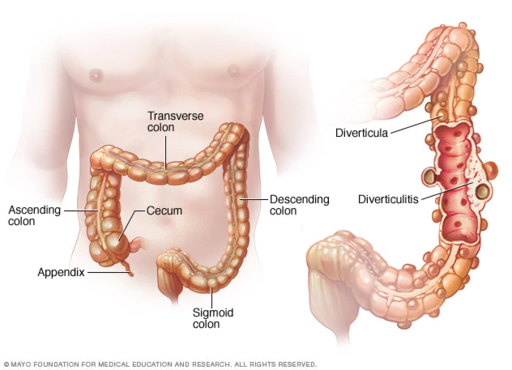
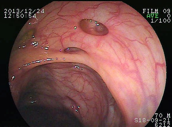
Diverticulitis can usually be diagnosed with a CT scan and can be monitored with a colonoscopy (as seen in Figure 15.7.3). Mild diverticulitis may be treated with oral antibiotics and a short-term liquid diet. For severe cases, intravenous antibiotics, hospitalization, and complete bowel rest (no nourishment via the mouth) may be recommended. Complications such as abscess formation or perforation of the colon require surgery.
Peptic Ulcer
A peptic ulcer is a sore in the lining of the stomach or the duodenum (first part of the small intestine). If the ulcer occurs in the stomach, it is called a gastric ulcer. If it occurs in the duodenum, it is called a duodenal ulcer. The most common symptoms of peptic ulcers are upper abdominal pain that often occurs in the night and improves with eating. Other symptoms may include belching, vomiting, weight loss, and poor appetite. Many people with peptic ulcers, particularly older people, have no symptoms. Peptic ulcers are relatively common, with about ten per cent of people developing a peptic ulcer at some point in their life.
The most common cause of peptic ulcers is infection with the bacterium Helicobacter pylori, which may be transmitted by food, contaminated water, or human saliva (for example, by kissing or sharing eating utensils). Surprisingly, the bacterial cause of peptic ulcers was not discovered until the 1980s. The scientists who made the discovery are Australians Robin Warren and Barry J. Marshall. Although the two scientists eventually won a Nobel Prize for their discovery, their hypothesis was poorly received at first. To demonstrate the validity of their discovery, Marshall used himself in an experiment. He drank a culture of bacteria from a peptic ulcer patient and developed symptoms of peptic ulcer in a matter of days. His symptoms resolved on their own within a couple of weeks, but, at his wife's urging, he took antibiotics to kill any remaining bacteria. Marshall’s self-experiment was published in the Australian Medical Journal, and is among the most cited articles ever published in the journal. Figure 15.7.4 shows how H. pylori cause peptic ulcers.
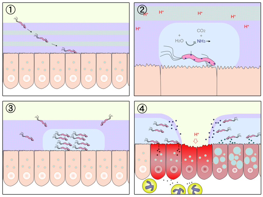
Another relatively common cause of peptic ulcers is chronic use of non-steroidal anti-inflammatory drugs (NSAIDs), such as aspirin or ibuprofen. Additional contributing factors may include tobacco smoking and stress, although these factors have not been demonstrated conclusively to cause peptic ulcers independent of H. pylori infection. Contrary to popular belief, diet does not appear to play a role in either causing or preventing peptic ulcers. Eating spicy foods and drinking coffee and alcohol were once thought to cause peptic ulcers. These lifestyle choices are no longer thought to have much (if any) of an effect on the development of peptic ulcers.
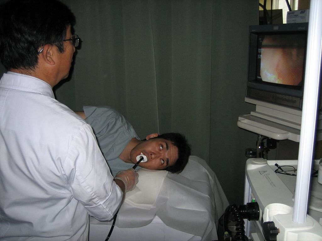
Peptic ulcers are typically diagnosed on the basis of symptoms or the presence of H. pylori in the GI tract. However, endoscopy (shown in Figure 15.7.5), which allows direct visualization of the stomach and duodenum with a camera, may be required for a definitive diagnosis. Peptic ulcers are usually treated with antibiotics to kill H. pylori, along with medications to temporarily decrease stomach acid and aid in healing. Unfortunately, H. pylori has developed resistance to commonly used antibiotics, so treatment is not always effective. If a peptic ulcer has penetrated so deep into the tissues that it causes a perforation of the wall of the stomach or duodenum, then emergency surgery is needed to repair the damage.
Gastroenteritis
Gastroenteritis, also known as infectious diarrhea or stomach flu, is an acute and usually self-limiting infection of the GI tract by pathogens. Symptoms typically include some combination of diarrhea, vomiting, and abdominal pain. Fever, lack of energy, and dehydration may also occur. The illness generally lasts less than two weeks, even without treatment, but in young children it is potentially deadly. Gastroenteritis is very common, especially in poorer nations. Worldwide, up to five billion cases occur each year, resulting in about 1.4 million deaths.
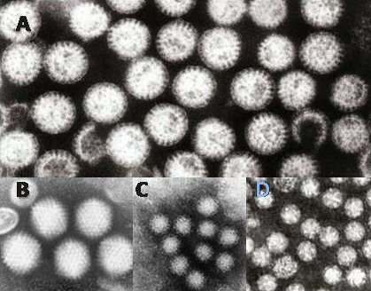
Commonly called “stomach flu,” gastroenteritis is unrelated to the influenza virus, although viruses are the most common cause of the disease (see Figure 15.7.6). In children, rotavirus is most often the cause which is why the British Columbia immunization schedule now includes a rotovirus vaccine. Norovirus is more likely to be the cause of gastroenteritis in adults. Besides viruses, other potential causes of gastroenteritis include fungi, bacteria (most often E. coli or Campylobacter jejuni), and protozoa(including Giardia lamblia, more commonly called Beaver Fever, described below). Transmission of pathogens may occur due to eating improperly prepared foods or foods left to stand at room temperature, drinking contaminated water, or having close contact with an infected individual.
Gastroenteritis is less common in adults than children, partly because adults have acquired immunity after repeated exposure to the most common infectious agents. Adults also tend to have better hygiene than children. If children have frequent repeated incidents of gastroenteritis, they may suffer from malnutrition, stunted growth, and developmental delays. Many cases of gastroenteritis in children can be avoided by giving them a rotavirus vaccine. Frequent and thorough handwashing can cut down on infections caused by other pathogens.
Treatment of gastroenteritis generally involves increasing fluid intake to replace fluids lost in vomiting or diarrhea. Oral rehydration solution, which is a combination of water, salts, and sugar, is often recommended. In severe cases, intravenous fluids may be needed. Antibiotics are not usually prescribed, because they are ineffective against viruses that cause most cases of gastroenteritis.
Giardiasis
Giardiasis, popularly known as beaver fever, is a type of gastroenteritis caused by a GI tract parasite, the single-celled protozoan Giardia lamblia (pictured in Figure 15.7.7). In addition to human beings, the parasite inhabits the digestive tract of a wide variety of domestic and wild animals, including cows, rodents, and sheep, as well as beavers (hence its popular name). Giardiasis is one of the most common parasitic infections in people the world over, with hundreds of millions of people infected worldwide each year.
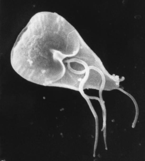
Transmission of G. lamblia is via a fecal-oral route (as in, you got feces in your food). Those at greatest risk include travelers to countries where giardiasis is common, people who work in child-care settings, backpackers and campers who drink untreated water from lakes or rivers, and people who have close contact with infected people or animals in other settings. In Canada, Giardia is the most commonly identified intestinal parasite and approximately 3,000 Canadians will contract the parasite annually.
Symptoms of giardiasis can vary widely. About one-third third of people with the infection have no symptoms, whereas others have severe diarrhea with poor absorption of nutrients. Problems with absorption occur because the parasites inhibit intestinal digestive enzyme production, cause detrimental changes in microvilli lining the small intestine, and kill off small intestinal epithelial cells. The illness can result in weakness, loss of appetite, stomach cramps, vomiting, and excessive gas. Without treatment, symptoms may continue for several weeks. Treatment with anti-parasitic medications may be needed if symptoms persist longer or are particularly severe.
15.7 Summary
- Inflammatory bowel disease is a collection of inflammatory conditions primarily affecting the intestines. The diseases involve the immune system attacking the GI tract, and they have multiple genetic and environmental causes. Typical symptoms include abdominal pain and diarrhea, which show a pattern of repeated flare-ups interrupted by periods of remission. Lifestyle changes and medications may control flare-ups and extend remission. Surgery is sometimes required.
- The two principal inflammatory bowel diseases are Crohn’s disease and ulcerative colitis. Crohn’s disease may affect any part of the GI tract from the mouth to the anus, among other body tissues. Ulcerative colitis affects the colon and/or rectum.
- Some people have little pouches, called diverticula, in the lining of their large intestine, a condition called diverticulosis. People with diverticulosis may develop diverticulitis, in which one or more of the diverticula become infected and inflamed. Diverticulitis is generally treated with antibiotics and bowel rest. Sometimes, surgery is required.
- A peptic ulcer is a sore in the lining of the stomach (gastric ulcer) or duodenum (duodenal ulcer). The most common cause is infection with the bacterium Helicobacter pylori. NSAIDs (such as aspirin) can also cause peptic ulcers, and some lifestyle factors may play contributing roles. Antibiotics and acid reducers are typically prescribed, and surgery is not often needed.
- Gastroenteritis, or infectious diarrhea, is an acute and usually self-limiting infection of the GI tract by pathogens, most often viruses. Symptoms typically include diarrhea, vomiting, and/or abdominal pain. Treatment includes replacing lost fluids. Antibiotics are not usually effective.
- Giardiasis is a type of gastroenteritis caused by infection of the GI tract with the protozoa parasite Giardia lamblia. It may cause malnutrition. Generally self-limiting, severe or long-lasting cases may require antibiotics.
15.7 Review Questions
-
-
- Compare and contrast Crohn’s disease and ulcerative colitis.
- How are diverticulosis and diverticulitis related?
- Identify the cause of giardiasis. Why may it cause malabsorption?
- Name three disorders of the GI tract that can be caused by bacteria.
- Name one disorder of the GI tract that can be helped by anti-inflammatory medications, and one that can be caused by chronic use of anti-inflammatory medications.
- Describe one reason why it can be dangerous to drink untreated water.
15.7 Explore More
https://youtu.be/H5zin8jKeT0
Who's at risk for colon cancer? - Amit H. Sachdev and Frank G. Gress, TED-Ed, 2018.
https://youtu.be/V_U6czbDHLE
The surprising cause of stomach ulcers - Rusha Modi, TED-Ed, 2017.
Attributions
Figure 15.7.1
BADAS_Crohn by Dayavathi Ashok and Patrick Kiely/ Journal of medical case reports on Wikimedia Commons is used under a CC BY 2.0 (https://creativecommons.org/licenses/by/2.0) license.
Figure 15.7.2
512px-Ds00070_an01934_im00887_divert_s_gif.webp by Lfreeman04 on Wikimedia Commons is used under a CC BY-SA 4.0 (https://creativecommons.org/licenses/by-sa/4.0) license.
Figure 15.7.3
Colon_diverticulum by melvil on Wikimedia Commons is used under a CC BY-SA 4.0 (https://creativecommons.org/licenses/by-sa/4.0) license.
Figure 15.7.4
H_pylori_ulcer_diagram by Y_tambe on Wikimedia Commons is used under a CC BY-SA 3.0 (http://creativecommons.org/licenses/by-sa/3.0/) license.
Figure 15.7.5
1024px-Endoscopy_training by Yuya Tamai on Wikimedia Commons is used under a CC BY 2.0 (https://creativecommons.org/licenses/by/2.0) license.
Figure 15.7.6
Gastroenteritis_viruses by Dr. Graham Beards [en:User:Graham Beards] at en.wikipedia on Wikimedia Commons is used under a CC BY 3.0 (https://creativecommons.org/licenses/by/3.0) license.
Figure 15.7.7
Giardia_lamblia_SEM_8698_lores by Janice Haney Carr from CDC/ Public Health Image Library (PHIL) ID# 8698 on Wikimedia Commons is in the public domain (https://en.wikipedia.org/wiki/public_domain).
References
Ashok, D., & Kiely, P. (2007). Bowel associated dermatosis - arthritis syndrome: a case report. Journal of medical case reports, 1, 81. https://doi.org/10.1186/1752-1947-1-81
Marshall, B. J., Armstrong, J. A., McGechie, D. B., & Glancy, R. J. (1985). Attempt to fulfil Koch's postulates for pyloric Campylobacter. The Medical Journal of Australia, 142(8), 436–439.
Marshall, B. J., McGechie, D. B., Rogers, P. A., & Glancy, R. J. (1985). Pyloric campylobacter infection and gastroduodenal disease. The Medical Journal of Australia, 142(8), 439–444.
TED-Ed. (2017, September 28). The surprising cause of stomach ulcers - Rusha Modi. YouTube. https://www.youtube.com/watch?v=V_U6czbDHLE&feature=youtu.be
TED-Ed. (2018, January 4). Who's at risk for colon cancer? - Amit H. Sachdev and Frank G. Gress. YouTube. https://www.youtube.com/watch?v=H5zin8jKeT0&feature=youtu.be
Created by CK-12 Foundation/Adapted by Christine Miller
Case Study Conclusion: Please Don’t Pass the Bread
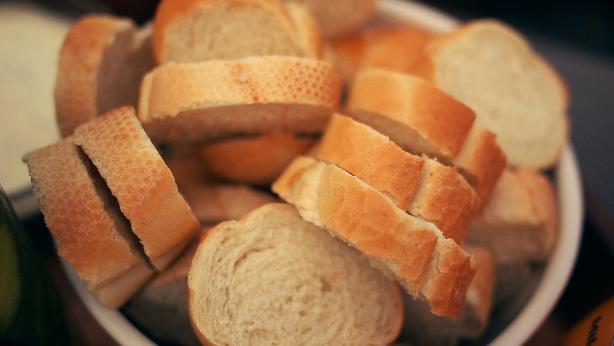
The bread above may or may not look appetizing to you, but for people with celiac disease, it is certainly off limits. Bread and pasta are traditionally made with wheat, which contains proteins called gluten. As you learned in the beginning of the chapter, even trace amounts of gluten can damage the digestive system of people with celiac disease. When Angela and Saloni met for lunch, Angela chose a restaurant that she knew could provide her with gluten-free food because she has this disease.
When people with celiac disease eat gluten, it causes an autoimmune reaction that results in inflammation and flattening of the villi of the small intestine. What do you think happens if the villi are inflamed and flattened? Think about what you have learned about the functions of the villi and small intestine. The small intestine is where most chemical digestion and absorption of nutrients occurs in the body. The villi increase the surface area in the small intestine to maximize the digestion of food and absorption of nutrients into the blood and lymph. If the villi are inflamed and flattened, there is less surface area where digestion and absorption can occur. Therefore, damage from celiac disease can result in an inadequate absorption of nutrients called malabsorption.
Malabsorption explains why there can be so many different types of symptoms of celiac disease, ranging from diarrhea and other forms of digestive distress, to anemia, nutritional deficiencies, skin rashes, osteoporosis and bone pain, depression and anxiety, and rarer — but potentially serious — complications, such as cancer. Our bodies need to digest and absorb adequate amounts of nutrients in order to function properly and stay healthy. Lack of nutrients can affect and damage cells, tissues, and organs throughout the body, sometimes seriously and irreversibly. A person with celiac disease can limit and often heal intestinal damage just by not eating gluten. In fact, eliminating all gluten from the diet is the main treatment for celiac disease. In some people with celiac disease, a gluten-free diet may not be enough, and steroids and other medications may be used to reduce the inflammation in the small intestine.
Celiac disease is an autoimmune disorder in which the body’s immune system attacks its own tissues. It is thought to be caused by the presence of particular genes in combination with exposure to gluten. What are some other autoimmune disorders that you read about in this chapter that affect the digestive system? The two main inflammatory bowel diseases, Crohn’s disease and ulcerative colitis, are both due to the body’s immune system attacking the digestive system, resulting in inflammation. Crohn’s disease can affect any part of the GI tract, most commonly the ileum of the small intestine, while ulcerative colitis mainly affects the colon and rectum. Similar to celiac disease, treatments for these diseases also focus on reducing GI tract damage through lifestyle changes and medications.
Gluten is clearly dangerous for people with celiac disease, but should people who do not have celiac disease or other diagnosed medical problem with gluten also eliminate gluten from their diet? Many medical experts say no, because gluten-free diets are so restrictive, they may cause nutritional deficiencies without providing any proven health benefits. They can also be expensive and, as Saloni’s cousin found out, difficult to maintain, given that gluten is present in so many foods. It is estimated that only one per cent of the population has celiac disease. Most people should enjoy a varied diet and consult with their doctor if they are concerned about celiac disease, other types of gluten intolerance, or food allergies.
Watch this TED-Ed video "What's the big deal with gluten? - William D. Chey" to learn more:
https://youtu.be/uEM2iDT-VAk
What’s the big deal with gluten? - William D. Chey, TED-Ed, 2015.
Chapter 15 Summary
In this chapter, you learned about the digestive system, which allows the body to obtain needed nutrients from food. Specifically, you learned that:
- The digestive system consists of organs that break down food, absorb its nutrients, and expel any remaining food waste.
- Most digestive organs form a long, continuous tube through which food passes, called the gastrointestinal (GI) tract. It starts at the mouth, which is followed by the pharynx, esophagus, stomach, small intestine, and large intestine.
- Organs of the GI tract have walls that consist of several tissue layers that enable them to carry out digestion and/or absorption. For example, the inner mucosa has cells that secrete digestive enzymes and other digestive substances and also cells that absorb nutrients. The muscle layer of the organs enables them to contract and relax in waves of peristalsis to move food through the GI tract.
- Digestion is a form of catabolism, in which food is broken down into small molecules that the body can absorb and use for energy, growth, and repair. Digestion occurs when food moves through the GI tract. The digestive process is controlled by both hormones and nerves.
-
- Mechanical digestion is a physical process in which food is broken into smaller pieces without becoming chemically changed. It occurs mainly in the mouth and stomach.
- Chemical digestion is a chemical process in which macromolecules — including carbohydrates, proteins, lipids, and nucleic acids — in food are changed into simple nutrient molecules that can be absorbed into body fluids. Carbohydrates are chemically digested to sugars, proteins to amino acids, lipids to fatty acids, and nucleic acids to individual nucleotides. Chemical digestion requires digestive enzymes. Gut flora carry out additional chemical digestion.
- Absorption occurs when the simple nutrient molecules that result from digestion are absorbed into blood or lymph. They are then circulated through the body.
- Organs of the upper gastrointestinal (GI) tract include the mouth, pharynx, esophagus, and stomach.
-
- The mouth is the first organ of the GI tract. It has several structures that are specialized for digestion, including salivary glands, tongue, and teeth. Both mechanical digestion and chemical digestion of carbohydrates and fats begin in the mouth.
- The pharynx and esophagus move food from the mouth to the stomach but are not directly involved in the process of digestion or absorption. Food moves through the esophagus by peristalsis.
- Mechanical and chemical digestion continue in the stomach. Acid and digestive enzymes secreted by the stomach start the chemical digestion of proteins. The stomach turns masticated food into a semi-fluid mixture called chyme.
- The lower GI tract includes the small intestine and large intestine. The small intestine is where most chemical digestion and virtually all absorption of nutrients occur. The large intestine contains huge numbers of beneficial bacteria, and removes water and salts from food waste before it is eliminated.
-
- The small intestine consists of three parts: the duodenum, jejunum, and ileum. All three parts of the small intestine are lined with mucosa that is very wrinkled and covered with villi and microvilli, giving the small intestine a huge surface area for digestion and absorption.
-
-
- The duodenum secretes digestive enzymes and also receives bile from the liver or gallbladder and digestive enzymes and bicarbonate from the pancreas. These digestive substances neutralize acidic chyme and allow for the chemical digestion of carbohydrates, proteins, lipids, and nucleic acids in the duodenum.
- The jejunum carries out most of the absorption of nutrients in the small intestine, including the absorption of simple sugars, amino acids, fatty acids, and many vitamins.
- The ileum carries out any remaining digestion and absorption of nutrients, but its main function is to absorb vitamin B12 and bile salts.
- The large intestine consists of the colon (which in turn includes the cecum, ascending colon, transverse colon, descending colon, and sigmoid colon), rectum, and anus. The appendix is attached to the cecum of the colon.
-
-
-
- The main function of the large intestine is to remove water and salts from chyme for recycling within the body and eliminating the remaining solid feces from the body through the anus. The large intestine is also the site where trillions of bacteria help digest certain compounds, produce vitamins, stimulate the immune system, and break down toxins, among other important functions.
-
- Accessory organs of digestion are organs that secrete substances needed for the chemical digestion of food, but through which food does not actually pass as it is digested. The accessory organs include the liver, gallbladder, and pancreas. These organs secrete or store substances that are carried to the duodenum of the small intestine as needed for digestion.
-
- The liver is a large organ in the abdomen that is divided into lobes and smaller lobules, which consist of metabolic cells called hepatic cells, or hepatocytes. The liver receives oxygen in blood from the aorta through the hepatic artery. It receives nutrients in blood from the GI tract and wastes in blood from the spleen through the portal vein.
-
-
- The main digestive function of the liver is the production of the alkaline liquid called bile. Bile is carried directly to the duodenum by the common bile duct or to the gallbladder first for storage. Bile neutralizes acidic chyme that enters the duodenum from the stomach and also emulsifies fat globules into smaller particles (micelles) that are easier to digest chemically.
- Other vital functions of the liver include regulating blood sugar levels by storing excess sugar as glycogen; storing many vitamins and minerals; synthesizing numerous proteins and lipids; and breaking down waste products and toxic substances.
- The gallbladder is a small pouch-like organ near the liver. It stores and concentrates bile from the liver until it is needed in the duodenum to neutralize chyme and help digest lipids.
- The pancreas is a glandular organ that secretes both endocrine hormones and digestive enzymes. As an endocrine gland, the pancreas secretes insulin and glucagon to regulate blood sugar. As a digestive organ, the pancreas secretes digestive enzymes into the duodenum through ducts. Pancreatic digestive enzymes include amylase (starches); trypsin and chymotrypsin (proteins); lipase (lipids); and ribonucleases and deoxyribonucleases (RNA and DNA).
-
- Inflammatory bowel disease is a collection of inflammatory conditions primarily affecting the intestines. The diseases involve the immune system attacking the GI tract, and they have multiple genetic and environmental causes. Typical symptoms include abdominal pain and diarrhea, which show a pattern of repeated flare-ups interrupted by periods of remission. Lifestyle changes and medications may control flare-ups and extend remission. Surgery is sometimes required.
-
- The two principal inflammatory bowel diseases are Crohn’s disease and ulcerative colitis. Crohn’s disease may affect any part of the GI tract from the mouth to the anus, among other body tissues. Ulcerative colitis affects the colon and/or rectum.
- Some people have little pouches, called diverticula, in the lining of their large intestine, a condition called diverticulosis. People with diverticulosis may develop diverticulitis, in which one or more of the diverticula become infected and inflamed. Diverticulitis is generally treated with antibiotics and bowel rest. Sometimes, surgery is required.
- A peptic ulcer is a sore in the lining of the stomach (gastric ulcer) or duodenum (duodenal ulcer). The most common cause is infection with the bacterium Helicobacter pylori. NSAIDs such as aspirin can also cause peptic ulcers, and some lifestyle factors may play contributing roles. Antibiotics and acid reducers are typically prescribed. Surgery is not often needed.
- Gastroenteritis, or infectious diarrhea, is an acute and usually self-limiting infection of the GI tract by pathogens, most often viruses. Symptoms typically include diarrhea, vomiting, and/or abdominal pain. Treatment includes replacing lost fluids. Antibiotics are not usually effective.
- Giardiasis is a type of gastroenteritis caused by infection of the GI tract with the protozoa parasite Giardia lamblia. It may cause malnutrition. Generally self-limiting, severe or long-lasting cases may require antibiotics.
As you have learned, the process of digestion, in which food is broken down and nutrients are absorbed by the body, also involves the elimination of solid wastes. Elimination of waste is also called excretion. Some of the organs of the digestive system, such as the liver and large intestine, are also considered part of the excretory system. Read the next chapter to learn more about the excretory system and how wastes are eliminated from our bodies.
Chapter 15 Review
-
- Explain how the accessory organs of digestion interact with the GI tract.
- If the pH in the duodenum was too low (acidic), what effect do you think this would have on the processes of the digestive system?
- Discuss whether digestion occurs in the large intestine.
- Lipids are digested at different points in the digestive system. Describe how lipids are digested at two of these points.
- Describe two different functions of stomach acid.
- Name and describe the location and function of three of the valves of the GI tract.
- What is the name of the rhythmic muscle contractions that move food through the GI tract?
- What are the major roles of the upper GI tract?
- What is the physiological cause of heartburn?
- What are two ways in which the tongue participates in digestion?
- Where is the epiglottis located? If the epiglottis were to not close properly, what might happen?
Attribution
Figure 15.8.1
Sliced Bread/ Bread Basket by Daria Nepriakhina on Unsplash is used under the Unsplash License (https://unsplash.com/license).
Reference
TED-Ed. (2015, June 2). What’s the big deal with gluten? - William D. Chey. YouTube. https://www.youtube.com/watch?v=uEM2iDT-VAk&feature=youtu.be
A peptide hormone, produced by alpha cells of the pancreas. It works to raise the concentration of glucose and fatty acids in the bloodstream, and is considered to be the main catabolic hormone of the body. It is also used as a medication to treat a number of health conditions.
Created by: CK-12/Adapted by: Christine Miller
What Is a Scientific Theory?
Germ theory, which is described in detail below, is one of several scientific theories you will read about in human biology. A scientific theory is a broad explanation for events. Scientific theories are widely accepted by the scientific community. To become a theory, an explanation must be strongly supported by a great deal of evidence.
People commonly use the word theory to describe a guess or hunch about how or why something happens. For example, you might say, "I think a woodchuck dug this hole in the ground, but it's just a theory." Using the word theory in this way is different from the way it is used in science. A scientific theory is not just a guess or hunch that may or may not be true. In science, a theory is an explanation that has a high likelihood of being correct because it is so well supported by evidence.
https://www.youtube.com/watch?v=GyN2RhbhiEU
What is the difference between a scientific law and theory? by Matt Anticole, TEDEd, 2015
Germ Theory: A Human Biology Example
![Image by Francesco Redenti 1820-1876. (Wellcome Library Record no. 3120i) [Public domain], via Wikimedia Commons A black and white side-profile caricature of Girolamo Fracastoro wearing tradition middle-18th century attire.](https://pressbooks.ccconline.org/acchumanbio/wp-content/uploads/sites/152/2019/06/Redenti_-_Girolamo_Fracastoro-2.jpg)
The germ theory of disease states that contagious diseases are caused by germs, or microorganisms, which are organisms that are too small to be seen without magnification. Microorganisms which cause disease are called pathogens. Human pathogens include bacteria and viruses, among other microscopic entities. When pathogens invade humans or other living hosts, they grow, reproduce, and make their hosts sick. Diseases caused by germs are contagious because the microorganisms that cause them can spread from person to person.
First Statement of Germ Theory
Germ theory was first clearly stated by an Italian physician named Girolamo Fracastoro (pictured in Figure 1.5.1) in the mid-1500s. Fracastoro proposed that contagious diseases are caused by transferable "seed-like entities," which we now call germs. According to Fracastoro, germs spread through populations through direct or indirect contact between individuals, making people sick.
Fracastoro's idea, though essentially correct, was disregarded by other physicians. Instead, Hippocrates' and Galen's idea of miasma remained the accepted explanation for the spread of disease for another 300 years. However, evidence for Fracastoro's idea accumulated during that time. Some of the earliest evidence was provided by the Dutch lens and microscope maker Anton van Leeuwenhoek, who is considered by many to be the father of microbiology. By the 1670s, van Leeuwenhoek had directly observed many different types of microorganisms, including bacteria.
Evidence from Puerperal Fever
One of the first physicians to demonstrate that a microorganism is the cause of a specific human disease was the Hungarian obstetrician Ignaz Semmelweis in the 1840s. The disease was puerperal fever, an often-fatal infection of the female reproductive organs. Puerperal fever is also called childbed fever, because it usually affects women who have just given birth.
![Graph by Power.corrupts [Public domain], from Wikimedia Commons](https://pressbooks.ccconline.org/acchumanbio/wp-content/uploads/sites/152/2023/10/1024px-Yearly_mortality_rates_1833-1858-1.png)
Semmelweis observed that deaths from puerperal fever occurred much more often when women had been attended by doctors at his hospital than by midwives at home. Semmelweis also noticed that doctors often came directly from autopsies to the beds of women about to give birth. From his observations, Semmelweis inferred that puerperal fever was a contagious disease caused by some type of matter carried to pregnant patients on the hands of doctors from autopsied bodies. As a consequence, Semmelweis urged doctors and medical students at his hospital to wash their hands with chlorinated lime water before examining pregnant women. After this change, the hospital's death rate for women who had just given birth fell from 18 to 2 per cent, which was a 90 per cent reduction. Some of Semmelweis' findings are presented in the graph above-right.
Semmelweis published his results, but they were derided by the medical profession. The idea that doctors themselves were the carriers of a fatal disease was taken as a personal affront by his fellow physicians. One of Semmelweis' peers protested indignantly that doctors are gentlemen and that gentlemen's hands are always clean. As a result of attitudes such as this, Semmelweis became the target of a vicious smear campaign. Eventually, Semmelweis had a mental breakdown and was committed to a mental hospital, where he died.
Father of Germ Theory
![Image by By Photo Credit:Content Providers(s): [Public domain], via Wikimedia A view through a microscope showing larger irregularly oval blue cells, and strings of smaller yellow round cells. The chains of small yellow cells are the Streptococcus pyogenes.](https://pressbooks.ccconline.org/acchumanbio/wp-content/uploads/sites/152/2023/10/Streptococcus_pyogenes_01_thumbnail-1.png)
![Image by Albert Edelfelt [Public domain], via Wikimedia Commons A painting showing Louis Pasteur sitting in his lab examining a substance in a bottle](https://pressbooks.ccconline.org/acchumanbio/wp-content/uploads/sites/152/2023/10/Tableau_Louis_Pasteur-1.jpg)
Throughout the later 1800s, more formal investigations were conducted about the relationship between germs and disease. Some of the most important were undertaken by Louis Pasteur. Pasteur (right) was a French chemist who did careful experiments to show that fermentation, food spoilage, and certain diseases are caused by microorganisms. He discovered the cause of puerperal fever in 1879. He determined it was an infection caused by the bacterium Streptococcus pyogenes, shown under magnification (Figure 1.5.3).
Although Pasteur was not the first person to propose germ theory, his investigations clearly supported it. He also became a strong proponent of the theory and managed to convince most of the scientific community of its validity. For these reasons, Pasteur is often regarded as the father of germ theory.
1.5 Summary
- A scientific theory is a broad explanation that is widely accepted because it is strongly supported by a great deal of evidence.
- An example of a scientific theory is the germ theory of disease. According to this theory, contagious diseases are caused by germs, or microorganisms.
- The germ theory of disease was first proposed in the mid-1500s. It was not widely accepted until the late 1800s, when it was strongly supported by experimental evidence from Louis Pasteur.
1.5 Review Questions
- Define scientific theory.
- Compare the way the word theory is used in science versus in everyday language.
- What is the germ theory of disease? How did it develop?
- Explain why Pasteur, rather than Fracastoro or Semmelweis, is called the father of germ theory.
- Galen and Fracastoro may have come up with different explanations for how disease is spread, but what observations do you think they made that were similar?
- Use the explanation of Semmelweis’ research and the graph in Figure 1.9 to answer the following questions:
- What was Semmelweis’ observation that led him to undertake this study? What question was he trying to answer?
- What was the hypothesis (i.e. proposed answer for a scientific question) that Semmelweis was testing?
- Why did Semmelweis track death rates from puerperal fever at Dublin Maternity Hospital, where autopsies were not performed?
- What were two pieces of evidence shown in the graph that supported Semmelweis’ hypothesis?
- Why do you think it was important that Semmelweis compared Dublin Maternity Hospital and Wien Maternity Clinic over the same years?
- What is the difference between a microorganism and a pathogen?
- Explain why the development of the microscope lent support to the germ theory of disease.
- Does the observation of microorganisms alone conclusively prove that germ theory is correct? Why or why not?
- Who do you think was using more scientific reasoning: Semmelweis or the physicians that derided his results? Explain your answer.
1.5 Explore More
https://www.youtube.com/watch?v=rPiW6Y_oDJo&feature=emb_logo
Sammelweis - USA/ Austria Film Belvedere Film, Semmelweis Orvostörténeti Múzeum, 2013
Attributions
Figure 1.5.1
Fracastoro, Girolamo, 1478-1553,. by Francesco Redenti 1820-1876, from Wellcome Library Record no. 3120i, is in the public domain (https://en.wikipedia.org/wiki/Public_domain).
Figure 1.5.2
Puerperal fever yearly mortality rates, 1833-1858, by Power.corrupts, has been released into the public domain (https://en.wikipedia.org/wiki/Public_domain).
Figure 1.5.3
Streptococcus pyogenes 01, from Centers for Disease Control and Prevention's Public Health Image Library (PHIL), ID #2110, is in the public domain (https://en.wikipedia.org/wiki/Public_domain).
Figure 1.5.4
Albert Edelfelt - Louis Pasteur - 1885, photograph by Ondra Havala, is in the public domain (https://en.wikipedia.org/wiki/Public_domain).
References
Semmelweis Orvostörténeti Múzeum. (2013, October 31). Sammelweiz. YouTube. https://www.youtube.com/watch?v=rPiW6Y_oDJo&feature=emb_logo
TEDEd. (2015). What's the difference between a scientific law and a theory? - Matt Anticole. YouTube. https://www.youtube.com/watch?v=GyN2RhbhiEU&t=91s
Wikipedia contributors. (2020, August 3). Antonie van Leeuwenhoek. In Wikipedia. https://en.wikipedia.org/w/index.php?title=Antonie_van_Leeuwenhoek&oldid=970998908
Wikipedia contributors. (2020, July 28). Galen. In Wikipedia. https://en.wikipedia.org/w/index.php?title=Galen&oldid=969901897
Wikipedia contributors. (2020, July 1). Girolamo Fracastoro. In Wikipedia. https://en.wikipedia.org/w/index.php?title=Girolamo_Fracastoro&oldid=965417568
Wikipedia contributors. (2020, July 30). Hippocrates. In Wikipedia. https://en.wikipedia.org/w/index.php?title=Hippocrates&oldid=970254565
Wikipedia contributors. (2020, July 21). Ignaz Semmelweis. In Wikipedia. https://en.wikipedia.org/w/index.php?title=Ignaz_Semmelweis&oldid=968773367
Wikipedia contributors. (2020, August 5). Louis Pasteur. In Wikipedia. https://en.wikipedia.org/w/index.php?title=Louis_Pasteur&oldid=971330056
Wikipedia contributors. (2020, August 5). Miasma theory. In Wikipedia. https://en.wikipedia.org/w/index.php?title=Miasma_theory&oldid=971286379
A type of immune cell that has granules (small particles) with enzymes that are released during infections, allergic reactions, and asthma. An eosinophil is a type of white blood cell and a type of granulocyte.
Created by: Christine Miller
Definition
In order to truly understand the concept of Traditional Ecological Knowledge (TEK), it is important to gather as many definitions as possible- this gives us an accurate breadth of the term with all its nuances. Click through the images below to read several descriptions of TEK.
Value
People who have lived in a community for generations are often the first to notice any signs of environmental change. Information about a particular region's climate and ecology is retained when the people from the region take on location as part of their cultural identity across generations. This traditional knowledge is passed from generation to generation through story telling and mentorship.
TEK shares some similarities with what is termed "Western Science". Both recognize that knowledge is always growing and changing and that observations are critical to recognizing patterns and causalities in nature. In addition, both TEK and Western Science recognize interdependence in biological systems and the need to treat ecology as a complex system. TEK differs in some ways from Western Science: knowledge is passed on orally, partly through metaphor and story, and this learned knowledge is embedded into daily living. TEK also differs from Western Science in that TEK is tied in to morality, spirituality and individual identity, making it more than just knowledge; it is sacred knowledge.
Examples

People who are indigenous to the province of British Columbia have been managing natural resources in this area for time immemorial. Numerous examples of sustainable harvesting methods can be found across the province, but our example, harvesting and management of the Avalanche Lily, comes from the Secwepemc peoples of the interior of British Columbia. Many thanks to Nancy Turner, Marianne Boelscher Ignace and Ronald Ignace for their documentation of these practices in their paper: Traditional Ecological Knowledge and Wisdom of Aboriginal Peoples in British Columbia
The Avalanche Lily is a yellow-flowered member of the lily family native to Western North America. This flower grows from an edible bulb which ranges in size from 3-5 centimetres. The Secwepemc people have harvested these bulbs as an important food source for generations. Oral transmission of knowledge allowed the Secwepemc people to use thousands of years of accumulated data around growing cycles, seasons and management practices to harvest these plants effectively while maintaining a healthy population of lilies for future use. Very intentional conservation strategies were/are practiced when harvesting the bulbs:
- Bulbs were harvested according to elevation in order to collect bulbs in the right stage of maturity and to spread out harvesting and yield over several months. Bulbs picked early in the season were too soft, and bulbs picked late were too watery.
- Only the largest bulbs were selected by choosing stems with multiple fruiting bodies.
- As bulbs were harvested, smaller bulbs were replanted, seeds were scattered and the soil was tilled by the harvesters.
- Areas which had been extensively harvested were often left for several years to allow the lily population time to recover.
The two videos below show how knowledge of a particular ecosystem is handed down through the traditions of mentorship and storytelling:
Modern science, native knowledge, by The Nature Conservancy, 2015

First Stories - Nganawendaanan Nde'ing (I Keep Them in My Heart), by Shannon Letande, 2006
TEK is Part of Place
The Traditional Ecological Knowledge held by Indigenous communities often includes very location specific knowledge. There are many diverse groups of First Peoples in British Columbia, each with expert knowledge about the ecology of their specific ancestral regions. The link to the map from the Native Land Digital website shows some of the traditional boundaries of the Indigenous people in British Columbia. Click on the areas to see where First Nations communities are located.
1.6 Summary
- Traditional Ecological Knowledge (TEK) is an important and valuable body of knowledge
- People groups who have lived in an area over generations pass down TEK through storytelling and mentorship
- TEK and Western Science share certain characteristics, including use of observations, and identification of patterns and causalities in nature
- TEK and Western Science differ in that TEK is passed down through oral storytelling and is deeply rooted in morality, spirituality and individual identity
1.6 Review Questions
Type your exercises here.
- Define Traditional Ecological Knowledge.
- How is TEK passed down through generations?
- How does TEK differ from Western Science?
- What are some ways in which TEK can inform resource management?
- What are some of the ramifications of loss of TEK? How can TEK be maintained?
1.6 Explore More
https://www.youtube.com/watch?time_continue=2&v=pHNlel72eQc&feature=emb_logo
TEDxTC - Winona LaDuke - Seeds of Our Ancestors, Seeds of Life, by TEDx-TC, 2012
Attributions
Definitions of Traditional Ecological Knowledge
- Kamloops, BC, Canada by Louis Paulin on Unsplash is used under the Unsplash License (https://unsplash.com/license).
- The Totem Poles of Capilano, Vancouver by Udayaditya Barua on Unsplash is used under the Unsplash License (https://unsplash.com/license).
- Alouette Lake by Chloe Evans on Unsplash is used under the Unsplash License (https://unsplash.com/license).
Figure 1.6.1
Erythronium grandiflorum 5077, by Walter Siegmund on Wikimedia Commons is used under a CC BY-SA 3.0 (https://creativecommons.org/licenses/by-sa/3.0) license.
References
Herkes, J. (n.d.). Continuing studies: Traditional ecological knowledge. University of Northern British Columbia. Retrieved from https://www.unbc.ca/continuing-studies/courses/traditional-ecological-knowledge
(1993). Traditional ecological knowledge: Concepts and cases (p. vi). Canadian Museum of Nature, Ottawa, Ontario, Canada.
Letande, S. (2006). First Stories - Nganawendaanan Nde'ing (I keep them in my heart). https://www.nfb.ca/film/first_stories_nganawendaanan_ndeing/
Minerals Management Service (n.d.). What is Traditional Knowledge [online]. Government of Alaska.https://web.archive.org/web/20030328053734/http://www.mms.gov/alaska/native/tradknow/tk_mms2.htm
TEDx-TC. (2012, March 4). TEDxTC - Winona LaDuke - Seeds of our ancestors, seeds of life. https://www.youtube.com/watch?v=pHNlel72eQc&feature=youtu.be
The Nature Conservancy. (2015, February 25). Modern science, native knowledge. YouTube. https://www.youtube.com/watch?v=1QRpnHoGivk
Turner, N., Ignace, M., & Ignace, R. (2000, October 1). Traditional ecological knowledge and wisdom of Aboriginal peoples in British Columbia. Ecological Applications, 10(5), 1275-1287. doi:10.2307/2641283
Wakefield, A.J. (1999, September 11). MMR vaccination and autism. Lancet, 354(9182), 949-950. https://www.thelancet.com/journals/lancet/article/PIIS0140-6736(05)75696-8/fulltext doi:https://doi.org/10.1016/S0140-6736(05)75696-8
Created by CK-12 Foundation/Adapted by Christine Miller

Fashion Statement
This colourful hairstyle makes quite a fashion statement. Many people spend a lot of time and money on their hair, even if they don’t have an exceptional hairstyle like this one. Besides its display value, hair actually has important physiological functions.
What is Hair?
Hair is a filament that grows from a hair follicle in the dermis of the skin. It consists mainly of tightly packed, keratin-filled cells called keratinocytes. The human body is covered with hair follicles, with the exception of a few areas, including the mucous membranes, lips, palms of the hands, and soles of the feet.
Structure of Hair
The part of the hair located within the follicle is called the hair root. The root is the only living part of the hair. The part of the hair that is visible above the surface of the skin is the hair shaft. The shaft of the hair has no biochemical activity and is considered dead.
Follicle and Root
Hair growth begins inside a follicle (see Figure 10.5.2 below). Each hair follicle contains stem cells that can keep dividing, which allows hair to grow. The stem cells can also regrow a new hair after one falls out. Another structure associated with a hair follicle is a sebaceous gland that produces oily sebum. The sebum lubricates and helps to waterproof the hair. A tiny arrector pili muscle is also attached to the follicle. When it contracts, the follicle moves, and the hair in the follicle stands up.
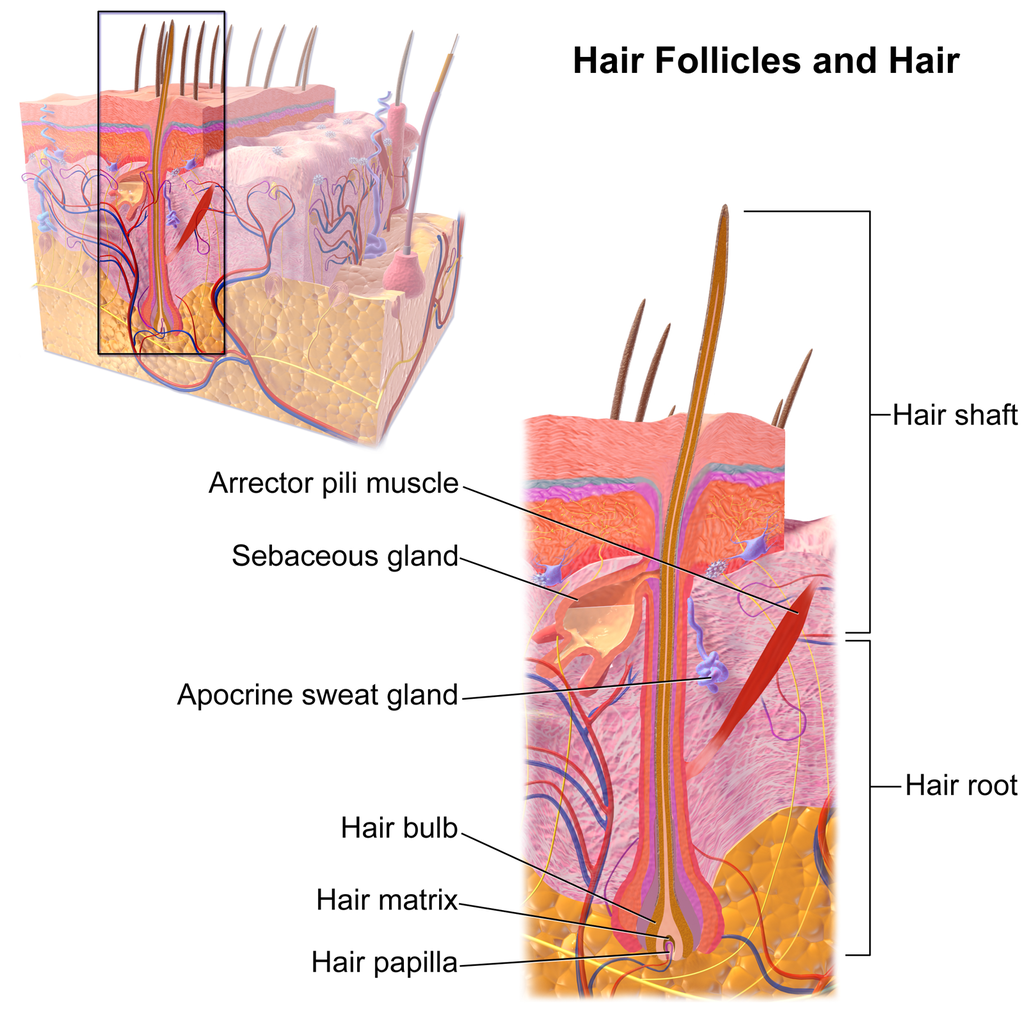
Shaft
The hair shaft is a hard filament that may grow very long. Hair normally grows in length by about half an inch a month. In cross-section, a hair shaft can be divided into three zones, called the cuticle, cortex, and medulla.
- The cuticle (or outer coat) is the outermost zone of the hair shaft. It consists of several layers of flat, thin keratinocytes that overlap one another like shingles on a roof. This arrangement helps the cuticle repel water. The cuticle is also covered with a layer of lipids, just one molecule thick, which increases its ability to repel water. This is the zone of the hair shaft that is visible to the eye.
- The cortex is the middle zone of the hair shaft, and it is also the widest part. The cortex is highly structured and organized, consisting of keratin bundles in rod-like structures. These structures give hair its mechanical strength. The cortex also contains melanin, which gives hair its colour.
- The medulla is the innermost zone of the hair shaft. This is a small, disorganized, and more open area at the center of the hair shaft. The medulla is not always present. When it is present, it contains highly pigmented cells full of keratin.
Characteristics of Hair
Two visible characteristics of hair are its colour and texture. In adult males, the extent of balding is another visible characteristic. All three characteristics are genetically controlled.
Hair Colour
All natural hair colours are the result of melanin, which is produced in hair follicles and packed into granules in the hair. Two forms of melanin are found in human hair: eumelanin and pheomelanin. Eumelanin is the dominant pigment in brown hair and black hair, and pheomelanin is the dominant pigment in red hair. Blond hair results when you have only a small amount of melanin in the hair. Gray and white hair occur when melanin production slows down, and eventually stops.
Figure 10.5.3 Variation in hair colouration. Which types of melanin are present for each hair colour shown?
Hair Texture
Hair exists in a variety of textures. The main aspects of hair texture are the curl pattern, thickness, and consistency.
- The shape of the hair follicle determines the shape of the hair shaft. The shape of the hair shaft, in turn, determines the curl pattern of the hair. Round hair shafts produce straight hair. Hair shafts that are oval or have other shapes produce wavy or curly hair .
- The size of the hair follicle determines the thickness of hair. Thicker hair has greater volume than thinner hair.
- The consistency of hair is determined by the hair follicle volume and the condition of the hair shaft. The consistency of hair is generally classified as fine, medium, or coarse. Fine hair has the smallest circumference, and coarse hair has the largest circumference. Medium hair falls in between these two extremes. Coarse hair also has a more open cuticle than thin or medium hair does, which causes it to be more porous.

Figure 10.5.4 Curly hair has a differently shaped shaft than straight hair.
Functions of Hair
In humans, one function of head hair is to provide insulation and help the head retain heat. Head hair also protects the skin on the head from damage by UV light.
The function of hair in other locations on the body is debated. One idea is that body hair helps keep us warm in cold weather. When the body is too cold, arrector pili muscles contract and cause hairs to stand up (shown in Figure 10.5.5), trapping a layer of warm air above the epidermis. However, this is more effective in mammals that have thick hair or fur than it is in relatively hairless human beings.
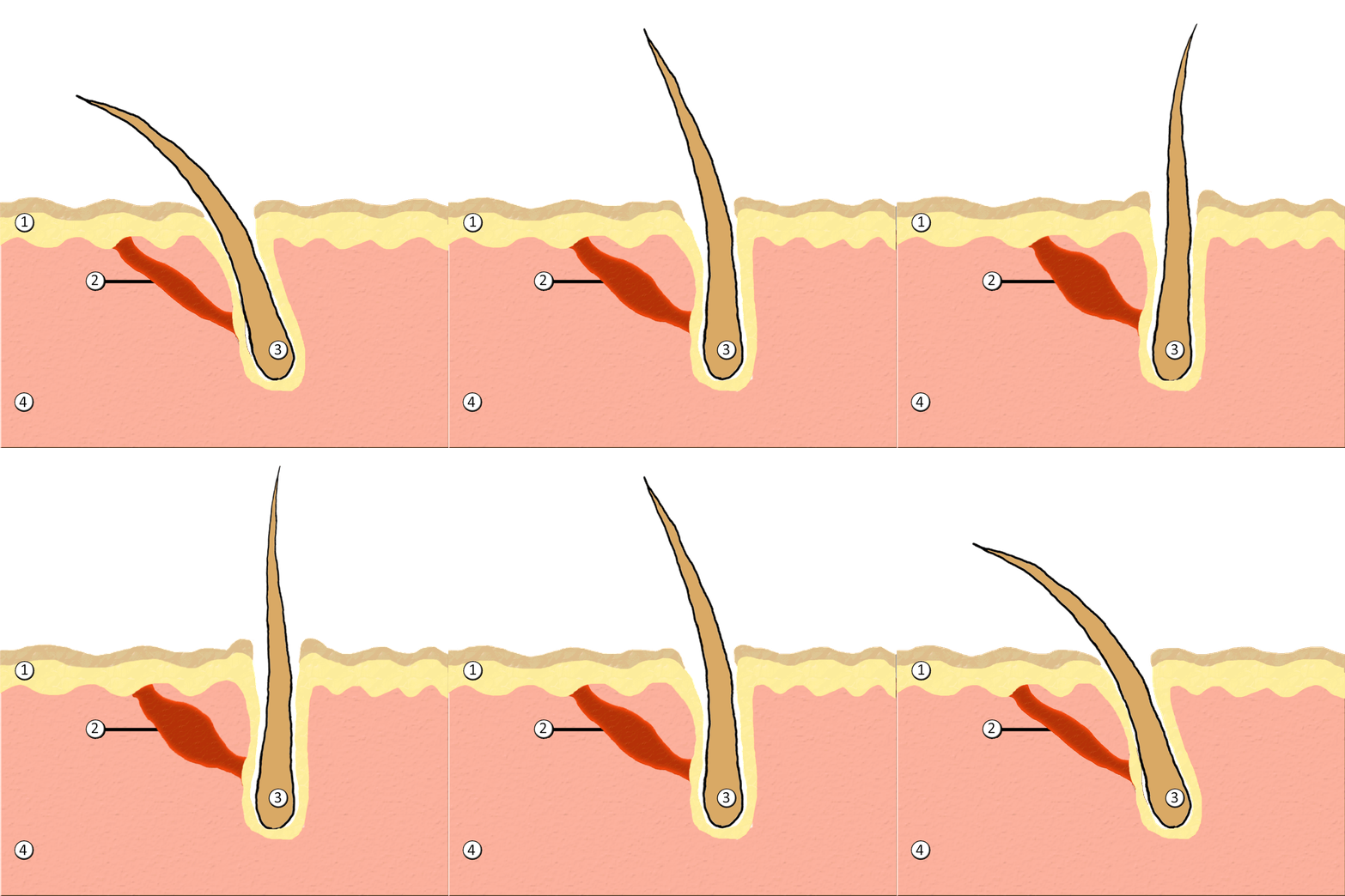
Human hair has an important sensory function, as well. Sensory receptors in the hair follicles can sense when the hair moves, whether it moves because of a breeze, or because of the touch of a physical object. The receptors may also provide sensory awareness of the presence of parasites on the skin.

Some hairs, such as the eyelashes, are especially sensitive to the presence of potentially harmful matter. The eyelashes grow at the edge of the eyelid and can sense when dirt, dust, or another potentially harmful object is too close to the eye. The eye reflexively closes as a result of this sensation. The eyebrows also provide some protection to the eyes. They protect the eyes from dirt, sweat, and rain. In addition, the eyebrows play a key role in nonverbal communication (see Figure 10.5.6). They help express emotions such as sadness, anger, surprise, and excitement.
Hair in Human Evolution
Among mammals, humans are nearly unique in having undergone significant loss of body hair during their evolution. Humans are also unlike most other mammals in having curly hair as one variation in hair texture. Even non-human primates (see Figure 10.5.7) all have straight hair. This suggests that curly hair evolved at some point during human evolution.
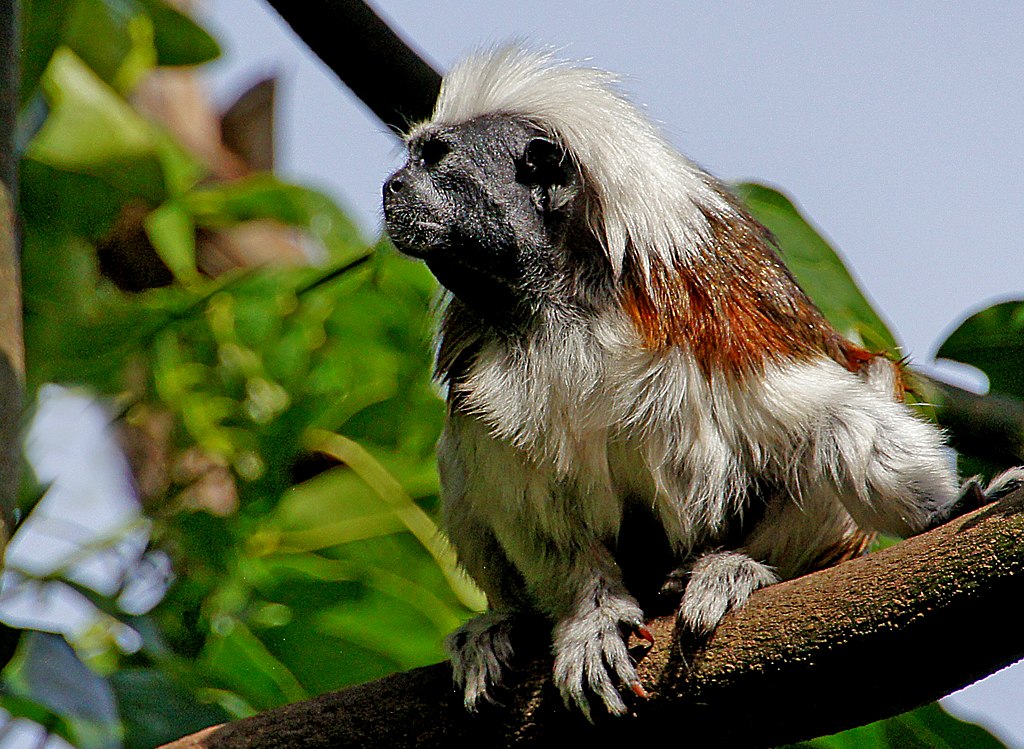
Loss of Body Hair
One hypothesis for the loss of body hair in the human lineage is that it would have facilitated cooling of the body by the evaporation of sweat. Humans also evolved far more sweat glands than other mammals, which is consistent with this hypothesis, because sweat evaporates more quickly from less hairy skin. Another hypothesis for human hair loss is that it would have led to fewer parasites on the skin. This might have been especially important when humans started living together in larger, more crowded social groups.
These hypotheses may explain why we lost body hair, but they can’t explain why we didn’t also lose head hair and hair in the pubic region and armpits. It is possible that head hair was retained because it protected the scalp from UV light. As our bipedal ancestors walked on the open savannas of equatorial Africa, the skin on the head would have been an area exposed to the most direct rays of sunlight in an upright hominid. Pubic and armpit hair may have been retained because they served as signs of sexual maturity, which would have been important for successful mating and reproduction.
Evolution of Curly Hair
Greater protection from UV light has also been posited as a possible selective agent favoring the evolution of curly hair. Researchers have found that straight hair allows more light to pass into the body through the hair shaft via the follicle than does curly hair. In this way, human hair is like a fibre optic cable. It allows light to pass through easily when it is straight, but it impedes the passage of light when it is kinked or coiled. This is indirect evidence that UV light may have been a selective agent leading to the evolution of curly hair.
Social and Cultural Significance of Hair
Hair has great social significance for human beings. Body hair is an indicator of biological sex, because hair distribution is sexually dimorphic. Adult males are generally hairier than adult females, and facial hair in particular is a notable secondary male sex characteristic. Hair may also be an indicator of age. White hair is a sign of older age in both males and females, and male pattern baldness is a sign of older age in males. In addition, hair colour and texture can be a sign of ethnic ancestry.
Hair also has great cultural significance. Hairstyle and colour may be an indicator of social group membership and for better or worse can be associated with specific stereotypes. Head shaving has been used in many times and places as a punishment, especially for women. On the other hand, in some cultures, cutting off one’s hair symbolizes liberation from one’s past. In other cultures, it is a sign of mourning. There are also many religious-based practices involving hair. For example, the majority of Muslim women hide their hair with a headscarf. Sikh men grow their hair long and cover it with a turban. Amish men (like the one pictured in Figure 10.5.8) grow facial hair only after they marry — but just a beard, and not a mustache.

Unfortunately, sometimes hairstyle, colour and characteristics are used to apply stereotypes, particularly with respect to women. "Blonde jokes" are a good example of how negative stereotypes are maintained despite having no actual truth behind them. Many stereotypes related to hair are hidden, even from persons perpetrating the stereotype. Often a hairstyle is judged by another as having ties to gender, sexuality, worldview and/or socioeconomic status; even when these inferences are woefully inaccurate. It is important to be aware of our own biases and determine if these biases are appropriate - take a look at the collage in Figure 10.5.9. What are your initial reactions? Are these reactions founded in fact? Do you harbor an unfair bias?
Figure 10.5.9 What are your biases? Are they fair?
10.5 Summary
- Hair is a filament that grows from a hair follicle in the dermis of the skin. It consists mainly of tightly packed, keratin-filled cells called keratinocytes. The human body is almost completely covered with hair follicles.
- The part of a hair that is within the follicle is the hair root. This is the only living part of a hair. The part of a hair that is visible above the skin surface is the hair shaft. It consists of dead cells.
- Hair growth begins inside a follicle when stem cells within the follicle divide to produce new keratinocytes. An individual hair may grow to be very long.
- A hair shaft has three zones: the outermost zone called the cuticle; the middle zone called the cortex; and the innermost zone called the medulla.
- Genetically controlled, visible characteristics of hair include hair colour, hair texture, and the extent of balding in adult males. Melanin (eumelanin and/or pheomelanin) is the pigment that gives hair its colour. Aspects of hair texture include curl pattern, thickness, and consistency.
- Functions of head hair include providing insulation and protecting skin on the head from UV light. Hair everywhere on the body has an important sensory function. Hair in eyelashes and eyebrows protects the eyes from dust, dirt, sweat, and other potentially harmful substances. The eyebrows also play a role in nonverbal communication.
- Among mammals, humans are nearly unique in having undergone significant loss of body hair during their evolution, probably because sweat evaporates more quickly from less hairy skin. Curly hair also is thought to have evolved at some point during human evolution, perhaps because it provided better protection from UV light.
- Hair has social significance for human beings, because it is an indicator of biological sex, age, and ethnic ancestry. Human hair also has cultural significance. Hairstyle may be an indicator of social group membership, for example.
10.5 Review Questions
-
- Compare and contrast the hair root and hair shaft.
- Describe hair follicles.
-
-
- Explain variation in human hair colour.
- What factors determine the texture of hair?
- Describe two functions of human hair.
- What hypotheses have been proposed for the loss of body hair during human evolution?
- Discuss the social and cultural significance of human hair.
- Describe one way in which hair can be used as a method of communication in humans.
- Explain why waxing or tweezing body hair, which typically removes hair down to the root, generally keeps the skin hair-free for a longer period of time than shaving, which cuts hair off at the surface of the skin.
10.5 Explore More
https://www.youtube.com/watch?v=8diYLhl8bWU
Why do some people go bald? - Sarthak Sinha, TED-Ed, 2015.
https://www.youtube.com/watch?v=kNw8V_Fkw28
Hair Love | Oscar®-Winning Short Film (Full) | Sony Pictures Animation, 2019.
https://www.youtube.com/watch?v=hDW5e3NR1Cw
Why do we care about hair | Naomi Abigail | TEDxBaDinh, TEDx Talks, 2015.
Attributions
Figure 10.5.1
Hair by jessica-dabrowski-TETR8YLSqt4 [photo] by Jessica Dabrowski on Unsplash is used under the Unsplash License (https://unsplash.com/license).
Figure 10.5.2
Blausen_0438_HairFollicleAnatomy_02 by BruceBlaus on Wikimedia Commons is used under a CC BY 3.0 (https://creativecommons.org/licenses/by/3.0) license.
Figure 10.5.3
- Standing tall by Ilaya Raja on Unsplash is used under the Unsplash License (https://unsplash.com/license).
- Blond-haired woman smiling by Carlos Lindner on Unsplash is used under the Unsplash License (https://unsplash.com/license).
- Smith Mountain Lake redhead by Chris Ross Harris on Unsplash is used under the Unsplash License (https://unsplash.com/license).
- Through the look of experience by Laura Margarita Cedeño Peralta on Unsplash is used under the Unsplash License (https://unsplash.com/license).
Figure 10.5.4
Curly hair by chris-benson-clvEami9RN4 [photo] by Chris Benson on Unsplash is used under the Unsplash License (https://unsplash.com/license).
Figure 10.5.5
1024px-PilioerectionAnimation by AnthonyCaccese on Wikimedia Commons is used under a CC BY-SA 4.0 (https://creativecommons.org/licenses/by-sa/4.0/deed.en) license.
Figure 10.5.6
Pout by alexander-dummer-Em8I8Z_DwA4 [photo] by Alexander Dummer on Unsplash is used under the Unsplash License (https://unsplash.com/license).
Figure 10.5.7
Cotton_top_tamarin_monkey._(12046035746) by Bernard Spragg. NZ, from Christchurch, New Zealand on Wikimedia Commons is used under a CC0 1.0 Universal
Public Domain Dedication license (https://creativecommons.org/publicdomain/zero/1.0/deed.en).
Figure 10.5.8
Amish hairstyle by CK-12 Foundation is used under a CC BY-NC 3.0 (https://creativecommons.org/licenses/by-nc/3.0/) license.
 ©CK-12 Foundation Licensed under
 ![]() • Terms of Use • Attribution
• Terms of Use • Attribution
Figure 10.5.9
- Rainbow Hair Bubble Man by Behrouz Jafarnezhad on Unsplash is used under the Unsplash License (https://unsplash.com/license).
- Pink hair in Atlanta, United States by Tammie Allen on Unsplash is used under the Unsplash License (https://unsplash.com/license).
- Magdalena 2 by Valerie Elash on Unsplash is used under the Unsplash License (https://unsplash.com/license).
- Perfect Style by Daria Volkova on Unsplash is used under the Unsplash License (https://unsplash.com/license)
- Stay Classy by Fayiz Musthafa on Unsplash is used under the Unsplash License (https://unsplash.com/license)
- Take your time by Jan Tinneberg on Unsplash is used under the Unsplash License (https://unsplash.com/license)
References
Blausen.com staff. (2014). Medical gallery of Blausen Medical 2014. WikiJournal of Medicine 1 (2). DOI:10.15347/wjm/2014.010. ISSN 2002-4436.
Brainard, J/ CK-12 Foundation. (2016). Figure 7 This style of facial hair is adopted by most Amish men after they marry [digital image]. In CK-12 College Human Biology (Section 12.5) [online Flexbook]. CK12.org. https://www.ck12.org/book/ck-12-college-human-biology/section/12.5/
Sony Pictures Animation. (2019, December 5). Hair love | Oscar®-winning short film (Full) | Sony Pictures Animation. YouTube. https://www.youtube.com/watch?v=kNw8V_Fkw28
TED-Ed. (2015, August 25). Why do some people go bald? – Sarthak Sinha. YouTube. https://www.youtube.com/watch?v=8diYLhl8bWU
TEDx Talks. (2015, February 4). Why do we care about hair | Naomi Abigail | TEDxBaDinh. YouTube. https://www.youtube.com/watch?v=hDW5e3NR1Cw
Created by CK-12 Foundation/Adapted by Christine Miller
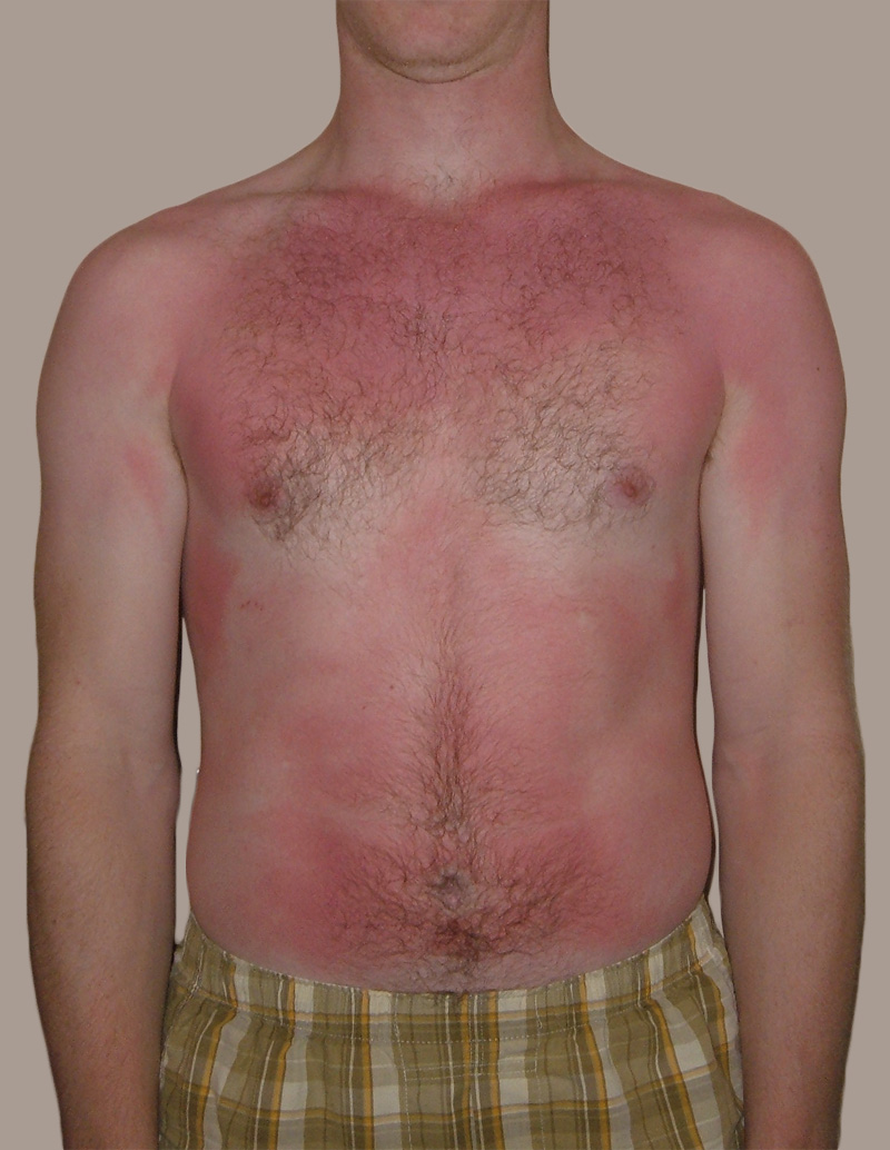
Feel the Burn
The person in Figure 10.3.1 is no doubt feeling the burn — sunburn, that is. Sunburn occurs when the outer layer of the skin is damaged by UV light from the sun or tanning lamps. Some people deliberately allow UV light to burn their skin, because after the redness subsides, they are left with a tan. A tan may look healthy, but it is actually a sign of skin damage. People who experience one or more serious sunburns are significantly more likely to develop skin cancer. Natural pigment molecules in the skin help protect it from UV light damage. These pigment molecules are found in the layer of the skin called the epidermis.
What is the Epidermis?
The epidermis is the outer of the two main layers of the skin. The inner layer is the dermis. It averages about 0.10 mm thick, and is much thinner than the dermis. The epidermis is thinnest on the eyelids (0.05 mm) and thickest on the palms of the hands and soles of the feet (1.50 mm). The epidermis covers almost the entire body surface. It is continuous with — but structurally distinct from — the mucous membranes that line the mouth, anus, urethra, and vagina.
Structure of the Epidermis
There are no blood vessels and very few nerve cells in the epidermis. Without blood to bring epidermal cells oxygen and nutrients, the cells must absorb oxygen directly from the air and obtain nutrients via diffusion of fluids from the dermis below. However, as thin as it is, the epidermis still has a complex structure. It has a variety of cell types and multiple layers.
Cells of the Epidermis
There are several different types of cells in the epidermis. All of the cells are necessary for the important functions of the epidermis.
- The epidermis consists mainly of stacks of keratin-producing epithelial cells called keratinocytes. These cells make up at least 90 per cent of the epidermis. Near the top of the epidermis, these cells are also called squamous cells.
- Another eight per cent of epidermal cells are melanocytes. These cells produce the pigment melanin that protects the dermis from UV light.
- About one per cent of epidermal cells are Langerhans cells. These are immune system cells that detect and fight pathogens entering the skin.
- Less than one per cent of epidermal cells are Merkel cells, which respond to light touch and connect to nerve endings in the dermis.
Layers of the Epidermis
The epidermis in most parts of the body consists of four distinct layers. A fifth layer occurs in the palms of the hands and soles of the feet, where the epidermis is thicker than in the rest of the body. The layers of the epidermis are shown in Figure 10.3.2, and described in the following text.
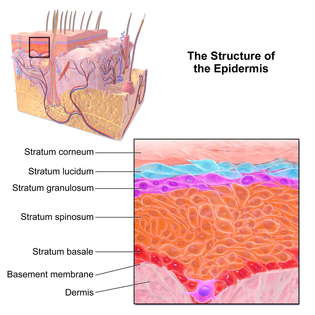
Stratum Basale
The stratum basale is the innermost (or deepest) layer of the epidermis. It is separated from the dermis by a membrane called the basement membrane. The stratum basale contains stem cells — called basal cells — which divide to form all the keratinocytes of the epidermis. When keratinocytes first form, they are cube-shaped and contain almost no keratin. As more keratinocytes are produced, previously formed cells are pushed up through the stratum basale. Melanocytes and Merkel cells are also found in the stratum basale. The Merkel cells are especially numerous in touch-sensitive areas, such as the fingertips and lips.
Stratum Spinosum
Just above the stratum basale is the stratum spinosum. This is the thickest of the four epidermal layers. The keratinocytes in this layer have begun to accumulate keratin, and they have become tougher and flatter. Spiny cellular projections form between the keratinocytes and hold them together. In addition to keratinocytes, the stratum spinosum contains the immunologically active Langerhans cells.
Stratum Granulosum
The next layer above the stratum spinosum is the stratum granulosum. In this layer, keratinocytes have become nearly filled with keratin, giving their cytoplasm a granular appearance. Lipids are released by keratinocytes in this layer to form a lipid barrier in the epidermis. Cells in this layer have also started to die, because they are becoming too far removed from blood vessels in the dermis to receive nutrients. Each dying cell digests its own nucleus and organelles, leaving behind only a tough, keratin-filled shell.
Stratum Lucidum
Only on the palms of the hands and soles of the feet, the next layer above the stratum granulosum is the stratum lucidum. This is a layer consisting of stacks of translucent, dead keratinocytes that provide extra protection to the underlying layers.
Stratum Corneum
The uppermost layer of the epidermis everywhere on the body is the stratum corneum. This layer is made of flat, hard, tightly packed dead keratinocytes that form a waterproof keratin barrier to protect the underlying layers of the epidermis. Dead cells from this layer are constantly shed from the surface of the body. The shed cells are continually replaced by cells moving up from lower layers of the epidermis. It takes a period of about 48 days for newly formed keratinocytes in the stratum basale to make their way to the top of the stratum corneum to replace shed cells.
Functions of the Epidermis
The epidermis has several crucial functions in the body. These functions include protection, water retention, and vitamin D synthesis.
Protective Functions
The epidermis provides protection to underlying tissues from physical damage, pathogens, and UV light.
Protection from Physical Damage
Most of the physical protection of the epidermis is provided by its tough outer layer, the stratum corneum. Because of this layer, minor scrapes and scratches generally do not cause significant damage to the skin or underlying tissues. Sharp objects and rough surfaces have difficulty penetrating or removing the tough, dead, keratin-filled cells of the stratum corneum. If cells in this layer are pierced or scraped off, they are quickly replaced by new cells moving up to the surface from lower skin layers.
Protection from Pathogens
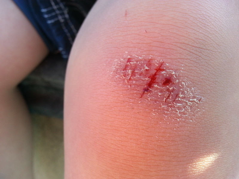
When pathogens such as viruses and bacteria try to enter the body, it is virtually impossible for them to enter through intact epidermal layers. Generally, pathogens can enter the skin only if the epidermis has been breached, for example by a cut, puncture, or scrape (like the one pictured in Figure 10.3.3). That’s why it is important to clean and cover even a minor wound in the epidermis. This helps ensure that pathogens do not use the wound to enter the body. Protection from pathogens is also provided by conditions at or near the skin surface. These include relatively high acidity (pH of about 5.0), low amounts of water, the presence of antimicrobial substances produced by epidermal cells, and competition with non-pathogenic microorganisms that normally live on the epidermis.
Protection from UV Light
UV light that penetrates the epidermis can damage epidermal cells. In particular, it can cause mutations in DNA that lead to the development of skin cancer, in which epidermal cells grow out of control. UV light can also destroy vitamin B9 (in forms such as folate or folic acid), which is needed for good health and successful reproduction. In a person with light skin, just an hour of exposure to intense sunlight can reduce the body’s vitamin B9 level by 50 per cent.
Melanocytes in the stratum basale of the epidermis contain small organelles called melanosomes, which produce, store, and transport the dark brown pigment melanin. As melanosomes become full of melanin, they move into thin extensions of the melanocytes. From there, the melanosomes are transferred to keratinocytes in the epidermis, where they absorb UV light that strikes the skin. This prevents the light from penetrating deeper into the skin, where it can cause damage. The more melanin there is in the skin, the more UV light can be absorbed.
Water Retention
Skin's ability to hold water and not lose it to the surrounding environment is due mainly to the stratum corneum. Lipids arranged in an organized way among the cells of the stratum corneum form a barrier to water loss from the epidermis. This is critical for maintaining healthy skin and preserving proper water balance in the body.
Although the skin is impermeable to water, it is not impermeable to all substances. Instead, the skin is selectively permeable, allowing certain fat-soluble substances to pass through the epidermis. The selective permeability of the epidermis is both a benefit and a risk.
- Selective permeability allows certain medications to enter the bloodstream through the capillaries in the dermis. This is the basis of medications that are delivered using topical ointments, or patches (see Figure 10.3.4) that are applied to the skin. These include steroid hormones, such as estrogen (for hormone replacement therapy), scopolamine (for motion sickness), nitroglycerin (for heart problems), and nicotine (for people trying to quit smoking).
- Selective permeability of the epidermis also allows certain harmful substances to enter the body through the skin. Examples include the heavy metal lead, as well as many pesticides.
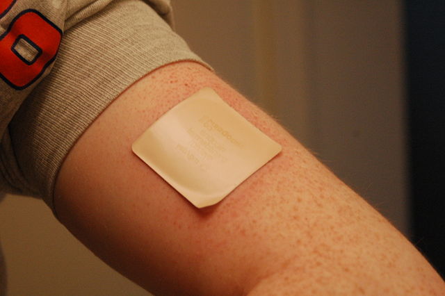
Vitamin D Synthesis
Vitamin D is a nutrient that is needed in the human body for the absorption of calcium from food. Molecules of a lipid compound named 7-dehydrocholesterol are precursors of vitamin D. These molecules are present in the stratum basale and stratum spinosum layers of the epidermis. When UV light strikes the molecules, it changes them to vitamin D3. In the kidneys, vitamin D3 is converted to calcitriol, which is the form of vitamin D that is active in the body.
What Gives Skin Its Colour?
Melanin in the epidermis is the main substance that determines the colour of human skin. It explains most of the variation in skin colour in people around the world. Two other substances also contribute to skin colour, however, especially in light-skinned people: carotene and hemoglobin.
- The pigment carotene is present in the epidermis and gives skin a yellowish tint, especially in skin with low levels of melanin.
- Hemoglobin is a red pigment found in red blood cells. It is visible through skin as a pinkish tint, mainly in skin with low levels of melanin. The pink colour is most visible when capillaries in the underlying dermis dilate, allowing greater blood flow near the surface.
Hear what Bill Nye has to say about the subject of skin colour in the video here.
Bacteria on Skin
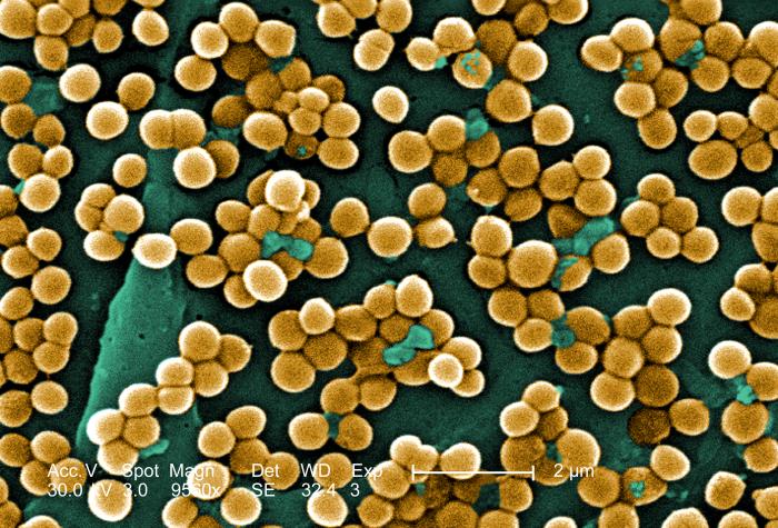
The surface of the human skin normally provides a home to countless numbers of bacteria. Just one square inch of skin normally has an average of about 50 million bacteria. These generally harmless bacteria represent roughly one thousand bacterial species (including the one in Figure 10.3.5) from 19 different bacterial phyla. Typical variations in the moistness and oiliness of the skin produce a variety of rich and diverse habitats for these microorganisms. For example, the skin in the armpits is warm and moist and often hairy, whereas the skin on the forearms is smooth and dry. These two areas of the human body are as diverse to microorganisms as rainforests and deserts are to larger organisms. The density of bacterial populations on the skin depends largely on the region of the skin and its ecological characteristics. For example, oily surfaces, such as the face, may contain over 500 million bacteria per square inch. Despite the huge number of individual microorganisms living on the skin, their total volume is only about the size of a pea.
In general, the normal microorganisms living on the skin keep one another in check, and thereby play an important role in keeping the skin healthy. If the balance of microorganisms is disturbed, however, there may be an overgrowth of certain species, and this may result in an infection. For example, when a patient is prescribed antibiotics, it may kill off normal bacteria and allow an overgrowth of single-celled yeast. Even if skin is disinfected, no amount of cleaning can remove all of the microorganisms it contains. Disinfected areas are also quickly recolonized by bacteria residing in deeper areas (such as hair follicles) and in adjacent areas of the skin.
Feature: Myth vs. Reality
Because of the negative health effects of excessive UV light exposure, it is important to know the facts about protecting the skin from UV light.
Myth |
Reality |
| "Sunblock and sunscreen are just different names for the same type of product. They both work the same way and are equally effective." | Sunscreens and sunblocks are different types of products that protect the skin from UV light in different ways. They are not equally effective. Sunblocks are opaque, so they do not let light pass through. They prevent most of the rays of UV light from penetrating to the skin surface. Sunblocks are generally stronger and more effective than sunscreens. Sunblocks also do not need to be reapplied as often as sunscreens. Sunscreens, in contrast, are transparent once they are applied the skin. Although they can prevent most UV light from penetrating the skin when first applied, the active ingredients in sunscreens tend to break down when exposed to UV light. Sunscreens, therefore, must be reapplied often to remain effective. |
| "The skin needs to be protected from UV light only on sunny days. When the sky is cloudy, UV light cannot penetrate to the ground and harm the skin." | Even on cloudy days, a significant amount of UV radiation penetrates the atmosphere to strike Earth’s surface. Therefore, using sunscreens or sunblocks to protect exposed skin is important even when there are clouds in the sky. |
| "People who have dark skin, such as African Americans, do not need to worry about skin damage from UV light." | No matter what colour skin you have, your skin can be damaged by too much exposure to UV light. Therefore, even dark-skinned people should use sunscreens or sunblocks to protect exposed skin from UV light. |
| "Sunscreens with an SPF (sun protection factor) of 15 are adequate to fully protect the skin from UV light." | Most dermatologists recommend using sunscreens with an SPF of at least 35 for adequate protection from UV light. They also recommend applying sunscreens at least 20 minutes before sun exposure and reapplying sunscreens often, especially if you are sweating or spending time in the water. |
| "Using tanning beds is safer than tanning outside in natural sunlight." | The light in tanning beds is UV light, and it can do the same damage to the skin as the natural UV light in sunlight. This is evidenced by the fact that people who regularly use tanning beds have significantly higher rates of skin cancer than people who do not. It is also the reason that the use of tanning beds is prohibited in many places in people who are under the age of 18, just as youth are prohibited from using harmful substances, such as tobacco and alcohol. |
10.3 Summary
- The epidermis is the outer of the two main layers of the skin. It is very thin, but has a complex structure.
- Cell types in the epidermis include keratinocytes that produce keratin and make up 90 per cent of epidermal cells, melanocytes that produce melanin, Langerhans cells that fight pathogens in the skin, and Merkel cells that respond to light touch.
- The epidermis in most parts of the body consists of four distinct layers. A fifth layer occurs only in the epidermis of the palms of the hands and soles of the feet.
- The innermost layer of the epidermis is the stratum basale, which contains stem cells that divide to form new keratinocytes. The next layer is the stratum spinosum, which is the thickest layer and contains Langerhans cells and spiny keratinocytes. This is followed by the stratum granulosum, in which keratinocytes are filling with keratin and starting to die. The stratum lucidum is next, but only on the palms and soles. It consists of translucent dead keratinocytes. The outermost layer is the stratum corneum, which consists of flat, dead, tightly packed keratinocytes that form a tough, waterproof barrier for the rest of the epidermis.
- Functions of the epidermis include protecting underlying tissues from physical damage and pathogens. Melanin in the epidermis absorbs and protects underlying tissues from UV light. The epidermis also prevents loss of water from the body and synthesizes vitamin D.
- Melanin is the main pigment that determines the colour of human skin. The pigments carotene and hemoglobin, however, also contribute to skin colour, especially in skin with low levels of melanin.
- The surface of healthy skin normally is covered by vast numbers of bacteria representing about one thousand species from 19 phyla. Different areas of the body provide diverse habitats for skin microorganisms. Usually, microorganisms on the skin keep each other in check unless their balance is disturbed.
10.3 Review Questions
- What is the epidermis?
- Identify the types of cells in the epidermis.
- Describe the layers of the epidermis.
-
- State one function of each of the four epidermal layers found all over the body.
- Explain three ways the epidermis protects the body.
- What makes the skin waterproof?
- Why is the selective permeability of the epidermis both a benefit and a risk?
- How is vitamin D synthesized in the epidermis?
- Identify three pigments that impart colour to skin.
- Describe bacteria that normally reside on the skin, and explain why they do not usually cause infections.
- Explain why the keratinocytes at the surface of the epidermis are dead, while keratinocytes located deeper in the epidermis are still alive.
- Which layer of the epidermis contains keratinocytes that have begun to die?
-
- Explain why our skin is not permanently damaged if we rub off some of the surface layer by using a rough washcloth.
10.3 Explore More
https://www.youtube.com/watch?v=27lMmdmy-b8
Jonathan Eisen: Meet your microbes, TED, 2015.
https://www.youtube.com/watch?v=9AcQXnOscQ8
Why Do We Blush?, SciShow, 2014.
https://www.youtube.com/watch?v=_r4c2NT4naQ
The science of skin colour - Angela Koine Flynn, TED-Ed, 2016.
Attributions
Figure 10.3.1
Sunburn by QuinnHK at English Wikipedia on Wikimedia Commons is released into the public domain (https://en.wikipedia.org/wiki/Public_domain).
Figure 10.3.2
Blausen_0353_Epidermis by BruceBlaus on Wikimedia Commons is used under a CC BY 3.0 (https://creativecommons.org/licenses/by/3.0) license.
Figure 10.3.3
Isaac's scraped knee close-up by Alpha on Flickr is used under a CC BY-NC-SA 2.0 (https://creativecommons.org/licenses/by-nc-sa/2.0/) license.
Figure 10.3.4
Nicoderm by RegBarc on Wikimedia Commons is used under a CC BY-SA 3.0 (http://creativecommons.org/licenses/by-sa/3.0/) license. (No machine-readable author provided for original.)
Figure 10.3.5
Staphylococcus aureus bacteria, MRSA by Microbe World on Flickr is used under a CC BY-NC-SA 2.0 (https://creativecommons.org/licenses/by-nc-sa/2.0/) license.
References
Blausen.com staff. (2014). Medical gallery of Blausen Medical 2014. WikiJournal of Medicine 1 (2). DOI:10.15347/wjm/2014.010. ISSN 2002-4436.
Jeff Bone 'n' Pookie. (2020, July 19). Bill Nye the science guy explains we have different skin color. Youtube. https://www.youtube.com/watch?v=zOkj5jgC4sM&feature=youtu.be
SciShow. (2014, July 15). Why do we blush? YouTube. https://www.youtube.com/watch?v=9AcQXnOscQ8
TED. (2015, July 17). Jonathan Eisen: Meet your microbes. YouTube. https://www.youtube.com/watch?v=27lMmdmy-b8
TED-Ed. (2016, February 16). The science of skin color - Angela Koine Flynn. YouTube. https://youtu.be/_r4c2NT4naQ
The ability of an organism to maintain constant internal conditions despite external changes.
Biological molecules that lower amount the energy required for a reaction to occur.
A hormone is a signaling molecule produced by glands in multicellular organisms that target distant organs to regulate physiology and behavior.
The process by which DNA is copied.
Image shows a diagram of Thrombocytes in their normal state and activated.
Thrombocytes (platelets) are typically ovoid during normal circulation, but when activated become super fibrous. The not activated platelets look like very smooth and the activated platelets look like sea anemones- lots of little projects sticking out of their surface.
Created by CK-12/Adapted by Christine Miller
The Thinker
![Auguste Rodin [CC0] The Thinker (French: Le Penseur) is a bronze sculpture by Auguste Rodin, usually placed on a stone pedestal. The work shows a nude male figure of over life-size sitting on a rock with his chin resting on one hand as though deep in thought, often used as an image to represent philosophy.](https://pressbooks.ccconline.org/acchumanbio/wp-content/uploads/sites/152/2019/06/The-thinker-1.jpg)
You've probably seen this famous statue created by the French sculptor Auguste Rodin. Rodin's skill as a sculptor is especially evident here because the statue — which is made of bronze — looks so lifelike. How does a bronze statue differ from a living, breathing human being or other living organism? What is life? What does it mean to be alive? Science has answers to these questions.
Characteristics of Living Things
To be classified as a living thing, most scientists agree that an object must have all seven of the traits listed below. Humans share these characteristics with other living things.
- Homeostasis
- Organization
- Metabolism
- Growth
- Adaptation
- Response to stimuli
- Reproduction
Homeostasis
All living things are able to maintain a more-or-less constant internal environment. Regardless of the conditions around them, they can keep things relatively stable on the inside. The condition in which a system is maintained in a more-or-less steady state is called homeostasis. Human beings, for example, maintain a stable internal body temperature. If you go outside when the air temperature is below freezing, your body doesn't freeze. Instead, by shivering and other means, it maintains a stable internal temperature.
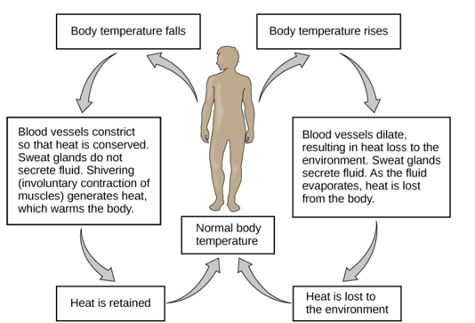
Organization
Living things have multiple levels of organization. Their molecules are organized into one or more cells. A cell is the basic unit of the structure and function of living things. Cells are the building blocks of living organisms. An average adult human being, for example, consists of trillions of cells. Living things may appear very different from one another on the outside, but their cells are very similar. Compare the human cells and onion cells in Figures 2.2.3 and 2.2.4. What similarities do you see?
![Joseph Elsbernd [CC BY-SA 2.0 (https://creativecommons.org/licenses/by-sa/2.0)] Shows the image through a microscope of human cheek cells. The cells are oval in shape and light blue, with a darker blue spot close to the centre. The light blue shows the cell membrane and cytoplasm and the darker blue shows the nucleus of the cell.](https://pressbooks.ccconline.org/acchumanbio/wp-content/uploads/sites/152/2023/10/Cheek-Cells-1.jpg)
![kaibara87 [CC BY 2.0 (https://creativecommons.org/licenses/by/2.0)] Shows an image through a microscope of onion cells. The cells are packed together and are rectangular in shape. Their cell walls and nuclei are stained a darker blue and the cytoplasm is whitish.](https://pressbooks.ccconline.org/acchumanbio/wp-content/uploads/sites/152/2023/10/Onion-Cells-1.jpg)
Metabolism
All living things can use energy. They require energy to maintain internal conditions (homeostasis), to grow, and to execute other processes. Living cells use the "machinery" of metabolism, which is the building up and breaking down of chemical compounds. Living things can transform energy by building up large molecules from smaller ones. This form of metabolism is called anabolism. Living things can also break down, or decompose, large organic molecules into smaller ones. This form of metabolism is called catabolism.
Consider weight lifters who eat high-protein diets. A protein is a large molecule made up of several small amino acids. When we eat proteins, our digestive system breaks them down into amino acids (catabolism), so that they are small enough to be absorbed by the digestive system and into the blood. From there, amino acids are transported to muscles, where they are converted back to proteins (anabolism).

Growth
All living things have the capacity for growth. Growth is an increase in size that occurs when there is a higher rate of anabolism than catabolism. A human infant, for example, has changed dramatically in size by the time it reaches adulthood, as is apparent from the image below. In what other ways do we change as we grow from infancy to adulthood?
A human infant has a lot of growing to do before adulthood.
Adaptations and Evolution
An adaptation is a characteristic that helps living things survive and reproduce in a given environment. It comes about because living things have the ability to change over time in response to the environment. A change in the characteristics of living things over time is called evolution. It develops in a population of organisms through random genetic mutations and natural selection.
Response to Stimuli
All living things detect changes in their environment and respond to them. These stimuli can be internal or external, and the response can take many forms, from the movement of a unicellular organism in response to external chemicals (called chemotaxis) to complex reactions involving all the senses of a multicellular organism. A response is often expressed by motion; for example, the leaves of a plant turning toward the sun (called phototropism).
Click through the images below: the venus fly trap, the cat, and the flower are all showing response to a stimuli.
Figure 2.2.6 Examples of responses to environmental stimuli.
Reproduction
All living things are capable of reproduction, the process by which living things give rise to offspring. Reproduction may be as simple as a single cell dividing to form two daughter cells, which is how bacteria reproduce. Reproduction in human beings and many other organisms, of course, is much more complicated. Nonetheless, whether a living thing is a human being or a bacterium, it is normally capable of reproduction.
Feature: Myth vs. Reality
Myth: Viruses are living things.
Reality: The traditional scientific view of viruses is that they originate from bits of DNA or RNA shed from the cells of living things, but that they are not living things themselves. Scientists have long argued that viruses are not living things because they do not exhibit most of the defining traits of living organisms. A single virus, called a virion, consists of a set of genes (DNA or RNA) inside a protective protein coat, called a capsid. Viruses have organization, but they are not cells, and they do not possess the cellular "machinery" that living things use to carry out life processes. As a result, viruses cannot undertake metabolism, maintain homeostasis, or grow.
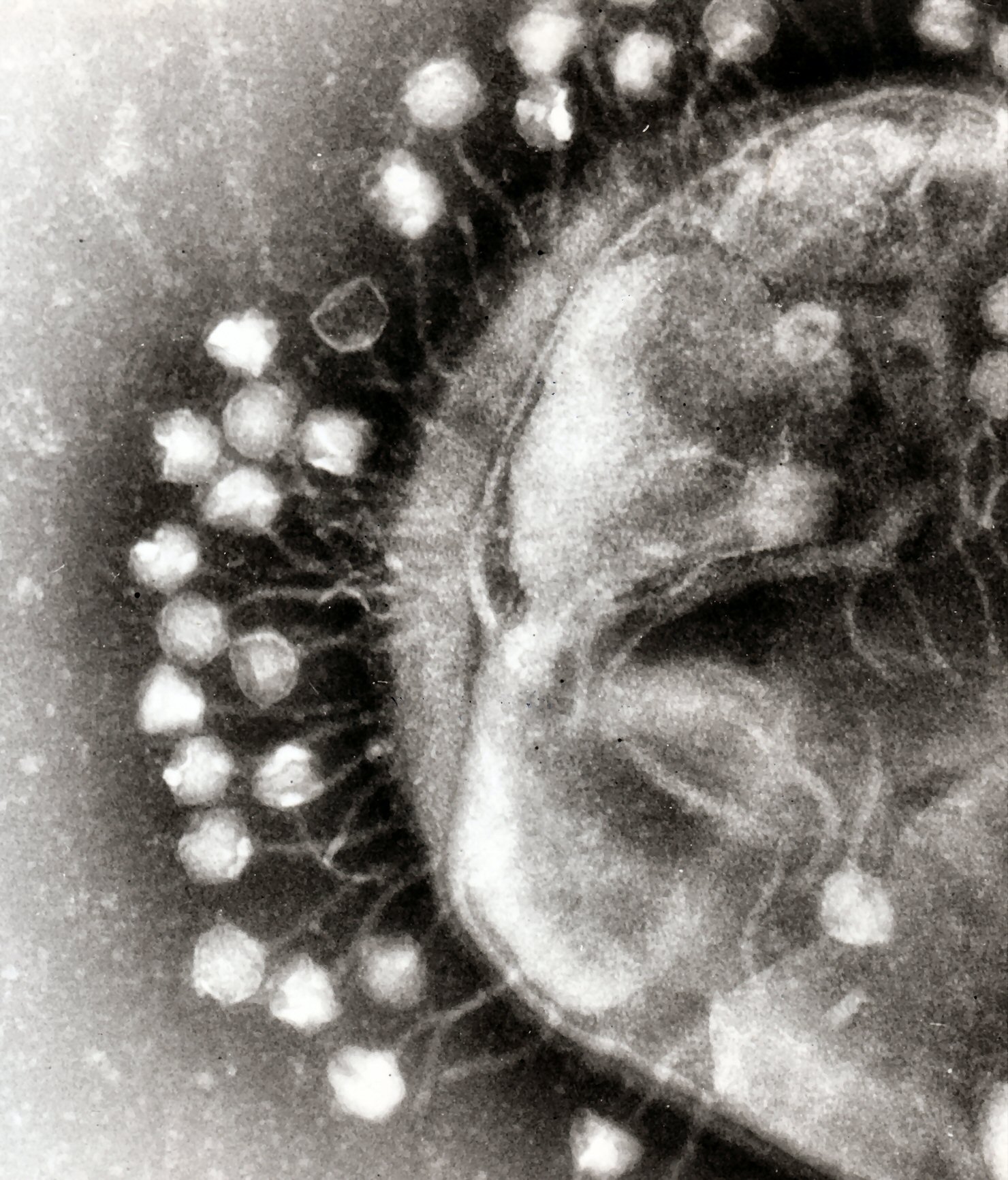
They do not seem to respond to their environment, and they can reproduce only by invading and using "tools" inside host cells to produce more virions. The only traits viruses seem to share with living things is the ability to evolve adaptations to their environment. In fact, some viruses evolve so quickly that it is difficult to design drugs and vaccines against them! That's why maintaining protection from the viral disease influenza, for example, requires a new flu vaccine each year.
Within the last decade, new discoveries in virology (the study of viruses) suggest that this traditional view about viruses may be incorrect, and that the "myth" that viruses are living things may be the reality. Researchers have discovered giant viruses that contain more genes than cellular life forms, such as bacteria. Some of the genes code for proteins needed to build new viruses, which suggests that these giant viruses may be able — or were once able — to reproduce without a host cell. Some of the strongest evidence that viruses are living things comes from studies of their proteins, which show that viruses and cellular life share a common ancestor in the distant past. Viruses may have once existed as primitive cells, but at some point they lost their cellular nature and became modern viruses that require host cells to reproduce. This idea is not so far-fetched when you consider that many other species require a host to complete their life cycle.
2.2 Summary
- To be classified as a living thing, most scientists agree that an object must exhibit seven characteristics. Humans share these traits with all other living things.
- All living things:
- Can maintain a more-or-less constant internal environment, which is called homeostasis.
- Have multiple levels of organization and consist of one or more cells.
- Can use energy and are capable of metabolism.
- Grow and develop.
- Can evolve adaptations to their environment.
- Can detect and respond to environmental stimuli.
- Are capable of reproduction, which is the process by which living things give rise to offspring.
2.2 Review Questions
- Identify the seven traits that most scientists agree are shared by all living things.
- What is homeostasis? What is one way humans fulfill this criterion of living things?
- Define reproduction and describe two different examples.
- Assume that you found an object that looks like a dead twig. You wonder if it might be a stick insect. How could you ethically determine if it is a living thing?
- Describe viruses and which traits they do and do not share with living things. Do you think viruses should be considered living things? Why or why not?
- People who are biologically unable to reproduce are certainly still considered alive. Discuss why this situation does not invalidate the criteria that living things must be capable of reproduction.
- What are the two types of metabolism described here. What are their differences?
- What are some similarities between the cells of different organisms? If you are not familiar with the specifics of cells, simply describe the similarities you see in the pictures above.
- What are two processes in a living thing that use energy?
- Give an example of a response to stimuli in humans.
- Do unicellular organisms (such as bacteria) have an internal environment that they maintain through homeostasis? Why or why not?
- Evolution occurs through natural ____________ .
- If alien life is found on other planets, do you think the aliens will have cells? Discuss your answer.
- Movement in response to an external chemical is called ___________, while movement towards light is called ___________ .
2.2 Explore More
https://www.youtube.com/watch?v=cQPVXrV0GNA&t=354s
Characteristics of Life, Ameoba Sisters, 2017.
Attributions
Figure 2.2.1
The Thinker MET 131262, by Auguste Rodin, 1910, from the Metropolitan Museum of Art, is in the public domain (https://en.wikipedia.org/wiki/Public_domain).
Figure 2.2.2
Homeostasis: Figure 4, by OpenStax College, Biology is used under a CC BY 4.0 (https://creativecommons.org/licenses/by/4.0) license. Download for free at http://cnx.org/contents/04fdb865-17a1-43d8-bb33-36f821ddd119@7.
Figure 2.2.3
Human cheek cells, by Joseph Elsbernd, 2012, on Flickr, is used under a CC BY-SA 2.0 (https://creativecommons.org/licenses/by-sa/2.0/) license.
Figure 2.2.4
Onion cells 2, by Umberto Salvagnin, 2009, on Flickr, is used under a CC BY 2.0 (https://creativecommons.org/licenses/by/2.0/) license.
Figure 2.2.5
Photo (family) by Jakob Owens on Unsplash is used under the Unsplash License (https://unsplash.com/license).
Figure 2.2.6
- Trap of Dionaea muscipula by che on Wikimedia Commons is used under a CC BY-SA 2.5 (https://creativecommons.org/licenses/by-sa/2.5/deed.en) license.
- Plants leaning towards the sunlight from Pxhere is used under a CC0 1.0 universal
public domain dedication license (https://creativecommons.org/publicdomain/zero/1.0/). - Surprised young cat by Watchduck (a.k.a. Tilman Piesk) on Wikimedia Commons is used under a CC BY 3.0 (https://creativecommons.org/licenses/by/3.0) license.
Figure 2.2.7
Bacteriophages, by Dr. Graham Beards, is used under a CC BY-SA 3.0 (https://creativecommons.org/licenses/by-sa/3.0) license.
References
Ameoba Sisters. (2017, October 26). Characteristics of life. YouTube. https://www.youtube.com/watch?v=cQPVXrV0GNA&feature=youtu.be
OpenStax. (2016, March 23). Figure 4 The body is able to regulate temperature in response to signals from the nervous system. In OpenStax, Biology (Section 33.3). OpenStax CNX. http://cnx.org/contents/185cbf87-c72e-48f5-b51e-f14f21b5eabd@10.8.
Wikipedia contributors. (2020, June 14). Adaptation. Wikipedia. https://en.wikipedia.org/w/index.php?title=Adaptation&oldid=962556016
Wikipedia contributors. (2020, June 21). Auguste Rodin. Wikipedia. https://en.wikipedia.org/w/index.php?title=Auguste_Rodin&oldid=963668399
Wikipedia contributors. (2020, June 22). Chemotaxis. Wikipedia. https://en.wikipedia.org/w/index.php?title=Chemotaxis&oldid=963884872
Wikipedia contributors. (2020, June 22). Evolution. Wikipedia. https://en.wikipedia.org/w/index.php?title=Evolution&oldid=963929880
Wikipedia contributors. (2020, June 20). Phototropism. Wikipedia. https://en.wikipedia.org/w/index.php?title=Phototropism&oldid=963567791
Wikipedia contributors. (2020, June 22). Virus. Wikipedia. https://en.wikipedia.org/w/index.php?title=Virus&oldid=963829311
A rigid organ that constitutes part of the vertebrate skeleton in animals.
Created by CK-12 Foundation/Adapted by Christine Miller

Art for All Eras
Pictured in Figure 10.2.1, is Maud Stevens Wagner, a tattoo artist from 1907. Tattoos are not just a late 20th and early 21st century trend. They have been popular in many eras and cultures. Tattoos literally illustrate the biggest organ of the human body: the skin. The skin is very thin, but it covers a large area — about 2 m2 in adults. The skin is the major organ in the integumentary system.
What Is the Integumentary System?
In addition to the skin, the integumentary system includes the hair and nails, which are organs that grow out of the skin. Because the organs of the integumentary system are mostly external to the body, you may think of them as little more than accessories, like clothing or jewelry, but they serve vital physiological functions. They provide a protective covering for the body, sense the environment, and help the body maintain homeostasis.
The Skin
The skin is remarkable not only because it is the body’s largest organ: the average square inch of skin has 20 blood vessels, 650 sweat glands, and more than 1,000 nerve endings. Incredibly, it also has 60,000 pigment-producing cells. All of these structures are packed into a stack of cells that is just 2 mm thick. Although the skin is thin, it consists of two distinct layers: the epidermis and dermis, as shown in the diagram (Figure 10.2.2).
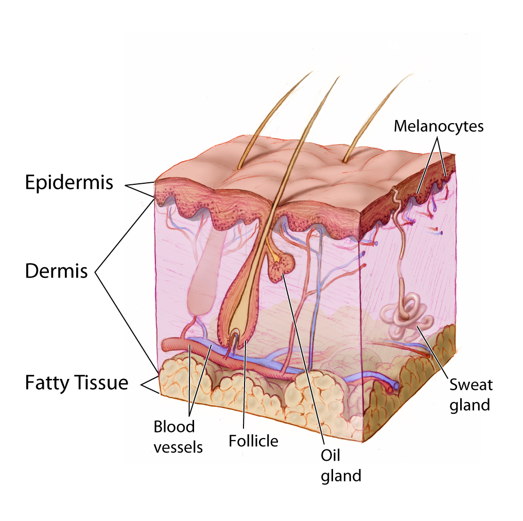
Outer Layer of Skin
The outer layer of skin is the epidermis. This layer is thinner than the inner layer (the dermis). The epidermis consists mainly of epithelial cells, called keratinocytes, which produce the tough, fibrous protein keratin. The innermost cells of the epidermis are stem cells that divide continuously to form new cells. The newly formed cells move up through the epidermis toward the skin surface, while producing more and more keratin. The cells become filled with keratin and die by the time they reach the surface, where they form a protective, waterproof layer. As the dead cells are shed from the surface of the skin, they are replaced by other cells that move up from below. The epidermis also contains melanocytes, the cells that produce the brown pigment melanin, which gives skin most of its colour. Although the epidermis contains some sensory receptor cells — called Merkel cells — it contains no nerves, blood vessels, or other structures.
Inner Layer of Skin
The dermis is the inner, thicker layer of skin. It consists mainly of tough connective tissue, and is attached to the epidermis by collagen fibres. The dermis contains many structures (as shown in Figure 10.2.2), including blood vessels, sweat glands, and hair follicles, which are structures where hairs originate. In addition, the dermis contains many sensory receptors, nerves, and oil glands.
Functions of the Skin
The skin has multiple roles in the body. Many of these roles are related to homeostasis. The skin’s main functions are preventing water loss from the body and serving as a barrier to the entry of microorganisms. Another function of the skin is synthesizing vitamin D, which occurs when the skin is exposed to ultraviolet (UV) light. Melanin in the epidermis blocks some of the UV light and protects the dermis from its damaging effects.
Another important function of the skin is helping to regulate body temperature. When the body is too warm, for example, the skin lowers body temperature by producing sweat, which cools the body when it evaporates. The skin also increases the amount of blood flowing near the body surface through vasodilation (widening of blood vessels), bringing heat from the body core to radiate out into the environment. The sweaty hair and flushed skin of the young man pictured in Figure 10.2.3 reflect these skin responses to overheating.

Hair
Hair is a fibre found only in mammals. It consists mainly of keratin-producing keratinocytes. Each hair grows out of a follicle in the dermis. By the time the hair reaches the surface, it consists mainly of dead cells filled with keratin. Hair serves several homeostatic functions. Head hair is important in preventing heat loss from the head and protecting its skin from UV radiation. Hairs in the nose trap dust particles and microorganisms in the air, and prevent them from reaching the lungs. Hair all over the body provides sensory input when objects brush against it, or when it sways in moving air. Eyelashes and eyebrows (see Figure 10.2.4) protect the eyes from water, dirt, and other irritants.
Nails
Fingernails and toenails consist of dead keratinocytes filled with keratin. The keratin makes them hard but flexible, which is important for the functions they serve. Nails prevent injury by forming protective plates over the ends of the fingers and toes. They also enhance sensation by acting as a counterforce to the sensitive fingertips when objects are handled. In addition, the fingernails can be used as tools.
Interactions with Other Organ Systems
The skin and other parts of the integumentary system work with other organ systems to maintain homeostasis.
- The skin works with the immune system to defend the body from pathogens by serving as a physical barrier to microorganisms.
- Vitamin D is needed by the digestive system to absorb calcium from food. By synthesizing vitamin D, the skin works with the digestive system to ensure that calcium can be absorbed.
- To control body temperature, the skin works with the cardiovascular system to either lose body heat, or to conserve it through vasodilation or vasoconstriction.
- To detect certain sensations from the outside world, the nervous system depends on nerve receptors in the skin.
10.2 Summary
- The integumentary system consists of the skin, hair, and nails. Functions of the integumentary system include providing a protective covering for the body, sensing the environment, and helping the body maintain homeostasis.
- The skin consists of two distinct layers: a thinner outer layer called the epidermis, and a thicker inner layer called the dermis.
- The epidermis consists mainly of epithelial cells called keratinocytes, which produce keratin. New keratinocytes form at the bottom of the epidermis. They become filled with keratin and die as they move upward toward the surface of the skin, where they form a protective, waterproof layer.
- The dermis consists mainly of tough connective tissues and many structures, including blood vessels, sensory receptors, nerves, hair follicles, and oil and sweat glands.
- The skin’s main functions are preventing water loss from the body, serving as a barrier to the entry of microorganisms, synthesizing vitamin D, blocking UV light, and helping to regulate body temperature.
- Hair consists mainly of dead keratinocytes and grows out of follicles in the dermis. Hair helps prevent heat loss from the head, and protects its skin from UV light. Hair in the nose filters incoming air, and the eyelashes and eyebrows keep harmful substances out of the eyes. Hair all over the body provides tactile sensory input.
- Like hair, nails also consist mainly of dead keratinocytes. They help protect the ends of the fingers and toes, enhance the sense of touch in the fingertips, and may be used as tools.
10.2 Review Questions
- Name the organs of the integumentary system.
- Compare and contrast the epidermis and dermis.
- Identify functions of the skin.
-
- What is the composition of hair?
- Describe three physiological roles played by hair.
- What do nails consist of?
- List two functions of nails.
- In terms of composition, what do the outermost surface of the skin, the nails, and hair have in common?
- Identify two types of cells found in the epidermis of the skin. Describe their functions.
- Which structure and layer of skin does hair grow out of?
- Identify three main functions of the integumentary system. Give an example of each.
- What are two ways in which the integumentary system protects the body against UV radiation?
10.2 Explore More
https://www.youtube.com/watch?v=OxPlCkTKhzY
The science of skin - Emma Bryce, TED-Ed, 2018.
https://www.youtube.com/watch?v=ZSJITdsTze0&feature=emb_logo
Why do we have to wear sunscreen? - Kevin P. Boyd, TED-Ed, 2013.
https://www.youtube.com/watch?time_continue=1&v=Lfhot7tQcWs&feature=emb_logo
Scarification | National Geographic, 2008.
Attributions
Figure 10.2.1
Maud_Stevens_Wagner -The Plaza Gallery, Los Angeles, 1907 from the Library of Congress on Wikimedia Commons is in the public domain (https://en.wikipedia.org/wiki/public_domain).
Figure 10.2.2
Anatomy_The_Skin_-_NCI_Visuals_Online by Don Bliss (artist) from National Cancer Institute, on Wikimedia Commons is in the public domain (https://en.wikipedia.org/wiki/public_domain).
Figure 10.2.3
shashank-shekhar-Db1J_qp_ctc [photo] by Shashank Shekhar on Unsplash is used under the Unsplash License (https://unsplash.com/license).
Figure 10.2.4
Eyelashes by aryan-dhiman-93NBu0zG_H4 [photo] by Aryan Dhiman on Unsplash is used under the Unsplash License (https://unsplash.com/license).
Reference
National Geographic. (2008). Scarification | National Geographic. YouTube. https://www.youtube.com/watch?v=Lfhot7tQcWs&t=1s
TED-Ed. (2018, March 12). The science of skin - Emma Bryce. YouTube. https://www.youtube.com/watch?v=OxPlCkTKhzY&feature=youtu.be
TED-Ed. (2013, August 6). Why do we have to wear sunscreen? - Kevin P. Boyd. YouTube. https://www.youtube.com/watch?v=ZSJITdsTze0&feature=youtu.be

