10.8 Case Study Conclusion: Wearing His Heart on His Sleeve
Case Study Conclusion: Wearing His Heart on His Sleeve
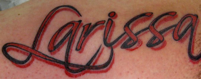
Are you still wondering whether Ayko, who you read about in the beginning of this chapter, actually got a tattoo of his new girlfriend’s name on his arm? Figure 10.8.1 is your answer! Let’s hope his love for Larissa — and for the artwork — lasts as long as his tattoo. According to a poll conducted for Global TV by Ipsos Reid in 2012, 10% of Canadian and 11% of American adults regret getting a tattoo. Although laser tattoo removal is available, it does not always work fully, can cause pain and scarring, and is expensive and time-consuming. Some people who regret a tattoo opt instead (or additionally) to cover it with another tattoo, see Figure 10.8.2 below.
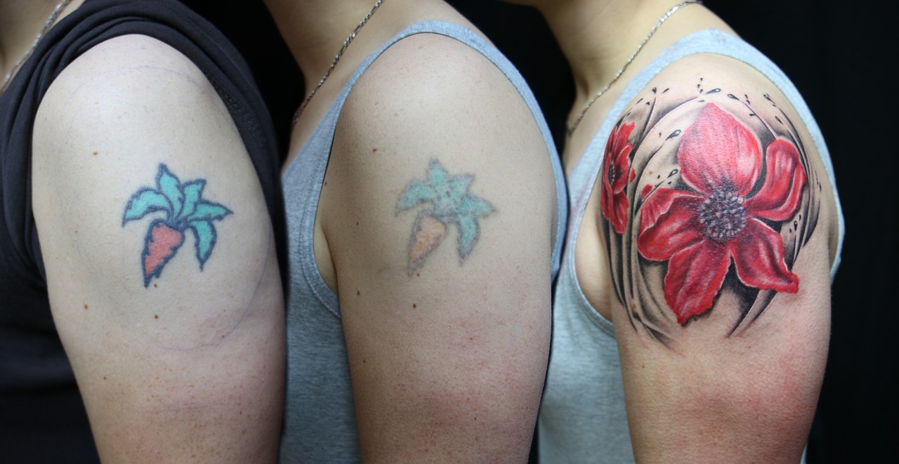
Why are tattoos essentially permanent? Tattoos are created by inserting a needle containing pigment through the epidermis and into the dermis of the skin. The pigment is injected into the dermal layer, creating the design. The pigment can remain in the dermal layer for a person’s lifetime for a few reasons. One, unlike the thinner outer epidermal layer, the dermis is not continually shed and replaced, so the pigment generally stays put. Two, the pigments used in tattooing mainly consist of large particles. When you get a tattoo, the penetration of the skin and insertion of foreign particles causes an immune response in which white blood cells attempt to engulf and remove the pigment. Because most of the pigment particles are so large, however, they cannot be removed from the dermis by the immune cells, and the design remains.
In laser tattoo removal, pulses from a high-intensity laser are applied to the tattoo and absorbed by the pigments. This breaks up the large pigment particles into particles that are small enough to be removed by the immune system. The pigments may then be excreted out of the body, or moved to other areas of the body, such as the lymph nodes. Different wavelengths of laser energy are often required to remove different colours of pigments, because they absorb different wavelengths of light. Generally, blue and black are the easiest colours to remove. Green, red, and yellow tend to be the hardest to remove. It may take as many as six to ten laser treatments — with a few weeks of recovery time in between — to remove a tattoo. Some tattoos can never be completely removed.
Why are mehndi designs (like Ayko’s trial “henna tattoo”) not permanent? Unlike real tattoos, henna paste is applied on the surface of the skin (shown below in Figure 10.8.3), and not injected into the skin with a needle. The dye molecules simply migrate from the paste into the top layer of the epidermis, the stratum corneum.
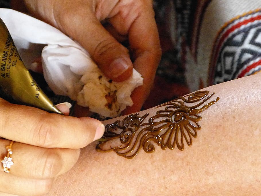
As you have learned, the stratum corneum consists of dead, keratin-filled keratinocytes, which are continually shed and replaced with new cells from the layers below. As a result, mehndi is not permanent. The design is lost as the cells that contain the dye are shed and replaced.
As you read in the beginning of this chapter, mehndi is often applied to the palms of the hands and soles of the feet, which generally results in a darker stain than other areas of the body. This is because the stratum corneum is thicker in these regions, so the dye penetrates through more layers of cells, making the design appear darker. What else is different about the epidermis of the palms and soles? You may recall that these regions are the only place where there is a fifth layer of epidermis — the stratum lucidum — making the skin in these areas even thicker and tougher.
Hopefully, Ayko thought carefully about the potential emotional and social implications of getting a tattoo — and learned how difficult they are to remove — before getting a real one. Health and safety should also be of utmost concern to anyone considering getting a tattoo. As you have learned in this chapter, the skin acts as a barrier against dangerous pathogens and substances. When you penetrate the skin using a needle, it can introduce harmful viruses and bacteria directly into the dermis, where the blood vessels are. Tattoo artists and shops need to take precautions to protect their clients against diseases that can be transmitted through blood (such as HIV and hepatitis), as well as bacterial infections. The tattoo artist should wear disposable gloves and a mask, use new and unopened needles and ink tubes, and properly sterilize other equipment. Even if the artist takes all the proper precautions, there is still a chance that the unopened ink could have been contaminated with pathogens during the production process. The shop should be aware of any ink recalls. Anyone getting a tattoo should make sure their artist and shop strictly adhere to all local health and safety regulations.
The risk of disease is not the only risk from tattoos. The pigments in tattoos may contain heavy metals and other potentially toxic substances. Tattoo parlours are regulated by provincial guidelines in Canada, and these guidelines vary from province to province — but these guidelines are mainly concerned with sterilization of equipment and don’t address anything about pigments. A recent study published in the scientific journal Nature (Scientific Reports) showed that pigments from tattoos may migrate from a person’s tattoos into their lymph nodes. Among the substances that make up the tattoo ink that migrated were aluminum, chromium, iron, nickel and copper – all considered “toxic”.
Additionally, people can sometimes have an allergic reaction to the pigments, or develop scarring or granulomas (small bumps of tissue due to an immune response) around the tattoo. Rarely, people can experience temporary swelling or burning of their tattoos when they get scanned in an MRI machine for a medical procedure. Clearly, people should think carefully about the potential health implications before getting a tattoo.
Fortunately, Ayko found a reputable and safe tattoo artist, and is not experiencing any ill effects from his tattoo. He is happy with his tattoo, at least for now. Tattoos — and other kinds of decoration of the integumentary system — are forms of artistic, personal, and cultural expression that have been used by many cultures over the course of human history. The system that protects us from the elements, helps us maintain homeostasis, and mediates our interactions with the outside world also happens to be easily modifiable! Whether it is a haircut, makeup, beard style, nail polish, piercing or a tattoo, humans have a variety of ways of altering our integumentary system, which changes our outward appearance and what we communicate to others.
Chapter 10 Summary
In this chapter, you learned about the structures and functions of the organs of the integumentary system. Specifically, you learned that:
- The integumentary system consists of the skin, hair, and nails. Functions of the integumentary system include providing a protective covering for the body, sensing the environment, and helping the body maintain homeostasis.
- The skin’s main functions include preventing water loss from the body, serving as a barrier to the entry of microorganisms, synthesizing vitamin D, blocking UV light, and helping to regulate body temperature.
- The skin consists of two distinct layers: a thinner outer layer called the epidermis, and a thicker inner layer called the dermis.
-
- The epidermis consists mainly of epithelial cells called keratinocytes, which produce keratin. New keratinocytes form at the bottom of the epidermis. They become filled with keratin and die as they move upward toward the surface of the skin, where they form a protective, waterproof layer.
- The dermis consists mainly of tough connective tissues that provide strength and stretch, as well as almost all skin structures, including blood vessels, sensory receptors, hair follicles, and oil and sweat glands.
- Cell types in the epidermis include keratinocytes (which make up 90 per cent of epidermal cells), melanocytes that produce melanin, Langerhans cells that fight pathogens in the skin, and Merkel cells that respond to light touch.
- In most parts of the body, the epidermis consists of four distinct layers. A fifth layer occurs only in the epidermis of the palms of the hands and soles of the feet.
-
- The innermost layer of the epidermis is the stratum basale, which contains stem cells that divide to form new keratinocytes.
- The next layer is the stratum spinosum, which is the thickest layer, and contains Langerhans cells and spiny keratinocytes.
- This is followed by the stratum granulosum, in which keratinocytes are filling with keratin and beginning to die.
- The stratum lucidum is next, but only on the palms and soles. It consists of translucent dead keratinocytes.
- The outermost layer is the stratum corneum, which consists of flat, dead, tightly packed keratinocytes that form a tough, waterproof barrier for the rest of the epidermis.
- The epidermis protects underlying tissues from physical damage and pathogens. Melanin in the epidermis absorbs and protects underlying tissues from UV light. The epidermis also prevents loss of water from the body and synthesizes vitamin D.
-
- Melanin is the main pigment that determines the colour of human skin. However, the pigments carotene and hemoglobin also contribute to skin colour, especially in skin with low levels of melanin.
- The surface of healthy skin normally is covered by vast numbers of bacteria representing about one thousand species from 19 phyla. Different areas of the body provide diverse habitats for skin microorganisms. Usually, microorganisms on the skin keep each other in check unless their balance is disturbed.
- The thicker inner layer of the skin — the dermis — has two layers. The upper papillary layer has papillae extending upward into the epidermis and loose connective tissues. The lower reticular layer has denser connective tissues and structures, such as glands and hair follicles. Glands in the dermis include eccrine and apocrine sweat glands, as well as sebaceous glands. Hair follicles are structures where hairs originate.
- Functions of the dermis include cushioning subcutaneous tissues, regulating body temperature, sensing the environment, and excreting wastes. The dense connective tissues of the dermis provide cushioning. The dermis regulates body temperature mainly by sweating and by vasodilation or vasoconstriction. The many tactile sensory receptors in the dermis make it the main organ for the sense of touch. Wastes excreted in sweat include excess water, electrolytes, and certain metabolic wastes.
- Hair is a filament that grows from a hair follicle in the dermis of the skin. It consists mainly of tightly packed, dead keratinocytes that are filled with keratin. The human body is almost completely covered with hair follicles.
- Hair helps prevent heat loss from the head and protects its skin from UV light. Hair in the nose filters incoming air, and the eyelashes and eyebrows keep harmful substances out of the eyes. Hair all over the body provides tactile sensory input. The eyebrows also play a role in nonverbal communication.
- The part of a hair that is within the follicle is the hair root. This is the only living part of a hair. The part of a hair that is visible above the skin surface is the hair shaft. It consists of dead cells.
-
- Hair growth begins inside a follicle when stem cells within the follicle divide to produce new keratinocytes.
- A hair shaft has three zones: the outermost zone called the cuticle, the middle zone called the cortex, and the innermost zone called the medulla.
- Genetically controlled, visible characteristics of hair include hair colour, hair texture, and the extent of balding in adult males. Melanin (eumelanin and/or pheomelanin) is the pigment that gives hair its colour. Aspects of hair texture include curl pattern, thickness, and consistency.
- Among mammals, humans are nearly unique in having undergone significant loss of body hair during their evolution, probably because sweat evaporates more quickly from less hairy skin. Curly hair also is thought to have evolved at some point during human evolution, perhaps because it provided better protection from UV light.
- Hair has social significance for human beings, being an indicator of biological sex, age, and ethnic ancestry. Human hair also has cultural significance. For example, hairstyle may be an indicator of social group membership.
- Nails consist of sheets of dead, keratin-filled keratinocytes. The keratin in nails makes them hard but flexible. They help protect the ends of the fingers and toes, enhance the sense of touch in the fingertips, and may be used as tools.
- A nail has three main parts: the nail root, which is under the epidermis; the nail plate, which is the visible part of the nail; and the free margin, which is the distal edge of the nail. Other structures under or around a nail include the nail bed, cuticle, and nail fold.
- A nail grows from a deep layer of living epidermal tissues, called the nail matrix, at the proximal end of the nail. Stem cells in the nail matrix keep dividing to allow nail growth, forming first the nail root and then the nail plate as the nail continues to grow longer and emerges from the epidermis.
- Fingernails grow faster than toenails. Actual rates of growth depend on many factors, such as age, sex, and season.
- The colour of the nail bed can be used to quickly assess oxygen and blood flow in a patient. How the nail plate grows out can reflect recent health problems, such as illness or nutrient deficiency. Nails — and especially toenails — are prone to fungus infections. Nails are more permeable than skin and can absorb several harmful substances, such as herbicides.
- Skin cancer is a disease in which skin cells grow out of control. It is caused mainly by excessive exposure to UV light, which damages DNA.
- There are three common types of skin cancer: basal cell carcinoma, squamous cell carcinoma, and melanoma. Carcinomas are more common and unlikely to metastasize. Melanoma is rare and likely to metastasize. It causes most skin cancer deaths.
- Besides exposure to UV light, risk factors for skin cancer include having light coloured skin, having many moles, and a family history of skin cancer, among several others.
Now that you have learned about the organs on surface of the body, read the next chapter to travel inside and learn about the skeletal system, which protects and supports us internally, among other functions.
Chapter 10 Review
- Describe one way in which the integumentary system works with another organ system to carry out a particular function.
-
- Describe two types of waterproofing used in the integumentary system. Include the types of molecules and where they are located.
- Explain why nails enhance touch sensations.
- Why do you think light coloured skin is a risk factor for skin cancer?
- Describe the similarities between how the epidermis, hair, and nails all grow.
- What does the whitish crescent-shaped area at the base of your nails (toward your hands) represent? What is its function?
- What is one difference between human hair and the hair of non-human primates?
- Describe the relationship between skin and hair.
- What kind of skin cancer is a cancer of a type of stem cell?
- For the skin and hair, describe one way in which they each protect the body against pathogens.
- If sweat glands are in the dermis, how is sweat released to the surface of the body?
- Explain why you think that physicians usually insist that patients remove any nail polish before having surgery.
- Describe generally how the brain gets touch information from the skin.
Attributions
Figure 10.8.1
Larissa Tattoo4039922685_46bf0bcfe5_c by Micael Faccio on Flickr is used under a CC BY 2.0 (https://creativecommons.org/licenses/by/2.0/) license.
Figure 10.8.2
Tattoo laser and cover-631211_1280 [photo] by Herco Roelofs on Pixabay is used under the Pixabay License (https://pixabay.com/ja/service/license/).
Figure 10.8.3
henna-tattoo-abu-dhabi by MarieFrance on Pixabay is used under the Pixabay License (https://pixabay.com/ja/service/license/).
References
Global News Staff. (2017, September 15). Health: ‘Toxic’ tattoo ink particles can travel to your lymph nodes: study. Globalnews.ca. https://globalnews.ca/news/3746925/tattoo-ink-safety-lymph-nodes/
Ipsos Reid. (2012). “Two in ten Canadians (22%), Americans (21%)
have a tattoo; One in ten tattooed Canadians (10%), Americans (11%) regret it” [News release]. Ipsos.com. https://www.ipsos.com/sites/default/files/publication/2012-01/5490.pdf
Rideout, K. (2010, July). Comparison of guidelines and regulatory frameworks for personal services establishments. National Collaborating Centre for Environmental Health. https://www.ncceh.ca/sites/default/files/PSE_Guidelines_Comparison_Table_July%202010.pdf
Schreiver, I., Hesse, B., Seim, C. et al. Synchrotron-based ν-XRF mapping and μ-FTIR microscopy enable to look into the fate and effects of tattoo pigments in human skin. Scientific Reports 7,11395. https://doi.org/10.1038/s41598-017-11721-z
The outer layer of skin that consists mainly of epithelial cells and lacks nerve endings, blood vessels, and other structures.
The inner layer of skin that is made of tough connective tissue and contains blood vessels, nerve endings, hair follicles, and glands.
A joint in which the opposing bony surfaces are covered with a layer of hyaline cartilage or fibrocartilage and in which some degree of free movement is possible.
Respiration using electron acceptors other than molecular oxygen. Although oxygen is not the final electron acceptor, the process still uses a respiratory electron transport chain.
Acquired Immunodeficiency Syndrome - a chronic, potentially life-threatening condition caused by the human immunodeficiency virus (HIV). By damaging your immune system, HIV interferes with your body's ability to fight infection and disease.
Mutations acquired by a cell that can be passed to future cells arising from the mutated cell in the course of cell division.
A microorganism which causes disease.
Created by: CK-12/Adapted by Christine Miller
Figure 4.2.1 Human cells viewed with a very powerful tool called a scanning electron microscope.
Amazing Cells
What are these incredible objects? Would it surprise you to learn that they are all human cells? Cells are actually too small to see with the unaided eye. It is visible here in such detail because it is being viewed with a very powerful tool called a scanning electron microscope. Cells may be small in size, but they are extremely important to life. Like all other living things, you are made of cells. Cells are the basis of life, and without cells, life as we know it would not exist. You will learn more about these amazing building blocks of life in this section.
What Are Cells?
If you look at living matter with a microscope — even a simple light microscope — you will see that it consists of cells. Cells are the basic units of the structure and function of living things. They are the smallest units that can carry out the processes of life. All organisms are made up of one or more cells, and all cells have many of the same structures and carry out the same basic life processes. Knowing the structure of cells and the processes they carry out is necessary to an understanding of life itself.
Discovery of Cells
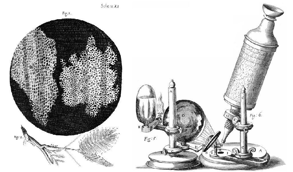
The first time the word cell was used to refer to these tiny units of life was in 1665 by a British scientist named Robert Hooke. Hooke was one of the earliest scientists to study living things under a microscope. The microscopes of his day were not very strong, but Hooke was still able to make an important discovery. When he looked at a thin slice of cork under his microscope, he was surprised to see what looked like a honeycomb. Hooke made the drawing in the figure to the right to show what he saw. As you can see, the cork was made up of many tiny units. Hooke called these units cells because they resembled cells in a monastery.
Soon after Robert Hooke discovered cells in cork, Anton van Leeuwenhoek in Holland made other important discoveries using a microscope. Leeuwenhoek made his own microscope lenses, and he was so good at it that his microscope was more powerful than other microscopes of his day. In fact, Leeuwenhoek’s microscope was almost as strong as modern light microscopes. Using his microscope, Leeuwenhoek was the first person to observe human cells and bacteria.
Cell Theory
By the early 1800s, scientists had observed cells of many different organisms. These observations led two German scientists named Theodor Schwann and Matthias Jakob Schleiden to propose cells as the basic building blocks of all living things. Around 1850, a German doctor named Rudolf Virchow was studying cells under a microscope, when he happened to see them dividing and forming new cells. He realized that living cells produce new cells through division. Based on this realization, Virchow proposed that living cells arise only from other living cells.
The ideas of all three scientists — Schwann, Schleiden, and Virchow — led to cell theory, which is one of the fundamental theories unifying all of biology.
Cell theory states that:
- All organisms are made of one or more cells.
- All the life functions of organisms occur within cells.
- All cells come from existing cells.
Seeing Inside Cells
Starting with Robert Hooke in the 1600s, the microscope opened up an amazing new world — a world of life at the level of the cell. As microscopes continued to improve, more discoveries were made about the cells of living things, but by the late 1800s, light microscopes had reached their limit. Objects much smaller than cells (including the structures inside cells) were too small to be seen with even the strongest light microscope.
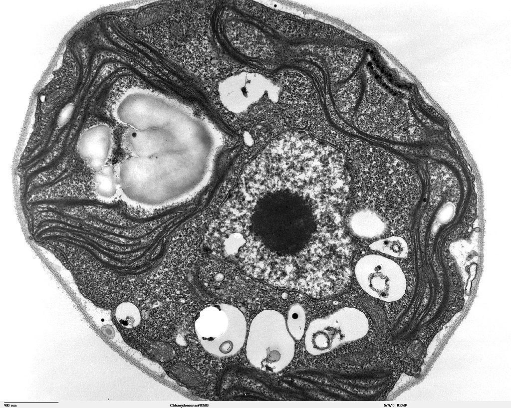
Then, in the 1950s, a new type of microscope was invented. Called the electron microscope, it used a beam of electrons instead of light to observe extremely small objects. With an electron microscope, scientists could finally see the tiny structures inside cells. They could even see individual molecules and atoms. The electron microscope had a huge impact on biology. It allowed scientists to study organisms at the level of their molecules, and it led to the emergence of the molecular biology field. With the electron microscope, many more cell discoveries were made.
Structures Shared By All Cells
Although cells are diverse, all cells have certain parts in common. These parts include a plasma membrane, cytoplasm, ribosomes, and DNA.
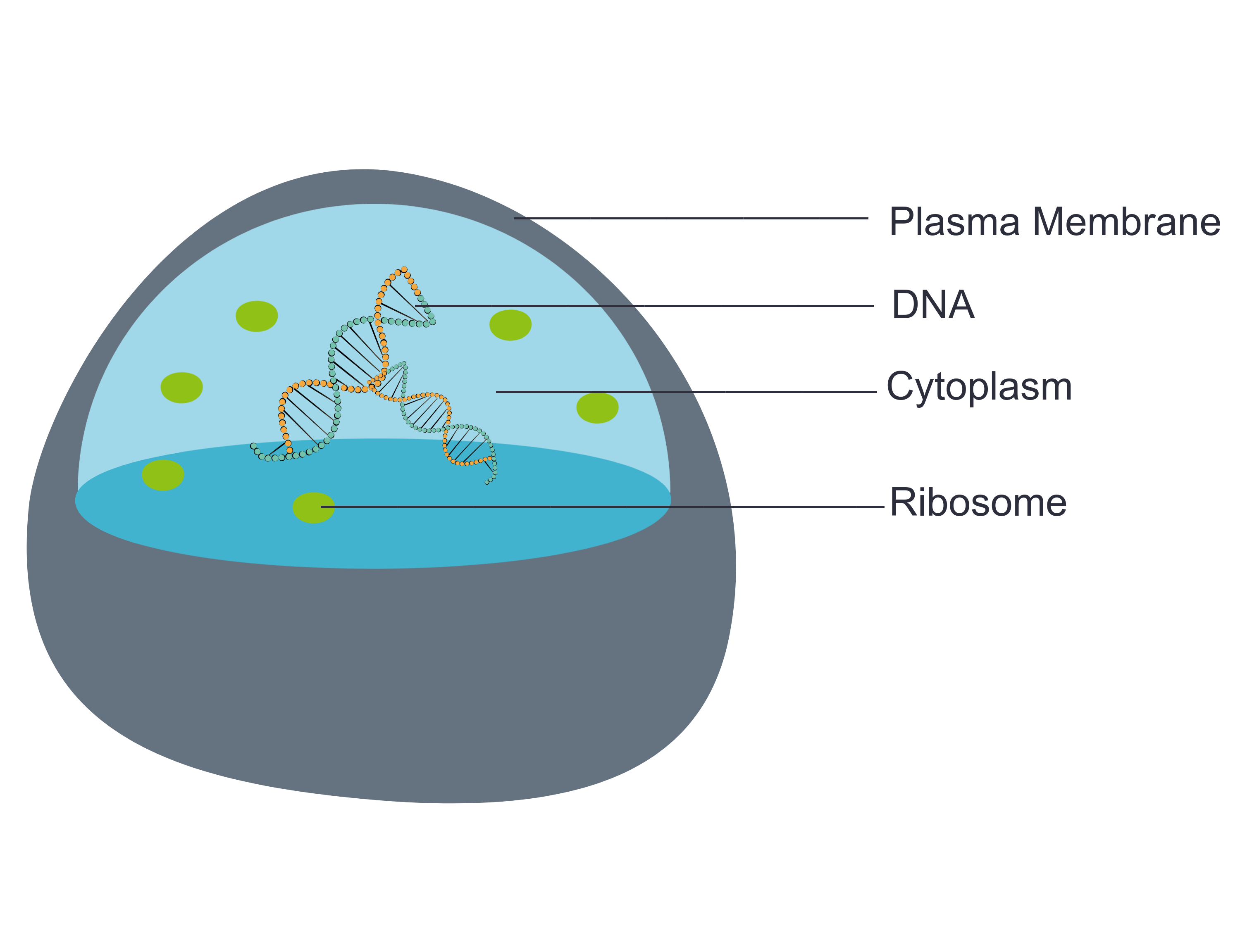
- The plasma membrane (a type of cell membrane) is a thin coat of lipids that surrounds a cell. It forms the physical boundary between the cell and its environment. You can think of it as the “skin” of the cell.
- Cytoplasm refers to all of the cellular material inside of the plasma membrane. Cytoplasm is made up of a watery substance called cytosol, and it contains other cell structures, such as ribosomes.
- Ribosomes are the structures in the cytoplasm in which proteins are made.
- DNA is a nucleic acid found in cells. It contains the genetic instructions that cells need to make proteins.
These four parts are common to all cells, from organisms as different as bacteria and human beings. How did all known organisms come to have such similar cells? The similarities show that all life on Earth has a common evolutionary history.
4.2 Summary
- Cells are the basic units of structure and function in living things. They are the smallest units that can carry out the processes of life.
- In the 1600s, Hooke was the first to observe cells from an organism (cork). Soon after, microscopist van Leeuwenhoek observed many other living cells.
- In the early 1800s, Schwann and Schleiden theorized that cells are the basic building blocks of all living things. Around 1850, Virchow observed cells dividing. To previous learnings, he added that living cells arise only from other living cells. These ideas led to cell theory, which states that all organisms are made of cells, that all life functions occur in cells, and that all cells come from other cells.
- It wasn't until the 1950s that scientists could see what was inside the cell. The invention of the electron microscope allowed them to see organelles and other structures smaller than cells.
- There is variation in cells, but all cells have a plasma membrane, cytoplasm, ribosomes, and DNA. These similarities show that all life on Earth has a common ancestor in the distant past.
4.2 Review Questions
- Describe cells.
- Explain how cells were discovered.
- Outline the development of cell theory.
-
- Identify the structures shared by all cells.
- Proteins are made on _____________ .
-
- Robert Hooke sketched what looked like honeycombs — or repeated circular or square units — when he observed plant cells under a microscope.
- What is each unit?
- Of the shared parts of all cells, what makes up the outer surface of each unit?
- Of the shared parts of all cells, what makes up the inside of each unit?
4.2 Explore More
https://www.youtube.com/watch?v=8IlzKri08kk
Introduction to Cells: The Grand Cell Tour, by The Amoeba Sisters, 2016.
Attributions
Figure 4.2.1
- A white blood cell (WBC) known as a neutrophil by National Institute of Allergy and Infectious Diseases (NIAID) on the CDC/ Public Health Image Library (PHIL) Photo ID# 18129. is in the public domain (https://en.wikipedia.org/wiki/Public_domain).
- Healthy Human T Cell by NIAID on Flickr. is used under a CC BY 2.0 (https://creativecommons.org/licenses/by/2.0/) license.
- Human natural killer cell by NIAID on Flickr. is used under a CC BY 2.0 (https://creativecommons.org/licenses/by/2.0/) license.
- Human blood with red blood cells, T cells (orange) and platelets (green) by ZEISS Microscopy on Flickr. is used under a CC BY-NC-ND 2.0 (https://creativecommons.org/licenses/by-nc-nd/2.0/) license.
- Developing nerve cells by ZEISS Microscopy on Flickr. is used under a CC BY-NC-ND 2.0 (https://creativecommons.org/licenses/by-nc-nd/2.0/) license.
Figure 4.2.2
Hooke-microscope-cork by Robert Hooke (1635-1702) [uploaded by Alejandro Porto] on Wikimedia Commons is released into the public domain (https://en.wikipedia.org/wiki/Public_domain).
Figure 4.2.3
Electron Microscope image of a cell by Dartmouth Electron Microscope Facility, Dartmouth College on Wikimedia Commons is released into the public domain (https://en.wikipedia.org/wiki/Public_domain).
Figure 4.2.4
Basic-Components-of-a-cell by Christine Miller is used under a CC0 1.0 (https://creativecommons.org/publicdomain/zero/1.0/) license.
References
Amoeba Sisters. (2016, November 1). Introduction to cells: The grand cell tour. YouTube. https://www.youtube.com/watch?v=8IlzKri08kk&feature=youtu.be
National Institute of Allergy and Infectious Diseases (NIAID). (2011). A white blood cell (WBC) known as a neutrophil, as it was in the process of ingesting a number of spheroid shaped, methicillin-resistant, Staphylococcus aureus (MRSA) bacteria [digital image]. CDC/ Public Health Image Library (PHIL) Photo ID# 18129. https://phil.cdc.gov/Details.aspx?pid=18129.
Wikipedia contributors. (2020, June 24). Antonie van Leeuwenhoek. In Wikipedia. https://en.wikipedia.org/w/index.php?title=Antonie_van_Leeuwenhoek&oldid=964339564
Wikipedia contributors. (2020, May 25). Matthias Jakob Schleiden. In Wikipedia. https://en.wikipedia.org/w/index.php?title=Matthias_Jakob_Schleiden&oldid=958819219
Wikipedia contributors. (2020, June 4). Rudolf Virchow. In Wikipedia,. https://en.wikipedia.org/w/index.php?title=Rudolf_Virchow&oldid=960708716
Wikipedia contributors. (2020, May 16). Theodor Schwann. In Wikipedia. https://en.wikipedia.org/w/index.php?title=Theodor_Schwann&oldid=956919239
The ability of an organism to maintain constant internal conditions despite external changes.
visible part of a nail that is external to the skin
a colorless cell that circulates in the blood and body fluids and is involved in counteracting foreign substances and disease; a white (blood) cell. There are several types, all amoeboid cells with a nucleus, including lymphocytes, granulocytes, monocytes, and macrophages.
accessory organ of the skin made of sheets of dead keratinocytes at the distal ends of the fingers and toes
The space occurring between two or more membranes. In cell biology, it's most commonly described as the region between the inner membrane and the outer membrane of a mitochondrion or a chloroplast.
Created by CK-12 Foundation/Adapted by Christine Miller
Goose Bumps
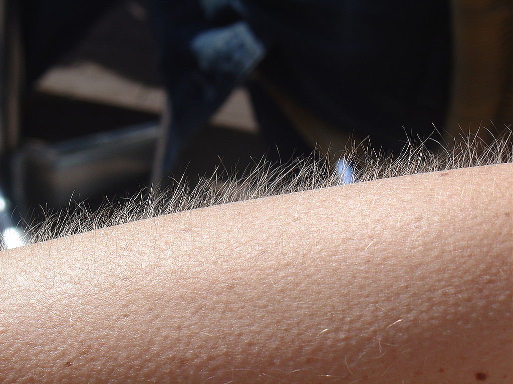
No doubt you’ve experienced the tiny, hair-raising skin bumps called goose bumps, like those you see in Figure 10.4.1. They happen when you feel chilly. Do you know what causes goose bumps, or why they pop up when you are cold? The answers to these questions involve the layer of skin known as the dermis.
What is the Dermis?
The dermis is the inner of the two major layers that make up the skin, the outer layer being the epidermis. The dermis consists mainly of connective tissues. It also contains most skin structures, such as glands and blood vessels. The dermis is anchored to the tissues below it by flexible collagen bundles that permit most areas of the skin to move freely over subcutaneous (“below the skin”) tissues. Functions of the dermis include cushioning subcutaneous tissues, regulating body temperature, sensing the environment, and excreting wastes.
Anatomy of the Dermis
The basic anatomy of the dermis is a matrix, or sort of scaffolding, composed of connective tissues. These tissues include collagen fibres — which provide toughness — and elastin fibres, which provide elasticity. Surrounding these fibres, the matrix also includes a gel-like substance made of proteins. The tissues of the matrix give the dermis both strength and flexibility.
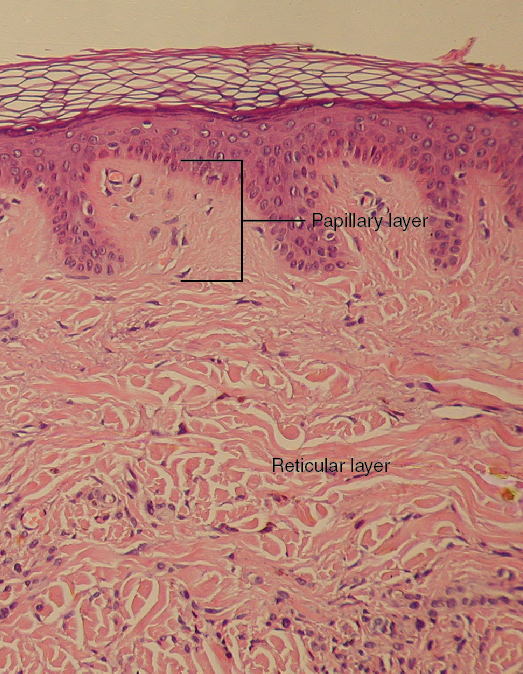
The dermis is divided into two layers: the papillary layer and the reticular layer. Both layers are shown in Figure 10.4.2 below and described in the text that follows.
Papillary Layer
The papillary layer is the upper layer of the dermis, just below the basement membrane that connects the dermis to the epidermis above it. The papillary layer is the thinner of the two dermal layers. It is composed mainly of loosely arranged collagen fibres. The papillary layer is named for its fingerlike projections — or papillae — that extend upward into the epidermis. The papillae contain capillaries and sensory touch receptors.
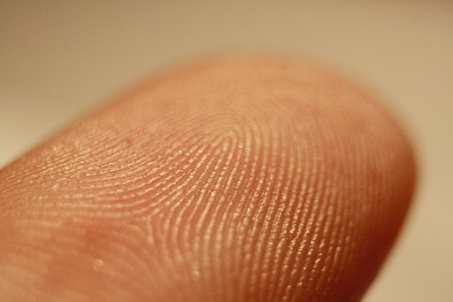
The papillae give the dermis a bumpy surface that interlocks with the epidermis above it, strengthening the connection between the two layers of skin. On the palms and soles, the papillae create epidermal ridges. Epidermal ridges on the fingers are commonly called fingerprints (see Figure 10.4.3). Fingerprints are genetically determined, so no two people (other than identical twins) have exactly the same fingerprint pattern. Therefore, fingerprints can be used as a means of identification, for example, at crime scenes. Fingerprints were much more commonly used forensically before DNA analysis was introduced for this purpose.
Reticular Layer
The reticular layer is the lower layer of the dermis, located below the papillary layer. It is the thicker of the two dermal layers. It is composed of densely woven collagen and elastin fibres. These protein fibres give the dermis its properties of strength and elasticity. This layer of the dermis cushions subcutaneous tissues of the body from stress and strain. The reticular layer of the dermis also contains most of the structures in the dermis, such as glands and hair follicles.
Structures in the Dermis
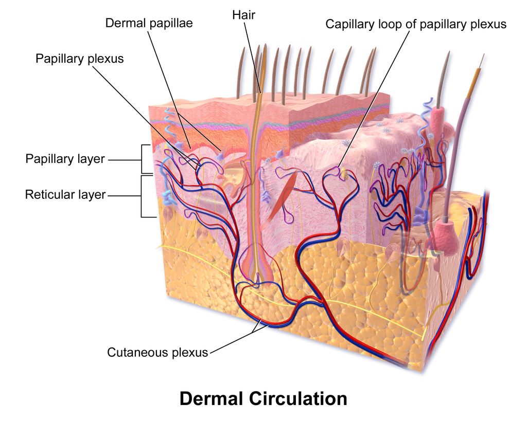
Both papillary and reticular layers of the dermis contain numerous sensory receptors, which make the skin the body’s primary sensory organ for the sense of touch. Both dermal layers also contain blood vessels. They provide nutrients to remove wastes from dermal cells, as well as cells in the lowest layer of the epidermis, the stratum basale. The circulatory components of the dermis are shown in Figure 10.4.4 below.
Glands
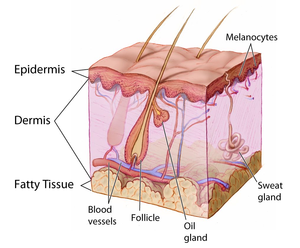
Glands in the reticular layer of the dermis include sweat glands and sebaceous (oil) glands. Both are exocrine glands, which are glands that release their secretions through ducts to nearby body surfaces. The diagram in Figure 10.4.5 shows these glands, as well as several other structures in the dermis.
Sweat Glands
Sweat glands produce the fluid called sweat, which contains mainly water and salts. The glands have ducts that carry the sweat to hair follicles, or to the surface of the skin. There are two different types of sweat glands: eccrine glands and apocrine glands.
- Eccrine sweat glands occur in skin all over the body. Their ducts empty through tiny openings called pores onto the skin surface. These sweat glands are involved in temperature regulation.
- Apocrine sweat glands are larger than eccrine glands, and occur only in the skin of the armpits and groin. The ducts of apocrine glands empty into hair follicles, and then the sweat travels along hairs to reach the surface. Apocrine glands are inactive until puberty, at which point they start producing an oily sweat that is consumed by bacteria living on the skin. The digestion of apocrine sweat by bacteria causes body odor.
Sebaceous Glands
Sebaceous glands are exocrine glands that produce a thick, fatty substance called sebum. Sebum is secreted into hair follicles and makes its way to the skin surface along hairs. It waterproofs the hair and skin, and helps prevent them from drying out. Sebum also has antibacterial properties, so it inhibits the growth of microorganisms on the skin. Sebaceous glands are found in every part of the skin — except for the palms of the hands and soles of the feet, where hair does not grow.
Hair Follicles
Hair follicles are the structures where hairs originate (see the diagram above). Hairs grow out of follicles, pass through the epidermis, and exit at the surface of the skin. Associated with each hair follicle is a sebaceous gland, which secretes sebum that coats and waterproofs the hair. Each follicle also has a bed of capillaries, a nerve ending, and a tiny muscle called an arrector pili.
Functions of the Dermis
The main functions of the dermis are regulating body temperature, enabling the sense of touch, and eliminating wastes from the body.
Temperature Regulation
Several structures in the reticular layer of the dermis are involved in regulating body temperature. For example, when body temperature rises, the hypothalamus of the brain sends nerve signals to sweat glands, causing them to release sweat. An adult can sweat up to four litres an hour. As the sweat evaporates from the surface of the body, it uses energy in the form of body heat, thus cooling the body. The hypothalamus also causes dilation of blood vessels in the dermis when body temperature rises. This allows more blood to flow through the skin, bringing body heat to the surface, where it can radiate into the environment.
When the body is too cool, sweat glands stop producing sweat, and blood vessels in the skin constrict, thus conserving body heat. The arrector pili muscles also contract, moving hair follicles and lifting hair shafts. This results in more air being trapped under the hairs to insulate the surface of the skin. These contractions of arrector pili muscles are the cause of goose bumps.
Sensing the Environment
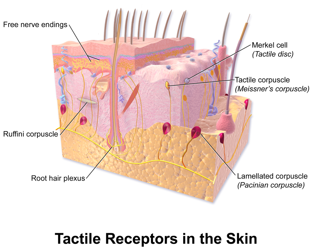
Sensory receptors in the dermis are mainly responsible for the body’s tactile senses. The receptors detect such tactile stimuli as warm or cold temperature, shape, texture, pressure, vibration, and pain. They send nerve impulses to the brain, which interprets and responds to the sensory information. Sensory receptors in the dermis can be classified on the basis of the type of touch stimulus they sense. Mechanoreceptors sense mechanical forces such as pressure, roughness, vibration, and stretching. Thermoreceptors sense variations in temperature that are above or below body temperature. Nociceptors sense painful stimuli. Figure 10.4.6 shows several specific kinds of tactile receptors in the dermis. Each kind of receptor senses one or more types of touch stimuli.
- Free nerve endings sense pain and temperature variations.
- Merkel cells sense light touch, shapes, and textures.
- Meissner’s corpuscles sense light touch.
- Pacinian corpuscles sense pressure and vibration.
- Ruffini corpuscles sense stretching and sustained pressure.
Excreting Wastes
The sweat released by eccrine sweat glands is one way the body excretes waste products. Sweat contains excess water, salts (electrolytes), and other waste products that the body must get rid of to maintain homeostasis. The most common electrolytes in sweat are sodium and chloride. Potassium, calcium, and magnesium electrolytes may be excreted in sweat, as well. When these electrolytes reach high levels in the blood, more are excreted in sweat. This helps to bring their blood levels back into balance. Besides electrolytes, sweat contains small amounts of waste products from metabolism, including ammonia and urea. Sweat may also contain alcohol in someone who has been drinking alcoholic beverages.
Feature: My Human Body
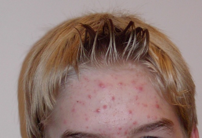
Acne is the most common skin disorder in the Canada. At least 20% of Canadians have acne at any given time and it affects approximately 90% of adolescents (as in Figure 10.4.7). Although acne occurs most commonly in teens and young adults, but it can occur at any age. Even newborn babies can get acne.
The main sign of acne is the appearance of pimples (pustules) on the skin, like those in the photo above. Other signs of acne may include whiteheads, blackheads, nodules, and other lesions. Besides the face, acne can appear on the back, chest, neck, shoulders, upper arms, and buttocks. Acne can permanently scar the skin, especially if it isn’t treated appropriately. Besides its physical effects on the skin, acne can also lead to low self-esteem and depression.
Acne is caused by clogged, sebum-filled pores that provide a perfect environment for the growth of bacteria. The bacteria cause infection, and the immune system responds with inflammation. Inflammation, in turn, causes swelling and redness, and may be associated with the formation of pus. If the inflammation goes deep into the skin, it may form an acne nodule.
Mild acne often responds well to treatment with over-the-counter (OTC) products containing benzoyl peroxide or salicylic acid. Treatment with these products may take a month or two to clear up the acne. Once the skin clears, treatment generally needs to continue for some time to prevent future breakouts.
If acne fails to respond to OTC products, nodules develop, or acne is affecting self-esteem, a visit to a dermatologist is in order. A dermatologist can determine which treatment is best for a given patient. A dermatologist can also prescribe prescription medications (which are likely to be more effective than OTC products) and provide other medical treatments, such as laser light therapies or chemical peels.
What can you do to maintain healthy skin and prevent or reduce acne? Dermatologists recommend the following tips:
- Wash affected or acne-prone skin (such as the face) twice a day, and after sweating.
- Use your fingertips to apply a gentle, non-abrasive cleanser. Avoid scrubbing, which can make acne worse.
- Use only alcohol-free products and avoid any products that irritate the skin, such as harsh astringents or exfoliants.
- Rinse with lukewarm water, and avoid using very hot or cold water.
- Shampoo your hair regularly.
- Do not pick, pop, or squeeze acne. If you do, it will take longer to heal and is more likely to scar.
- Keep your hands off your face. Avoid touching your skin throughout the day.
- Stay out of the sun and tanning beds. Some acne medications make your skin very sensitive to UV light.
10.4 Summary
- The dermis is the inner and thicker of the two major layers that make up the skin. It consists mainly of a matrix of connective tissues that provide strength and stretch. It also contains almost all skin structures, including sensory receptors and blood vessels.
- The dermis has two layers. The upper papillary layer has papillae extending upward into the epidermis and loose connective tissues. The lower reticular layer has denser connective tissues and structures, such as glands and hair follicles. Glands in the dermis include eccrine and apocrine sweat glands and sebaceous glands. Hair follicles are structures where hairs originate.
- Functions of the dermis include cushioning subcutaneous tissues, regulating body temperature, sensing the environment, and excreting wastes. The dense connective tissues of the dermis provide cushioning. The dermis regulates body temperature mainly by sweating and by vasodilation or vasoconstriction. The many tactile sensory receptors in the dermis make it the main organ for the sense of touch. Wastes excreted in sweat include excess water, electrolytes, and certain metabolic wastes.
10.4 Review Questions
- What is the dermis?
- Describe the basic anatomy of the dermis.
- Compare and contrast the papillary and reticular layers of the dermis.
- What causes epidermal ridges, and why can they be used to identify individuals?
- Name the two types of sweat glands in the dermis, and explain how they differ.
- What is the function of sebaceous glands?
- Describe the structures associated with hair follicles.
- Explain how the dermis helps regulate body temperature.
- Identify three specific kinds of tactile receptors in the dermis, along with the type of stimuli they sense.
- How does the dermis excrete wastes? What waste products does it excrete?
- What are subcutaneous tissues? Which layer of the dermis provides cushioning for subcutaneous tissues? Why does this layer provide most of the cushioning, instead of the other layer?
- For each of the functions listed below, describe which structure within the dermis carries it out.
- Brings nutrients to and removes wastes from dermal and lower epidermal cells
- Causes hairs to move
- Detects painful stimuli on the skin
10.4 Explore More
https://www.youtube.com/watch?v=FX-FwK0IIrE
How do you get rid of acne? SciShow, 2016.
https://www.youtube.com/watch?v=VcHQWMAClhQ&feature=emb_logo
When You Can't Scratch Away An Itch, Seeker, 2013.
Attributions
Figure 10.4.1
Goose_bumps by EverJean on Wikimedia Commons is used under a CC BY 2.0 (https://creativecommons.org/licenses/by/2.0) license.
Figure 10.4.2
Layers_of_the_Dermis by OpenStax College on Wikimedia Commons is used under a CC BY 3.0 (https://creativecommons.org/licenses/by/3.0) license.
Figure 10.4.3
Fingerprint_detail_on_male_finger_in_Třebíč,_Třebíč_District by Frettie on Wikimedia Commons is used under a CC BY 3.0 (https://creativecommons.org/licenses/by/3.0) license.
Figure 10.4.4
Blausen_0802_Skin_Dermal Circulation by BruceBlaus on Wikimedia commons is used under a CC BY 3.0 (https://creativecommons.org/licenses/by/3.0) license.
Figure 10.4.5
Anatomy_The_Skin_-_NCI_Visuals_Online by Don Bliss (artist) / National Cancer Institute (National Institutes of Health, with the ID 4604) is in the public domain (https://en.wikipedia.org/wiki/public_domain).
Figure 10.4.6
Blausen_0809_Skin_TactileReceptors by BruceBlaus on Wikimedia commons is used under a CC BY 3.0 (https://creativecommons.org/licenses/by/3.0) license.
Figure 10.4.7
Akne-jugend by Ellywa on Wikimedia Commons is released into the public domain (https://en.wikipedia.org/wiki/public_domain). (No machine-readable author provided. Ellywa assumed, based on copyright claims).
References
Betts, J. G., Young, K.A., Wise, J.A., Johnson, E., Poe, B., Kruse, D.H., Korol, O., Johnson, J.E., Womble, M., DeSaix, P. (2013, June 19). Figure 5.7 Layers of the dermis [digital image]. In Anatomy and Physiology (Section 5.1 Layers of the skin). OpenStax. https://openstax.org/books/anatomy-and-physiology/pages/5-1-layers-of-the-skin
Blausen.com staff. (2014). Medical gallery of Blausen Medical 2014. WikiJournal of Medicine 1 (2). DOI:10.15347/wjm/2014.010. ISSN 2002-4436.
SciShow. (2016, October 26). How do you get rid of acne? YouTube. https://www.youtube.com/watch?v=FX-FwK0IIrE
Seeker. (2013, October 26). When you can't scratch away an itch. YouTube. https://www.youtube.com/watch?v=VcHQWMAClhQ&feature=emb_logo
Created by CK-12 Foundation/Adapted by Christine Miller

Vampires
From Bram Stoker’s famous novel about Count Dracula, to films such as Van Helsing and the Twilight Saga, fantasies featuring vampires (like the one in Figure 14.5.1) have been popular for decades. Vampires, in fact, are found in centuries-old myths from many cultures. In such myths, vampires are generally described as creatures that drink blood — preferably of the human variety — for sustenance. Dracula, for example, is based on Eastern European folklore about a human who attains immortality (and eternal damnation) by drinking the blood of others.
What Is Blood?
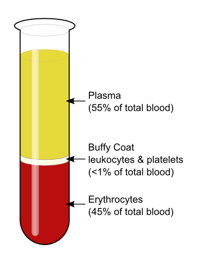
The average adult body contains between 4.7 and 5.7 litres of blood. More than half of that amount is fluid. Most of the rest of that amount consists of blood cells. The relative amounts of the various components in blood are illustrated in Figure 14.5.2. The components are also described in detail below.
Blood is a fluid connective tissue that circulates throughout the body through blood vessels of the cardiovascular system. What makes blood so special that it features in widespread myths? Although blood accounts for less than 10% of human body weight, it is quite literally the elixir of life. As blood travels through the vessels of the cardiovascular system, it delivers vital substances (such as nutrients and oxygen) to all of the cells, and carries away their metabolic wastes. It is no exaggeration to say that without blood, cells could not survive. Indeed, without the oxygen carried in blood, cells of the brain start to die within a matter of minutes.
Functions of Blood
Blood performs many important functions in the body. Major functions of blood include:
- Supplying tissues with oxygen, which is needed by all cells for aerobic cellular respiration.
- Supplying cells with nutrients, including glucose, amino acids, and fatty acids.
- Removing metabolic wastes from cells, including carbon dioxide, urea, and lactic acid.
- Helping to defend the body from pathogens and other foreign substances.
- Forming clots to seal broken blood vessels and stop bleeding.
- Transporting hormones and other messenger molecules.
- Regulating the pH of the body, which must be kept within a narrow range (7.35 to 7.45).
- Helping regulate body temperature (through vasoconstriction and vasodilation).
Blood Plasma
Plasma is the liquid component of human blood. It makes up about 55% of blood by volume. It is about 92% water, and contains many dissolved substances. Most of these substances are proteins, but plasma also contains trace amounts of glucose, mineral ions, hormones, carbon dioxide, and other substances. In addition, plasma contains blood cells. When the cells are removed from plasma, as in Figure 14.5.2 above, the remaining liquid is clear but yellow in colour.
Blood Cells
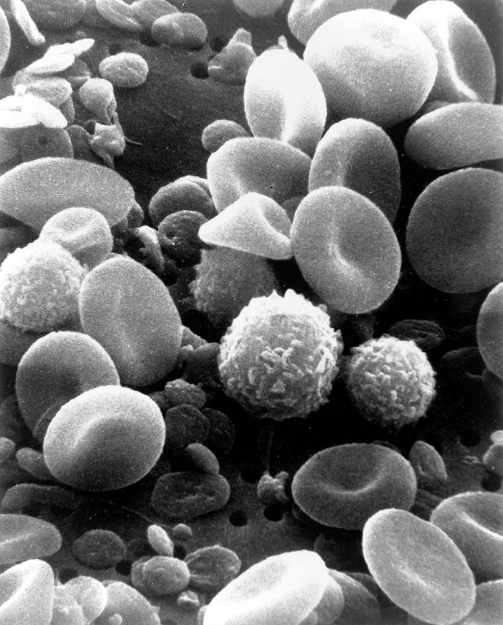
The cells in blood include erythrocytes, leukocytes, and thrombocytes. These different types of blood cells are shown in the photomicrograph (Figure 14.5.3) and described in the sections that follow.
Erythrocytes
The most numerous cells in blood are red blood cells, also called erythrocytes. One microlitre of blood contains between 4.2 and 6.1 million red blood cells, and red blood cells make up about 25% of all the cells in the human body. The cytoplasm of a mature erythrocyte is almost completely filled with hemoglobin, the iron-containing protein that binds with oxygen and gives the cell its red colour. In order to provide maximum space for hemoglobin, mature erythrocytes lack a cell nucleus and most organelles. They are little more than sacks of hemoglobin.
Erythrocytes also carry proteins called antigens that determine blood type. Blood type is a genetic characteristic. The best known human blood type systems are the ABO and Rhesus systems.
- In the ABO system, there are two common antigens, called antigen A and antigen B. There are four ABO blood types, A (only A antigen), B (only B antigen), AB (both A and B antigens), and O (neither A nor B antigen). The ABO antigens are illustrated in Figure 14.5.4.
- In the Rhesus system, there is just one common antigen. A person may either have the antigen (Rh+) or lack the antigen (Rh-).
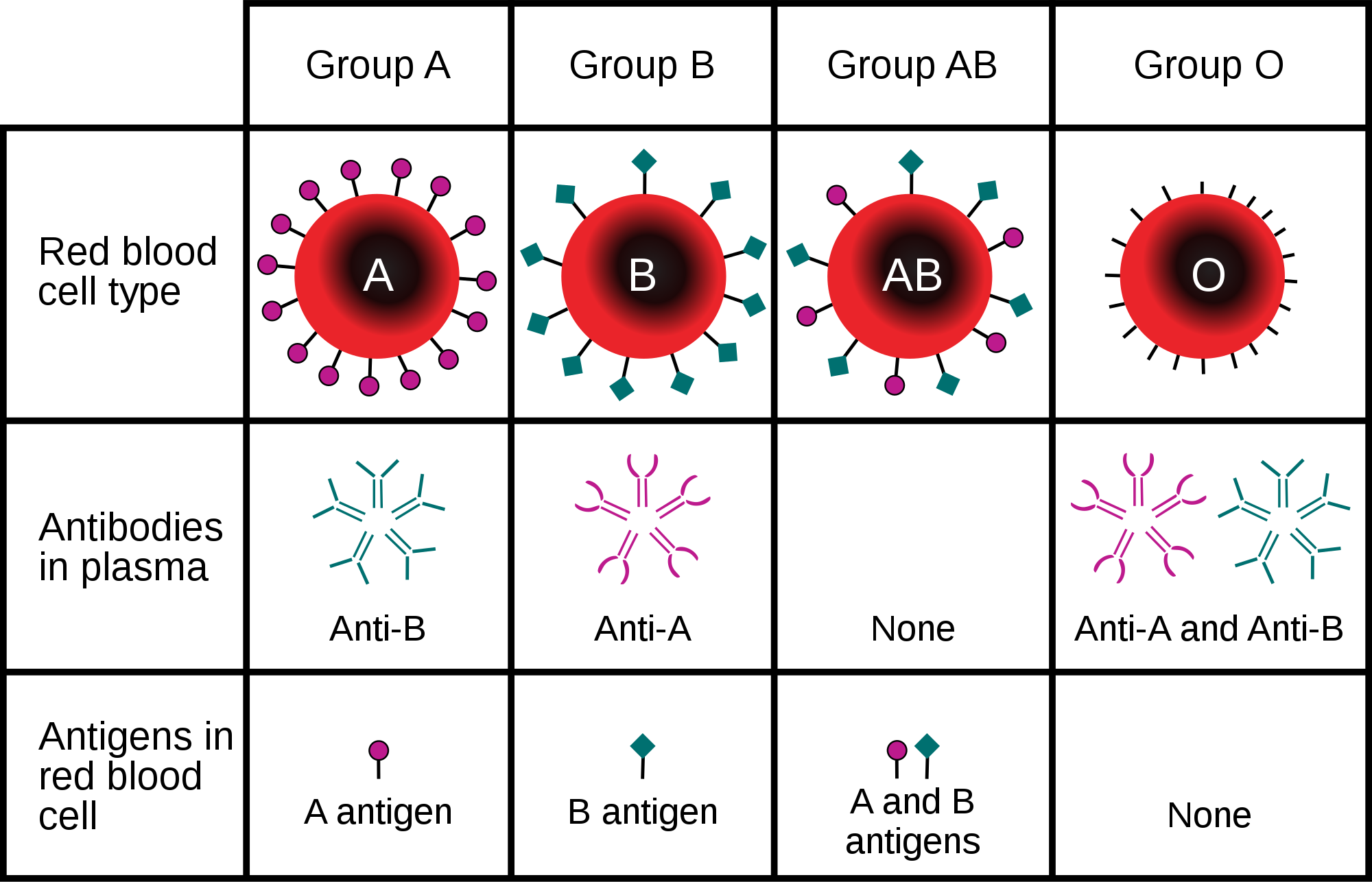
Blood type is important for medical reasons. A person who needs a blood transfusion must receive blood of a compatible type. Blood that is compatible lacks antigens that the patient's own blood also lacks. For example, for a person with type A blood (no B antigen), compatible types include any type of blood that lacks the B antigen. This would include type A blood or type O blood, but not type AB or type B blood. If incompatible blood is transfused, it may cause a potentially life-threatening reaction in the patient’s blood.
Leukocytes
Leukocytes (also called white blood cells) are cells in blood that defend the body against invading microorganisms and other threats. There are far fewer leukocytes than red blood cells in blood. There are normally only about 1,000 to 11,000 white blood cells per microlitre of blood. Unlike erythrocytes, leukocytes have a nucleus. White blood cells are part of the body’s immune system. They destroy and remove old or abnormal cells and cellular debris, as well as attack pathogens and foreign substances. There are five main types of white blood cells, which are described in Table 14.5.1: neutrophils, eosinophils, basophils, lymphocytes, and monocytes. The five types differ in their specific immune functions.
| Type of Leukocyte | Per cent of All Leukocytes | Main Function(s) |
|---|---|---|
| Neutrophil | 62% | Phagocytize (engulf and destroy) bacteria and fungi in blood. |
| Eosinophil | 2% | Attack and kill large parasites; carry out allergic responses. |
| Basophil | less than 1% | Release histamines in inflammatory responses. |
| Lymphocyte | 30% | Attack and destroy virus-infected and tumor cells; create lasting immunity to specific pathogens. |
| Monocyte | 5% | Phagocytize pathogens and debris in tissues. |
Thrombocytes
Thrombocytes, also called platelets, are actually cell fragments. Like erythrocytes, they lack a nucleus and are more numerous than white blood cells. There are about 150 thousand to 400 thousand thrombocytes per microlitre of blood.
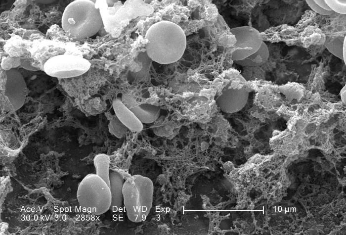
The main function of thrombocytes is blood clotting, or coagulation. This is the process by which blood changes from a liquid to a gel, forming a plug in a damaged blood vessel. If blood clotting is successful, it results in hemostasis, which is the cessation of blood loss from the damaged vessel. A blood clot consists of both platelets and proteins, especially the protein fibrin. You can see a scanning electron microscope photomicrograph of a blood clot in Figure 14.5.5.
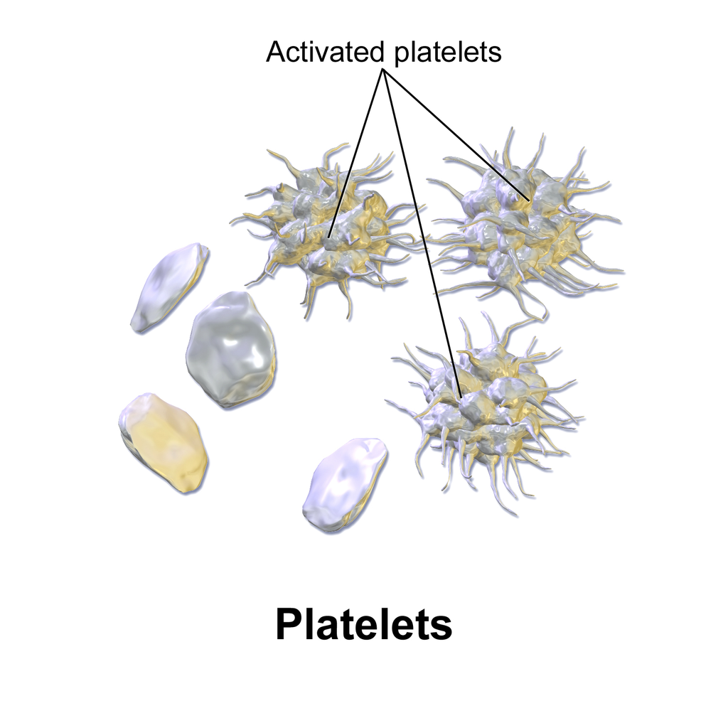
Coagulation begins almost instantly after an injury occurs to the endothelium of a blood vessel. Thrombocytes become activated and change their shape from spherical to star-shaped, as shown in Figure 14.5.6. This helps them aggregate with one another (stick together) at the site of injury to start forming a plug in the vessel wall. Activated thrombocytes also release substances into the blood that activate additional thrombocytes and start a sequence of reactions leading to fibrin formation. Strands of fibrin crisscross the platelet plug and strengthen it, much as rebar strengthens concrete.

Formation and Degradation of Blood Cells
Blood is considered a connective tissue, because blood cells form inside bones. All three types of blood cells are made in red marrow within the medullary cavity of bones in a process called hematopoiesis. Formation of blood cells occurs by the proliferation of stem cells in the marrow. These stem cells are self-renewing — when they divide, some of the daughter cells remain stem cells, so the pool of stem cells is not used up. Other daughter cells follow various pathways to differentiate into the variety of blood cell types. Once the cells have differentiated, they cannot divide to form copies of themselves.
Eventually, blood cells die and must be replaced through the formation of new blood cells from proliferating stem cells. After blood cells die, the dead cells are phagocytized (engulfed and destroyed) by white blood cells, and removed from the circulation. This process most often takes place in the spleen and liver.
Blood Disorders
Many human disorders primarily affect the blood. They include cancers, genetic disorders, poisoning by toxins, infections, and nutritional deficiencies.
- Leukemia is a group of cancers of the blood-forming tissues in the bone marrow. It is the most common type of cancer in children, although most cases occur in adults. Leukemia is generally characterized by large numbers of abnormal leukocytes. Symptoms may include excessive bleeding and bruising, fatigue, fever, and an increased risk of infections. Leukemia is thought to be caused by a combination of genetic and environmental factors.
- Hemophilia refers to any of several genetic disorders that cause dysfunction in the blood clotting process. People with hemophilia are prone to potentially uncontrollable bleeding, even with otherwise inconsequential injuries. They also commonly suffer bleeding into the spaces between joints, which can cause crippling.
- Carbon monoxide poisoning occurs when inhaled carbon monoxide (in fumes from a faulty home furnace or car exhaust, for example) binds irreversibly to the hemoglobin in erythrocytes. As a result, oxygen cannot bind to the red blood cells for transport throughout the body, and this can quickly lead to suffocation. Carbon monoxide is extremely dangerous, because it is colourless and odorless, so it cannot be detected in the air by human senses.
- HIV is a virus that infects certain types of leukocytes and interferes with the body’s ability to defend itself from pathogens and other causes of illness. HIV infection may eventually lead to AIDS (acquired immunodeficiency syndrome). AIDS is characterized by rare infections and cancers that people with a healthy immune system almost never acquire.
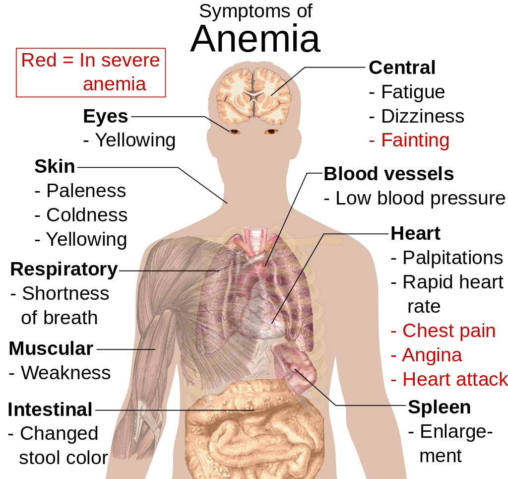
- Anemia is a disorder in which the blood has an inadequate volume of erythrocytes, reducing the amount of oxygen that the blood can carry, and potentially causing weakness and fatigue. These and other signs and symptoms of anemia are shown in Figure 14.5.8. Anemia has many possible causes, including excessive bleeding, inherited disorders (such as sickle cell hemoglobin), or nutritional deficiencies (iron, folate, or B12). Severe anemia may require transfusions of donated blood.
Feature: Myth vs. Reality
Donating blood saves lives. In fact, with each blood donation, as many as three lives may be saved. According to Government Canada, up to 52% of Canadians have reported that they or a family member have needed blood or blood products at some point in their lifetime. Many donors agree that the feeling that comes from knowing you have saved lives is well worth the short amount of time it takes to make a blood donation. Nonetheless, only a minority of potential donors actually donate blood. There are many myths about blood donation that may help explain the small percentage of donors. Knowing the facts may reaffirm your decision to donate if you are already a donor — and if you aren’t a donor already, getting the facts may help you decide to become one.
| Myth | Reality |
|---|---|
| "Your blood might become contaminated with an infection during the donation." | There is no risk of contamination because only single-use, disposable catheters, tubing, and other equipment are used to collect blood for a donation. |
| "You are too old (or too young) to donate blood." | There is no upper age limit on donating blood, as long as you are healthy. The minimum age is 16 years. |
| "You can’t donate blood if you have high blood pressure." | As long as your blood pressure is below 180/100 at the time of donation, you can give blood. Even if you take blood pressure medication to keep your blood pressure below this level, you can donate. |
| "You can’t give blood if you have high cholesterol." | Having high cholesterol does not affect your ability to donate blood. Taking cholesterol-lowering medication also does not disqualify you. |
| "You can’t donate blood if you have had a flu shot." | Having a flu shot has no effect on your ability to donate blood. You can even donate on the same day that you receive a flu shot. |
| "You can’t donate blood if you take medication." | As long as you are healthy, in most cases, taking medication does not preclude you from donating blood. |
| "Your blood isn’t needed if it’s a common blood type." | All types of blood are in constant demand. |
14.5 Summary
- Blood is a fluid connective tissue that circulates throughout the body in the cardiovascular system. Blood supplies tissues with oxygen and nutrients and removes their metabolic wastes. Blood helps defend the body from pathogens and other threats, transports hormones and other substances, and helps keep the body’s pH and temperature in homeostasis.
- Plasma is the liquid component of blood, and it makes up more than half of blood by volume. It consists of water and many dissolved substances. It also contains blood cells, including erythrocytes, leukocytes and thrombocytes.
- Erythrocytes, (also known as red blood cells) are the most numerous cells in blood. They consist mostly of hemoglobin, which carries oxygen. Erythrocytes also carry antigens that determine blood type.
- Leukocytes (also referred to as white blood cells) are less numerous than erythrocytes and are part of the body’s immune system. There are several different types of leukocytes that differ in their specific immune functions. They protect the body from abnormal cells, microorganisms, and other harmful substances.
- Thrombocytes (also called platelets) are cell fragments that play important roles in blood clotting, or coagulation. They stick together at breaks in blood vessels to form a clot and stimulate the production of fibrin, which strengthens the clot.
- All blood cells form by proliferation of stem cells in red bone marrow in a process called hematopoiesis. When blood cells die, they are phagocytized by leukocytes and removed from the circulation.
- Disorders of the blood include leukemia, which is cancer of the bone-forming cells; hemophilia, which is any of several genetic blood-clotting disorders; carbon monoxide poisoning, which prevents erythrocytes from binding with oxygen and causes suffocation; HIV infection, which destroys certain types of leukocytes and can cause AIDS; and anemia, in which there are not enough erythrocytes to carry adequate oxygen to body tissues.
14.5 Review Questions
- What is blood? Why is blood considered a connective tissue?
- Identify four physiological roles of blood in the body.
- Describe plasma and its components.
-
14.5 Explore More
https://youtu.be/e-5wqwp64MM
Joe Landolina: This gel can make you stop bleeding instantly, TED, 2014.
https://youtu.be/hgp8LtwFSBA
Can Synthetic Blood Help The World's Blood Shortage? Science Plus, 2016.
https://youtu.be/1Qfmkd6C8u8
How bones make blood - Melody Smith, TED-Ed, 2020.
Attributions
Figure 14.5.1
vampire_PNG32 from pngimg.com is used under a CC BY-NC 4.0 (https://creativecommons.org/licenses/by-nc/4.0/) license.
Figure 14.5.2
Blood-centrifugation-scheme by KnuteKnudsen at English Wikipedia on Wikimedia Commons is used under a CC BY 3.0 (https://creativecommons.org/licenses/by/3.0) license.
Figure 14.5.3
SEM_blood_cells by Bruce Wetzel and Harry Schaefer (Photographers)/ NCI AV-8202-3656 on Wikimedia Commons is in the public domain (https://en.wikipedia.org/wiki/en:Public_domain).
Figure 14.5.4
ABO_blood_type.svg by InvictaHOG on Wikimedia Commons is in the public domain (https://en.wikipedia.org/wiki/en:Public_domain).
Figure 14.5.5
Blood_clot_in_scanning_electron_microscopy by Janice Carr from CDC/ Public Health Image LIbrary (PHIL) ID #7308 on Wikimedia Commons is in the public domain (https://en.wikipedia.org/wiki/en:Public_domain).
Figure 14.5.6
Blausen_0740_Platelets by BruceBlaus on Wikimedia Commons is used under a CC BY 3.0 (https://creativecommons.org/licenses/by/3.0) license.
Figure 14.5.7
Platelet_Party_900x by Awkward Yeti (used with permission of the author) © All Rights Reserved
Figure 14.5.8
Symptoms_of_anemia.svg by Mikael Häggström on Wikimedia Commons is in the public domain (https://en.wikipedia.org/wiki/en:public_domain).
References
Blausen.com Staff. (2014). Medical gallery of Blausen Medical 2014. WikiJournal of Medicine 1 (2). DOI:10.15347/wjm/2014.010. ISSN 2002-4436.
Blood, organ and tissue donation. (2020, April 28). Government of Canada. https://www.canada.ca/en/public-health/services/healthy-living/blood-organ-tissue-donation.html#a3
Canadian Blood Services. (n.d.). There is an immediate need for blood as demand is rising. https://www.blood.ca
Science Plus. (2016, March 2). Can synthetic blood help the world's blood shortage? https://www.youtube.com/watch?v=hgp8LtwFSBA&feature=youtu.be
TED. (2014, November 20). Joe Landolina: This gel can make you stop bleeding instantly. YouTube. https://www.youtube.com/watch?v=e-5wqwp64MM&feature=youtu.be
TED-Ed. (2020, January 27). How bones make blood - Melody Smith. YouTube. https://www.youtube.com/watch?v=1Qfmkd6C8u8&feature=youtu.be
The movement of water or other solvent through a plasma membrane from a region of low solute concentration to a region of high solute concentration.
Long chains of hydrocarbons with a carboxyl group and a methyl group at opposite ends. Can be either saturated, containing mostly single bonds between adjacent carbons, or unsaturated, containing many double bonds between adjacent carbons.
Respiration using electron acceptors other than molecular oxygen. Although oxygen is not the final electron acceptor, the process still uses a respiratory electron transport chain.
A brown pigment produced by melanocytes in the skin that gives skin most of its color and prevents UV light from penetrating the skin.
A cardiac muscle cell. The cell is striated, containing thick and thin proteins arranged linearly. These filaments are composed, like other striated muscle cells, largely of actin and myosin. The cell has an abundant supply of mitochondria that supply the energy needed by the cell for regular muscular contraction.
A threadlike structure of nucleic acids and protein found in the nucleus of most living cells, carrying genetic information in the form of genes.
The process by which a parent cell divides into two or more daughter cells. Cell division usually occurs as part of a larger cell cycle.
a bone cell, formed when an osteoblast becomes embedded in the matrix it has secreted.
Mutations acquired by a cell that can be passed to future cells arising from the mutated cell in the course of cell division.
A pigment in the epidermis that gives skin a yellowish tint, especially in skin with low levels of melanin.
The way in which scientists and researchers use a systematic approach to answer questions about the world around us.
Created by CK-12 Foundation/Adapted by Christine Miller

Case Study: Flight Risk
Nineteen-year-old Malcolm is about to take his first plane flight. Shortly after he boards the plane and sits down, a man in his late sixties sits next to him in the aisle seat. About half an hour after the plane takes off, the pilot announces that she is turning the seat belt light off, and that it is safe to move around the cabin.
The man in the aisle seat — who has introduced himself to Malcolm as Willie — immediately unbuckles his seat belt and paces up and down the aisle a few times before returning to his seat. After about 45 minutes, Willie gets up again, walks some more, then sits back down and does some foot and leg exercises. After the third time Willie gets up and paces the aisles, Malcolm asks him whether he is walking so much to accumulate steps on a pedometer or fitness tracking device. Willie laughs and says no. He is actually trying to do something even more important for his health — prevent a blood clot from forming in his legs.
Willie explains that he has a chronic condition: heart failure. Although it sounds scary, his condition is currently well-managed, and he is able to lead a relatively normal lifestyle. However, it does put him at risk of developing other serious health conditions, such as deep vein thrombosis (DVT), which is when a blood clot occurs in the deep veins, usually in the legs. Air travel — and other situations where a person has to sit for a long period of time — increases the risk of DVT. Willie’s doctor said that he is healthy enough to fly, but that he should walk frequently and do leg exercises to help avoid a blood clot.
As you read this chapter, you will learn about the heart, blood vessels, and blood that make up the cardiovascular system, as well as disorders of the cardiovascular system, such as heart failure. At the end of the chapter you will learn more about why DVT occurs, why Willie has to take extra precautions when he flies, and what can be done to lower the risk of DVT and its potentially deadly consequences.
Chapter Overview: Cardiovascular System
In this chapter, you will learn about the cardiovascular system, which transports substances throughout the body. Specifically, you will learn about:
- The major components of the cardiovascular system: the heart, blood vessels, and blood.
- The functions of the cardiovascular system, including transporting needed substances (such as oxygen and nutrients) to the cells of the body, and picking up waste products.
- How blood is oxygenated through the pulmonary circulation, which transports blood between the heart and lungs.
- How blood is circulated throughout the body through the systemic circulation.
- The components of blood — including plasma, red blood cells, white blood cells, and platelets — and their specific functions.
- Types of blood vessels — including arteries, veins, and capillaries — and their functions, similarities, and differences.
- The structure of the heart, how it pumps blood, and how contractions of the heart are controlled.
- What blood pressure is and how it is regulated.
- Blood disorders, including anemia, HIV, and leukemia.
- Cardiovascular diseases (including heart attack, stroke, and angina), and the risk factors and precursors — such as high blood pressure and atherosclerosis — that contribute to them.
As you read the chapter, think about the following questions:
- What is heart failure?Why do you think it increases the risk of DVT?
- What is a blood clot? What are possible health consequences of blood clots?
- Why do you think sitting for long periods of time increases the risk of DVT? Why does walking and exercising the legs help reduce this risk?
Attribution
Figure 14.1.1
aircraft-1583871_1920 [photo] by olivier89 from Pixabay is used under the Pixabay License (https://pixabay.com/de/service/license/).
The upper layer of the dermis with papillae extending upward into the epidermis.
The lower layer of the dermis that gives the dermis strength and elasticity and contains many dermal structures such as glands and hair follicles.
An antibody, also known as an immunoglobulin, is a large, Y-shaped protein produced mainly by plasma cells that is used by the immune system to neutralize pathogens such as pathogenic bacteria and viruses.
A hormone is a signaling molecule produced by glands in multicellular organisms that target distant organs to regulate physiology and behavior.
A hormone is a signaling molecule produced by glands in multicellular organisms that target distant organs to regulate physiology and behavior.
Created by: CK-12/Adapted by Christine Miller

Bring on the S'mores!
This inviting camp fire can be used for both heat and light. Heat and light are two forms of energy that are released when a fuel like wood is burned. The cells of living things also get energy by "burning." They "burn" glucose in a process called cellular respiration.
What Is Cellular Respiration?
Cellular respiration is the process by which living cells break down glucose molecules and release energy. The process is similar to burning, although it doesn’t produce light or intense heat as a campfire does. This is because cellular respiration releases the energy in glucose slowly and in many small steps. It uses the energy released to form molecules of ATP, the energy-carrying molecules that cells use to power biochemical processes. In this way, cellular respiration is an example of energy coupling: glucose is broken down in an exothermic reaction, and then the energy from this reaction powers the endothermic reaction of the formation of ATP. Cellular respiration involves many chemical reactions, but they can all be summed up with this chemical equation:
C6H12O6 6O2 → 6CO2 6H2O Chemical Energy (in ATP)
In words, the equation shows that glucose (C6H12O6) and oxygen (O2) react to form carbon dioxide (CO2) and water (H2O), releasing energy in the process. Because oxygen is required for cellular respiration, it is an aerobic process.
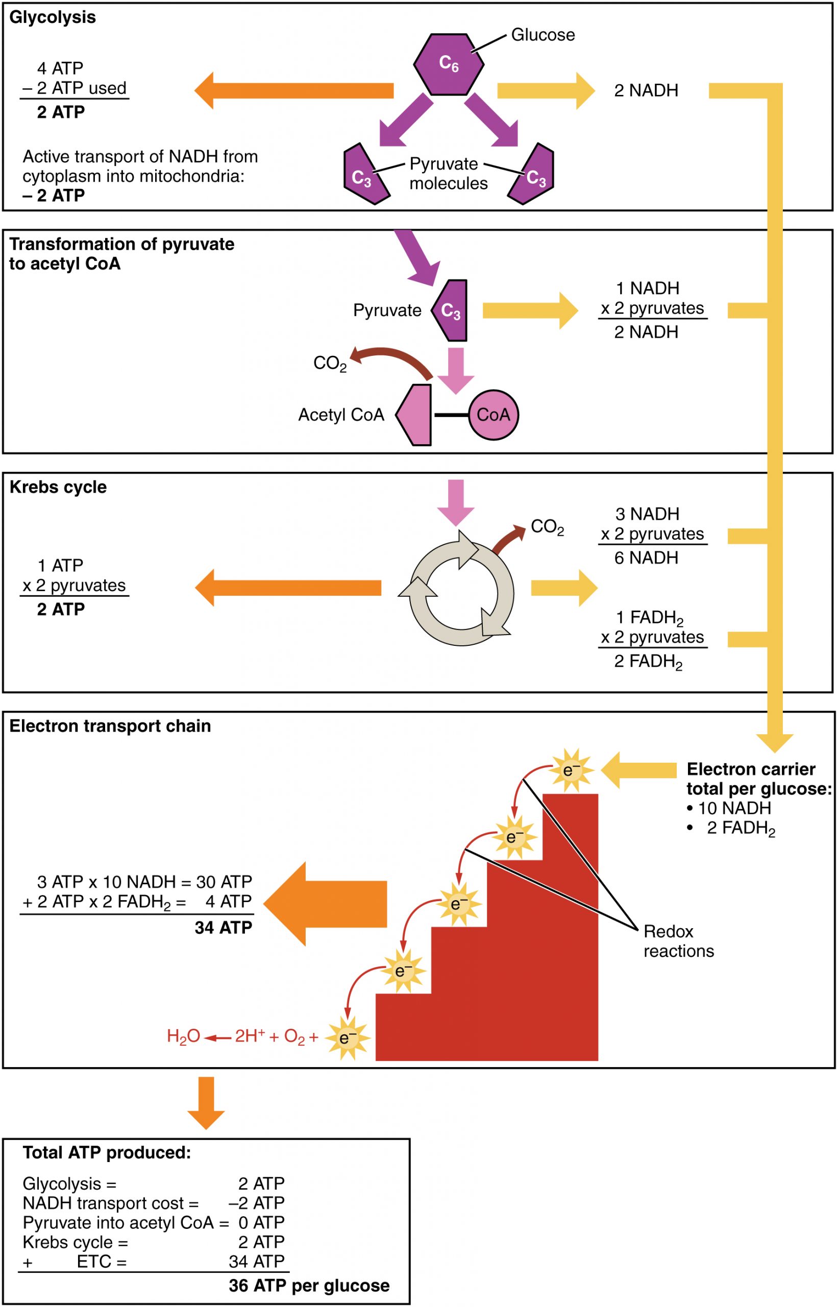
Cellular respiration occurs in the cells of all living things, both autotrophs and heterotrophs. All of them burn glucose to form ATP. The reactions of cellular respiration can be grouped into three stages: glycolysis, the Krebs cycle (also called the citric acid cycle), and electron transport. Figure 4.10.2 gives an overview of these three stages, which are also described in detail below.
Cellular Respiration Stage I: Glycolysis
The first stage of cellular respiration is glycolysis, which happens in the cytosol of the cytoplasm.
Splitting Glucose
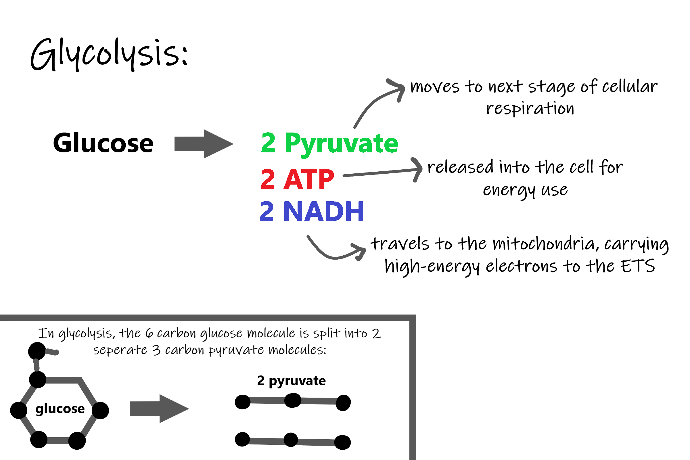
The word glycolysis literally means “glucose splitting,” which is exactly what happens in this stage. Enzymes split a molecule of glucose into two molecules of pyruvate (also known as pyruvic acid). This occurs in several steps, as summarized in the following diagram.
Results of Glycolysis
Energy is needed at the start of glycolysis to split the glucose molecule into two pyruvate molecules which go on to stage II of cellular respiration. The energy needed to split glucose is provided by two molecules of ATP; this is called the energy investment phase. As glycolysis proceeds, energy is released, and the energy is used to make four molecules of ATP; this is the energy harvesting phase. As a result, there is a net gain of two ATP molecules during glycolysis. During this stage, high-energy electrons are also transferred to molecules of NAD to produce two molecules of NADH, another energy-carrying molecule. NADH is used in stage III of cellular respiration to make more ATP.
Transition Reaction
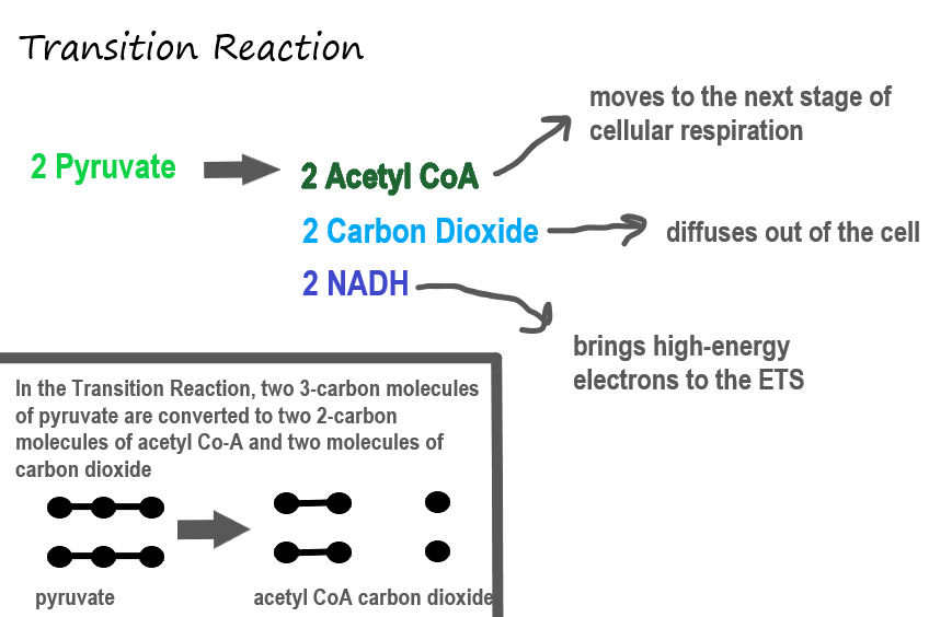
Before pyruvate can enter the next stage of cellular respiration it needs to be modified slightly. The transition reaction is a very short reaction which converts the two molecules of pyruvate to two molecules of acetyl CoA, carbon dioxide, and two high energy electron pairs convert NAD to NADH. The carbon dioxide is released, the acetyl CoA moves to the mitochondria to enter the Kreb's Cycle (stage II), and the NADH carries the high energy electrons to the Electron Transport System (stage III).
Structure of the Mitochondrion
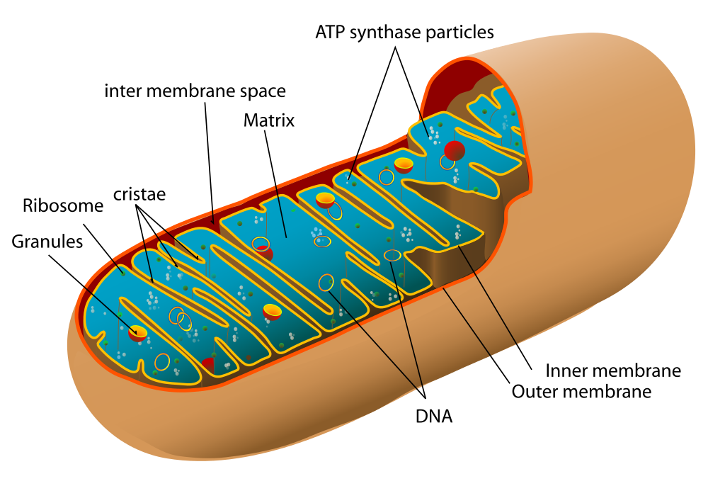
Before you read about the last two stages of cellular respiration, you need to know more about the mitochondrion, where these two stages take place. A diagram of a mitochondrion is shown in Figure 4.10.5.
The structure of a mitochondrion is defined by an inner and outer membrane. This structure plays an important role in aerobic respiration.
As you can see from the figure, a mitochondrion has an inner and outer membrane. The space between the inner and outer membrane is called the intermembrane space. The space enclosed by the inner membrane is called the matrix. The second stage of cellular respiration (the Krebs cycle) takes place in the matrix. The third stage (electron transport) happens on the inner membrane.
Cellular Respiration Stage II: The Krebs Cycle
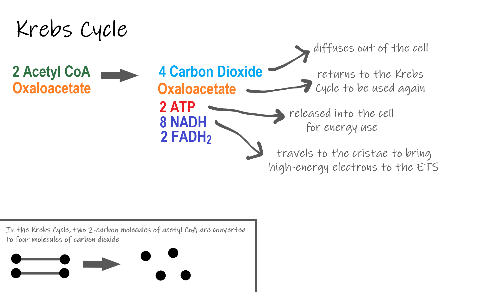
Recall that glycolysis produces two molecules of pyruvate (pyruvic acid), which are then converted to acetyl CoA during the short transition reaction. These molecules enter the matrix of a mitochondrion, where they start the Krebs cycle (also known as the Citric Acid Cycle). The reason this stage is considered a cycle is because a molecule called oxaloacetate is present at both the beginning and end of this reaction and is used to break down the two molecules of acetyl CoA. The reactions that occur next are shown in Figure 4.10.6.
Steps of the Krebs Cycle
The Krebs cycle itself actually begins when acetyl-CoA combines with a four-carbon molecule called OAA (oxaloacetate) (see Figure 4.10.6). This produces citric acid, which has six carbon atoms. This is why the Krebs cycle is also called the citric acid cycle.
After citric acid forms, it goes through a series of reactions that release energy. The energy is captured in molecules of NADH, ATP, and FADH2, another energy-carrying coenzyme. Carbon dioxide is also released as a waste product of these reactions.
The final step of the Krebs cycle regenerates OAA, the molecule that began the Krebs cycle. This molecule is needed for the next turn through the cycle. Two turns are needed because glycolysis produces two pyruvic acid molecules when it splits glucose.
Results of the Glycolysis, Transition Reaction and Krebs Cycle
After glycolysis, transition reaction, and the Krebs cycle, the glucose molecule has been broken down completely. All six of its carbon atoms have combined with oxygen to form carbon dioxide. The energy from its chemical bonds has been stored in a total of 16 energy-carrier molecules. These molecules are:
- 4 ATP (2 from glycolysis, 2 from Krebs Cycle)
- 12 NADH (2 from glycolysis, 2 from transition reaction, and 8 from Krebs cycle)
- 2 FADH2 (both from the Krebs cycle)
The events of cellular respiration up to this point are exergonic reactions- they are releasing energy that had been stored in the bonds of the glucose molecule. This energy will be transferred to the third and final stage of cellular respiration: the Electron Transport System, which is an endergonic reaction. Using an exothermic reaction to power an endothermic reaction is known as energy coupling.
Cellular Respiration Stage III: Electron Transport Chain
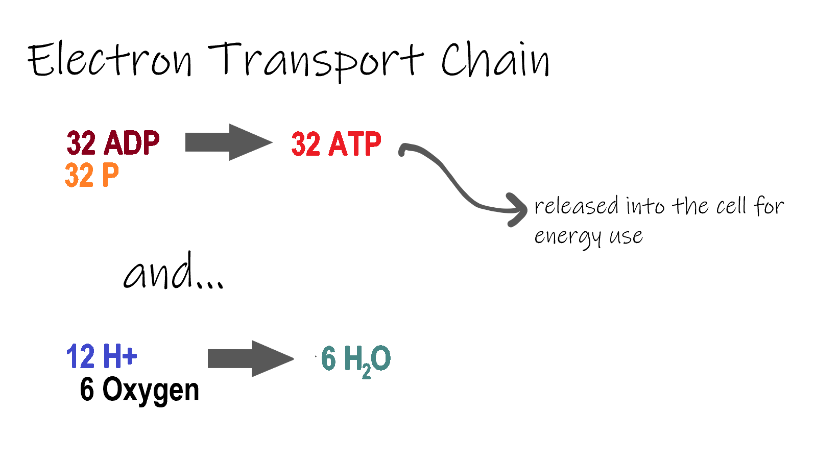
ETC, the final stage in cellular respiration produces 32 ATP. The Electron Transport Chain is the final stage of cellular respiration. In this stage, energy being transported by NADH and FADH2 is transferred to ATP. In addition, oxygen acts as the final proton acceptor for the hydrogens released from all the NADH and FADH2, forming water. Figure 4.10.8 shows the reactants and products of the ETC.
Transporting Electrons
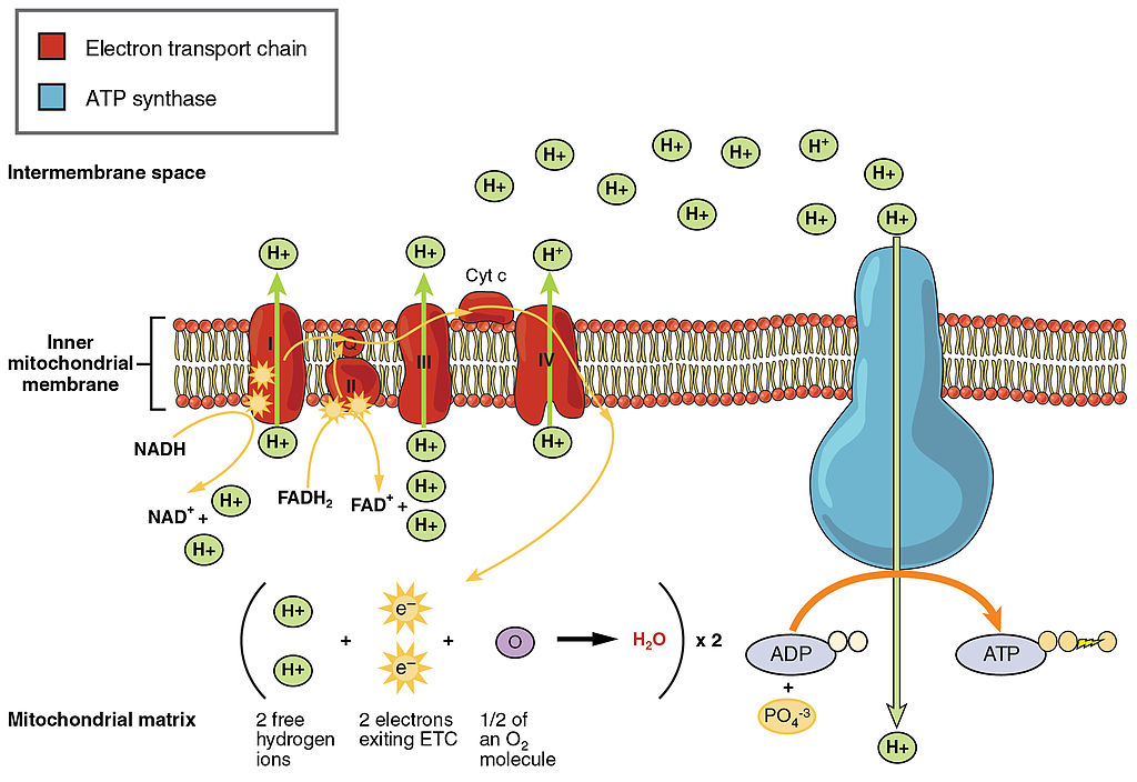
The Electron transport chain is the third stage of cellular respiration and is illustrated in Figure 4.10.8. During this stage, high-energy electrons are released from NADH and FADH2, and they move along electron-transport chains on the inner membrane of the mitochondrion. An electron-transport chain is a series of molecules that transfer electrons from molecule to molecule by chemical reactions. Some of the energy from the electrons is used to pump hydrogen ions (H ) across the inner membrane, from the matrix into the intermembrane space. This ion transfer creates an electrochemical gradient that drives the synthesis of ATP.
Making ATP
As shown in Figure 4.10.8, the pumping of hydrogen ions across the inner membrane creates a greater concentration of the ions in the intermembrane space than in the matrix. This gradient causes the ions to flow back across the membrane into the matrix, where their concentration is lower. ATP synthase acts as a channel protein, helping the hydrogen ions cross the membrane. It also acts as an enzyme, forming ATP from ADP and inorganic phosphate in a process called oxidative phosphorylation. After passing through the electron-transport chain, the “spent” electrons combine with oxygen to form water.
How Much ATP?
You have seen how the three stages of aerobic respiration use the energy in glucose to make ATP. How much ATP is produced in all three stages combined? Glycolysis produces two ATP molecules, and the Krebs cycle produces two more. Electron transport begins with several molecules of NADH and FADH2 from the Krebs cycle and transfers their energy into as many as 34 more ATP molecules. All told, then, up to 38 molecules of ATP can be produced from just one molecule of glucose in the process of cellular respiration.
4.10 Summary
- Cellular respiration is the aerobic process by which living cells break down glucose molecules, release energy, and form molecules of ATP. Generally speaking, this three-stage process involves glucose and oxygen reacting to form carbon dioxide and water.
- The first stage of cellular respiration, called glycolysis, takes place in the cytoplasm. In this step, enzymes split a molecule of glucose into two molecules of pyruvate, which releases energy that is transferred to ATP. Following glycolysis, a short reaction called the transition reaction converts the pyruvate into two molecules of acetyl CoA.
- The organelle called a mitochondrion is the site of the other two stages of cellular respiration. The mitochondrion has an inner and outer membrane separated by an intermembrane space, and the inner membrane encloses a space called the matrix.
- The second stage of cellular respiration, called the Krebs cycle, takes place in the matrix of a mitochondrion. During this stage, two turns through the cycle result in all of the carbon atoms from the two pyruvate molecules forming carbon dioxide and the energy from their chemical bonds being stored in a total of 16 energy-carrying molecules (including two from glycolysis and two from transition reaction).
- The third and final stage of cellular respiration, called electron transport, takes place on the inner membrane of the mitochondrion. Electrons are transported from molecule to molecule down an electron-transport chain. Some of the energy from the electrons is used to pump hydrogen ions across the membrane, creating an electrochemical gradient that drives the synthesis of many more molecules of ATP.
- In all three stages of cellular respiration combined, as many as 38 molecules of ATP are produced from just one molecule of glucose.
4.10 Review Questions
- What is the purpose of cellular respiration? Provide a concise summary of the process.
- State what happens during glycolysis.
- Describe the structure of a mitochondrion.
- What molecule is present at both the beginning and end of the Krebs cycle?
- What happens during the electron transport stage of cellular respiration?
- How many molecules of ATP can be produced from one molecule of glucose during all three stages of cellular respiration combined?
- Do plants undergo cellular respiration? Why or why not?
- Explain why the process of cellular respiration described in this section is considered aerobic.
- Name three energy-carrying molecules involved in cellular respiration.
-
- Which stage of aerobic cellular respiration produces the most ATP?
-
4.10 Explore More
https://www.youtube.com/watch?time_continue=2&v=00jbG_cfGuQ&feature=emb_logo
ATP & Respiration: Crash Course Biology #7, CrashCourse, 2012.
https://www.youtube.com/watch?v=4Eo7JtRA7lg&t=3s
Cellular Respiration and the Mighty Mitochondria, The Amoeba Sisters, 2014.
Attributions
Figure 4.10.1
Smores by Jessica Ruscello on Unsplash is used under the Unsplash License (https://unsplash.com/license).
Figure 4.10.2
Carbohydrate_Metabolism by OpenStax College on Wikimedia Commons is used under a CC BY 3.0 (https://creativecommons.org/licenses/by/3.0) license.
Figure 4.10.3
Glycolysis by Christine Miller is used under a CC BY 4.0 (https://creativecommons.org/licenses/by/4.0/) license.
Figure 4.10.4
Transition Reaction by Christine Miller is used under a CC BY 4.0 (https://creativecommons.org/licenses/by/4.0/) license.
Figure 4.10.5
Mitochondrion by Mariana Ruiz Villarreal [LadyofHats] on Wikimedia Commons is released into the public domain (https://en.wikipedia.org/wiki/Public_domain).
Figure 4.10.6
Krebs cycle by Christine Miller is used under a CC BY 4.0 (https://creativecommons.org/licenses/by/4.0/) license.
Figure 4.10.7
Electron Transport Chain (ETC) by Christine Miller is used under a CC BY 4.0 (https://creativecommons.org/licenses/by/4.0/) license.
Figure 4.10.8
The_Electron_Transport_Chain by OpenStax College on Wikimedia Commons is used under a CC BY 3.0 (https://creativecommons.org/licenses/by/3.0) license.
References
CrashCourse. (2012, March 12). ATP & Respiration: Crash Course Biology #7. YouTube. https://www.youtube.com/watch?time_continue=2&v=00jbG_cfGuQ&feature=emb_logo
Betts, J. G., Young, K.A., Wise, J.A., Johnson, E., Poe, B., Kruse, D.H., Korol, O., Johnson, J.E., Womble, M., DeSaix, P. (2013, April 25). Figure 24.8 Electron Transport Chain [digital image]. In Anatomy & Physiology, Connexions (Section ). OpenStax. https://openstax.org/books/anatomy-and-physiology/pages/24-2-carbohydrate-metabolism
Betts, J. G., Young, K.A., Wise, J.A., Johnson, E., Poe, B., Kruse, D.H., Korol, O., Johnson, J.E., Womble, M., DeSaix, P. (2013, April 25). Figure 24.9 Carbohydrate Metabolism [digital image]. In Anatomy & Physiology, Connexions (Section 24.2). OpenStax. https://openstax.org/books/anatomy-and-physiology/pages/24-2-carbohydrate-metabolism
The Amoeba Sisters. (2014, October 22). Cellular Respiration and the Mighty Mitochondria. YouTube. https://www.youtube.com/watch?v=4Eo7JtRA7lg&t=3s
The transfer of genetic variation from one population to another. If the rate of gene flow is high enough, then two populations are considered to have equivalent allele frequencies and therefore effectively be a single population.
The part of a hair that is located within the hair follicle and consists of living keratinocytes.
A part of a hair that is visible above the surface of the skin and consists of dead keratinocytes.
The outermost part of the hair shaft. It is formed from dead cells, overlapping in layers, which form scales that strengthen and protect the hair shaft.
Located between the hair cuticle and medulla and is the thickest hair layer. It also contains most of the hair's pigment, giving the hair its color. The pigment in the cortex is melanin, which is also found in skin.
The innermost layer of the hair shaft. This nearly invisible layer is the most soft and fragile, and serves as the pith or marrow of the hair.
The body system which acts as a chemical messenger system comprising feedback loops of the hormones released by internal glands of an organism directly into the circulatory system, regulating distant target organs. In humans, the major endocrine glands are the thyroid gland and the adrenal glands.
A division of the autonomic nervous system that controls digestive functions.
A metabolic process that produces chemical changes in organic substrates through the action of enzymes. In biochemistry, it is narrowly defined as the extraction of energy from carbohydrates in the absence of oxygen.
visible part of a nail that is external to the skin
A loop system wherein the system responds to a perturbation. The response may be in the same direction (as in positive feedback) or in the opposite direction (as in negative feedback). A feedback mechanism may be observed at the level of cells, organisms, ecosystems, or the biosphere.
A three-dimensional network of extracellular macromolecules, such as collagen, enzymes, and glycoproteins, that provide structural and biochemical support to surrounding cells.
The fusion of haploid gametes, egg and sperm, to form the diploid zygote.
A loop system wherein the system responds to a perturbation. The response may be in the same direction (as in positive feedback) or in the opposite direction (as in negative feedback). A feedback mechanism may be observed at the level of cells, organisms, ecosystems, or the biosphere.
A group of diseases involving abnormal cell growth with the potential to invade or spread to other parts of the body.
A genetic mutation caused by a deletion or insertion in a DNA sequence that shifts the way the sequence is read.
A mature haploid male or female germ cell which is able to unite with another of the opposite sex in sexual reproduction to form a zygote.
The process whereby a haploid cell (n) is formed from a diploid cell (2n) through meiosis and cell differentiation. Gametogenesis in the male is known as spermatogenesis and produces spermatozoa. Gametogenesis in the female is known as oogenesis and result in the formation of ova.

