10.2 Introduction to the Integumentary System

Art for All Eras
Pictured in Figure 10.2.1, is Maud Stevens Wagner, a tattoo artist from 1907. Tattoos are not just a late 20th and early 21st century trend. They have been popular in many eras and cultures. Tattoos literally illustrate the biggest organ of the human body: the skin. The skin is very thin, but it covers a large area — about 2 m2 in adults. The skin is the major organ in the integumentary system.
What Is the Integumentary System?
In addition to the skin, the integumentary system includes the hair and nails, which are organs that grow out of the skin. Because the organs of the integumentary system are mostly external to the body, you may think of them as little more than accessories, like clothing or jewelry, but they serve vital physiological functions. They provide a protective covering for the body, sense the environment, and help the body maintain homeostasis.
The Skin
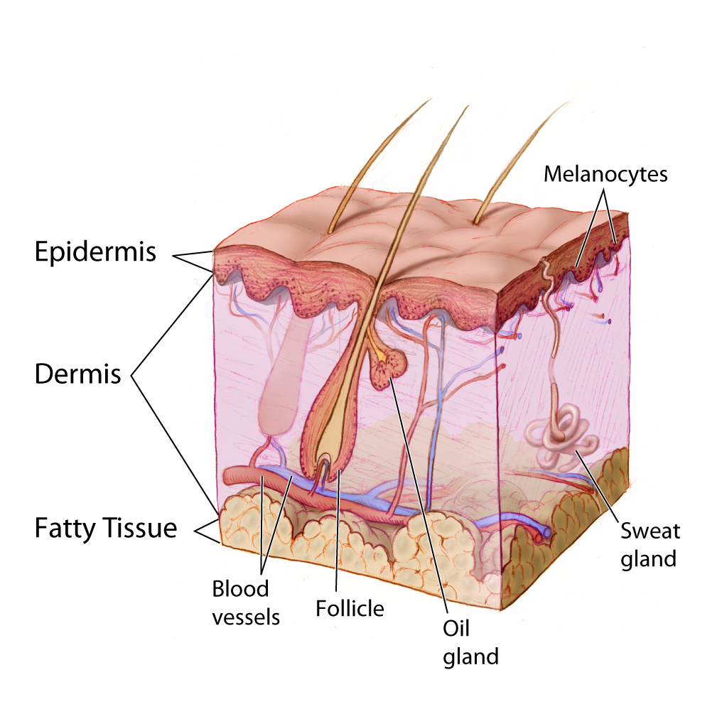
The skin is remarkable not only because it is the body’s largest organ: the average square inch of skin has 20 blood vessels, 650 sweat glands, and more than 1,000 nerve endings. Incredibly, it also has 60,000 pigment-producing cells. All of these structures are packed into a stack of cells that is just 2 mm thick. Although the skin is thin, it consists of two distinct layers: the epidermis and dermis, as shown in the diagram (Figure 10.2.2).
Outer Layer of Skin
The outer layer of skin is the epidermis. This layer is thinner than the inner layer (the dermis). The epidermis consists mainly of epithelial cells, called keratinocytes, which produce the tough, fibrous protein keratin. The innermost cells of the epidermis are stem cells that divide continuously to form new cells. The newly formed cells move up through the epidermis toward the skin surface, while producing more and more keratin. The cells become filled with keratin and die by the time they reach the surface, where they form a protective, waterproof layer. As the dead cells are shed from the surface of the skin, they are replaced by other cells that move up from below. The epidermis also contains melanocytes, the cells that produce the brown pigment melanin, which gives skin most of its colour. Although the epidermis contains some sensory receptor cells — called Merkel cells — it contains no nerves, blood vessels, or other structures.
Inner Layer of Skin
The dermis is the inner, thicker layer of skin. It consists mainly of tough connective tissue, and is attached to the epidermis by collagen fibres. The dermis contains many structures (as shown in Figure 10.2.2), including blood vessels, sweat glands, and hair follicles, which are structures where hairs originate. In addition, the dermis contains many sensory receptors, nerves, and oil glands.
Functions of the Skin
The skin has multiple roles in the body. Many of these roles are related to homeostasis. The skin’s main functions are preventing water loss from the body and serving as a barrier to the entry of microorganisms. Another function of the skin is synthesizing vitamin D, which occurs when the skin is exposed to ultraviolet (UV) light. Melanin in the epidermis blocks some of the UV light and protects the dermis from its damaging effects.
Another important function of the skin is helping to regulate body temperature. When the body is too warm, for example, the skin lowers body temperature by producing sweat, which cools the body when it evaporates. The skin also increases the amount of blood flowing near the body surface through vasodilation (widening of blood vessels), bringing heat from the body core to radiate out into the environment. The sweaty hair and flushed skin of the young man pictured in Figure 10.2.3 reflect these skin responses to overheating.

Hair
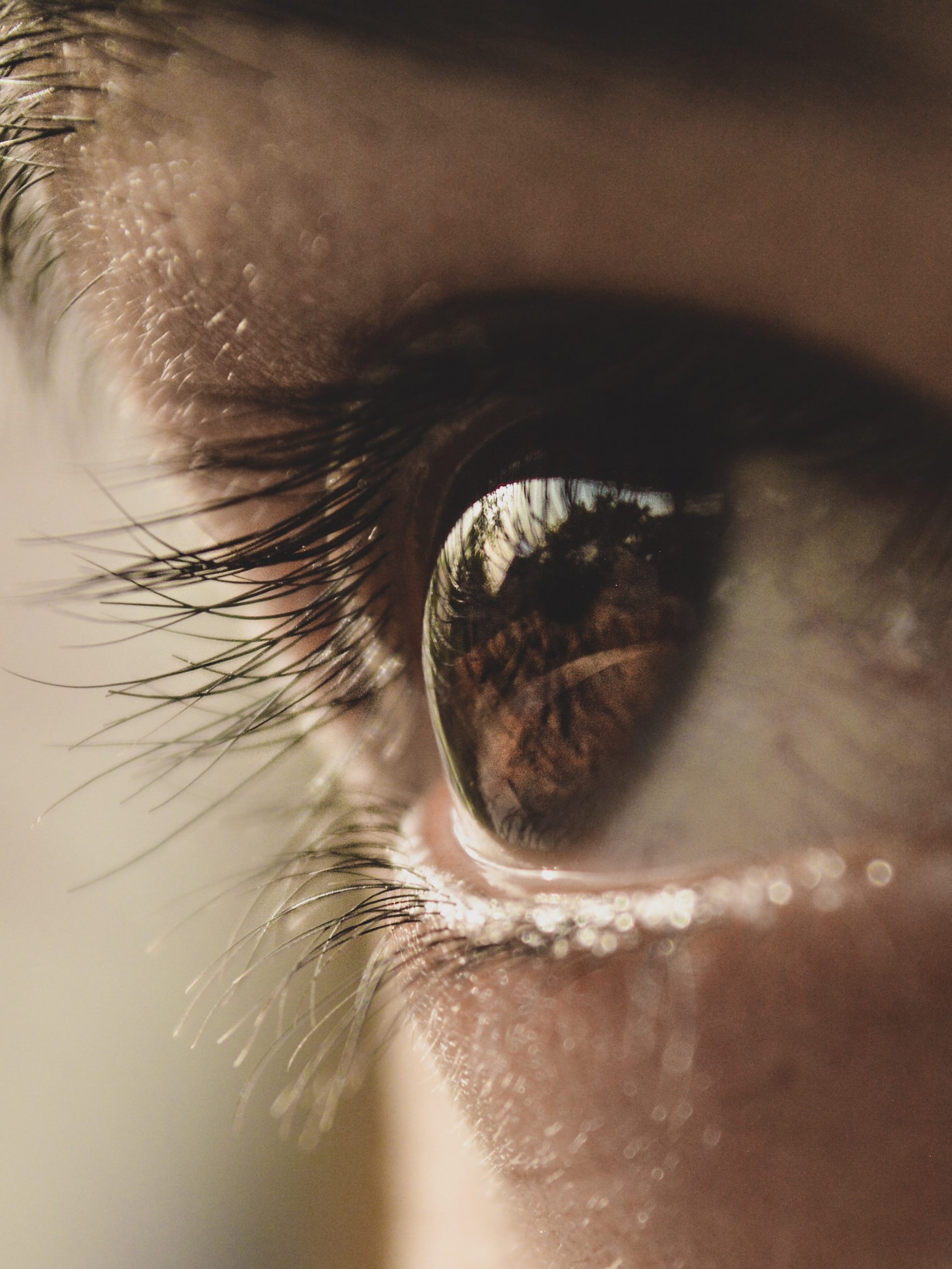
Hair is a fibre found only in mammals. It consists mainly of keratin-producing keratinocytes. Each hair grows out of a follicle in the dermis. By the time the hair reaches the surface, it consists mainly of dead cells filled with keratin. Hair serves several homeostatic functions. Head hair is important in preventing heat loss from the head and protecting its skin from UV radiation. Hairs in the nose trap dust particles and microorganisms in the air, and prevent them from reaching the lungs. Hair all over the body provides sensory input when objects brush against it, or when it sways in moving air. Eyelashes and eyebrows (see Figure 10.2.4) protect the eyes from water, dirt, and other irritants.
Nails
Fingernails and toenails consist of dead keratinocytes filled with keratin. The keratin makes them hard but flexible, which is important for the functions they serve. Nails prevent injury by forming protective plates over the ends of the fingers and toes. They also enhance sensation by acting as a counterforce to the sensitive fingertips when objects are handled. In addition, the fingernails can be used as tools.
Interactions with Other Organ Systems
The skin and other parts of the integumentary system work with other organ systems to maintain homeostasis.
- The skin works with the immune system to defend the body from pathogens by serving as a physical barrier to microorganisms.
- Vitamin D is needed by the digestive system to absorb calcium from food. By synthesizing vitamin D, the skin works with the digestive system to ensure that calcium can be absorbed.
- To control body temperature, the skin works with the cardiovascular system to either lose body heat, or to conserve it through vasodilation or vasoconstriction.
- To detect certain sensations from the outside world, the nervous system depends on nerve receptors in the skin.
10.2 Summary
- The integumentary system consists of the skin, hair, and nails. Functions of the integumentary system include providing a protective covering for the body, sensing the environment, and helping the body maintain homeostasis.
- The skin consists of two distinct layers: a thinner outer layer called the epidermis, and a thicker inner layer called the dermis.
- The epidermis consists mainly of epithelial cells called keratinocytes, which produce keratin. New keratinocytes form at the bottom of the epidermis. They become filled with keratin and die as they move upward toward the surface of the skin, where they form a protective, waterproof layer.
- The dermis consists mainly of tough connective tissues and many structures, including blood vessels, sensory receptors, nerves, hair follicles, and oil and sweat glands.
- The skin’s main functions are preventing water loss from the body, serving as a barrier to the entry of microorganisms, synthesizing vitamin D, blocking UV light, and helping to regulate body temperature.
- Hair consists mainly of dead keratinocytes and grows out of follicles in the dermis. Hair helps prevent heat loss from the head, and protects its skin from UV light. Hair in the nose filters incoming air, and the eyelashes and eyebrows keep harmful substances out of the eyes. Hair all over the body provides tactile sensory input.
- Like hair, nails also consist mainly of dead keratinocytes. They help protect the ends of the fingers and toes, enhance the sense of touch in the fingertips, and may be used as tools.
10.2 Review Questions
- Name the organs of the integumentary system.
- Compare and contrast the epidermis and dermis.
- Identify functions of the skin.
-
- What is the composition of hair?
- Describe three physiological roles played by hair.
- What do nails consist of?
- List two functions of nails.
- In terms of composition, what do the outermost surface of the skin, the nails, and hair have in common?
- Identify two types of cells found in the epidermis of the skin. Describe their functions.
- Which structure and layer of skin does hair grow out of?
- Identify three main functions of the integumentary system. Give an example of each.
- What are two ways in which the integumentary system protects the body against UV radiation?
10.2 Explore More
The science of skin – Emma Bryce, TED-Ed, 2018.
Why do we have to wear sunscreen? – Kevin P. Boyd, TED-Ed, 2013.
Scarification | National Geographic, 2008.
Attributions
Figure 10.2.1
Maud_Stevens_Wagner -The Plaza Gallery, Los Angeles, 1907 from the Library of Congress on Wikimedia Commons is in the public domain (https://en.wikipedia.org/wiki/public_domain).
Figure 10.2.2
Anatomy_The_Skin_-_NCI_Visuals_Online by Don Bliss (artist) from National Cancer Institute, on Wikimedia Commons is in the public domain (https://en.wikipedia.org/wiki/public_domain).
Figure 10.2.3
shashank-shekhar-Db1J_qp_ctc [photo] by Shashank Shekhar on Unsplash is used under the Unsplash License (https://unsplash.com/license).
Figure 10.2.4
Eyelashes by aryan-dhiman-93NBu0zG_H4 [photo] by Aryan Dhiman on Unsplash is used under the Unsplash License (https://unsplash.com/license).
Reference
National Geographic. (2008). Scarification | National Geographic. YouTube. https://www.youtube.com/watch?v=Lfhot7tQcWs&t=1s
TED-Ed. (2018, March 12). The science of skin – Emma Bryce. YouTube. https://www.youtube.com/watch?v=OxPlCkTKhzY&feature=youtu.be
TED-Ed. (2013, August 6). Why do we have to wear sunscreen? – Kevin P. Boyd. YouTube. https://www.youtube.com/watch?v=ZSJITdsTze0&feature=youtu.be
Created by: CK-12/Adapted by Christine Miller
Figure 4.2.1 Human cells viewed with a very powerful tool called a scanning electron microscope.
Amazing Cells
What are these incredible objects? Would it surprise you to learn that they are all human cells? Cells are actually too small to see with the unaided eye. It is visible here in such detail because it is being viewed with a very powerful tool called a scanning electron microscope. Cells may be small in size, but they are extremely important to life. Like all other living things, you are made of cells. Cells are the basis of life, and without cells, life as we know it would not exist. You will learn more about these amazing building blocks of life in this section.
What Are Cells?
If you look at living matter with a microscope — even a simple light microscope — you will see that it consists of cells. Cells are the basic units of the structure and function of living things. They are the smallest units that can carry out the processes of life. All organisms are made up of one or more cells, and all cells have many of the same structures and carry out the same basic life processes. Knowing the structure of cells and the processes they carry out is necessary to an understanding of life itself.
Discovery of Cells
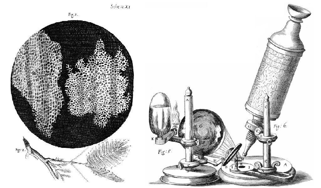
The first time the word cell was used to refer to these tiny units of life was in 1665 by a British scientist named Robert Hooke. Hooke was one of the earliest scientists to study living things under a microscope. The microscopes of his day were not very strong, but Hooke was still able to make an important discovery. When he looked at a thin slice of cork under his microscope, he was surprised to see what looked like a honeycomb. Hooke made the drawing in the figure to the right to show what he saw. As you can see, the cork was made up of many tiny units. Hooke called these units cells because they resembled cells in a monastery.
Soon after Robert Hooke discovered cells in cork, Anton van Leeuwenhoek in Holland made other important discoveries using a microscope. Leeuwenhoek made his own microscope lenses, and he was so good at it that his microscope was more powerful than other microscopes of his day. In fact, Leeuwenhoek’s microscope was almost as strong as modern light microscopes. Using his microscope, Leeuwenhoek was the first person to observe human cells and bacteria.
Cell Theory
By the early 1800s, scientists had observed cells of many different organisms. These observations led two German scientists named Theodor Schwann and Matthias Jakob Schleiden to propose cells as the basic building blocks of all living things. Around 1850, a German doctor named Rudolf Virchow was studying cells under a microscope, when he happened to see them dividing and forming new cells. He realized that living cells produce new cells through division. Based on this realization, Virchow proposed that living cells arise only from other living cells.
The ideas of all three scientists — Schwann, Schleiden, and Virchow — led to cell theory, which is one of the fundamental theories unifying all of biology.
Cell theory states that:
- All organisms are made of one or more cells.
- All the life functions of organisms occur within cells.
- All cells come from existing cells.
Seeing Inside Cells
Starting with Robert Hooke in the 1600s, the microscope opened up an amazing new world — a world of life at the level of the cell. As microscopes continued to improve, more discoveries were made about the cells of living things, but by the late 1800s, light microscopes had reached their limit. Objects much smaller than cells (including the structures inside cells) were too small to be seen with even the strongest light microscope.
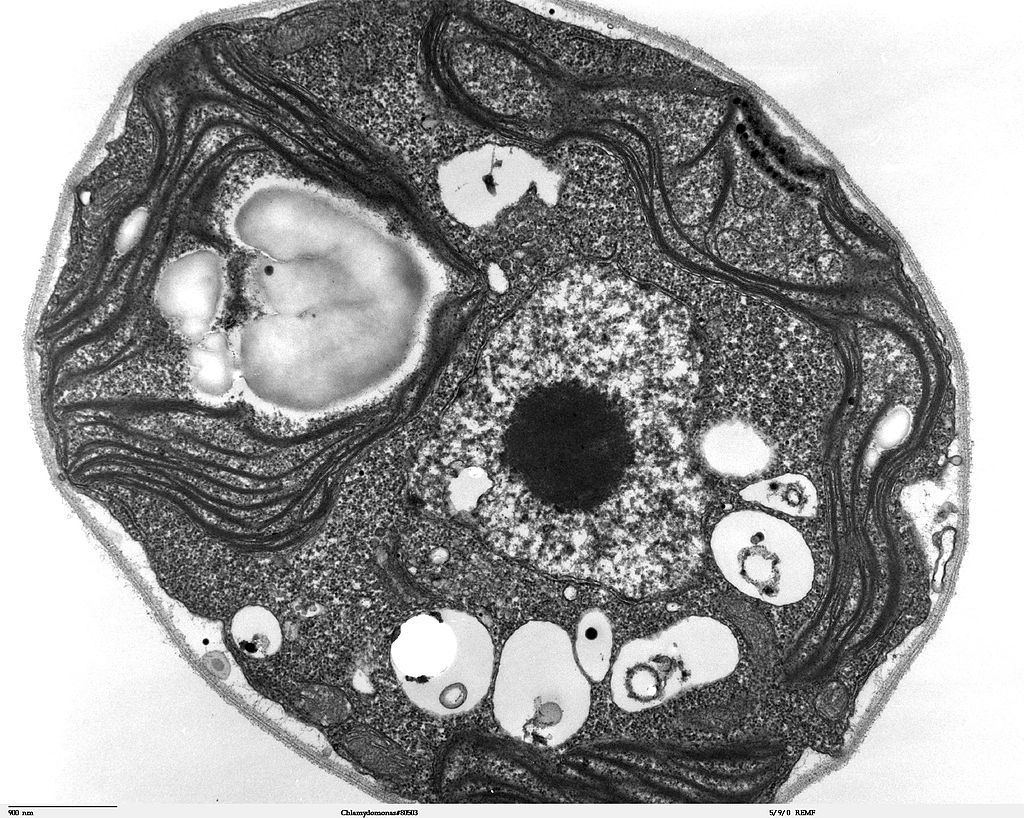
Then, in the 1950s, a new type of microscope was invented. Called the electron microscope, it used a beam of electrons instead of light to observe extremely small objects. With an electron microscope, scientists could finally see the tiny structures inside cells. They could even see individual molecules and atoms. The electron microscope had a huge impact on biology. It allowed scientists to study organisms at the level of their molecules, and it led to the emergence of the molecular biology field. With the electron microscope, many more cell discoveries were made.
Structures Shared By All Cells
Although cells are diverse, all cells have certain parts in common. These parts include a plasma membrane, cytoplasm, ribosomes, and DNA.
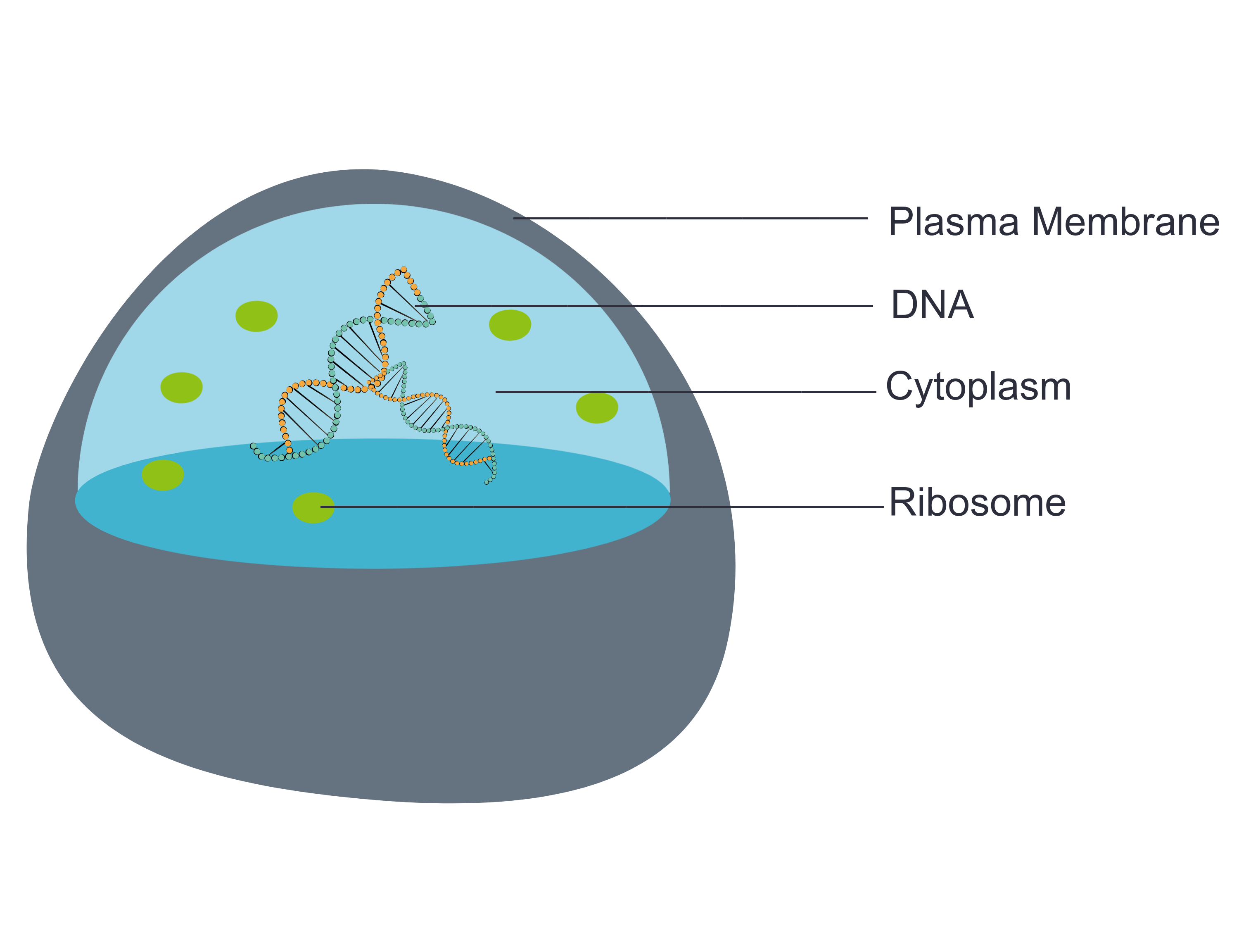
- The plasma membrane (a type of cell membrane) is a thin coat of lipids that surrounds a cell. It forms the physical boundary between the cell and its environment. You can think of it as the “skin” of the cell.
- Cytoplasm refers to all of the cellular material inside of the plasma membrane. Cytoplasm is made up of a watery substance called cytosol, and it contains other cell structures, such as ribosomes.
- Ribosomes are the structures in the cytoplasm in which proteins are made.
- DNA is a nucleic acid found in cells. It contains the genetic instructions that cells need to make proteins.
These four parts are common to all cells, from organisms as different as bacteria and human beings. How did all known organisms come to have such similar cells? The similarities show that all life on Earth has a common evolutionary history.
4.2 Summary
- Cells are the basic units of structure and function in living things. They are the smallest units that can carry out the processes of life.
- In the 1600s, Hooke was the first to observe cells from an organism (cork). Soon after, microscopist van Leeuwenhoek observed many other living cells.
- In the early 1800s, Schwann and Schleiden theorized that cells are the basic building blocks of all living things. Around 1850, Virchow observed cells dividing. To previous learnings, he added that living cells arise only from other living cells. These ideas led to cell theory, which states that all organisms are made of cells, that all life functions occur in cells, and that all cells come from other cells.
- It wasn't until the 1950s that scientists could see what was inside the cell. The invention of the electron microscope allowed them to see organelles and other structures smaller than cells.
- There is variation in cells, but all cells have a plasma membrane, cytoplasm, ribosomes, and DNA. These similarities show that all life on Earth has a common ancestor in the distant past.
4.2 Review Questions
- Describe cells.
- Explain how cells were discovered.
- Outline the development of cell theory.
-
- Identify the structures shared by all cells.
- Proteins are made on _____________ .
-
- Robert Hooke sketched what looked like honeycombs — or repeated circular or square units — when he observed plant cells under a microscope.
- What is each unit?
- Of the shared parts of all cells, what makes up the outer surface of each unit?
- Of the shared parts of all cells, what makes up the inside of each unit?
4.2 Explore More
https://www.youtube.com/watch?v=8IlzKri08kk
Introduction to Cells: The Grand Cell Tour, by The Amoeba Sisters, 2016.
Attributions
Figure 4.2.1
- A white blood cell (WBC) known as a neutrophil by National Institute of Allergy and Infectious Diseases (NIAID) on the CDC/ Public Health Image Library (PHIL) Photo ID# 18129. is in the public domain (https://en.wikipedia.org/wiki/Public_domain).
- Healthy Human T Cell by NIAID on Flickr. is used under a CC BY 2.0 (https://creativecommons.org/licenses/by/2.0/) license.
- Human natural killer cell by NIAID on Flickr. is used under a CC BY 2.0 (https://creativecommons.org/licenses/by/2.0/) license.
- Human blood with red blood cells, T cells (orange) and platelets (green) by ZEISS Microscopy on Flickr. is used under a CC BY-NC-ND 2.0 (https://creativecommons.org/licenses/by-nc-nd/2.0/) license.
- Developing nerve cells by ZEISS Microscopy on Flickr. is used under a CC BY-NC-ND 2.0 (https://creativecommons.org/licenses/by-nc-nd/2.0/) license.
Figure 4.2.2
Hooke-microscope-cork by Robert Hooke (1635-1702) [uploaded by Alejandro Porto] on Wikimedia Commons is released into the public domain (https://en.wikipedia.org/wiki/Public_domain).
Figure 4.2.3
Electron Microscope image of a cell by Dartmouth Electron Microscope Facility, Dartmouth College on Wikimedia Commons is released into the public domain (https://en.wikipedia.org/wiki/Public_domain).
Figure 4.2.4
Basic-Components-of-a-cell by Christine Miller is used under a CC0 1.0 (https://creativecommons.org/publicdomain/zero/1.0/) license.
References
Amoeba Sisters. (2016, November 1). Introduction to cells: The grand cell tour. YouTube. https://www.youtube.com/watch?v=8IlzKri08kk&feature=youtu.be
National Institute of Allergy and Infectious Diseases (NIAID). (2011). A white blood cell (WBC) known as a neutrophil, as it was in the process of ingesting a number of spheroid shaped, methicillin-resistant, Staphylococcus aureus (MRSA) bacteria [digital image]. CDC/ Public Health Image Library (PHIL) Photo ID# 18129. https://phil.cdc.gov/Details.aspx?pid=18129.
Wikipedia contributors. (2020, June 24). Antonie van Leeuwenhoek. In Wikipedia. https://en.wikipedia.org/w/index.php?title=Antonie_van_Leeuwenhoek&oldid=964339564
Wikipedia contributors. (2020, May 25). Matthias Jakob Schleiden. In Wikipedia. https://en.wikipedia.org/w/index.php?title=Matthias_Jakob_Schleiden&oldid=958819219
Wikipedia contributors. (2020, June 4). Rudolf Virchow. In Wikipedia,. https://en.wikipedia.org/w/index.php?title=Rudolf_Virchow&oldid=960708716
Wikipedia contributors. (2020, May 16). Theodor Schwann. In Wikipedia. https://en.wikipedia.org/w/index.php?title=Theodor_Schwann&oldid=956919239
The ability of an organism to maintain constant internal conditions despite external changes.
visible part of a nail that is external to the skin
The outer layer of skin that consists mainly of epithelial cells and lacks nerve endings, blood vessels, and other structures.
Acquired Immunodeficiency Syndrome - a chronic, potentially life-threatening condition caused by the human immunodeficiency virus (HIV). By damaging your immune system, HIV interferes with your body's ability to fight infection and disease.
Respiration using electron acceptors other than molecular oxygen. Although oxygen is not the final electron acceptor, the process still uses a respiratory electron transport chain.
Created by CK-12 Foundation/Adapted by Christine Miller
Case Study: Defending Your Defenses

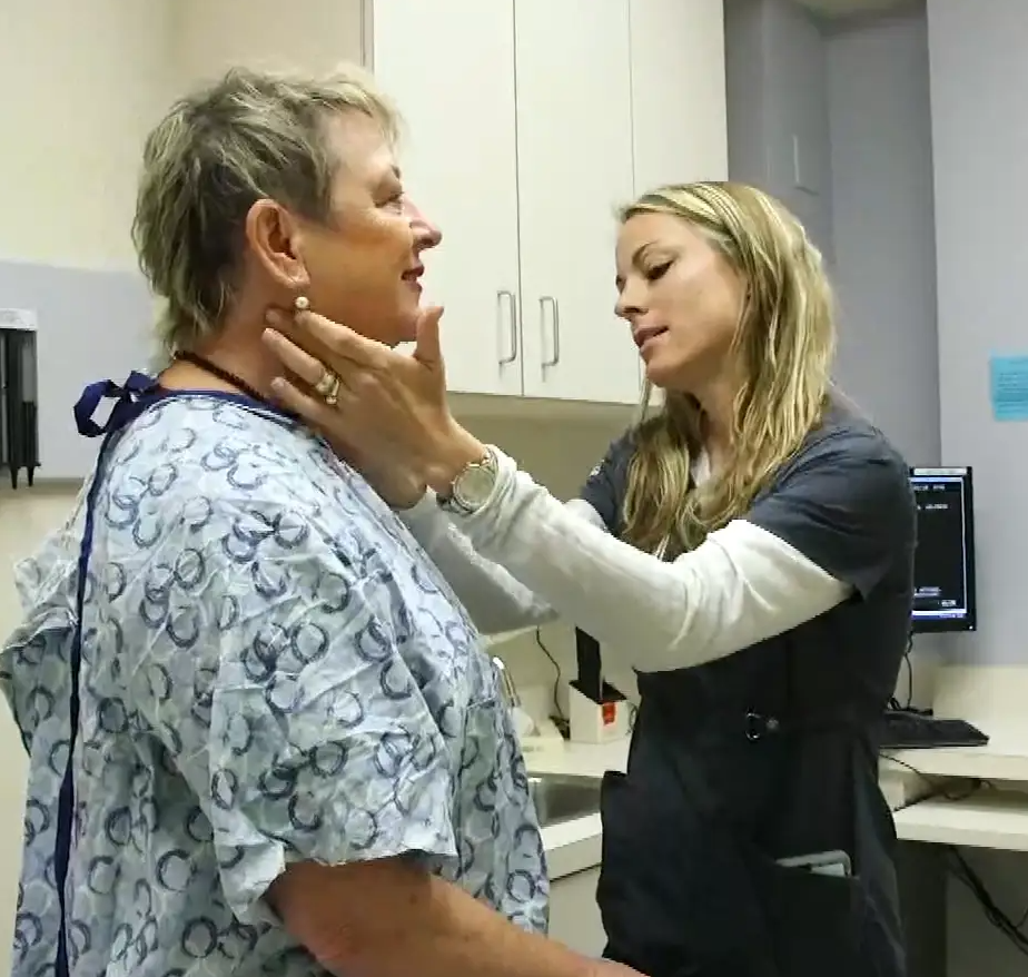
Twenty-six-year-old Hakeem wasn’t feeling well. He was more tired than usual, dragging through his workdays despite going to bed earlier, and napping on the weekends. He didn’t have much of an appetite, and had started losing weight. When he pressed on the side of his neck, like the doctor is doing in Figure 17.1.1, he noticed an unusual lump.
Hakeem went to his doctor, who performed a physical exam and determined that the lump was a swollen lymph node. Lymph nodes are part of the immune system, and they will often become enlarged when the body is fighting off an infection. Dr. Hayes thinks that the swollen lymph node and fatigue could be signs of a viral or bacterial infection, although he is concerned about Hakeem’s lack of appetite and weight loss. All of those symptoms combined can indicate a type of cancer called lymphoma. An infection, however, is a more likely cause, particularly in a young person like Hakeem. Dr. Hayes prescribes an antibiotic in case Hakeem has a bacterial infection, and advises him to return in a few weeks if his lymph node does not shrink, or if he is not feeling better.
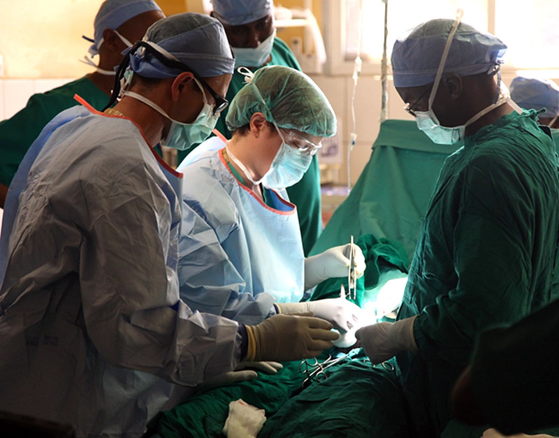
Hakeem returns a few weeks later. He is not feeling better and his lymph node is still enlarged. Dr. Hayes is concerned, and orders a biopsy of the enlarged lymph node. A lymph node biopsy for suspected lymphoma often involves the surgical removal of all or part of a lymph node. This helps to determine whether the tissue contains cancerous cells.
The initial results of the biopsy indicate that Hakeem does have lymphoma. Although lymphoma is more common in older people, young adults and even children can get this disease. There are many types of lymphoma, with the two main types being Hodgkin's lymphoma and non-Hodgkin's lymphoma. Non-Hodgkin lymphoma (NHL), in turn, has many subtypes. The subtype depends on several factors, including which cell types are affected. Some subtypes of NHL, for example, affect immune system cells called B cells, while others affect different immune system cells called T cells.
Dr. Hayes explains to Hakeem that it is important to determine which type of lymphoma he has, in order to choose the best course of treatment. Hakeem’s biopsied tissue will be further examined and tested to see which cell types are affected, as well as which specific cell-surface proteins — called antigens — are present. This should help identify his specific type of lymphoma.
As you read this chapter, you will learn about the functions of the immune system, and the specific roles that its cells and organs — such as B and T cells and lymph nodes — play in defending the body. At the end of this chapter, you will learn what type of lymphoma Hakeem has and what some of his treatment options are, including treatments that make use of the biochemistry of the immune system to fight cancer with the immune system itself.
Chapter Overview: Immune System
In this chapter, you will learn about the immune system — the system that defends the body against infections and other causes of disease, such as cancerous cells. Specifically, you will learn about:
- How the immune system identifies normal cells of the body as “self” and pathogens and damaged cells as “non-self.”
- The two major subsystems of the general immune system: the innate immune system — which provides a quick, but non-specific response — and the adaptive immune system, which is slower, but provides a specific response that often results in long-lasting immunity.
- The specialized immune system that protects the brain and spinal cord, called the neuroimmune system.
- The organs, cells, and responses of the innate immune system, which includes physical barriers (such as skin and mucus), chemical and biological barriers, inflammation, activation of the complement system of molecules, and non-specific cellular responses (such as phagocytosis).
- The lymphatic system — which includes white blood cells called lymphocytes, lymphatic vessels (which transport a fluid called lymph), and organs (such as the spleen, tonsils, and lymph nodes) — and its important role in the adaptive immune system.
- Specific cells of the immune system and their functions, including B cells, T cells, plasma cells, and natural killer cells.
- How the adaptive immune system can generate specific and often long-lasting immunity against pathogens through the production of antibodies.
- How vaccines work to generate immunity.
- How cells in the immune system detect and kill cancerous cells.
- Some strategies that pathogens employ to evade the immune system.
- Disorders of the immune system, including allergies, autoimmune diseases (such as diabetes and multiple sclerosis), and immunodeficiency resulting from conditions such as HIV infection.
As you read the chapter, think about the following questions:
- What are the functions of lymph nodes?
- What are B and T cells? How do they relate to lymph nodes?
- What are cell-surface antigens? How do they relate to the immune system and to cancer?
Attributions
Figure 17.1.1
Lymph nodes/Is it a Cold or the Flu by Lee Health on Vimeo is used under Vimeo's Terms of Service (https://vimeo.com/terms#licenses).
Figure 17.1.2
mitchell-luo-ymo_yC_N_2o-unsplash [photo] by Mitchell Luo on Unsplash is used under the Unsplash License (https://unsplash.com/license).
Figure 17.1.3
Lymph node biopsy by US Army Africa on Flickr is used under a CC BY 2.0 (https://creativecommons.org/licenses/by/2.0/) license.
References
Mayo Clinic Staff. (n.d.). Hodgkin's lymphoma [online article]. MayoClinic.org. https://www.mayoclinic.org/diseases-conditions/hodgkins-lymphoma/symptoms-causes/syc-20352646
Mayo Clinic Staff. (n.d.). Non-Hodgkin's lymphoma [online article]. MayoClinic.org. https://www.mayoclinic.org/diseases-conditions/non-hodgkins-lymphoma/symptoms-causes/syc-20375680
Respiration using electron acceptors other than molecular oxygen. Although oxygen is not the final electron acceptor, the process still uses a respiratory electron transport chain.
A threadlike structure of nucleic acids and protein found in the nucleus of most living cells, carrying genetic information in the form of genes.
The inner layer of skin that is made of tough connective tissue and contains blood vessels, nerve endings, hair follicles, and glands.
Created by CK-12 Foundation/Adapted by Christine Miller
Goose Bumps
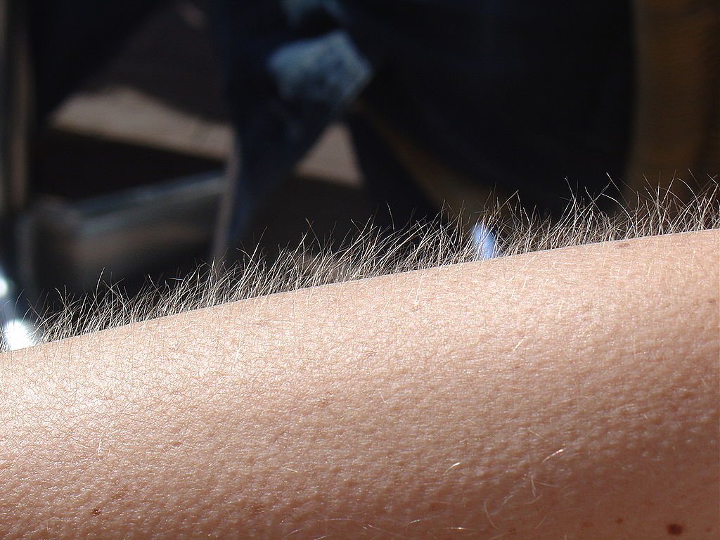
No doubt you’ve experienced the tiny, hair-raising skin bumps called goose bumps, like those you see in Figure 10.4.1. They happen when you feel chilly. Do you know what causes goose bumps, or why they pop up when you are cold? The answers to these questions involve the layer of skin known as the dermis.
What is the Dermis?
The dermis is the inner of the two major layers that make up the skin, the outer layer being the epidermis. The dermis consists mainly of connective tissues. It also contains most skin structures, such as glands and blood vessels. The dermis is anchored to the tissues below it by flexible collagen bundles that permit most areas of the skin to move freely over subcutaneous (“below the skin”) tissues. Functions of the dermis include cushioning subcutaneous tissues, regulating body temperature, sensing the environment, and excreting wastes.
Anatomy of the Dermis
The basic anatomy of the dermis is a matrix, or sort of scaffolding, composed of connective tissues. These tissues include collagen fibres — which provide toughness — and elastin fibres, which provide elasticity. Surrounding these fibres, the matrix also includes a gel-like substance made of proteins. The tissues of the matrix give the dermis both strength and flexibility.
The dermis is divided into two layers: the papillary layer and the reticular layer. Both layers are shown in Figure 10.4.2 below and described in the text that follows.
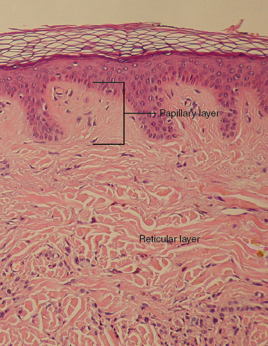
Papillary Layer
The papillary layer is the upper layer of the dermis, just below the basement membrane that connects the dermis to the epidermis above it. The papillary layer is the thinner of the two dermal layers. It is composed mainly of loosely arranged collagen fibres. The papillary layer is named for its fingerlike projections — or papillae — that extend upward into the epidermis. The papillae contain capillaries and sensory touch receptors.
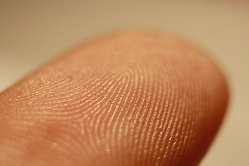
The papillae give the dermis a bumpy surface that interlocks with the epidermis above it, strengthening the connection between the two layers of skin. On the palms and soles, the papillae create epidermal ridges. Epidermal ridges on the fingers are commonly called fingerprints (see Figure 10.4.3). Fingerprints are genetically determined, so no two people (other than identical twins) have exactly the same fingerprint pattern. Therefore, fingerprints can be used as a means of identification, for example, at crime scenes. Fingerprints were much more commonly used forensically before DNA analysis was introduced for this purpose.
Reticular Layer
The reticular layer is the lower layer of the dermis, located below the papillary layer. It is the thicker of the two dermal layers. It is composed of densely woven collagen and elastin fibres. These protein fibres give the dermis its properties of strength and elasticity. This layer of the dermis cushions subcutaneous tissues of the body from stress and strain. The reticular layer of the dermis also contains most of the structures in the dermis, such as glands and hair follicles.
Structures in the Dermis
Both papillary and reticular layers of the dermis contain numerous sensory receptors, which make the skin the body’s primary sensory organ for the sense of touch. Both dermal layers also contain blood vessels. They provide nutrients to remove wastes from dermal cells, as well as cells in the lowest layer of the epidermis, the stratum basale. The circulatory components of the dermis are shown in Figure 10.4.4 below.
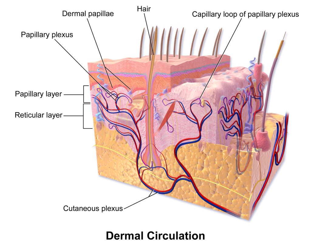
Glands
Glands in the reticular layer of the dermis include sweat glands and sebaceous (oil) glands. Both are exocrine glands, which are glands that release their secretions through ducts to nearby body surfaces. The diagram in Figure 10.4.5 shows these glands, as well as several other structures in the dermis.
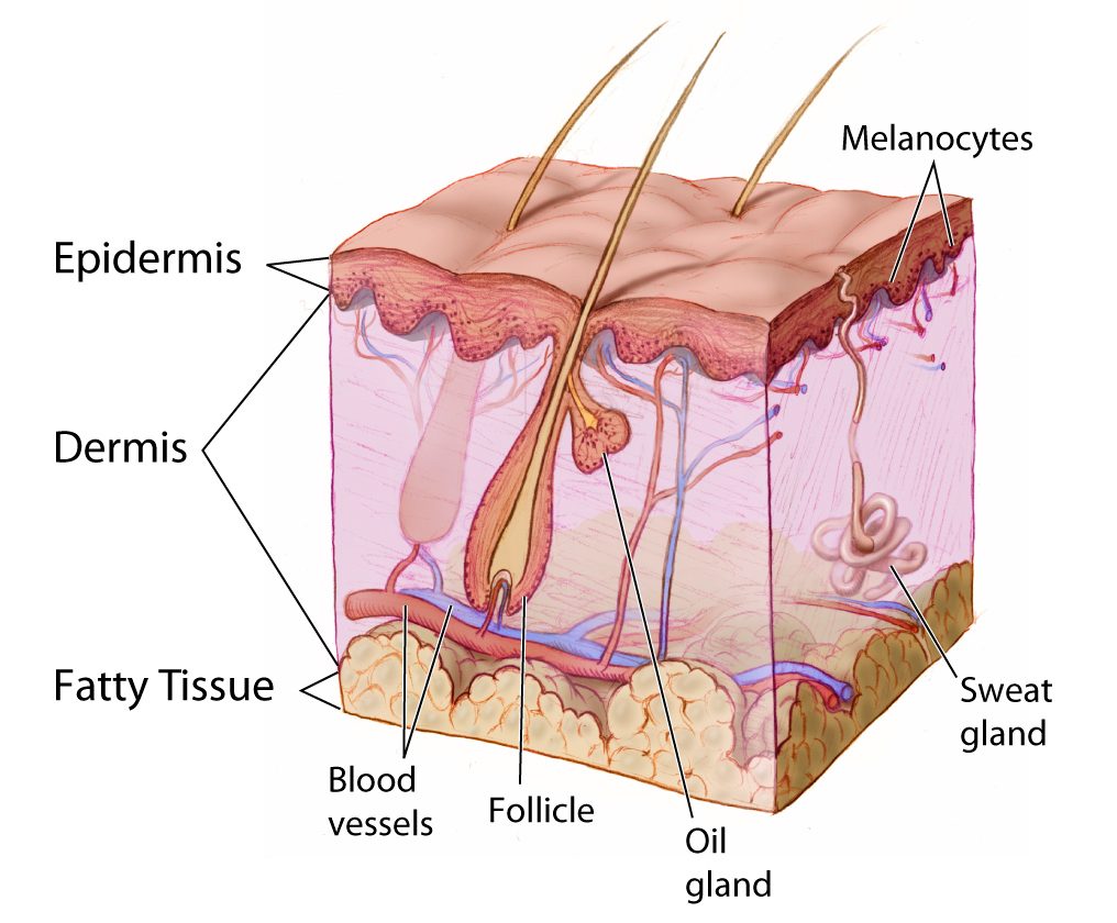
Sweat Glands
Sweat glands produce the fluid called sweat, which contains mainly water and salts. The glands have ducts that carry the sweat to hair follicles, or to the surface of the skin. There are two different types of sweat glands: eccrine glands and apocrine glands.
- Eccrine sweat glands occur in skin all over the body. Their ducts empty through tiny openings called pores onto the skin surface. These sweat glands are involved in temperature regulation.
- Apocrine sweat glands are larger than eccrine glands, and occur only in the skin of the armpits and groin. The ducts of apocrine glands empty into hair follicles, and then the sweat travels along hairs to reach the surface. Apocrine glands are inactive until puberty, at which point they start producing an oily sweat that is consumed by bacteria living on the skin. The digestion of apocrine sweat by bacteria causes body odor.
Sebaceous Glands
Sebaceous glands are exocrine glands that produce a thick, fatty substance called sebum. Sebum is secreted into hair follicles and makes its way to the skin surface along hairs. It waterproofs the hair and skin, and helps prevent them from drying out. Sebum also has antibacterial properties, so it inhibits the growth of microorganisms on the skin. Sebaceous glands are found in every part of the skin — except for the palms of the hands and soles of the feet, where hair does not grow.
Hair Follicles
Hair follicles are the structures where hairs originate (see the diagram above). Hairs grow out of follicles, pass through the epidermis, and exit at the surface of the skin. Associated with each hair follicle is a sebaceous gland, which secretes sebum that coats and waterproofs the hair. Each follicle also has a bed of capillaries, a nerve ending, and a tiny muscle called an arrector pili.
Functions of the Dermis
The main functions of the dermis are regulating body temperature, enabling the sense of touch, and eliminating wastes from the body.
Temperature Regulation
Several structures in the reticular layer of the dermis are involved in regulating body temperature. For example, when body temperature rises, the hypothalamus of the brain sends nerve signals to sweat glands, causing them to release sweat. An adult can sweat up to four litres an hour. As the sweat evaporates from the surface of the body, it uses energy in the form of body heat, thus cooling the body. The hypothalamus also causes dilation of blood vessels in the dermis when body temperature rises. This allows more blood to flow through the skin, bringing body heat to the surface, where it can radiate into the environment.
When the body is too cool, sweat glands stop producing sweat, and blood vessels in the skin constrict, thus conserving body heat. The arrector pili muscles also contract, moving hair follicles and lifting hair shafts. This results in more air being trapped under the hairs to insulate the surface of the skin. These contractions of arrector pili muscles are the cause of goose bumps.
Sensing the Environment
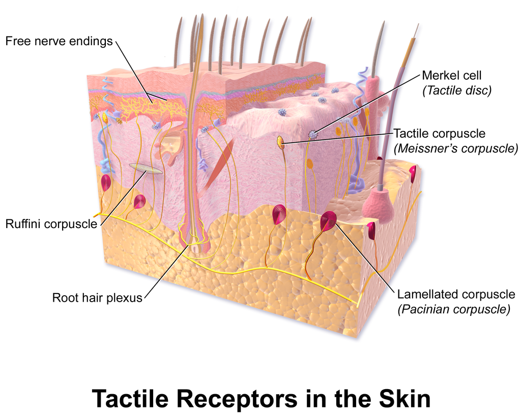
Sensory receptors in the dermis are mainly responsible for the body’s tactile senses. The receptors detect such tactile stimuli as warm or cold temperature, shape, texture, pressure, vibration, and pain. They send nerve impulses to the brain, which interprets and responds to the sensory information. Sensory receptors in the dermis can be classified on the basis of the type of touch stimulus they sense. Mechanoreceptors sense mechanical forces such as pressure, roughness, vibration, and stretching. Thermoreceptors sense variations in temperature that are above or below body temperature. Nociceptors sense painful stimuli. Figure 10.4.6 shows several specific kinds of tactile receptors in the dermis. Each kind of receptor senses one or more types of touch stimuli.
- Free nerve endings sense pain and temperature variations.
- Merkel cells sense light touch, shapes, and textures.
- Meissner’s corpuscles sense light touch.
- Pacinian corpuscles sense pressure and vibration.
- Ruffini corpuscles sense stretching and sustained pressure.
Excreting Wastes
The sweat released by eccrine sweat glands is one way the body excretes waste products. Sweat contains excess water, salts (electrolytes), and other waste products that the body must get rid of to maintain homeostasis. The most common electrolytes in sweat are sodium and chloride. Potassium, calcium, and magnesium electrolytes may be excreted in sweat, as well. When these electrolytes reach high levels in the blood, more are excreted in sweat. This helps to bring their blood levels back into balance. Besides electrolytes, sweat contains small amounts of waste products from metabolism, including ammonia and urea. Sweat may also contain alcohol in someone who has been drinking alcoholic beverages.
Feature: My Human Body
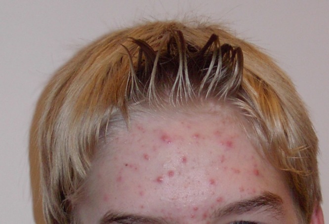
Acne is the most common skin disorder in the Canada. At least 20% of Canadians have acne at any given time and it affects approximately 90% of adolescents (as in Figure 10.4.7). Although acne occurs most commonly in teens and young adults, but it can occur at any age. Even newborn babies can get acne.
The main sign of acne is the appearance of pimples (pustules) on the skin, like those in the photo above. Other signs of acne may include whiteheads, blackheads, nodules, and other lesions. Besides the face, acne can appear on the back, chest, neck, shoulders, upper arms, and buttocks. Acne can permanently scar the skin, especially if it isn’t treated appropriately. Besides its physical effects on the skin, acne can also lead to low self-esteem and depression.
Acne is caused by clogged, sebum-filled pores that provide a perfect environment for the growth of bacteria. The bacteria cause infection, and the immune system responds with inflammation. Inflammation, in turn, causes swelling and redness, and may be associated with the formation of pus. If the inflammation goes deep into the skin, it may form an acne nodule.
Mild acne often responds well to treatment with over-the-counter (OTC) products containing benzoyl peroxide or salicylic acid. Treatment with these products may take a month or two to clear up the acne. Once the skin clears, treatment generally needs to continue for some time to prevent future breakouts.
If acne fails to respond to OTC products, nodules develop, or acne is affecting self-esteem, a visit to a dermatologist is in order. A dermatologist can determine which treatment is best for a given patient. A dermatologist can also prescribe prescription medications (which are likely to be more effective than OTC products) and provide other medical treatments, such as laser light therapies or chemical peels.
What can you do to maintain healthy skin and prevent or reduce acne? Dermatologists recommend the following tips:
- Wash affected or acne-prone skin (such as the face) twice a day, and after sweating.
- Use your fingertips to apply a gentle, non-abrasive cleanser. Avoid scrubbing, which can make acne worse.
- Use only alcohol-free products and avoid any products that irritate the skin, such as harsh astringents or exfoliants.
- Rinse with lukewarm water, and avoid using very hot or cold water.
- Shampoo your hair regularly.
- Do not pick, pop, or squeeze acne. If you do, it will take longer to heal and is more likely to scar.
- Keep your hands off your face. Avoid touching your skin throughout the day.
- Stay out of the sun and tanning beds. Some acne medications make your skin very sensitive to UV light.
10.4 Summary
- The dermis is the inner and thicker of the two major layers that make up the skin. It consists mainly of a matrix of connective tissues that provide strength and stretch. It also contains almost all skin structures, including sensory receptors and blood vessels.
- The dermis has two layers. The upper papillary layer has papillae extending upward into the epidermis and loose connective tissues. The lower reticular layer has denser connective tissues and structures, such as glands and hair follicles. Glands in the dermis include eccrine and apocrine sweat glands and sebaceous glands. Hair follicles are structures where hairs originate.
- Functions of the dermis include cushioning subcutaneous tissues, regulating body temperature, sensing the environment, and excreting wastes. The dense connective tissues of the dermis provide cushioning. The dermis regulates body temperature mainly by sweating and by vasodilation or vasoconstriction. The many tactile sensory receptors in the dermis make it the main organ for the sense of touch. Wastes excreted in sweat include excess water, electrolytes, and certain metabolic wastes.
10.4 Review Questions
- What is the dermis?
- Describe the basic anatomy of the dermis.
- Compare and contrast the papillary and reticular layers of the dermis.
- What causes epidermal ridges, and why can they be used to identify individuals?
- Name the two types of sweat glands in the dermis, and explain how they differ.
- What is the function of sebaceous glands?
- Describe the structures associated with hair follicles.
- Explain how the dermis helps regulate body temperature.
- Identify three specific kinds of tactile receptors in the dermis, along with the type of stimuli they sense.
- How does the dermis excrete wastes? What waste products does it excrete?
- What are subcutaneous tissues? Which layer of the dermis provides cushioning for subcutaneous tissues? Why does this layer provide most of the cushioning, instead of the other layer?
- For each of the functions listed below, describe which structure within the dermis carries it out.
- Brings nutrients to and removes wastes from dermal and lower epidermal cells
- Causes hairs to move
- Detects painful stimuli on the skin
10.4 Explore More
https://www.youtube.com/watch?v=FX-FwK0IIrE
How do you get rid of acne? SciShow, 2016.
https://www.youtube.com/watch?v=VcHQWMAClhQ&feature=emb_logo
When You Can't Scratch Away An Itch, Seeker, 2013.
Attributions
Figure 10.4.1
Goose_bumps by EverJean on Wikimedia Commons is used under a CC BY 2.0 (https://creativecommons.org/licenses/by/2.0) license.
Figure 10.4.2
Layers_of_the_Dermis by OpenStax College on Wikimedia Commons is used under a CC BY 3.0 (https://creativecommons.org/licenses/by/3.0) license.
Figure 10.4.3
Fingerprint_detail_on_male_finger_in_Třebíč,_Třebíč_District by Frettie on Wikimedia Commons is used under a CC BY 3.0 (https://creativecommons.org/licenses/by/3.0) license.
Figure 10.4.4
Blausen_0802_Skin_Dermal Circulation by BruceBlaus on Wikimedia commons is used under a CC BY 3.0 (https://creativecommons.org/licenses/by/3.0) license.
Figure 10.4.5
Anatomy_The_Skin_-_NCI_Visuals_Online by Don Bliss (artist) / National Cancer Institute (National Institutes of Health, with the ID 4604) is in the public domain (https://en.wikipedia.org/wiki/public_domain).
Figure 10.4.6
Blausen_0809_Skin_TactileReceptors by BruceBlaus on Wikimedia commons is used under a CC BY 3.0 (https://creativecommons.org/licenses/by/3.0) license.
Figure 10.4.7
Akne-jugend by Ellywa on Wikimedia Commons is released into the public domain (https://en.wikipedia.org/wiki/public_domain). (No machine-readable author provided. Ellywa assumed, based on copyright claims).
References
Betts, J. G., Young, K.A., Wise, J.A., Johnson, E., Poe, B., Kruse, D.H., Korol, O., Johnson, J.E., Womble, M., DeSaix, P. (2013, June 19). Figure 5.7 Layers of the dermis [digital image]. In Anatomy and Physiology (Section 5.1 Layers of the skin). OpenStax. https://openstax.org/books/anatomy-and-physiology/pages/5-1-layers-of-the-skin
Blausen.com staff. (2014). Medical gallery of Blausen Medical 2014. WikiJournal of Medicine 1 (2). DOI:10.15347/wjm/2014.010. ISSN 2002-4436.
SciShow. (2016, October 26). How do you get rid of acne? YouTube. https://www.youtube.com/watch?v=FX-FwK0IIrE
Seeker. (2013, October 26). When you can't scratch away an itch. YouTube. https://www.youtube.com/watch?v=VcHQWMAClhQ&feature=emb_logo
a colorless cell that circulates in the blood and body fluids and is involved in counteracting foreign substances and disease; a white (blood) cell. There are several types, all amoeboid cells with a nucleus, including lymphocytes, granulocytes, monocytes, and macrophages.
An anatomical structure that consists of a small cluster of cells, surrounding a central cavity.
accessory organ of the skin made of sheets of dead keratinocytes at the distal ends of the fingers and toes

