9.6 Adrenal Glands
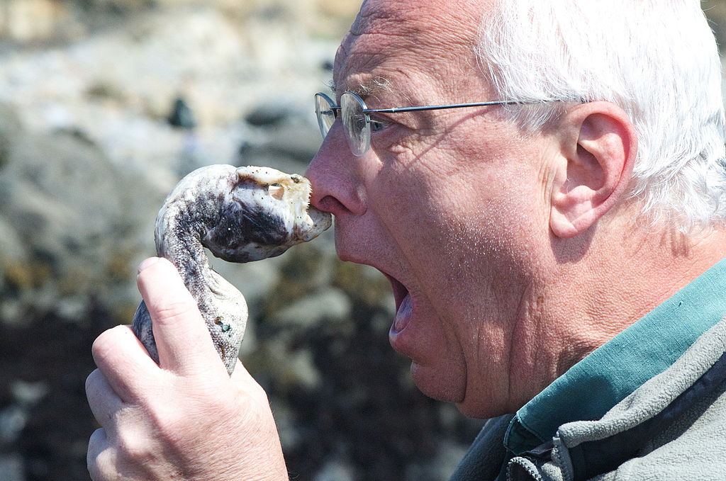
Eek!
Being bitten on the nose by an eel certainly qualifies as a frightening experience! The fear this man is experiencing produces the same physiological responses in most people — racing heart, rapid breathing, clammy hands. These and other fight-or-flight responses prepare the body to either defend itself or run away from danger. Why does fear elicit these changes in the body? The responses occur in large part because of hormones secreted by the adrenal glands.
Introduction to the Adrenal Glands
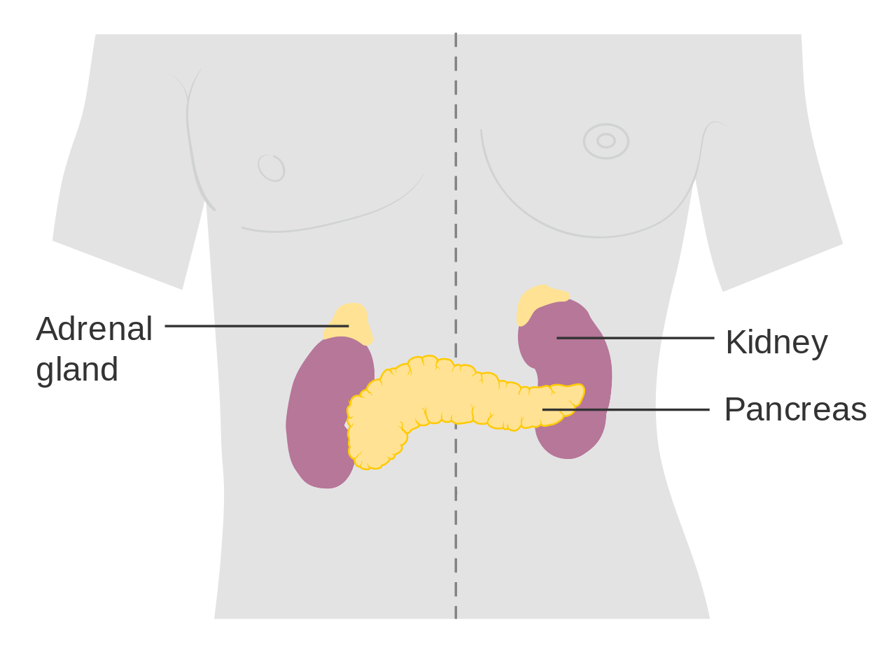
The adrenal glands are endocrine glands that produce a variety of hormones. Adrenal hormones include the fight-or-flight hormone adrenaline and the steroid hormone cortisol. The two adrenal glands are located on both sides of the body, just above the kidneys, as shown in Figure 9.6.2. The right adrenal gland (on the left in the figure) is smaller and has a pyramidal shape. The left adrenal gland (on the right in the figure) is larger and has a half-moon shape.
Each adrenal gland has two distinct parts, and each part has a different function, although both parts produce hormones. There is an outer layer, called the adrenal cortex, which produces steroid hormones including cortisol. There is also an inner layer, called the adrenal medulla, which produces non-steroid hormones including adrenaline.
Adrenal Cortex
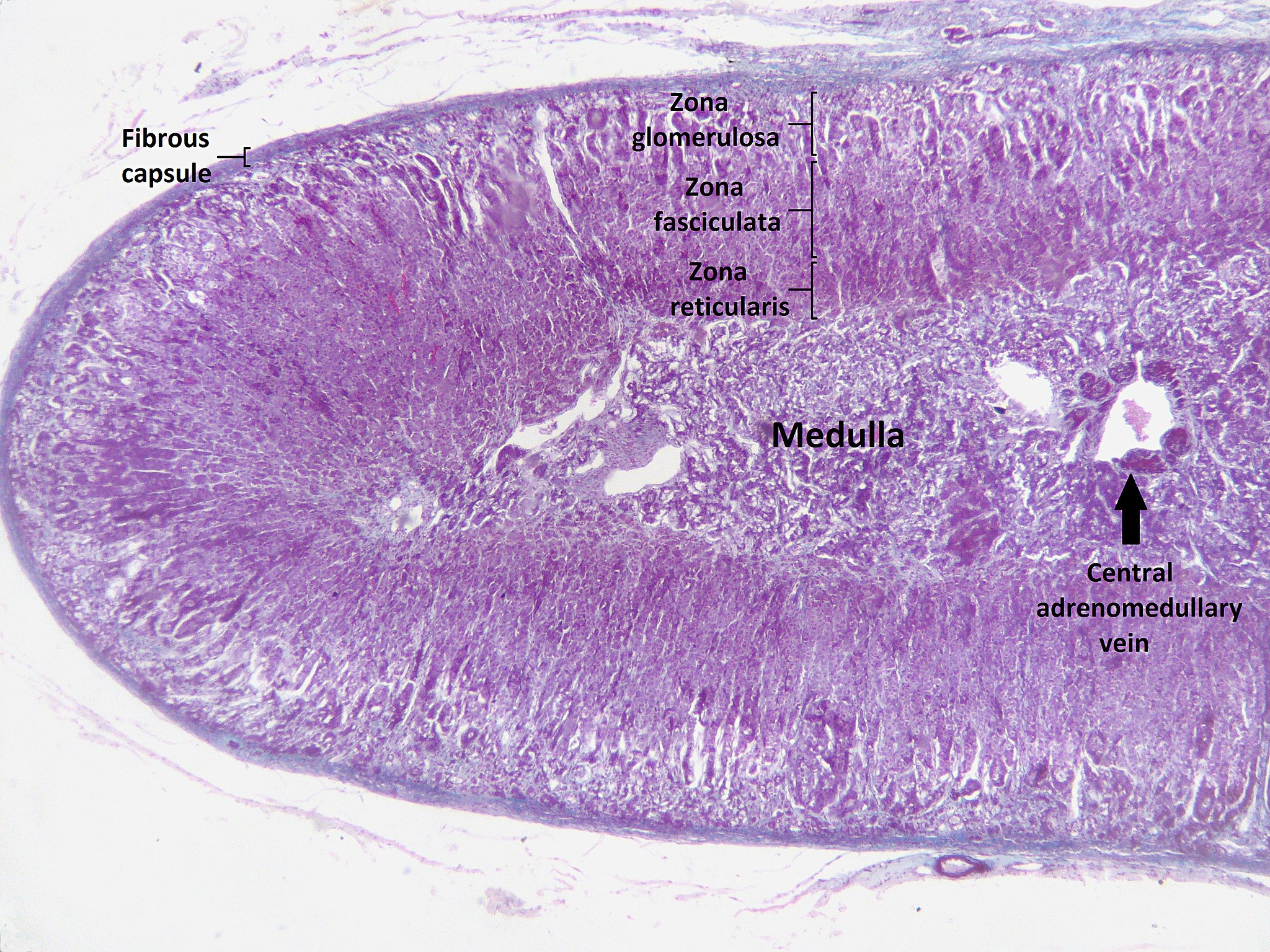
The adrenal cortex, or outer layer of the adrenal gland, is divided into three additional layers, called zones (see Figure 9.6.3). Each zone has distinct enzymes that produce different hormones from the common precursor molecule cholesterol, which is a lipid.
- Zona glomerulosa is the outermost layer of the adrenal cortex. It lies immediately under the outer fibrous capsule that encloses the adrenal gland.
- Zona fasciculata is the middle layer of the adrenal cortex. It is the largest of the three zones, accounting for nearly 80 per cent of the adrenal cortex.
- Zona reticularis is the innermost layer of the adrenal cortex. It is directly adjacent to the medulla of the adrenal gland.
Types of Adrenal Cortex Hormones
Hormones produced by the adrenal cortex are known by the general term corticosteroids. As steroid hormones, corticosteroids are endocrine hormones that are made of lipids and exert their effects on target cells by crossing the plasma membrane and binding with receptors within the cytoplasm. A steroid hormone and its receptor form a complex that enters the cell nucleus and affects gene expression. There are three types of corticosteroids synthesized and secreted by the adrenal cortex. Each type is produced by a different zone of the adrenal cortex, as shown in Figure 9.6.4.

Mineralocorticoids
Mineralocorticoids are produced in the zona glomerulosa and include the hormone aldosterone. These hormones help control the balance of mineral salts (electrolytes) in the body. In the kidneys, aldosterone increases the reabsorption of sodium ions and the excretion of potassium ions. Aldosterone also stimulates the retention of sodium ions by cells in the colon and by the sweat glands. The amount of sodium in the body affects the volume of extracellular fluids (including the blood) and thereby affects blood pressure. In this way, mineralocorticoids help control blood volume and blood pressure.
Glucocorticoids
Glucocorticoids are produced in the zona fasciculata and include the hormone cortisol, which is released in repsonse to stress and is considered the primary stress hormone. Glucocorticoids help control the rate of metabolism of proteins, fats, and sugars. In general, they increase the level of glucose and fatty acids circulating in the blood. Cells rely primarily on glucose for energy, but they can also use fatty acids for energy as an alternative to glucose. Glucocorticoids are also involved in suppression of the immune system, having a potent anti-inflammatory effect. In addition, cortisol reduces the production of new bone and decreases absorption of calcium from the gastrointestinal tract.
Androgens
Androgens are produced in the zona reticularis and include the hormone DHEA (dehydroepiandrosterone). Androgens are a general term for male sex hormones, although this is somewhat misleading, as adrenal cortex androgens are produced by both males and females. In adult males, they are converted to more potent androgens, such as testosterone in the male gonads (testes). In adult females, they are converted to female sex hormones called estrogens in the female gonads (ovaries).
Regulation of Adrenal Cortex Hormones
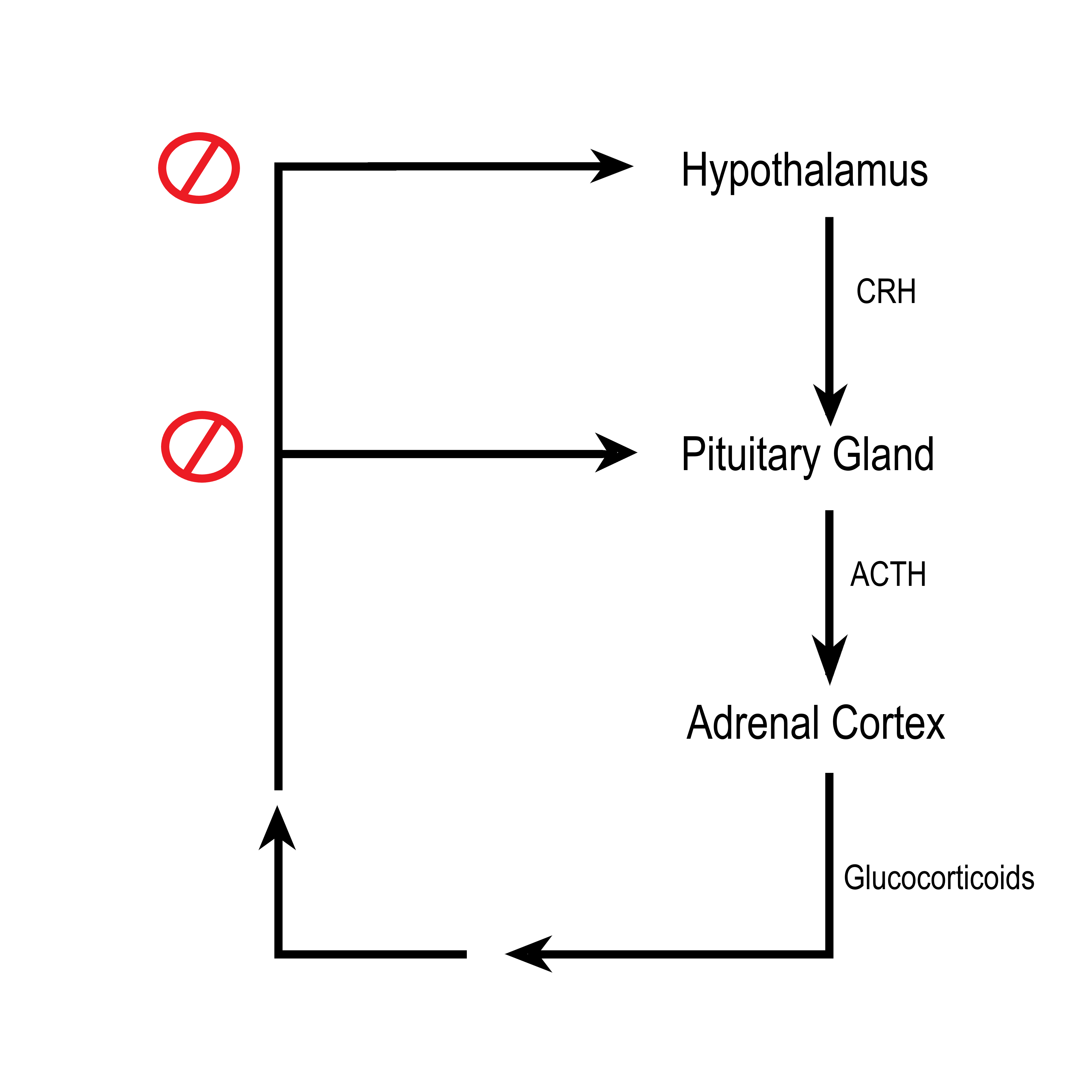
Steroid hormone production by the three zones of the adrenal cortex is regulated by hormones secreted by the anterior lobe of the pituitary gland, as well as by other physiological stimuli. For example, the production of glucocorticoids such as cortisol is stimulated by adrenocorticotropic hormone (ACTH) from the anterior pituitary, which in turn is stimulated by corticotropin releasing hormone (CRH) from the hypothalamus. When levels of glucocorticoids start to rise too high, they provide negative feedback to the hypothalamus and pituitary gland to stop secreting CRH and ACTH, respectively. This negative feedback mechanism is illustrated in Figure 9.6.5. The opposite occurs when levels of glucocorticoids start to fall too low.
Adrenal Medulla
The adrenal medulla is at the center of each adrenal gland and is surrounded by the adrenal cortex. It contains a dense network of blood vessels into which it secretes its hormones. The hormones synthesized and secreted by the adrenal medulla are generally known as catecholamines, and they include adrenaline (also called epinephrine) and noradrenaline (also called norepinephrine). These water-soluble, non-steroid hormones are made of amino acids. As non-steroid hormones, they cannot cross the plasma membrane of target cells. Instead, they exert their effects by binding to receptors on the surface of target cells. The binding of hormone and receptor activates an enzyme in the plasma membrane that controls a second messenger. It is the second messenger that influences processes inside the cell.
Catecholamines function to produce a rapid response throughout the body in stressful situations. They bring about such changes as increased heart rate, more rapid breathing, constriction of blood vessels in certain parts of the body, and an increase in blood pressure. The release of catecholamines by the adrenal medulla is stimulated by activation of the sympathetic division of the autonomic nervous system.
Disorders of the Adrenal Glands
Disorders of the adrenal glands generally include either hypersecretion or hyposecretion of adrenal hormones. The underlying cause of the abnormal secretion may be a problem with the adrenal glands or with the pituitary gland, which controls adrenal cortex hormone production. Both adrenal and pituitary glands are subject to the formation of tumors, which may cause adrenal disorders. The adrenal gland may also be affected by infections or autoimmune diseases.
Adrenal Hypersecretion: Cushing’s Syndrome
Hypersecretion of the glucocorticoid hormone cortisol leads to a disorder called Cushing’s syndrome. The most common cause of Cushing’s syndrome is a pituitary tumor, which causes excessive production of ACTH. The disease produces a wide variety of signs and symptoms, which may include obesity, diabetes, high blood pressure (hypertension), excessive body hair, osteoporosis, and depression. A distinctive sign of Cushing’s syndrome is the appearance of stretch marks in the skin, as the skin becomes progressively thinner. Another distinctive sign is a moon face, in which fat deposits give the face a rounded appearance. Treatment of Cushing’s syndrome depends on its cause and may include surgery to remove a tumor or medications to suppress activity of the adrenal glands.
Adrenal Hyposecretion: Addison’s Disease
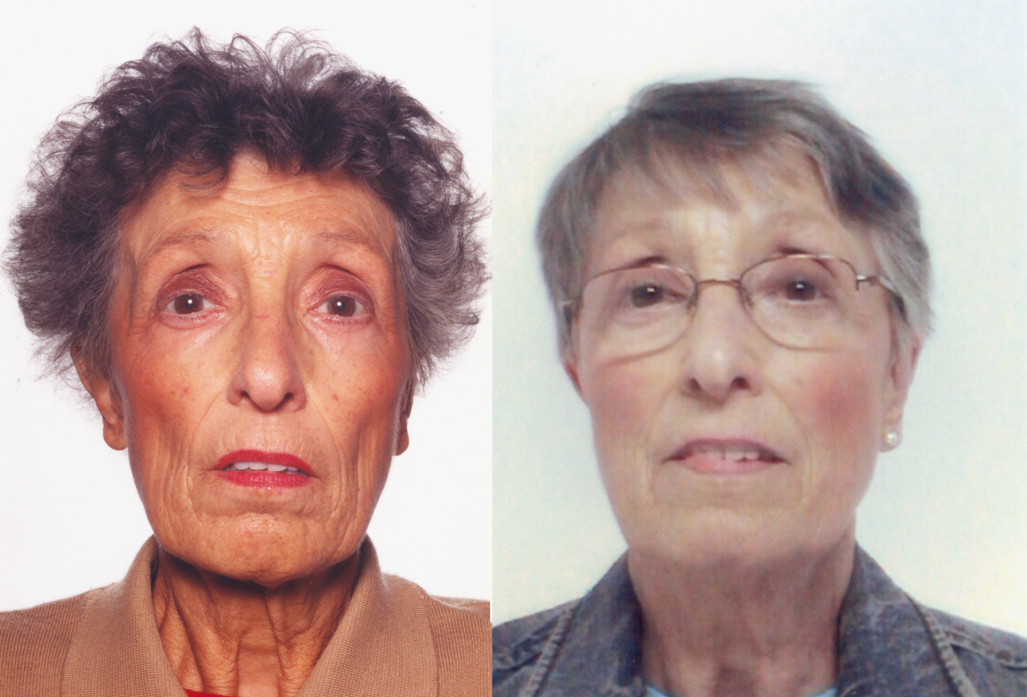
Hyposecretion of the glucocorticoid hormone cortisol leads to a disorder called Addison’s disease. There may also be hyposecretion of mineralocorticoids with this disorder. Addison’s disease is generally an autoimmune disorder, in which the immune system produces abnormal antibodies that attack cells of the adrenal cortex. Untreated infections, especially of tuberculosis, may also damage the adrenal cortex and cause Addison’s disease. A third possible cause is decreased output of ACTH by the pituitary gland, generally due to a pituitary tumor. A distinctive sign of Addison’s disease is hyperpigmentation of the skin (see the photos in Figure 9.6.6). Other symptoms tend to be nonspecific and include excessive fatigue. Addison’s disease is generally treated with replacement hormones in pill form.
Feature: My Human Body

Does just looking at this photo (Figure 9.6.7) cause you to break out in a cold sweat and experience heart palpitations? Imagine how scary it would be to actually fling yourself backward off a tall building like the BASE jumper in the photo! There would be very little time to use a parachute to slow your fall before you hit the ground. BASE jumping is called the most dangerous sport on Earth. In fact, it is so dangerous that it is outlawed in some places.
People who participate in such dangerous activities as BASE jumping are likely to be adrenaline “junkies.” They are addicted to the adrenaline rush and euphoria — or “high” — it causes when their fight-or-flight response is triggered by danger. Why does adrenaline have this effect? Adrenaline is closely related to dopamine, a chemical messenger in the brain that plays a major role in pleasure and addiction.
Adrenaline addicts don’t have to participate in BASE jumping or other dangerous sports to get an adrenaline rush. They might choose a dangerous occupation like firefighting, participate in risky behaviors like reckless driving or bank robbing, or just pick fights with other people. They might even create their own stress by always taking on too much work or delaying projects until close to their deadline.
While some excitement in one’s life is generally a good thing, always putting oneself in danger or constantly being under stress are obviously not good things. If you think you might be an adrenaline addict, note that there are healthier ways to experience a hormonal “high.” Running, biking, or participating in some other form of vigorous aerobic exercise causes the pituitary gland and hypothalamus to produce opiate-like endorphins, leading to a so-called “runner’s high.” Like the euphoric feeling adrenaline causes, a runner’s high may last for hours.
9.6 Summary
- The adrenal glands are endocrine glands that produce a variety of hormones. The two adrenal glands are located on both sides of the body, just above the kidneys. Each gland has two layers: an outer layer called the adrenal cortex and an inner layer called the adrenal medulla.
- The adrenal cortex produces steroid hormones called by the general term corticosteroids, of which there are three types: mineralocorticoids (such as aldosterone), which helps control electrolyte balance; glucocorticoids (such as cortisol), which helps control the rate of metabolism, suppresses the immune system, and is the major stress hormone; and androgens (such as DHEA), which is converted to sex hormones in the gonads.
- The adrenal medulla produces non-steroid catecholamine hormones, including adrenaline and noradrenaline. These hormones stimulate the fight-or-flight response.
- Disorders of the adrenal glands generally include either hypersecretion or hyposecretion of adrenal hormones. The cause may be a problem with the adrenal glands or with the pituitary gland, which controls adrenal cortex hormone production. Examples include Cushing’s syndrome, in which there is hypersecretion of cortisol, and Addison’s disease, in which there is hyposecretion of cortisol and mineralocorticoids.
9.6 Review Questions
- Describe the structure and location of the adrenal glands.
-
- Compare and contrast the adrenal cortex and adrenal medulla.
- Identify the three layers of the adrenal cortex and the type of hormones each layer produces.
- Give an example of each type of corticosteroid and state its function.
- Explain how the production of glucocorticoids is regulated.
- What is a catecholamine? Give an example of a catecholamine and state its function.
- Compare and contrast Cushing’s syndrome and Addison’s disease.
- What are two ways in which the nervous system (which includes the brain, spinal cord, and nerves) controls the adrenal gland?
- Explain why a pituitary tumor can cause either hypersecretion or hyposecretion of cortisol.
9.6 Explore More
How stress affects your body – Sharon Horesh Bergquist, TED-Ed, 2015.
How stress affects your brain – Madhumita Murgia, TED-Ed, 2015.
Adrenaline: Fight or Flight Response, Henk van ‘t Klooster, 2013.
Attributions
Figure 9.6.1
Attack from wikimedia commons by Jerry Kirkhart from Los Osos, Calif. on Wikimedia Commons is used under a CC BY 2.0 (https://creativecommons.org/licenses/by/2.0) license.
Figure 9.6.2
Diagram_showing_where_the_adrenal_glands_are_in_the_body_CRUK_415.svg by Cancer Research UK uploader on Wikimedia Commons is used under a CC BY-SA 4.0 (https://creativecommons.org/licenses/by-sa/4.0) license.
Figure 9.6.3
Adrenal_cortex_labelled by Jpogi at English Wikipedia on Wikimedia Commons is used under a CC0 1.0 Universal Public Domain Dedication (https://creativecommons.org/publicdomain/zero/1.0/deed.en) license.
Figure 9.6.4
The_Adrenal_Glands by OpenStax College is used under a CC BY 4.0 (https://creativecommons.org/licenses/by/4.0/) license.
Figure 9.6.5
ACTH negative feedback loop by Christinelmiller is used under a CC BY 4.0 (https://creativecommons.org/licenses/by/4.0/) license.
Figure 9.6.6
A_69-Year-Old_Female_with_Tiredness_and_a_Persistent_Tan_01 by Petros Perros on Wikimedia Commons is used under a CC BY 2.5 (https://creativecommons.org/licenses/by/2.5/deed.en) license.
Figure 9.6.7
BASE_Jumping_from_Sapphire_Tower_in_Istanbul by Kontizas Dimitrios on Wikimedia Commons is used under a CC BY-SA 3.0 (https://creativecommons.org/licenses/by-sa/3.0) license.
References
Betts, J. G., Young, K.A., Wise, J.A., Johnson, E., Poe, B., Kruse, D.H., Korol, O., Johnson, J.E., Womble, M., DeSaix, P. (2013, June 19). Figure 17.17 Adrenal glands [digital image]. In Anatomy and Physiology (Section 17.6). OpenStax College. https://openstax.org/books/anatomy-and-physiology/pages/17-6-the-adrenal-glands
Henk van ‘t Klooster. (2013). Adrenaline: Fight or flight response. YouTube. https://www.youtube.com/watch?v=FBnBTkcr6No&feature=youtu.be
Perros, P. (2005). A 69-year-old female with tiredness and a persistent tan. PLoS Medicine, 2(8): e229. https://doi.org/10.1371/journal.pmed.0020229
TED-Ed. (2015, October 22). How stress affects your body – Sharon Horesh Bergquist. YouTube. https://www.youtube.com/watch?v=v-t1Z5-oPtU&feature=youtu.be
TED-Ed. (2015, November 6). How stress affects your brain – Madhumita Murgia. YouTube. https://www.youtube.com/watch?v=WuyPuH9ojCE&feature=youtu.be
An involuntary human body response mediated by the nervous and endocrine systems that prepares the body to fight or flee from perceived danger.
one of a pair of glands located on top of the kidneys that secretes hormones such as cortisol and adrenaline
The outer layer of the adrenal gland that produces steroid hormones such as cortisol and aldosterone.
An organelle found in eukaryotic cells with the function of making cellular products such as hormones and lipids. The smooth endoplasmic reticulum is a part of the endoplasmic reticulum that does not have attached ribosomes.
A type of immune cell that has granules (small particles) with enzymes that are released during allergic reactions and asthma. A basophil is a type of white blood cell and a type of granulocyte.
A type of immune cell that has granules (small particles) with enzymes that are released during allergic reactions and asthma. A basophil is a type of white blood cell and a type of granulocyte.
Any steroid hormone produced by the cortex of the adrenal gland; includes mineralocorticoids, glucocorticoids, and androgens.
A type of immune cell that has granules (small particles) with enzymes that can kill tumor cells or cells infected with a virus. A natural killer cell is a type of white blood cell.
A substance that is insoluble in water. Examples include fats, oils and cholesterol. Lipids are made from monomers such as glycerol and fatty acids.
A chemical reaction that releases energy through light or heat.
A semi-permeable lipid bilayer that separates the interior of all cells from their surroundings.
A central organelle containing hereditary material.
The process by which information from a gene is used in the synthesis of a functional protein.
The main mineralocorticoid hormone which is responsible for sodium conservation in the kidney, salivary glands, sweat glands and colon.
A glucocorticoid hormone produced by the cortex of the adrenal gland that is released in response to stress and also helps control metabolic rate, suppression of the immune system, and other functions
A class of biological molecule consisting of linked monomers of amino acids and which are the most versatile macromolecules in living systems and serve crucial functions in essentially all biological processes.
Glucose (also called dextrose) is a simple sugar with the molecular formula C6H12O6. Glucose is the most abundant monosaccharide, a subcategory of carbohydrates. Glucose is mainly made by plants and most algae during photosynthesis from water and carbon dioxide, using energy from sunlight.
Long chains of hydrocarbons with a carboxyl group and a methyl group at opposite ends. Can be either saturated, containing mostly single bonds between adjacent carbons, or unsaturated, containing many double bonds between adjacent carbons.
a hormonal disorder common among women of reproductive age. Women with PCOS may have infrequent or prolonged menstrual periods or excess male hormone (androgen) levels. The ovaries may develop numerous small collections of fluid (follicles) and fail to regularly release eggs.
A substance that takes part in and undergoes change during a chemical reaction.
a hormonal disorder common among women of reproductive age. Women with PCOS may have infrequent or prolonged menstrual periods or excess male hormone (androgen) levels. The ovaries may develop numerous small collections of fluid (follicles) and fail to regularly release eggs.
The female sex hormone secreted mainly by the ovaries.
A testable proposed explanation for a phenomenon.
The front lobe of the pituitary gland that synthesizes and secretes pituitary hormones.
Created by CK-12 Foundation/Adapted by Christine Miller

One Piano, Four Hands
Did you ever see two people play the same piano? How do they coordinate all the movements of their own fingers — let alone synchronize them with those of their partner? The peripheral nervous system plays an important part in this challenge.
What Is the Peripheral Nervous System?
The peripheral nervous system (PNS) consists of all the nervous tissue that lies outside of the central nervous system (CNS). The main function of the PNS is to connect the CNS to the rest of the organism. It serves as a communication relay, going back and forth between the CNS and muscles, organs, and glands throughout the body.
Tissues of the Peripheral Nervous System
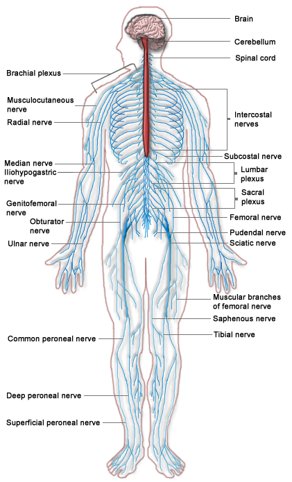
The PNS is mostly made up of cable-like bundles of axons called nerves, as well as clusters of neuronal cell bodies called ganglia (singular, ganglion). Nerves are generally classified as sensory, motor, or mixed nerves based on the direction in which they carry nerve impulses.
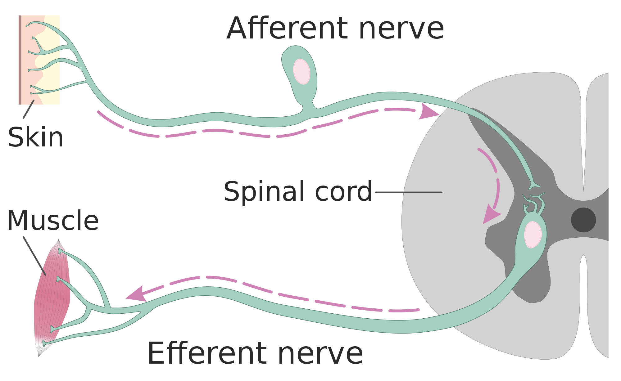
- Sensory nerves transmit information from sensory receptors in the body to the CNS. Sensory nerves are also called afferent nerves. You can see an example in the figure below.
- Motor nerves transmit information from the CNS to muscles, organs, and glands. Motor nerves are also called efferent nerves. You can see one in the figure below.
- Mixed nerves contain both sensory and motor neurons, so they can transmit information in both directions. They have both afferent and efferent functions.
Divisions of the Peripheral Nervous System
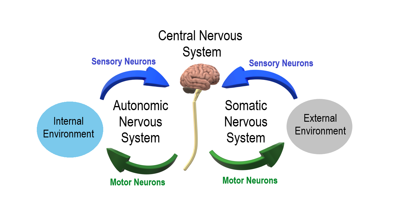
The PNS is divided into two major systems, called the autonomic nervous system and the somatic nervous system. In the diagram below, the autonomic system is shown on the left, and the somatic system on the right. Both systems of the PNS interact with the CNS and include sensory and motor neurons, but they use different circuits of nerves and ganglia.
Somatic Nervous System
The somatic nervous system primarily senses the external environment and controls voluntary activities about which decisions and commands come from the cerebral cortex of the brain. When you feel too warm, for example, you decide to turn on the air conditioner. As you walk across the room to the thermostat, you are using your somatic nervous system. In general, the somatic nervous system is responsible for all of your conscious perceptions of the outside world, as well as all of the voluntary motor activities you perform in response. Whether it’s playing a piano, driving a car, or playing basketball, you can thank your somatic nervous system for making it possible.
Somatic sensory and motor information is transmitted through 12 pairs of cranial nerves and 31 pairs of spinal nerves. Cranial nerves are in the head and neck and connect directly to the brain. Sensory components of cranial nerves transmit information about smells, tastes, light, sounds, and body position. Motor components of cranial nerves control skeletal muscles of the face, tongue, eyeballs, throat, head, and shoulders. Motor components of cranial nerves also control the salivary glands and swallowing. Four of the 12 cranial nerves participate in both sensory and motor functions as mixed nerves, having both sensory and motor neurons.
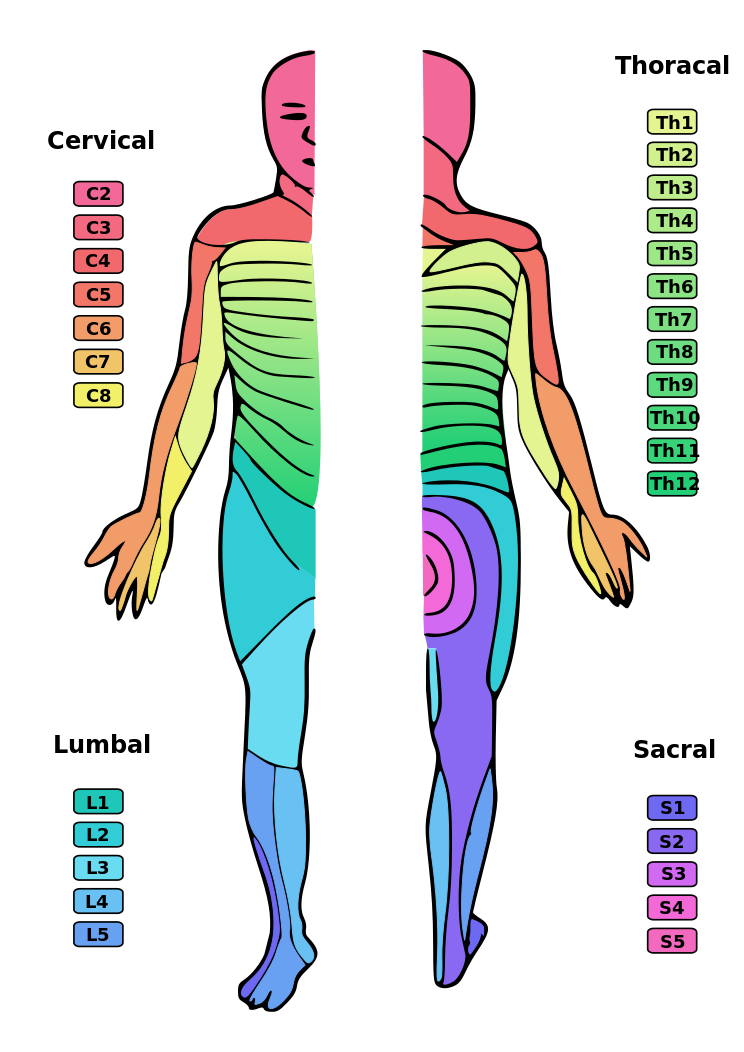
Spinal nerves emanate from the spinal column between vertebrae. All of the spinal nerves are mixed nerves, containing both sensory and motor neurons. The areas of skin innervated by the 31 pairs of spinal nerves are shown in the figure below. These include sensory nerves in the skin that sense pressure, temperature, vibrations, and pain. Other sensory nerves are in the muscles, and they sense stretching and tension. Spinal nerves also include motor nerves that stimulate skeletal muscles to contract, allowing for voluntary body movements.
Autonomic Nervous System
The autonomic nervous system primarily senses the internal environment and controls involuntary activities. It is responsible for monitoring conditions in the internal environment and bringing about appropriate changes in them. In general, the autonomic nervous system is responsible for all the activities that go on inside your body without your conscious awareness or voluntary participation.
Structurally, the autonomic nervous system consists of sensory and motor nerves that run between the CNS (especially the hypothalamus in the brain), internal organs (such as the heart, lungs, and digestive organs), and glands (such as the pancreas and sweat glands). Sensory neurons in the autonomic system detect internal body conditions and send messages to the brain. Motor nerves in the autonomic system affect appropriate responses by controlling contractions of smooth or cardiac muscle, or glandular tissue. For example, when sensory nerves of the autonomic system detect a rise in body temperature, motor nerves signal smooth muscles in blood vessels near the body surface to undergo vasodilation, and the sweat glands in the skin to secrete more sweat to cool the body.
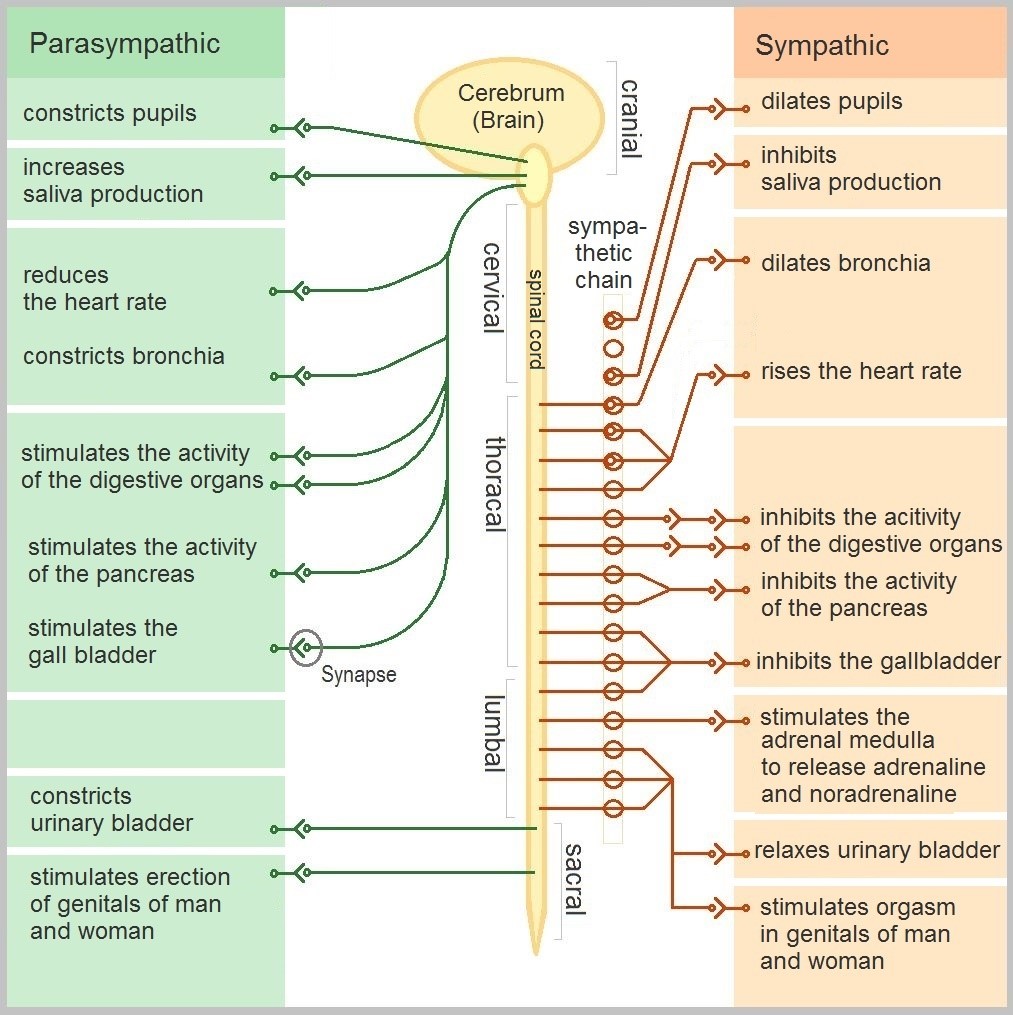
The autonomic nervous system, in turn, has three subdivisions: the sympathetic division, parasympathetic division, and enteric division. The first two subdivisions of the autonomic system are summarized in the figure below. Both affect the same organs and glands, but they generally do so in opposite ways.
- The sympathetic division controls the fight-or-flight response. Changes occur in organs and glands throughout the body that prepare the body to fight or flee in response to a perceived danger. For example, the heart rate speeds up, air passages in the lungs become wider, more blood flows to the skeletal muscles, and the digestive system temporarily shuts down.
- The parasympathetic division returns the body to normal after the fight-or-flight response has occurred. For example, it slows down the heart rate, narrows air passages in the lungs, reduces blood flow to the skeletal muscles, and stimulates the digestive system to start working again. The parasympathetic division also maintains internal homeostasis of the body at other times.
- The enteric division is made up of nerve fibres that supply the organs of the digestive system. This division allows for the local control of many digestive functions.
Disorders of the Peripheral Nervous System
Unlike the CNS — which is protected by bones, meninges, and cerebrospinal fluid — the PNS has no such protections. The PNS also has no blood-brain barrier to protect it from toxins and pathogens in the blood. Therefore, the PNS is more subject to injury and disease than is the CNS. Causes of nerve injury include diabetes, infectious diseases (such as shingles), and poisoning by toxins (such as heavy metals). PNS disorders often have symptoms like loss of feeling, tingling, burning sensations, or muscle weakness. If a traumatic injury results in a nerve being transected (cut all the way through), it may regenerate, but this is a very slow process and may take many months.
Two other diseases of the PNS are Guillain-Barre syndrome and Charcot-Marie-Tooth disease.
- Guillain-Barre syndrome is a rare disease in which the immune system attacks nerves of the PNS, leading to muscle weakness and even paralysis. The exact cause of Guillain-Barre syndrome is unknown, but it often occurs after a viral or bacterial infection. There is no known cure for the syndrome, but most people eventually make a full recovery. Recovery can be slow, however, lasting anywhere from several weeks to several years.
- Charcot-Marie-Tooth disease is a hereditary disorder of the nerves, and one of the most common inherited neurological disorders. It affects predominantly the nerves in the feet and legs, and often in the hands and arms, as well. The disease is characterized by loss of muscle tissue and sense of touch. It is presently incurable.
Feature: My Human Body
The autonomic nervous system is considered to be involuntary because it doesn't require conscious input. However, it is possible to exert some voluntary control over it. People who practice yoga or other so-called mind-body techniques, for example, can reduce their heart rate and certain other autonomic functions. Slowing down these otherwise involuntary responses is a good way to relieve stress and reduce the wear-and-tear that stress can place on the body. Such techniques may also be useful for controlling post-traumatic stress disorder and chronic pain. Three types of integrative practices for these purposes are breathing exercises, body-based tension modulation exercises, and mindfulness techniques.
Breathing exercises can help control the rapid, shallow breathing that often occurs when you are anxious or under stress. These exercises can be learned quickly, and they provide immediate feelings of relief. Specific breathing exercises include paced breath, diaphragmatic breathing, and Breathe2Relax or Chill Zone on MindShift™ CBT, which are downloadable breathing practice mobile applications, or "Apps". Try syncing your breathing with Eric Klassen's "Triangle breathing, 1 minute" video:
https://www.youtube.com/watch?v=u9Q8D6n-3qw
Triangle breathing, 1 minute, Erin Klassen, 2015.
Body-based tension modulation exercises include yoga postures (also known as “asanas”) and tension manipulation exercises. The latter include the Trauma/Tension Release Exercise (TRE) and the Trauma Resiliency Model (TRM). Watch this video for a brief — but informative — introduction to the TRE program:
https://www.youtube.com/watch?v=67R974D8swM&feature=youtu.be
TRE® : Tension and Trauma Releasing Exercises, an Introduction with Jessica Schaffer, Jessica Schaffer Nervous System RESET, 2015.
Mindfulness techniques have been shown to reduce symptoms of depression, as well as those of anxiety and stress. They have also been shown to be useful for pain management and performance enhancement. Specific mindfulness programs include Mindfulness Based Stress Reduction (MBSR) and Mindfulness Mind-Fitness Training (MMFT). You can learn more about MBSR by watching the video below.
https://www.youtube.com/watch?v=0TA7P-iCCcY&feature=youtu.be
Mindfulness-Based Stress Reduction (UMass Medical School, Center for Mindfulness), Palouse Mindfulness, 2017.
8.6 Summary
- The peripheral nervous system (PNS) consists of all the nervous tissue that lies outside the central nervous system (CNS). Its main function is to connect the CNS to the rest of the organism.
- The PNS is made up of nerves and ganglia. Nerves are bundles of axons, and ganglia are groups of cell bodies. Nerves are classified as sensory, motor, or a mix of the two.
- The PNS is divided into the somatic and autonomic nervous systems. The somatic system controls voluntary activities, whereas the autonomic system controls involuntary activities.
- The autonomic nervous system is further divided into sympathetic, parasympathetic, and enteric divisions. The sympathetic division controls fight-or-flight responses during emergencies, the parasympathetic system controls routine body functions the rest of the time, and the enteric division provides local control over the digestive system.
- The PNS is not as well protected physically or chemically as the CNS, so it is more prone to injury and disease. PNS problems include injury from diabetes, shingles, and heavy metal poisoning. Two disorders of the PNS are Guillain-Barre syndrome and Charcot-Marie-Tooth disease.
8.6 Review Questions
- Describe the general structure of the peripheral nervous system. State its primary function.
- What are ganglia?
- Identify three types of nerves based on the direction in which they carry nerve impulses.
- Outline all of the divisions of the peripheral nervous system.
- Compare and contrast the somatic and autonomic nervous systems.
- When and how does the sympathetic division of the autonomic nervous system affect the body?
- What is the function of the parasympathetic division of the autonomic nervous system? Specifically, how does it affect the body?
- Name and describe two peripheral nervous system disorders.
- Give one example of how the CNS interacts with the PNS to control a function in the body.
- For each of the following types of information, identify whether the neuron carrying it is sensory or motor, and whether it is most likely in the somatic or autonomic nervous system:
- Visual information
- Blood pressure information
- Information that causes muscle contraction in digestive organs after eating
- Information that causes muscle contraction in skeletal muscles based on the person’s decision to make a movement
-
8.6 Explore More
https://www.youtube.com/watch?v=ySIDMU2cy0Y&feature=emb_logo
Phantom Limbs Explained, Plethrons, 2015.
https://www.youtube.com/watch?time_continue=1&v=73yo5nJne6c&feature=emb_logo
Why Do Hot Peppers Cause Pain? Reactions, 2015.
Attributions
Figure 8.6.1
Kid’s piant duet by PJMixer on Flickr is used under a CC BY-NC-ND 2.0 (https://creativecommons.org/licenses/by-nc-nd/2.0/) license.
Figure 8.6.2
Nervous_system_diagram by ¤~Persian Poet Gal on Wikimedia Commons is released into the public domain (https://en.wikipedia.org/wiki/Public_domain).
Figure 8.6.3
Afferent_and_efferent_neurons_en.svg by Helixitta on Wikimedia Commons is used under a CC BY-SA 4.0 (https://creativecommons.org/licenses/by-sa/4.0) license.
Figure 8.6.4
Autonomic and Somatic Nervous System by Christinelmiller on Wikimedia Commons is used under a CC BY-SA 4.0 (https://creativecommons.org/licenses/by-sa/4.0) license.
Figure 8.6.5
Dermatoms.svg by Ralf Stephan (mailto:ralf@ark.in-berlin.de) on Wikimedia Commons is released into the public domain (https://en.wikipedia.org/wiki/Public_domain).
Figure 8.6.6
The_Autonomic_Nervous_System by Geo-Science-International on Wikimedia Commons is used and adapted by Christine Miller under a CC0 1.0 Universal
Public Domain Dedication license (https://creativecommons.org/publicdomain/zero/1.0/).
References
Erin Klassen. (2015, December 15). Triangle breathing, 1 minute. YouTube. https://www.youtube.com/watch?v=u9Q8D6n-3qw&feature=youtu.be
Jessica Schaffer Nervous System RESET. (2015, January 15). TRE® : Tension and trauma releasing exercises, an Introduction with Jessica Schaffer. YouTube. https://www.youtube.com/watch?v=67R974D8swM&feature=youtu.be
Mayo Clinic Staff. (n.d.). Charcot-Marie-Tooth disease [online article]. MayoClinic.org. https://www.mayoclinic.org/diseases-conditions/charcot-marie-tooth-disease/symptoms-causes/syc-20350517
Mayo Clinic Staff. (n.d.). Diabetes [online article]. MayoClinic.org. https://www.mayoclinic.org/diseases-conditions/diabetes/symptoms-causes/syc-20371444
Mayo Clinic Staff. (n.d.). Guillain-Barre syndrome [online article]. MayoClinic.org. https://www.mayoclinic.org/diseases-conditions/guillain-barre-syndrome/symptoms-causes/syc-20362793
Mayo Clinic Staff. (n.d.). Shingles [online article]. MayoClinic.org. https://www.mayoclinic.org/diseases-conditions/shingles/symptoms-causes/syc-20353054
Mayo Clinic Staff. (n.d.). Stroke [online article]. MayoClinic.org. https://www.mayoclinic.org/diseases-conditions/stroke/symptoms-causes/syc-20350113
Palouse Mindfulness. (2017, March 25). Mindfulness-based stress reduction (UMass Medical School, Center for Mindfulness), YouTube. https://www.youtube.com/watch?v=0TA7P-iCCcY&feature=youtu.be
Plethrons, (2015, March 23). Phantom limbs explained. YouTube. https://www.youtube.com/watch?v=ySIDMU2cy0Y&feature=youtu.be
Reactions. (2015, December 1). Why do hot peppers cause pain? YouTube. https://www.youtube.com/watch?v=73yo5nJne6c&feature=youtu.be
Created by: CK-12/Adapted by Christine Miller

Oh, the Agony!
Wearing braces can be very uncomfortable, but it is usually worth it. Braces and other orthodontic treatments can re-align the teeth and jaws to improve bite and appearance. Braces can change the position of the teeth and the shape of the jaws because the human body is malleable. Many phenotypic traits — even those that have a strong genetic basis — can be molded by the environment. Changing the phenotype in response to the environment is just one of several ways we respond to environmental stress.
Types of Responses to Environmental Stress
There are four different types of responses that humans may make to cope with environmental stress:
- Adaptation
- Developmental adjustment
- Acclimatization
- Cultural responses
The first three types of responses are biological in nature, and the fourth type is cultural. Only adaptation involves genetic change and occurs at the level of the population or species. The other three responses do not require genetic change, and they occur at the individual level.
Adaptation
An adaptation is a genetically-based trait that has evolved because it helps living things survive and reproduce in a given environment. Adaptations generally evolve in a population over many generations in response to stresses that last for a long period of time. Adaptations come about through natural selection. Those individuals who inherit a trait that confers an advantage in coping with an environmental stress are likely to live longer and reproduce more. As a result, more of their genes pass on to the next generation. Over many generations, the genes and the trait they control become more frequent in the population.
A Classic Example: Hemoglobin S and Malaria
Probably the most frequently-cited example of a genetic adaptation to an environmental stress is sickle cell trait. As you read in the previous section, people with sickle cell trait have one abnormal allele (S) and one normal allele (A) for hemoglobin, the red blood cell protein that carries oxygen in the blood. Sickle cell trait is an adaptation to the environmental stress of malaria, because people with the trait have resistance to this parasitic disease. In areas where malaria is endemic (present year-round), the sickle cell trait and its allele have evolved to relatively high frequencies. It is a classic example of natural selection favoring heterozygotes for a gene with two alleles. This type of selection keeps both alleles at relatively high frequencies in a population.
To Taste or Not to Taste
Another example of an adaptation in humans is the ability to taste bitter compounds. Plants produce a variety of toxic compounds in order to protect themselves from being eaten, and these toxic compounds often have a bitter taste. The ability to taste bitter compounds is thought to have evolved as an adaptation, because it prevented people from eating poisonous plants. Humans have many different genes that code for bitter taste receptors, allowing us to taste a wide variety of bitter compounds.
A harmless bitter compound called phenylthiocarbamide (PTC) is not found naturally in plants, but it is similar to toxic bitter compounds that are found in plants. Humans' ability to taste this harmless substance has been tested in many different populations. In virtually every population studied, there are some people who can taste PTC (called tasters), and some people who cannot taste PTC, (called nontasters). The ratio of tasters to non-tasters varies among populations, but on average, 75 per cent of people can taste PTC and 25 per cent cannot.
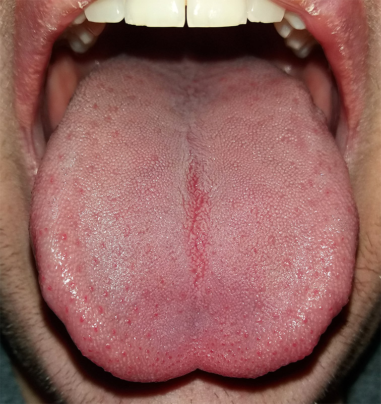
Like many scientific discoveries, human variation in PTC-taster status was discovered by chance. Around 1930, a chemist named Arthur Fox was working with powdered PTC in his lab. Some of the powder accidentally blew into the air. Another lab worker noticed that the powdered PTC tasted bitter, but Fox couldn't detect any taste at all. Fox wondered how to explain this difference in PTC-tasting ability. Geneticists soon determined that PTC-taster status is controlled by a single gene with two common alleles, usually represented by the letters T and t. The T allele encodes a chemical receptor protein (found in taste buds on the tongue, as illustrated in Figure 6.4.2) that can strongly bind to PTC. The other allele, t, encodes a version of the receptor protein that cannot bind as strongly to PTC. The particular combination of these two alleles that a person inherits determines whether the person finds PTC to taste very bitter (TT), somewhat bitter (Tt), or not bitter at all (tt).
If the ability to taste bitter compounds is advantageous, why does every human population studied contain a significant percentage of people who are nontasters? Why has the nontasting allele been preserved in human populations at all? Some scientists hypothesize that the nontaster allele actually confers the ability to taste some other, yet-to-be identified, bitter compound in plants. People who inherit both alleles would presumably be able to taste a wider range of bitter compounds, so they would have the greatest ability to avoid plant toxins. In other words, the heterozygote genotype for the taster gene would be the most fit and favored by natural selection.
Most people no longer have to worry whether the plants they eat contain toxins. The produce you grow in your garden or buy at the supermarket consists of known varieties that are safe to eat. However, natural selection may still be at work in human populations for the PTC-taster gene, because PTC tasters may be more sensitive than nontasters to bitter compounds in tobacco and vegetables in the cabbage family (that is, cruciferous vegetables, such as the broccoli, cauliflower, and cabbage pictured in Figure 6.4.3).
- People who find PTC to taste very bitter are less likely to smoke tobacco, presumably because tobacco smoke has a stronger bitter taste to these individuals. In this case, selection would favor taster genotypes, because tasters would be more likely to avoid smoking and its serious health risks.
- Strong tasters find cruciferous vegetables to taste bitter. As a result, they may avoid eating these vegetables (and perhaps other foods, as well), presumably resulting in a diet that is less varied and nutritious. In this scenario, natural selection might work against taster genotypes.
Figure 6.4.3 Cruciferous vegetables.
Developmental Adjustment
It takes a relatively long time for genetic change in response to environmental stress to produce a population with adaptations. Fortunately, we can adjust to some environmental stresses more quickly by changing in nongenetic ways. One type of nongenetic response to stress is developmental adjustment. This refers to phenotypic change that occurs during development in infancy or childhood, and that may persist into adulthood. This type of change may be irreversible by adulthood.
Phenotypic Plasticity
Developmental adjustment is possible because humans have a high degree of phenotypic plasticity, which is the ability to alter the phenotype in response to changes in the environment. Phenotypic plasticity allows us to respond to changes that occur within our lifetime, and it is particularly important for species (like our own) that have a long generation time. With long generations, evolution of genetic adaptations may occur too slowly to keep up with changing environmental stresses.
Developmental Adjustment and Cultural Practices
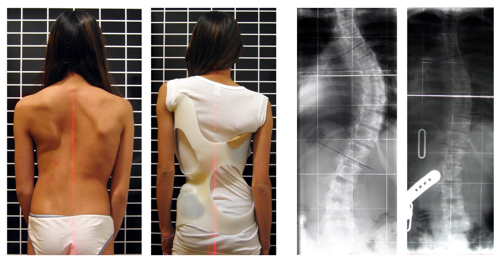
Developmental adjustment may be the result of naturally occurring environmental stresses or cultural practices, including medical or dental treatments. Like our example at the beginning of this section, using braces to change the shape of the jaw and the position of the teeth is an example of a dental practice that brings about a developmental adjustment. Another example of developmental adjustment is the use of a back brace to treat scoliosis (see images in Figure 6.4.4). Scoliosis is an abnormal curvature from side to side in the spine. If the problem is not too severe, a brace, if worn correctly, should prevent the curvature from worsening as a child grows, although it cannot straighten a curve that is already present. Surgery may be required to do that.
Developmental Adjustment and Nutritional Stress
An important example of developmental adjustment that results from a naturally occurring environmental stress is the cessation of physical growth that occurs in children who are under nutritional stress. Children who lack adequate food to fuel both growth and basic metabolic processes are likely to slow down in their growth rate — or even to stop growing entirely. Shunting all available calories and nutrients into essential life functions may keep the child alive at the expense of increasing body size.
Table 6.4.1 shows the effects of inadequate diet on children's' growth in several countries worldwide. For each country, the table gives the prevalence of stunting in children under the age of five. Children are considered stunted if their height is at least two standard deviations below the median height for their age in an international reference population.
Table 6.4.1
Percentage of Stunting in Young Children in Selected Countries (2011-2015)
| Percentage of Stunting in Young Children in Selected Countries (2011-2015) | |
| Country | Per cent of Children Under Age 5 with Stunting |
| United States | 2.1 |
| Turkey | 9.5 |
| Mexico | 13.6 |
| Thailand | 16.3 |
| Iraq | 22.6 |
| Philippines | 33.6 |
| Pakistan | 45.0 |
| Papua New Guinea | 49.5 |
After a growth slow-down occurs and if adequate food becomes available, a child may be able to make up the loss of growth. If food is plentiful, the child may grow more rapidly than normal until the original, genetically-determined growth trajectory is reached. If the inadequate diet persists, however, the failure of growth may become chronic, and the child may never reach his or her full potential adult size.
Phenotypic plasticity of body size in response to dietary change has been observed in successive generations within populations. For example, children in Japan were taller, on average, in each successive generation after the end of World War II. Boys aged 14-15 years old in 1986 were an average of about 18 cm (7 in.) taller than boys of the same age in 1959, a generation earlier. This is a highly significant difference, and it occurred too quickly to be accounted for by genetic change. Instead, the increase in height is a developmental adjustment, thought to be largely attributable to changes in the Japanese diet since World War II. During this period, there was an increase in the amount of animal protein and fat, as well as in the total calories consumed.
Acclimatization
Other responses to environmental stress are reversible and not permanent, whether they occur in childhood or adulthood. The development of reversible changes to environmental stress is called acclimatization. Acclimatization generally develops over a relatively short period of time. It may take just a few days or weeks to attain a maximum response to a stress. When the stress is no longer present, the acclimatized state declines, and the body returns to its normal baseline state. Generally, the shorter the time for acclimatization to occur, the more quickly the condition is reversed when the environmental stress is removed.
Acclimatization to UV Light
A common example of acclimatization is tanning of the skin (see Figure 6.4.5). This occurs in many people in response to exposure to ultraviolet radiation from the sun. Special pigment cells in the skin, called melanocytes, produce more of the brown pigment melanin when exposed to sunlight. The melanin collects near the surface of the skin where it absorbs UV radiation so it cannot penetrate and potentially damage deeper skin structures. Tanning is a reversible change in the phenotype that helps the body deal temporarily with the environmental stress of high levels of UV radiation. When the skin is no longer exposed to the sun’s rays, the tan fades, generally over a period of a few weeks or months.
Figure 6.4.5 Tanning of the skin occurs in many people in response to exposure to ultraviolet radiation from the sun.
Acclimatization to Heat
Another common example of acclimatization occurs in response to heat. Changes that occur with heat acclimatization include increased sweat output and earlier onset of sweat production, which helps the body stay cool because evaporation of sweat takes heat from the body’s surface in a process called evaporative cooling. It generally takes a couple of weeks for maximum heat acclimatization to come about by gradually working out harder and longer at high air temperatures. The changes that occur with acclimatization just as quickly subside when the body is no longer exposed to excessive heat.
Acclimatization to High Altitude

Short term acclimatization to high altitude occurs as a response to low levels of oxygen in the blood. This reduced level of oxygen is detected by carotid bodies, which will trigger in increase in breathing and heart rate. Over a period of weeks the body will compensate by increasing red blood cell production, thereby improving the oxygen-carrying capacity of the blood. This is why mountaineers wishing to climb to the peak of Mount Everest must complete the full climb in portions; it is recommended that climbers spend 2-3 days acclimatizing for every 600 metres of elevation increase. In addition, the higher to altitude, the longer it make take to acclimatize; climbers are advised to spend 4-5 days acclimatizing at base camp (whether the base camp in Nepal or China) before completing the final leg of the climb to the peak. The concentration of red blood cells gradually decreases to normal levels once a climber returns to their normal elevation.
Cultural Responses
More than any other species, humans respond to environmental stresses with learned behaviors and technology. These cultural responses allow us to change our environments to control stresses, rather than changing our bodies genetically or physiologically to cope with the stresses. Even archaic humans responded to some environmental stresses in this way. For example, Neanderthals used shelters, fires, and animal hides as clothing to stay warm in the cold climate in Europe during the last ice age. Today, we use more sophisticated technologies to stay warm in cold climates while retaining our essentially tropical-animal anatomy and physiology. We also use technology (such as furnaces and air conditioners) to avoid temperature stress and stay comfortable in hot or cold climates.
6.4 Summary
- Humans may respond to environmental stress in four different ways: adaptation, developmental adjustment, acclimatization, and cultural responses.
- An adaptation is a genetically based trait that has evolved because it helps living things survive and reproduce in a given environment. Adaptations evolve by natural selection in populations over a relatively long period to time. Examples of adaptations include sickle cell trait as an adaptation to the stress of endemic malaria and the ability to taste bitter compounds as an adaptation to the stress of bitter-tasting toxins in plants.
- A developmental adjustment is a non-genetic response to stress that occurs during infancy or childhood, and that may persist into adulthood. This type of change may be irreversible. Developmental adjustment is possible because humans have a high degree of phenotypic plasticity. It may be the result of environmental stresses (such as inadequate food), which may stunt growth, or cultural practices (such as orthodontic treatments), which re-align the teeth and jaws.
- Acclimatization is the development of reversible changes to environmental stress that develop over a relatively short period of time. The changes revert to the normal baseline state after the stress is removed. Examples of acclimatization include tanning of the skin and physiological changes (such as increased sweating) that occur with heat acclimatization.
- More than any other species, humans respond to environmental stress with learned behaviors and technology, which are cultural responses. These responses allow us to change our environment to control stress, rather than changing our bodies genetically or physiologically to cope with stress. Examples include using shelter, fire, and clothing to cope with a cold climate.
6.4 Review Questions
- List four different types of responses that humans may make to cope with environmental stress.
- Define adaptation.
-
- Explain how natural selection may have resulted in most human populations having people who can and people who cannot taste PTC.
- What is a developmental adjustment?
- Define phenotypic plasticity.
- Explain why phenotypic plasticity may be particularly important in a species with a long generation time.
- Why may stunting of growth occur in children who have an inadequate diet? Why is stunting preferable to the alternative?
- What is acclimatization?
- How does acclimatization to heat come about, and what are two physiological changes that occur in heat acclimatization?
- Give an example of a cultural response to heat stress.
- Which is more likely to be reversible — a change due to acclimatization, or a change due to developmental adjustment? Explain your answer.
6.4 Explore More
https://www.youtube.com/watch?v=upp9-w6GPhU
Could we survive prolonged space travel? - Lisa Nip, TED-Ed, 2016.
https://www.youtube.com/watch?v=hRnrIpUMyZQ&t=182s
How this disease changes the shape of your cells - Amber M. Yates, TED-Ed, 2019.
Attributions
Figure 6.4.1
Free_Awesome_Girl_With_Braces_Close_Up by D. Sharon Pruitt from Hill Air Force Base, Utah, USA on Wikimedia Commons is used under a CC BY 2.0 (https://creativecommons.org/licenses/by/2.0/deed.en) license.
Figure 6.4.2
Tongue by Mahdiabbasinv on Wikimedia Commons is used under a CC BY-SA 4.0 (https://creativecommons.org/licenses/by-sa/4.0/deed.en) license.
Figure 6.4.3
- White cauliflower on brown wooden chopping board by Louis Hansel @shotsoflouis on Unsplash is used under the Unsplash License (https://unsplash.com/license).
- Broccoli on wooden chopping board by Louis Hansel @shotsoflouis on Unsplash is used under the Unsplash License (https://unsplash.com/license).
- Green cabbage close up by Craig Dimmick on Unsplash is used under the Unsplash License (https://unsplash.com/license).
- Cabbage hybrid/ brussel sprouts by Solstice Hannan on Unsplash is used under the Unsplash License (https://unsplash.com/license).
- Kale by Laura Johnston on Unsplash is used under the Unsplash License (https://unsplash.com/license).
- Tiny bok choy at the Asian market by Jodie Morgan on Unsplash is used under the Unsplash License (https://unsplash.com/license).
Figure 6.4.4
Scoliosis_patient_in_cheneau_brace_correcting_from_56_to_27_deg by Weiss H.R. from Scoliosis Journal/BioMed Central Ltd. on Wikimedia Commons is used under a CC BY 2.0 (https://creativecommons.org/licenses/by/2.0) license.
Figure 6.4.5
- Tan Lines by k.steudel on Flickr is used under a CC BY 2.0 (https://creativecommons.org/licenses/by/2.0/) license.
- Twin tan lines (all sizes) by Quinn Dombrowski on Flickr is used under a CC BY-SA 2.0 (https://creativecommons.org/licenses/by-sa/2.0/) license.
- Wedding ring tan line by Quinn Dombrowski on Flickr is used under a CC BY-SA 2.0 (https://creativecommons.org/licenses/by-sa/2.0/) license.
- Tan by Evil Erin on Flickr is used under a CC BY 2.0 (https://creativecommons.org/licenses/by/2.0/) license.
Figure 6.4.6
Nepalese base camp by Mark Horrell on Flickr is used under a CC BY-NC-SA 2.0 (https://creativecommons.org/licenses/by-nc-sa/2.0/) license.
References
TED-Ed. (2016, October 4). Could we survive prolonged space travel? - Lisa Nip. YouTube. https://www.youtube.com/watch?v=upp9-w6GPhU&feature=youtu.be
TED-Ed. (2019, May 6). How this disease changes the shape of your cells - Amber M. Yates. YouTube. https://www.youtube.com/watch?v=hRnrIpUMyZQ&feature=youtu.be
Weiss, H. (2007). Is there a body of evidence for the treatment of patients with Adolescent Idiopathic Scoliosis (AIS)? [Figure 2 - digital photograph], Scoliosis, 2(19). https://doi.org/10.1186/1748-7161-2-19
The central part of an adrenal gland that is surrounded by the adrenal cortex and that produces catecholamine hormones including adrenaline.
A class of molecules that includes the non-steroid hormones produced by the medulla of the adrenal gland, such as adrenaline, that stimulate the fight-or-flight response.
A non-steroid catecholamine hormone produced by the medulla of the adrenal glands that stimulates the fight-or-flight response.
Facts and statistics collected together for reference or analysis.
Any reaction which requires or absorbs energy from its surroundings, usually in the form of heat.
Amino acids are organic compounds that combine to form proteins.
Created by CK-12 Foundation/Adapted by Christine Miller
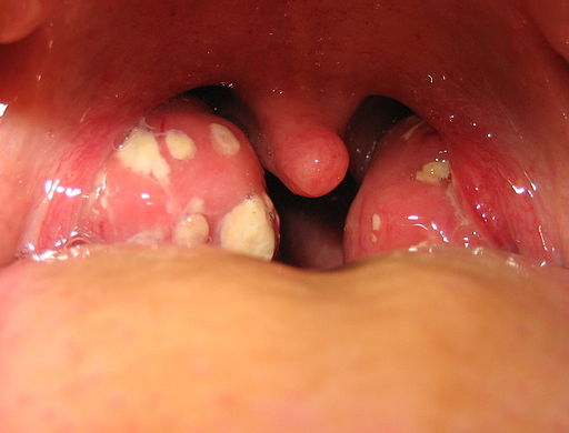
Tonsillitis
The white patches on either side of the throat in Figure 17.3.1 are signs of tonsillitis. The tonsils are small structures in the throat that are very common sites of infection. The white spots on the tonsils pictured here are evidence of infection. The patches consist of large amounts of dead bacteria, cellular debris, and white blood cells — in a word: pus. Children with recurrent tonsillitis may have their tonsils removed surgically to eliminate this type of infection. The tonsils are organs of the lymphatic system.
What Is the Lymphatic System?
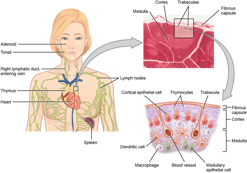
The lymphatic system is a collection of organs involved in the production, maturation, and harboring of white blood cells called lymphocytes. It also includes a network of vessels that transport or filter the fluid known as lymph in which lymphocytes circulate. Figure 17.3.2 shows major lymphatic vessels and other structures that make up the lymphatic system. Besides the tonsils, organs of the lymphatic system include the thymus, the spleen, and hundreds of lymph nodes distributed along the lymphatic vessels.
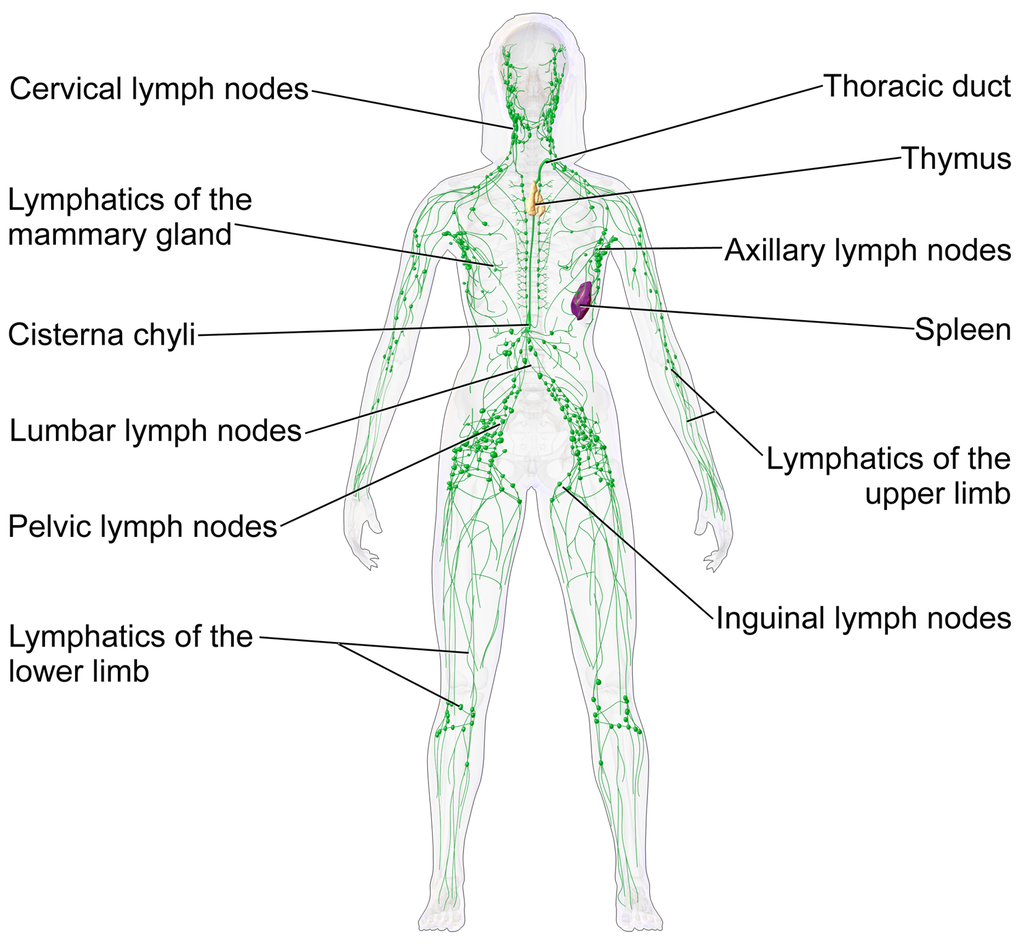
The lymphatic vessels form a transportation network similar in many respects to the blood vessels of the cardiovascular system. However, unlike the cardiovascular system, the lymphatic system is not a closed system. Instead, lymphatic vessels carry lymph in a single direction — always toward the upper chest, where the lymph empties from lymphatic vessels into blood vessels.
Cardiovascular Function of the Lymphatic System
The return of lymph to the bloodstream is one of the major functions of the lymphatic system. When blood travels through capillaries of the cardiovascular system, it is under pressure, which forces some of the components of blood (such as water, oxygen, and nutrients) through the walls of the capillaries and into the tissue spaces between cells, forming tissue fluid, also called interstitial fluid (see Figure 17.3.3). Interstitial fluid bathes and nourishes cells, and also absorbs their waste products. Much of the water from interstitial fluid is reabsorbed into the capillary blood by osmosis. Most of the remaining fluid is absorbed by tiny lymphatic vessels called lymph capillaries. Once interstitial fluid enters the lymphatic vessels, it is called lymph. Lymph is very similar in composition to blood plasma. Besides water, lymph may contain proteins, waste products, cellular debris, and pathogens. It also contains numerous white blood cells, especially the subset of white blood cells known as lymphocytes. In fact, lymphocytes are the main cellular components of lymph.
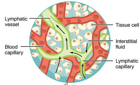
The lymph that enters lymph capillaries in tissues is transported through the lymphatic vessel network to two large lymphatic ducts in the upper chest. From there, the lymph flows into two major veins (called subclavian veins) of the cardiovascular system. Unlike blood, lymph is not pumped through its network of vessels. Instead, lymph moves through lymphatic vessels via a combination of contractions of the vessels themselves and the forces applied to the vessels externally by skeletal muscles, similarly to how blood moves through veins. Lymphatic vessels also contain numerous valves that keep lymph flowing in just one direction, thereby preventing backflow.
Digestive Function of the Lymphatic System
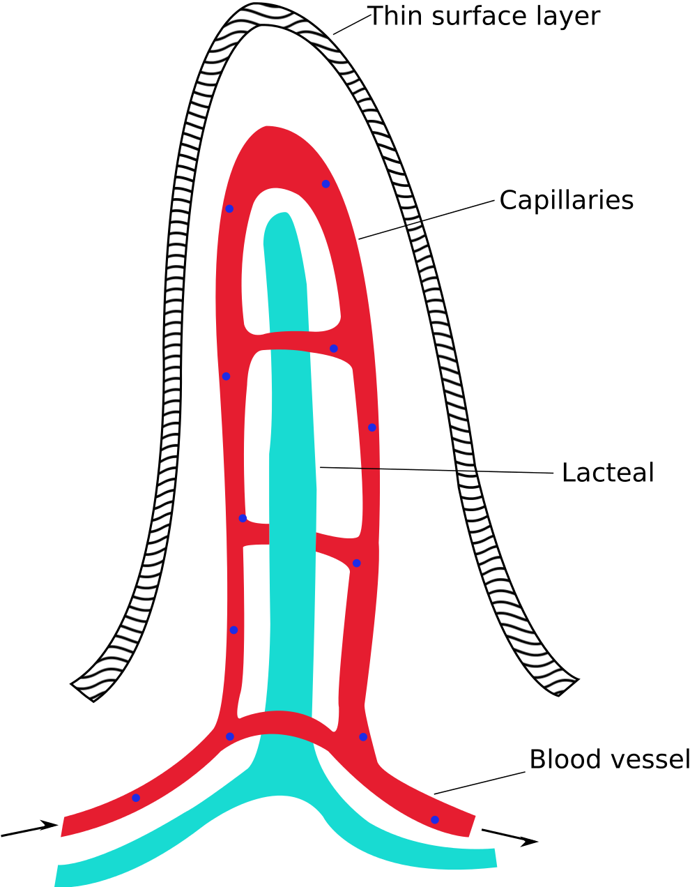
Lymphatic vessels called lacteals (see Figure 17.3.4) are present in the lining of the gastrointestinal tract, mainly in the small intestine. Each tiny villus in the lining of the small intestine has an internal bed of capillaries and lacteals. The capillaries absorb most nutrients from the digestion of food into the blood. The lacteals absorb mainly fatty acids from lipid digestion into the lymph, forming a fatty-acid-enriched fluid called chyle. Vessels of the lymphatic network then transport chyle from the small intestine to the main lymphatic ducts in the chest, from which it drains into the blood circulation. The nutrients in chyle then circulate in the blood to the liver, where they are processed along with the other nutrients that reach the liver directly via the bloodstream.
Immune Function of the Lymphatic System
The primary immune function of the lymphatic system is to protect the body against pathogens and cancerous cells. This function of the lymphatic system is centred on the production, maturation, and circulation of lymphocytes. Lymphocytes are leukocytes that are involved in the adaptive immune system. They are responsible for the recognition of — and tailored defense against — specific pathogens or tumor cells. Lymphocytes may also create a lasting memory of pathogens, so they can be attacked quickly and strongly if they ever invade the body again. In this way, lymphocytes bring about long-lasting immunity to specific pathogens.
There are two major types of lymphocytes, called B cells and T cells. Both B cells and T cells are involved in the adaptive immune response, but they play different roles.
Production and Maturation of Lymphocytes
Like all other types of blood cells (including erythrocytes), both B cells and T cells are produced from stem cells in the red marrow inside bones. After lymphocytes first form, they must go through a complicated maturation process before they are ready to search for pathogens. In this maturation process, they “learn” to distinguish self from non-self. Only those lymphocytes that successfully complete this maturation process go on to actually fight infections by pathogens.
B cells mature in the bone marrow, which is why they are called B cells. After they mature and leave the bone marrow, they travel first to the circulatory system and then enter the lymphatic system to search for pathogens. T cells, on the other hand, mature in the thymus, which is why they are called T cells. The thymus is illustrated in Figure 17.3.5. It is a small lymphatic organ in the chest that consists of an outer cortex and inner medulla, all surrounded by a fibrous capsule. After maturing in the thymus, T cells enter the rest of the lymphatic system to join B cells in the hunt for pathogens. The bone marrow and thymus are called primary lymphoid organs because of their role in the production and/or maturation of lymphocytes.
Lymphocytes in Secondary Lymphoid Organs
The tonsils, spleen, and lymph nodes are referred to as secondary lymphoid organs. These organs do not produce or mature lymphocytes. Instead, they filter lymph and store lymphocytes. It is in these secondary lymphoid organs that pathogens (or their antigens) activate lymphocytes and initiate adaptive immune responses. Activation leads to cloning of pathogen-specific lymphocytes, which then circulate between the lymphatic system and the blood, searching for and destroying their specific pathogens by producing antibodies against them.
Tonsils
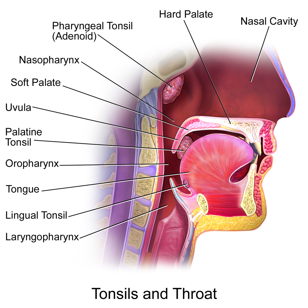
There are four pairs of human tonsils. Three of the four are shown in Figure 17.3.6. The fourth pair, called tubal tonsils, is located at the back of the nasopharynx. The palatine tonsils are the tonsils that are visible on either side of the throat. All four pairs of tonsils encircle a part of the anatomy where the respiratory and gastrointestinal tracts intersect, and where pathogens have ready access to the body. This ring of tonsils is called Waldeyer's ring.
Spleen
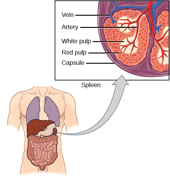
The spleen (Figure 17.3.7) is the largest of the secondary lymphoid organs, and is centrally located in the body. Besides harboring lymphocytes and filtering lymph, the spleen also filters blood. Most dead or aged erythrocytes are removed from the blood in the red pulp of the spleen. Lymph is filtered in the white pulp of the spleen. In the fetus, the spleen has the additional function of producing red blood cells. This function is taken over by bone marrow after birth.
Lymph Nodes
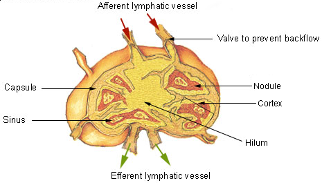
Each lymph node is a small, but organized collection of lymphoid tissue (see Figure 17.3.8) that contains many lymphocytes. Lymph nodes are located at intervals along the lymphatic vessels, and lymph passes through them on its way back to the blood.
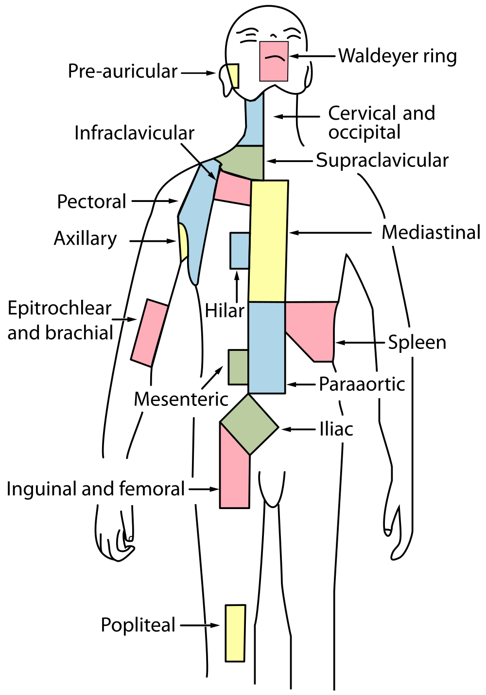
There are at least 500 lymph nodes in the human body. Many of them are clustered at the base of the limbs and in the neck. Figure 17.3.9 shows the major lymph node concentrations, and includes the spleen and the region named Waldeyer’s ring, which consists of the tonsils.
Feature: Myth vs. Reality
When lymph nodes become enlarged and tender to the touch, they are obvious signs of immune system activity. Because it is easy to see and feel swollen lymph nodes, they are one way an individual can monitor his or her own health. To be useful in this way, it is important to know the myths and realities about swollen lymph nodes.
Myth
|
Reality
|
| "You should see a doctor immediately whenever you have swollen lymph nodes." | Lymph nodes are constantly filtering lymph, so it is expected that they will change in size with varying amounts of debris or pathogens that may be present. A minor, unnoticed infection may cause swollen lymph nodes that may last for a few weeks. Generally, lymph nodes that return to their normal size within two or three weeks are not a cause for concern. |
| "Swollen lymph nodes mean you have a bacterial infection." | Although an infection is the most common cause of swollen lymph nodes, not all infections are caused by bacteria. Mononucleosis, for example, commonly causes swollen lymph nodes, and it is caused by viruses. There are also other causes of swollen lymph nodes besides infections, such as cancer and certain medications. |
| "A swollen lymph node means you have cancer." | Cancer is far less likely to be the cause of a swollen lymph node than is an infection. However, if a lymph node remains swollen longer than a few weeks — especially in the absence of an apparent infection — you should have your doctor check it. |
| "Cancer in a lymph node always originates somewhere else. There is no cancer of the lymph nodes." | Cancers do commonly spread from their site of origin to nearby lymph nodes and then to other organs, but cancer may also originate in the lymph nodes. This type of cancer is called lymphoma. |
17.3 Summary
- The lymphatic system is a collection of organs involved in the production, maturation, and harboring of leukocytes called lymphocytes. It also includes a network of vessels that transport or filter the fluid called lymph in which lymphocytes circulate.
- The return of lymph to the bloodstream is one of the functions of the lymphatic system. Lymph flows from tissue spaces — where it leaks out of blood vessels — to the subclavian veins in the upper chest, where it is returned to the cardiovascular system. Lymph is similar in composition to blood plasma. Its main cellular components are lymphocytes.
- Lymphatic vessels called lacteals are found in villi that line the small intestine. Lacteals absorb fatty acids from the digestion of lipids in the digestive system. The fatty acids are then transported through the network of lymphatic vessels to the bloodstream.
- The primary immune function of the lymphatic system is to protect the body against pathogens and cancerous cells. It is responsible for producing mature lymphocytes and circulating them in lymph. Lymphocytes, which include B cells and T cells, are the subset of white blood cells involved in adaptive immune responses. They may create a lasting memory of and immunity to specific pathogens.
- All lymphocytes are produced in bone marrow and then go through a process of maturation in which they “learn” to distinguish self from non-self. B cells mature in the bone marrow, and T cells mature in the thymus. Both the bone marrow and thymus are considered primary lymphatic organs.
- Secondary lymphatic organs include the tonsils, spleen, and lymph nodes. There are four pairs of tonsils that encircle the throat. The spleen filters blood, as well as lymph. There are hundreds of lymph nodes located in clusters along the lymphatic vessels. All of these secondary organs filter lymph and store lymphocytes, so they are sites where pathogens encounter and activate lymphocytes and initiate adaptive immune responses.
17.3 Review Questions
- What is the lymphatic system?
-
- Summarize the immune function of the lymphatic system.
- Explain the difference between lymphocyte maturation and lymphocyte activation.
17.3 Explore More
https://youtu.be/RMLPwOiYnII
What is Lymphoedema or Lymphedema? Compton Care, 2016.
https://youtu.be/ah74jT00jBA
Spleen physiology What does the spleen do in 2 minutes, Simple Nursing, 2015.
https://youtu.be/L4KexZZAdyA
How to check your lymph nodes, University Hospitals Bristol and Weston NHS FT, 2020.
Attributions
Figure 17.3.1
512px-Tonsillitis by Michaelbladon at English Wikipedia on Wikimedia Commons is in the public domain (https://en.wikipedia.org/wiki/Public_domain). (Transferred from en.wikipedia to Commons by Kauczuk)
Figure 17.3.2
Blausen_0623_LymphaticSystem_Female by BruceBlaus on Wikimedia Commons is used under a CC BY 3.0 (https://creativecommons.org/licenses/by/3.0) license.
Figure 17.3.3
2201_Anatomy_of_the_Lymphatic_System (cropped) by OpenStax College on Wikimedia Commons is used under a CC BY 3.0 (https://creativecommons.org/licenses/by/3.0) license.
Figure 17.3.4
1000px-Intestinal_villus_simplified.svg by Snow93 on Wikimedia Commons is in the public domain (https://en.wikipedia.org/wiki/Public_domain).
Figure 17.3.5
2206_The_Location_Structure_and_Histology_of_the_Thymus by OpenStax College on Wikimedia Commons is used under a CC BY 3.0 (https://creativecommons.org/licenses/by/3.0) license.
Figure 17.3.6
Blausen_0861_Tonsils&Throat_Anatomy2 by BruceBlaus on Wikimedia Commons is used under a CC BY 3.0 (https://creativecommons.org/licenses/by/3.0) license.
Figure 17.3.7
Figure_42_02_14 by CNX OpenStax on Wikimedia Commons is used under a CC BY 4.0 (https://creativecommons.org/licenses/by/4.0) license.
Figure 17.3.8
Illu_lymph_node_structure by NCI/ SEER Training on Wikimedia Commons is in the public domain (https://en.wikipedia.org/wiki/Public_domain). (Archives: https://web.archive.org/web/20070311015818/http://training.seer.cancer.gov/module_anatomy/unit8_2_lymph_compo1_nodes.html)
Figure 17.3.9
1000px-Lymph_node_regions.svg by Fred the Oyster (derivative work) on Wikimedia Commons is in the public domain (https://en.wikipedia.org/wiki/Public_domain). (Original by NCI/ SEER Training)
References
Betts, J. G., Young, K.A., Wise, J.A., Johnson, E., Poe, B., Kruse, D.H., Korol, O., Johnson, J.E., Womble, M., DeSaix, P. (2013, June 19). Figure 21.2 Anatomy of the lymphatic system [digital image]. In Anatomy and Physiology (Section 21.1). OpenStax. https://openstax.org/books/anatomy-and-physiology/pages/21-1-anatomy-of-the-lymphatic-and-immune-systems
Betts, J. G., Young, K.A., Wise, J.A., Johnson, E., Poe, B., Kruse, D.H., Korol, O., Johnson, J.E., Womble, M., DeSaix, P. (2013, June 19). Figure 21.7 Location, structure, and histology of the thymus [digital image]. In Anatomy and Physiology (Section 21.1). OpenStax. https://openstax.org/books/anatomy-and-physiology/pages/21-1-anatomy-of-the-lymphatic-and-immune-systems
Blausen.com Staff. (2014). Medical gallery of Blausen Medical 2014". WikiJournal of Medicine 1 (2). DOI:10.15347/wjm/2014.010. ISSN 2002-4436
Compton Care. (2016, March 7). What is lymphoedema or lymphedema? YouTube. https://www.youtube.com/watch?v=RMLPwOiYnII&feature=youtu.be
OpenStax. (2016, May 27) Figure 14. The spleen is similar to a lymph node but is much larger and filters blood instead of lymph [digital image]. In Open Stax, Biology (Section 42.2). OpenStax CNX. https://cnx.org/contents/GFy_h8cu@10.8:etZobsU-@6/Adaptive-Immune-Response
Simple Nursing. (2015, June 28). Spleen physiology What does the spleen do in 2 minutes. YouTube. https://www.youtube.com/watch?v=ah74jT00jBA&feature=youtu.be
University Hospitals Bristol and Weston NHS FT. (2020, May 13). How to check your lymph nodes. YouTube. https://www.youtube.com/watch?v=L4KexZZAdyA&feature=youtu.be
division of the peripheral nervous system that controls involuntary activities
A lasting attraction between atoms, ions or molecules that enables the formation of chemical compounds.
A lasting attraction between atoms, ions or molecules that enables the formation of chemical compounds.
Facts and statistics collected together for reference or analysis.
An explanation of an aspect of the natural world that can be repeatedly tested and verified in accordance with the scientific method.

