8.4 Nerve Impulses

When Lightning Strikes
This amazing cloud-to-surface lightning occurred when a difference in electrical charge built up in a cloud relative to the ground. When the buildup of charge was great enough, a sudden discharge of electricity occurred. A nerve impulse is similar to a lightning strike. Both a nerve impulse and a lightning strike occur because of differences in electrical charge, and both result in an electric current.
Generating Nerve Impulses
A nerve impulse, like a lightning strike, is an electrical phenomenon. A nerve impulse occurs because of a difference in electrical charge across the plasma membrane of a neuron. How does this difference in electrical charge come about? The answer involves ions, which are electrically-charged atoms or molecules.
Resting Potential
When a neuron is not actively transmitting a nerve impulse, it is in a resting state, ready to transmit a nerve impulse. During the resting state, the sodium-potassium pump maintains a difference in charge across the cell membrane of the neuron. The sodium-potassium pump is a mechanism of active transport that moves sodium ions (Na+) out of cells and potassium ions (K+) into cells. The sodium-potassium pump moves both ions from areas of lower to higher concentration, using energy in ATP and carrier proteins in the cell membrane. The video below, “Sodium Potassium Pump” by Amoeba Sisters, describes in greater detail how the sodium-potassium pump works. Sodium is the principal ion in the fluid outside of cells, and potassium is the principal ion in the fluid inside of cells. These differences in concentration create an electrical gradient across the cell membrane, called resting potential. Tightly controlling membrane resting potential is critical for the transmission of nerve impulses.
Sodium Potassium Pump, Amoeba Sisters, 2020.
Action Potential
A nerve impulse is a sudden reversal of the electrical gradient across the plasma membrane of a resting neuron. The reversal of charge is called an action potential. It begins when the neuron receives a chemical signal from another cell or some other type of stimulus. If the stimulus is strong enough to reach threshold, an action potential will take place is a cascade along the axon.
This reversal of charges ripples down the axon of the neuron very rapidly as an electric current, which is illustrated in the diagram below (Figure 8.4.2). A nerve impulse is an all-or-nothing response depending on if the stimulus input was strong enough to reach threshold. If a neuron responds at all, it responds completely. A greater stimulation does not produce a stronger impulse.
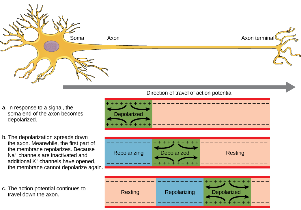
In neurons with a myelin sheath on their axon, ions flow across the membrane only at the nodes between sections of myelin. As a result, the action potential appears to jump along the axon membrane from node to node, rather than spreading smoothly along the entire membrane. This increases the speed at which the action potential travels.
Transmitting Nerve Impulses
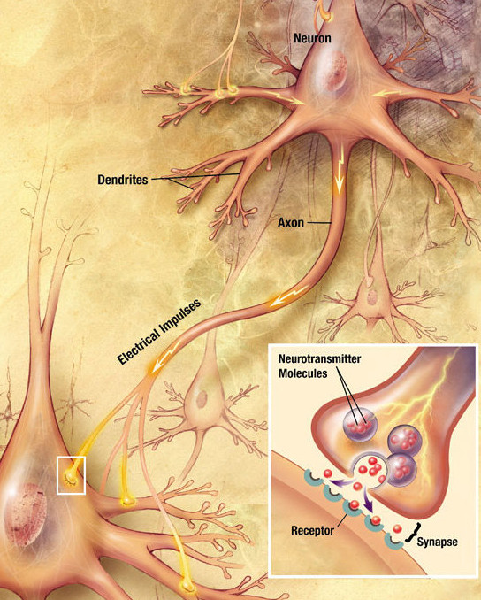
The place where an axon terminal meets another cell is called a synapse. This is where the transmission of a nerve impulse to another cell occurs. The cell that sends the nerve impulse is called the presynaptic cell, and the cell that receives the nerve impulse is called the postsynaptic cell.
Some synapses are purely electrical and make direct electrical connections between neurons. Most synapses, however, are chemical synapses. Transmission of nerve impulses across chemical synapses is more complex.
Chemical Synapses
At a chemical synapse, both the presynaptic and postsynaptic areas of the cells are full of molecular machinery that is involved in the transmission of nerve impulses. As shown in Figure 8.4.3, the presynaptic area contains many tiny spherical vessels called synaptic vesicles that are packed with chemicals called neurotransmitters. When an action potential reaches the axon terminal of the presynaptic cell, it opens channels that allow calcium to enter the terminal. Calcium causes synaptic vesicles to fuse with the membrane, releasing their contents into the narrow space between the presynaptic and postsynaptic membranes. This area is called the synaptic cleft. The neurotransmitter molecules travel across the synaptic cleft and bind to receptors, which are proteins embedded in the membrane of the postsynaptic cell.
Neurotransmitters and Receptors
There are more than a hundred known neurotransmitters, and more than one type of neurotransmitter may be released at a given synapse by a presynaptic cell. For example, it is common for a faster-acting neurotransmitter to be released, along with a slower-acting neurotransmitter. Many neurotransmitters also have multiple types of receptors to which they can bind. Receptors, in turn, can be divided into two general groups: chemically gated ion channels and second messenger systems.
- When a chemically gated ion channel is activated, it forms a passage that allows specific types of ions to flow across the cell membrane. Depending on the type of ion, the effect on the target cell may be excitatory or inhibitory.
- When a second messenger system is activated, it starts a cascade of molecular interactions inside the target cell. This may ultimately produce a wide variety of complex effects, such as increasing or decreasing the sensitivity of the cell to stimuli, or even altering gene transcription.
The effect of a neurotransmitter on a postsynaptic cell depends mainly on the type of receptors that it activates, making it possible for a particular neurotransmitter to have different effects on various target cells. A neurotransmitter might excite one set of target cells, inhibit others, and have complex modulatory effects on still others, depending on the type of receptors. However, some neurotransmitters have relatively consistent effects on other cells. Consider the two most widely used neurotransmitters, glutamate and GABA (gamma-aminobutyric acid). Glutamate receptors are either excitatory or modulatory in their effects, whereas GABA receptors are all inhibitory in their effects in adults.
Problems with neurotransmitters or their receptors can cause neurological disorders. The disease myasthenia gravis, for example, is caused by antibodies from the immune system blocking receptors for the neurotransmitter acetylcholine in postsynaptic muscle cells. This inhibits the effects of acetylcholine on muscle contractions, producing symptoms, such as muscle weakness and excessive fatigue during simple activities. Some mental illnesses (including depression) are caused, at least in part, by imbalances of certain neurotransmitters in the brain. One of the neurotransmitters involved in depression is thought to be serotonin, which normally helps regulate mood, among many other functions. Some antidepressant drugs are thought to help alleviate depression in many patients by normalizing the activity of serotonin in the brain.
8.4 Summary
- A nerve impulse is an electrical phenomenon that occurs because of a difference in electrical charge across the plasma membrane of a neuron.
- The sodium-potassium pump maintains an electrical gradient across the plasma membrane of a neuron when it is not actively transmitting a nerve impulse. This gradient is called the resting potential of the neuron.
- An action potential is a sudden reversal of the electrical gradient across the plasma membrane of a resting neuron. It begins when the neuron receives a chemical signal from another cell or some other type of stimulus. The action potential travels rapidly down the neuron’s axon as an electric current and occurs in three stages: Depolarization, Repolarization and Recovery.
- A nerve impulse is transmitted to another cell at either an electrical or a chemical synapse. At a chemical synapse, neurotransmitter chemicals are released from the presynaptic cell into the synaptic cleft between cells. The chemicals travel across the cleft to the postsynaptic cell and bind to receptors embedded in its membrane.
- There are many different types of neurotransmitters. Their effects on the postsynaptic cell generally depend on the type of receptor they bind to. The effects may be excitatory, inhibitory, or modulatory in more complex ways. Both physical and mental disorders may occur if there are problems with neurotransmitters or their receptors.
8.4 Review Questions
- Define nerve impulse.
- What is the resting potential of a neuron, and how is it maintained?
- Explain how and why an action potential occurs.
- Outline how a signal is transmitted from a presynaptic cell to a postsynaptic cell at a chemical synapse.
- What generally determines the effects of a neurotransmitter on a postsynaptic cell?
- Identify three general types of effects that neurotransmitters may have on postsynaptic cells.
- Explain how an electrical signal in a presynaptic neuron causes the transmission of a chemical signal at the synapse.
- The flow of which type of ion into a neuron results in an action potential? How do these ions get into the cell? What does this flow of ions do to the relative charge inside the neuron compared to the outside?
- Name three neurotransmitters.
-
-
8.4 Explore More
Action Potentials, Teacher’s Pet, 2018.
TED Ed| What is depression? – Helen M. Farrell, Parta Learning, 2017.
5 Weird Involuntary Behaviors Explained!, It’s Okay To Be Smart, 2015.
Attributions
Figure 8.4.1
Lightening/ Purple Lightning, Dee Why by Jeremy Bishop on Unsplash is used under the Unsplash License (https://unsplash.com/license).
Figure 8.4.2
Action Potential by CNX OpenStax, Biology on Wikimedia Commons is used under a CC BY 4.0 (https://creativecommons.org/licenses/by/4.0/deed.en) license.
Figure 8.4.3
Chemical_synapse_schema_cropped by Looie496 created file (adapted from original from US National Institutes of Health, National Institute on Aging) is in the public domain (https://en.wikipedia.org/wiki/Public_domain).
References
Amoeba Sisters. (2020, January 29). Sodium potassium pump. YouTube. https://www.youtube.com/watch?v=7NY6XdPBhxo&feature=youtu.be
CNX OpenStax. (2016, May 27) Figure 4 The action potential is conducted down the axon as the axon membrane depolarizes, then repolarizes [digital image]. In Open Stax, Biology (Section 35.2). OpenStax CNX. https://cnx.org/contents/GFy_h8cu@10.53:cs_Pb-GW@6/How-Neurons-Communicate
It’s Okay To Be Smart. (2015, January 26). 5 Weird involuntary behaviors explained! YouTube. https://www.youtube.com/watch?v=ZE8sRMZ5BCA&feature=youtu.be
Mayo Clinic Staff. (n.d.). Depression (major depressive disorder) [online article]. MayoClinic.org. https://www.mayoclinic.org/diseases-conditions/depression/symptoms-causes/syc-20356007
Mayo Clinic Staff. (n.d.). Myasthenia gravis [online article]. MayoClinic.org. https://www.mayoclinic.org/diseases-conditions/myasthenia-gravis/symptoms-causes/syc-20352036
National Institute on Aging. (2006, April 8). Alzheimers disease: Unraveling the mystery. National Institutes of Health. https://www.nia.nih.gov/ (archived version)
Parta Learning. (2017, December 8). TED Ed| What is depression? – Helen M. Farrell. YouTube. https://www.youtube.com/watch?v=rBcU_apy0h8&t=291s
Teacher’s Pet. (2018, August 26). Action potentials. YouTube. https://www.youtube.com/watch?v=FEHNIELPb0s&feature=youtu.be
A signal transmitted along a nerve fiber.
An atom or molecule with a net electric charge due to the loss or gain of one or more electrons.
The smallest particle of an element that still has the properties of that element.
A molecule is an electrically neutral group of two or more atoms held together by chemical bonds.
Created by CK-12 Foundation/Adapted by Christine Miller
Stroller Moms
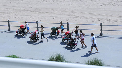
These moms (Figure 12.5.1) are setting a great example for their children by engaging in physical exercise. Adopting a habit of regular physical exercise is one of the most important ways to maintain fitness and good health. From higher self-esteem to a healthier heart, physical exercise can have a positive effect on virtually all aspects of health, including physical, mental, and emotional health.
What Is Physical Exercise?
Physical exercise is any bodily activity that enhances or maintains physical fitness and overall health and wellness. We generally think of physical exercise as activities that are undertaken for the main purpose of improving physical fitness and health. However, physical activities that are undertaken for other purposes may also count as physical exercise. Scrubbing a floor, raking a lawn, or playing active games with young children or a pet are all activities that can have fitness and health benefits, even though they generally are not done mainly for this purpose.
How much physical exercise should people get? In the Canada, both the Canadian Food Guide and the Canadian Society for Exercise Physiology recommend that every child and adult who is able should participate in moderate exercise for a minimum of 60 minutes a day. This might include walking, swimming, and/or household or yard work.
Types of Physical Exercise
Physical exercise can be classified into three types, depending on the effects it has on the body: aerobic exercise, anaerobic exercise, and flexibility exercise. Many specific examples of physical exercise (including playing soccer and rock climbing) can be classified as more than one type.
Aerobic Exercise

Aerobic exerciseis any physical activity in which muscles are used at well below their maximum contraction strength, but for long periods of time. Aerobic exercise uses a relatively high percentage of slow-twitch muscle fibres that consume a large amount of oxygen. The main goal of aerobic exercise is to increase cardiovascular endurance, although it can have many other benefits, including muscle toning. Examples of aerobic exercise include cycling, swimming, brisk walking, jumping rope, rowing, hiking, tennis, and kayaking as shown in Figure 12.5.2 .
Anaerobic Exercise
Anaerobic exercise is any physical activity in which muscles are used at close to their maximum contraction strength, but for relatively short periods of time. Anaerobic exercise uses a relatively high percentage of fast-twitch muscle fibres that consume a small amount of oxygen. Goals of anaerobic exercise include building and strengthening muscles, as well as improving bone strength, balance, and coordination. Examples of anaerobic exercise include push-ups, lunges, sprinting, interval training, resistance training, and weight training (such as biceps curls with a dumbbell, as pictured in Figure 12.5.3).

Flexibility Exercise

Flexibility exercise is any physical activity that stretches and lengthens muscles. Goals of flexibility exercise include increasing joint flexibility, keeping muscles limber, and improving the range of motion, all of which can reduce the risk of injury. Examples of flexibility exercise include stretching, yoga (as in Figure 12.5.4), and tai chi.
Health Benefits of Physical Exercise
Many studies have shown that physical exercise is positively correlated with a diversity of health benefits. Some of these benefits include maintaining physical fitness, losing weight and maintaining a healthy weight, regulating digestive health, building and maintaining healthy bone density, increasing muscle strength, improving joint mobility, strengthening the immune system, boosting cognitive ability, and promoting psychological well-being. Some studies have also found a significant positive correlation between exercise and both quality of life and life expectancy. People who participate in moderate to high levels of physical activity have been shown to have lower mortality rates than people of the same ages who are not physically active and daily exercise has been shown to increase life expectancy up to an average of five years.
The underlying physiological mechanisms explaining why exercise has these positive health benefits are not completely understood. However, developing research suggests that many of the benefits of exercise may come about because of the role of skeletal muscles as endocrine organs. Contracting muscles release hormones called myokines, which promote tissue repair and the growth of new tissue. Myokines also have anti-inflammatory effects, which, in turn, reduce the risk of developing inflammatory diseases. Exercise also reduces levels of cortisol, the adrenal cortex stress hormone that may cause many health problems — both physical and mental — at sustained high levels.
Cardiovascular Benefits of Physical Exercise
The beneficial effects of exercise on the cardiovascular system are well documented. Physical inactivity has been identified as a risk factor for the development of coronary artery disease. There is also a direct correlation between physical inactivity and cardiovascular disease mortality. Physical exercise, in contrast, has been demonstrated to reduce several risk factors for cardiovascular disease, including hypertension (high blood pressure), “bad” cholesterol (low-density lipoproteins), high total cholesterol, and excess body weight. Physical exercise has also been shown to increase “good” cholesterol (high-density lipoproteins), insulin sensitivity, the mechanical efficiency of the heart, and exercise tolerance, which is the ability to perform physical activity without undue stress and fatigue.
Cognitive Benefits of Physical Exercise
Physical exercise has been shown to help protect people from developing neurodegenerative disorders, such as dementia. A 30-year study of almost 2,400 men found that those who exercised regularly had a 59 per cent reduction in dementia when compared with those who did not exercise. Similarly, a review of cognitive enrichment therapies for the elderly found that physical activity — in particular, aerobic exercise — can enhance the cognitive function of older adults. Anecdotal evidence suggests that frequent exercise may even help reverse alcohol-induced brain damage. There are several possible reasons why exercise is so beneficial for the brain. Physical exercise:
- Increases blood flow and oxygen availability to the brain.
- Increases growth factors that promote new brain cells and new neuronal pathways in the brain.
- Increases levels of neurotransmitters (such as serotonin), which increase memory retention, information processing, and cognition.
Mental Health Benefits of Physical Exercise
Numerous studies suggest that regular aerobic exercise works as well as pharmaceutical antidepressants in treating mild-to-moderate depression. A possible reason for this effect is that exercise increases the biosynthesis of at least three neurochemicals that may act as euphoriants. The euphoric effect of exercise is well known. Distance runners may refer to it as “runner’s high,” and people who participate in crew (as in Figure 12.5.5) may refer to it as “rower’s high.” Because of these effects, health care providers often promote the use of aerobic exercise as a treatment for depression.

Additional mental health benefits of physical exercise include reducing stress, improving body image, and promoting positive self-esteem. Conversely, there is evidence to suggest that being sedentary is associated with increased risk of anxiety.
Sleep Benefits of Physical Exercise
A recent review of published scientific research suggests that exercise generally improves sleep for most people, and helps sleep disorders, such as insomnia. In fact, exercise is the most recommended alternative to sleeping pills for people with insomnia. For sleep benefits, the optimum time to exercise may be four to eight hours before bedtime, although exercise at any time of day seems to be beneficial. The only possible exception is heavy exercise undertaken shortly before bedtime, which may actually interfere with sleep.
Other Benefits of Physical Exercise
Some studies suggest that physical activity may benefit the immune system. For example, moderate exercise has been found to be associated with a decreased incidence of upper respiratory tract infections. Evidence from many studies has found a correlation between physical exercise and reduced death rates from cancer, specifically breast cancer and colon cancer. Physical exercise has also been shown to reduce the risk of type 2 diabetes and obesity.
Variation in Responses to Physical Exercise

Not everyone benefits equally from physical exercise. When participating in aerobic exercise, most people will have a moderate increase in their endurance, but some people will as much as double their endurance. Some people, on the other hand, will show little or no increase in endurance from aerobic exercise. Genetic differences in slow-twitch and fast-twitch skeletal muscle fibres may play a role in these different results. People with more slow-twitch fibres may be able to develop greater endurance, because these muscle fibres have more capillaries, mitochondria, and myoglobin than fast-twitch fibres. As a result, slow-twitch fibres can carry more oxygen and sustain aerobic activity for a longer period of time than fast-twitch fibres. Studies show that endurance athletes (like the marathoner pictured in Figure 12.5.6) generally do tend to have a higher proportion of slow-twitch fibres than other people.
There is also great variation in individual responses to muscle building as a result of anaerobic exercise. Some people have a much greater capacity to increase muscle size and strength, whereas other people never develop large muscles, no matter how much they exercise them. People who have more fast-twitch than slow-twitch muscle fibres may be able to develop bigger, stronger muscles, because fast-twitch muscle fibres contribute more to muscle strength and have greater potential to increase in mass. Evidence suggests that athletes who excel at power activities (such as throwing and jumping) tend to have a higher proportion of fast-twitch fibres than do endurance athletes.
Can You “Overdose” on Physical Exercise?
Is it possible to exercise too much? Can too much exercise be harmful? Evidence suggests that some adverse effects may occur if exercise is extremely intense and the body is not given proper rest between exercise sessions. Athletes who train for multiple marathons have been shown to develop scarring of the heart and heart rhythm abnormalities. Doing too much exercise without prior conditioning also increases the risk of injuries to muscles and joints. Damage to muscles due to overexertion is often seen in new military recruits (see Figure 12.5.7). Too much exercise in females may cause amenorrhea, which is a cessation of menstrual periods. When this occurs, it generally indicates that a woman is pushing her body too hard.
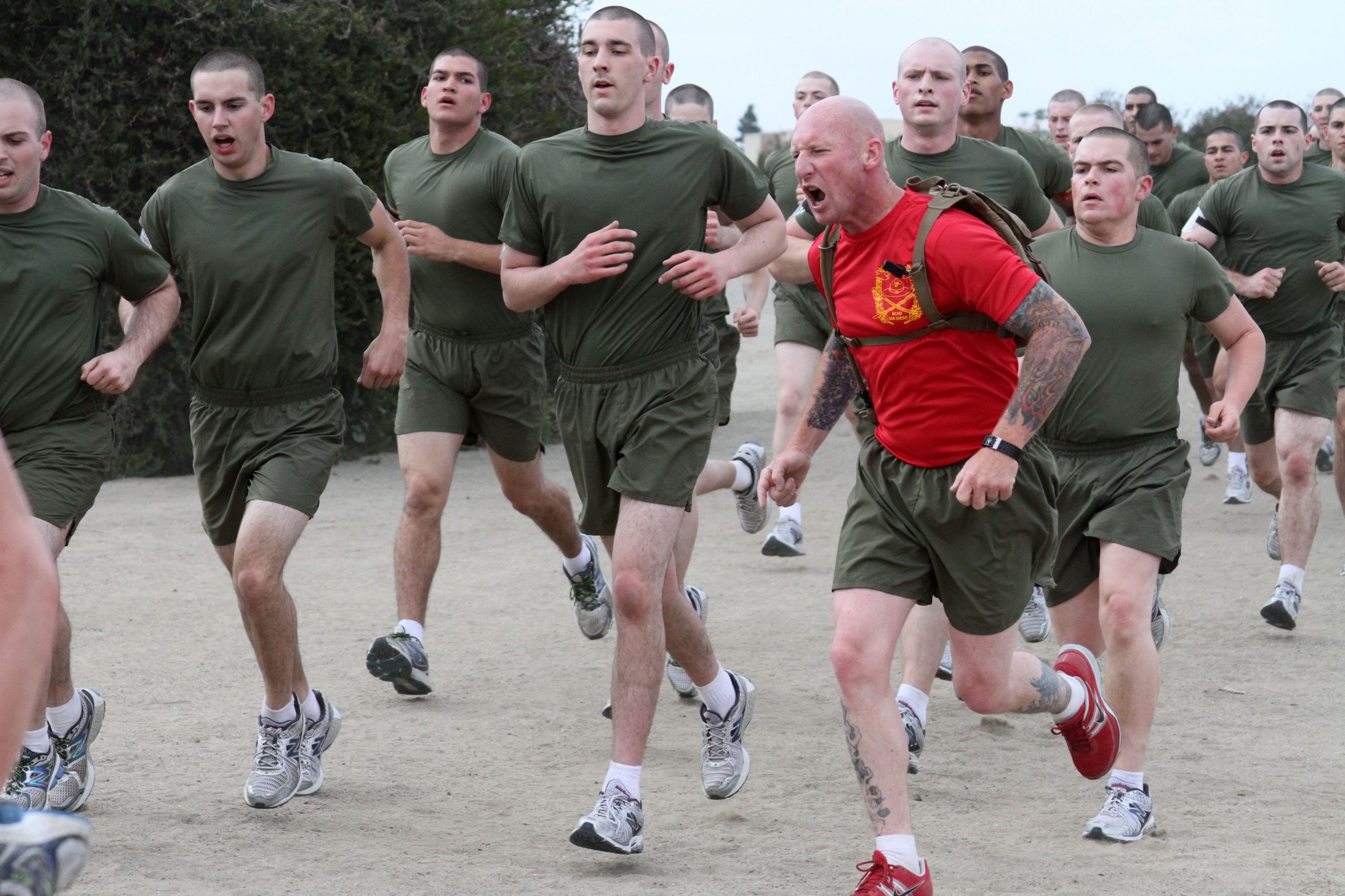
Many people develop delayed onset muscle soreness (DOMS), which is pain or discomfort in muscles that is felt one to three days after exercising, and generally subsides two or three days later. DOMS was once thought to be caused by the buildup of lactic acid in the muscles. Lactic acid is a product of anaerobic respiration in muscle tissues. However, lactic acid disperses fairly rapidly, so it is unlikely to explain pain experienced several days after exercise. The current theory is that DOMS is caused by tiny tears in muscle fibres, which occur when muscles are used at too high a level of intensity.
Feature: My Human Body
Most people know that exercise is important for good health, and it’s easy to find endless advice about exercise programs and fitness plans. What is not so easy to find is the motivation to start exercising — and to stick with it. This is the main reason why so many people fail to get regular exercise. Practical concerns like a busy schedule and bad weather can certainly make exercising more of a challenge, but the biggest barriers to adopting a regular exercise routine are mental. If you want to exercise but find yourself making excuses or getting discouraged and giving up, here are some tips that may help you get started and stay moving:
- Avoid an all-or-nothing point of view. Don’t think you need to spend hours sweating at the gym or training for a marathon to get healthy. Even a little bit of exercise is better than nothing at all. Start out with ten or 15 minutes of moderate activity each day. Taking a walk around your neighborhood is a great way to begin! From there, gradually increase the amount of time until you are exercising to at least 30 minutes a day, five days a week. Make it a routine.
- Be kind to yourself, and reinforce positive behaviors with rewards. Don’t be down on yourself because you are overweight or out of shape. Don’t beat yourself up because of a supposed lack of willpower. Instead, look at any past failures as opportunities to learn and do better. When you do achieve even small exercise goals, treat yourself to something special. Did you just complete your first workout? Reward yourself with a relaxing bath or other treat.
- Don’t make excuses for not exercising. Common complaints include being too busy or tired or not athletic enough. Such excuses are not valid reasons to avoid exercising, and they will sabotage any plans to improve your fitness. If you can’t find a 30-minute period to work out, try to find ten minutes, three times a day. If you’re feeling tired, know that exercise can actually reduce fatigue and boost your energy level. If you feel clumsy and uncoordinated, remind yourself that you don’t need to be athletic to take a walk or engage in vigorous house or yard work.
- Find an activity that you truly enjoy doing. Don’t think you have to lift weights or run on a treadmill to exercise your muscles. If you find such activities boring or unpleasant, you won’t stick with them. Any activity that increases your heart rate and uses large muscles can provide a workout, especially if you’re not in the habit of exercising, so find something you like to do. Do you like to dance? Put on some music and dance up a sweat! Do you enjoy gardening? Get out in the yard and dig up some dirt! Still not interested? Try an activity-based video game, such as Wii or Kinect. You may find it so much fun that it doesn’t seem like exercise until you realize you’ve worked up a sweat.
- Make yourself accountable. Tell friends and family members that you’re going to start exercising. You’ll be letting them — as well as yourself — down if you don’t follow through. Some people find that keeping an exercise log to track their progress is a good way to be accountable and stick to an exercise program. Perhaps the best way to keep at it is to find an exercise partner. If you’ve got someone waiting to exercise with you, you will be less likely to make excuses for not exercising.
- Add more physical activity to your daily life. You don’t need to follow a structured exercise program to increase your activity level. Do your house or yard work briskly for a workout. Park your car further than necessary from work or the mall, and walk the extra distance. If you live close enough, leave the car at home and walk to and from your destination. Rather than taking elevators or escalators, walk up and down stairs. When you take breaks at work, take a walk instead of sitting. Every time a commercial comes on while you’re watching TV, take a quick exercise break — run in place or do some curls with hand weights.
12.5 Summary
- Physical exercise is any bodily activity that enhances or maintains physical fitness and overall health. Activities such as household chores may count as physical exercise, even if they are not done for their health benefits. Current recommendations for adults are 30 minutes a day of moderate exercise.
- Aerobic exercise is any physical activity that uses muscles at less than their maximum contraction strength, but for long periods of time. This type of exercise uses a relatively high percentage of slow-twitch muscle fibres that consume large amounts of oxygen. Aerobic exercises increase cardiovascular endurance and include cycling and brisk walking.
- Anaerobic exercise is any physical activity that uses muscles at close to their maximum contraction strength, but for short periods of time. This type of exercise uses a relatively high percentage of fast-twitch muscle fibres that consume small amounts of oxygen. Anaerobic exercises increase muscle and bone mass and strength, and they include push-ups and sprinting.
- Flexibility exercise is any physical activity that stretches and lengthens muscles, thereby improving range of motion and reducing risk of injury. Examples include stretching and yoga.
- Many studies have shown that physical exercise is positively correlated with a diversity of physical, mental, and emotional health benefits. Physical exercise also increases quality of life and life expectancy.
- Many of the benefits of exercise may come about because contracting muscles release hormones called myokines, which promote tissue repair and growth and have anti-inflammatory effects.
- Physical exercise can reduce risk factors for cardiovascular disease, including hypertension and excess body weight. Physical exercise can also increase factors associated with cardiovascular health, such as mechanical efficiency of the heart.
- Physical exercise has been shown to offer protection from dementia and other cognitive problems, perhaps because it increases blood flow or neurotransmitters in the brain, among other potential effects.
- Numerous studies suggest that regular aerobic exercise works as well as pharmaceutical antidepressants in treating mild-to-moderate depression, possibly because it increases synthesis of natural euphoriants in the brain.
- Research shows that physical exercise generally improves sleep for most people and helps sleep disorders, such as insomnia. Other health benefits of physical exercise include better immune system function and reduced risk of type 2 diabetes and obesity.
- There is great variation in individual responses to exercise, partly due to genetic differences in proportions of slow-twitch and fast-twitch skeletal muscle fibres. People with more slow-twitch fibres may be able to develop greater endurance from aerobic exercise, whereas people with more fast-twitch fibres may be able to develop greater muscle size and strength from anaerobic exercise.
- Some adverse effects may occur if exercise is extremely intense and the body is not given proper rest between exercise sessions. Many people who overwork their muscles develop delayed onset muscle soreness (DOMS), which may be caused by tiny tears in muscle fibres.
12.5 Review Questions
- How do we define physical exercise?
- What are current recommendations for physical exercise for adults?
-
- Define flexibility exercise, and state its benefits. What are two examples of flexibility exercises?
- In general, how does physical exercise affect health, quality of life, and longevity?
- What mechanism may underlie many of the general health benefits of physical exercise?
- Relate physical exercise to cardiovascular disease risk.
- What may explain the positive benefits of physical exercise on cognition?
- How does physical exercise compare with antidepressant drugs in the treatment of depression?
- Identify several other health benefits of physical exercise.
- Explain how genetics may influence the way individuals respond to physical exercise.
- Can too much physical exercise be harmful?
12.5 Explore More
https://www.youtube.com/watch?v=hmFQqjMF_f0
How playing sports benefits your body ... and your brain - Leah Lagos and Jaspal Ricky Singh, TED-Ed, 2016.
https://www.youtube.com/watch?v=rLsimrBoYXc&t=12s
The surprising reason our muscles get tired - Christian Moro, TED-Ed, 2019.
https://youtu.be/2tM1LFFxeKg
What makes muscles grow? - Jeffrey Siegel, TED-Ed, 2015.
https://www.youtube.com/watch?v=QeIrdqU0o9s
Why some people find exercise harder than others | Emily Balcetis, TED, 2014.
Attributions
Figure 12.5.1
stroller fit by Serge Melki from Indianapolis, USA on Wikimedia Commons is used under a CC BY 2.0 (https://creativecommons.org/licenses/by/2.0) license.
Figure 12.5.2
Children kayaking young sport by Hagerty Ryan, USFWS on Pixnio is used under a public domain (CC0) Certification (https://creativecommons.org/licenses/publicdomain/).
Figure 12.5.3
Bicep curls [photo] by Senior Airman Jarrod Grammel from U.S. Moody Air Force Base is released into the public domain (https://en.wikipedia.org/wiki/Public_domain).
Figure 12.5.4
Flexibility exercise by carl-barcelo-nqUHQkuVj3c [photo] by Carl Barcelo on Unsplash is used under the Unsplash License (https://unsplash.com/license).
Figure 12.5.5
Canadian women’s double scull silver Rio 2016 by Gerhard Pratt on Flickr is used under a CC BY-NC 2.0 (https://creativecommons.org/licenses/by-nc/2.0/) license.
Figure 12.5.6
Toronto Marathon 2012 by Marc Roberts on Flickr is used under a CC BY-NC-SA 2.0 (https://creativecommons.org/licenses/by-nc-sa/2.0/) license.
Figure 12.5.7
Muscle damage in military recruits by Lance Cpl. Bridget M. Keane from the United States Marine Corps Recruit Depot is in the public domain (https://en.wikipedia.org/wiki/Public_domain).
References
Elwood, P., Galante, J., Pickering, J., Palmer, S., Bayer, A., Ben-Shlomo, Y., Longley, M., & Gallacher, J. (2013). Healthy lifestyles reduce the incidence of chronic diseases and dementia: evidence from the Caerphilly cohort study. PloS one, 8(12), e81877. https://doi.org/10.1371/journal.pone.0081877
Mayo Clinic Staff. (n.d.). Amenorrhea [online article]. MayoClinic.org. https://www.mayoclinic.org/diseases-conditions/amenorrhea/symptoms-causes/syc-20369299#
Mayo Clinic Staff. (n.d.). Coronary artery disease [online article]. MayoClinic.org. https://www.mayoclinic.org/diseases-conditions/coronary-artery-disease/symptoms-causes/syc-20350613
TED-Ed. (2016, June 28). How playing sports benefits your body ... and your brain - Leah Lagos and Jaspal Ricky Singh. YouTube. https://www.youtube.com/watch?v=hmFQqjMF_f0&feature=youtu.be
TED-Ed. (2019, April 18). The surprising reason our muscles get tired - Christian Moro. YouTube. https://www.youtube.com/watch?v=rLsimrBoYXc&feature=youtu.be
TED-Ed. (2015, November 3). What makes muscles grow? - Jeffrey Siegel. YouTube https://www.youtube.com/watch?v=2tM1LFFxeKg&feature=youtu.be
TED. (2014, November 14). Why some people find exercise harder than others | Emily Balcetis, YouTube. https://www.youtube.com/watch?v=QeIrdqU0o9s&feature=youtu.be
Wikipedia contributors. (2020, August 1). Delayed onset muscle soreness. In Wikipedia. https://en.wikipedia.org/w/index.php?title=Delayed_onset_muscle_soreness&oldid=970682631
A solute pump that pumps potassium into cells while pumping sodium out of cells, both against their concentration gradients. This pumping is active and occurs at the ratio of 2 potassium for every 3 calcium.
The semipermeable membrane surrounding the cytoplasm of a cell.
The movement of ions or molecules across a cell membrane into a region of higher concentration, assisted by enzymes and requiring energy.
A complex organic chemical that provides energy to drive many processes in living cells, e.g. muscle contraction, nerve impulse propagation, and chemical synthesis. Found in all forms of life, ATP is often referred to as the "molecular unit of currency" of intracellular energy transfer.
Image shows several baguettes laying on a counter
Created by CK-12 Foundation/Adapted by Christine Miller
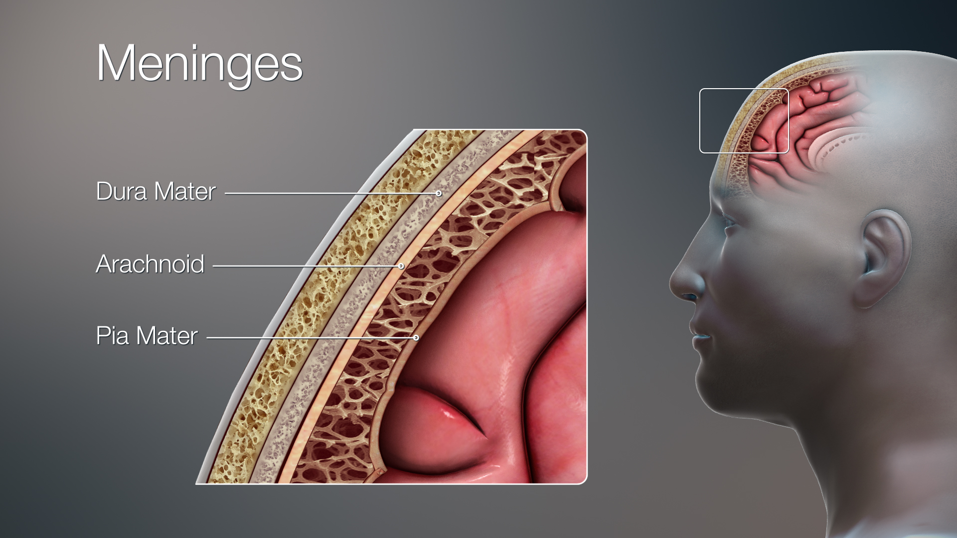
As you learned in this chapter, the human body consists of many complex systems that normally work together efficiently — like a well-oiled machine — to carry out life’s functions. For example, the image above (Figure 7.9.1) illustrates how the brain and spinal cord are protected by layers of membrane called meninges and fluid that flows between the meninges and in spaces called ventricles inside the brain. This fluid is called cerebrospinal fluid, and as you have learned, one of its important functions is to cushion and protect the brain and spinal cord, which make up most of the central nervous system (CNS). Additionally, cerebrospinal fluid circulates nutrients and removes waste products from the CNS. Cerebrospinal fluid is produced continually in the ventricles, circulates throughout the CNS, and is then reabsorbed by the bloodstream. If too much cerebrospinal fluid is produced, its flow is blocked, or not enough is reabsorbed, the system becomes out of balance and it can build up in the ventricles. This causes an enlargement of the ventricles called hydrocephalus that can put pressure on the brain, resulting in the types of neurological problems that former professional football player Jayson, described in the beginning of this chapter, is suffering from.
Recall that Jayson’s symptoms included loss of bladder control, memory loss, and difficulty walking. The cause of his symptoms was not immediately clear, although his doctors suspected that it related to the nervous system, since the nervous system acts as the control centre of the body, controlling and regulating many other organ systems. Jayson’s memory loss directly implicated the brain's involvement, since that is the site of thoughts and memory. The urinary system is also controlled in part by the nervous system, so the inability to hold urine appropriately can also be a sign of a neurological issue. Jayson’s trouble walking involved the muscular system, which works alongside the skeletal system to enable movement of the limbs. In turn, the contraction of muscles is regulated by the nervous system. You can see why a problem in the nervous system can cause a variety of different symptoms by affecting multiple organ systems in the human body.
To try to find the exact cause of Jayson’s symptoms, his doctors performed a lumbar puncture (or spinal tap), which is the removal of some cerebrospinal fluid through a needle inserted into the lower part of the spinal canal. They then analyzed Jayson’s cerebrospinal fluid for the presence of pathogens (such as bacteria) to determine whether an infection was the cause of his neurological symptoms. When no evidence of infection was found, they used an MRI to observe the structures of his brain. This is when they discovered his enlarged ventricles, which are a hallmark of hydrocephalus.
To treat Jayson’s hydrocephalus, a surgeon implanted a device called a shunt in his brain to remove the excess fluid. An illustration of a brain shunt is shown in Figure 9.7.2 . One side of the shunt consists of a small tube, called a catheter, which was inserted into Jayson’s ventricles. Excess cerebrospinal fluid is then drained through a one-way valve to the other end of the shunt, which was threaded under his skin to his abdominal cavity, where the fluid is released and can be reabsorbed by the bloodstream.
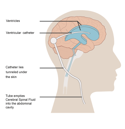
Implantation of a shunt is the most common way to treat hydrocephalus, and for some people, it can allow them to recover almost completely. However, there can be complications associated with a brain shunt. The shunt can have mechanical problems or cause an infection. Also, the rate of draining must be carefully monitored and adjusted to balance the rate of cerebrospinal fluid removal with the rate of its production. If it is drained too fast, it is called overdraining, and if it is drained too slowly, it is called underdraining. In the case of underdraining, the pressure on the brain and associated neurological symptoms will persist. In the case of overdraining, the ventricles can collapse, which can cause serious problems, such as the tearing of blood vessels and hemorrhaging. To avoid these problems, some shunts have an adjustable pressure valve, where the rate of draining can be adjusted by placing a special magnet over the scalp. You can see how the proper balance between cerebrospinal fluid production and removal is so critical – both in the causes of hydrocephalus and in its treatment.
In what other ways does your body regulate balance, or maintain a state of homeostasis? In this chapter you learned about the feedback loops that keep body temperature and blood glucose within normal ranges. Other important examples of homeostasis in the human body are the regulation of the pH in the blood and the balance of water in the body. You will learn more about homeostasis in different body systems in the coming chapters.
Thanks to Jayson’s shunt, his symptoms are starting to improve, but he has not fully recovered. Time may tell whether the removal of the excess cerebrospinal fluid from his ventricles will eventually allow him to recover normal functioning or whether permanent damage to his nervous system has already been done. The flow of cerebrospinal fluid might seem simple, but when it gets out of balance, it can easily wreak havoc on multiple organ systems because of the intricate interconnectedness of the systems within the human “machine."
To learn more about hydrocephalus and its treatment, watch this video from Boston Children's Hospital:
https://www.youtube.com/watch?v=bHD8zYImKqA
Hydrocephalus and its treatment | Boston Children’s Hospital, 2011.
Chapter 7 Summary
This chapter provided an overview of the organization and functioning of the human body. You learned that:
- The human body consists of multiple parts that function together to maintain life. The biology of the human body incorporates the body’s structure — or anatomy — and the body’s functioning, or physiology.
- The organization of the human body is a hierarchy of increasing size and complexity, starting at the level of atoms and molecules and ending at the level of the entire organism.
- Cells are the level of organization above atoms and molecules, and they are the basic units of structure and function of the human body. Each cell carries out basic life functions, as well as other specific roles. Cells of the human body show a lot of variation.
-
- Variations in cell function are generally reflected in variations in cell structure.
- Some cells are unattached to other cells and can move freely. Others are attached to each other and cannot move freely. Some cells can divide readily and form new cells, and others can divide only under exceptional circumstances. Many cells are specialized to produce and secrete particular substances.
- All the different cell types within an individual have the same genes. Cells can vary because different genes are expressed depending on the cell type.
- Many common types of human cells consist of several subtypes of cells, each of which has a special structure and function. For example, subtypes of bone cells include osteocytes, osteoblasts, osteogenic cells, and osteoclasts.
- A tissue is a group of connected cells that have a similar function. There are four basic types of human tissues that make up all the organs of the human body: epithelial, muscle, nervous, and connective tissues.
-
- Connective tissues, such as bone, tendons and blood, are made up of a scattering of living cells that are separated by non-living material, called extracellular matrix.
- Epithelial tissues, such as skin and mucous membranes, protect the body and its internal organs and secrete or absorb substances.
- Muscular tissues are made up of cells that have the unique ability to contract. They include skeletal, smooth, and cardiac muscle tissues.
- Nervous tissues are made up of neurons, which transmit messages, and neuroglia of various types, which play supporting roles.
- An organ is a structure that consists of two or more types of tissues that work together to do the same job. The brain and the heart are two examples.
-
- Many organs are composed of a major tissue that performs the organ’s main function, as well as other tissues that play supporting roles.
- The human body contains five organs that are considered vital for survival: the heart, brain, kidneys, liver, and lungs. If any of these five organs stops functioning, death of the organism is imminent without medical intervention.
- An organ system is a group of organs that work together to carry out a complex overall function. For example, the skeletal system provides structure to the body and protects internal organs.
-
- There are 11 major organ systems in the human organism. They are the integumentary, skeletal, muscular, nervous, endocrine, cardiovascular, lymphatic, respiratory, digestive, urinary, and reproductive systems. Only the reproductive system varies significantly between males and females.
- The human body is divided into a number of body cavities. A body cavity is a fluid-filled space in the body that holds and protects internal organs. The two largest human body cavities are the ventral cavity and dorsal cavity.
-
- The ventral cavity is at the anterior (or front) of the trunk. It is subdivided into the thoracic cavity, abdominal cavity and the pelvic cavity.
- The dorsal cavity is at the posterior (or back) of the body, and includes the head and the back of the trunk. It is subdivided into the cranial cavity and spinal cavity.
- Organ systems of the human body must work together to keep the body alive and functioning normally. This requires communication among organ systems. This is controlled by the autonomic nervous system and endocrine system. The autonomic nervous system controls involuntary body functions, such as heart rate and digestion. The endocrine system secretes hormones into the blood that travel to body cells and influence their activities.
-
- Cellular respiration is a good example of organ system interactions, because it is a basic life process that occurs in all living cells. It is the intracellular process that breaks down glucose with oxygen to produce carbon dioxide and energy. Cellular respiration requires the interaction of the digestive, cardiovascular, and respiratory systems.
- The fight-or-flight response is a good example of how the nervous and endocrine systems control other organ system responses. It is triggered by a message from the brain to the endocrine system and prepares the body for flight or a fight. Many organ systems are stimulated to respond, including the cardiovascular, respiratory, and digestive systems.
- Playing softball or doing other voluntary physical activities may involve the interaction of nervous, muscular, skeletal, respiratory, and cardiovascular systems.
- Homeostasis is the condition in which a system such as the human body is maintained in a more or less steady state. It is the job of cells, tissues, organs, and organ systems throughout the body to maintain homeostasis.
-
- For any given variable (such as body temperature), there is a particular set point that is the physiological optimum value. The spread of values around the set point that is considered insignificant is called the normal range.
- Homeostasis is generally maintained by a negative feedback loop that includes a stimulus, sensor, control centre, and effector. Negative feedback serves to reduce an excessive response and to keep a variable within the normal range. Negative feedback loops control body temperature and the blood glucose level.
- Sometimes homeostatic mechanisms fail, resulting in homeostatic imbalance. Diabetes is an example of a disease caused by homeostatic imbalance. Aging can bring about a reduction in the efficiency of the body’s control system, making the elderly more susceptible to disease.
- Positive feedback loops are not common in biological systems. Positive feedback serves to intensify a response until an end point is reached. Positive feedback loops control blood clotting and childbirth.
The severe and broad impact of hydrocephalus on the body’s systems highlights the importance of the nervous system and its role as the master control system of the body. In the next chapter, you will learn much more about the structures and functioning of this fascinating and important system.
Chapter 7 Review
-
- Compare and contrast tissues and organs.
-
-
- Which type of tissue lines the inner and outer surfaces of the body?
- What is a vital organ? What happens if a vital organ stops working?
- Name three organ systems that transport or remove wastes from the body.
- Name two types of tissue in the digestive system.
-
- Describe one way in which the integumentary and cardiovascular systems work together to regulate homeostasis in the human body.
-
- True or False: Body cavities are filled with air.
- In which organ system is the pituitary gland? Describe how the pituitary gland increases metabolism.
- When the level of thyroid hormone in the body gets too high, it acts on other cells to reduce production of more thyroid hormone. What type of feedback loop does this represent?
- Hypothetical organ A is the control centre in a feedback loop that helps maintain homeostasis. It secretes molecule A1 which reaches organ B, causing organ B to secrete molecule B1. B1 negatively feeds back onto organ A, reducing the production of A1 when the level of B1 gets too high.
- What is the stimulus in this feedback loop?
- If the level of B1 falls significantly below the set point, what do you think happens to the production of A1? Why?
- What is the effector in this feedback loop?
- If organs A and B are part of the endocrine system, what type of molecules do you think A1 and B1 are likely to be?
- What are the two main systems that allow various organ systems to communicate with each other?
- What are two functions of the hypothalamus?
Attributions
Figure 7.9.1
3D Medical Illustration Meninges Details by Scientific Animations on Wikimedia Commons is used under a CC BY-SA 4.0 (https://creativecommons.org/licenses/by-sa/4.0/deed.en) license.
Figure 7.9.2
Hydrocephalus with Shunt from CK-12 Foundation is used under a CC BY-NC 3.0 (https://creativecommons.org/licenses/by-nc/3.0/) license.
 ©CK-12 Foundation Licensed under
 ![]() • Terms of Use • Attribution
• Terms of Use • Attribution
References
Betts, J. G., Young, K.A., Wise, J.A., Johnson, E., Poe, B., Kruse, D.H., Korol, O., Johnson, J.E., Womble, M., DeSaix, P. (2013, April 25). Figure 1.3 Levels of structural organization of the human body [digital image]. In Anatomy and Physiology (Section 1.2). OpenStax. https://openstax.org/books/anatomy-and-physiology/pages/1-2-structural-organization-of-the-human-body
Betts, J. G., Young, K.A., Wise, J.A., Johnson, E., Poe, B., Kruse, D.H., Korol, O., Johnson, J.E., Womble, M., DeSaix, P. (2013, April 25). Figure 1.4 Organ systems of the human body [digital image]. In Anatomy and Physiology (Section 1.2). OpenStax. https://openstax.org/books/anatomy-and-physiology/pages/1-2-structural-organization-of-the-human-body
Betts, J. G., Young, K.A., Wise, J.A., Johnson, E., Poe, B., Kruse, D.H., Korol, O., Johnson, J.E., Womble, M., DeSaix, P. (2013, April 25). Figure 1.15 Dorsal and ventral body cavities [digital image]. In Anatomy and Physiology (Section 1.2). OpenStax. https://openstax.org/books/anatomy-and-physiology/pages/1-6-anatomical-terminology
Boston Children's Hospital. (2011, ). Hydrocephalus and its treatment | Boston Children’s Hospital. YouTube. https://www.youtube.com/watch?v=bHD8zYImKqA&feature=youtu.be
Brainard, J/ CK-12 Foundation. (2016). Figure 2 An illustration of a brain shunt [digital image]. In CK-12 College Human Biology (Section 9.8) [online Flexbook]. CK12.org. https://www.ck12.org/book/ck-12-college-human-biology/section/9.8/
File:Body cavities lateral view labeled.jpg. (2018, January 4). Wikimedia Commons. https://commons.wikimedia.org/w/index.php?title=File:Body_Cavities_Lateral_view_labeled.jpg&oldid=276851269. (Original image: Figure 1.15 Dorsal and ventral body cavities, from OpenStax, Anatomy and Physiology.)
File:Body cavities lateral view labeled.jpg. (2018, January 4). Wikimedia Commons. https://commons.wikimedia.org/w/index.php?title=File:Body_Cavities_Lateral_view_labeled.jpg&oldid=276851269. (Original image: OpenStax [Version 8.25 from the textbook OpenStax Anatomy and Physiology] adapted for Review questions by Christine Miller].
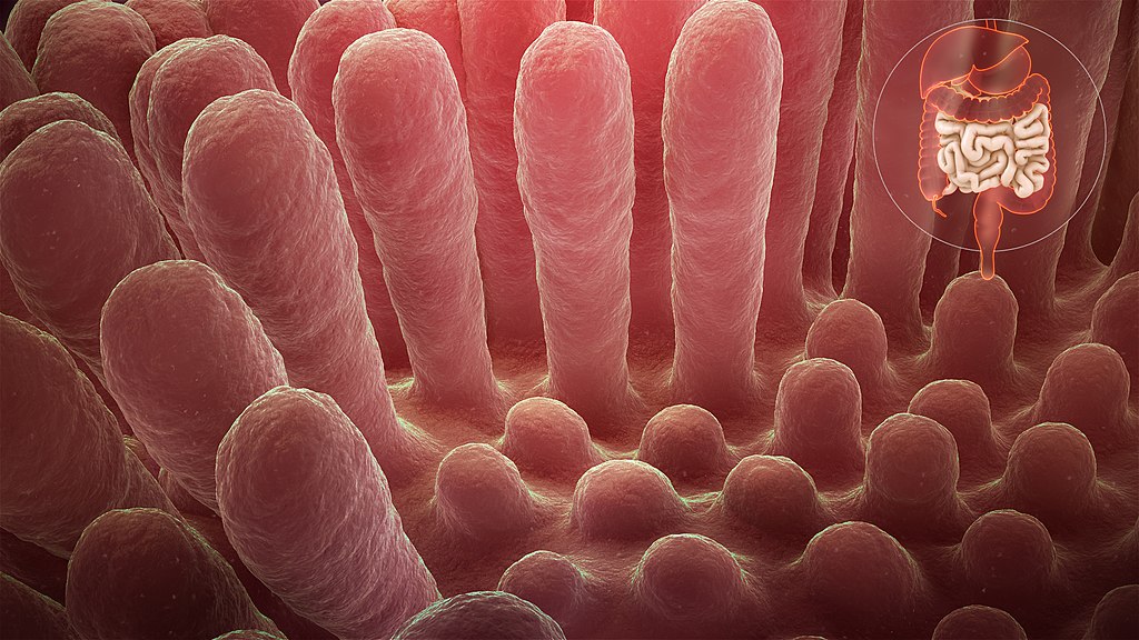
Image shows a computer rendering of villia. The healthy villi are tall narrow projections growing out of the wall of the small intestine, similar to trees in a forest. The celiac-affected villi appear as little more than bumps- like stumps where a forest used to be.
Image shows a leg affect by PAD. Plaques in the leg arteries have caused reduced blood flow to the leg.
Created by CK-12 Foundation/Adapted by Christine Miller

Case Study: Please Don’t Pass the Bread
Angela and Saloni are college students who met in physics class. They decide to study together for their upcoming midterm, but first, they want to grab some lunch. Angela says there is a particular restaurant she would like to go to, because they are able to accommodate her dietary restrictions. Saloni agrees and they head to the restaurant.
At lunch, Saloni asks Angela what is special about her diet. Angela tells her that she can’t eat gluten. Saloni says, “My cousin did that for a while because she heard that gluten is bad for you. But it was too hard for her to not eat bread and pasta, so she gave it up.” Angela tells Saloni that avoiding gluten isn’t optional for her — she has celiac disease. Eating even very small amounts of gluten could damage her digestive system. It can be difficult for people living with celiac disease to find foods when eating out.
You have probably heard of gluten, but what is it, and why is it harmful to people with celiac disease? Gluten is a protein present in wheat and some other grains (such as barley, rye, and oats), so it is commonly found in foods like bread, pasta, baked goods, and many packaged foods, like the ones pictured in Figure 15.1.2.
Figure 15.1.2 Gluten is a protein present in foods like bread, pasta, and baked goods.
For people with celiac disease, eating gluten causes an autoimmune reaction that results in damage to the small, finger-like villi lining the small intestine, causing them to become inflamed and flattened (see Figure 15.1.3). This damage interferes with the digestive process, which can result in a wide variety of symptoms including diarrhea, anemia, skin rash, bone pain, depression, and anxiety, among others. The degree of damage to the villi can vary from mild to severe, with more severe damage generally resulting in more significant symptoms and complications. Celiac disease can have serious long-term consequences, such as osteoporosis, problems in the nervous and reproductive systems, and the development of certain types of cancers.
Why does celiac disease cause so many different types of symptoms and have such significant negative health consequences? As you read this chapter and learn about how the digestive system works, you will see just how important the villi of the small intestine are to the body as a whole. At the end of the chapter, you will learn more about celiac disease, why it can be so serious, and whether it is worth avoiding gluten for people who do not have a diagnosed medical issue with it.
Chapter Overview: Digestive System
In this chapter, you will learn about the digestive system, which processes food so that our bodies can obtain nutrients. Specifically, you will learn about:
- The structures and organs of the gastrointestinal (GI) tract through which food directly passes. This includes the mouth, pharynx, esophagus, stomach, small intestine, and large intestine.
- The functions of the GI tract, including mechanical and chemical digestion, absorption of nutrients, and the elimination of solid waste.
- The accessory organs of digestion — the liver, gallbladder, and pancreas — which secrete substances needed for digestion into the GI tract, in addition to performing other important functions.
- Specializations of the tissues of the digestive system that allow it to carry out its functions.
- How different types of nutrients (such as carbohydrates, proteins, and fats) are digested and absorbed by the body.
- Beneficial bacteria that live in the GI tract and help us digest food, produce vitamins, and protect us from harmful pathogens and toxic substances.
- Disorders of the digestive system, including inflammatory bowel diseases, ulcers, diverticulitis, and gastroenteritis (commonly known as “stomach flu”).
As you read this chapter, think about the following questions related to celiac disease:
- What are the general functions of the small intestine? What do the villi in the small intestine do?
- Why do you think celiac disease causes so many different types of symptoms and potentially serious complications?
- What are some other autoimmune diseases that involve the body attacking its own digestive system?
Attributions
Figure 15.1.1
Bread [photo] by Sergio Arze on Unsplash is used under the Unsplash License (https://unsplash.com/license).
Figure 15.1.2
- Paste cu sos de roșii by Sestrjevitovschii Ina on Unsplash is used under the Unsplash License (https://unsplash.com/license).
- Cookies and More by Sarah Shaffer on Unsplash is used under the Unsplash License (https://unsplash.com/license).
- Raspberry waffles by Izabelle Acheson on Unsplash is used under the Unsplash License (https://unsplash.com/license).
- Homemade croissant & pain au chocolat by Cristiano Pinto on Unsplash is used under the Unsplash License (https://unsplash.com/license).
Figure 15.1.3
Inflammed_mucous_layer_of_the_intestinal_villi_depicting_Celiac_disease by www.scientificanimations.com (image 140/191) on Wikimedia Commons is used under a CC BY-SA 4.0 (https://creativecommons.org/licenses/by-sa/4.0) license.
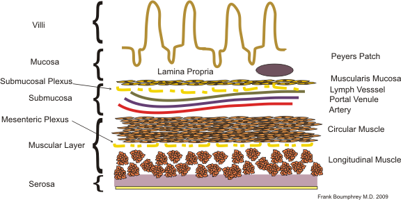
Image shows a diagram of the layers of the GI tract, including the mucosa, submucosa, muscularis and serosa. The mucosa is highly enfolded and contains villi, the submucosa hosts vessels from the cardiovascular and lymphatic system. The muscularis is made of smooth muscle, and the serosa includes cells that secrete serous fluid.
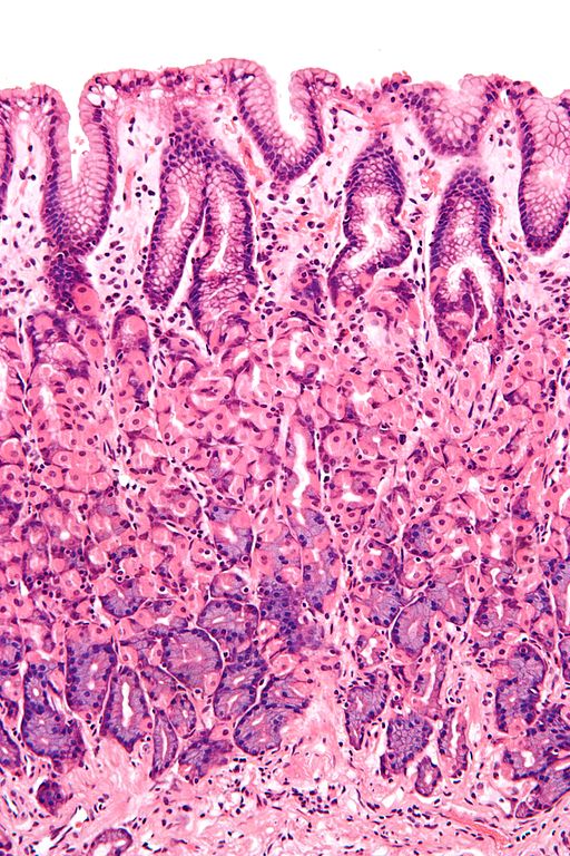
Image shows a pictomicrograph of the layers of the GI tract. Each of the mucosa, submucosa, muscularis and serosa are differentiated with respect to colouration and cell shape/size.
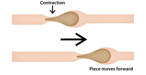
Image shows a diagram illustrating how peristalsis pushes food through the digestive tract by squeezing just behind the food, pushing it forward.
As per caption
A neurotransmitter that will have excitatory effects on the neuron, meaning it will increase the likelihood that a neuron will fire an action potential.
Created by CK-12 Foundation/Adapted by Christine Miller

We All Scream for Ice Cream
If you’re an ice cream lover, then just the sight of this yummy ice cream cone may make your mouth water. The “water” in your mouth is actually saliva, a fluid released by glands that are part of the digestive system. Saliva contains digestive enzymes, among other substances important for digestion. When your mouth waters at the sight of a tasty treat, it’s a sign that your digestive system is preparing to digest food.
What Is the Digestive System?
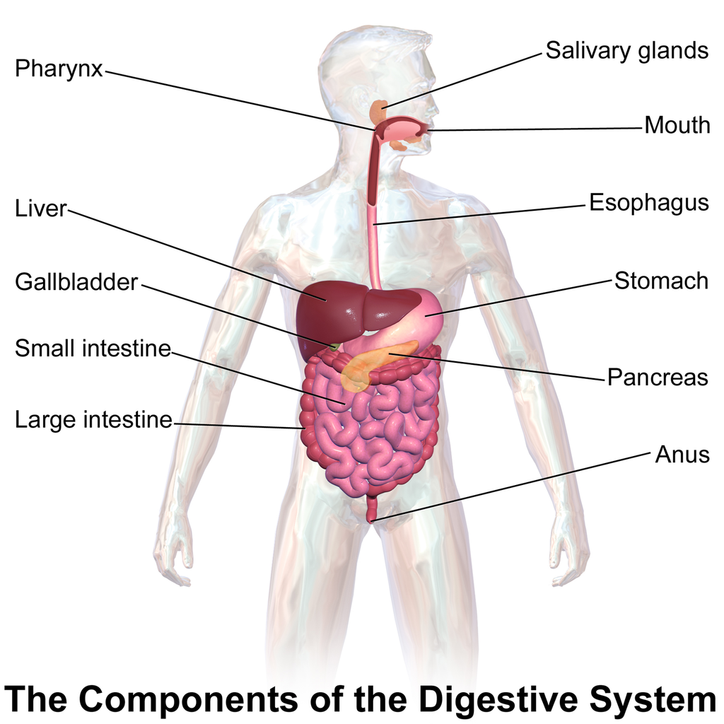
The digestive system consists of organs that break down food, absorb its nutrients, and expel any remaining waste. Organs of the digestive system are shown in Figure 15.2.2. Most of these organs make up the gastrointestinal (GI) tract, through which food actually passes. The rest of the organs of the digestive system are called accessory organs. These organs secrete enzymes and other substances into the GI tract, but food does not actually pass through them.
Functions of the Digestive System
The digestive system has three main functions relating to food: digestion of food, absorption of nutrients from food, and elimination of solid food waste. Digestion is the process of breaking down food into components the body can absorb. It consists of two types of processes: mechanical digestion and chemical digestion. Mechanical digestion is the physical breakdown of chunks of food into smaller pieces, and it takes place mainly in the mouth and stomach. Chemical digestion is the chemical breakdown of large, complex food molecules into smaller, simpler nutrient molecules that can be absorbed by body fluids (blood or lymph). This type of digestion begins in the mouth and continues in the stomach, but occurs mainly in the small intestine.
After food is digested, the resulting nutrients are absorbed. Absorption is the process in which substances pass into the bloodstream or lymph system to circulate throughout the body. Absorption of nutrients occurs mainly in the small intestine. Any remaining matter from food that is not digested and absorbed passes out of the body through the anus in the process of elimination.
Gastrointestinal Tract
The gastrointestinal (GI) tract is basically a long, continuous tube that connects the mouth with the anus. If it were fully extended, it would be about nine metres long in adults. It includes the mouth, pharynx, esophagus, stomach, and small and large intestines. Food enters the mouth, and then passes through the other organs of the GI tract, where it is digested and/or absorbed. Finally, any remaining food waste leaves the body through the anus at the end of the large intestine. It takes up to 50 hours for food or food waste to make the complete trip through the GI tract.
Tissues of the GI Tract
The walls of the organs of the GI tract consist of four different tissue layers, which are illustrated in Figure 15.2.3: mucosa, submucosa, muscularis externa, and serosa.
- The mucosa is the innermost layer surrounding the lumen (open space within the organs of the GI tract). This layer consists mainly of epithelium with the capacity to secrete and absorb substances. The epithelium can secret digestive enzymes and mucus, and it can absorb nutrients and water.
- The submucosa layer consists of connective tissue that contains blood and lymph vessels, as well as nerves. The vessels are needed to absorb and carry away nutrients after food is digested, and nerves help control the muscles of the GI tract organs.
- The muscularis externa layer contains two types of smooth muscle: longitudinal muscle and circular muscle. Longitudinal muscle runs the length of the GI tract organs, and circular muscle encircles the organs. Both types of muscles contract to keep food moving through the tract by the process of peristalsis, which is described below.
- The serosa layer is the outermost layer of the walls of GI tract organs. This is a thin layer that consists of connective tissue and separates the organs from surrounding cavities and tissues.
 |
 |
Peristalisis in the GI Tract
The muscles in the walls of GI tract organs enable peristalsis, which is illustrated in Figure 15.2.5. Peristalsis is a continuous sequence of involuntary muscle contraction and relaxation that moves rapidly along an organ like a wave, similar to the way a wave moves through a spring toy. Peristalsis in organs of the GI tract propels food through the tract.
Watch the video "What is peristalsis?" by Mister Science to see peristalsis in action:
https://youtu.be/kVjeNZA5pi4
What is peristalsis?, Mister Science, 2018.
Immune Function of the GI Tract
The GI tract plays an important role in protecting the body from pathogens. The surface area of the GI tract is estimated to be about 32 square metres (105 square feet), or about half the area of a badminton court. This is more than three times the area of the exposed skin of the body, and it provides a lot of area for pathogens to invade the tissues of the body. The innermost mucosal layer of the walls of the GI tract provides a barrier to pathogens so they are less likely to enter the blood or lymph circulations. The mucus produced by the mucosal layer, for example, contains antibodies that mark many pathogenic microorganisms for destruction. Enzymes in some of the secretions of the GI tract also destroy pathogens. In addition, stomach acids have a very low pH that is fatal for many microorganisms that enter the stomach.
Divisions of the GI Tract
The GI tract is often divided into an upper GI tract and a lower GI tract. For medical purposes, the upper GI tract is typically considered to include all the organs from the mouth through the first part of the small intestine, called the duodenum. For our instructional purposes, it makes more sense to include the mouth through the stomach in the upper GI tract, and all of the small intestine — as well as the large intestine — in the lower GI tract.
Upper GI Tract
The mouth is the first digestive organ that food enters. The sight, smell, or taste of food stimulates the release of digestive enzymes and other secretions by salivary glands inside the mouth. The major salivary gland enzyme is amylase. It begins the chemical digestion of carbohydrates by breaking down starches into sugar. The mouth also begins the mechanical digestion of food. When you chew, your teeth break, crush, and grind food into increasingly smaller pieces. Your tongue helps mix the food with saliva and also helps you swallow.
A lump of swallowed food is called a bolus. The bolus passes from the mouth into the pharynx, and from the pharynx into the esophagus. The esophagus is a long, narrow tube that carries food from the pharynx to the stomach. It has no other digestive functions. Peristalsis starts at the top of the esophagus when food is swallowed and continues down the esophagus in a single wave, pushing the bolus of food ahead of it.
From the esophagus, food passes into the stomach, where both mechanical and chemical digestion continue. The muscular walls of the stomach churn and mix the food, thus completing mechanical digestion, as well as mixing the food with digestive fluids secreted by the stomach. One of these fluids is hydrochloric acid (HCl). In addition to killing pathogens in food, it gives the stomach the low pH needed by digestive enzymes that work in the stomach. One of these enzymes is pepsin, which chemically digests proteins. The stomach stores the partially digested food until the small intestine is ready to receive it. Food that enters the small intestine from the stomach is in the form of a thick slurry (semi-liquid) called chyme.
Lower GI Tract
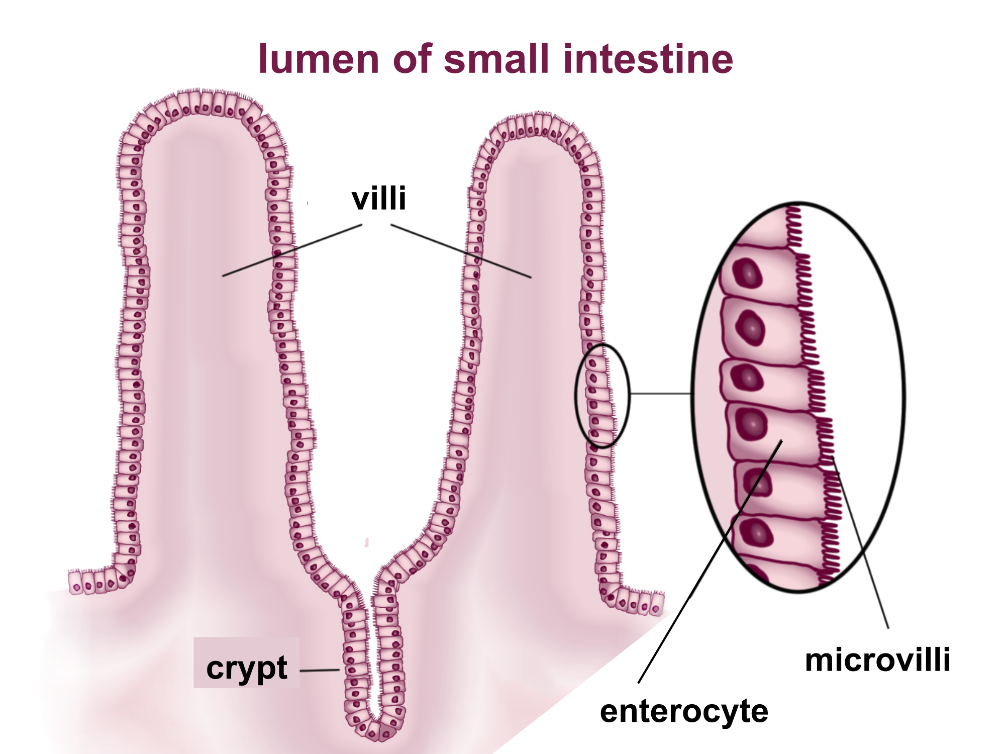
The small intestine is a narrow, but very long tubular organ. It may be almost seven metres long in adults. It is the site of most chemical digestion and virtually all absorption of nutrients. Many digestive enzymes are active in the small intestine, some of which are produced by the small intestine itself, and some of which are produced by the pancreas, an accessory organ of the digestive system. Much of the inner lining of the small intestine is covered by tiny finger-like projections called villi, each of which is covered by even tinier projections called microvilli. These projections, shown in the drawing below (Figure 15.2.6), greatly increase the surface area through which nutrients can be absorbed from the small intestine.
From the small intestine, any remaining nutrients and food waste pass into the large intestine. The large intestine is another tubular organ, but it is wider and shorter than the small intestine. It connects the small intestine and the anus. Waste that enters the large intestine is in a liquid state. As it passes through the large intestine, excess water is absorbed from it. The remaining solid waste — called feces — is eventually eliminated from the body through the anus.
Accessory Organs of the Digestive System
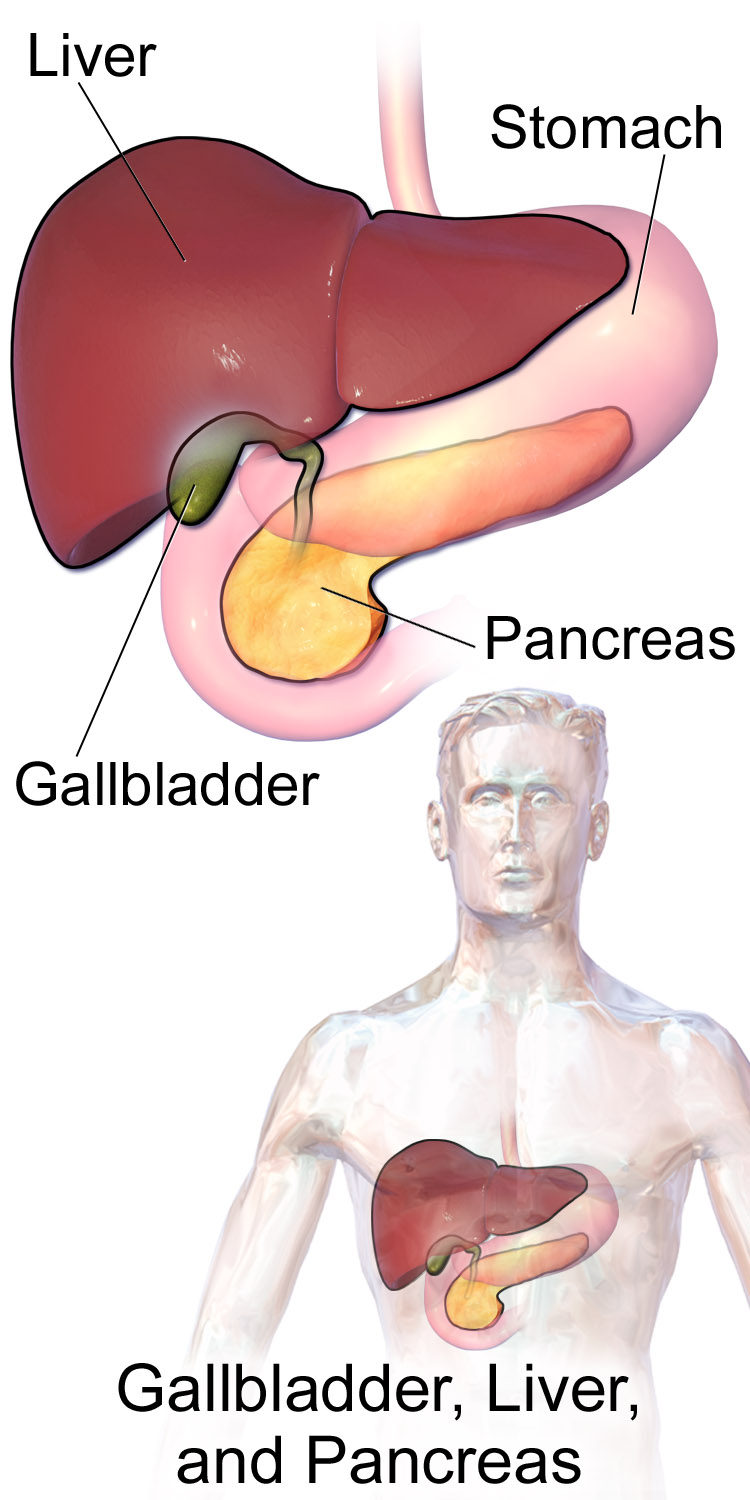
Accessory organs of the digestive system are not part of the GI tract, so they are not sites where digestion or absorption take place. Instead, these organs secrete or store substances needed for the chemical digestion of food. The accessory organs include the liver, gallbladder, and pancreas. They are shown in Figure 15.2.7 and described in the text that follows.
- The liver is an organ with multitude of functions. Its main digestive function is producing and secreting a fluid called bile, which reaches the small intestine through a duct. Bile breaks down large globules of lipids into smaller ones that are easier for enzymes to chemically digest. Bile is also needed to reduce the acidity of food entering the small intestine from the highly acidic stomach, because enzymes in the small intestine require a less acidic environment in order to work.
- The gallbladder is a small sac below the liver that stores some of the bile from the liver. The gallbladder also concentrates the bile by removing some of the water from it. It then secretes the concentrated bile into the small intestine as needed for fat digestion following a meal.
- The pancreas secretes many digestive enzymes, and releases them into the small intestine for the chemical digestion of carbohydrates, proteins, and lipids. The pancreas also helps lessen the acidity of the small intestine by secreting bicarbonate, a basic substance that neutralizes acid.
15.2 Summary
- The digestive system consists of organs that break down food, absorb its nutrients, and expel any remaining food waste.
- Digestion is the process of breaking down food into components that the body can absorb. It includes mechanical digestion and chemical digestion. Absorption is the process of taking up nutrients from food by body fluids for circulation to the rest of the body. Elimination is the process of excreting any remaining food waste after digestion and absorption are finished.
- Most digestive organs form a long, continuous tube called the gastrointestinal (GI) tract. It starts at the mouth, which is followed by the pharynx, esophagus, stomach, small intestine, and large intestine. The upper GI tract consists of the mouth through the stomach, while the lower GI tract consists of the small and large intestines.
- Digestion and/or absorption take place in most of the organs of the GI tract. Organs of the GI tract have walls that consist of several tissue layers that enable them to carry out these functions. The inner mucosa has cells that secrete digestive enzymes and other digestive substances, as well as cells that absorb nutrients. The muscle layer of the organs enables them to contract and relax in waves of peristalsis to move food through the GI tract.
- Three digestive organs — the liver, gallbladder, and pancreas — are accessory organs of digestion. They secrete substances needed for chemical digestion into the small intestine.
15.2 Review Questions
- What is the digestive system?
- What are the three main functions of the digestive system? Define each function.
-
- Relate the tissues in the walls of GI tract organs to the functions the organs perform.
15.2 Explore More
https://youtu.be/Og5xAdC8EUI
How your digestive system works - Emma Bryce, TED-Ed, 2017.
https://youtu.be/YVfyYrEmzgM
How does your body know you're full? - Hilary Coller, TED-Ed, 2017.
Attributions
Figure 15.2.1
Ice Cream [photo] by Mark Cruz on Unsplash is used under the Unsplash License (https://unsplash.com/license).
Figure 15.2.2
Blausen_0316_DigestiveSystem by BruceBlaus on Wikimedia Commons is used under a CC BY 3.0 (https://creativecommons.org/licenses/by/3.0) license.
Figure 15.2.3
Intestinal_layers by Boumphreyfr on Wikimedia Commons is used under a CC BY-SA 3.0 (https://creativecommons.org/licenses/by-sa/3.0) license.
Figure 15.2.4
512px-Normal_gastric_mucosa_intermed_mag by Nephron on Wikimedia Commons is used under a CC BY-SA 3.0 (https://creativecommons.org/licenses/by-sa/3.0) license.
Figure 15.2.5
Peristalsis pushes food through the GI tract by CK-12 Foundation is used under a CC BY NC 3.0 (https://creativecommons.org/licenses/by-nc/3.0/) license.
Figure 15.2.6
Villi_&_microvilli_of_small_intestine.svg by BallenaBlanca on Wikimedia Commons is used under a CC BY-SA 4.0 (https://creativecommons.org/licenses/by-sa/4.0) license.
Figure 15.2.7
Blausen_0428_Gallbladder-Liver-Pancreas_Location by BruceBlaus on Wikimedia Commons is used under a CC BY 3.0 (https://creativecommons.org/licenses/by/3.0) license.
References
Blausen.com Staff. (2014). Medical gallery of Blausen Medical 2014. WikiJournal of Medicine 1 (2). DOI:10.15347/wjm/2014.010. ISSN 2002-4436.
Brainard, J/ CK-12 Foundation. (2016). Figure 4 Peristalsis pushes food through the GI tract. [digital image]. In CK-12 College Human Biology (Section 17.2) [online Flexbook]. CK12.org. https://www.ck12.org/book/ck-12-college-human-biology/section/17.2/
Mister Science. (2018). What is peristalsis? YouTube. https://www.youtube.com/channel/UCxTlkZfjArUobBAeVwzJjYg/videos
TED-Ed. (2017, November 13). How does your body know you're full? - Hilary Coller. YouTube. https://www.youtube.com/watch?v=YVfyYrEmzgM&feature=youtu.be
TED-Ed. (2017, December 14). How your digestive system works - Emma Bryce. YouTube. https://www.youtube.com/watch?v=Og5xAdC8EUI&feature=youtu.be
Created by CK-12 Foundation/Adapted by Christine Miller

We All Scream for Ice Cream
If you’re an ice cream lover, then just the sight of this yummy ice cream cone may make your mouth water. The “water” in your mouth is actually saliva, a fluid released by glands that are part of the digestive system. Saliva contains digestive enzymes, among other substances important for digestion. When your mouth waters at the sight of a tasty treat, it’s a sign that your digestive system is preparing to digest food.
What Is the Digestive System?
The digestive system consists of organs that break down food, absorb its nutrients, and expel any remaining waste. Organs of the digestive system are shown in Figure 15.2.2. Most of these organs make up the gastrointestinal (GI) tract, through which food actually passes. The rest of the organs of the digestive system are called accessory organs. These organs secrete enzymes and other substances into the GI tract, but food does not actually pass through them.

Functions of the Digestive System
The digestive system has three main functions relating to food: digestion of food, absorption of nutrients from food, and elimination of solid food waste. Digestion is the process of breaking down food into components the body can absorb. It consists of two types of processes: mechanical digestion and chemical digestion. Mechanical digestion is the physical breakdown of chunks of food into smaller pieces, and it takes place mainly in the mouth and stomach. Chemical digestion is the chemical breakdown of large, complex food molecules into smaller, simpler nutrient molecules that can be absorbed by body fluids (blood or lymph). This type of digestion begins in the mouth and continues in the stomach, but occurs mainly in the small intestine.
After food is digested, the resulting nutrients are absorbed. Absorption is the process in which substances pass into the bloodstream or lymph system to circulate throughout the body. Absorption of nutrients occurs mainly in the small intestine. Any remaining matter from food that is not digested and absorbed passes out of the body through the anus in the process of elimination.
Gastrointestinal Tract
The gastrointestinal (GI) tract is basically a long, continuous tube that connects the mouth with the anus. If it were fully extended, it would be about nine metres long in adults. It includes the mouth, pharynx, esophagus, stomach, and small and large intestines. Food enters the mouth, and then passes through the other organs of the GI tract, where it is digested and/or absorbed. Finally, any remaining food waste leaves the body through the anus at the end of the large intestine. It takes up to 50 hours for food or food waste to make the complete trip through the GI tract.
Tissues of the GI Tract
The walls of the organs of the GI tract consist of four different tissue layers, which are illustrated in Figure 15.2.3: mucosa, submucosa, muscularis externa, and serosa.
- The mucosa is the innermost layer surrounding the lumen (open space within the organs of the GI tract). This layer consists mainly of epithelium with the capacity to secrete and absorb substances. The epithelium can secret digestive enzymes and mucus, and it can absorb nutrients and water.
- The submucosa layer consists of connective tissue that contains blood and lymph vessels, as well as nerves. The vessels are needed to absorb and carry away nutrients after food is digested, and nerves help control the muscles of the GI tract organs.
- The muscularis externa layer contains two types of smooth muscle: longitudinal muscle and circular muscle. Longitudinal muscle runs the length of the GI tract organs, and circular muscle encircles the organs. Both types of muscles contract to keep food moving through the tract by the process of peristalsis, which is described below.
- The serosa layer is the outermost layer of the walls of GI tract organs. This is a thin layer that consists of connective tissue and separates the organs from surrounding cavities and tissues.
 |
 |
Peristalisis in the GI Tract
The muscles in the walls of GI tract organs enable peristalsis, which is illustrated in Figure 15.2.5. Peristalsis is a continuous sequence of involuntary muscle contraction and relaxation that moves rapidly along an organ like a wave, similar to the way a wave moves through a spring toy. Peristalsis in organs of the GI tract propels food through the tract.

Watch the video "What is peristalsis?" by Mister Science to see peristalsis in action:
https://youtu.be/kVjeNZA5pi4
What is peristalsis?, Mister Science, 2018.
Immune Function of the GI Tract
The GI tract plays an important role in protecting the body from pathogens. The surface area of the GI tract is estimated to be about 32 square metres (105 square feet), or about half the area of a badminton court. This is more than three times the area of the exposed skin of the body, and it provides a lot of area for pathogens to invade the tissues of the body. The innermost mucosal layer of the walls of the GI tract provides a barrier to pathogens so they are less likely to enter the blood or lymph circulations. The mucus produced by the mucosal layer, for example, contains antibodies that mark many pathogenic microorganisms for destruction. Enzymes in some of the secretions of the GI tract also destroy pathogens. In addition, stomach acids have a very low pH that is fatal for many microorganisms that enter the stomach.
Divisions of the GI Tract
The GI tract is often divided into an upper GI tract and a lower GI tract. For medical purposes, the upper GI tract is typically considered to include all the organs from the mouth through the first part of the small intestine, called the duodenum. For our instructional purposes, it makes more sense to include the mouth through the stomach in the upper GI tract, and all of the small intestine — as well as the large intestine — in the lower GI tract.
Upper GI Tract
The mouth is the first digestive organ that food enters. The sight, smell, or taste of food stimulates the release of digestive enzymes and other secretions by salivary glands inside the mouth. The major salivary gland enzyme is amylase. It begins the chemical digestion of carbohydrates by breaking down starches into sugar. The mouth also begins the mechanical digestion of food. When you chew, your teeth break, crush, and grind food into increasingly smaller pieces. Your tongue helps mix the food with saliva and also helps you swallow.
A lump of swallowed food is called a bolus. The bolus passes from the mouth into the pharynx, and from the pharynx into the esophagus. The esophagus is a long, narrow tube that carries food from the pharynx to the stomach. It has no other digestive functions. Peristalsis starts at the top of the esophagus when food is swallowed and continues down the esophagus in a single wave, pushing the bolus of food ahead of it.
From the esophagus, food passes into the stomach, where both mechanical and chemical digestion continue. The muscular walls of the stomach churn and mix the food, thus completing mechanical digestion, as well as mixing the food with digestive fluids secreted by the stomach. One of these fluids is hydrochloric acid (HCl). In addition to killing pathogens in food, it gives the stomach the low pH needed by digestive enzymes that work in the stomach. One of these enzymes is pepsin, which chemically digests proteins. The stomach stores the partially digested food until the small intestine is ready to receive it. Food that enters the small intestine from the stomach is in the form of a thick slurry (semi-liquid) called chyme.
Lower GI Tract
The small intestine is a narrow, but very long tubular organ. It may be almost seven metres long in adults. It is the site of most chemical digestion and virtually all absorption of nutrients. Many digestive enzymes are active in the small intestine, some of which are produced by the small intestine itself, and some of which are produced by the pancreas, an accessory organ of the digestive system. Much of the inner lining of the small intestine is covered by tiny finger-like projections called villi, each of which is covered by even tinier projections called microvilli. These projections, shown in the drawing below (Figure 15.2.6), greatly increase the surface area through which nutrients can be absorbed from the small intestine.

From the small intestine, any remaining nutrients and food waste pass into the large intestine. The large intestine is another tubular organ, but it is wider and shorter than the small intestine. It connects the small intestine and the anus. Waste that enters the large intestine is in a liquid state. As it passes through the large intestine, excess water is absorbed from it. The remaining solid waste — called feces — is eventually eliminated from the body through the anus.
Accessory Organs of the Digestive System

Accessory organs of the digestive system are not part of the GI tract, so they are not sites where digestion or absorption take place. Instead, these organs secrete or store substances needed for the chemical digestion of food. The accessory organs include the liver, gallbladder, and pancreas. They are shown in Figure 15.2.7 and described in the text that follows.
- The liver is an organ with multitude of functions. Its main digestive function is producing and secreting a fluid called bile, which reaches the small intestine through a duct. Bile breaks down large globules of lipids into smaller ones that are easier for enzymes to chemically digest. Bile is also needed to reduce the acidity of food entering the small intestine from the highly acidic stomach, because enzymes in the small intestine require a less acidic environment in order to work.
- The gallbladder is a small sac below the liver that stores some of the bile from the liver. The gallbladder also concentrates the bile by removing some of the water from it. It then secretes the concentrated bile into the small intestine as needed for fat digestion following a meal.
- The pancreas secretes many digestive enzymes, and releases them into the small intestine for the chemical digestion of carbohydrates, proteins, and lipids. The pancreas also helps lessen the acidity of the small intestine by secreting bicarbonate, a basic substance that neutralizes acid.
15.2 Summary
- The digestive system consists of organs that break down food, absorb its nutrients, and expel any remaining food waste.
- Digestion is the process of breaking down food into components that the body can absorb. It includes mechanical digestion and chemical digestion. Absorption is the process of taking up nutrients from food by body fluids for circulation to the rest of the body. Elimination is the process of excreting any remaining food waste after digestion and absorption are finished.
- Most digestive organs form a long, continuous tube called the gastrointestinal (GI) tract. It starts at the mouth, which is followed by the pharynx, esophagus, stomach, small intestine, and large intestine. The upper GI tract consists of the mouth through the stomach, while the lower GI tract consists of the small and large intestines.
- Digestion and/or absorption take place in most of the organs of the GI tract. Organs of the GI tract have walls that consist of several tissue layers that enable them to carry out these functions. The inner mucosa has cells that secrete digestive enzymes and other digestive substances, as well as cells that absorb nutrients. The muscle layer of the organs enables them to contract and relax in waves of peristalsis to move food through the GI tract.
- Three digestive organs — the liver, gallbladder, and pancreas — are accessory organs of digestion. They secrete substances needed for chemical digestion into the small intestine.
15.2 Review Questions
- What is the digestive system?
- What are the three main functions of the digestive system? Define each function.
-
- Relate the tissues in the walls of GI tract organs to the functions the organs perform.
15.2 Explore More
https://youtu.be/Og5xAdC8EUI
How your digestive system works - Emma Bryce, TED-Ed, 2017.
https://youtu.be/YVfyYrEmzgM
How does your body know you're full? - Hilary Coller, TED-Ed, 2017.
Attributions
Figure 15.2.1
Ice Cream [photo] by Mark Cruz on Unsplash is used under the Unsplash License (https://unsplash.com/license).
Figure 15.2.2
Blausen_0316_DigestiveSystem by BruceBlaus on Wikimedia Commons is used under a CC BY 3.0 (https://creativecommons.org/licenses/by/3.0) license.
Figure 15.2.3
Intestinal_layers by Boumphreyfr on Wikimedia Commons is used under a CC BY-SA 3.0 (https://creativecommons.org/licenses/by-sa/3.0) license.
Figure 15.2.4
512px-Normal_gastric_mucosa_intermed_mag by Nephron on Wikimedia Commons is used under a CC BY-SA 3.0 (https://creativecommons.org/licenses/by-sa/3.0) license.
Figure 15.2.5
Peristalsis pushes food through the GI tract by CK-12 Foundation is used under a CC BY NC 3.0 (https://creativecommons.org/licenses/by-nc/3.0/) license.
Figure 15.2.6
Villi_&_microvilli_of_small_intestine.svg by BallenaBlanca on Wikimedia Commons is used under a CC BY-SA 4.0 (https://creativecommons.org/licenses/by-sa/4.0) license.
Figure 15.2.7
Blausen_0428_Gallbladder-Liver-Pancreas_Location by BruceBlaus on Wikimedia Commons is used under a CC BY 3.0 (https://creativecommons.org/licenses/by/3.0) license.
References
Blausen.com Staff. (2014). Medical gallery of Blausen Medical 2014. WikiJournal of Medicine 1 (2). DOI:10.15347/wjm/2014.010. ISSN 2002-4436.
Brainard, J/ CK-12 Foundation. (2016). Figure 4 Peristalsis pushes food through the GI tract. [digital image]. In CK-12 College Human Biology (Section 17.2) [online Flexbook]. CK12.org. https://www.ck12.org/book/ck-12-college-human-biology/section/17.2/
Mister Science. (2018). What is peristalsis? YouTube. https://www.youtube.com/channel/UCxTlkZfjArUobBAeVwzJjYg/videos
TED-Ed. (2017, November 13). How does your body know you're full? - Hilary Coller. YouTube. https://www.youtube.com/watch?v=YVfyYrEmzgM&feature=youtu.be
TED-Ed. (2017, December 14). How your digestive system works - Emma Bryce. YouTube. https://www.youtube.com/watch?v=Og5xAdC8EUI&feature=youtu.be
Image shows a man participating in a hot-dog eating contest. His mouth is so full of hot dog that he can't close his lips.
An antibody, also known as an immunoglobulin, is a large, Y-shaped protein produced mainly by plasma cells that is used by the immune system to neutralize pathogens such as pathogenic bacteria and viruses.
An organic chemical that functions in the brain and body of many types of animals (and humans) as a neurotransmitter—a chemical message released by nerve cells to send signals to other cells, such as neurons, muscle cells and gland cells.
Image shows a woman taking a bit of a soft taco. She is using her teeth to break off her bite.

